MORPHOLOGICAL OBSERVATIONS ON THE TESTES ... - Unesp
MORPHOLOGICAL OBSERVATIONS ON THE TESTES ... - Unesp
MORPHOLOGICAL OBSERVATIONS ON THE TESTES ... - Unesp

Create successful ePaper yourself
Turn your PDF publications into a flip-book with our unique Google optimized e-Paper software.
Rev. Chi!o Anat.,20(3):263-268,2002.<strong>MORPHOLOGICAL</strong> <strong>OBSERVATI<strong>ON</strong>S</strong> <strong>ON</strong> <strong>THE</strong> <strong>TESTES</strong>Physalaemus cuvieri (AMPHIBIA, ANURA)OFESTUDIO MORFOLÓGICO DE LOS TESTÍCULOS DE Physalaemus cuvieri (AMPHIBIA, ANURA)CIassius de Oliveira; Cristiani Zanetoni & Rodrigo ZieriOLIVEIRA, C.; ZANET<strong>ON</strong>I, C. & ZIERI, R. Morphological observations on the testes of Physalaemus cuvieri (Amphibia, Anura).Rev. Chi!oAnat., 20(3):263-268, 2002.SUMMARY: In general, the testes in the anurans are paired ovoid organs, constituted by a mass of seminiferous structuressurrounded by a layer of fibrous connective tissue, which holds a germ epithelium with characteristic cellular types. This study describesthe testicular histoJogical architecture and anatomy, as well as the organization and morphology of germ cells. Five male samples, fromthe Botucatu area (São Paulo State, Brazil), of Physalaemus cuvieri (Leptodactylidae) were used. After macroscopic analyses and obtainmentofthe testicularfragments, the material was submitted to the histological routine to inclusion in paraffin and coloration with haematoxylin/eosin. Arare peculiarity is the presence of numerous pigment-containing cells randomly distributed in the albuginea tunic and testicularinterstitium, giving the testis a dark brown coloration. In the germ tissue the spermatogonia I are biggest spermatogenetic cells. With thecellular differentiation and proliferation, succeded the other cellular types (spermatogonia lI, spermatocytes I and lI, spermatids I and JI,and spermatooza) with a cystic organization, that is, groups of cells associated with Sertoli cells, forming the spermatogenetic cysts orspermatocysts. The spermatogenetic lineage cells were differentiated and identified according to the cellular and cystic morphology.KEY WORDS: 1. Anura; 2. Leptodactylidae; 3. Physalaemus cuvieri; 4. Testis; 5. Spermatogenesis.INTRODUCTI<strong>ON</strong>In the anurans, the testes are paired ovoid organsconstituted by seminiferous tubules convoluted surroundedby the fibrous connective tissue, which canstitutes thealbuginea tunic. Cancerning to the histological architectureof the seminiferous elements, to the amphibians and, in ageneral sense, the germ epithelium may be arranged inseminiferous locules in the Apoda (Wake, 1969) and Anura(Duellman & Trueb, 1994) ar in seminiferous ampoules artesticular lobules the Urodela.The germ tissue, which constitutes the testicularparenchyma, has different cellular types: spermatagonia in theepithelium boundary; spermatocytes and spermatids in thesequence af the cellular differentiation; and spermatozoa inthe lumen or in their adjacencies. In this epithelium, there is acystic arrangement, that is, groups of sexual cells assaciatedwith the Sertoli cells forming spermatagenetic cysts orspermatocysts. Therefore, each one of these units c\usters cellsin the same stage of differentiatian and with a synchronismdevelapment, common characteristic in the amphibians (Wake;Lofts, 1974; Rastogi et ai., 1988; Oliveira et ai., 2002).There are few researches about the organs andstructures that constitute the anuran male repraductivesystem, specifically in animaIs of neatropical areas, like inBrazil. Beside that, even if there is not a large number ofamphibians in this taxon, several different reproducti vestrategies will occur (Duellman & Trueb), which seem ta besuggestive afmorphological and functional variations in thereproductive organs.Thus, this study tried to analyze the testesmorphological arrangement, describing histological andanatomical aspects, as well as some structural characteristicsaf the germ cells and their cystic arrangement in the anuranPhysalaemus cuvieri (Fitzinger, 1826 - Leptodactylidae).Department of Biology - Instituto de Biociências, Letras e Ciências Exatas - UNESP, São José do Rio Preto - São Paulo State, Brazil.263
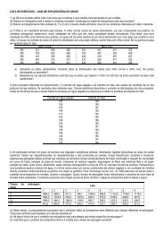
OLIVEIRA, c.; ZANET<strong>ON</strong>I, C. & ZIE)H, R. Morphological observations on the testes of Physalaemus euvieri (Amphibia, Anura). Rev. Chil. Anaf., 20(3):263-268, 2002.MATERIAL AND METHODFive male samples ofPhysalaemus cuvieri were used. Thespecimens were collected in Botucatu(São Paulo State - Brazil), between Juneand December, when they were in thereproductive activity period. Aftercaptured and transported to thelaboratory, the individuaIs wereanesthetized with ether and submitted tomorphologicalstudies.AThe animaIs were opened throughmedium incision from the cloaca as faras anterior limbs, exposing thereproductive organs to macroscopicanalyses. After the reduction of the testesin small pieces, they wereimmediately immersed in Bouin fixativesolution during 20 hours, washed, andtransferred to 70% ethanol solution.Then, the .material was sent to thehistological routine to be dehydrated inethanol, clarified with xylol, andembedded in paraffin. Sections of 6 ~mwere stained with haematoxylin/eosin forhistological analyses, The individuaIswere appropriately preserved as proofmaterial.RESULTSConcerning the gonadmacroscopic aspect of Physalaemuscuvieri, as to color, shape, and size, wecan conspicuous anatomic variations.However, histologically there is not adifference in the germ epitheliumarrangement. Covering the testis we canfind a thin capsule of connective tissueand smooth muscle, the albuginea tunic.The blood vessels, which exist in thistesticular capsule, are maínly destinedto the parenchyma, where a germepithelium is arranged in seminiferouslocules, Delimited by a loose connectivetissue, these locules form morphologicalunits, that is, the seminiferousFigure I, A) Testes anatomic aspect of the Physalaemus cuvieri with dark brownpigmentation in the albuginea tunic, Dissected gonad ( ) putting in evidence the internalpigmentation and the units denominated seminiferous [ocules (27x). B) Histology of theseminiferous locule (HlE, 215x) and C) Generalized representation of the cystic germepithelium: locular wall (I); interlocular tissue (2) with Leydig cells, blood capillary andpigment-containing cells; primary (3) and secondary (4) spermatogonia; primary (5) andsecondary (6) spermatocytes; primary (7) and secondary (8) spermatids; spermatozoonbundles (9) and Sertoli cells (10).264
Figure 2. Detail 01'the histologicaJ arrangement 01'the semini1'erous 10cuJi: I) spermatogonia I; 2)spermatogonia lI; 3) spermatocytes I; 4) spermatocytes 11; 5) spermatids I; 6) spermatids lI; 7)spermatozoon bundles; 8) spermatozoon in the locular lume. (H/E, A and B - 430x; C and D - 850x).elements of the gonads. In Physalaemus cuvieri, the interloculartis sue is relatively scarce, presenting apigmentation in this tissue and also in the albuginea tunic,giving the testis a dark brown coloration. Between theseminiferous units, they have a inter-Iocular tissuecomposed by Leydig interstitial cells, fibroblasts,pigment-containing cells, blood vessels, and some efferentductules (Fig. I).Each seminiferous loculehas a germ tissue with a lot ofspermatogenetic cysts orspermatocysts, that is, c1usters ofgerm celIs in different stages ofditlerentiation that occur due toa intimate morphofunctionalrelation with the Sertoli cells. Inthe cysts, cells are joined in thesame differentiation status,therefore, with a developmentsynchrony. Some cysts seIdompresent cells with smalldifferences among themselvesand, since these asynchronismsare very small, we consider thecysts as a cellular group in whichali the sexual cells are in thesame stage of differentiation(Figs. 1 and 2).The different cellulartypes were differentiated andidentified according to themorphology and cystic arrangement(Fig. 2), also representedschematicalIy (Fig. 1c).Spermatogonia - three stagesaf differentiation weredistinguished. The primordialspermatogonia or primordialgerm cells are the mostvoluminous cells of thespennatogenetic lineage, whosenuclei are irregular and with amultilobular aspect. Theypresent chromatinic granulations,beside a single,eccentric nucleolus. In general,they are very c10se to the locularwalI, and are associate to theSertoli cells which stilI haveaspect of folIicular cells and thenuc1eus is falciform. It is alsocommon that t~ese spermatogoniabe isolated or joined insmalI groups, but this do notcharacterize genn cysts. From mitotic divisions, the primary(I) and the secondary (11)spermatogonia are originated. Thesecondary ones are smaller, more irregular, and they wereidentified due to the nuclear compactation degree. Thespermatogonia I are organized as celIular cysts with a irregularaspect andhard delimitation, they are located nearthe locular wall. The spennatogonia 11suffer some aIterations,which make thenuc1ei to become more compacted and give265
OLIVEIRA, c.; ZANET<strong>ON</strong>I, C. & ZIERI, R. Morphological observatioos 00 the testes of Phvsalael11l1s ellvieri (Amphibia, Ao"ra). ReI'. Chil. Anat., 20(3):263-268, 2002.them typical shapes. The Sertoli cells, when associated withthe spermatogonia lI, present a nucleus with more elongatedshape, still semilunar or falciform.Spermatocytes - they are the result from the spermatogoniaII ditferentiation, the spermatocytes I are cells just smallerthan the spermatogonia I. In its nucleus, the chromatin isslightly condensed. The spermatocytes II are haploid cells andquite smaller than the previous, and they are originated in thefirst meiotic division. Generally, the spermatocytes areobserved in different phases of the first meiotic division,presenting different degrees of compactation of the nuclearmaterial. In this stage, the Sertoli cells are more voluminous,the nuclei are less condensed and present the tendency toassume intermediate shape, between elongated and ovoid.Spermatids - whenthe spermatocytesII pass throughthesecond meiotic division, they form the spermatids. These, inthe primary stage or as round spermatids I, are differentiatedfrom the cysts of the spermatocytes II when some cells showthemselves slightly elongated. The spermatids II or elongatedare cells with compacted and elongated nuclei, whose cysticarrangement is altered to be organized in bundles sustainedby the Sertoli cells. In this moment, the nucleus of the Sertolicells finds itself as oval or rounded, with chromatin looselydistributed, besides the evident and central nucleolus.Spermatozoa - they are characterized by a extraordinary nuclearcompactation and a cytoplasmic reduction. Thespermatozoon head, represented mainly by the nucleus, isslender; and, in general, it turns to the nucleus of the Sertolicell, in opposition, the tail has a filamentous appearance andis slightly stain, and, generally, turns to the luminar area of theseminiferous locule. The spermatozoa in development arearranged in bundles very well organized, due to the associationwith the Sertoli cells. When they reach maturity, they arereleased from the bundle and reach the locular lumen and theetferent ductule.DISCUSSI<strong>ON</strong>The unusual dark brown pigmentation of the testes ofPhysalaemus cuvieri is a peculiarity, which occur due to apresence of numerous pigmented cells (melano-macrophagesor typical melanocytes?) randomly distributed in the testicularcapsule and interstitium. This unusual characteristic, atesticular pigmentation, rarely is observed in others species,as described in Physalaemus fuscomaculatus (Aoki et aI.,1969) and Bombina bombina (Gollmann et aI., 1993). In theanuran Xenopus laevis, and other lower vertebrates, thepigment cells can be found in the different organs, constitutingan extracutaneous pigmentary system of unknown function(Zuasti et ai., 1998).Concerning the general histological architectureobserved in Physalaemus cuvieri, it was verified a greatsimilarity to the descriptions of the other species of samefamily, like Physalaemus fuscomaculatus (Aoki et aI.) andCaudiverbera caudiverbera (Hermosilla et aI., 1983); and alsowith species of the Hylidae family - Hyla ranki (Taboga &Dolder, 1991), Hyla japonica (Lee & Kwon, 1992); Hylapulchella andina (Montero & Pisanó, 1992); and Scinaxfuscovarius (Oliveira & Vicentini, 1998; Oliveira et al.). Inthe species that show a continuous spermatogenesis for ali thereproductive period, differerH cellular types may be identifiedin only one seminiferous locule, that is, ali the cells of thegerm lineage occupy the same locule and differentiatethemselves simultaneously.To Leptodactylids, Telmatobius laticeps andTelmatobius pisanoi (Montero & Pisanó, 1990), the histologicalarrangement is similar, however a potentially continuous cycleoccurs, considering that there is a well-defined phase of theintense spermatogenetic activity and other of rest, as it wasalso described to the hylidae: Hyla pulchella andina (Montero& Pisanó, 1992). In anurans like Nectophrynoides occidentalis(Zuber- Vogeli & Xavier, 1966), a temperate area bufonidae,the cycle is discontinuous, it influence even more in the gonadalactivity and consequently in the dynamic of spermatogenesis.For the neotropical area species, occurs preferentially acontinuous reproduction, in which ali the cellular types arepresent for whole year.With the cystic arrangement, the spermatogeneticlineage cells together with the Sertoli cells form delimitedgroups by a membranous capsule, which is denominated germcyst or spermatocyst. We stress the fact that independently ofthe reproductive cycle type of the species, the cysticarrangement of the germ cells is confirmed as an importantcharacteristic of the anuran amphibians, as well as to otheranamniotes vertebrates (Lofts; Grier et aI., 1980; Grier, 1992).In a similar way to what was obtained for Bufo woodhouse(Atherton, 1974), Caudiverbera caudiverbera (Hermosilla etai.) and Scinaxfuscovarius (Oliveira & Vicentini), we also donot observe any specific area of the testis where predominatecertain cellular kinds. It is corroborated by the apparentlyrandom distribution of the spermatocysts in the seminiferousstructures.Concerning the spermatogenetic lineage cells, the primordialspermatogonia are the biggest cells and rest on theadjacencies of the basallamina of the seminiferous structures,associated with the follicular cells (Lofts). As to the266
OLIVEIRA, C.; ZANET<strong>ON</strong>I, C. & ZIERI, R. Morphological observations on the testes of Physalaemus cllvieri (Amphibia, Anura). Rev. Chi!o Allal., 20(3):263-268. 2002.spermatogonial types, similar observations to the Bufoarenarun (Cavicchia & Moviglia, 1983), Hyla ranki (Taboga& Dolder) and to the Scinaxfuscovarius (Oliveira et ai.) werealso verified in the species of this research.The spermatocytes generally are observed in theprophase of the first meiotic division, with different levels ofchromatinic and chromosomal condensation. Thespermatocytes l are commonly bigger than the spermatogoniaII (Lofts; Rastogi et ai. and Oliveira et ai.). With thespermatocytes l division, smaller cells are originated, thespermatocytes lI, which present half the diameter of the origincells, as we observed in the Physalaemus cuvieri.ln Caudiverbera caudiverbera (Hermosilla et ai.),Odontophrynus cultripes (Báo et ai., 1991), Scinaxfuscovarius(Oliveira et ai.) and in our analysis, the spermatids l, in a firststage, show a spherical nucleus and thin granulation, after thenuclei become oval and the granular chromatin is distributedhomogeneously. The following stages are characterized by acellular and nuclear elongation, which occur simultaneouslywith the chromatinic condensation, culminating with thespermatids II formation.At the end of the spermatogenesis, the spermatozoaremain united in the bundles, they are still sustained by theSertoli cells, however with their liberation to the lumen of theseminiferous locule, the arrangement is dissolved and theseremain in the spermatic path until the liberation.Although the architecture of the germ epithelium ofali mentioned species represents a general and typical modelfor the anurans, the final germ cell- the spermatozoon - showsa great heterogeneity in the species. This can be related tophylogenetic aspects (Kwon & Lee, 1995) and to reproductivestrategies of the anurans (Duellman & Trueb).ACKNOWLEDGMENTS : We are grateful to the CNPq forthe award of Scientific lnitiation Fellowship (PIBlC-CNPq96 to 98), to Cristiani Zanetoni, to Umberto Jorge Alves deAndrade for schematizing in translucent tracer paper, toRoberta Faria Femandes and Prof. Dr. Álvaro Luiz Hattnherfor the translation into English, to Profa. Dra. Rosa Maria daSilva and Dra. Roxana Guadalupe Herrera Álvarez fortranslation into castellano, and to Prof. Dr. Sebastião RobertoTaboga for the suggestions to the manuscript.OLIVEIRA, c.; ZANET<strong>ON</strong>I, C. & ZIERI, R. Estudio morfológico de los testículos de Physalaemus cuvieri (Amphibia, Anura). Rev.Chi!oAnat., 20(3):263-268, 2002.RESUMEN: Generalmente, los testículos de anuros constituyen órganos ovoides, pares, formados por masa de estructurasseminíferas, envueltas en una capa de tejido conjuntivo fibroso, que contiene un epitelio germinativo provisto de tipos celulares característicos.En este trabajo se describe Ia anatomía y Ia arquitectura histológica de los testículos, así como también Ia organización ymorfología de Ias células germinativas. Se utilizaron cinco ejemplares machos de Ia especie Physalaemus cuvieri (Leptodactylidae),provenientes de Ia zona de Botucatu (São Paulo, Brasil). Tras el análisis macroscópico y Ia obtención de los fragmentos testiculares, elmaterial se sometió a Ias técnicas histológicas rutinarias para su inclusión en parafina y coloración con hematoxilina/eosina. Se constatócomo rara peculiaridad Ia presencia de numerosas células con pigmento distribuidas de forma aleatoria en Ia túnica albugínea e intersticiotesticular, Ias cuales otorgan ai testículo una coloración marrón oscura. En el tejido germinativo Ias espermatogonias I son Ias mayorescélulas espermatogenéticas. En Ia secuencia de Ia diferenciación y proliferación ce]ulares, se siguen ]os demás tipos de células(espermatogonias lI, espermatocitos I y lI, espermátidas I y lI, y espermatozoides) que presentan una organización cística, es decir,grupos de cé]ulas se asocian a ]as célu]as de Sertoli, formando ]os cistos espermatogenéticos o espermatocistos. Se diferenciaron eidentificaron Ias células dellinaje espermatogenético, según ]a morfología de ]as células y dei propio cisto.PALABRAS CLAVE: 1.Anura; 2. Leptodactylidae; 3. Physalaemus cuvieri, 4. Testículo; 5. Espermatogénesis.REFERENCESAoki, A.; Vitale-Calpe, R. & Pisano, A. The testicularinterstitial tissue of the amphibian Physalaemusfuscumaculatus. Z. Zellforsch. Mikrosk. Anat., 98:9-16, 1969.Atherton, R. W. A gradient analysis of spennatogenesis inthe toad Bufo woodhousei Girard (1854). Herpetologica,30:240-4, 1974.Báo, S. N.; Dalton, G. C. & Oliveira, S. F. Spermiogenesis inOdontophrynus cultripes (Amphibia, Anura, Leptodactylidae):ultrastructural and cytochemical studies ofproteins using E-PTA. 1. Morphol., 207:303-14, 1991.Cavicchia, 1. C. & Moviglia, G. A. The blood-testis barrier inthe toad (Bufo arenarum, HenseI): a freeze-fracture andlanthanum tracer study. Anat. Rec., 205:387-96, 1983.267
OLIVEIRA, C.; ZANET<strong>ON</strong>I, C. & ZIERI, R. Morphological observations on the testes of Plzysa!ael11l1sellvieri (Amphibia, Anura). Rev. Chi!o Allla., 20(3):263-268, 2002.Duellman, W. E. & Trueb, L. Biology of amphibians. NewYork, McGraw-Hill, 670p, 1994.Gollmann, G.; Borkin, L. J. & Roth, P. Genic andmorphological variation in the fire-bellied toad, Bombinabombina (Anura, Discoglossidae). Zoolog. Jahrb. Abtei!.Fuer System. Oeko!. Geograp. Tiere, 120: 129-36, 1993.Grier, H. J. Chordate tesis: the extracelIular matrixhypothesis. J. Exp. Zoo!., 261:151-60, 1992.Grier, H. J.; Linton, R. l; Leatherland, J. F. & De Vlaming,V. L. Structural evidence for two different testicular typesin teleost fishes. Am. J. Anat., 159:331-45, 1980.HermosilIa, L B.; Urbina, A. P. & Cabrera, J. C. P.Espermatogenesis en Ia rana Chilena Caudiverberacaudiverbera (Linne, 1758) (Anura, Leptodactylidae).Bo!' Soc. Bio!' Concepción, 54:103-15.1983.Taboga, S. R. & Dolder, M. A. H. Análise histológica daespermatogênese de Hyla ranki (Amphibia, Anura,Hylidae). Rev. Bras. Cienc. Moifo!., 8:66-71,1991.Wake, M. H. Evolutionary morfology of the caecilianurogenital system. I. The gonads and the fat bodies. 1.Morph., 126:291-331, 1969.Zuasti, A.; Jiménez-Cervantes, c.; García-Borrón, l C. &Ferrer, C. The melanogenic system of Xenopus laevis.Archives Histology Cytology, 61:305-16,1998.Zuber- Vogeli, M. & Xavier, F. La spermatogénese deNectophrynoides occidentalis au cours du cycle annuel.Bul!. Soc. Zoo!. France, 90:261-7, 1966.Kwon, A. S. & Lee, Y. H. Comparative spermatology ofanurans with special references to phylogeny. Mém. Mus.natn. Hist. nat., 166:321-32, 1995.Lee, Y. H. & Kwon, A. S. UItrastructure of spermiogenesisin Hyla japonica (Anura, Amphibia). Acta Zool.(Stcokolm), 73:49-55, 1992.Lofts, B. Reproduction. In: Physiology ofthe amphibia. NewYork: Academic Press, 2:107-218,1974.Montero, R. & Pisanó, A. Ciclo espermatogénico de dos especiesde Telmatobius deI noroeste Argentino. Amphibia-Reptilia, II :97-110, 1990.Montero, R. & Pisanó, A. El ciclo espermatogénico anualde Hyla pulchella andina: un análisis numérico. ActaZoológica Lilloana, 41:173-80, 1992.Oliveira, C. & Vicentini, C. A. Descrição anatômica dos testículose corpos adiposos de Scinaxfuscovarius (Anura,Hylidae). Biociências, 6:79-88, 1998.Oliveira, C.; Vicentini, C. A. & Taboga, S. R. Structuralcharacterization of nuclear phenotypes during Scinaxfuscovarius spermatogenesis (Anura, Hylidae).Caryologia in press, 2003.Corresponáeru:e to:Prof 'Dr. C[assius áe Ofiveira'Department of 'BiofogyInstituto áe 'Biociêru:ias, Letras e Ciêru:ias 'hcatas'llr;{'E5PC'EP: 15040--000São José áo 'Rio PretoSão Paufo State, 'B1(.9IZIL:faie (55-17)221-2390e-maif: cfassius@5io.i5ilÚ.unesp.5r:R.fci5iáo : 22-09-20025Ueptaáo: 29-10-2002Rastogi, R. K.; Bagnara, J. T.; leIa, L. & Krasovich, M. A.Reproduction in the mexican leaf frog, Pachymedusadacnicolor. IV. Spermatogenesis: a light and uItrasonicstudy.1. Morpho!., 197:277-302, 1988.268