Obstetric Ultrasound - RadiologyInfo
Obstetric Ultrasound - RadiologyInfo
Obstetric Ultrasound - RadiologyInfo

Create successful ePaper yourself
Turn your PDF publications into a flip-book with our unique Google optimized e-Paper software.
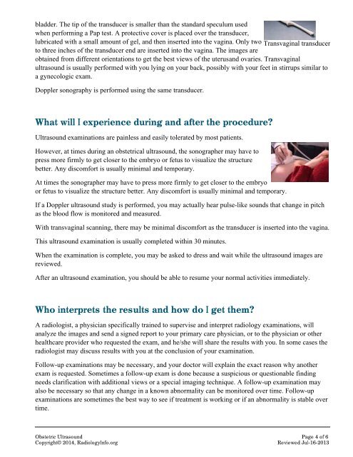
ladder. The tip of the transducer is smaller than the standard speculum usedwhen performing a Pap test. A protective cover is placed over the transducer,lubricated with a small amount of gel, and then inserted into the vagina. Only two Transvaginal transducerto three inches of the transducer end are inserted into the vagina. The images areobtained from different orientations to get the best views of the uterus and ovaries. Transvaginalultrasound is usually performed with you lying on your back, possibly with your feet in stirrups similar toa gynecologic exam.Doppler sonography is performed using the same transducer.What will I experience during and after the procedure?<strong>Ultrasound</strong> examinations are painless and easily tolerated by most patients.However, at times during an obstetrical ultrasound, the sonographer may have topress more firmly to get closer to the embryo or fetus to visualize the structurebetter. Any discomfort is usually minimal and temporary.At times the sonographer may have to press more firmly to get closer to the embryoor fetus to visualize the structure better. Any discomfort is usually minimal and temporary.If a Doppler ultrasound study is performed, you may actually hear pulse-like sounds that change in pitchas the blood flow is monitored and measured.With transvaginal scanning, there may be minimal discomfort as the transducer is inserted into the vagina.This ultrasound examination is usually completed within 30 minutes.When the examination is complete, you may be asked to dress and wait while the ultrasound images arereviewed.After an ultrasound examination, you should be able to resume your normal activities immediately.Who interprets the results and how do I get them?A radiologist, a physician specifically trained to supervise and interpret radiology examinations, willanalyze the images and send a signed report to your primary care physician, or to the physician or otherhealthcare provider who requested the exam, and he/she will share the results with you. In some cases theradiologist may discuss results with you at the conclusion of your examination.Follow-up examinations may be necessary, and your doctor will explain the exact reason why anotherexam is requested. Sometimes a follow-up exam is done because a suspicious or questionable findingneeds clarification with additional views or a special imaging technique. A follow-up examination mayalso be necessary so that any change in a known abnormality can be monitored over time. Follow-upexaminations are sometimes the best way to see if treatment is working or if an abnormality is stable overtime.<strong>Obstetric</strong> <strong>Ultrasound</strong> Page 4 of 6Copyright© 2014, <strong>RadiologyInfo</strong>.orgReviewed Jul-16-2013