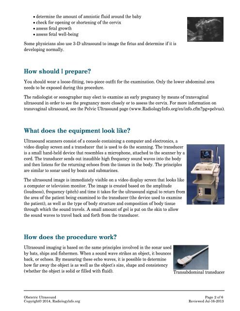
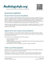
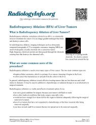
determine the amount of amniotic fluid around the babycheck for opening or shortening of the cervixassess fetal growthassess fetal well-beingSome physicians also use 3-D ultrasound to image the fetus and determine if it isdeveloping normally.How should I prepare?You should wear a loose-fitting, two-piece outfit for the examination. Only the lower abdominal areaneeds to be exposed during this procedure.The radiologist or sonographer may elect to examine an early pregnancy by means of transvaginalultrasound in order to see the pregnancy more closely or to assess the cervix. For more information ontransvaginal ultrasound, see the Pelvic <strong>Ultrasound</strong> page (www.<strong>RadiologyInfo</strong>.org/en/info.cfm?pg=pelvus).What does the equipment look like?<strong>Ultrasound</strong> scanners consist of a console containing a computer and electronics, avideo display screen and a transducer that is used to do the scanning. The transduceris a small hand-held device that resembles a microphone, attached to the scanner by acord. The transducer sends out inaudible high frequency sound waves into the bodyand then listens for the returning echoes from the tissues in the body. The principlesare similar to sonar used by boats and submarines.The ultrasound image is immediately visible on a video display screen that looks likea computer or television monitor. The image is created based on the amplitude(loudness), frequency (pitch) and time it takes for the ultrasound signal to return fromthe area of the patient being examined to the transducer (the device used to examinethe patient), as well as the type of body structure and composition of body tissuethrough which the sound travels. A small amount of gel is put on the skin to allowthe sound waves to travel back and forth from the transducer.How does the procedure work?<strong>Ultrasound</strong> imaging is based on the same principles involved in the sonar usedby bats, ships and fishermen. When a sound wave strikes an object, it bouncesback, or echoes. By measuring these echo waves, it is possible to determinehow far away the object is as well as the object's size, shape and consistency(whether the object is solid or filled with fluid).In medicine, ultrasound is used to detect changes in appearance, size orTransabdominal transducer<strong>Obstetric</strong> <strong>Ultrasound</strong> Page 2 of 6Copyright© 2014, <strong>RadiologyInfo</strong>.orgReviewed Jul-16-2013
In medicine, ultrasound is used to detect changes in appearance, size orcontour of organs, tissues, and vessels or detect abnormal masses, such as tumors.In an ultrasound examination, a transducer both sends the sound waves and receives the echoing waves.When the transducer is pressed against the skin, it directs small pulses of inaudible, high-frequencysound waves into the body. As the sound waves bounce off internal organs, fluids and tissues, thesensitive microphone in the transducer records tiny changes in the sound's pitch and direction. Thesesignature waves are instantly measured and displayed by a computer, which in turn creates a real-timepicture on the monitor. One or more frames of the moving pictures are typically captured as still images.Small loops of the moving “real time” images may also be saved.The movement of the embryo or fetus and his or her heartbeat can be seen as anongoing ultrasound movie. Most ultrasound devices also have an audio componentthat processes the echoes produced by blood flowing through the fetal heart, bloodvessels and umbilical cord. This sound can be made audible to human ears and hasbeen described by patients as a whooshing noise.Doppler ultrasound, a special application of ultrasound, measures the direction andspeed of blood cells as they move through vessels. The movement of blood cellscauses a change in pitch of the reflected sound waves (called the Doppler effect). Acomputer collects and processes the sounds and creates graphs or color pictures that represent the flow ofblood through the blood vessels.How is the procedure performed?For most ultrasound exams, you will be positioned lying face-up on an examination table that can betilted or moved.After you are positioned on the examination table, the radiologist or sonographer will apply a warmwater-based gel to the area of the body being studied. The gel will help the transducer make securecontact with the body and eliminate air pockets between the transducer and the skin that can block thesound waves from passing into your body. The transducer is placed on the body and moved back andforth over the area of interest until the desired images are captured.There is usually no discomfort from pressure as the transducer is pressed against the area beingexamined. However, if scanning is performed over an area of tenderness, you may feel pressure or minorpain from the transducer.Once the imaging is complete, the clear ultrasound gel will be wiped off your skin. Any portions that arenot wiped off will dry to a powder. The ultrasound gel does not stain or discolor clothing.Sometimes the radiologist determines that a transvaginal scan needs to be performed. This techniqueoften provides improved, more detailed images of the uterus and ovaries. This method of scanning isespecially useful in early pregnancy.Transvaginal ultrasound is performed very much like a gynecologic exam andinvolves the insertion of the transducer into the vagina after you empty your<strong>Obstetric</strong> <strong>Ultrasound</strong> Page 3 of 6Copyright© 2014, <strong>RadiologyInfo</strong>.orgReviewed Jul-16-2013