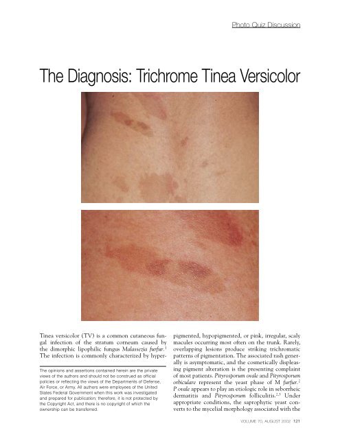
The Diagnosis: Trichrome Tinea Versicolor
The Diagnosis: Trichrome Tinea Versicolor
The Diagnosis: Trichrome Tinea Versicolor
- No tags were found...

Create successful ePaper yourself
Turn your PDF publications into a flip-book with our unique Google optimized e-Paper software.
Photo Quiz Discussion<strong>The</strong> <strong>Diagnosis</strong>: <strong>Trichrome</strong> <strong>Tinea</strong> <strong>Versicolor</strong><strong>The</strong> opinions and assertions contained herein are the privateviews of the authors and should not be construed as officialpolicies or reflecting the views of the Departments of Defense,Air Force, or Army. All authors were employees of the UnitedStates Federal Government when this work was investigatedand prepared for publication; therefore, it is not protected bythe Copyright Act, and there is no copyright of which theownership can be transferred.<strong>Tinea</strong> versicolor (TV) is a common cutaneous fungalinfection of the stratum corneum caused bythe dimorphic lipophilic fungus Malassezia furfur. 1<strong>The</strong> infection is commonly characterized by hyperpigmented,hypopigmented, or pink, irregular, scalymacules occurring most often on the trunk. Rarely,overlapping lesions produce striking trichromaticpatterns of pigmentation. <strong>The</strong> associated rash generallyis asymptomatic, and the cosmetically displeasingpigment alteration is the presenting complaintof most patients. Pityrosporum ovale and Pityrosporumorbiculare represent the yeast phase of M furfur. 2P ovale appears to play an etiologic role in seborrheicdermatitis and Pityrosporum folliculitis. 2,3 Underappropriate conditions, the saprophytic yeast convertsto the mycelial morphology associated with theVOLUME 70, AUGUST 2002 121
Photo Quiz Discussioncharacteristic scaling macules. Conditions associatedwith the mycelial transition are a warm humidenvironment, oily skin or an inherited predispositionfor it, Cushing disease, depressed cellularimmunity, or malnutrition. 4M furfur is readily identified by examining a simplepotassium hydroxide mount of skin scrapings.<strong>The</strong> characteristic “spaghetti and meatballs” appearanceof the hyphal and yeast elements confirms thediagnosis of TV, which makes culture unnecessary.Wood’s lamp examination reveals a yellowish toyellowish-green fluorescence and helps excludeerythrasma and tinea corporis from the differentialdiagnosis. <strong>The</strong> clinical differential diagnosis includesother entities that result in dyspigmentation, such asvitiligo, pityriasis alba, psoriasis, seborrheic dermatitis,confluent and reticulate (Gougerot-Carteaud)papillomatosis, erythrasma, and dermatophytosis. 4Several theories have been proposed to explainthe dyspigmentation seen in pityriais versicolor.Hypopigmentation may result from dicarboxylicacids such as azelaic acid produced by the fungus. 5Azelaic acid produces strong inhibition of the dopatyrosinereaction in vitro. <strong>The</strong>se same acids alsoexhibit a cytotoxic effect on melanosomes. 6-8Another possibility is that the scale of TV preventstanning in these areas. 9 Hyperpigmentation in TV isnot fully understood. An increased thickness of thekeratin layer in hyperpigmented individuals 7 and aninflammatory response resulting in stimulation ofmelanocytes are possible mechanisms. 4Treatment regimens include 2.5% seleniumsulfide shampoo applied liberally and allowed toremain for 10 minutes to overnight before washingoff. Ten-minute applications may be repeated dailyfor 2 weeks and then weekly to monthly to preventrecurrence. Overnight applications generally are performedmonthly as needed. Topical azole antifungals,such as clotrimazole and ketoconazole are effectivein treating TV, and topical terbinafine has beenshown to be effective in both cream and gelforms. 10,11 Terbinafine spray has been marketed forthe treatment of TV. It is unclear how large a contributionthe vehicle makes to the therapeutic effect ofthe product. Although topical treatment is ideal forcases with limited involvement, systemic therapyoffers greater convenience when treating widespreaddisease. Systemic therapy with a single dose of ketoconazole(400 mg) was shown to be as effective as200 mg daily for 10 days. 12 Pityriasis versicolor oftenis recurrent, and repeat or maintenance treatmentmay be necessary. This can be accomplished withweekly or monthly topical selenium sulfide treatments,weekly washing with benzoyl peroxide or zincpyrithione soap, or periodic systemic therapy.REFERENCES1. Gordon AM. Lipophilic yeast-like organisms associatedwith tinea versicolor. J Invest Dermatol. 1951;17:267-270.2. Martin AG, Kobayashi GS. “Pityrosporum infections” ofthe skin: papulosquamous tinea versicolor, “Pityrosporum”folliculitis, and invers tinea versicolor. In: Freedberg IM,Eisen AZ, Wolff K, et al, eds. Fitzpatrick’s Dermatology inGeneral Medicine. 5th ed. New York, NY: McGraw HillCo.: 1999;2358-2371.3. Faergemann J. Pityrosporum yeasts—what’s new? Mycoses.1997;40(suppl 1):29-32.4. Sunenshine PJ, Schwartz, RA, Janniger CK. <strong>Tinea</strong><strong>Versicolor</strong>. Int J Dermatol. 1998;37:648-655.5. Galardi I, elKomy M, Mousa A, et al. <strong>Tinea</strong> versicolor: histologicand ultrastructural investigation of pigmentarychanges. Int J Dermatol. 1992;31:253-256.6. Hattori M, Ogawa H, Takamori K, et al. De-(hypo)pigmentationmechanism of affected area of pityriasis versicolor.J Dermatol. 1984;11:63-66.7. Charles CR, Sire DJ, Johnson BL, et al. Hypopigmentationin tinea versicolor: a histological and electron microscopicstudy. Int J Dermatol. 1973;12:48-58.8. Nazzaro-Porro M, Passi S, Zina G, et al. Ten years’ experienceof treating lentigo maligna with azelaic acid. J InvestDermatol. 1989;143:49-57.9. Allen HB, Charles CR, Johnson BL. Hyperpigmented tineaversicolor. Arch Dermatol. 1976;112:1110-1112.10. Faergemann J, Hersle K, Nordin P. Pityriasis versicolor:clinical experience with Lamisil cream and Lamisil DermGel. Dermatology. 1997;194(suppl 1):19-21.11. Savin R, Eisen D, Fradin MS, et al. <strong>Tinea</strong> versicolor treatedwith terbinafine 1% solution. Int J Derm. 1999;38:863-865.12. Fernandez-Nava HD, Laya-Cuadra B, Tianco EA. Comparisonof single dose 400 mg versus 10-day 200 mg dailydose ketoconazole in the treatment of tinea versicolor.Int J Dermatol. 1997;36:64-66.122 CUTIS ®