Create successful ePaper yourself
Turn your PDF publications into a flip-book with our unique Google optimized e-Paper software.
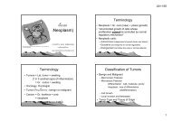
11/22/2011Esophageal varicesFigure 17-7 Barrett esophagus. Microscopic view showing squamous mucosa and intestinal-type columnar epithelial cells (goblet cells) in a glandular mucosa.Downloaded from: Robbins & Cotran Pathologic Basis of Disease (on 12 November 2007 02:58 AM)© 2007 Elsevier• เปนภาวะที่มีการโปงพองของหลอดเลือดที่ submucosa ของหลอดอาหารสวนลาง• สวนใหญพบในผูปวยที่มีตับแข็ง/Cirrhosis เนื่องจากมีProlonged and severe portalhypertension• สวนใหญเปนสาเหตุทําใหผูปวยอาเจียนเปนเลือดรุนแรง/ Severehematemesis• เนื่องจากเลือดใน portal vein ไมสามารถผานจากตับเขาสู inferiorvana cava แบบปกติได ผลทําใหเลือดไหลยอนผานทาง coronaryveins ของกระเพาะอาหารไปที่esophageal subepithelial andsubmucosal veins (varices) มากขึ้นเพื่อไปเชื่อมกับ azygous veinsและsystemic circulation• ผูปวยตับแข็ง นอกจาก esophageal varices แลวจะมีเสนเลือดโปงพองที่ rectal canal (hemorrhoids) และ falciform ligament (caputmedusa) เชนกันNeoplasm of Esophagus• BENIGN TUMORS พบไดทั้งในผนังหลอดอาหาร /Intramural และ ที่ submucosal ไดแก Leiomyoma, fibroma,lipoma, hemangioma, neurofibroma, lymphangioma หรือ ที่mucosa ไดแก Squamous papilloma, fibroepithelial polyp,และinflammatory polyp• MALIGNANT TUMORS ไดแก มะเร็งของเยื่อบุที่เรียกวาEsophageal carcinomasและ Malignant stromal tumors5