View/Open
View/Open
View/Open
- No tags were found...

You also want an ePaper? Increase the reach of your titles
YUMPU automatically turns print PDFs into web optimized ePapers that Google loves.
DOSEMETRIC SURVEY INDIAGNOSTIC RADIOLOGYA THESIS SUBMITTED IN FULL RERSRCH OF THEREQUIREMENTS FOR THE DEGREE OF Ph.D. OF SCIENCEIN NUCLEAR PHYSICSBYKOUTHER ELHAJ MOHAMED MOHAMADAINUNIVERSITY OF KHARTOMFACULITY OF SCIENCEDEPARTMENT OF PHYSICSOCTOBER 2004
DEDICATIONTO WHOMI HAVE ALWAYS LOVED MY PARENTSAND MY LOVELY FAMILLYII
AcknowledgementsI wish to express my sincere appreciation to Dr. Faroug Habbani ( khartoumuniversity -Sudan ), Dr. Ana Cecilia (FUCRUZ), and Dr. Luis Rosa (IRD) Brazilfor their supervision.My appreciation to IAEA in Sudan and the Institute of Radiation Protection (IRD)and to the medical physics head department Dr. Helvaiso Mota for their keen interestand nessary assitance during the practical workThanks are due to the staff of the Diagnostic Radiology Departments at IPPMG IFF,HGB hospitals in Brazil and Ahmed gasim, Khartoum,Ummdurman hospitals inSudanI am gratefully Thanks Dr. Mohamed Elmusallmy and Dr. Amal Elzubair for theirsupportThanks are due to ICTP ,TWOWS , Trieste , Ministry of Higher Education, andSudan universitry of science and technology -Sudan for the faintail supportOf curse many thanks must go also to my wonderful family for their encouragementeasptially my husband BurhanIII
AbstractA dosimetric survey in pediatric radiology and adults patients is currentlybeing carried out at the pediatrics units of two large hospitals in Rio de Janeiro city:IPPMG (Instituto de Pediatria e Puericultura Martagão Gesteira, UniversityHospital of Federal University of Rio de Janeiro), IFF (Instituto FernandesFigueira, FIOCRUZ) and Hospital Geral de Bonsucesso, a large public hospital inRio de Janeiro city (HGB)Brazil. And three pediatrics’ units in Sudan namelyAhmed gasim ,Khartoum and Umdurman hospitalsFor Chest x-ray examination the ESD for AP, PA and LAT projections ofpediatrics patients, and the scattered dose at the thyroid ,ovary and gonads havebeen obtained with thermo luminescent dosimeters (TLD) and with use of asoftware package DoseCal in the Brazilian hospitals , and with the SoftwareDosecal in the Sudanese hospitals. The aim of this work is to estimate the EntranceSkin Dose (ESD), the effective dose (ED) and the Body Organ Doses (BOD) forchest x-ray exposure in pediatric patients, and different exams for adults patients,and to compare the results obtained in the two countries Sudan and Brazil with thereference dose level .For ESD evaluation of the chest X-ray , three different TL dosimeters havebeen used, namely LIF:Mg,Ti (TLD100), CaSO 4 :Dy and LiF:Mg,Cu,P (TLD100H).The age intervals considered were: 0-1 year, 1-5 years, 5-10 years and 10-15 years.The results obtained with all dosimeters are similar, and it is in good agreementwith the DoseCal software, especially for AP and PA projections. However, somelarger discrepancies are presented for the LAT projection. And the results withinBrazil are somewhat consistent while in Sudan, large difference was found.IV
Therefore, a wide distribution of doses has been obtained, also the Dose in Brazil isless than the reference dose level while in some Sudanese hospitals it is higher thanthe reference dose levelFor adult’s patients only the Software Dosecale has been used to measure theESD, ED and BOD. for different exams and projections Abdomen , skull, Lumbarspine ,cervical spine , pelvis ,chest for AP,PA and LAT projectionsV
Contents:AbstractAcknowledgementsList of figures:-List of tables :-page NumberIIIIIIXXCHAPTER ONE1.1 Introduction 11.2 Objectives 91.3 lietrurer reiview 10References 18CHAPTER TWOTHEORETICAL BACKGROUND2.1 Radiation Quantities and Units 202.1.0 Introduction and overview: 202.1.1 Exposure:2.1.2 Energy Fluence ψ: 212.1.3. Kerma: 212.1.4 Relation of kerma to energy fluence for photons 232.1.5 Surface Integral Exposure: 232.1.6 Absorbed Dose 242.1.7 Integral Dose : 252.1.8 Biological Impact: 262.1.9 Dose Equivalent: 262.2 Radiation measurement :2.2.0 Introduction: 272.2.1Ionization in air as the primary radiation standard 272.2.2 The ionization Chamber : 282.2.3 The Geiger Muller Counter 302.2.4 The Geiger - Muller tube 322.2.5 Secondary ionization chambers 342.2.6 Dose -Area product meters 372.2.7 Pocket exposure meters for personal monitoring 382.3.Thermo Luminescence Dosimetry2.3.0 The thermoluminescence process 392.3.1 The Glow curve 402.3.2 Calibration of thermoluminescence 422.3.3TheThermoluminescence process in Lithium 42fluoride(LiF:Mg,Ti)2.3.4 The Role of Magnesium ion 422.3.5 Advantages and disadvantages of TLD's 432.4 DoseCal Software2.4.1 Overview 432.4.2 Installation 442.4.3 Minimum Requirements 442.4.4 Limitations 44VI
References 45CHAPTER THREEExperimental Technique and Material Properties:3.0 Introduction 463.1 Linearity 463.2 Dose response 483.3Response to photon 493.4 Fading 503.5Sensitivity 533.6Annealing process 533.7Stability and Reproducibility 543.8 Dose rate dependence 553.9Light Sensitivity 553.10Environmental factors 563.11Tribothermolumence 563.12Precision and accuracy 573.12.1Precision 573.12.2 Accuracy 573.12.3Random uncertainties 583.12. 4 systematic uncertainties 593.12. 5 Accuracy of TL measurements 603.12.6 Accuracy in low dose dosimeter 603.13 TLD Reader3.13.1Basic TLD Reader 613.13.2 Automatic TLD reader system 623.13.3 Heating system 633.14 LiF:Mg,Ti.3.14.1Introduction 643.14.2General Physical Properties 653.14.3 Tl glow curves 663.15 LiF,Mg,Cu,p3.15.1General Physical Properties 683.15.2 Tl glow curves 693.16 CaSO 4 :Dy3.16.1Introduction 723.16.2General Physical Properties 723.16.3 Tl glow curves 73References 75CHAPTER FOURMaterial and methods4.0 Thermolmiescent Dosimeters4.0.1 The reading conditions 794.0.2 The annealing conditions 794.1 DoseCal Software4.1.1 Using the software 814.1.2 Dose Cal input page 834.1.3 Dose Cal result page 844.1.4 Dose Cal organ dose 85VII
CHAPTER FIVEResults5, 1 - Test of reducibility, sensitivity and calculation of Si factor 865, 2 - Calibration Factor of the TLD’S and the threshold dose 865, 2, 1 - Calibration of LiF:Mg, Ti, CaSO 4 :Dy & LiF:Mg, Cu, P 865, 2, 2 - The Threshold Dose 875, 3 - The reading and annealing conditions of the TLD’S 875,4 ESD& Scatter Dose of chest x-ray examination 885, 5 - Results obtained in IFF pediatric Hospital using the TLD’S 895, 5, 1 - Technical factors used for chest x-ray examination , ESD, andscatter Dos for mobile X-ray 915, 5, 2 - ESD & Scatter Dose for Chest X-ray Examination in IFF hospital5, 6 - The software results in Brazil 925, 6, 1 - Results of ESD for chest x-ray using the TLD’s and software In 98IPPMG hospital5, 6, 2 – Results of ESD for chest x-ray using the TLD’s and software 98In IFF hospital5, 7 - The software results in Sudan5, 7, 1 - Oumdurman Hospital 1005, 7, 2 - Khartoum Hospital 1015,7,3-AhmedGasimHospital 1025, 8- Comparison of ESD, ED and BOD in Sudan and Brazil hospitals 102for chest X-ray AP and PA projections using the software Doscal5,9 - ESD and EDobtained for adults patients in IFF and HGB 107hospitals using the software5,10- Values of ESD and ED for mobile equipment for the AP 108projections of chest and abdomen in IFF5,11- ESD and ED for several projections in AP, PA and LAT 109for peadiatrics patients in IFF.CHAPTER SIXDiscussions and conclusions 110Action to be taken in each hospitalsReferences 120Appendix A, B and C 121122VIII
List of figures:-Fig (1.1) Dose Response curve 8Fig (2.1) Relationship 0f absorbed dose to exposure 25Fig (2.2) The free air ionization chamber.(From Whyte1959.) 28Fig (2.3) Simplified electrical circuits for measuring(a) current flow (exposure rate), (b) total charge exposure. 29Fig (2.4) Variation in current appearing across capacitor plateswith applied potential difference . 31Fig(2.5) Amplification of ionization by the electric field 32Fig (2.6) Essential features of an end -window Geiger-Muller 32tube suitable for detecting beta particles .Fig (2.7) secondary electron flux in a gas -filled cavity. 35Fig (2.8) A schematic demonstration that the flux of secondaryelectrons at equilibrium is in dependent of the density of the stoppingmaterial 36Fig (2 .9) a simple compact secondary ionization chamber thatmakes use of the 'air equivalent wall ' principle 37Figure(3.1) A representative glow curve from LiF;Mg,Ti.This curve was obtained from TLD-100 66Fig (3.2) (a) An isometric plot of thermo luminescenceemission from TLD-100 67Fig (3.3) Representive glow curve fromLiF;Mg,Cu,P 70Fig (3.4) (a) An isometric plot of thermo luminescenceemission from LiF:Mg,Cu,p 71Fig (3.5)(a) Glow curves from CaSO4:Dy with or without impurity.(b) Glow curves of CaSO4:Dy (high Na)for various annealing treatments 73Fig (3. 6 ) show the TL emission spectrum from CaSo4:DyHeating rate = 2.5 0Cs-1 pre irradiation anneal :1h at 400 0C 74Fig 4.1 TLDs reader and Oven used in this thesis 80Fig (4.1 ) DoseCal Input Page.Fig (4.2) DoseCal Results Page. 83Fig (4.3) DoseCal Organ Doses Page. 84Fig (5.1) shows the ESD obatin in IPPMG hospital using the 90TLD' for chest AP,PA and LAT projectionsFig 5.2) shows the Body Organ dose (BOD) 99for chest AP/PA in IPPMG hospitalFig (5.3) shows the ESD in mGy obtained in IFF hospital 99for the convential X-ray and fluoroscopy for chest AP,PA and LAT 101Fig (5.4) shows the result obtained in Sudan and Brazilcompared with the reference dose levelFig (5.5) ) shows the minimum, maximum and meanvalues of ESD as compared to reference values and to 106results from other European countries. Most valuesare comparable with the reference level adopted in EuropeIX
List of tables :-Table (2.1) Reported from literature for LiF :Mg ,Ti and CaSO 4 :Dy 41Table (3.1) Linearity ranges 47Table (3.2) glow peak temperature and half –livefor different TL phosphor 51Table (3.3) Fading Characteristics. 52Table (3.4) Annealing procedures. 54Table (4. 1) The reading conditions. 79Table (4. 2) The annealing conditions. 79X
1.1 INTRODUCTIONAfter the discovery of the X-ray in 1895 by W.C.Roentgen, the use of X-ray in medicinehas increased fast. More recently, new technologies and modern equipments have beenincluded as part of diagnostic resources ofradiology departments. However the necessary condition and control for the use of suchdevices has not been taken into account in most installation accordingly.In most courtiers, several initiatives have been implemented in order to regulate the use ofthe ionizing radiation with the best image quality lowest doses and reduce the cost to thedepartment; the most efficient imitative is the implementation of QAPThe World Health Organization (WHO) has defined Quality Assurance in x-ray medicaldiagnosis as “an organized effort by the staff operating a facility to insure that thediagnostic images produce by the facilities are of sufficient high quality so that theyconsistantly provide adequate diagnostic information at the lowest possible cost and withthe least possible exposure of the patient to radiatio”. However the most important inliterature on the design and implementation of Quality Asuurance (QA)programmess fordiagnostic radiology exists(NCRP,1988;WHO, 1982)Quality assurance (QA) is a program used by management to maintain optimal diagnosticimage quality with minimum hazard and suffering to patients. The program includesperiodic quality control tests, preventive maintenance procedures, administrative methodsand training. It is also includes continuous assessment of the efficacy of the imagingservice and the means to initiate corrective action, the primary goal of radiology qualityassurance is to insure the consistent provision of prompt and accurate diagnosis of patients.A QA program having the following three objectives will adequately meet this goal• To maintain the quality of diagnostic images• To minimize the radiation exposure to patient and staff1
• To be cost effectiven 1998, the Brazilian Sanitary Surveillance and the Ministry of Health of Brazilpublished the decree 453 [1] establishing radiation protection guidelines for diagnosticradiology in medicine and odontology. Among the legal exigencies contained in the decree,it is mandatory the implantation of Quality Assurance Programs (QAP) in all medicalinstitutions that use ionising radiations. In the field of odontology, the Instituto deRadioproteção e Dosimetria (IRD), Brazil, has been developing a very importantprogramme since 1980 [2] with good results.In Sudan, the National Assembly in 1996, issued the Sudan Atomic EnergyCommission (SAEC) Act. Under this Act, a policymaking Board was established by theCouncil of Ministers. The SAEC Board, within its mandate, established the RegulatoryAuthority Radiation Protection Technical Committee (RPTC). It is a national committeeresponsible for the development of the radiation protection legal framework, licensingprocedures, policy making and approving inspections.Biological effects of Ionizing radiationTo minimize the radiation exposure to patient first we must consider the effect ofionizing radiation which may be immediate or delayed . Some tissues are highlyradiosenstive and each tissue has its own risk factor. Ionizing radiation acts on the cells ofthe human body. If the cells do not repair themselves, permanent effects of radiationdamage can be observed as biological changes in a tissue or organ. These changes may bemanifested as medical symptoms, which are classified into deterministic or stochasticeffectsIn Deterministic effects the most common result of radiation damage isfor the cell to die. If only a few cells are affected, it is not usually a problem as there are2
many cells in the body and new cells will replace the dead cells. However, as the amount ofradiation absorbed (later referred to as the dose) increases, a point will be reached wheresufficient cells are killed to affect the overall operation of the organ. The result of this is aloss of organ function, which will become more serious as the number of affected cells isincreased.The different types of radiation damage resulting from the loss of organ function are knownas deterministic effects. These effects are characterized by having a threshold dose (belowwhich there is no observable effect) followed by a response where the severity of the effectincreases with increasing radiation dose.Examples of deterministic effects are: erythematic or skin reddening, cataracts (followingirradiation of the eye lens).Early effects are normally observed within a few days or weeks after the exposure. Lateeffects take several years to develop (cataract).For Stochastic effects sometimes, the effect of radiation is not to kill the cell but toalter it in some way. In most cases this alteration will not affect the cell significantly sothere will be no observable effect. However there is a possibility that the injury might affectthe control system of the cell, subsequently causing it to divide more rapidly than normal.If the affected cell does begin to divide in this way, an increasing number of abnormaldaughter cells will be produced. If these abnormal cells invade normal tissue they are calledmalignant cells and this results in cancer.The type of cancer formed is dependent on the type of the original cell, which was altered.Cancers do not appear immediately after radiation exposure but appear after a latencyperiod in which no effects are observable. The latency period is dependent on the type of3
cancer but can vary from two years for leukemia to thirty years or possibly longer for somesolid cancers. Hence, cancer is classified as a late effect.Unlike deterministic effects, the amount of radiation exposure does not change the severityof the cancer but it does alter the chance of getting cancer. In other words, exposure to ahigh dose can increase the risk of getting a cancer but if cancer occurs (whether it be at lowor high dose) the severity of the cancer is the same. The same is true with hereditaryeffects. Such effects relying on chance are referred to as stochastic effects. For the purposesof radiation protection, it is assumed that the probability of a stochastic effect increaseslinearly as the dose increases and that there is no threshold dose. If there is no thresholddose then it is considered that even small doses of radiation might cause cancer.Stochastic effects are the only effects possible at low doses and hence, radiation protectionis aimed at preventing deterministic effects and reducing the chances of stochastic effectsoccurring to both the public and radiation workers.In a fetus, or unborn child, the number of cells is less than in the adult and the cellsare rapidly dividing. As radiation damage is more extensive on dividing cells, the fetus isparticularly sensitive to radiation damage and exposure to high doses could result inpossible malformation or even death. For this reason and because it is possible thatexposure to radiation at any time in pregnancy may increase the probability of cancer in thechild, special radiation protection measures are necessary to protect the fetus.Studies have indicated that exposure to radiation during the first two weeks of pregnancydoes not result in deterministic or stochastic effects to the child. But between week two andtwenty four of pregnancy, high doses to the mother can cause major organ damage to thedeveloping fetus. The central nervous system could also be severely damaged resulting inmental retardation. At very high doses, effects such as spontaneous abortion, malformationand reduction in intelligence have been observed, the frequency and severity of these4
effects varying with both dose and stage of fetal developmentThe need for special quality assurance (QA) programmes for pediatric patients werefirst realized early in 1980s for the reason that radiation protection in pediatric radiologydeserves special attention since it is generally assumed that children are at ages up to 10years are about factor three more sensitive to radiation than the average population and therisk for late effects after exposure to X-rays is higher for infants and children than for theadults, also the children may live for long time andExaggerated by delayed effects of radiation. Besides cancer, which is the main delayedeffect, hereditary effects are caused by damage to the reproductive cells by ionizingradiation: there is a chance that this damage may affect either the immediate child orsubsequent generations. Hereditary effects are based on chance and hence are stochastic.However, the risk of hereditary effects is much lower than the risk of cancer.The relationship between dose and effect for given conditions of exposure (type ofradiation and dose-rate) allows the definition of the tissue specific risk factor, which is theratio between the increase in probability of a given stochastic effect (tissue or organspecific cancer) and the corresponding dose. It is usually expressed in excess probability ofa given cancer attached to a given tissue or organ for a unit increase of dose. To take intoaccount the fact that some tissues and body organs are more sensitive to radiation thanothers and a dose in one organ may be more risky than the same dose in another organ., theICRP recommends tissue-weighting factors (w T ), which are applied to specific bodyorgans. These dimensionless factors take into consideration the different radiosensitivitiesof the different organs and tissues.Very high doses of radiation to the whole body can cause sufficientdamage to the organs to stop their function and this may ultimately cause death. The acuteradiation syndrome (nausea, vomiting, diarrhea,) is an early deterministic effect resulting5
from an acute high dose to the whole body (over one or a few Sv). Other effects resultingfrom acute exposure are damage to white blood cells, and ultimately to the central nervoussystem (about 50 Sv).Radiation illness follows a three-stage progression. Shortly after the exposure the personfeels sick and may vomit. This is caused by the build-up of toxins caused by the exposure.After about one day the person begins to recover; this recovery stage might last severalweeks. The third stage of the illness is caused by the more serious damaging effects of theradiation exposure, blood count depletion, gastrointestinal damage etc, and may result indeath depending on the magnitude of the dose and the availability of prompt specialistmedical treatment.Ionizing radiations cannot be seen, felt or sensed by the human body in any way butexcessive exposure to them may have adverse health effects. In order to avoid excessiveexposure, appropriate and efficient radiation measuring instruments are needed, such asThermolumence Dosimetry (TLD), which is widely used for patient dosimetry indiagnostic radiology; these dosimeters are very sensitive to radiation. TLD dosimetrymaterials such as lithium fluoride or lithium borate are approximately tissue equivalentand consequently are practically invisible on most radiographs. This means that the useof these dosimeters does not interfere with the clinical diagnosis.Also it is possible to calculate the patient dose using knowledge of the X-ray output,tube voltage, exposure time, focus skin distance and the backscatter factorPatient dosimetery is now regarded as integral part of the QA process as well asradiological audit. It is fundamental end point by which the effect of audit or qualityassurance procedure can be assessed6
Fig (1.1) Dose Response curve%EffectRepairing cellstructures is stillpossiblePractically all thecells are deadNo repairing: a low ∆dosemeans a great damage∆dosDose7
1.2 Objective :In the light of the above mentioned factors of the biological effects of ionizingradiation in patients and the need of including patients dosimetry in QAP this workpropsed to1. Make survey of radiation doses in pediatrics patients in some public anduniversity hospital.2. To measure the Entrance surface Dose (ESD). Effective dose (ED) and BodyOrgan dose (BOD), for different examinations.3. Compare the results obtained for the different hospitals in Brazil and Sudan.4. Identify the most probable causes of dose value differences5. Proposed appropriate actions to be taken in each hospitals6. Compare the results obtained using the TLDs as dose measurements andDosecal software as dose calculations.8
1.3 Literature ReviewSimilar work were done in different countries this work are presented according to thetime of publication, starting with recent one2002Radiation dose quantities and risk in neonates in a special care baby unit. Ref.(1)Abstract:Radiographs are taken in the neonatal period most commonly to assist in the diagnosis andmanagement of respiratory difficulties. Frequent accurate radiographic assessment isrequired and a knowledge of the radiation dose is necessary to justify such exposures. Asurvey of radiation doses to neonates from diagnostic radiography (chest and abdomen) hasbeen carried out in the special care baby unit of the Royal Free Hospital. Entrance surfacedose (ESD) was calculated from quality control measurements on the X-ray unit itself. Directmeasurement of radiation doses was also performed using highly sensitivethermoluminescent dosemeters (TLDs) (LiF:Mg,Cu,P), calibrated and tested for consistencyin sensitivity. ESD, as calculated from exposure parameters, was found to range from 28µGy to 58 µGy, with a mean ESD per radiograph of 36±6 µGy averaged over 95examinations. ESDs as derived from TLD crystals ranged from 18 µGy to 58 µGy for 30radiographic examinations. The mean energy imparted, the mean whole body dose perradiograph and the mean effective dose were estimated to be 14±8 µJ, 10±4 µGy and 8±2µSv, respectively. Assuming that neonates and fetuses are equally susceptible tocarcinogenic effects of radiation, which involve an overestimation of risk, the radiation riskof childhood cancer from a single radiograph was estimated to be of the order (0.3–1.3) x10 -6 . Radiation doses compared favourably with the reference values of 80 µGy ESDpublished by the Commission of the European Communities in 1996, and 50 µGy publishedby the National Radiological Protection Board in 2000.2002Irish X-ray departments demonstrate varying levels of adherence to Europeanguidelines on good radiographic technique Ref. (2)Abstract:The Commission of European Communities (CEC) publication "European Guidelines onQuality Criteria for Diagnostic Radiographic Images" includes examples of goodradiographic technique for a number of common X-ray examinations. If these guidelines arefollowed, compliance with dose and image quality criteria as specified in the CEC documentshould be demonstrated. Studies in England, Germany and Greece have shown that anumber of X-ray departments are not using optimum techniques. The aim of this study is todemonstrate the level of adherence to CEC guidelines in Irish hospitals 3–4 years followingpublication of the above document. 16 hospitals were randomly chosen and the followingdetails on technique and equipment were recorded for chest, abdomen, pelvis and lumbarspine examinations of standard sized patients: tube potential, focus-to-film distance,automatic exposure control (AEC), film–screen combination, X-ray tube filtration andsecondary radiation grid. Varying levels of adherence to the guidelines were evident9
depending on the parameter being investigated, with no hospital demonstrating 100%compliance and no hospital demonstrating 100% non-compliance. For all parameters, withthe exception of AEC use, the majority of hospitals exhibited non-adherence for at least oneprojection. The results suggest that if hospitals in Ireland observe the straightforwardexamples of good radiographic technique described in the CEC publication, significantreductions in collective dose can be achieved.2001The effect of beam tube potential variation on gonad dose to patients during chestradiography investigated using high sensitivity LiF: Mg, Cu, P thermo luminescentdosimeters Ref. (3)Abstract:Optimization of X-ray beam tube potential (kVp) in radiological examinations canminimize patient dose. This research aims to investigate the effect of tube potentialvariation on gonad doses to patients during posteroanterior (PA) chest radiographyexaminations. This study was carried out using a Toshiba general purpose X-ray unit and aRando phantom. Dose measuring equipment included an ion chamber system, a dose–areaproduct (DAP) meter and a thermoluminescent dosemeter (TLD) reader system with highsensitivity TLD pellets of LiF:Mg,Cu,P for low level gonad dose measurement. PA chestexposures of the phantom to produce a constant exit dose were made using a standard lowtube potential (range 60–100 kVp) non-grid technique and a high tube potential (range 95–150 kVp) grid technique. Entrance surface doses (ESDs) and DAPs were also included inthe measurements. Effective doses (EDs) were computed from ESD and DAPmeasurements using NRPB-SR262 and Xdose software. Results show that with the lowtube potential technique both ovary dose and testes dose increase with increasing tubepotential; statistically significant correlations of r=0.994 (p=0.0006) and r=0.998 (p=0.001),respectively, were found. For both organs, doses increase at a rate of approximately 2% perkVp. With the high tube potential technique there is insignificant correlation between gonaddoses and tube potential. When comparing patient doses from typical exposures made at 70kVp. (low tube potential non-grid technique) with doses from exposures made at 120 kVp(high tube potential grid technique), the high tube potential technique delivers significantlyhigher values for ESD, and ovary, testes and effective doses by factors of 1.7, 5.2, 5.5 and2.7, respectively.2000Assessment of Effective Dose in Paediatric Radiology: A Survey at 14 DutchHospitals Ref.(4)Abstract:Radiation protection in paediatric radiology deserves special attention since it is assumedthat children are more sensitive to radiation than are adults. This study aimed atestablishing the dose for three frequently applied projections in paediatric radiology at 14Dutch hospitals. Measured entrance doses are a factor 2.5 to 4.5 lower in comparison withthe criteria of the European Commission for radiation dose to the patient. An evaluation oftechniques applied at the different hospitals shows that there is no consensus on the optimalradiographic technique. In consequence, variations in entrance dose with a factor 3 to 1010
were observed. The highest effective doses were found for pelvis radiographs andradiographs of the abdomen of 5-year-old children, i.e. 26 µSv and 43 µSv respectively.For the other investigations average effective doses vary from 5 to 10 µSv.2000The Establishment of Reference Doses in Paediatric Radiology as a Functionof Patient Size. Ref.(5)Abstract:There is a wide range in paediatric patient size from a newborn baby to a 15-year-oldadolescent. Reference doses for paediatric radiology can sensibly be established only forspecific sizes of children. Five standard sizes of patient have been chosen at ages 0(newborn), 1, 5, 10 and 15 years. Standard AP and lateral thickness for the head and trunkfor the reference ages were derived from published measurements on children.Normalization factors for entrance surface dose and dose-area product measurements werecalculated which depend on the thickness of the real patient, the thickness of the neareststandard 'patient', and an effective linear attenuation coefficient (µ). These normalizationfactors were applied to European data to derive some preliminary reference doses.2000Reference dose levels for patients undergoing common diagnostic X-ray examinations inIrish hospitals. Ref. (6)Abstract:Wide variations in patient dose for the same type of X-ray examination have been evidentfrom various international dose surveys. Reference dose levels provide a framework toreduce this variability and aid in the optimization of radiation protection. The aim of thisstudy was to establish, for the first time, a baseline for national reference dose levels inIreland for four of the most common X-ray examinations: chest, abdomen, pelvis and lumbarspine. Measurements of entrance surface dose using thermoluminescent dosemeters (TLDs)for these four X-ray examinations were performed on 10 patients in each of 16 randomlyselected hospitals. This represented 42% of Irish hospitals applicable to this study. Resultshave shown wide variation of mean hospital doses, from a factor of 3 for an anteroposteriorlumbar spine to a factor of 23 for the chest X-ray. The difference between maximum andminimum individual patient dose values varied up to a factor of 75. Reasons for these dosevariations were complex but, in general, low tube potential, high mAs and low filtrationwere associated with high-dose hospitals. This study also demonstrated lower reference doselevels of up to 40% when compared with those established by the UK and the Commissionof the European Communities for four out of six projections. Only the chest X-ray exhibited asimilar reference level to those established elsewhere. This emphasizes the importance ofeach country establishing its own reference dose levels that are appropriate to their ownradiographic techniques and practices in order to optimize patient protection.200011
Measurement of Entrance Skin Doses to Patients in Four CommonDiagnostic Examinations by Thermoluminescence Dosimetry in Nigeria. Ref.(7)Abstract:Entrance surface doses (ESD) to patients were monitored in four common diagnostic X rayexaminations (chest, hand and wrist, lumbar spine and skull) at the outpatient X raydepartment of the University College Hospital, Ibadan. Measurement was based onthermoluminescence dosimetry (TLD) techniques using lithium fluoride discs and a VintenSolaro TLD reader. Some spread was observed in the values of doses received by patientsfor each of the examinations. It is greatest for the examination of the skull PA and least forthe examination of the skull LAT and lumbar spine AP; due mainly to variations in thesizes of the patients examined. For all examinations the values of the mean ESD obtainedrange from a minimum of 0.310 ± 0.071 mGy to a maximum of 5.66 ± 0.782 mGy for theexamination of the hand and wrist PA and lumbar spine LAT, respectively.2000A Regional Dose and Image Quality Survey for Chest, Abdomen and PelvisRadiographs in Paediatrics. Ref. (8)Abstract:A dosimetric survey in paediatric radiology is currently being carried out, aiming at theassessment of patient dose and image quality for chest, abdomen and pelvis radiographs insome age categories at five hospitals in the Tarragona area. Entrance surface dosemeasurements were performed using homogeneous PMMA phantoms. Effective doseswere assessed through the application of published conversion factors. The range ofentrance doses averaged by sites was 75-729 µGy for pelvis radiographs of children aged 5months, 813-1600 µGy for pelvis radiographs of children aged 5 years, 94-250 µGy forchest radiographs of children aged 5 years and 980-2300 µGy for abdomen radiographs ofchildren aged 5 years. The reference dose values given in the European Guidelines onQuality Criteria for Diagnostic Radiographic Images in Paediatrics were exceeded at two ormore hospitals for all projections. The range of average effective dose for the analyzedexaminations was 14-245 µSv. The maximum ratios of effective dose by sites variedbetween 2.2 and 11 for the analyzed projections. By examination type, average values inthe range 100 to 245 µSv were estimated for 5 year pelvis and abdomen examinations.12
1998Analysis of the status of X-ray diagnosis in Ghana. Ref. (9)Abstract:In Ghana's healthcare system there are about 4200 people to each physician. The annualfrequency of X-ray examinations during the period 1990 to 1996 ranges from six to 11 perthousand population. Chest radiograph examinations account for 46.5% of the annualfrequency. The survey revealed that there are no established acceptance testing proceduresfor newly installed X-ray equipment. Neither institutional level performance checksfollowing major repairs of faulty equipment, nor routine checks at regular intervals to ensureself-consistency of equipment performance are conducted. The results of the film retakeanalysis undertaken indicates a need for quality assurance programmes to be taken seriouslyto avert considerable cost and high patient doses. Radiographers and X-ray-technical officerswho physically perform X-ray examinations should receive adequate training in the selectionof procedures so as to ensure that doses to patients are as low as reasonably practicable inorder to achieve the desired diagnostic objective1998A study of chest radiography with mobile X-ray units. Ref. (10)Abstract:A survey of radiographic technique and estimated entrance surface dose has been carried outfor 364 chest radiographs performed with mobile X-ray equipment in the Intensive TherapyUnit (ITU) and 30 wards at Aberdeen Royal Infirmary. Data for these two types of locationwere compared, as were those for two film/screen systems used on the wards. Image qualityassessments were made on sets of radiographs for two patients. Entrance skin doses for chestradiographs performed in the ITU were 50% greater than on the wards with the samefilm/screen system. The main technique difference was the use of shorter focus-to-skindistances (FSDs) in ITU. Doses with the Kodak Insight system were 20% higher than thoseusing Du Pont Quanta III in similar locations. No correlation was found between imagequality and entrance surface dose (ESD). Results from the survey were used to recommendexposure factors for shorter FSDs. A follow-up study revealed a 35-45% reduction in ESDfor Kodak Insight and a 20% reduction for Quanta III1996Influence of dose reduction recommendations on changes in chest radiographytechniques. Ref. (11)Abstract:A previous dosimetric study on chest radiography identified ways to reduce patient entrancesurface dose (ESD). This present study was designed to monitor changes that had occurredin the use of applied potential and film-screen sensitivity, after a series of recommendationswere issued. The study falls into two parts: (1) an assessment of the impact of therecommendations and (2) what factors were responsible for change. Where changes hadoccurred, exposure factors were collected for 30 patients per tube and the mean ESD wascalculated for each tube. Intercomparison (r = 0.93, p < 0.001) was made betweencalculated and measured (TLDs) values of mean ESD for 10 X-ray units, to ensure that thecalculated values provided accurate estimates of the new mean ESDs. 89% of unitspreviously monitored for patient ESD now use average applied potentials greater than 9013
kVp and 51% are using film-screen sensitivities of 400. The mean ESD has been reduced onaverage by 47%, from 0.15 mGy to 0.08 mGy. It has been estimated that the annualcollective dose from diagnostic radiology procedures in 30 hospitals in the West Midlandshas been reduced by a value in excess of 40 man Sv. Reasons for change could be attributedto some of the following factors: (a) a knowledge of dose levels in comparison with othercentres; (b) personal contact with departments; (c) feedback in terms of results and dosesavings and (d) positive encouragement to make changes.1995Dose Distribution in Children at Chest Radiography Ref. (12)Abstract:The aim of this work was to estimate the absorbed dose and its distribution for frontal andlateral chest X rays in paediatric patients. A study of 126 children at one hospital inSweden was carried out. Two examinations techniques were used. For the patientmeasurements dose-area product meters and TL dosemeters were used. Entrance surfacedose, energy imparted, organ doses and effective dose were calculated. The mean entrancesurface dose for the younger children was 0.10 mGy for the frontal view and 0.16 mGy forthe lateral view. For the older children the values were 0.13 mGy for the frontal view and0.30 mGy for the lateral view. The mean absorbed dose to the total body for oneexamination (one frontal projection + one lateral projection) was estimated to be 0.02 mGyfor the younger children and 0.03 mGy for the older.1995Evolution of Quality Assurance in Paediatric Radiology Ref. (13)Abstract:Quality assurance in diagnostic radiology has become increasingly important over the last10 years. It is vital, especially in paediatric radiology, to make the right selection ofradiographic examinations and their proper sequence and, furthermore, to adapt theradiographic technique to the special requirements of paediatric patients. As part of aquality control program, several field study surveys were conducted and the X rayequipment was evaluated in a total of 180 clinics and doctors' offices in Germany in theperiod between 1988 and 1992. For this purpose, measurements of the entrance surfacedose (ESD) using a patient equivalent Teflon phantom were taken for seven frequent X rayexaminations in infancy. The dose values varied extensively in all examinations. Similarvariations were found for general radiologists and other physicians who X ray children, butthe mean doses were three times higher than the mean doses found for paediatricradiologists. An initiative of the Commission of the European Communities (CEC) led tothe definition of image quality criteria and parameters for good radiographic technique forfrequent X rays as part of the development of Quality Criteria in Paediatrics (WorkingDocument of the CEC). This allowed a direct evaluation of the relation between themeasured ESD and image quality. The analysis from two European Community-widesurveys in about 90 children’s' clinics showed similarly wide dose variations as in theabove mentioned national surveys. The Commission of the European Community now hasthe responsibility to make use of these results to induce effective quality assuranceprogrammes in its member states.14
1992Adult and Child Doses in Standardised X Ray Examinations. Ref. (14)Abstract:Data are presented on patient doses measured during standard hospital routine in sevenradiological departments in the Province of Brescia. This study is part of a QualityAssurance Programme, carried out to assess the possibility and validity of a regionalprotocol. Before collecting dose data, tests on the performance of the X ray units andprocessors were performed in every department according to a Quality Control Protocol.The following examinations were considered: chest, knee, lumbar spine, pelvis, skull andbarium meal. The surface entrance doses of 314 adults and 216 children were measured.The sample size for barium meal was lower: 65 adults and only 10 children. The patientsanthropometric data and the technical parameters used were collected at the same time. Foradults the organ doses and effective dose equivalent (EDE) were calculated. A side range ofentrance doses were obtained both for adults and children. The reasons can be: patient size,performance of the equipment and processors, film-screen combination, use of AEC, use offluoroscopy and grid, training and skill of the staff.1992Results of a Dosimetry Study in the European Community on Frequent X RayExamination in Infants. Ref. (15)Abstract:This Europe-wide dosimetry study, covering 89 departments in 11 EC countries, measuredentrance surface dose (ESD) using TLDs, and surveyed X ray equipment and radiographictechniques used for frequent paediatric X ray examinations of the chest, abdomen, pelvis,skull and spine. The survey was limited to infants (10 months, 4 months and prematures of" 1 kg). Data analysis shows that radiographic techniques differed widely. This was one ofthe reasons for the large variations in ESD of an order of magnitude of 1:50. A substantialnumber of departments used either very old X ray generators and/or techniques which arepoorly suited for paediatric radiology. A significant dose reduction was seen whenrecommended guidelines for good radiographic technique were followed. The results ofthis study emphasize the necessity for the adherence to easily followed guidelines for theimprovement of training and equipment in paediatric radiology.15
1991Measurement of radiation doses in the most frequent simple examinations inpaediatric radiology and its dependence on patient age. Ref. (16)Abstract:Radiation doses to patients were measured in four X-ray rooms specifically devoted topaediatric radiology, from two hospitals. The study was performed for the most frequentsimple examinations, namely abdomen, hip and pelvis, skull, spine and chest. Patients wereclassed into four different age groups: 0.1-1 year, greater than 1-5 years, greater than 5-10years and greater than 10-14 years. Operating X-ray generator parameters and entrancesurface doses were recorded for all groups. Representative values were obtained for standardworking conditions prior to any correcting action being taken. Dose values are reported, andsome of the differences between the results found in the rooms for each examination arediscussed. Without attempting to relate adult and paediatric radiology, the entrance surfacedoses measured and the provisionally recommended CEC values for similar examinations inadult patients are compared.16
References:[1].C I Armpilia, BSc(Hons), MScP L Croa, I A J Fife, BSc(Hons), MSc and sdaleMedical Physics Department, Royal Free Hampstead NHS Trust, Pond Street, LondonNW3 2QG, UKBritish Journal of Radiology 75 (2002),590-595[2].P C Brennan, PhD and D Johnston, MSc, BScUCD School of Diagnostic Imaging, Herbert Avenue, Dublin4, IrelandBritish Journal of Radiology 75 (2002),243-248 © 2002[3].K K L Fung, PhD, FIR, MSc 1 and W B Gilboy, PhD, FInstP, CPhys 21 Department of Optometry and Radiography, The Hong Kong Polytechnic University,Hung Hom, Kowloon, Hong Kong2 Department of Physics, The University of Surrey, Guildford GU2 7XH, UKBritish Journal of Radiology 74 (2001),358-367[4]. J. Geleijns, J.J. Broerse, M. van Vliet, M. L َ◌pez and H.M. ZonderlandRadiat. Prot. Dosim. 90(1-2), pp 135-140 (2000)[5]. D. Hart, B.F. Wall, P.C. Shrimpton and D.R. DanceRadiat. Prot. Dosim. 90(1-2), pp 235-238 (2000)[6]. DA Johnston and PC BrennanSchool of Diagnostic Imaging, University College Dublin, Ireland.The British Journal of Radiology, Vol 73, Issue 868 396-402,2000[7]. I.R. Ajayi and A. AkinwumijuRadiat. Prot. Dosim. 87(3), pp 217-220 (2000)[8]. M. L َ◌pez, J.J. Morant, K. Geleijns and A. CalzadoRadiat. Prot. Dosim. 90(1-2), pp 275-278 (2000)17
[9]. C Schandorf and GK TettehRadiation Protection Board, Ghana Atomic Energy Commission, Ghana.The British Journal of Radiology, Vol 71, Issue 850 1040-1048,1998[10]. PD Simpson, CJ Martin, CL Darragh and R AbelWessex Regional Medical Physics Department, Royal United Hospital, Bath, Avon, UK.The British Journal of Radiology, Vol 71, Issue 846 640-645,1998[11]. HM Warren-Forward, MJ Haddaway, IW McCall and DH TempertonDepartment of Radiology, Robert Jones & Agnes Hunt Orthopaedic Hospital, Oswestry,Shropshire, UK.The British Journal of Radiology, Vol 69, Issue 824 755-761,1996[12]. A. Almén and S. MattssonRadiat. Prot. Dosim. 57(1-4), pp 463-467 (1995)[13]. K. Schneider (INVITED)Radiat. Prot. Dosim. 57(1-4), pp 119-123 (1995)[14]. R.E. Gallini, S. Belletti, V. Berna and U. GiugniRadiat. Prot. Dosim. 43(1-4), pp 41-47 (1992)[15]. K. Schneider, H. Fendel, C. Bakawski, E. Stein, M. Kohn, M. Kellner, K.Schweighofer, G. Cartagena, R. Padovani, W. Panzer, C. Scheurer and B. WallRadiat. Prot. Dosim. 43(1-4), pp 31-36 (1992)[16]. MJ Ruiz, L Gonzalez, E Vano and A MartinezMedical Physics Group, School of Medicine, Complutense University of Madrid, Spain.The British Journal of Radiology, Vol 64, Issue 766 929-933,199118
CHAPTER TWOTHEORETICAL BACKGROUND2.1 Radiation Quantities and Units2.1.0 Introduction and overview :There are many different quantities and units used to quantify radiation, becausethere are a number of different aspects of an X-ray beam or gamma radiation that can beused to express the amount of radiation .The selection of the most appropriate quantitydepends on the specific application.2.1.1 Exposure:Exposure is the quantity most commonly used to express the amount of radiationdelivered to a point. The conventional unit for exposure is the Roentgen R, and the SI unitis the coulomb per kilogram of air (C/kg):15B1R = 2.58 x10 -4 C/kg (2,1)16B1C/kg = 3876 R (2,2)The reason exposure is such a widely used radiation quantity is that it can be readilymeasured[1]. All forms of radiation measured are based on an effect produced when theradiation interacts with a material. The specific effect used to measure exposure is theionization in air produced by radiation.An exposure of 1 Roentgen produces 2.08x10 9 ion pairs per cm 3of air at standardtemperature and pressure (STP);1cm 3 of air at STP has a mass of 0.001293g. The officialdefinition of the Roentgen is the amount of exposure that will produced 2.58x10 -4 C(ofionization) per kg of air. A coulomb is the unit of electrical charge. Since ionizationproduces charged particles (ions), the amount of ionization produced can be expressed inCoulombs. 1 Coulomb of charge is produced by 6.24 x10 18 ionization. Exposure is19
quantity of radiation concentration .For a specific photon energy, exposure is proportionalto photon concentration or fluence.2.1.2 Energy Fluence ψ:The simplest field -descriptive quantity which takes into account the energies of theindividual rays is the energy fluence ψ, for which the energies of all the rays are summed .Let R be the expectation value of the total energy (exclusive of rest mass energy ) carriedby the N e rays striking a finite sphere surrounding a point P during a time intervalextending from an arbitrary starting time t 0 to a later time t * .If the sphere is reduced to aninfinite at P with a great circle area of da, we may define a quantity called energy fluenceψ, as the quotient of the differential of R by da :ψ = dR/da (2,3)Which is usually expressed in units of J m -2 or erg cm -2individual particle and photon energies are ordinarily given in MeV or keV, which is thekinetic energy acquired by a singly charged particle in falling through a potentialdifference of one million or one thousand volts respectively .Energies in MeV can beconverted into ergs and Joules through the following statements of equivalence:17B1 MeV = 1.602 x10 -6 erg = 1.602 x10 -13 J (2,4)1 erg = 10 -7 J =6.24 x 10 5 MeV (2,5)18B1J = 6.24x10 12 MeV =10 7 erg (2,6)2.1.3 Kerma:This quantity is relevant only for fields of indirectly ionizing radiation's (photons orneutrons) or for any ionizing radiation source distributed within the absorbing medium.20
Definition :The kerma K can be defined in terms of the related stochastic quantity energytransferred ε tr, [2] , and the radiant energy R[3],The energy transferred in volume V is:ε tr = (R in ) u-(R out ) unonr+∑Q (2,7)Where (R in ) u =radiant energy of uncharged particles entering a volume V,(R out ) u nonr =radiant energy of uncharged particle leaving a volume V+∑Q = net energy derived from rest mass in volume VRadiant energy R is defined as the energy of particle (excluding rest energy) emitted,transferred, or received [3]. Upon consideration of Equation (2,7) it will be seen thatenergy transferred is just the kinetic energy received by charged particles in the specifiedvolume V, regardless of where or how they in turn spend that energy .How ever any kineticenergy passed from one charged particle to another is not to be counted in ε tr as defined .We may now define the kerma K at a point of interest P in V asK = d(ε tr ) e /dm = dε tr /dm (2,8)Where (ε tr ) eis the expectation value of the energy transferred in the finite volume Vduring some time interval , d(ε tr ) e is that for the infinitesimal volume dv at the internalpoint P, and dm is the mass in dv. Since the argument of any legitimate differential quotientmay always be taken to be non stochastic, the symbol , d(ε tr ) e may be simplified to dε tras indicated in equation (2,8).21
Thus the kerma is the expectation value of the energy transferred to charged particleper unit mass at a point of interest, including radiative loss energy but excludingenergy passed from charged particle to anotherKerma can be expressed in units of erg/g, (rad), or J/kg , Gray (Gy)1Gy = 1J/kg =10 2 rad =10 4 erg/g (2,9)2.1.4 Relation of kerma to energy fluence for photonsFor monoenergetic photons the kerma at a point P is related to the energy fluencethere by the mass energy transfer coefficient, (µ tr /ρ) E,Z , which is characteristic of the photonenergy E and atomic number Z of the matter at pK= ψ. (µ tr /ρ) E,Z (2,10)Here µ tr is called the linear energy transfer coefficient in units of m -1 or cm -1 , and ρ is thedensity in kg/m 3 or g/cm 3 . ψ is the energy fluence at P in J/m 2 or erg/cm 2 , K is the kermaat P expressed in J/kg or erg /g, respectively either can be converted into rads if desired byequation (2,9)2.1.5 Surface Integral Exposure:Since exposure expressed in Roentgens or Coulomb per kilogram, it is aconcentration, it does not express the total amount of radiation delivered to a body, the totalradiation delivered, or Surface Integral Exposure (SIE), is determined by the exposure and22
the dimensions of the exposed area .It is also referred to as the exposure –Area product.The SIE is expressed in the conventional units of roentgen –square centimeter (R-Cm 2 ).2.1.6 Absorbed Dose:A human body absorbs most of the radiation energy delivered to it .The portion ofan X-ray beam that is absorbed depends on the penetrating ability of the radiation and thesize and the density of the body section .In the most clinical situations more than 90%isabsorbed (1). Two aspects of the absorbed radiation energy must be considered: the amount(concentration) absorbed at various locations throughout the body and the total amountabsorbed.Absorbed dose is the quantity that expresses the concentration of radiation , energyabsorbed at specific point within the body tissue, since an X-ray beam is attenuated byabsorption as it passes through the body, all tissues within the beam will not absorb thesame dose .The absorbed dose will be much greater for the tissues near the entrance surfacethan those deeper within the body.Units:The conventional unit for absorbed dose is the rad, which is equivalent to 100ergsof absorbed energy per gram of tissue .The SI unit is the Gray (Gy).For a specific type of tissue and photon energy spectrum, the absorbed dose isproportional to exposure delivered to the tissue .The ratio between dose (rads) and exposure(roentgens) is shown in the figure (2,1) for soft tissue and bone, over the photon energyrange normally encountered in diagnostic procedure. The absorbed dose in soft tissue isslightly less than 1rad/R of exposure through the photon energy range. The relationship forbone undergoes a considerable variation with the photon energy. For a typical diagnosticX-ray spectrum, a bone exposure of 1 R will produce an absorbed dose of an approximately3 rad [1].23
Fig (2,1)fig (2.1) Relationship 0f absorbed dose to exposure2.1.7Integral Dose:Integral dose is the total amount of energy absorbed in the body .It is determined notonly by the absorbed dose values but also by the total mass of tissue exposed.The conventional unit for integral dose is the gram –rad, which is equivalent to 100ergs of absorbed energy .The concept behind the use of this unit is that if we add theabsorbed doses (rads) for each gram of tissue in the body, we will have an indication oftotal absorbed energy. Since the integral dose is quantity of energy, the SI unit used is thejoule .The relationship between the two units is1J= 1,000 gram-rad (2.11)Integral dose (total absorbed radiation energy) is probably the radiation quantity that mostclosely correlates with the potential radiation damage during diagnostic procedure. Thisbecause it reflects not only the concentration of the radiation absorbed in the tissue but alsothe amount of tissue affected by the radiation.24
2.1.8 Biological Impact:It is sometimes desirable to express the actual or relative biological impact ofradiation. It is necessary to develop a distinction between the biological impact and thephysical quantity of radiation, because all types of radiation do not have the same potentialfor producing biological change. For example, one rad of one type of radiation mightproduce significantly more radiation damage than one rad of another type. In other words,the biological impact is determined by both the quantity of radiation and its ability toproduce biological affect.2.1.9 Dose Equivalent:Dose equivalent (H) is quantity commonly used to express the biological impact ofradiation on persons receiving occupational or environmental exposure. Personnel exposurein a clinical facility is often determined and recorded as dose equivalent.Dose equivalent is proportional to the absorbed dose (D), the quality factor (Q), andthe modifying factors (N) of the specific type of radiation. Most radiation encountered indiagnostic procedure (X-ray, gamma, and beta) has quality and modifying factor values of(1). Therefore, the dose equivalent is numerically equal to the absorbed dose. Someradiation types consisting of large (relative to electrons) particles have quality factor valuesgreater than 1. For example, alpha particles have quality factor value of approximately 20.The conventional unit for dose equivalent is the rem, and the SI unit is the Sievert(Sv). When the quantity factor is 1, the relationship between the dose equivalent (H) andabsorbed dose (D) areH (rem) = D (rad) (2.12)H(Sv) = D (Gy) (2.13)Dose equivalent values can be converted from one system of units to the other by1 Sv = 100 rem. (2.14)25
2.2 Radiation measurement:2.2.0 Introduction:With respect to measurement, three separate features of an X-ray beam must beidentified .The first consideration is the flux of photons travelling through air from theanode towards the patient .The ionization produced by this flux is a measure of theRadiation exposure. If expressed per unit area per second it is the intensity of morefundamental importance as far as the biological risk is concerned is the absorbed dose ofradiation. This is a measure of the amount of energy deposited as a result of ionizationprocesses.Clearly the intensity of an X-ray beam must be measured in terms of observablephysical, chemical or biological change that the beam my cause,2.2.1 Ionization in air as the primary radiation standardThere are a number of important prerequisites for the property chosen as the basis forradiation measurement(1) It must be accurate and unequivocal.(2) It must be very sensitive to producing a large response for a small amount ofradiation energy.(3) It must be reproducible.(4) The measurement should be independent of intensity.(5) The method must apply equally well to very large and very small doses.(6) It must be reliable to all radiation energies.(7) The answer must convert readily into a value for the absorbed energy in biologicaltissues or absorbed dose.26
None of the properties of ionizing radiation satisfies all these requirements perfectlybut ionization in air comes closest and has been internationally accepted as the basis forradiation dosimetry [6]2.2.2 The ionization Chamber :Fig (2,2) The free air ionization chamber.(From Whyte1959.)Fig (2,2) shows a direct experimental interpretation of the definition of radiation exposure.The diaphragm, constructed of a heavy metal such as tungsten or gold, defines an X-raybeam of accurately known cross section A .This beam passes between a pair of parallelplates in an air -filled enclosure .The upper plate is maintain at a high potential relative tothe lower and, in the electric field arising from the potential difference between the plates,all the ions of one sign produced in the region between the dashed lines move to thecollecting electrode .This is generally referred to as a free air ionization chamber.Either the current flow (exposure rate) or the total charge (exposure) may be27
measured using the simplified electrical circuit shown in figure (2,3)(a),(b).Since 1ml of air weighs 1.3x10 -6 kg at STP, a chamber of capacity 100-ml contains 1.3x10 -4 kg of air. A typical exposure rate might be 2.5 µckg -1 h -1 (a dose rate of approximately 0.1mGyh -1 which corresponds to a current flow of (2.5x10 -6 /3600) x1.3 x10 -4 cs -1 or about 10 -13 ASince the free air ionization chamber is a primary standard for radiation measurement,accuracy is required ,also great care is required to achieve such precision and a number ofcorrections must be made if the air in the chamber is not at STP for air at pressure P andtemperature T, the true reading R t is related to the observed reading byR t = R 0 (P 0 /P)(T/T 0 ) Where P 0 ,T 0 are STP values.Fig (2,3) Simplified electrical circuits for measuring (a) current flow(exposure rate), (b) total charge exposure.28
Also great care must be taken, using guard rings and guard wires to insure that the electricfield is always precisely normal to the plates. Otherwise, electrons within the definedvolume may miss the collecting plate or, conversely, may reach the collecting plates afterbeing produced outside the defined volumeMajor difficulties arise as the X-ray photon energy increases, especially above 300keV,because of the ranges of the secondary electrons, recall that all the secondary ionizationmust occur with in the air volume, if the collecting volume is increased, eventually itbecomes impossible to maintain field uniformityThus the free air ionization chamber is very sensitive in the sense that one ion pair iscreated for the deposition of a very small amount of energy.However it is insensitivewhen compared to solid detectors that work on the ionization principle because air is a poorstopping material for X-rays .It is also bulky and operates over only a limited range of X-rays energies.However it is primary measuring device and all other devices must be calibrated against it.2.2.3 The Geiger Muller CounterWhen using the ionization chamber, if the X-ray beam intensity was fixed but thepotential difference between the plates was gradually increased from zero, the currentflowing from the collector plate would vary as shown in figure (2,4). Initially, all ion pairsrecombine and no current is registered. As the potential difference increases (region AB)more and more electrons are drawn to the collector, until, at the first plateau BC all the ionpairs are being collected. This is the region in which the ionization chamber operates andits potential difference must be in the range BC.29
Fig (2,4) Variation in current appearing across capacitor plates with applied potential differencefor a fixed X-ray beam intensity.(AB) loss of ions by recombination . (BC) ionizationplateau. (CD) proportional counting .(EF)Geiger -Muller region .(Beyond F)continuos discharge .The voltage axis shows typical values onlyBeyond C, the current increases again. This is because secondary electrons gainenergy from the electric field between the plates and eventually acquire enough energy tocause further ionization see fig (2,5) Proportional Counters operate in the region of CD.They have the advantage of increased sensitivity, the extra energy having been drawn fromthe electric field, and as the name implies, the strength of signal is still proportional to theamount of primary and secondary ionization. Hence, proportional counters can be used tomeasure radiation exposure .However, very precise voltage stabilization is required, sincethe amplification factor is changing rapidly with small voltage changes, and such device isunsuitable for precision work with portable measuring device.30
Beyond D the amplification increases rapidly until the so-called Geiger Muller (GM)plateau is reached at EF. Beyond F there is continous discharge.Fig (2,5) Amplification of ionization by the electric field2.2.4 The Geiger - Muller tubeEssential features of a GM tube are shown in figure (2,6) and the most mportant details ofits design and operation are as follows:Fig (2,6) Essential features of an end -window Geiger-Muller tube suitable fordetecting beta particles .The thin window is not necessary when detecting X-orGamma -rays31
1. In figure (2,4), the GM plateau was attained by applying a high voltage, V, betweenparallel plates. In fact it is the electric field E=V/d, where d is the distance between theplates, that accelerates electrons. High field can be achieved more readily using a wireanode since the wire E varies as 1/r, where r is the radius of the wire. Thus the electricfield is very high close to a wire anode even for working voltage of 300 - 400 V. Whenworking on the GM plateau, EF, count rate change only slowly with applied voltage sovery precise voltage stabilization is not necessary.2. The primary electrons are accelerated to produce an avalanche as in the proportionalcounter .but in the avalanche discharge, excited atoms as well as ions are formed. Theylose this excitation energy by emittingX-ray and ultra-violet photons which liberateouter electrons from other gas atoms creating further ion pairs by a process ofphotoionization. As these events my occur some distance from the initial avalanche,the discharge is spread over the whole of the wire, because of the high electric fields inthe GM tube ,the positive ions reach the cathode in sufficient numbers and withsufficient energies to eject electrons. These electrons initiate other pulses which recyclein the counter thus producing a continuos discharge3. The continuous discharge must be stopped before another pulse can be detected. This isdone by adding a little alcohol or bromine to the counting gas, which is either helium orargon, at reduced pressure. During collisions between the counting gas ions and thequenching gas molecules, the ionization is transferred to the latter, when these reach thecathode, they are neutralized by electrons extracted by field emission from the cathode.The electron energy is used up in dissociating quenching some of the ultra-violetphotons.4. The discharge is also quenched because a space charge of positive ions develops roundthe anode, thereby reducing of the force on the electrons.32
5. Finally quenching can be achieved by reducing the external anode voltage using anexternal resistor and this is triggered by the early part of discharge.Once discharge has been initiated, and during the time it is being quenched, any furtherprimary ionization will not be recorded as the a separate count .The instrument iseffectively "dead" until the externally applied voltage is restored to it's full value, typicallyafter about 300 µs. This is known as the Dead time .Thus the true count is always higher than the measured count .The difference isminimal at 10 counts per second but at 1000 counts per second the monitor is dead for 1000X 300X10 -6 = 0.3 s in every second and losses become appreciable.2.2.5 Secondary ionization chambersOne of the problems identified for the free air ionization chamber was its large volume.Fortunately, there is a technique, which, to an acceptable level of accuracy for laboratoryinstruments, eliminates this problemImagine a large volume of ethylene gas with dimensions much bigger than the range ofsecondary electrons .Now compress the gas to solid polyethylene, leaving only a smallvolume gas at the center Fig (2,7). The radiation exposure will be determined by thedensity of electrons within the gas and if the gas volume is small, the number of secondaryelectrons either being created in the gas or coming to the end of their range there will benegligible compared to the electron density in the solidHow ever, the electron density in the solid is the same as it would be at the center of thelarge gas volume. This is because the electron flux across any plane is the product of rate ofproduction per unit thickness and range33
Fig (2,7) secondary electron flux in a gas -filled cavity. Consider the solid as a series oflayers starting at the edge of the cavity. Layers 1,2 and 3 contribute t the ionization inthe cavity but layer 4 does not.The rate of production depends on the number of interactions and is higher in the solidthan in the gas by ρ gas /ρ solid(the ratio of the densities ).The ratio range of electrons insolid /range of electrons in gas is in inverse ratio ρ gas /ρ solidHence the product (rate of production of secondary electrons X range of secondaryelectrons) is independent of density as shown schematically in figure (2,8)34
Fig (2,8) A schematic demonstration that the flux of secondary electrons atequilibrium is in dependent of the density of the stopping material, when equilibriumis established, shown by a * for each material, ten secondary electrons are crossingeach vertical slice (the electron density) in each caseAn alternative way to view this situation is that, provided the atomic composition is thesame, the gas in the cavity does not know whether it is surrounded by a big volume of gasor by much smaller volume of solidresulting from compression of the gas.The result is precise for polyethylene and ethylene gas because the materials differonly in density, good approximation to air equivalent walls have been constructed and acorrection can be made for the discrepancy35
Fig (2,9) a simple compact secondary ionization chamber that makes use of the 'airequivalent wall ' principleA simple, compact instrument ,suitable for exposure rate measurements around anX- ray set, is shown in figure (2,9) .The dimensions now only require that the wallthickness should be greater than the range of secondary electrons in the solid medium. Notethat a correction must also be made for attenuation of the primary beam in the wallsurrounding the measurement cavity .2.2.6 Dose -area product metersDose area product is defined as the absorbed dose to air averaged over the area ofthe X-ray beam in a plane perpendicular to the beam axis multiplied by the area of thebeam in the same plane. It is usually measured on Gy cm 2and radiation backscatteredfrom the patient is excludedProvided that the cross sectional area of the beam lies completely with in the detector, itmay be shown by simple application of the inverse square law that the reading will not varywith the distance from the tube focus.Thus the dose area product can be measured at any point between the diaphragm housingon the X-ray tube and the patient, but not so close to the patient that there is significant36
ackscattered radiation.Dose area product meters consist of flat, large area parallel plate ionization chambersconnected to suitable electrometers which respond to total charge collected over the wholearea of the chamber .The meter is mounted close to the tube focus where the area of the X-ray beam is relatively small and dose rates are high. It is normally mounted on thediaphragm housing where it does not interfere with the examination and is usuallytransparent so that when fitted to an over -couch X- ray tube the light beam diaphragmdevice can still be used .2.2.7 Pocket exposure meters for personal monitoringAlthough the secondary ionization chamber is much more compact than a freeionization chamber, it is still too large to be readily portable. However, an individualworking in an Un known radiation field clearly may need to have an immediate reading oftheir accumulated dose or instantaneous dose rate.An early device for personal monitoring, based on the ionization principle, was the"fountain pen " dosimeter. This used the principle of the gold leaf electroscope. When theelectroscope was charged the leaves diverged, if the gas around leaves become ionized theinstrument was discharge and the leaves collapsed.More widely used nowadays, as a practical ionization instrument is the portableradiation monitor or "bleeper ". As its name implies, It is light enough to carry around inthe pocket and gives both an audible warning of radiation dose rate and displays the dosereceived. The bleep sounds every 15-30 min on background and the bleep rate increaseswith the dose rate becoming continuous in high radiation fields the instrument is quitesensitive, registering doses as low as 1µGy X-rays and giving approximately one bleepevery 20 s at 10 µGy h -1 .37
Note that however this instrument appears to be an ionization chamber since it gives adirect reading of absorbed dose, it works on modifying Gieger-Muller principle and therange of this instrument is from 45kev up to mega voltage range it may be unsuitable foruse at the lowest diagnostic energies. This poor sensitivity at law energies is a feature of'compensated Geiger’s see (2.2.3).2.3.Thermo Luminescent DosimetryMany crystalline materials exhibit phenomena of thermo luminescence .When sucha crystal is irradiated, a very minute fraction of the absorbed energy is stored in crystallattice. Some of this energy can be recovered latter as visible light if the material is heated.This phenomena of release of visible photon by thermal means is known asthermoluminescence2.3.0 The thermoluminescent processThe sensitive volume of thermoluminescent dosimeter (TLD) consists of a small mass(1 - 100 mg) of crystalline dielectric material containing suitable activators to make itperforms as the thermoluminescent phosphor .The activator which may be present only inthe trace amount, provide two kinds of centers or crystal lattice imperfectionsa) Trap for electrons and "holes" which can capture and hold the charge carries in anelectrical potential well for usefully long period of time.b) Luminescence centers located either at electron traps or at holes traps, which emitlight when electrons &holes are permitted to recombine at such a center.38
2.3.1 The Glow curveConsider a material contains defects, which give rise to a single electron trap, theenergy depth of the ground state is" E” below the bottom of the Conducting Bond .if at thesame time " t “ a single electron trap contains (n) electrons, the energy distribution ofelectrons within the trap will be described by the Boltzman distribution, and hence theprobability of release of a single electron is given byP = S e -E/KT (2,15)Where (K) is Boltzman constant (S) is frequency factor associated with particular latticedefect and (T) is temperature of materialThe rate of release of electrons from the trap is- dn/dt = n S e -E/KT (2,16)Where (n) is number of electronsAssuming that no electrons released from the traps are retraped (all underthermoluminescence transitions), the intensity of thermoluminecence glow "I" depends onthe rate of photon emission and therefore on the rate of release of electrons from traps andthe rate of arrival at luminescence centers.I = - C dn/dt = C n S e -E/KT (2,17)Where C is constant related to luminescence efficiency, if the material is heated at uniformrateR = dT/dt (2,18)Then dn /dt = (dn/dT). (dT/dt) = R dn/dT (2,19)39
By substituting in equation (2,16) we getdn/dT = (-1/R) n S e -E/KT (2,20)By integrating one getLn (n/n 0 ) =T∫T0(-1/R)S e-E/KTdT(2,21)Where n 0 is number of electrons present in the trap at time t 0 and at temp T 0Finally substituting in equation (2,17) we getI = n 0 C exp. [T∫T0(-1/R)S e-E/KTdT]S e -E/KT ( 2,22)This is the expression for the glow intensity I from electrons trapped at a single trappinglevel E [5]Many different TLD phosphors have been studied and reported in the literature, two ofthem are represented in the table belowTable (1.1) Reported in the literature of LiF :Mg ,Ti and CaSO 4 :DyLiF :Mg ,Ti CaSO 4 :DyDensity in (gm/cm 3 ) 2.64 2.61Photon effective atomic number 8.2 15.3Ratio of TL response 30KeV CO 60 1.3 10 - 12Mean glow peak temp C 0 190 - 210 220 - 250Spectral emission peak (nm) 400 478 - 571Fading dosimetry peak at normal ambient 5% in 3 -12 month 6%in 6 monthtempUseful range of absorbed dose in Gy 5X10 -5 - 10 3 10 -6 - 10 3Chemical stability Good GoodToxicity High if ingested LowPhysical formCrystals , powder, Powder, discs,chips, micro rods chips40
2.3.2 Calibration of thermoluminescenceA thermoluminescent dosimeter must be calibrated before it can be used formeasuring unknown dose, since the response of the TLD material is affected by theirprevious radiation history and thermal history. The material must be suitably annealed toremove residual effect2.3.3The Thermoluminescence process in LithiumFluoride(LiF:Mg,Ti )The thermoluminescence phosphor most widely and intensively studied is LithiumFluoride doped with Magnesium and Titanium (LiF:Mg,Ti)The thermoluminescence process in (LiF:Mg,Ti) is complex and is critically dependenton a number of factors including!- The amount and the type of impurities present.2- the chemical form and method of introduction into the lattice, and the thermalproperties.3-Optical and mechanical treatment of the phosphor during it's manufacture and use.2.3.4 The Role of Magnesium ionIf LiF:Mg,Ti is given a pre - irradiation anneal at 400 0 C for one hour and cooledquickly to normal ambient temperature the resulting glow curve contain at least six peaksbetween normal ambient temperature and 300 0 C, by convention these are named peak1(60 0 C) ,2 (120 0 C),3 (170 0 C) ,4(190 0 C), 5(210 0 C )and 6 (285 0 C). Peak 5 is onenormally used for practically dosimetry it is possible however, effectively to reduce thenumber annealing the material for 1--2 h at 400 0 C or 16--24 h at 80 0 C prior to irradiation.41
2.3.5 Advantages and disadvantages of TLD'sThe advantages are the following(1) response is linear with dose over a wide range .(2) sensitivity is almost energy independent(3) adequate sensitivity is achieved in very small volume(4) the TLD's are small and of low atomic number therefore are unlikely to obscurediagnostic informationDisadvantages of TLD's are(1) they must be calibrated against standard radiation source.(2) Careful annealing is required after read -out to ensure that the TLD material return tothe same condition in respect of the number available traps otherwise the sensitivityand hence calibration factor of the TLD my change(3) Delay between exposure and read out(4) Expensive TLD read out equipment is required2.4 Dosecal Software2.4.1 OverviewDoseCal is a software system designed to calculate and report entrance surface, organ andeffective doses from manually entered tube output data and exposure factors. Data is storedon disk in an Excel spreadsheet format and study set-ups may be stored for subsequentrecall. This system reads entries from NRPB data files, which must be correctly installedfor the software to operate. DoseCal checks the integrity of the NRPB data files each timethe software is run as part of the initialization process.42
(2.4.2) InstallationInsert the DoseCal CD into your CD ROM drive and run the Setup.exe file located inx:\ESD\disks, where x = the CD drive, and follow the prompts. Make sure that the softwareis installed in the directory C:\ESD. NRPB database version xx must also be installed indirectories C:\Child0, C:\Child1, C:\Child5, C:\Child10 and C:\Child15 for the child dataand C:\Sr262 for adult data.(2.4.3) Minimum requirementsThe system is designed to run on a Pentium PC running Windows 95/98/NT with aminimum of 32M bytes of RAM and ~10Mbytes (plus ~8Mbytes for the NRPB data) offree disk space.(2.4.4) LimitationsWhilst considerable effort has been taken to ensure the correct operation of this software,correct operation under all possible conditions is not implied or guaranteed. The operatorshould therefore assess that the generated data is within an acceptable operating range. Thesoftware is not designed, intended or authorized for use in any application in whichmalfunction or failure might result in or contribute to damage to health, injury or death.43
References :[1] Physical principles of medical imaging, second edition by Perry Sprawals,Jr.Ph.D.FACR.1995[2] Attix, (1979,1983)[3]International commision on Radiation Units and measurments,(ICRU, 1980).[4]Introduction to radiological physics and radiation dosimetery by Frank Herbert AttixUSA[5]AF – Mckinley (1981)[6]Physics for diagnostic radiology, second edition by P P Dendy and B Heaton ,UK1999[7]Harrison .R M 1997ionizing radiation safety in diagnostic radiology imaging 9 3-[8]Hart d, hillier M C , Wall B F Shrimpton P C and bungay d 1996 D oses to patentsfrom medical examinations in UK[9] NRPB 1990 patients dose reduction in diagnostic radiology report by Royal collegeof radiologist and the national radiological protection board[10] DosCal V2.31 User Manual ,Radiological protection Centre , Department ofmedical physics &Bioengineering St.George Hospital ,London44
deviation from linearity is due to the presence of supralinearty. The origin of thisphenomenon is not yet well knownTable (3.1) Linearity rangesTL materialLinearity for 60 Co gamma rays(order of magnitude )(rad)LiF:Mg,Ti 10 -2 -10 2Li 2 B 4 O 7 :Mn 10 -2 -10 2CaF 2 :Mn 10 -4 -10 3CaF 2 :Dy 10 -5 -10 3BeO 10 -2 -10 2For an explanation of the phenomena, different hypotheses have been put forward, such asthe creation of new traps as an effect of the irradiation [2] or an increase of the intrinsic TLefficiency, etc.The supralinearity an be a function of the linear energy transfer(LET)of the radiation andthe dose limit at which supralinearity becomes evident is larger for high LET particles,attention should be paid to the fact that if the supralinearity region is reached, the TLdetector maintains the new sensitivity even after readout process this fact is problem forthose detectors that are re –used, as in the case of personal dosimetry if a detector has beenirradiated in supralinearity range, in order to restore its previous sensitivity it will benecessary to perform a complete annealing cycle. beyond the supralinearity range due tothe decrease in the number of available traps the saturation effect of the phosphor becomesapparent. the saturation effect automatically determines the upper dose limit for eachdetector, which is usually taken as 20% below the saturation valueFinally another interesting point to discuss is the lower dose limit or threshold dose, thedetection threshold, which can be defined as the smallest dose that can be distinguishedsignificantly from zero dose, can be take as three times the standard deviation of the zerodose reading, expressed in unit of absorbed dose.46
The zero dose reading is calculated from the signal obtained when non irradiated detector isread this signal may be due to triboluminescence and chemiluminescence stimulation of thedetector by visible and ultraviolet light, infrared emission of the heating element and darkcurrent fluctuations of the photomultiplier tube and residual signals due to previousirradiation3.2 Dose ResponseThe Dose response F(D) is define as the functional dependence of the intensity of themeasured TL signal upon the absorbed dose. The ideal dosemetric material would have alinear dose response over a wide dose range ;however, most materials used in practicaldosimetry display a variety of non linear effect. In particular, one often finds that theresponse of TlD material is linear, then supralinear,t hen sublinear as the dose increased.We defined the normalized dose response function (or supralinearity index)f(D) such that :F(D) =(F(D)/D)/(FD 1)/D1) (3.3)Where F(D) is the dose response at a dose D. and D1is low dose at which the dose responseis linear. Thus our ideal dosimeter would satisfy f(D)=1 over a wide dose. Unfortunately,f(D)=1 is found only over a narrow dose range, up to few GY, many TLD materials.Supralinearity defined as f(D) >1,while Sublinearity (f(D)
of the supralinearty dependent upon LET, but the dose at which the onset of supralineartybecomes evident is also LET dependent[12.13]and even the spectra may change3.3Response to photonsIf a TL material is to be used for any dosemetric applications in the field of photonradiation, one of he main characteristic that must be known is its energy response. For thepurpose of this subsection, energy response is define as follows :the energy response ismeasure of the energy absorbed in the Tl material used comparison to the energy absorbedin a material taken as the reference, when irradiated at the same exposure (normal referencematerials in dosimetry are air and tissue.The energy response can easily be calculated as the ratio between the mass energyabsorption coefficient of the detector and of air respectively in the energy range up to3Mev, i.e. where exposure is still defined. calculations can also be performed for highenergies but in practice they have no meaning as electronic equilibrium no longer exist [6]Air is normally taken as the reference medium because a well defined quantity, theexposure can easily and accurately be measured for it and because the ratio betweenabsorbed dose and exposure for air is a constant. If S(E) is the energy response one has(where d stands for ‘detector’)( µ / ρ )endS( E)= (3.4)( µen/ ρ)airThis formula is simply derived from the Bragg principle to large cavities since (μ en /ρ)dcommonly refers to a compound or mixture different elements the additivity rule must beused to calculate it at every energy value [5]:(μ en /ρ)d=(μ en /ρ) 1 W 1 +(μ en /ρ) 2 w 2 +(μ en /ρ) 3 W 3 +..+(μ en /ρ) i w i (3.5)where (μ en /ρ) i is the mass energy absorption coefficient of the i th element and w i is itsfractions by weight48
in practical calculation dealing with phosphor doped with or containing various impurities,these must be considered on a count of their high Z valuesThe simple formula used for energy response calculations is valid under the followingconditionsElectronic equilibrium.1No self –absorption in the detector.2Light yield per rad independent of the LET.3For dosimetric purpose a material with an energy response which is a constant as possibleover the energy range of interest is desirable. This ca be a chivied by choosing materialwith a low effective atomic number Z. For special application, however, high Z materialscoupled with flitters can be used.Sometimes, for special applications, a large energy response can be useful for determiningthe energy components of the radiation field. In fact by using several detectors withdifferent filtration, Tl responses that are different in the presence of different radiationenergies are obtained. Of course, if detailed information on the different energies is neededa large number of detectors should be used. In this case the dosimeter well be verycomplicated ;moreover, it will be more likely to introduce large error into the evaluation.therefore the solution of using too many detectors in the same dosimeter is to be carefullyconsidered.3.4 FadingThe release of electrons and holes from their traps and consequently their recombination isa statistical phenomenon, the probability of which is a function of temperature, thisprobability is given by :P = S exp.(-E/KT) (3.6)With P = transition probability, S = Vibration factor characteristic of the center, E =49
activation energy, k = Boltzman’s constant and T = absolute temperature, the half –life ofthe phenomenon is given by ζ= 0.693p -1Table (3,2) gives the temperate of the different peaks and information on the stability of thecarriers in the corresponding traps at 29 o C for some phosphor.Table (3,2) glow peak temperature and half –live for different TL phosphorTL material Peak Emission Half -lifenumber temperature( o C)LiF:Mg I 70 5minII 130 10hIII 170 0.5 yr.IV 200 7 yr.V 225 80yrVI 275 -CaF 2 :Mn I 260 1%per dayCaF 2 :Dy I 120II 140III 200} 25%per monthIV 240}BeO I 70II 160III 180} 0%per 5 monthIV 220}Li 2 B 4 O 7 :Mn I 50II 90III 200} 10%per 2 monthIV 220}As well known, the unintentional loss of the latent information is called fading temperatureis normally responsible for this loss, but other quantities such as light ca greatly influencethe latent information in the TL material, in the table (3,3) the thermal fading measured forthe most commonly used TL materials is given [1]. As can be seen, the majority of thelisted material do not have a large degree of fading ;one exception is CaSO 4 :Mn as it50
presents a peak at relatively low temperature. Therefore this material cannot be used oversuch long periods of time as those normally accepted for routine personal dosimetry.Table (3,3) Fading CharacteristicsTL material Thermal fading (25 o C) Optical fadingLiF:Mg ~5% in 1 year WeekLi 2 B 4 O 7 :Mn ~10% i 2 month WeekCaF 2 :Mn ~1% in 1 day -CaF 2 :Dy ~13% in 1 month StrongBeO ~8% in 3 month StrongCaSO 4 :Mn 50-85 in 3 days -Concerning thermal fading, the shallow traps will fade more rapidly the deep ones due tolarger transition probability. this fact can produce large errors in dose assessment in orderto avoid them the shallow traps can be emptied internationally by post irradiation heattreatment either in a separate oven or in the reader as part of readout process, this thermaltreatment, when applicable, will increase the stability of the latent information even oververy long periodsTable (3.3) also gives some qualitative indications of optical fading, i.e. the modificationdue to artificial light or sun light [1]. in fact, electron transitions may be stimulated by light(particularly near UV light ) giving rise to two effects that ca take place simultaneously :the creation of a spurious signal(a)the loss of the latent dose information(b)51
3.5 SensitivityThe sensitivity of particular TLD material defined as the TL signal strength per unitabsorbed dose. To over come uncertinanties associated with the absolute measurement ofsensitivity one normally defines relative sensitivity by comparing the TL signal from thematerial of interest with TL signal from LiF TLD-100. Thus TLD-100 has sensitivity of 1;asenstivty of S(D) is defined:S(D) =FF( D)material( D) TLD −100(3.7)‘As grown ‘materials often are found to have poor sensitivity. sensitization is the ability toincrease the sensitivity of a TLD material by pre exposure and annealing treatments variousrecipes have been recommended depending upon the material where possible .3.6 Annealing processFor each TL material used in dosimetric applications it is extremely important to know theprocedure for restoring its basic conditions after an irradiation. This procedure is calledannealing and has tow aims : the first s to empty the traps of the phosphor completely afterthe irradiation and read out cycle ;the second is not stabilize the electron traps in order toobtain, within narrow limits, the same glow curve even after repeated irradiation’s andthermal treatments, the annealing procedure is similar for every TL material and, in somecases such as LiF, it is very critical because if the procedure is not strictly the same, onecan obtain significantly different results from repeat irradiation’s to the same exposure.In table (3.4) are summarized the annealing procedure for several TL materials used inpractice, as can be seen, the heat treatment needed to anneal LiF(TLD100) and to stabilizeits response is somewhat complex and it must be performed in very rigorous andreproducible way52
table (3.4)Annealing proceduresTL material Annealing procedures Pre –read annealingLiF(TLD100) 1h at 400 o C+24hat 80 o C 10 min at 100 o C(or2hat 100 o C )Li 2 B 4 O 7 :Mn 15minat 300 o C 10 min at 100 o CBeO 15 min at 600 o C -CaF 2 :Dy(TLD200) 1hat 400 o C -CaF 2 :Mn(TLD100) 1hat 400 o C -LiF(PTL700) 240-250 o C in the reader performed in the readerThe different annealing given in the table are necessary in order to empty all the traps(shallow and deep ) of the phosphor. Many materials also have different peaks at lowtemperature these peaks are likely to be emptied even at room temperature (particularlypeaks below 100 o C) so in many cases it is necessary to perform partial annealing beforethe reading in order to avoid significant loss of information this procedure is known as preread annealing in table (3,4)these treatments are also givenFinally, in order to perform a quick assessment of the dose recorded by the TL defectors,particular set ups have bee introduced in the readers for obtaining, in simple and rabid waythe reading of the TL light and afterwards, the annealing of the defector, its reproducibilityremaining the same, this procedure, how ever can only be applied when the radiation doseis not too high and the residual signal is comparable to or lower than the background signalof the non irradiated phosphorIn the case of high irradiation it is therefore necessary to perform a high –temperatureanneal of the TL detector3.7 Stability and ReproducibilityThe term stability here means physicochemical stability. a phosphor used for dosimetricpurposes should not under go any physiochemical change during the repeated annealingprocesses and repeated exposure. this means that the glow curve, as well as the non53
adiation –induces light emission and TL yield, must not change during extended storage ofthe material, repeated irradiation and reading. If this conditions are fulfilled the TL materialcan be used for dosimetric applications.Several materials can be re –used many times without any noticeable changes in thematerial as long as it is not exposed to high doses.As consequence of the stability of the phosphor one can evaluate the reproducibility of eachmaterial to a certain dose level by calculating the standard deviation of a repeated set ofmeasurements under the same exposure and reading conditions3.8 Dose rate dependenceThe dose rate dependence of LiF,Li 2 B 4 O 7 :Mn and BeO was studied by exposing thesedetectors t an x-ray beam which emits very short pulses of high intensity [6] the pulseduration was 10 -7 s. the result obtained have shown that :the response of LiF is not modified up to an exposure of 1.5X10 11 Rs -1 (a)the response of Beo is not modified up to an exposure of 5X10 11 Rs -1 (b))the response of Li 2 B 4 O 7 :Mn is not modified up to an exposure10 12 Rs -1(cthe non dependence on dose rate is an important requirement that demonstrates thepossibility of using the TL detectors for measurements near installations deliveringphotons in very short pulses it has demonstrated that for radiological protectionpurposes the dose rate dependence of TL materials can be taken as insignificant3.9 Light SensitivityUnwanted fading of the TL signal can also occur by optical excitation of the charge fromthe traps. Absorption of photons of energy which is greater or equal the optical trap depthwill result in a release of trapped charge and corresponding reduction in the TL signal,other mechanisms have also been suggested to explain the reduction of the TL signal due tooptical bleaching Mckeever and Colleagues [15,16]demonstrated that the charge does not54
have to be optically emptied from the dose metric trap directly during the bleachingprocess. Instead, emptying of electrons from deeper, thermally disconnected states cancause the TL signal from the trap of interest to decrease because of reduction in theavailable concentration of recombination sites. Whatever the mechanism is for particularTLD material, optical fading is certainly detrimental to the material performance and needsto be tested for and characterized.3.10 Environmental factorsAbsorbed dose range and radiation energy considerations a part, environmental factors suchas temperature, humidity, contact with body fluids, insertion into catheters, sterilization,etc, influence the choice of dosimeter form and packing if dosimeters are not protectedfrom their environment, the result is often low precision and some times gross error inabsorbed dose measurement.During exposure under clinical conditions, dosimeter may come into contact wit heat(human body core temperature is 37 o C)and high humidity environments. If implanted orintroduced into body cavities, they can come into contact with body fluids. Somephosphors (spatially in powder form)have been affected by humidity [19].3.11 Tribothermolumence(or triboluminescence)Tribothermolumenceis a spurious signal that should be avoided, as otherwise it would beincluded in the measurement, thus increasing both detection threshold and the error in thedose assessment.The mechanism of this phenomenon is not well known :it is believed to be produced by themutual friction of the crystals, the surface tensions so created release their energy as lightthe heating process.The Tribothermolumence depends on the physical state of the detector. microcrystallinepowder presents a large Tribothermolumence in comparison with extruded or single crystal55
detectors For LiF for example, the light emitted due to tribbo –phenomenon can beapproximately equivalent to 1rad or 20 m rad, depending o the physical sate of the detector.In order to eliminate this phenomenon, an experiment was carried out [18] which showedthat it is sufficient to heat the sample in the absence of oxygen, very good results can beobtained by putting the detector in an atmosphere of inert gas such as argon or nitrogen.Of course, the Tribothermolumence is more or less important depending on the dosereceived by the detectors so in the case of the dose measurement at therapy levels it is notimportant to se inert gas during the read out process. Conversely, in routine personaldosemetry where the dose is not known a priori, it is essential to heat the defector in thepresence of the inert gas (normally nitrogen)3.12 Precision and accuracy of TLD measurements3.12.1 PrecisionPrecision is a term related to the random uncertainties with the measurement, i.e. theuncertainties that have been derived by statistical methods from a number of repeatedreadings. in order to define the precision of a set of measurements, the standard deviationmay be used. ’Low precision ‘means that random uncertainties are very high.3.12.2 AccuracyAccuracy is a statement of closeness with which a measurement is expected to approach thetrue value. accuracy includes the effect of both systematic and random uncertainties. thevalue of a quantity is understood to be considered as ‘true’ either by theoreticalconsideration or by comparison with fundamental measurement. The indicated value is thevalue of a quantity as indicated by the relevant measuring device sometimes also calledreading or measured value‘high accuracy’ means that the indicated and the actual values are nearly the same56
3.12.3 Random uncertaintiesIf the measured value of a quantity is represented by parameter Y then, for a normaldistribution the probability of Y having a value lying between Y+dY is given byP(Y)dY =1σ 2π( Y − µ )exp {-22σ2}dY (3.8)where µ is a constant, equal the value of Y at the maximum of the distribution curve and σis measure of the dispersion or width of the curve. the quantity σ 2is called the variance of the distribution, the quantity σ can be estimated from an analysisof the observations and this estimate, together with the number of degrees of freedom, areused to derive the random uncertaintiesIf n measurements of the same quantity are performed, the best estimate of the constant µof the distribution is given by the mean valueY1 n2Y = ∑ ( Y Y )i 1 i−n =(3.9)and the best estimate of the variance σ 2 of the distribution is given by the variance S 2 (Y):S 2 12(Y) = ∑ n( Y Y )i 1 i−(3.10)=n −1The quantity S(Y) is called the standard deviation of the measurement.Because any mean value Y comes from a limited number of measurements, repeateddetermination of Y will produce a series of different values, these for a large n will have adistribution of Y. The standard deviation of this distribution can be determined and it iscalled the standard error of the mean S(Y ),given byS 2 (Y ) =1∑ n( Yi=1n(n −1)i−Y )2S 2 ( Y )=n(3.11)In many circumstances experiments consist of measurements that involve severalquantities. Therefore, the value Y of a physical quantity is linked to other separate physical57
quantities by the relation ship Y =f( a,b,c,...) with the variances of the single quantitiesS 2 (a), S 2 (b),....The estimated variance of Y is given byS 2 (Y) =(∂Y/∂a) 2 S 2 (a)+( ∂Y/∂b) 2 S 2 (b)+( ∂Y/∂c) 2 S 2 (c)+.... (3.12)The same holds true for Y = f( a , b,c (3.13)l;;3.12. 4 Systematic uncertaintieswhere as in the treatment of random uncertainties a straight for word statistical procedurecan be applied, for systematic uncertainties this is not possible, since the probabilitystribution is not known if the value Y of physical quantity is a function of a number ofmeasurements a,b,c,.... of separate physical quantity, i.e.Y=f(a,b,c,....) (3.14)The if the measurement are all independent, the systematic uncertainty (∆Y)a of Y, due tothe systematic uncertainty ∆a on a, is given by(∆Y)a = δ Y / δa∆a (3.15)In practice there tow methods used to combine the different components in order to givethe overall systemic uncertainty ∆Y. The first is by a simple arithmetic addition :∆Y=(∆Y) a +(∆Y) b +(∆Y) c +... (3.16)= δ Y / δa∆ a + δ Y / δb∆ b + δ Y / δc∆ c +... . (3.17)the second is to combine them in quadrature :∆Y 2 ==(∆Y) 2 a+(∆Y) 2 b+(∆Y) 2 c+... (3.18)2222= ( ) ( ) ( )2δ Y / δa∆a+ δY/ δb∆b+ δY/ δc∆c(3.19)The first method probably overestimate the total systematic uncertanitinty, while thesecond tends to underestimate it, therefore, in stating the systematic uncertainty of physicalquantity the component parts should be listed, together with the actual value of anyconstants and correction factors used ;the method of summing the component parts shouldalso be indicated.258
3.12. 5 Accuracy of TL measurementsThe accuracy of a particular dose measurement is defined by the difference between themeasured value of the dose and the true dose with which the dosimeter was irradiated.The most important variables that influence the accuracy are associated with the calibrationof the dosimetric system and with the behavior of the dosimeter when exposed to deferentkinds of radiation, i.e. energy dependence, dose rate dependence, etc.To avoid, for example inaccuracies due to the energy dependence, it would be desirable toperform the calibration of the dosimeters with the same radiation quality as the control. Inmany practical situations this is not a achievable because the value of the energy isunknown a priori since the dosimeters have been exposed to a mixed x-ray and gamma rayfield as in the case of routine dosimeter. In this case a systematic error is unavoidable evenif very sophisticated techniques for energy correction have been applied.If however, the radiation field contains mainly Low –energy x-rays, as in the case for manypractical situations, the error will be much higherAnother example of systematic uncertainty comes from inaccurate positioning of thedosimeter in the heating tray ;this particularly true for powder dosimeter if appropriate careis not taken.3.12.6 Accuracy in low dose dosimeterOne of the recent applications of TL materials for the measurement of low doses such as inthe case of environmental monitoring around nuclear power plantsFor this application, the signal which is obtained in many cases comparable with the background signal, even if using high –sensitivity materials. Therefore espial attention must begiven during calibrationReproducibility will be determine by giving one TLD repeated exposure equal to thatresulting from an exposure rate of59
10µRh -1 during the field cycle. The responses will have a relative standard deviation of lessthan 3.0%Uniformity will be determine by giving TLD from the same batch an exposure equal to thatresulting from an exposure rate of10µRh- 1 during the field cycle the response obtained will have relative standard deviation ofless than 7.5%if the level of uniformity specified above is not obtained, a selection of TLDor individual calibration will be necessaryAs for accuracy, 95% of the final corrected values (after all appropriate corrections to themeasured values are applied, including those for error expected under field conditions)willdiffer from the true exposure value by less than 30%of the true exposure value.3.13.1 Basic TLD ReaderBack in the years 1930-1950, the technique of measuring TL by glow curves wasdeveloped [20] since then this technique has been simplified so that many TL materials canbe studied easily. TL can be observed visually, for example, by pouring an irradiatedphosphor onto heated electric iron in the dark. Instrumentation is therefore not verycomplicated :it consists of a heat source and a quantitative light defector. during early days,most researches in the field built their own readout equipment, usually based on electricalresistance heaters, photomltipliers (PM),and filter systems for discrimination againstspurious signals and the infrared emission from heated componentsThe essential features common to every TLD Reader are [21-23] (1)a phosphor heatingsystem, (2) a light collection and detectionsystem, (3)a signal masuring system and (4) adispay and recording systemAsimple TLD reader has been described by Cameron et al [24,25]. In 1967 its cost was lessthan US$200 for parts, itrequired about20 h of construction time, had a variety of samplesizes,. Shutter action was accomplished by moving the Pmtube mounted on ametal sheet,60
and the planchet was heated by a standard projecton lihgt bulbAfew simple,generalrulesfor the design of a TLDreaser can gived here.For the heating ofthe phosphor, electeic resistance heating ans hot has heating have been widely used. Thereader should be disined in such a way thatthe PM tube sees as much of the phosphor, andas litle od the otherheated areas aspossible, the spectral response of the PM tube shouldcover spectrum of modern TL to be measuredMost TL readersnowadays employ na integrator because the integratedTl light more closelyrelated to the dose and esier to measure reproducibly than the Tl peak height. Theinstruments usually provide a digital data disply with automatic switch of range. Aschematic diagram illustrating features common to allTLD readers is shown in fig(3.1)3.13.2 Automatic TLD reader systemThe existing commercial reader system can be roughly categorized as follows:Moderately priced, rugged, simple –to-operate readers for routine use by.1technicians with little training mostly in medical physics departments, but alsofor training or routine personnel dosimetry in small installationsIn the second category are the more sophisticated, versatile, and expensive.2laboratory type instruments which an be used for a wide variety of dosimetertypes, and which normally offer adjustable heating programs, cooled PM tubesprecise temperature control, and higher stability. Their main use is in research,and for special problems such as accurate measurement of low dosed.The final category is hat of automatic system for large scale TLD personnel.3dosimetry for evaluation of a special type of badge. Such readers, whose usemat become more widespread in the future in countries with high labour costs,usually also provide for automatic reading and print – out of the number of theindividual dosimeter.61
3.13.3 Heating systemIn the TLD system, various types of heating are used. They may be classified as planchetheating, gas heating, microwave heating, and laser heating.Planchet heatingPlanchet heating, or ohmic heating, is simply the heating of a read out pan or trayon which the TLD is placed, either directly by the passage of a current through thepan or tray, or indirectly by bringing it into contact with an electrically heatedelement or block. The heating element referred to has low thermal capacity whilethe methods, temperature regulation is achieved by means of a thermocouple.Planchet heating is by far the most widespread form of heatingGas heatingSome TLD readers have been built utilizing heated nitrogen as the heat transfermedium. This is only suitable for solid forms of TLD and not for loose powder.The heating is very rapid, and is particularly suited to automated TLD readers, The,generally brought into the heating cavity by means of a vacuum needle, is heatedby one or more hot nitrogen jets. The advantages of gas heating are a short heatingcycle of a bout 10 s, low background signal, good reproducibility, easy loadingprocedures, and high versatilityMicrowave heatingMicrowave heating, or RF heating is the heating of graphite or other suitablematerial in which the TLD is placed. The graphite is heated by the induced currentproduced by a microwave induction heating coil. In one design the TLD is sealedonto a graphite tray using silicone resin adhesive. The TLD is positioned by adrawer system above a flat, water- cooled microwave coil(1MHz), where it62
eaches its maximum temperature of 300 0 C in about 10 s. Disadvantages ofmicrowave heating are the large amount of microwave power needed, and thedifficulties of controlling the heating cycle. Direct temperature measurement ispossibleLaser systemA laser system is an active electron device that converts input power into a verynarrow, intense beam of coherent visible or infrared light. The input power excites theatoms of an optical resonator to a higher energy level, and the resonator forces the excitedatoms to radiate in phaseLaser heating is one type of optical heating, the use of infrared (IR) lasers hasnumber of advantages over visible or ultraviolet (UV) lasers. they areNo inter -band transitions occur, and defect level – band transition are•egligible at the power densities employedIR photons produce no new defects that may take place as a result of excitation•recombinationOptical discrimination between the IR photons and the TL emission is readily•achieved with proper choice of filters, e.g. a thin sapphire or quartz windowbetween the PM tube and the emitting phosphor3.14 LiF:Mg,Ti.3.14.1IntroductionT he first family of TLD materials is lithium fluoride. LiF, in the formLiF:Mg,Ti,(TLD-100) it is most widely used TLD material on the market since its firstintroduction more than 40 years ago (United states Patents 322248,7,11(1963)and3,320,18091967). Even today it remains the most popular TLD material, especially forpersonal dosemetry. This popularity appears to be due to the near tissue equivalence of the63
material, along with its overall reliability and in spite of the complexity of the glow curveand only a moderate sensitivity3.14.2General Physical PropertiesLiF:Mg,Ti. dosimeter materials come in Varity of physical forms, including singlecrystal , extruded rods, hot pressed chips and powder. The most popular form is probablythe 3.2x3.2x0.9 mm 3hot pressed chip and many commercial manufactures of TLDdosimeter badges use chips of this size as the central element of their badge design. The useof powders is rare in the personal dosimetry because of the difficulty in automating routinehandling procedures.The most sensitive LiF:Mg,Ti material is obtained with approximately 180 ppmMg 2+ and10 ppmTi 4+ . Most samples contain several ppm of OH - ions as an additional impurity. TLD-100 consists of Li in its natural isotopic abundance (7.5% 6 Liand 92.5% 7 Li)Sources of LiF:Mg,Ti TLDs include Bicron –NE(Harshaw), USA (chips, powders androds, solds as TLD-100, TLD-600or TLD-700, depending upon the isotopic content of Li).USA (sold as the 2600-53/58 series depending upon size and isotopic content), RussianAcademy of sciences (single crystals, sold as DTG-4in various sizes and shapes and in6 Liand 7 Lienriched versions), the institute of Nuclear physics, Poland (sold as MT-N, MT-6or MT-7, depending upon the isotopic content )TLD LiF:Mg,Ti is also sold by Radon’s(Sweden), CEC(France), the institute of Nuclear sciences, Belgrade (Yugoslavia) andGermany sold as LiF200T3.14.3 TL glow curvesA large number of glow peaks are observed from LiF;Mg,Ti, dependingupon such factors as the pre- and post irradiation annealing schemes, the dose and type ofradiation used ,the spectral wave length over which the data are being recorded and theprecise manufacture and batch from which the samples were obtained64
Fig (3.1) A representative glow curve from LiF;Mg,Ti. This curve was obtained from TLD-100 after a pre –irradiation anneal at 400 0 C for 1h and rapid cool to room temperature at6.0 0 C.s -1 , irradiation with 1 Gy of 137 Cs.Fig ( 3. 1 ) Represents the glow curve of LiF;Mg,Ti. The main TL peak (the so calleddosemtric peak) is peak 5, the lower temperature peak peaks 1-3 appearing between 50 0 Cand 150 0 C there are also several peaks which appear below room temperature, the typicalglow curve illustrated in fig (3.1) May not be exactly reproduced for all samples of LiF;Mg, Ti, following the particular annealing scheme described. There is a certain non65
university among the LiF;Mg,Ti family of materials . the non universality of the glow curvestructure has been reviewed by Horowitz [26]From the point of view of dosimetry, the variations in the relative sizes of peaks 4 and 5 arepotentially the most troublesome. These variations give rise inevitably, to correspondingvariation in, and non -universality of, several of the detailed dosimetric properties of thismaterial . Characteristics such as fading rate, response to annealing temperatures, doseresponse, etc. can all be affected, sometimes dramatically [27] . These variations mean thatcalibration of eachindividual dosemeter is necessary if precise dosimetry is desiredFig (3.2) (a) An isometric plot of thermo luminescence emission from TLD-100 following66
(I) an anneal at 400 0 C for 1h and a rapid cool to room temperature, (ii) irradiation at roomtemperature (3Gy),(iii) heating at 2.5 0 C.s -1 (b) Contour plot of the emission shown in (a)The TL emission spectrum for TLD-100 is shown in isometric form in figure (3.2)(a) for a sample annealed at 400 0 C for 1 h and irradiated with 3 Gy. The heating rate usedwas 2.5 0 C.s -1The main dosimetric peak has an emission maximum between 410 and 415 nm, but there isevidence for the presence of more than one band ;from contour plots figure (3, 0(b) there isclearly a shift in the peak wavelength as one goes from the lower temperature peaks to themain peak. Detailed analyses of emission spectrum for TL fromTLD-100 has been carriedout by Fairchild et al [28].3.15 LiF:Mg,Cu,p3.15.1General Physical PropertiesFirst described by Nakajima and colleagues [29,30] this material has been producedfor dosimetric use in the form of powder [31], polycrystalline line hot pressed chips (4.0 x4.0 x0.8 mm 3 ), sintered circular chips (4.5mm diameter x0.8 mm) and thin films (3.5-5mg.cm -1 ). Attempts to produce dosimetric quality single crystals have not provedsuccessful because of extremely high concentration of Mg, Cu and especially P [32]. Thepreparations described below are taken primary from the published reports of groups at thesolid dosimetric and detector laboratory, Beijing, chinaPreparation of powders proceeds by mixing optical grade LiF powder with MgF 2 , CuF 2and NH 4 H 2 PO 4 powder in a platinum crucible at 1000-1050 0 C under N 2 or N 2 /O 2atmosphere. After 30 min in the molten stage the sample is fast cooled to room temperatureand the polycrystalline material is powdered, sieved and washed. To make chips thepowders are then either hot pressed or sintered, then sliced67
There have been several studies to determine the optimum concentrations of Mg, Cuand P for maximum sensitivity and reproducibility. There are two major issues ofimportance :firstly it is desirable to obtain the maximum possible sensitivity and for thishigh P content has been found to be essential. Secondly, it is important to reduce the hightemperature TL peaks to minimum since it is found that heating LiF:Mg,Cu,p samplesbeyond 240 0 C has a deleterious effect upon the reproducibility of the sample. Thus, glowpeaks remaining above this temperature can cause an accumulation of residual signalLiF:Mg,Cu,p is sold commercially by the solid dosimetric and detectorlaboratory(china)as GR-200, Nemoto&Co.LtD (Japan)as NTL-500, the Institute of NuclearPhysics (Poland)as MCP-N, Victorian Inc(USA) as 2600-82, Bicron-NE (harshow) (USA)as TLD-100H, TLD-600H,TLD-700H and the Technological Institute St Petersburg(Russia) as TLD-370.3.15.2 TL glow curvesA glow curve typical of LiF:Mg,Cu,p is shown in Fig(3.3 ) it is typified by anintense peak(peak 4)near 200 0 C, with lower intensity satellite peaks at lower and highertemperature. Peak 4 is the main dosimetery peak and, peak 5is the most troublesome interms of the residual signal and the consequently re-usability of this material [33]. Thedifficulty stems from the loss of sensitivity observed if the material is heated above 240±50 C [34]. Some recent results, how ever, indicate that short duration heating above 240 0 C,as in the case during Tl readout, can significantly reduce the residual signal withoutaffecting the sensitivity[35]68
Fig (3.3) Represent glow curve from LiF;Mg,Cu,P.The sample was pre-irradiation annealedat 240 0 C for 10 minutes and cooled to room temperature at approximately 6 0 C s -1 theheating rate was 1 0 C s -1 . The absorbed dose was 1Gyfrom 137Cs source.As with the LiF:Mg,Ti, there is certain amount of non universality in the glow curvestructure of LiF:Mg,Cu,p and this manifests itself as high variability in the size of peak 5,as well as the sensitivity of peak 4[36], the variability in both the sensitivity and the size ofthe residual may require careful sample calibration for accurate dosimetry, and evenselection/rejection for law dose applications, coupled with the choices of the annealingprotocols adopted69
Fig (3.4) (a) An isometric plot of thermo luminescence emission from LiF:Mg,Cu,pfollowing (I)an anneal at 240 0 C for 10 min and rapid cool to room temperature (II)irradiation at room temperature (50 mGy);and (III) heating at 2.5 0 C.s -1 . The resolution is10 nm. The sample is GR-200A (b)A contour plot of emission shown in (a)The TL emission spectrum of LiF:Mg,Cu,p is shown in isometric form in fig(3.4).The specimen has been pre-irradiation annealed at 240 0 C for 10 min before being rapidlycooled to room temperature and irradiated (50 mGy). The main emission appears at ~ 370nm. Study by Mckeever [37] has followed the alterations in the emission spectrum whichoccur as function of do pant concentration, pre irradiation annealing treatments, and re-use.This work reveals that P is the luminescent activator and that if the samples are heatedabove 240 0 C either before irradiation or during TL readout, then changes apparentlyirreversible, occur in both glow curve structure and the emission wave length. In particularpeak 4 decrease in sensitivity, the higher temperature TL peaks( in the region of peak 5) become more intense and the emission shifts slightly to lawerwave length peak, more detailed study of the emission from GR-200 and MCP-N byMeijvogle and Bos[38]Whilst LiF has been an extremely popular and well characterized material forpersonnel dosimetry because of its close equivalent to body tissue, it is neither the onlydosimetric material for personnel use, nor is it the most sensitive material that is readilyavailable. another type of alternative is sulfates (CaSo 4 :Dy). The interest in calcium sulfate70
dosimeter has an even longer history and variants of the material have been proposed anddemonstrated since 1990s. in this case the material differs significantly from body tissue interms of effective atomic number and so the applications will rarely include personnelmonitoring as function3.16.2. General physical propertiesThe method of preparation was fist described by Yamashita et al [39]but later variants [40]produced equally high sensitivity material. In early work the approach was to use analargrade CaSo 4 combined with rare earth Oxides in concentrated sulfuric acid. This results inwhite polycrystalline material which can then be sieved to select a particular grain size(between 1.86and 1.24µm). This grains are then fired at 600 0 C for two hours. In somecases the material at this point is again crushed and subjected to a further heat treatment upto 700 0 C. Although rarely specified it is assumed that the furnace atmosphere is air ornitrogen. Furnace cooling rates are not quoted in the published literature although this ispresumably an important factor if the solubility of isolated rare earth doped sites differsgreatly between the anneal temperature and the final frozen –in conditions. In the laterpaper an optimum responsivity was obtained by using 600 0 C anneal which was some10%superior to heating at 400 or 700 0 C. How ever, one notes that Abubaker et al[41]improved the over all sensitivity of their CaSo 4 :Dy material by a factor of 50%onraising the anneal temperature from 400 to 700 0 C, perhaps by allowing greater solubilityand separation of Dy ions CaSo 4 :Dy and CaSo 4 :Tm are available from the institute ofNuclear sciences (Belgrade, Yugoslavia)as hot sintered pellets, either 4.3 mm or 0.9 mmthick. CaSo 4 :Dy can also be obtained in powder from (80 and 200 mesh) from Bicron- NE(Harshaw)(Solon, USA), sold as TLD-900. Teflon impregnated discs are also available .Powder CaSo 4 :Dy for TLD application is also available from Renentech Chemicals PVT,Ltd (Bombay, India)71
3.16.3. TL glow CurveAn example of TL glow curve obtained with CaSo 4 :Dy is shown in fig (3, 5). The materialis a CaSo 4 :Dy sample from the institute of Nuclear sciences (Belgrade, Yugoslavia). Otherpublished example for rare earth doped Ca So 4 (e.g.Yamashita et al ) show very similarglow curves with the mean peak at~220 0 C being observed independently of the dopant ion, but with minor differences in the law and high temperature features. Many researchersemphasis that grain size and impurities influence the relative intensities of the broad glowpeaks as well as over all sensitivity [42]As observed in fig(3, 6 )the glow curves from CaSo 4 :Dy and CaSo 4 :Tm can be quite variedand consist of several overlapping peaks fig (3, ) (a) show glow curves from CaSo 4 :Dy withand without impurity (b) glow curves of CaSo 4 :Dy(high Na) for various annealingtreatmentsFig (3.5)(a) Glow curves from CaSO 4 :Dy with or without impurity. (b) Glow curves ofCaSO 4 :Dy (high Na)for various annealing treatments72
Fig (3, 6 ) show the TL emission spectrum from CaSo 4 :Dy after iGy a 137 C s Heating rate =2.5 0 Cs -1 pre irradiation anneal :1h at 400 0 Cfig (3.7) TL emission spectrum from CaSO 4 :Dy after 1Gy 137 Cs. Heating rate =2.5 0 C.S -1Pre-irradiation anneal :1h at 400 0 C : (a) isometric view ; (b) contour plot73
References:[1] techincal recommendation for the use of thermoluminescence for dosimetry inindividual Monitoring for photons and electrons from external sources. commission of theEuropean Communities doc. EUR5358 .1975[2]Cameron J R, Zimmerman D and Blond R 1967 proc. ist Int. Conf. on LuminescenceDosimetry, Stanford university, June 1965.USAEC Div Tech. inf AEC 8[3]Attix FH Health phys.15 49.1968[4]Bassi P, Busuoli G and Rimondi O 1976Int.J.Appl. Rad.istopes27 291[5] Morgan K Z and turner J E 1967 principles of radiation protection (new York :Wiley)[6]Goldsteion N Health phys 22 90.1972[7] Schulman T H, Attix F H, West E J and Ginther R J 1960 Rev. Sci.Instrum.31 1263[8] Townsend, P. D., Ahmed, K., Chandler, P. J., Mckeever,S.W.S.and Whiton, H.J.,Radiat Effects 72,245 (1983)[9] Horowitz, Y. S. and Moscovvitch, M.,Nucl.Instrum.MethodsA234,207(1986)[10] Horowitz, Y. S. and bem Shachar, B., Radiat. Dosim.23,401(1987)[11] Mckeever,S.W.S and Horowitz,Y.S.,Radiant.Phys.Chem.36,35(1990)[12]Moscovvitch, M. and Horowitz,Y.S.,J.Phys.D:Appl.Phys.21,804(1988)[13] Mische,E. and Mckeever,S.W.s.,J.Radiat.Prot.Dosim.29,159(1989)[14]Levy, P. w., PACT J.6,224(1982)[15] Mckeever,S.W.S.,Nucl.Tracksradiat.Meas.18,5(1993)[16]Morris, M.W. and MackeeverS.W.S.,Radiat.port.dosim.47,637(1993)[17]Chen, R., Hornyak, W.F. and Mathur,V.K.,J.Ohys.d:Appl.Phys.23,724(1990)[18]Jain, V.K. In :themoluminescence Dosimetery,ed.Y.S.Horowitz(CRC Press, BocaRaton)P.173(1984)[19]Mason E.W., McKinley A F, Clark I and Saunders D1974 proc. 4th international. conf.74
on Luminescence Dosimetery, Karokow Augest ed T Niewiadomski (Karakow :Institute ofNuclear phys)p 219[20]Daniel,F.,boyd,C. A.and Saunders, D.F.Thermoluminescence as aresearchTool.Science177,343(1953)[21] Spanne, P.TL.Readout Intrumentation. IN thermoluminescence andThermoluminescence Dosimetry Vol.III.ed.Y.S.Horowitz (boca Raton,Folirda:CRC Press)(1984)[22] Mckinaly, A. F.Thermoluminescence Dosimetery (Bristol: Adam hilger) P.118(1981)[23] Julis,H.W.Instrumentation inThrernoluminescence Dosimetry Radiat.port.dosim17,267(1986)[24]Becker, K.Solid State Dosimetry (Cleveland, OH: CRC Press )P.48(1973)[25] Cameron, J.R.,Suntharalingam, N.and Kinney, G.N. Thermoluminescence Dosimetry(University of Wisons Press,Madiason, Wiscons, USA )P.75(1968)[26]Horowitz, Y.S.In:Thermoluminescence Dosimetry,VOL,I,ed Y.S Horowitz,(CRCPress,Boca Raton).[27] Horowitz, Y.S., Radiat.prot.Dosim.35, 3 (1991)[28] Fairchild, R.G.,Mattern,P.L.,Lengweiler,K.andLevy,p.w.,J.Appl.Phys,49,4512,4532(1987)[29] Nakajima, T., Muraayama, Y., Matsuzawa,T. and Koyano., Nucl. Instum Methods157, 155 (1978)[30] Nakajima, T., Muraayama, Y. And Matsuzawa, T.Health Phys. 36,79(1979)[31] Horowitz, A. and Horowitz, Y.S., Radiat.Prot.Dosim, 33, 267 (1990)[32] Mckeever, J., Macintyre, D., Taylor, S.R., Mckeever, S.W.S., Horowitz, A. andHorowitz,Y.S., Radiat.Prot.Dosim. 47, 123 (1993)75
[33] Mckeever, J., Walker, F.Dand Mckeever, S.W.S., Nucl. Tracks Radiat. Meas.21.179(1993)[34] Zha, Z.Y., Wang, S.S., Shen, W.X., Zhu, J.H and Cai,G.g., Radiat.Prot.Dosim.47,11(1993)[35] Oster, L., Horowitz, Y.S. and Horowitz, A., Radiat.Prot.Dosim 49, 407 (1993)[36] Horowitz,Y.S., Radiat.Prot.Dosim 47,133 (1993)[37] Mckeever, S.W.S., j. Phys. D: Appl. Phys. 24, 988 (1991)[38] Meijvogel, K. and Bos, A.J.J., Radiat.Meas.24, 239 (1995)[39] Yamashita ,T.,Nada, N., Onishi,H. and Kitamura, S.,In:Proc. 2 nd Conf .onLuminescence Dosimetry, Gatlinburg, CONF 680920, P.4(1986);Healthphys.,21,295(1971)[40] Azion, J. and Gatierrez, ª, Health phys. 56, 551 (1986)[41]Abubaker,R., Untung, S. and Oberhofer, M., Radiat. Prot. Dosim. 33,95 (1990)[42] Drazic, G. and Tronetelj, M., Appl. Radiat. Isto.37,337 (1986)76
CHAPTERFOURMaterial and Methods:4.0 Thermolmiescent DosimetersThe thermo luminescent dosimeters used were manufactured by Bicorn Company,LiF: Mg, Ti (TLD-100) and LiF:Mg,Cu,P (TLD-100H), and by the Instituto de PesquisasEnergéticas e Nucleares (IPEN), in Brazil, for CaSO4: Dy.The individual relative sensitive factors and repeatability for all dosimeters used inthis work were investigated for gamma radiation, 1 cGy, 137 Cs source. The TL dosimetercalibration factors have been determined, in air, for different X-rays beam kVp obtainedwith Siemens Polymat 50 equipment. Dose evaluations for TL dosimeter calibration werecarried out with a dissymmetric system Radical, model 9015, with a6 cc ionizationchamber 90 X.The X-rays kilo voltages used were 63, 66, 70, 73, 77, 81, 85, 90 and96 kV. The energy dependence curves for the response of the three TLD’s used weredetermined using the above-mentioned qualities in terms of effective energy. Thisprocedure allowed the correction of the TLD response with respect to each beam qualityconsidered. This method is quite effective for TLD-100 and TLD-100H, since they presentlow energy dependence. However in the case of CaSO 4 : Dy, larger uncertainties may occur,due to the high-energy dependence of material response.All dosimeters were evaluated in a TL reader Harshaw 4000.The three types of the TLD’s were used to measure the ESD and scattered dose(Thyroid, Ovary and Gonads) for chest X-ray in paediatrics patients, in different age group0-1,1-5, 5-10 and 10-15 years. In IPPMG and IFF hospitals (Brazil)The reading and annealing conditions are presented, respectively, in tables (1.1) and77
(1.2).below4.0.1 The reading conditionsTable (1.1) : The reading conditionsLiF:Mg, Ti CaSO 4 :Dy LiF: Mg, Cu, PPreheat (°C) 100 100 140Temperature rate 10 10 10(°C/sec)Maximum temperature 300 400 240(°C)Anneal temperature 0 0 240(°C)Preheat time (sec) 10 10 5Acquire time (sec) 30 40 20Anneal time (sec) 0 0 12High Voltage (V) 680 680 6804.0.2 The annealing conditionsTable (1.2) : The annealing conditionsTLD Annealing Stabilization AnnealingLiF:Mg, Ti 400°C/1h + 100°C/2h 100°C / 15 minCaSO 4 :Dy 300°C/3h 100°C / 15 minLiF: Mg, Cu, P 240°C / 12 sec at the ______TL reader78
Fig (4.1) The TLDs reader used in this study4.1 Dose Cal Software79
For the Dose Cal software, the tube output, in mGy, of all X-rays machines wasmeasured using calibrated ionisation chambers: Radcheck Plus X-rays exposure meter,model 06-526 in IPPMG, Radical Model 2025 in IFF, Radical Model 2025 in HGB and aGammex Dose Rate model 06-526 in the Sudanese hospitals.Once the tube potential, the tube current, the exposure time and the focus to skindistance are known, formula 1 gives the ESD.⎛ kV ⎞ESD = Output x ⎜ ⎟⎝ 80 ⎠2⎛x ⎜⎝100FSD⎞⎟⎠2x mAs x BSF(4.1)Where Output is the output in mGy/mAs of the X-rays tube at 80kV at a distance of1m normalised to 10 mAs, kV is the tube potential (in kV), mAs is the product of the tubecurrent (in mA) and the exposure time (in seconds), FSD is the focus to skin distance (incm) and BSF is the back scatter factor.4.1.1 Using the softwareTo use the software we must:1. Enter a tube calibration set-up by filling in the tube kV and output table. Insert thecalibration focal distance and mAs. Insert the hospital details and tube filtration asshown in Figure 1, (Dose Cal input).2. Save the set-up file using the “Set-up” Menu on the toolbar. This data set will be storedand can be loaded in the future from the “Set-up” menu. Many tube data sets can bestored.3. Enter the patient details including DOB (dd/mm/yyyy) and weight in either kG or st andlbs. The programme requires this data to be entered. Enter dummy data if not known.80
4. Select the examination from the pull down menu. Select the projection. Enter theexamination kV, mAs and FSD5. Select the tube filtration if necessary.6. Press the “CALC DOSES” button to generate entrance skin dose (ESD) and effectivedose. (Figure 2. Dose cal Results Page).7. Press “VIEW ORGAN DOSES” button for individual organ doses (Figure 3. Dose calOrgan Doses Page).8. The doses can be saved, by pressing the “SAVE DATA” button. The data can be savedwith or without the organ doses by selecting / de-selecting the “NO ORGAN DATA /WITH ORGAN DATA” button before pressing ”SAVE DATA”.9. The data will be saved to 2 files on the C drive (C:\DATA\DATAESD &C:\DATA\RPRTESD). One is a database file containing all saved entries. The other is areport file with just the last entry data. These are text files that can be opened and savedas Excel files.10. If the ESD and DAP are already known, they can be entered in the input page and“CALC DOSES” will generate the corresponding effective dose and organ doses.81
4.2 Figure 1. DoseCal Input Page.Fourteen X-rays machines were used in this work. The X-raysThe output dataCalculated ESDBackscatterfactor usedEffective doseExaminationtechniques82
4.3 Figure 2. DoseCal Results Page.Calculated ESDBackscatterfactor usedEffective dose83
4.4 Figure 3. DoseCal Organ Doses Page.Fourteen X-rays machines were used in this work, with the Dosecal software, fordifferent examinations and projections (chest AP, PA and LAT; abdomen AP; skull AP andPA, cervical spine AP and LAT and lumbar spine AP and LAT) for adults and paediatricspatients. The X-rays equipment used in Sudan were FB-GT 22 (Ahmed Grasim Hospital),Siemens (Khartoum Hospital) and GEC Beaver R201 (Omdurman Hospital). In Brazil, aRORIX type DR124/30/50/O was used in IPPMG. In IFF, a RORIX DR154-3 and 6 mobileequipment have been used, namely, (2 FNX-85, 1 FNX-90 and 3 Mediroll-15). In HGBthree X-ray equipments have been used from Siemens (different models) Model NY.IB2B1.260 type 39557695, Siemens Type BI150/30/50 F-number 063051, Siemens TypeBI150/30/50 F-number 537622 used in HGB, for all the examinations above84
CHAPTER FIVEResults5, 1 - The test of reducibility, sensitivity and the calculation ofthe S i factorAll the TLD’S were exposed to 1R from 137 CS (γ-ray), to test the reducibility andsensitivity of the TLD’, this procedure were repeated five times and each time the responseof the TLD’S were measured using harshow 4000 and the result were tabulated in thetables below the mean of this five readings and the standard deviation were calculatedusing the excel program the percentage of the percentage of standard deviation and Sifactor were calculated as followSD%SD= ∗100AvegS i factor = mean for all the TLD’S/mean of each TLD’S individually (the results obtainedin Appendex A from table 1 -3d)5, 2 - Calibration of TLD’S & The Threshold Dose5, 2, 1 - Calibration of LiF:Mg,Ti, CaSO 4 :Dy & LiF:Mg,Cu,PTL dosimeter calibration factors were determined, in air, for different X-rays beams,obtained with a Siemens Polymat 50 equipment. Dose evaluations for TL dosimetercalibration were carried out with a dissymmetric system Radical, model 9015, with a 6 ccchamber 90 X. and the TL response were measured using Harshow 4000.The X-raysKilo voltages used were 48, 63, 66, 70, 73, 77, 81, 85, 90 and 96 kV. in x-ray machineusing different x-ray energy due to the energy dependent of CaSO 4 :Dy the results obtainedwere as followsTLD calibration factor = Dose in mGy/TLD response in nC85
Table 1Calibration for LiF, Mg,TiTLD response in nC Dose in mGy Calib/F in mGy/nC X-ray energy in kv27.74739 3.363 0.121201 6329.59828 3.585 0.121122 7332.33305 3.993 0.123496 7736.27726 4.399 0.121261 8134.70906 4.297 0.123801 9039.74004 4.893 0.123125 96Table 2Calibration for CaSO 4 :DyTLD Response in nc Dose in mGy calib /F in mGy/nC X-ray energy in kv1662.177 3.363 0.002023 631455.176 2.898 0.001992 661641.641 3.350 0.002041 701745.681 3.585 0.002054 731905.004 3.993 0.002096 771977.977 4.399 0.002226 811859.859 3.866 0.002079 851993.872 4.297 0.002155 902173.627 4.893 0.002251 96Table 3Calibration for LiF:Mg,Cu,P(TLD-100H)TLD Responce in nC Dose in mGy calib /F in mGy/nC X-ray energy in kv145.1700 1.46576 0.010097 48182.0867 1.88300 0.010341 63239.1533 2.54590 0.010645 66282.3956 2.94550 0.010430 70332.1922 3.50650 0.010556 77367.5244 3.87650 0.010548 81317.2189 3.41000 0.010750 85403.0575 4.30520 0.010681 96(5, 2, 2) The Threshold DoseTable 4The zero response of 36 LiF:Mg,Ti(TLD-100)TLD/No Res/nC TLD/No Res/nC TLD/No Res/nC1 0.29 13 0.23 25 0.242 0.32 14 0.24 26 0.263 0.33 15 0.24 27 0.254 0.29 16 0.24 28 0.265 0.3 17 0.24 29 0.256 0.25 18 0.26 30 0.257 0.24 19 0.27 31 0.288 0.25 20 0.27 32 0.249 0.25 21 0.28 33 0.2810 0.29 22 0.26 34 0.2711 0.24 23 0.24 35 0.2712 0.24 24 0.29 36 0.57Theroshld dose =3 ∗ S.D ∗ calibraion factor = 0,010801 mgy = 1 m rad86
5, 5 - Results obtained in IFF pediatric Hospital using the TLD’S5, 5, 1 - Technical factors used for AP chest x-ray examination and ESD, scatter Dose formobile X-rayTable 11W(Kg) Sex Kv mAs CenterLiFS.D CaS0 4 S.D Thyroid S.DGonad/Ovary1.75 M 42 0.042.5 F 60 0.12 F 60 0.12 F 44 0.041.25 M 42 0.4 0.036287 0.009597 0.041835 0.000848 0.002999 0.000558 0.03771 0.0031272.25 M 42 0.4 0.031151 0.003734 0.046507 0.003918 0.038396 0.004266 0.034186 0.0065172.5 F 60 0.1 0.068765 0.003899 0.064763 0.006081 0.016934 0.005074 0.003166 0.002042.5 F 60 0.15 M 70 0.14 F 65 0.19 M 65 0.15 0.072401 0.002731 0.066629 0.001242 0.063731 0.002306 0.001444 0.0007649 M 65 0.15 0.138187 0.008853 0.127648 0.001904 0.086793 0.002173 0.000486 0.0003347 M 70 0.17 M 65 0.17 M 70 0.15 F 70 0.1 0.056773 0.006006 0.050652 0.00372 0.004843 0.00044 0.050323 0.0031775.7 M 60 1.25 0.018577 0.002796 0.021707 0.00031 0.010894 0.003011 0.001623 0.0008585.7 M 60 1.25 0.067677 0.000435 0.073756 0.003385 0.003967 0.002196 0.00168 0.000385 M 60 1.251.8 F 65 0.12 F 70 1.612 M 70 216 M 70 0.1510 M 48 0.4 0.04704 0.00448 0.064186 0.001934 0.043948 0.002598 0.000523 0.00029115 M 70 2 0.112622 0.005196 0.110979 0.002364 0.079625 0.004251 0.002662 0.00010115 M 70 2 0.047426 0.001566 0.039939 0.000957 0.035918 0.002157 0.002414 0.00011612 F 70 0.111 M 70 1 0.072896 0.003031 0.084219 0.004848 0.013752 0.005893 0.00102 0.00075420 M 70 0.25 0.164602 0.008762 0.160783 0.00469 0.138354 0.019774 0.000325 0.00014520 F 70 0.15S.dTable 11 continueW(Kg) Sex Kv mAs cent/100H US.D Thyroid S.D Gonad S.D1.75 M 42 0.04 0.079486 0.003258 0.046677 0.021777 0.019088 0.0017812.5 F 60 0.1 0.109106 0.005434 0.074301 0.003795 0.027326 0.0026472 F 60 0.1 0.093884 0.015974 0.08808 0.002744 0.02587 0.0172712 F 44 0.04 0.092054 0.00234 0.080394 0.00358 0.030006 0.0169291.25 M 42 0.42.25 M 42 0.42.5 F 60 0.12.5 F 60 0.1 0.087996 0.013546 0.076833 0.006088 0.057696 0.0124375 M 70 0.1 0.101437 0.007286 0.082427 0.006494 0.022854 0.01380390
4 F 65 0.1 0.077004 0.005177 0.065857 0.008742 0.075864 0.0047259 M 65 0.159 M 65 0.157 M 70 0.1 0.105674 0.010607 0.064796 0.003093 0.023704 0.0133127 M 65 0.1 0.123959 0.008436 0.055136 0.008333 0.021899 0.0093777 M 70 0.1 0.173698 0.010857 0.16129 0.016651 0.130724 0.0096755 F 70 0.15.7 M 60 1.25 0.036105 0.002424 0.012272 0.0037 0.001631 0.0009885.7 M 60 1.25 0.087114 0.003418 0.004349 0.003438 0.004582 0.0006955 M 60 1.25 0.071345 0.014713 0.011017 0.007055 0.020072 0.0080721.8 F 65 0.1 0.16297 0.003386 0.159852 0.004463 0.109525 0.0037442 F 70 1.6 0.087516 0.007429 0.051517 0.002422 0.010965 0.00285112 M 70 2 0.302585 0.013117 0.239864 0.013095 0.13765 0.0128516 M 70 0.15 0.112165 0.008951 0.118463 0.006671 0.036637 0.00382110 M 48 0.415 M 70 215 M 70 212 F 70 0.1 0.126774 0.007504 0.043614 0.005419 0.035843 0.01091711 M 70 120 M 70 0.2520 F 70 0.15 0.091624 0.008298 0.029206 0.004988 0.027263 0.00715918 M 70 0.1 0.127932 0.002454 0.061586 0.020874 0.017936 0.0040695, 5, 2 - ESD & Scatter Dose for Chest X-ray Examination in IFF HospitalTable 12Chest AP ProjectionDose in mGyWCenterGonadSex Kv mAsS.D Cas04 S.D Thyroid S.D(Kg) LiF/OvaryS.d10 M 66 5 0.096591 0.014725 0.090222 0.000493 0.077293 0.004324 0.000816 0.00026811 F 65 5 0.08681 0.006288 0.067761 0.005615 0.047983 0.002652 0.001613 0.0005312 F 60 5 0.057692 0.005129 0.064294 0.002023 0.049186 0.007376 0.000857 0.00056812 F 60 5 0.076172 0.013973 0.063563 0.014505 0.013247 0.01011511 F 60 5 0.048694 0.007671 0.031235 0.014407 0.010098 0.00560312 M 66 50 0.066554 0.002181 0.075396 0.002264 0.061866 0.003264 0.001233 0.00091612 M 63 5 0.066364 0.003906 0.075595 0.000817 0.064831 0.007038 0.000116 5.48E-0512 M 66 50 0.093225 0.028816 0.069512 0.002346 0.003971 0.00307212 M 63 5 0.068333 0.00663 0.057703 0.014341 0.007731 0.00436415 F 66 5 0.081405 0.006417 0.076681 0.002243 0.070147 0.004619 0.00054 0.0008912 M 66 5 0.083574 0.008249 0.07983 0.004802 0.054642 0.010196 0.000826 0.00058312 M 66 5 0.121059 0.010126 0.061586 0.025716 0.00239 0.00076614 F 66 5 0.151407 0.004971 0.097725 0.006483 0.065052 0.00275512 F 63 4 0.035125 0.009226 0.045512 0.001905 0.037461 0.000413 0.003352 0.00058513 F 66 5 0.114044 0.008532 0.098174 0.006134 0.012778 0.00337621 M 73 5 0.061308 0.007643 0.043144 0.001959 0.037718 0.000599 0.000559 3.56E-0521 M 81 5 0.066342 0.005937 0.061732 0.0033 0.053394 0.003007 0.000626 4.34E-0591
Table 13Chest PA ProjectionDose in mGyWCenterGonadSex Kv mAsS.D Cas04 S.D Thyroid S.D(Kg) LiF/OvaryS.d33 M 66 10 0.139275 0.012194 0.031854 0.014176 0.020456 0.01540435 F 70 5 0.060177 0.005061 0.054075 0.000621 0.004009 0.000779 0.00053 0.00020731 M 73 8 0.113481 0.014148 0.017582 0.006253 0.010854 0.00470943 M 66 10 0.08893 0.002867 0.080621 0.004865 0.00643 0.000745 0.001341 0.0009635 F 70 5 0.060177 0.005061 0.054075 0.000621 0.004009 0.000779 0.00053 0.00020731 M 73 8 0.113481 0.014148 0.017582 0.006253 0.010854 0.00470933 M 66 10 0.139275 0.012194 0.031854 0.014176 0.020456 0.015404Table 14Chest LATDose in mGyWcenter/LiGonad/OSex Kv mAsS.D Cas04 S.D Thyroid S.DKgFvaryS.d10 M 85 8 0.272302 0.007727 0.262251 0.004481 0.071859 0.028313 0.05851 0.01070410 M 77 8 0.231822 0.010461 0.267848 0.023728 0.100516 0.004521 0.035522 0.0090675.5 M 73 4 0.09488 0.001696 0.05425 0.006357 0.035267 0.00180412 F 70 2.5 0.048576 0.003836 0.048144 0.003882 0.015653 0.008981 0.002424 0.00165912 F 70 2.5 0.052218 0.005231 0.038201 0.01799 0.007439 0.00311915 F 77 10 0.269837 0.018925 0.262243 0.000744 0.156072 0.038264 0.003971 0.00038612 M 73 5 0.107926 0.001671 0.102169 0.008656 0.014674 0.01011712 M 73 5 0.113988 0.001321 0.113359 0.001584 0.011937 0.005011 0.000383 0.00016714 F 81 8 0.329695 0.010101 0.123079 0.010224 0.072382 0.00376912 F 77 8 0.199751 0.003059 0.188727 0.00246 0.007312 0.002584 0.003255 0.00067913 F 77 10 0.311309 0.01285 0.078218 0.01647 0.012376 0.00620321 M 90 8 0.123009 0.002956 0.113787 0.003251 0.04042 0.002374 0.00049 0.00033533 M 77 20 0.396535 0.007654 0.292929 0.006299 0.019848 0.00752635 F 81 5 0.102024 0.012739 0.11002 0.002073 0.06045 0.006889 0.003379 0.00043931 M 85 16 0.397967 0.016545 0.106465 0.005615 0.020046 0.008918Table 15AP ProjectionDose in mGYWCenterGonadSex Kv mAsS.D Cas04 S.D Thyroid S.DKgLiFOvaryS.d5 M 67 2.5 1.070786 0.073496 1.098215 0.020483 0.772447 0.093536 0.007511 0.0013889 M 71 2.5 0.929743 0.076954 0.410517 0.051576 0.015478 0.0007054 M 71 2.5 0.86872 0.029788 0.690456 0.118229 0.031253 0.0034614 M 71 2.5 0.784629 0.051056 0.796817 0.011925 0.585261 0.090865 0.00232 0.0006195.5 M 70 2.5 0.687168 0.001505 0.495068 0.092344 0.020874 0.00792310 M 66 2.5 0.925308 0.006862 0.943415 0.000242 0.369575 0.199169 0.001312 0.0002438 M 68 2.5 0.373961 0.008636 0.34927 0.04901 0.18914 0.01273710 M 61 3.2 3.515331 0.056553 2.380567 0.131929 0.018608 0.00098710 F 75 2.5 0.352882 0.01374 0.36319 0.01869 0.046449 0.010921 0.090456 0.00541211 F 69 2.5 0.405476 0.018898 0.418645 0.022643 0.273129 0.013636 0.063461 0.00546992
11 F 73 2.5 1.480791 0.008224 1.525849 0.131502 0.690186 0.053368 0.060478 0.0077813 F 66 3.2 1.104267 0.059904 1.080577 0.007752 0.239033 0.094506 0.004586 0.00100412 M 75 3.2 1.798856 0.238407 0.613449 0.028627 0.017757 0.00203513.5 M 70 2.5 0.802315 0.050333 0.79399 0.00982 0.419665 0.05683 0.00379 0.00165411 F 72 2.5 1.679333 0.014602 0.227101 0.031493 0.022341 0.00791711 F 72 2.5 1.564172 0.006111 1.558377 0.027796 0.67043 0.14279 0.071929 0.04499414 F 70 3.2 0.806093 0.005219 0.860669 0.0227 0.504941 0.047375 0.132084 0.00269326 M 62 3.2 1.423168 0.008569 1.329928 0.025171 0.378115 0.076723 0.003756 0.00144817 F 68 3.2 0.641037 0.018388 0.59935 0.019744 0.203297 0.01883 0.004876 0.00026617 F 68 3.2 0.693198 0.021809 0.450359 0.108821 0.013278 0.00376114.5 F 76 2.5 1.032004 0.010261 0.817756 0.023436 0.015531 0.0016826 M 60 2.5 1.921343 0.238241 1.670394 0.196567 0.011489 0.00713212 M 67 2.5 2.875332 0.043105 0.631995 0.186316 0.209619 0.02757710 F 66 2.5 1.297226 0.019901 0.807157 0.038521 0.019859 0.00332212 M 66 2.5 0.838637 0.008126 0.357446 0.093945 0.012581 0.00220923 M 74 2.5 1.414712 0.019691 1.354701 0.030691 1.104841 0.079615 0.000924 0.0002714 M 73 3.2 0.769007 0.003341 0.529174 0.076296 0.020051 0.0006925 F 72 3.2 1.378611 0.044181 0.79514 0.058032 0.052843 0.01667520 M 66 2.5 1.693437 0.025897 1.24854 0.030958 0.04121 0.02802119 F 70 2.5 2.023889 0.012788 0.341335 0.0794 0.019292 0.00255319 F 63 2.5 0.817181 0.010491 0.163278 0.014115 0.033085 0.005475Table 16PA ProjectionWCenterGonad/OSex Kv mAsS.D Cas04 S.D Thyroid S.DKgLiFvaryS.d51 M 70 4 0.274462 0.011656 0.42051 0.068248 0.033539 0.003734 0.00244 0.00025243 F 55 10 0.859595 0.02276 0.844932 0.075087 0.036458 0.003775 0.000471 0.00065845 F 54 40 1.286123 0.011353 1.280866 0.021776 0.058722 0.011553 0.004206 0.00079545 F 54 40 1.383994 0.018711 0.10212 0.005746 0.030948 0.00129843 F 55 10 0.859595 0.02276 0.844932 0.075087 0.036458 0.003775 0.000471 0.00065845 F 54 40 1.286123 0.011353 1.280866 0.021776 0.058722 0.011553 0.004206 0.00079551 M 70 4 0.274462 0.011656 0.42051 0.068248 0.033539 0.003734 0.00244 0.000252PA Projection (16continue )WKgSex Kv mAs cent/100H US.D Thyroid S.D Gonad S.D51 M 70 4 1.150845 0.019842 0.063283 0.001536 0.033405 0.00818643 F 55 1045 F 54 4045 F 54 4043 F 55 1045 F 54 4051 M 70 4 1.150845 0.019842 0.063283 0.001536 0.033405 0.00818693
Table 17LAT projectionWCenterGonad/Sex Kv mAsS.D Cas04 S.D Thyroid S.DKgLiFOvaryS.d5 M 72 5.1 0.173867 0.019134 0.273726 0.085766 0.137124 0.007846 0.002215 0.00056410 M 77 5.1 1.419074 0.014336 1.575023 0.048151 0.112532 0.040108 0.005108 0.0014559 M 78 3.24 M 77 3.2 0.742428 0.003452 0.73971 0.080896 0.364328 0.038968 0.007733 0.0012615.5 M 78 5.110 M 66 2.5 3.13638 0.069205 3.188146 0.105162 0.41652 0.113212 0.005179 0.0002758 M 76 5.1 1.388806 0.022948 1.371906 0.023345 0.03824 0.006941 0.003487 0.0009312 M 76 312 M 76 310 F 69 2.5 0.243538 0.01307 0.248925 0.006412 0.166687 0.008373 0.000767 0.0006211 F 85 2.5 0.740132 0.032989 0.707212 0.02141 0.10584 0.021086 0.081914 0.0058213 F 76 4 0.554376 0.011122 0.580184 0.041205 0.321473 0.026096 0.00459 0.00113912 M 83 3.210 F 76 2.510 F 76 2.512 M 77 2.513.5 M 77 5.1 2.15829 0.016293 2.200421 0.129058 0.391221 0.022784 0.042621 0.01454811 F 81 3.2 0.866849 0.006211 1.014981 0.027457 0.244638 0.013799 0.006306 0.00073414 F 78 6.2 0.662027 0.011724 0.681879 0.0217 0.025782 0.00407 0.003017 0.00017617 F 77 6.2 0.598618 0.004201 0.611152 0.006917 0.292157 0.015602 0.001858 0.00013114.5 F 85 3.226 M 70 2.523 M 83 3.2 1.695245 0.082772 1.674141 0.049953 0.734893 0.086041 0.001142 0.00049714 M 85 3.225 F 84 3.220 M 76 3.219 F 80 2.522 M 83 6.2 0.978208 0.01685 1.047625 0.020296 0.280584 0.056218 0.003181 0.00094351 M 80 5 2.95253 0.102422 3.174418 0.15829 0.06027 0.002699 0.003003 0.00070145 F 72 10 2.199205 0.121915 2.221562 0.011384 0.079267 0.019204 0.005138 0.00033245 F 72 10 2.324011 0.039763 0.165028 0.016983 0.029951 0.00144951 M 80 5 3.108833 0.094101 0.09629 0.012392 0.037877 0.007734LAT projection (contiue)WKgSex Kv mAs cent/100H US.D Thyroid S.D Gonad S.D5 M 72 5.110 M 77 5.19 M 78 3.2 2.346169 0.065008 0.622006 0.097523 0.049525 0.0112524 M 77 3.2 0.81348 0.024783 0.339508 0.02517 0.079783 0.008915.5 M 78 5.1 0.625759 0.00544 0.124484 0.029426 0.02465 0.00338410 M 66 2.58 M 76 5.112 M 76 3 0.893758 0.034333 0.079549 0.027926 0.021624 0.00070712 M 76 3 0.40201 0.006567 0.152578 0.004601 0.022927 0.00329710 F 69 2.594
11 F 85 2.513 F 76 412 M 83 3.2 2.430189 0.022897 0.148898 0.005645 0.035735 0.00526610 F 76 2.5 0.276002 0.023732 0.214209 0.009883 0.02111 0.00641810 F 76 2.5 2.666172 0.080202 0.909886 0.14268 0.515126 0.02067312 M 77 2.5 3.967249 0.060332 2.325193 0.152866 0.020606 0.00196313.5 M 77 5.111 F 81 3.2 0.947705 0.048841 0.221863 0.030927 0.009847 0.00697714 F 78 6.217 F 77 6.2 0.630954 0.026826 0.428701 0.040464 0.028636 0.0018114.5 F 85 3.2 0.612342 0.007061 0.156179 0.015313 0.012376 0.003326 M 70 2.5 4.386366 0.188861 0.224406 0.038657 0.026876 0.00823623 M 83 3.214 M 85 3.2 1.092666 0.018683 0.506667 0.038314 0.0253 0.00249425 F 84 3.2 1.389402 0.007944 0.304922 0.035092 0.036062 0.00516820 M 76 3.2 3.330943 0.072511 0.296311 0.057855 0.03787 0.01396619 F 80 2.5 4.731544 0.055984 0.987532 0.769141 0.044302 0.00475622 M 83 6.251 M 80 545 F 72 1045 F 72 1051 M 80 5Cncoulsion Result of ESD & Scatter Dose for Chest X-ray Examination in IFFHospitalMóbil (FNX) 90 CTI PLUS Manutencaode Aparelhos Médicos Ltda.( Filtration 2.5 mmAL) For AP Projection ESD in mGyTable 18(a)Age in Years TLD 100 TLD100H CaSO 4(0-1) 0.056±0.004 0.090±0.015 0.056±0.003AP(1-5) 0.089±0.005 0.114±0.07 0.102±0.004Table 18(b)Age /Year Thyroid S.D Gonand S.D[0-1] 0.048 0.0046 0.027 0.0049[1-5] 0.053 0.0042 0, 014 0, 003695
Conventional X- Ray PORIXDR 154-3 TYP: Dr :124/30/50 Fabr Nr :6540 (1mm Al)Room 1 ESD in mGy Table 18(c)Table 18(d)Age in Years TLD 100 TLD100H CaSO 4AP(0-1) 0.08±0.009 0.062±0.011 0.07±0.003(1-5) 0.066±0.006 0.067±0.012 0.065±0.002PA (10-15) 0.075±0.004 — 0.065±0.014(0-1) 0.252±.0009 — 0.265±0.014LAT (1-5) 0.158±0.007 0.20±0.007 0.153±0.002(5-10) 0.113±0.008 — 0.112±0.003APAge/Y Thyroid S.D Gonand S.D[0-1] 0.053 0, 0086 0, 005 0, 00024[1-5] 0, 075 0, 007 0, 008 0, 0012PA [10-15] 0, 0149 0, 005 0, 008 0, 004[0-1] 0, 075 0, 013 0, 043 0, 007LAT [1-5] 0, 064 0, 013 0, 014 0, 002[10-15] 0, 115 0, 007 0, 011 0, 004Fluoroscopy Modelnumber Y-1B2B1-550 TYP 45290357 Location:MONZA SN24126YY5 Filtration 1, 0 mm Al Room 2Table 18(e) ESD in mGyAge in Years TLD 100 TLD100H CaSO 4AP(0-1) 0.79±0.035 0.83±0.035 0.796±0.02(1-5) 0.95±0.021 1.39±0.07 0.947±0.02PA(5-10) 1.41±0.019 1.33±0.019 1.35±0.030(10-15) 0.8±0.015 1.27±0.019 0.848±0.055(0-1) 1.39±0.025 1.016±0.027 1.43±0.068(1-5) 0.83±0.013 1.14±0.031 0.863±0.036LAT (5-10) 1.33±0.007 1.94±0.033 1.36±0.035(10-15) 2.57±.0.112 2.71±.0066 2.69±0.084Table 18(f)Projections Age/Y Thyroid S.D Gonad S.D[0-1] 0, 501 0, 068 0.0131 0, 0023AP [1-5] 0, 579 0, 071 0.044 0.007[5-10] 0, 697 0, 056 0.0279 0.0089PA [10-15] 0, 058 0.005 0.014 0, 00221[0-1] 0.227 0.040 0.015 0.0025LAT[1-5] 0.425 0.031 0.059 0.0025[5-10 0.518 0.071 0.024 0.004[10-15] 0.100 0.012 0.018 0.00296
5, 6, 1 – Results of ESD for chest x-ray using the TLD’s and software In IPPMG hospitalTable 19ProjectionAge(Year)TLD-100(mGy)CaSO 4 :Dy(mGy)DoseCalSoftware(mGy)AP 0 - 1 0.050 ± 0.01 0.070 ± 0.01 0.0451 - 5 0.060 ± 0.01 0.070 ± 0.01 0.066PA 1 - 5 0.047 ± 0.04 0.060 ± 0.01 0.0545 - 10 0.090 ± 0.002 0.060 ± 0.001 0.07610 - 15 0.120 ± 0.02 0.150 ± 0.03 0.096LAT 0 - 1 0.110 ± 0.009 0.106 ± 0.001 0.0541 - 5 0.135 ± 0.04 0.271 ± 0.01 0.0765 - 10 0.142 ± 0.002 0.145 ± 0.006 0.09610 - 15 0.330 ± 0.05 0.370 ± 0.01 0.1245, 6, 2 – Results of ESD for chest x-ray using the TLD’s and software In IFF hospitalEntrance skin doses evaluated at IFF. Mobil X-Rays Equipment. Table 20ProjectionAge(Year)TLD-100(mGy)TLD-100H(mGy)CaSO 4 :Dy(mGy)DoseCal(mGy)AP 0 -1 0.058 ± 0.004 0.090 ± 0.015 0.056 ± 0.003 0.0371 - 5 0.089 ± 0.005 0.114 ± 0.068 0.102 ± 0.004 0.051Entrance skin doses evaluated at IFF. Fluoroscopy Table 216BProjectionAge(Year)TLD 100(mGy)TLD100H(mGy)CaSO 4 :Dy(mGy)8BAP 0 - 1 0.790 ± 0.035 0.83 ± 0.035 0.796 ± 0.021 - 5 0.950 ± 0.021 1.39 ± 0.07 0.947 ± 0.025 - 10 1.41 ± 0.019 1.33 ± 0.019 1.35 ± 0.030PA 10 - 15 0.821 ± 0.015 1.27 ± 0.019 0.848 ± 0.055LAT0 - 1 1.39 ± 0.025 1.016 ± 0.027 1.43 ± 0.0681 - 5 0.83 ± 0.013 1.14 ± 0.031 0.863 ± 0.0365 - 10 1.33 ± 0.007 1.94 ± 0.033 1.36 ± 0.03510 - 15 2.57 ± 0.112 2.71 ± 0066 2.69 ± 0.084Entrance skin doses evaluated at IFF. (conventional X-ray ). Table 22ProjectionAge(Year)TLD 100(mGy)TLD100H(mGy)CaSO 4 :Dy(mGy)Software(mGy)AP0 - 1 0.080 ± 0.009 0.062 ± 0.011 0.070 ± 0.003 0.0361 - 5 0.066 ± 0.006 0.067 ± 0.012 0.065 ± 0.002 0.040PA 10 - 15 0.075 ± 0.004 —- 0.065 ± 0.014 0.0500 - 1 0.252 ± 0.009 — 0.265 ± 0.014 0.060LAT 1 - 5 0.158 ± 0.007 0.200 ± 0.007 0.153 ± 0.002 0.0635 - 10 0.113 ± 0.008 — 0.112 ± 0.003 0.08397
0.04 chest AP/PA Organ Dosein IPPMG HospitalPAAPESD in mGY0.030.020.010.00AdrenalsBrainBreastsEye lensesGall bladderStomachSm .intestineUp. L. intestineLw L intestineHeartKidneysLiverLungsOvariesPancreasSkinSpleenTesticlesThymusThyroidUrinary bladderUterusOesophagusResidueHead regionTrunk regionLeg regionTotal boneESD Red marrowDifferent OrganFig 5,2) shows the Body Organ dose (BOD) for chest AP/PA in IPPMG hospitalDose in mGy0.200.150.100.05Pediatric chest x-ray(in IFFHosiptal using SF)convention X-rayFlouroscopy0.00(0-1)Y (1-5) (5-10) (1-5) (5-10) (0-1)(1-5) (5-10) (0-1) (1-5) (5-10) (1-5)Y (5-10) (0-1)(1-5) (5-10)Age in YearAPPALATAPPALATFig (5,3) shows the ESD in mGy obtained in IFF hospital for the convential X-ray andfluoroscopy for chest AP, PA and LAT98
5, 7 - The software results in Sudan5, 7, 1 - Oumdurman HospitalTable 23(a)Umdurman HospitalWeight/kg kv mAs Dosein mGy sd(yEr+) se(yEr+) sum N(0-10)kg (53-60) (6.0 - 10) 0.15666 0.05101 0.01237 2.6632 17(10-20)kg (50-70) (6.0 - 12) 0.17705 0.12875 0.02954 3.364 19(20-30)kg (63-780 (10 - 16) 0.24638 0.24444 0.08642 1.971 8Table 23(b)Umdurman HospitalAge in Year ESD in mGy sd(yEr) se(yEr+) sum N type of exam(0-1) 0.13952 0.04375 0.01383 1.3952 10 AP(1-5) 0.209 0.14174 0.04092 2.508 12 AP(5-10) 0.23258 0.24137 0.08046 2.0932 9 PA(10-15) 0.15871 0.04621 0.01747 1.111 7 PATable 24(a)5, 7, 2 - Khartoum HospitalKhartoumHospitallWeight/kg kv mAs Dosein mGy sd(yEr+) se(yEr+) sum N(0-10)kg (36-36) (4.0 - 6.4) 0.37865 0.0608 0.01268 8.709 23(10-20)kg (36-38) (5.0 - 8.0) 0.31673 0.13595 0.04099 3.484 11(20-30)kg (38-40) (6.4 - 8.0) 0.262 0.04101 0.01834 1.31 5(30-40)kg (38-44) (6.4 - 10) 0.338 0.07622 0.03409 1.69 5Table 24(b)KhartoumHospitallAge in Year ESD in mGy sd(yEr+) se(yEr+) sum N type of exam(0-1) 0.33017 0.0663 0.02707 1.981 6 AP(1-5) 0.38445 0.08815 0.01879 8.458 22 AP(5-10) 0.279 0.0929 0.03097 2.511 9 PA(10-15) 0.32043 0.07351 0.02779 2.243 7 PA99
5, 7, 3 - Ahmed Gasim HospitalsTable25(a)Ahmed Gasim HospitalWeight/kg kv mAs Dosein mGy sd(yEr+) se(yEr+) sum N(0-10)kg (36-45) (1.8 - 3.0) 0.02163 0.00291 7.51E-04 0.3244 15(10-20)kg (42-54) (3.0 - 3.6) 0.02285 0.00467 0.0019 0.1371 6(20-30)kg (45-57) (3.0 - 3.6) 0.02204 0.00439 0.00139 0.2204 10Table25(b)Ahmed Gasim HospitalAge in Year ESD in mGy sd(yEr+) se(yEr+) sum N type of exam(0-1) 0.01963 0.00307 0.00125 0.1178 6 AP(1-5) 2.28E-02 3.24E-03 9.76E-04 0.251 11 AP(5-10) 0.02381 0.00377 0.00142 0.1667 7 PA(10-15) 0.02091 0.00405 0.00153 0.1464 7 PA0.400.35ESD pediatric Chest x-ray AP(1-5)Yindifferent Hospital in Sudan&Brazil0.30Dose in mGy0.250.200.150.10BrazilSudanRefernce level0.050.00IFF convential IFF flouroscopyIPPMG Ahmed GasimUmdurman KhartoumDifferent HospitalFig (5,4) shows the result obtained in Sudan and Brazil compared with the reference doselevelComparison of Sudan and Brazil hospitals for chest X-ray AP and PA projections100
Table26. ESD and ED for AP projectionAge (y)0BA Gasim Khartoum Umdurman 1BIPPMG IFFRoom1 IFF Room2 IFFmobile0-1ESD (µGy)Mean 20 330 146 45 32 74 50SD 3 51 35 5 7 19 38CV% 6 6 8 4 6 7 8Min 17 279 98 35 17 45 1Max 23 446 230 57 50 149 267Median 20 314 130 47 30 65 28Sample size 6 6 10 8 12 12 99ED (µSv) 3 51 21 8 6 15 71-5 ESD (µGy)Mean 23 395 161 66 41 86 68SD 2 49 39 27 7 23 57CV% 3 3 6 11 5 5 13Min 16 349 63 41 25 65 13Max 28 448 259 136 65 165 269Median 22 350 154 48 38 73 31Sample size 11 22 18 13 12 25 3919BED (µSv) 3 45 25 11 6 15 115-10ESD (µGy)Mean ** ** ** ** 38 101 47SD ** ** ** ** 2 39 36CV% ** ** ** ** 2 12 28Min ** ** ** ** 32 51 19Max ** ** ** ** 40 252 154Median ** ** ** ** 38 83 26Sample size ** ** ** ** 7 11 720BED (µSv) ** ** ** ** 6 16 8Table 27. ESD and ED for PA projectionAge (y) A Gasim Khartoum Umdurman IPPMG IFFRoom1 IFF Room21-5 27BESD (µGy)Mean ** ** ** 33 31 81SD ** ** ** 10 8 10CV% ** ** ** 9 8 5Min ** ** ** 21 17 72Max ** ** ** 69 43 95Median ** ** ** 28 34 74sSmple size ** ** ** 13 11 5ED (µSv) ** ** ** 4 6 95-10ESD (µGy)Mean 24 279 233 31 41 118101
SD 3 67 160 7 11 40CV% 5 8 22 8 8 15Min 19 209 88 21 31 82Max 28 498 843 44 68 179Median 23 233 125 28 31 94Sample size 8 10 10 10 11 521BED (µSv) 31 2 23 3 4 1410-15ESD (µGy)Mean 21 320 159 65 ** 119SD 3 50 36 26 ** 27CV% 6 7 8 14 ** 10Min 15 233 100 26 ** 91Max 25 437 231 97 ** 146Median 23 320 150 75 ** 119Sample size 8 8 8 7 ** 522BED (µSv) 11 1 15 6 ** 13Table28. ESD and ED for mobile x-ray equipment2BESD (µGy) Neonates (0-1) y (1-5) yMean 35.00 54.22 70.01SD 26.19 39.24 56.88CV% 12.30 11.20 10.9Max 156.10 254.80 268.00Min 0.55 6.07 12.72Median 28.30 28.32 31.88Sample size 37 42 56ED (µSv) 7.29 10.31 11.78Technical factors obtained for AP projection Table 290-1 y 1-5 y 5-10 yHospitalAge Weight VoltagemAs Age Weight VoltagemAs Age Weight Voltagemonth (kg) (kV)(y) (kg) (kV)(y) (kg) (kV)mAsA.Gasim Mean 5.88 5.50 40 2.1 2.3 9.6 40 2.6 ** ** ** **S.D 3.24 1.83 3 0.3 0.6 1.83 3.87 0.24 ** ** ** **CV% 23.02 13.66 3 5.9 8.4 5.7 2.9 2.8 ** ** ** **Min 0.96 3.00 36 1.8 1.8 6 37 2.4 ** ** ** **Max 12 9.00 43 2.4 4.0 16 51 3 ** ** ** **Median 5.76 5.50 40 2.1 1.9 9 37 2.4 ** ** ** **S. size 6 6 6 6 11 11 11 11 ** ** ** **Khartoum Mean 6 4.4 36 4.7 2.2 8.7 36.2 5.8 ** ** ** **S.D 2.4 0.7 0 0.7 0.9 1.9 0.3 0.8 ** ** ** **CV% 15.7 6.7 0 6.3 8.8 4.7 0.2 2.8 ** ** ** **Min 2.88 3.5 36 4 1 4 36 5 ** ** ** **Max 12 6 36 6.4 5 12 38 8 ** ** ** **Median 4.8 4 36 4.5 1.5 9 36 6.4 ** ** ** **S. size 6 6 6 6 22 22 22 22 ** ** ** **Umdurman Mean 8.76 5.40 54.40 7.20 3.2 11.7 59.5 9.4 ** ** ** **S.D 3 1.18 3.20 1.44 1.4 3.9 4.1 2.1 ** ** ** **102
CV% 10.97 6.83 1.84 6.25 9.9 7.9 1.6 5.3 ** ** ** **Min 1.92 3 50 6 1.32 4.5 50 6 ** ** ** **Max 12 8 60 10 5 20 70 20 ** ** ** **Median 9.78 5 54.5 6 3 12 60 9 ** ** ** **S. size 10 10 10 10 18 18 18 18 ** ** ** **IPPMG Mean 6 6.3 65.0 6.3 2.8 14.7 69.2 7.2 ** ** ** **S.D 4.8 2.2 2.3 0.0 0.9 3.4 4.4 1.3 ** ** ** **CV% 23.8 12.7 1.2 0.0 8.6 6.4 1.8 5.1 ** ** ** **Min 0.96 3.5 60 6.3 1.16 9 63 6.3 ** ** ** **Max 12 9 70 6.3 5 23 81 10 ** ** ** **Median 5.4 6.5 66 6.3 3 14 66 6.3 ** ** ** **S.size 8 8 8 8 13 13 13 13 ** ** ** **IFF Room1 Mean 6 6.8 62.8 3.8 2.5 11.7 70.7 3.2 6.5 18.6 74.6 2.5S.D 3.6 3.0 6.6 1.1 1.1 1.8 4.3 0.9 1.2 3.8 1.3 0.0CV% 17 12.9 3.0 8.4 13.1 4.4 1.8 8.5 7.0 7.7 0.7 0.0Min 0.96 2.4 52 2.5 1 8.8 63 2.5 5 12.9 70 2.5Max 12 18 75 5 4.75 17 75 5 10 25 77 2.5Median 4.8 6.2 63 4 2.12 11.15 72.5 2.5 6.16 18 75 2.5S.size 12 12 12 12 12 12 12 12 7 7 7 7IFF Room2 Mean 4.8 8.1 68.1 2.5 2.9 12.4 70.5 2.7 6.9 21.9 67.9 4.7S.D 2.4 3.2 2.6 0.0 0.8 2.7 3.0 0.3 1.0 7.1 5.9 3.2CV% 16.7 11.6 1.1 0.0 5.5 4.4 0.8 2.1 4.2 9.7 2.6 20.6Min 1.2 3.3 63.0 2.5 1.1 7.0 60.0 2.5 5.2 11.0 55.0 2.5Max 12 21.8 79 2.5 4.64 18 86 5 9.08 40 75 12Median 3.84 8 67 2.5 3.08 11 70 2.5 6.56 23 70 2.5S.size 12 12 12 12 25 25 25 25 11 11 11 11IFF Mobile Mean 3.6 4.7 58.3 2.0 2.4 12.5 64.6 2.4 8.5 17.3 68.5 3.0S.D 2.4 2.5 5.9 0.7 1.2 5.8 3.6 0.7 1.0 4.3 4.2 1.0CV% 6.9 5.5 1.0 3.4 7.9 7.3 0.9 4.8 4.8 10.2 2.5 13.6Min 1.2 1.0 40.0 0.3 1.0 3.2 60.0 1.0 6.9 13.0 56.0 1.5Max 12 11.6 90.0 8.0 4.7 110.6 75.0 5.2 10.0 25.0 75.0 6.0Median 2.4 3.5 60.0 2.0 1.7 10.0 65.0 2.0 8.1 16.5 70.0 2.8S. size 99 99 99 99 41 41 41 41 5 5 5 5Table 30. Technical factors obtained for PA projectionHospital23B1-5 y 5-10 y 25B10-15 yAge(y)Weight(kg)Voltage(kV)mAs Age(y)Weight(kg)Voltage(kV)mAs Age(y)Weight(kg)Voltage(kV)mAs26BA.Gasi Mean ** ** ** ** 8.2 20.6 51 3.3 12.14 29.71 52.86 3.43S.D ** ** ** ** 1.3 3.5 4 0.3 1.27 4.04 1.88 0.24CV% ** ** ** ** 5.6 6.1 2.9 3.1 3.7 4.8 1.3 2.5Min ** ** ** ** 6.5 16.0 42.0 3.0 10 25 50 3Max ** ** ** ** 10 25 57 3.6 14 36 56 3.6Median ** ** ** ** 8 18 52 3.6 13 30 54 3.6S. size ** ** ** ** 8 8 8 8 8 8 8 8Khartoum Mean ** ** ** ** 6.4 16.4 37.5 6.4 12.0 34.2 39.8 7.7S.D ** ** ** ** 1.3 3.5 0.8 0.0 2.2 6.7 1.6 0.9CV% ** ** ** ** 6.4 6.8 0.6 0.0 6.6 6.9 1.4 4.0Min ** ** ** ** 5.0 11.0 36.0 6.4 9.0 25.0 38.0 6.4Max ** ** ** ** 9.0 23.0 38.0 6.4 15.0 43.0 44.0 10.0Median ** ** ** ** 5.9 18.0 38.0 6.4 12.0 31.0 40.0 8.0103
S. size ** ** ** ** 10 10 10 10 8 8 8 8Umdurman Mean ** ** ** ** 7.6 19.7 68.3 11.6 12.8 28.6 69.6 12.6S.D ** ** ** ** 1.0 2.8 3.3 2.9 1.2 4.4 3.9 2.0CV% ** ** ** ** 4.3 4.5 1.5 7.8 3.4 5.4 2.0 5.5Min ** ** ** ** 6.0 16.0 63.0 8.0 10.1 20.0 63.0 10.0Max ** ** ** ** 10 30.0 78.0 20.0 15.0 40.0 78.0 16.0Median ** ** ** ** 7.2 18.0 66.0 10.0 12.6 28.0 70.0 12.0S.size ** ** ** ** 10 10 10 10 8 8 8 8IPPMG Mean 2.6 14.6 69.2 6.1 7.3 22.8 70.2 6.8 12.2 34.2 76.5 9S.D 1.0 4.2 5.3 1.6 1.1 3.4 6.4 2.4 0.9 7.6 11.0 1CV% 11 8.0 2.1 7.3 4.8 4.7 2.9 11.0 2.8 8.4 5.4 4Min 1.1 7.0 57.0 4.0 5.0 17.0 57.0 4.0 11.0 28.0 60.0 8Max 4.0 31.0 77.0 10.0 9.0 28.0 81.0 10.0 14.0 46.0 96.0 10Median 3.0 13.0 70.0 6.3 7.5 22.5 73.0 7.2 12.0 29.0 79.0 9S. size 13 13 13 13 10 10 10 10 7 7 7 7IFF Room1 Mean 3.0 18.7 63.6 3.2 6.8 20.6 70.4 3.4 ** ** ** **S.D 0.8 11.2 6.1 1.0 2.1 5.3 2.9 1.4 ** ** ** **CV% 8.2 18.0 2.9 9.0 9.5 7.7 1.2 12.3 ** ** ** **Min 1.7 8.8 55.0 2.5 5.0 16.0 60.0 2.5 ** ** ** **Max 4.2 80.0 77.0 5.0 10 35.0 78.0 8.0 ** ** ** **Median 3.4 13.0 60.0 2.5 5.0 17.5 70.0 2.5 ** ** ** **S. size 11 11 11 11 11 11 11 11 ** ** ** **IFF Room2 Mean 2.7 8.8 72.4 2.5 8.7 15.4 66.8 2.8 10.5 28.5 75.0 3.5S.D 0.6 0.4 3.0 0.0 1.1 4.4 2.7 0.0 0.4 5.6 4.0 1.2CV% 9.7 1.9 1.9 0.0 5.3 12.3 1.7 0.0 1.8 8.8 2.4 15.3Min 2.0 8.5 70.0 2.5 7.7 14.5 73.0 2.5 10.0 14.5 70.0 2.5Max 3.2 9.3 80.0 2.5 10 25.2 79.0 2.5 11.0 35.0 79.0 5.0Median 3.2 8.5 71.0 2.5 7.8 23.0 74.0 2.5 10.2 29.0 78.0 2.5S. size 5 5 5 5 5 5 5 5 5 5 5 5Table 31. Body Organ Dose in µGy for AP and PA projectionsAge (y) Organs IPPMG IFFRoom1IFFRoom 2AP PA AP PA AP PA1-5 Breasts 42 3 29 3 65 11Heart 18 5 13 4 27 15Thyroid 30 1.4 20 1.1 46 4.6Total bone 16 11 10 10 23 31Trunk region 13 7 9 6 21 185-10 Breasts 27 4 56 14Heart 3 5 5 19hyroid 18 0.9 38 5.2otal bone 7 11 15 34Trunk region 7 8 15 25Table 32Mobil X-ray kV mAs FSD Out- put in μGy at 50 kV104
FSD mAs Output3BAR FNX 50-80 2 - 8 50-110 100 10 37.6ENF MED 40-48 0.3 - 0.5 40-100 71 10 80.3UPG FNX 60-90 0.4 - 3 50-120 71 10 122.0DIP 56-75 1.5 - 12.5 50-120 71 10 79.0Table 33 Chest Ap(0-1)YESD in uGyDIP7BMean 29.73 61.70S.D 13.42 48.74CV% 16.00 13.76Min 12.57 6.07Max 70.21 267.10Median 26.37 33.31S.size 8 304BUPG FNXESD For Chest X-ray AP Projection (1-5)Y450400350MinMeanMaxRef ValueESD(µGy)300250200150100500Englan 00Itally1992spain 00sweden 95Germny95Sudanhollanda00A GasimKhartoumUmdurmanIPPMGBrazilIFF R1IFF R 2IFF MobileFig (5.5) ) shows the minimum, maximum and mean values of ESD as compared toreference values and to results from other European countries. Most values are comparablewith the reference level adopted in Europe105
5.9 Esd and Ed obtained for adult patient in IFF anf HGB hospitals using thge soft wareTable 34. The Entrance Surface Dose (mGy) and Effective Dose (mSv) for the threeexaminationsExaminations and Projection IFF Hospital HGB HospitalChest PA ESD (mGy)Mean 0.20 0.10SD 0.07 0.03CV(%) 5.0 2.0Minimum 0.07 0.03First Quartile 0.13 0.08Third Quartile 0.24 0.12Maximum 0.47 0.29Median 0.18 0.09Sample Size 61 215Effective Dose (mSv) 0.02 0.01Chest LAT ESD (mGy)Mean 0.47 0.28SD 0.25 0.15CV(%) 7.1 5.7Minimum 0.07 0.02First Quartile 0.21 0.19Third Quartile 0.66 0.30Maximum 1.33 0.91Median 0.45 0.25Sample Size 56 88Effective Dose (mSv) 0.03 0.03Skull AP ESD (mGy)Mean 1.25 0.66SD 0.41 0.31CV(%) 8.7 16.6Minimum 0.23 0.33First Quartile 0.83 0.40Third Quartile 1.47 0.56Maximum 2.47 1.77Median 0.40 0.52Sample Size 14 8Effective Dose (mSv) 0.01 0.01Lumbar Spine AP ESD (mGy)Mean 1.61 2.47SD 0.74 1.24CV(%) 14.5 2.2Minimum 0.14 0.29First Quartile 0.82 1.42Third Quartile 2.08 3.36Maximum 3.06 6.29Median 0.71 2.21Sample Size 10 57Effective Dose (mSv) 0.15 0.26106
5.10 The Values of ESD (µGy) and ED (µSv) for mobile equipment for IFF hospitalTable 35: Values of ESD (µGy) and ED (µSv) for mobile equipment for the AP projections ofchest and abdomen.Abdomen AP Chest APAge (years) 0-1 1-5 0-1 1-5 5-10ESD (µGy)Mean 57 83 39 58 56Min 2 1 1 2 15First quartile 20 37 20 24 36Third quartile 61 122 40 72 78Max 368 294 156 269 138SD 45 48 25 39 28Median 30 71 28 38 40Sample size 69 37 139 115 19ED (µSv) 13 16 8 9 95.11 and ED for several projections in AP, PA and LAT for pediatric patient in IFFTable 36: ESD and ED for several projections in AP, PA and LAT for pediatric patient in IFFAbdomen APAbdomen PA5BAge (years) 0-1 1-5 5-10 10-15 1 5 5 10 10 15ESD (µGy)Mean 242 277 308 454 ** ** **Min 81 114 129 138 ** ** **First quartile 151 228 207 376 ** ** **Third quartile 241 324 383 513 ** ** **Max 900 426 660 1004 ** ** **SD 93 71 128 187 ** ** **Median 218 301 245 411 ** ** **Sample size 17 28 16 7 ** ** **ED (µSv) 53 50 51 61 ** ** **Cervical Spine APCervical Spine PAESD (µGy)Mean ** 351 485 ** ** ** **Min ** 36 123 ** ** ** **First quartile ** 83 282 ** ** ** **Third quartile ** 572 572 ** ** ** **Max ** 1250 1327 ** ** ** **SD ** 245 236 ** ** ** **Median ** 251 378 ** ** ** **Sample size ** 29 13 ** ** ** **ED (µSv) ** 15 21 ** ** ** **107
Chest APChest PAESD (µGy)Mean 67 77 79 ** 32 62 83Min 13 17 32 ** 17 31 39First quartile 25 38 38 ** 26 31 42Third quartile 102 78 123 ** 39 87 91Max 227 648 174 ** 53 136 165SD 46 40 42 ** 9 28 33Median 43 63 58 ** 34 64 80Sample size 22 51 17 ** 11 11 6ED (µSv) 12 12 11 ** 3 6 69BSkull AP10BSkull PAESD (µGy)Mean 597 736 754 812 ** ** **Min 92 213 76 376 ** ** **First quartile 281 438 508 467 ** ** **Third quartile 985 1110 1080 1067 ** ** **Max 1202 1540 1440 1350 ** ** **SD 368 344 309 323 ** ** **Median 522 573 679 725 ** ** **Sample size 11 37 38 11 ** ** **ED (µSv) 30 12 8 6 ** ** **11BLumbar Spine AP12BLumbar Spine PAESD (µGy)Mean ** 335 843 ** ** ** **Min ** 4 219 ** ** ** **First quartile ** 141 698 ** ** ** **Third quartile ** 492 988 ** ** ** **Max ** 1229 1455 ** ** ** **SD ** 300 284 ** ** ** **Median ** 155 858 ** ** ** **Sample size ** 7 8 ** ** ** **ED (µSv) ** 28 65 ** ** ** **ESD (µGy) 13BChest LAT14BSkull LATMean 190 109 113 ** 979 915 **Min 33 27 39 ** 552 102 **First quartile 42 58 62 ** 879 566 **Third quartile 294 109 178 ** 1095 1450 **Max 682 785 249 ** 1320 1550 **SD 179 64 56 ** 209 443 **Median 55 73 90 ** 965 758 **Sample size 11 44 18 ** 8 11 **ED (µSv) 26 13 11 ** 22 12 **108
CHAPTER SIXDiscussions and Conclutions1) TLD’s results:The individual repeatability of TL dosimeters, was about 1% to 3%, (table 1-3) for thethree materials, for a dose of 1cGy due to 137 Cs gamma rays. Of course, when very lowdoses are intended to be measured, as in case of diagnostic radiology the repeatability willbe worse due to the higher influence of the TL dosimeter intrinsic background.It is important to mention that the results obtained with all types of dosimeters,LiF:Mg,Ti, LiF:Mg,Cu.P and CaSO 4 :Dy, present a good agreement, although it is not easyto find similar results for the same conditions due to problems with the classification of theradiological procedures as, for example, patient size, examination technique, clinicalcondition as well as the skill of the radiologist demonstrating that the dosimeters areproperly calibrated and the high energy dependence of CaSO 4 :Dy response is minimized.For IPPMG hospital only one X-ray machine was used, RORIX type DR124/30/50/O,with two types of TLD’s, TLD-100 and CaSo 4 :Dy (table 8,9) the mean ESD for AP chest is50, 60 µGy using TLD-100 and 70 µGy using CaSo 4 :Dy for age group 0-1 and 1-5. TheESD for the first group is greater than the reference dose level. And equal the referencedose level for the second group. For chest PA the mean ESD is 47, 60 µGy for age group 1-5, 90, 60 µGy for the agegroup 5-10 and 120, 150 µGy for the age group 10-15 for the TLD-100 and CaSo 4 :Dy,respectively, all the mean ESD for PA chest is lower than the reference dose level, for theLAT chest the mean ESD is 112, 106 µGy, 135, 271 µGy, 142, 145 µGy, 330, 370µGy,for the age group (0-1), (1-5), (5-10) and (10-150 Y , for TLD-100 and CaSo 4 :Dy,respectively , there is no reference dose level for this projectionAccording to NRPB 2000, reference levels for AP and PA chest radiographs in children109
are: 50 µGy for newborns and 1 year old children, 70 µGy for 5 year olds and 120 µGy for10 year old children. It is somewhat difficult to compare these reference values with ourresults, since in our case, age ranges were evaluated and from these ranges a mean valuewas calculatedFor the scattered dose (table 10a.10b,10c) very low dose were observed in the thyroid,ovary and gonad, except for new born baby when the field size is greater, higher dosewere observed for the thyroid with value near to the ESD.For IFF hospitals different types of X- ray have been used, a RORIX DR154-3,Fluoroscopy Modelnumber Y-1B2B1-550 TYP 45290357 and 6 mobile equipment namely,( 2 FNX-85, 1 FNX-90 and 3 Mediroll-15). With three different types of TLD’s ( table18a). For mobile x-ray the ESD for AP chest is (56-90) µGy, (89-114) µGy for age group(0-1) Y and (1-5)Y respectively, which is higher than EC reference level, and the scattereddose (table 18b)is 48 µGy for the thyroid , 27 µGy for gonad to the age group (0-1) Y and53 µGy for thyroid ,14 µGy for gonad to the age group (1-5) Y. Also higher dose wereobserved for the thyroid for new born.For Conventional X- Ray PORIXDR 154-3, (table 18c)the mean ESD for chest AP is (62-80) µGy,(65-67) µGy for age group (0-1), (1-5) Y respectively, for the first age group it ishigher than EC reference dose level and it is lower than EC reference the second age group,for PA chest only one group age (10-15)Y, the ESD for this age group is (65-75) µGy, andfor LAT chest the ESD is (252-256) µGy, (153-200) µGy, 113 µGy for the age group (0-1),(1-5)and (5-10) Y, respectively, for the scattered dose (table 18d) very low dose observedfor gonad and ovary, high dose for thyroid were observed for Ap projection (1-5)Y whichis 75 µGy, and, 115 µGy for LAT(0-1),and (10-15)y respectively .In IFF, results obtained for equipment (with fluoroscopy) table (18e, 18f)) using theTLD’s presented doses about 10 times higher than all other results. This was due to the fact110
that fluoroscopy was being used to locate the child before the exposure. This procedure wasreported to the head of the department who immediately forbade the use of fluoroscopy forpatient location. The use of fluoroscopy was maintained only for certain examinations, thatrequired this kind of technique, mainly dynamic examinations. Consequently, after thisintervention, the doses were reduced significantly.2)Software DoseCal results for chest X-ray:The results obtained with the software DoseCal as calculations methods, using the outputmeasurements for the same examinations shows good agreement with the results obtainedwith different types of TLD’s, as experimental results (Appendex -C) and this mayrecommend to use the software DoseCal as simple and fast way for dosimetery in QAP.3)Compare of results obtaibed in Sudan and brazil:The results of ESD, BOD and ED for the chest X-rays examinations, obtained infive hospitals, two in Brazil and three in Sudan are tabulated according to the age group,kV and mAs for the two projections AP and PA. ESD values were obtained for standardworking conditions prior to any correcting action being taken. AP projection has not beenused for the 10-15 years range, while PA projection has not been used for the 0-1 yearrange. In IFF, the mobile equipment has been used only for AP projection.Table (26) shows the statistical results of ESD and ED for AP projection, in all fivehospitals (for six rooms) and for the six mobile equipments. The minimum, maximum, SD(standard deviation), CV% (coefficient of variation), mean and median, as well as thesample size (number of patients) are presented. Table (27) shows similar results for the PAprojection.Since NRPB, provides reference levels for defined ages (newborns, 1 year, 5 years111
and 10 years.). However, NRPB serves as guidance, for the sake of comparison with ourresults. In tables (26) and (27), the results that are above the recommended limits arepresented in bold. A wide dose distribution has been found. For example, in table (26), forage range 0-1 year, in IFF hospital, the mobile equipment showed a min value of 1 µGy forthe ESD, while in Khartoum hospital, for the same age interval (1-5 years), the max ESDwas 446 µGy. Similarly, for the age range from 1-5 years, the ESD varied from 13 µGy inIFF (mobile equipment) to 448 µGy in Khartoum hospital. The mean values also showed awide distribution. For the age range 0-1 year the ESD in A. Gasim hospital was 20 µGy(mean value) whilst at Khartoum hospital it was 330 µGy. Similarly, for the age interval 1-5 year, the mean value at A. Gasim hospital was 23 µGy and at Khartoum hospital was 395µGy.A. Gasim hospital presented the lowest values for all age ranges probably due tothe fact that it is a university hospital for paediatric patients. On the other hand, Khartoumhospital systematically presented the highest values. This can be accounted for the fact thatprocessing conditions in that hospital are poor and there is no densitometry control ofprocessors. Additionally, X-ray equipments are very old with deficient maintenance.ED presented also a wide variation, ranging from 3-51 µSv for 0-1 year, 3-45 for1-5 years and from 6-16 µSv for 5-10 years.In table (26), although CV values are quite similar, analysing the mean and medianvalues for the 0-1 years range, it is evident that the distributions are quite different. It isimportant also to mention that the sample size is also different. In IFF hospital the FSD isgreater in room 1 than in room 2. This fact can explain the differences encountered withinthe same hospital.Table (27) similar results with variations in ESD from 24 µGy to 279 µGy in A.Gasim and Khartoum hospitals respectively for the age interval from 5-10 years and from112
21 µGy to 320 µGy for the age interval from 10-15 years in the same hospitals.Table (28) shows the average results for the ESD and ED for the six mobileequipments used in IFF. Age ranges are: neonates (up to 3 kg. of weight), 0-1 and 1-5 yearsold. The values of the ED are all below the recommended limits found in the literature. Forthe AP projection it can be seen that the mAs varied from 2.0 mAs (IFF mobile equipment)to 7.2 mAs (Umdurman hospital) for the age range from 0-1 years. For the age range from1-5 years, the mAs values ranged from 2.5 mAs to 9.4 mAs. For the PA projection and agerange from 5-10 it varied from 2.8 mAs (IFF) to 11.6 mAs (Umdurman hospital) and forthe age interval from 10-15 years, it varied from 3.43 mAs to 12.6 mAs.Tables (29) and (30) show the statistical data with respect to patientanthropometrical data (age and weight), kVp and mAs for AP and PA projectionsrespectively. Wide varieties of technical parameters have been found, reflecting the lack ofstandardization of procedures.In table (31), the BOD can be seen for AP and PA projections respectively. It isseen that AP projection delivers a high dose to the breasts and should be avoided wheneverpossible, especially for girls. The same applies for the thyroid.Figure (5,5) shows the minimum, maximum and mean values of ESD as compared toreference values and to results from other European countries. Most values arecomparable with the reference level adopted in Europe. The highest value comes fromKhartoum, Italy, Spain, Umdurman and IFF (room 2). Dose values measured in Rio deJaneiro are at the same levels of European results, except if compared with Danishresults, which are very low and completely different from the other countries. Thereforethey do not represent the European situation.An example of radiographic technique, output values (kV, mAS and FSD) and therespective ESD results in µGy are presented in tables (32) and (33). This example is for113
the mobile equipment used in IFF. Table (32) exemplifies the lack of standardizationwith respect to the radiographic technique. The most important factor in the increase ofdose is the mAs and it can be seen that it ranges from 0.3 mA to 12.5 mA. On the otherhand, the output values that are needed for running the Dose Cal software, influencedirectly in the final dose. The output values seen in table (32) vary from 37.6 to 122µGy. The variation of the above mentioned factors result in variation of ESD, BOD andED. An example of this variation is shown in table (33) where values of ESD for theage range 0-1 years are presented for two different mobile equipments. They are 29.73µGy for one equipment and 61.70 µGy for the other equipment.Results within Brazil are somewhat consistent while in Sudan, large difference werefound. Therefore, a wide distribution of doses has been obtained. The reasons can be:patient size, performance of the equipment and processors, film-screen combination, use ofgrid and/or training and skill of the staff. It can also be due to differences between theradiographic techniques used in each hospital (kVp, mAs and filtration).The results of this study emphasize the necessity for the adherence to easilyfollowed guidelines for the improvement of training and equipment in paediatric radiology.There is large scope for dose reduction, not necessarely associated with high investments.Dose differences of one order of magnitude, for the same type of examination, have beenreported. Several methods of dose reduction can be applied however, not all of theminfluence the effective dose in the same proportion. Therefore, each department shouldimplement the most appropriate/adequate method for its reality.For all age ranges Khartoum hospital presents, systematically, the highest dosesassociated to low CV values, meaning that the doses applied in this hospital are alwayshigh, indicating a permanent problem, probably associated with equipments and/orprocessing conditions. Opposite to this situation, A. Gasim presents the lowest dose values114
for all age ranges.Sample size (number of patients) is not the same due to several factors, especially to thelimited time for data collection.In IFF, in room two, the doses are systematically higher (twice up to three times) thanthey are in room one. The output of the equipment in room two was higher than the one ofroom one. This fact could be one of the reasons for the large differences in dose values.In Sudan extremely low and high dose values have been measured, while in Brazil thedifference between lower and higher doses is not so large.It has been observed that high dose values are, generally, associated with high mAsvalues.Khartoum and Umdurmam hospitals work with low kV techniques. This fact may,probably, explain the high doses encountered at these hospitals.In Brazil, the dose to the patient is, unfortunately, not yet considered as an importantlimiting factor in many departments. The most important parameter considered by thetechnicians is the image quality. Therefore, high mAs values are, normally, used in order toobtain a good image contrast, causing the delivery of higher avoidable doses to the patient.This philosophy should be changed in order to consider not only the adequate quality fordiagnosis of the radiography, attested by a radiologist but also, the dose delivered to thepatient, which should be maintained as low as reasonable achievable (ALARA principle).The present results do not reflect, necessarily, the Brazilian and Sudanese situation inpaediatrics radiology, but only the situation of the hospitals evaluated in this work. Thecomparison to the results obtained by some European countries was presented only to givean idea about the performance of the Brazilian and Sudanese hospitals when compared withthe one presented by hospitals of European developed countries.The absorbed doses in chest radiography are low compare with other paediatric X-rays115
examinations but chest examinations are ther most common projections, thereforesignificantly contribute to the collective dose. The patients are also very young, so it isdesirable to continue to optimise the paediatric chest radiography and to reduce the doses asmuch as reasonable. The ESD dose values evaluated for the different age intervalsconsidered are comparable with the values found in Sweden, Germany, Spain and Italy andthe reference dose level, except for the Khartoum hospital. The possible explanation forthis discrepancy is that in that hospital there is not yet an ongoing QAP. The X-rays tubesare also very old.QAP organized on a central basis can be a useful instrument to reach every hospital,with the aim of improving and optimising radiological practice.Absolute values of the ED are relatively low at all hospitals and below theinternational recommended limits.4) Results of the Software DoseCale for different examinations in IFF and HGBhospitals :The descriptive statistics of ESD (mGy), mean, standard deviation, CV%, minimum, firstand third quartile, maximum, and the mean effective dose, ED (mSv) is given in Table 34.The two hospitals give different ESD values for all the examinations and it is lower thanRDLs. Despite the fact that the exposure factors used in HGB are higher, IFF still giveshigher ESD values for all the examinations except lumbar spine AP. This may be as a resultof the difference in filtration and technical factors. Similar examinations carried out in the city of Sao Paulo, Brazil at 12 hospitals with atotal of 27 x-ray machines showed mean values of ESD for chest PA and LAT of 0.22 mGyand 0.98 mGy, respectively (6). These values are also lower than those of the RDLs.However, they are higher than those obtained in this study at the two Rio de Janeiro116
hospitals with 5 x-ray machines, for which the mean values of ESD were 0.15 mGy and0.37 mGy for chest PA and LAT projections, respectively.The mean ESD values of 1.25 and 0.66 mGy for skull AP and 1.61 and 2.47 mGy forlumbar spine AP for both hospitals IFF and HGB, respectively, showed a clear differencebetween the two hospitals. This difference may be due to the different technical factorsused. For lumbar spine AP, the mAs used in HGB were 3 times higher than in IFF, and theFSD was lower.The effective doses obtained at the two hospitals, IFF and HGB, were 0.02 and 0.01 mSvfor chest PA, 0.03 and 0.03 mSv for chest LAT, 0.01 and 0.01 mSv for skull AP, 0.15 and0.26 mSv for lumbar spine , respectively. The results obtained at the two hospitals werefound to be lower than the EC reference dose levels.5 Results of the Software Dose Cale for different examinations in IFF hospital forpaediatrics patients :The results of ESD and ED for the different examinations are tabulated according to theage group, ESD values were obtained for standard working conditions prior to anycorrecting action being taken.Table 35 shows the statistical results of ESD and ED for abdomen and chest in APprojection for the mobile equipment. The minimum, maximum, SD (standard deviation),mean and median, as well as the sample size (number of patients) are displayed. Forexample, in table 35 (mobile equipment), for chest AP it showed a variation from 1 to156µGy for the age range from 0-1 years. For the other age intervals similar differenceshave been reported. For abdomen AP, in the same age interval, results range from 2 to 368µGy.117
Table 36 shows the results for abdomen, cervical spine, chest, skull and lumbar spine forAP, PA and LAT projections. Again wide variations have been reported. For abdomen APin the age interval from 0-1 years the ESD varied from 81 to 900 µGy. For the age range 1-5 years, the ESD for cervical spine AP varied from 36 µGy to 1250 µGy. Similarly, for theage interval 1-5 years, for chest AP it varied from 17 to 648 µGy. For the same age rangeand skull AP the variation was from 213 to 1540 µGy. For lumbar spine AP, variationsranged from 4 to 1229 µGySimilarly the results for ED (µSv) presented also a widevariation for all age ranges.118
Action to be taken in each hospitalsSeveral actions can be implemented in the hospitals in order to improve image quality andreduce patient’s doses. Most of these action are not necessarilyassociated withhigh investments /cost. Some suggestions are• Implemented a preventive maintenance system with regular checking the X-rayequipments by scientific engineering• Implemented QAP with appreciate checking of x-ray equipment parameters(Kv.mA,t) and geometrical factors (collimation, alignment• Implemented the sensteometery of the processor, checking periodically :base+ fog,speed,, contrast and developer temp• Clean and replace bulbs of the view boxes• Attached to each equipment the technical factors chat for all the examination preparedby that equipment• Train the technicians how to use the” technical factors chat “ and other measures ofradio protection (beam collimation, use of protective lead clothes /equipments protectpatients and accompanying person)• Implemented system of radio protection applying the basic principle of justifications,optimization and dose limitations• Attached appreciate warning and orientation signs to the walls of the departmentincluding sign for pregnancy women and accompanying person• Implemented regular cleaning of the cassettes , process of the dark rooms• Some radiographic films in vertical positions away for radiation sources and in controlenvironment recommending (temp and humidity)• Installs warning signs and red lights as the entrance off control area119
References :[1]. K.E.M., Mohmadain, A.C.O.,Azevido, L.A.R., da Rosa, O.D., Gncalves,M.R.N., Guebel, H.C., Mota “Evaluation of the entrance skin dose due topaediatric chest x-ray examination carried out at a a great hospital in Rio de Janeirocity”, proceed in disk. Brazilian medical physics conference Rio de Janeiro 4-6October (2001).[2]. K.E.M., Mohmadain, A.C.O., Azevido, L.A.R., da Rosa, M.R.N .,Guebel, M.C.BBoechat “Dose measurement using thermo luminescent dosimeters and Dose calsoftware at two paediatrics hospitals in Rio de Janeiro”.Applied Radiation and isotopes 59 (2003), 53-57.[ 3]. K E M Mohmadain, AC O Azevido, LAR da Rosa, , MRN Guebel, Habani, F.”Avlaiacao de Doses Em Radiologia pediatrca de Torax : Uma Comparacao entre cincohospitals do Brazil e no Sudao”.Brazilian medical physics conference Bortlegery 13-16 may (2003).[4]. K.E.M., Mohmadain, A.C.O., Azevido, L.A.R., da Rosa, M.R.N .,Guebel,F.Habani, “Dose evaluation for paediatric chest X-rays examinations a comparisonamong five hospitals in Brazil and Sudan .Phys Med. Biol. 49 (2004), 1017-1031.[5]. K. E M Mohmadain, AC O Azevido, LAR da Rosa, , MRN Guebel, Habani, F.”A.C.P., Azevido, K.E.M., Mohmadain, A. O.,Osibot A.C., Paixo , A, Pires, A.L.,Filho,” Dose survey carried out in a large public hospital in Rio de Janeiro city”,World congers in medical physics and biomedical engineering August 24- 29,(2003), Sydney.[6]. M.B. Freitas and E.M.,Yoshimura An Overview of Doses to patients and irradiationconditions of Diagnostic X-ray examination carried out in hospital of the city Saopoulo, Brazil Radiat.Port. Dosim 103,2003,141.[ 7]. KEM Mohamadain. , AO Osibote. , ACP Azevedo, ,F.HabaniDosimtery study for Chest , Skull and Lumber spine Examination in adultsfrom two Brazil hospitals . First international meeting on appliedphysics 13-18th October 2003, Badajoz (Spain).Appendix A120
The test of reducibility, sensitivity and the calculation of the S i factorfor LiF: Mg, TiTable 128BCalculation of Si factor for LiF: Mg, TiRead(1) Read(2) Read(3) Read(4) Read(5) Mean S.D % S.D mean/all Si factor54.58 49.78 51.29 52.5 53.46 52.322 1.191 2% 52.81078 1.00934254.94 49.93 51.29 52.62 53.68 52.492 1.255 2% 52.81078 1.00607354.73 49.57 53.36 53.07 52.74 52.694 1.041 2% 52.81078 1.00221656.83 52.55 53.71 55.82 54.06 54.594 1.154 2% 52.81078 0.96733754.75 48.86 52.57 52.05 51.81 52.008 1.115 2% 52.81078 1.01543657.36 52.44 54.44 55.85 54.73 54.964 1.094 2% 52.81078 0.96082555.2 50.87 54.44 53.95 52.98 53.488 1.042 2% 52.81078 0.98733955.2 52.98 52.21 57.02 55.22 54.526 1.287 2% 52.81078 0.96854355.46 50.55 55.7 53.47 52.42 53.52 1.373 3% 52.81078 0.98674956.62 51.25 53.22 54.47 53.51 53.814 1.154 2% 52.81078 0.98135857.23 52.54 55.04 56.1 56.08 55.398 1.072 2% 52.81078 0.95329858.62 51.92 54.43 55.37 54.73 55.014 1.321 2% 52.81078 0.95995255.75 51.19 53.1 53.87 53.09 53.4 0.940 2% 52.81078 0.98896657.29 51.99 53.43 55.06 53.86 54.326 1.233 2% 52.81078 0.97210955.36 50.74 51.02 52.43 52.52 52.414 1.023 2% 52.81078 1.0075757.26 51.14 53.09 55.13 53.7 54.064 1.421 3% 52.81078 0.9768255.84 51.56 52.25 53.85 53.92 53.484 1.056 2% 52.81078 0.98741354.01 47.86 53.5 53.61 52.18 52.232 1.475 3% 52.81078 1.01108156.88 50.98 53.75 53.69 54.2 53.9 1.093 2% 52.81078 0.97979254.75 49.92 53.99 52.54 52.13 52.666 1.136 2% 52.81078 1.00274955.9 51.96 52.23 53.23 54.04 53.472 0.999 2% 52.81078 0.98763453.73 47.53 50.07 51.8 50.6 50.746 1.346 3% 52.81078 1.04068954.85 50.84 51.4 52.93 53.95 52.794 1.116 2% 52.81078 1.00031854.2 50.46 50.68 52.03 51.25 51.724 0.927 2% 52.81078 1.02101151.6 46.62 49.12 50.21 49.4 49.39 1.013 2% 52.81078 1.06926152.57 47.98 48.68 49.73 50.83 49.958 1.161 2% 52.81078 1.05710453.34 47.46 49.49 50.56 50.1 50.19 1.173 2% 52.81078 1.05221755.19 51.22 52.08 53.43 53.32 53.048 0.932 2% 52.81078 0.99552854.07 51.43 51.04 52.36 51.97 52.174 0.694 1% 52.81078 1.01220554.83 50.4 51.62 54.29 52.69 52.766 1.196 2% 52.81078 1.00084954.3 50.56 49.88 51.57 52.32 51.726 1.056 2% 52.81078 1.02097256.43 51.8 53.32 54.92 53.65 54.024 1.101 2% 52.81078 0.97754351.27 46.83 48.17 49.11 48.64 48.804 0.924 2% 52.81078 1.08209956.35 52.48 53.56 54.36 53.51 54.052 0.869 2% 52.81078 0.97703754.73 50.14 52.42 53.33 52.14 52.552 0.985 2% 52.81078 1.00492455.3 50.45 51.31 52.63 52.55 52.448 1.045 2% 52.81078 1.006917Table 2121
Calculation of Si factor for Caso 4: DyRead(1) Read(2) Read(3) Read(4) Read(5) Mean S.D %S.D mean/all Si factor412.42 413.03 415.26 403.23 394.25 407.638 7.118 2% 419.6345 1.029429449.99 452.99 446.73 438.71 435.41 444.766 6.165 1% 419.6345 0.943495413.16 410.56 409.17 399.78 397.25 405.984 5.975 1% 419.6345 1.033623380.31 381.77 383.23 380.62 377.73 380.732 1.414 0% 419.6345 1.102178452.8 452.44 449.34 441.32 437.12 446.604 5.907 1% 419.6345 0.939612438.01 435.64 437.07 433.29 426.33 434.068 3.406 1% 419.6345 0.966748438.13 427.59 430.36 414.63 416.41 425.424 7.923 2% 419.6345 0.986391455.1 427.86 434.83 432.27 420.53 434.118 8.677 2% 419.6345 0.966637433.5 438.94 438.04 422.93 412.39 429.16 9.200 2% 419.6345 0.977804445.08 453.25 451.73 443.07 431.35 444.896 6.149 1% 419.6345 0.943219401.11 401.07 417.47 378.85 364.73 392.646 16.685 4% 419.6345 1.068735409.53 406.47 406.1 388.76 364.01 394.974 14.871 4% 419.6345 1.062436449.37 449.43 443.37 430.64 409.85 436.532 13.030 3% 419.6345 0.961291412.97 403.55 405.43 384.33 347.57 390.77 19.856 5% 419.6345 1.073866420.71 412.72 411.62 391.79 354.35 398.238 20.134 5% 419.6345 1.053728438.26 431.77 426.47 411.38 393.88 420.352 14.178 3% 419.6345 0.998293395.14 392.93 391.59 380.35 362.86 384.574 10.375 3% 419.6345 1.091167403.63 397.95 394.03 391.04 381.17 393.564 5.967 2% 419.6345 1.066242417.42 391 402.74 376.35 376.61 392.824 13.805 4% 419.6345 1.068251432.84 426.35 434.55 427.26 412.84 426.768 5.738 1% 419.6345 0.983285450.39 450.25 437.99 434.33 416.77 437.946 9.917 2% 419.6345 0.958188426.29 429.42 431.93 428.42 431.2 429.452 1.690 0% 419.6345 0.977139468.2 473.56 468.23 459.17 460.71 465.974 4.827 1% 419.6345 0.900553437.07 441.48 437.89 432.7 424.4 434.708 4.926 1% 419.6345 0.965325448.75 450.05 443.29 439.65 425.04 441.356 7.208 2% 419.6345 0.950785483.95 444.57 429.55 433.8 416.33 441.64 18.096 4% 419.6345 0.950173442.09 442.21 436.65 430.05 418.58 433.916 7.681 2% 419.6345 0.967087419.41 413.49 411.98 390.85 395.63 406.272 10.426 3% 419.6345 1.032891410.15 411.43 410.54 388.6 384.21 400.986 11.665 3% 419.6345 1.046507454 459.71 458.47 448.53 447.58 453.658 4.482 1% 419.6345 0.925002416.09 414.64 415.05 397.61 390.66 406.81 10.140 2% 419.6345 1.031525438.42 433.81 430.1 400.92 409.43 422.536 13.889 3% 419.6345 0.993133452.1 453.44 455.41 443.19 440.27 448.882 5.722 1% 419.6345 0.934844397.85 391.75 393.65 391.57 390.21 393.006 2.195 1% 419.6345 1.067756433.94 428.38 429.22 420.46 412.08 424.816 6.837 2% 419.6345 0.987803426.11 420.71 409.88 402.35 387.87 409.384 11.420 3% 419.6345 1.025039416.49 399.36 402.54 389.92 385.39 398.74 8.868 2% 419.6345 1.052401412.54 411.85 407.45 404.65 385.15 404.328 7.671 2% 419.6345 1.037857453.41 451.238 441.66 440.62 428.16 440.962 6.573 1% 419.6345 0.951633360.21 354.6 345.62 398.93 345.32 360.936 15.198 4% 419.6345 1.162629453.68 450.75 445.49 441.59 428.24 443.95 7.228 2% 419.6345 0.945229427.34 420.97 416.78 415.45 379.79 412.066 12.910 3% 419.6345 1.018367447.43 444.17 443.18 442.44 423.92 440.228 6.523 1% 419.6345 0.953221434.19 424.81 420.34 425.51 381.91 417.352 14.177 3% 419.6345 1.005469409.24 401.76 398.78 396.39 376.84 396.602 7.990 2% 419.6345 1.058075442.28 433.8 428.14 427.44 415.38 429.408 6.905 2% 419.6345 0.97724414.36 421.35 404.06 418.69 390.93 409.878 9.906 2% 419.6345 1.023803444.97 431.09 427.69 435.54 391.85 426.228 13.751 3% 419.6345 0.984531484.54 490.65 484.81 395.51 477.16 466.534 28.410 6% 419.6345 0.899472410.19 408.83 399.43 390.56 376.12 397.026 10.949 3% 419.6345 1.056945432.37 427.78 422.88 414.95 395.66 418.728 10.738 3% 419.6345 1.002165122
452.95 443.82 436.9 437.6 420.62 438.378 8.006 2% 419.6345 0.957244445.87 429.31 431.59 419.94 384.15 422.172 16.102 4% 419.6345 0.993989438.71 438.08 425.15 429.86 410.2 428.4 8.580 2% 419.6345 0.979539420.02 413.77 405.76 404.51 389.57 406.726 8.135 2% 419.6345 1.031738425.67 414.86 406.72 405.95 395.78 409.796 8.375 2% 419.6345 1.024008436.5 430.44 417.43 411.68 397.34 418.678 11.834 3% 419.6345 1.002285417.7 423.36 404.18 407.9 398.79 410.386 8.115 2% 419.6345 1.022536448.6 451.53 449.93 434.96 428.69 442.742 8.734 2% 419.6345 0.947808445.85 458.09 443.24 443.27 438.93 445.876 4.886 1% 419.6345 0.941146415.38 415.29 391.97 393.13 380.99 399.352 12.786 3% 419.6345 1.050789417.44 421.51 396.32 398.06 388.58 404.382 12.074 3% 419.6345 1.037718455.92 463.58 449.74 450.97 449.45 453.932 4.654 1% 419.6345 0.924444428.16 428.57 407.29 412.02 395.44 414.296 11.255 3% 419.6345 1.012886401.53 399.72 380.01 385.85 383.81 390.184 8.353 2% 419.6345 1.075478413.16 412.11 380 390.67 378.18 394.824 14.249 4% 419.6345 1.062839437.03 439.19 424.87 427.48 417.46 429.206 7.123 2% 419.6345 0.9777438.97 443.91 424.91 425.31 423.7 431.36 8.064 2% 419.6345 0.972817458.87 466.03 444.49 444.98 434.68 449.81 10.112 2% 419.6345 0.932915470.43 476.43 460.23 451.54 445.39 460.804 10.101 2% 419.6345 0.910657455.9 456.89 444.02 446.55 440.54 448.78 6.092 1% 419.6345 0.935056417.78 413.71 395.16 386.68 377.77 398.22 14.020 4% 419.6345 1.053776440.19 447.07 431.14 424.87 403.73 429.4 12.080 3% 419.6345 0.977258434.54 428.23 403.09 408.33 388.47 412.532 15.082 4% 419.6345 1.017217411.18 408.75 384.45 375.01 358.65 387.608 17.886 5% 419.6345 1.082626443.95 453.64 430.94 431.92 421.7 436.43 9.892 2% 419.6345 0.961516435.36 430.38 408.84 411.21 391.61 415.48 13.912 3% 419.6345 1.009999444.37 447.86 428.12 433.09 414.63 433.614 10.001 2% 419.6345 0.96776465.04 475.16 458.1 466.68 442.12 461.42 9.048 2% 419.6345 0.909442412.47 414.09 383.26 400.13 379.19 397.828 13.282 3% 419.6345 1.054814429.55 430.74 400.72 414.9 398.17 414.816 12.297 3% 419.6345 1.011616432.37 435.97 403.2 417.41 409.75 419.74 11.544 3% 419.6345 0.999749465.71 468.33 426.79 443.06 430.35 446.848 16.138 4% 419.6345 0.939099425.18 426.16 380.22 405.75 386.45 404.752 17.134 4% 419.6345 1.036769438.77 442.15 412.81 421.7 404.66 424.018 13.154 3% 419.6345 0.989662424.96 422.37 391.22 396.03 388.17 404.55 15.292 4% 419.6345 1.037287427.2 426.72 396.57 405.26 393.42 409.834 13.701 3% 419.6345 1.023913412.29 418.35 385.95 391.46 384.69 398.548 13.418 3% 419.6345 1.052908372.17 375.61 354.75 357.65 357.45 363.526 8.291 2% 419.6345 1.154345455.93 460.51 431.3 435.92 436.72 444.076 11.315 3% 419.6345 0.944961434.67 441.03 416.76 423.29 415.21 426.192 9.326 2% 419.6345 0.984614419.14 425.9 396.25 409.82 401.94 410.61 9.528 2% 419.6345 1.021978407.47 408.92 383.6 390.12 384.28 394.878 10.654 3% 419.6345 1.062694446.95 445.68 424.22 439.52 421.24 435.522 10.234 2% 419.6345 0.963521407.83 409.75 389.94 401.93 394.01 400.692 6.974 2% 419.6345 1.047274438.13 432.05 398.87 409.2 395.5 414.75 16.272 4% 419.6345 1.011777For the LiF: Mg,Cu,P (TLD100-H) three different methods of annealing were used,123
the first method is standard annealing (240C o for 10 mints), a software program was usedfor this annealing. The second method is annealing using the TLD reader (240C o for 12sec) and the third method is (250C o for 12 sec), to test for the reducibility and sensitivity ofLiF:Mg,Cu,P(TLD100-H), the result were tabulated as followTable 3(b)Table 3(a)Standard annealingRead (1) Read(2) Read(3) Read(4) Read(5) Mean S.D %S.D740.88 745.99 756.81 752.00 748.92 7.863027 1%768.80 761.52 779.22 776.70 771.56 5.586144 1%743.76 736.08 735.54 748.89 739.67 748.92 2.538513 0%777.25 765.72 767.55 769.00 778.99 771.56 9.383307 1%725.12 716.85 716.95 724.92 725.06 740.788 5.805347 1%771.89 761.84 759.03 759.88 752.50 771.702 6.604377 1%741.47 737.37 732.25 744.64 735.88 721.78 1.053589 0%783.46 775.00 771.18 785.52 770.88 761.028 2.913280 0%749.66 741.65 738.24 755.74 752.10 738.322 7.389266 1%720.38 716.31 713.36 728.00 728.57 777.208 8.669129 1%Anneled in the reader 240C o for 12 secRead (1) Read(2) Read(3) Read(4) Read(5) Mean S.D %S.D875.39 887.67 887.09 892.35 897.21 887.942 8.119096 1%893.58 892.43 896.33 895.78 906.44 896.912 5.55863 1%912.96 906.13 911.76 908.00 922.89 912.348 6.508123 1%867.14 862.12 862.51 870.50 876.00 867.654 5.811745 1%853.75 845.65 852.35 858.40 860.69 854.168 5.841286 1%894.55 888.69 896.8 888.00 905.28 894.664 7.024467 1%834.33 842.33 850.8 847.00 859.91 846.874 9.531088 1%893.62 885.23 890.55 894.11 880.00 888.702 6.013067 1%818.79 819.56 825.32 820.00 827.32 822.198 3.853131 0%853.69 870.36 869.82 842.17 869.83 861.174 12.75955 1%Table 3(d)Anneled in the reader 250C o for 12 secRead (1) Read(2) Read(3) Read(4) Read(5) Mean S.D P/S.D914.74 904.01 910.55 885.51 915.12 905.986 12.28885 1%900.25 892.22 892.44 887.20 897.21 893.864 5.027826 1%876.16 866.22 872.48 864.49 871.8 870.230 4.788528 1%868.45 875.51 881.05 877.00 885.55 877.512 6.393686 1%851.00 833.98 838.40 833.42 839.96 839.352 7.088704 1%872.32 861.89 869.55 860.67 867.14 866.314 4.966199 1%845.72 827.28 829.97 822.51 831.11 831.318 8.705807 1%865.14 864.82 870.16 865.66 883.93 869.942 8.113897 1%866.95 851.89 853.63 853.11 856.395 7.074355 1%Appendx B124
Results obtained in IPPMG paediatric Hospital using the TLD’STechnical factors used in chest x-ray examination AP, PA and LATTable 1Ap projectionDose in mGyPatient NoAge in weight Sex Kv mAs mA Center S.D Cas04 S.D Thyroidyear /Kg/LiF0, 1 3, 6 F 66 6, 3 320 0, 0680 0, 0038 0, 0558E01 6627 0, 1 3, 5 M 66 6, 3 320 0, 0740 0, 0069 0, 0733186, 591 0, 2 4 F 63 6, 3 320 0, 0469 0, 0040 0, 0781 0, 0010 0, 0508A01888 0, 2 5 M 55 6.3 200 0, 0850 0, 0019 0, 0994 0, 0006 0, 0700E0113901 0, 4 7 M 66 4 200 0, 0438 0, 0009 0, 0643 0, 0007 0, 0491E018502 0, 4 7 F 66 4 200 0, 031 0, 002 0, 04774 0, 00231 0, 03315185918 0, 5 8 M 63 6, 3 320 0, 0849 0, 0027 0, 0796 0, 0007 0, 0647E0113911 0, 6 5 F 60 6, 3 320 0, 0399 0, 0039 0, 0615 0, 0026 0, 0530186987 0, 6 10 F 73 6, 3 200 0, 0550 0, 0011 0, 0797 0, 0017 0, 0611A012393 0, 8 8 M 63 6, 3 320 0, 0561 0, 0040 0, 0701 0, 0007 0, 0652E0112888 0, 8 9 F 66 6, 3 320 0, 0699 0, 0004 0, 0754 0, 0003 0, 0702185398 0, 9 6 M 57 6.3 200 0, 0885 0, 0038 0, 0993 0, 0012 0, 0846183612 0, 9 7 M 63 6, 3 320 0, 0612 0, 0006 0, 0675 0, 0004 0, 0458183612 1 9 M 66 6, 3 320 0, 0570 0, 0031 0, 06891 9 M 70 6, 3 320 0, 0860 0, 0080 0, 10380, 004597 1 13 F 73 4 200 0, 0742 0, 0018 0, 0877 0, 0012 0, 0580186, 549 1, 1 8 F 66 6, 3 320 0, 0757 0, 0075 0, 0756 0, 0035 0, 0693E0111832 1, 2 10 M 63 6, 3 320 0, 0494 0, 0049 0, 0818 0, 0007 0, 05951, 2 11 F 73 6, 3 320 0, 0985 0, 0074 0, 0870 0, 0012 0, 0398A011645 1, 2 10 F 70 4 200 0, 044 0, 004 0, 05487 533, 884 0, 045711848210 1, 3 12 M 63 6, 3 320 0, 0615 0, 0063 0, 0704 0, 0007 0, 0610E0114897 1, 6 14 M 66 6.3 320 0, 0677 0, 0057 0, 0810 0, 0003 0, 0739A01431 1, 6 11 M 70 4 200 0, 0643 0, 0013 0, 0800 0, 0021 0, 0340184006 1, 6 12 M 66 6, 3 320 0, 0692 0, 0013 0, 0746 0, 0009 0, 0088E0111183 2 11 F 66 6, 3 320 0, 0752 0, 0057 0, 0815 0, 0011 0, 0790185239 2 11 F 63 6, 3 320 0, 0636 0, 0057 0, 0745 0, 0012 0, 0655176706 2 14 F 66 6, 3 320 0, 0812 0, 0059 0, 0867 0, 0008 0, 0681183953 2 13 F 66 6, 3 320 0, 0779 0, 0023 0, 0825 0, 0015 0, 0729175792 3 15 F 77 10 320 0, 1549 0, 0117 0, 1779 0, 0083 0, 1741E0114005 3 17 F 77 10 320 0, 1695 0, 0027 0, 1688 0, 0039 0, 1642186053 3 19 M 66 6, 3 320 0, 0455 0, 0019 0, 0965 0, 0017 0, 0434A012700 3 13 F 66 6, 3 320 0, 0594 0, 0072 0, 0793 0, 0003 0, 0635185678 3 15 M 70 6, 3 320 0, 1073 0, 0059 0, 1040 0, 0016 0, 09094 15 M 66 6, 3 320 0, 0792 0, 0041 0, 0791 0, 0038 0, 0235A0188 4 21 F 70 6, 3 320 0, 1480 0, 0051 0, 1521 0, 0018 0, 1220Table 2125
PA ProjectionDose in mGyPatient NoAge in weightSex Kv mAyear /KgsmA center/LiF S.D Cas04 S.D Thyroid183145 1, 1 7 F 70 4 200 0, 0493 0, 0016 0, 0511 0, 0009 0, 0129E0212897 1, 3 11 F 63 6.2 320 0, 0294 0, 0032 0, 0342 0, 0011 0, 0034175353 2 10 M 70 6, 3 320 0, 0450 0, 0060 0, 0072E017217 2 15 F 70 4 200 0,0497 0,0035 0, 0538 0, 0026 0, 0064186750 2 10 F 63 6.3 320 0,0374 0,0058 0, 0453 0, 0010 0, 0041E016599 3 13 M 70 6, 3 320 0, 0330 0,0021 0, 0010A011987 3 16 F 63 8 200 0, 0696 0,0054 0, 0914 0, 0032 0, 0109176258 3 13 F 77 4 200 0, 0549 0,0019 0, 0708 0, 0017 0, 01333 20 F 77 4 200 0, 1039 0,0065 0, 1067 0, 0036 0, 0126172151 4 15 M 77 4 200 0, 0476 0,0041 0, 0606 0, 0016 0, 0100A012133 4 23 M 77 10 320 0, 0993 0,0061 0, 1320 0, 0014 0, 00954 31 M 66 8 200 0, 0808 0,0050 0, 1019 0, 0031 0, 0070A002298 4 15 M 77 4 200 0, 0544 0,0019 0, 0696 0, 0008 0, 0065177038 5 27 F 76 10 320 0, 1097 0,0112 0, 1300 0, 0002 0, 0118178864 6 27 M 66 10 200 0, 0972 0,0052 0, 1251 0, 0009 0, 0050178864 6 27 M 60 8 200 0, 0906 0,0020 0, 1039 0, 0002 0, 00526 22 M 57 8 200 0, 0646 0,0019 0, 0695 0, 0006 0, 0047A011320 7 17 F 73 4 200 0, 0383 0,0024 0, 0591 0, 0031 0, 0085186346 7 23 F 77 4 200 0, 0716 0,0029 0, 0668 0, 0012 0, 0076182969 8 28 M 66 10 320 0, 0944 0,0045 0, 1102 0, 0107 0, 0045A012600 8 26 M 57 8 200 0, 0668 0,0048 0, 0697 0, 0014 0, 0025156650 8 20 M 73 4 200 0, 040 0,0049 0,0455 0,0014 0,00598 22 M 74 4 200 0, 074 0,0049 0, 0867 0, 0047 0,0318146864 9 26 F 60 8 200 0, 0982 0,0062 0, 1158 0, 0021 0, 0086163269 9 18 M 73 6, 3 320 0, 0506 0,0039 0, 0468 0, 0005 0, 0032A011218 11 30 M 81 10 320 0, 1400 0,0082 0, 0139182804 11 28 M 60 8 200 0, 0546 0,0019 0, 0692 0, 0053 0, 0035163830 12 28 M 77 10 320 0, 1529 0,0022 0, 1671 0, 0114 0, 0178147581 12 28 F 60 8 200 0, 0698 0,0003 0, 0762 0, 0007 0, 0041129278 13 46 F 85 10 320 0, 1500 0,0105 0, 1593 0, 0038 0, 0177A012070 14 45 M 96 8 200 0, 1209 0,0025 0, 1588 0, 0028 0, 0164A012070 14 45 M 96 8 200 0, 1274 0,0007 0, 1582 0, 0010 0, 0124E01283 1.1 11 M 63 6.3 320 0, 0340 0,0031 0, 0451 0, 0005 0, 0010E01283 1.1 11 M 57 8 200 0, 0618 0,0036 0, 0689 0, 0009 0, 0023126
Table 3LAT prjection Dose in mGyPatient N o Age in y weight/KgSex Kv mAs mA center/LiF S.D Cas04 S.D Thyroid186, 591 0, 2 4 F 70 6, 3 320 0, 1132 0,0097 0, 1061 0. 0019 0, 0063A0188 0, 4 7 F 73 4 200 0, 061170 0,00319 0,07827 0,0042 0,01949E0113911 0, 6 10 F 73 6, 3 200 0, 1702 0, 0048 0,20040 0, 0070 0, 0325185398 0, 9 7 M 70 6, 3 320 0, 0972 0, 0069 0, 0949 0, 0019 0, 0610183181 1 10 M 73 6, 3 320 0, 1360 0, 0103 0, 1235 0, 0065 0, 00574597 1 8 F 73 6, 3 320 0, 1127 0,0124 0, 1323 0, 0072 0, 0594183145 1 7 F 77 4 200 0, 0474 0,0016 0, 0581 0, 0010 0, 00901 12 M 70 6, 3 320 0, 1189 0,0070 0, 1078 0, 0019 0, 02291 12 M 70 6, 3 320 0, 1069 0,0054 0, 1016 0, 0015 0, 0186183073 1, 2 10 F 70 6, 3 200 0, 1190 0,0112 0,12401 0,00440 0,04528E0212897 1, 3 11 F 73 6.3 320 0, 0688 0,0012 0, 0728 0, 0066 0, 02121848210 1, 6 11 M 73 4 200 0, 1570 0,0033 0, 1889 0, 0018 0, 0589E017217 2 15 F 77 6, 3 200 0, 1258 0,0115 0, 1468 0, 0050 0, 0332A01431 2 11 F 73 6, 3 320 0, 1229 0,0066 0, 1191 0, 0006 0, 0203176706 3 15 F 85 12,5 320 0, 2436 0,0056 0, 2787 0, 0138 0, 0484183953 3 17 F 90 10 320 0, 2618 0,0114 0, 2716 0, 0123 0, 0390175792 3 19 M 77 6, 3 320 0, 1356 0,0042 0, 1605 0, 0015 0, 0430E011390 3 15 F 77 6, 3 200 0, 1418 0,0039 0, 1613 0, 0010 0, 0911172151 4 15 M 81 6, 3 200 0, 1398 0,0065 0, 1769 0, 0043 0, 0372A0112133 4 23 M 85 16 320 0, 1321 0,0052 0, 2621 0, 0017 0, 02964 31 M 73 12.5 200 0, 1733 0,0077 0, 1902 0, 0062 0, 0631A002298 4 15 M 81 6, 3 200 0, 1352 0,0048 0, 1682 0, 0006 0, 0325177038 5 27 F 96 16 320 0, 2767 0,0161 0, 3593 0, 0038 0, 0358E017766 6 23 F 73 12, 5 320 0, 1832 0,0097 0, 1920 0, 0270 0, 0552178864 6 27 M 73 12.5 200 0, 1654 0,0129 0, 1926 0, 0035 0, 0253E01 7766 6 23 F 81 10 320 0, 1878 0,0051 0, 2013 0, 0059 0, 1937A011320 7 17 F 81 6, 3 200 0, 1426 0,0029 0, 1710 0, 0048 0, 1536186346 7 23 F 85 6, 3 200 0, 1992 0,0127 0, 1926 0, 0078 0, 0291182969 8 28 M 73 12.5 320 0, 1723 0,0092 0, 1942 0, 0014 0, 1099A012600 8 26 M 66 8 200 0, 1240 0,0006 0, 1393 0, 0091 0, 0912156650 8 20 M 85 6, 3 200 0, 129 0,0063 0,13167 0,00257 0,02330163269 9 18 M 77 10 320 0, 1505 0,0129 0, 1452 0, 0007 0,0208A011218 11 30 M 85 10 320 0, 1830 0,0047 0, 0051184006 11 11 F 70 6, 3 320 0, 0969 0,0051 0,1100 0, 0045 0, 0254182804 11 28 M 70 12.5 200 0, 1200 0,0087 0, 1409 0, 0014 0, 0267163830 12 28 M 90 12, 5 320 0, 3085 0,0076 0, 3761 0, 0182 0, 0617129278 13 46 F 96 16 320 0, 3345 0,0006 0, 3903 0, 0032 0, 1249A012070 14 45 M 109 16 200 0, 3557 0,0082 0, 3888 0, 0025 0, 2512173573 14 45 F 102 16 200 0, 2910 0,0012 0,27476 0,00422 0,02520E01283 1.1 11 M 70 10 320 0, 1360 0,0066 0, 1455 0, 0021 0, 0783Appendex C127
The software results in BrazilThe result obtained in IPPMG Hospital for chest and body organ doseTable 1Hospital RoomExam. Patient Weight Exam.FSDD.O.B.Projection Kv. mAsdate I.D.kg CodecmPPI 1 1/ 4/2002 4 03/01/02 3.6 Chest AP 66 6.3 100PPI 1 1/ 4/2002 5 03/01/02 3.5 Chest AP 66 6.3 100PPI 1 1/ 4/2002 9 02/01/02 4 Chest AP 63 6.3 100PPI 1 1/ 4/2002 11 11/01/01 8 Chest AP 63 6.3 100PPI 1 1/ 4/2002 13 10/01/01 5 Chest AP 60 6.3 100PPI 1 1/ 4/2002 2 04/01/01 9 Chest AP 66 6.3 100PPI 1 1/ 4/2002 3 04/01/01 9 Chest AP 70 6.3 100PPI 1 1/ 4/2002 15 04/01/01 8 Chest AP 66 6.3 100PPI 1 1/ 4/2002 10 02/01/01 10 Chest AP 63 6.3 100PPI 1 1/ 4/2002 16 02/01/01 11 Chest AP 66 6.3 100PPI 1 1/ 4/2002 1 04/01/00 9 Chest AP 66 6.3 100PPI 1 1/ 4/2002 12 04/01/00 11 Chest AP 66 6.3 100PPI 1 1/ 4/2002 17 04/01/00 14 Chest AP 66 6.3 100PPI 1 1/ 4/2002 6 04/01/99 15 Chest AP 77 10 100PPI 1 1/ 4/2002 7 04/01/99 17 Chest AP 77 10 100PPI 1 1/ 4/2002 8 04/01/99 19 Chest AP 66 6.3 100PPI 1 1/ 4/2002 20 04/01/99 13 Chest AP 66 6.3 100PPI 1 1/ 4/2002 21 04/01/99 13 Chest AP 70 6.3 100PPI 1 1/ 4/2002 18 04/01/98 15 Chest AP 66 6.3 100PPI 1 1/ 4/2002 19 04/01/98 21 Chest AP 70 6.3 100PPI 1 1/ 4/2002 6 04/01/97 23 Chest AP 81 10 100PPI 1 3/ 4/2002 23 03/01/01 7 Chest PA 70 4 130PPI 1 3/ 4/2002 26 03/01/01 11 Chest PA 57 8 130PPI 1 3/ 4/2002 21 01/01/01 11 Chest PA 63 6.3 130PPI 1 3/ 4/2002 2 04/01/00 10 Chest PA 70 6.3 130PPI 1 3/ 4/2002 4 04/01/00 15 Chest PA 70 4 130PPI 1 3/ 4/2002 24 04/01/00 10 Chest PA 63 6.3 130PPI 1 3/ 4/2002 1 04/01/99 13 Chest PA 70 6.3 130PPI 1 3/ 4/2002 13 04/01/99 16 Chest PA 63 8 130PPI 1 3/ 4/2002 17 04/01/99 13 Chest PA 77 4 130PPI 1 3/ 4/2002 6 04/01/98 15 Chest PA 77 4 130PPI 1 3/ 4/2002 11 04/01/98 23 Chest PA 77 10 130PPI 1 3/ 4/2002 15 04/01/98 31 Chest PA 66 8 130PPI 1 3/ 4/2002 22 04/01/98 15 Chest PA 77 4 130PPI 1 3/ 4/2002 9 04/01/97 27 Chest PA 76 10 130PPI 1 3/ 4/2002 14 04/01/96 27 Chest PA 66 10 130PPI 1 3/ 4/2002 27 04/01/96 22 Chest PA 57 8 130PPI 1 3/ 4/2002 5 04/01/95 17 Chest PA 73 4 130PPI 1 3/ 4/2002 7 04/01/95 23 Chest PA 77 4 130PPI 1 3/ 4/2002 12 04/01/94 28 Chest PA 66 10 130PPI 1 3/ 4/2002 29 04/01/94 20 Chest PA 73 4 130PPI 1 3/ 4/2002 30 04/01/94 20 Chest PA 81 4 130PPI 1 3/ 4/2002 25 04/01/93 26 Chest PA 60 8 130PPI 1 3/ 4/2002 28 04/01/93 18 Chest PA 73 6.3 130PPI 1 3/ 4/2002 3 04/01/91 30 Chest PA 81 10 130PPI 1 3/ 4/2002 19 04/01/91 28 Chest PA 60 8 130128
PPI 1 3/ 4/2002 8 04/01/90 28 Chest PA 77 10 130PPI 1 3/ 4/2002 20 04/01/90 28 Chest PA 60 8 130PPI 1 3/ 4/2002 10 04/01/89 46 Chest PA 85 10 130PPI 1 3/ 4/2002 16 04/01/88 45 Chest PA 96 8 130PPI 1 1/ 4/2002 4 02/01/02 4 Chest LLAT 70 6.3 100PPI 1 1/ 4/2002 14 12/01/01 7 Chest LLAT 73 4 100PPI 1 1/ 4/2002 12 10/01/01 10 Chest LLAT 73 6.3 100PPI 1 1/ 4/2002 11 06/01/01 7 Chest LLAT 70 6.3 100PPI 1 1/ 4/2002 13 04/01/01 10 Chest LLAT 73 6.3 100PPI 1 3/ 4/2002 16 04/01/01 15 Chest RLAT 70 10 130PPI 1 1/ 4/2002 15 02/01/01 10 Chest LLAT 70 6.3 100PPI 1 1/ 4/2002 7 01/01/01 11 Chest LLAT 70 6.3 100PPI 1 1/ 4/2002 8 01/01/01 11 Chest LLAT 70 6.3 100PPI 1 3/ 4/2002 14 01/01/01 11 Chest RLAT 73 6.3 130PPI 1 1/ 4/2002 10 10/01/00 11 Chest LLAT 70 4 100PPI 1 1/ 4/2002 5 04/01/00 11 Chest LLAT 73 6.3 100PPI 1 1/ 4/2002 6 04/01/00 11 Chest LLAT 70 6.3 100PPI 1 3/ 4/2002 3 04/01/00 15 Chest RLAT 77 6.3 130PPI 1 1/ 4/2002 1 04/01/99 15 Chest LLAT 85 12.5 100PPI 1 1/ 4/2002 2 04/01/99 17 Chest LLAT 90 10 100PPI 1 1/ 4/2002 3 04/01/99 19 Chest LLAT 77 6.3 100PPI 1 3/ 4/2002 4 04/01/98 15 Chest RLAT 81 6.3 130PPI 1 3/ 4/2002 9 04/01/98 23 Chest RLAT 85 16 130PPI 1 3/ 4/2002 15 04/01/98 15 Chest RLAT 81 6.3 130PPI 1 3/ 4/2002 7 04/01/97 27 Chest RLAT 96 16 130PPI 1 3/ 4/2002 6 04/01/96 23 Chest RLAT 96 12.5 130PPI 1 3/ 4/2002 11 04/01/96 27 Chest RLAT 73 12.5 130PPI 1 3/ 4/2002 2 04/01/95 17 Chest RLAT 81 6.3 130PPI 1 3/ 4/2002 10 04/01/94 28 Chest RLAT 73 12.5 130PPI 1 3/ 4/2002 17 04/01/94 20 Chest RLAT 85 6.3 130PPI 1 3/ 4/2002 16 04/01/93 18 Chest RLAT 81 10 130PPI 1 3/ 4/2002 1 04/01/91 30 Chest RLAT 85 10 130PPI 1 3/ 4/2002 13 04/01/91 45 Chest RLAT 70 12.5 130PPI 1 3/ 4/2002 13 04/01/91 28 Chest RLAT 70 12.5 130PPI 1 3/ 4/2002 5 04/01/90 28 Chest RLAT 90 12.5 130PPI 1 3/ 4/2002 8 04/01/89 46 Chest RLAT 96 16 130PPI 1 3/ 4/2002 12 04/01/88 45 Chest RLAT 109 16 130129
(cnotinue)Filtration ESD BSF Cf(ED) ED DAP Cf(ED) ED2.5 0.0 4.74E-02 1.24E+00 2.12E-01 1.00E-02 0.00E+00 0.00E+00 0.00E+002.5 0.0 4.74E-02 1.24E+00 2.12E-01 1.00E-02 0.00E+00 0.00E+00 0.00E+002.5 0.0 4.09E-02 1.23E+00 1.98E-01 8.12E-03 0.00E+00 0.00E+00 0.00E+002.5 0.0 4.10E-02 1.23E+00 1.84E-01 7.53E-03 0.00E+00 0.00E+00 0.00E+002.5 0.0 3.45E-02 1.23E+00 1.73E-01 5.96E-03 0.00E+00 0.00E+00 0.00E+002.5 0.0 4.77E-02 1.25E+00 1.53E-01 7.31E-03 0.00E+00 0.00E+00 0.00E+002.5 0.0 5.66E-02 1.25E+00 1.58E-01 8.95E-03 0.00E+00 0.00E+00 0.00E+002.5 0.0 4.77E-02 1.25E+00 1.53E-01 7.31E-03 0.00E+00 0.00E+00 0.00E+002.5 0.0 4.12E-02 1.24E+00 1.49E-01 6.14E-03 0.00E+00 0.00E+00 0.00E+002.5 0.0 4.77E-02 1.25E+00 1.53E-01 7.30E-03 0.00E+00 0.00E+00 0.00E+002.5 0.0 4.78E-02 1.25E+00 1.52E-01 7.25E-03 0.00E+00 0.00E+00 0.00E+002.5 0.0 4.78E-02 1.25E+00 1.52E-01 7.25E-03 0.00E+00 0.00E+00 0.00E+002.5 0.0 4.78E-02 1.25E+00 1.52E-01 7.25E-03 0.00E+00 0.00E+00 0.00E+002.5 0.0 1.19E-01 1.28E+00 1.70E-01 2.02E-02 0.00E+00 0.00E+00 0.00E+002.5 0.0 1.19E-01 1.28E+00 1.70E-01 2.02E-02 0.00E+00 0.00E+00 0.00E+002.5 0.0 4.79E-02 1.25E+00 1.50E-01 7.18E-03 0.00E+00 0.00E+00 0.00E+002.5 0.0 4.79E-02 1.25E+00 1.50E-01 7.18E-03 0.00E+00 0.00E+00 0.00E+002.5 0.0 5.69E-02 1.26E+00 1.55E-01 8.82E-03 0.00E+00 0.00E+00 0.00E+002.5 0.0 4.80E-02 1.25E+00 1.48E-01 7.12E-03 0.00E+00 0.00E+00 0.00E+002.5 0.0 5.71E-02 1.26E+00 1.54E-01 8.76E-03 0.00E+00 0.00E+00 0.00E+002.5 0.0 1.36E-01 1.29E+00 1.67E-01 2.27E-02 0.00E+00 0.00E+00 0.00E+002.5 0.0 2.10E-02 1.24E+00 1.03E-01 2.16E-03 0.00E+00 0.00E+00 0.00E+002.5 0.0 2.14E-02 1.21E+00 8.54E-02 1.83E-03 0.00E+00 0.00E+00 0.00E+002.5 0.0 2.40E-02 1.22E+00 9.42E-02 2.26E-03 0.00E+00 0.00E+00 0.00E+002.5 0.0 3.31E-02 1.24E+00 1.04E-01 3.45E-03 0.00E+00 0.00E+00 0.00E+002.5 0.0 2.10E-02 1.24E+00 1.04E-01 2.19E-03 0.00E+00 0.00E+00 0.00E+002.5 0.0 2.40E-02 1.22E+00 9.49E-02 2.28E-03 0.00E+00 0.00E+00 0.00E+002.5 0.0 3.31E-02 1.24E+00 1.06E-01 3.49E-03 0.00E+00 0.00E+00 0.00E+002.5 0.0 3.05E-02 1.22E+00 9.59E-02 2.93E-03 0.00E+00 0.00E+00 0.00E+002.5 0.0 2.76E-02 1.25E+00 1.15E-01 3.17E-03 0.00E+00 0.00E+00 0.00E+002.5 0.0 2.76E-02 1.26E+00 1.17E-01 3.22E-03 0.00E+00 0.00E+00 0.00E+002.5 0.0 6.91E-02 1.26E+00 1.17E-01 8.06E-03 0.00E+00 0.00E+00 0.00E+002.5 0.0 3.55E-02 1.23E+00 1.01E-01 3.58E-03 0.00E+00 0.00E+00 0.00E+002.5 0.0 2.76E-02 1.26E+00 1.17E-01 3.22E-03 0.00E+00 0.00E+00 0.00E+002.5 0.0 6.67E-02 1.25E+00 1.17E-01 7.81E-03 0.00E+00 0.00E+00 0.00E+002.5 0.0 4.44E-02 1.23E+00 1.01E-01 4.50E-03 0.00E+00 0.00E+00 0.00E+002.5 0.0 2.15E-02 1.21E+00 8.72E-02 1.87E-03 0.00E+00 0.00E+00 0.00E+002.5 0.0 2.39E-02 1.25E+00 1.11E-01 2.65E-03 0.00E+00 0.00E+00 0.00E+002.5 0.0 2.77E-02 1.26E+00 1.17E-01 3.24E-03 0.00E+00 0.00E+00 0.00E+002.5 0.0 4.45E-02 1.24E+00 9.95E-02 4.43E-03 0.00E+00 0.00E+00 0.00E+002.5 0.0 2.40E-02 1.25E+00 1.10E-01 2.64E-03 0.00E+00 0.00E+00 0.00E+002.5 0.0 3.17E-02 1.27E+00 1.22E-01 3.86E-03 0.00E+00 0.00E+00 0.00E+002.5 0.0 2.58E-02 1.22E+00 8.93E-02 2.31E-03 0.00E+00 0.00E+00 0.00E+002.5 0.0 3.78E-02 1.25E+00 1.09E-01 4.13E-03 0.00E+00 0.00E+00 0.00E+002.5 0.0 7.95E-02 1.28E+00 1.12E-01 8.92E-03 0.00E+00 0.00E+00 0.00E+002.5 0.0 2.59E-02 1.22E+00 8.19E-02 2.12E-03 0.00E+00 0.00E+00 0.00E+002.5 0.0 6.98E-02 1.27E+00 9.88E-02 6.89E-03 0.00E+00 0.00E+00 0.00E+002.5 0.0 2.59E-02 1.22E+00 7.56E-02 1.96E-03 0.00E+00 0.00E+00 0.00E+002.5 0.0 9.04E-02 1.29E+00 1.00E-01 9.08E-03 0.00E+00 0.00E+00 0.00E+002.5 0.0 9.75E-02 1.31E+00 1.03E-01 1.00E-02 0.00E+00 0.00E+00 0.00E+002.5 0.0 5.53E-02 1.23E+00 1.48E-01 8.16E-03 0.00E+00 0.00E+00 0.00E+00130
2.5 0.0 3.98E-02 1.23E+00 1.44E-01 5.71E-03 0.00E+00 0.00E+00 0.00E+002.5 0.0 6.28E-02 1.23E+00 1.36E-01 8.54E-03 0.00E+00 0.00E+00 0.00E+002.5 0.0 5.57E-02 1.23E+00 1.18E-01 6.54E-03 0.00E+00 0.00E+00 0.00E+002.5 0.0 6.31E-02 1.24E+00 1.13E-01 7.15E-03 0.00E+00 0.00E+00 0.00E+002.5 0.0 5.23E-02 1.23E+00 1.13E-01 5.91E-03 0.00E+00 0.00E+00 0.00E+002.5 0.0 5.58E-02 1.24E+00 1.09E-01 6.09E-03 0.00E+00 0.00E+00 0.00E+002.5 0.0 5.58E-02 1.24E+00 1.09E-01 6.07E-03 0.00E+00 0.00E+00 0.00E+002.5 0.0 5.58E-02 1.24E+00 1.09E-01 6.07E-03 0.00E+00 0.00E+00 0.00E+002.5 0.0 3.73E-02 1.24E+00 1.16E-01 4.32E-03 0.00E+00 0.00E+00 0.00E+002.5 0.0 3.54E-02 1.24E+00 1.08E-01 3.81E-03 0.00E+00 0.00E+00 0.00E+002.5 0.0 6.31E-02 1.24E+00 1.08E-01 6.84E-03 0.00E+00 0.00E+00 0.00E+002.5 0.0 5.58E-02 1.24E+00 1.05E-01 5.86E-03 0.00E+00 0.00E+00 0.00E+002.5 0.0 4.33E-02 1.25E+00 1.19E-01 5.13E-03 0.00E+00 0.00E+00 0.00E+002.5 0.0 1.87E-01 1.26E+00 1.15E-01 2.15E-02 0.00E+00 0.00E+00 0.00E+002.5 0.0 1.72E-01 1.27E+00 1.20E-01 2.06E-02 0.00E+00 0.00E+00 0.00E+002.5 0.0 7.31E-02 1.25E+00 1.07E-01 7.85E-03 0.00E+00 0.00E+00 0.00E+002.5 0.0 4.93E-02 1.26E+00 1.20E-01 5.93E-03 0.00E+00 0.00E+00 0.00E+002.5 0.0 1.42E-01 1.26E+00 1.24E-01 1.76E-02 0.00E+00 0.00E+00 0.00E+002.5 0.0 4.93E-02 1.26E+00 1.20E-01 5.93E-03 0.00E+00 0.00E+00 0.00E+002.5 0.0 1.91E-01 1.28E+00 1.33E-01 2.55E-02 0.00E+00 0.00E+00 0.00E+002.5 0.0 1.49E-01 1.29E+00 1.32E-01 1.97E-02 0.00E+00 0.00E+00 0.00E+002.5 0.0 7.44E-02 1.24E+00 1.09E-01 8.07E-03 0.00E+00 0.00E+00 0.00E+002.5 0.0 4.95E-02 1.26E+00 1.16E-01 5.73E-03 0.00E+00 0.00E+00 0.00E+002.5 0.0 7.46E-02 1.25E+00 1.05E-01 7.84E-03 0.00E+00 0.00E+00 0.00E+002.5 0.0 5.63E-02 1.27E+00 1.18E-01 6.65E-03 0.00E+00 0.00E+00 0.00E+002.5 0.0 7.89E-02 1.27E+00 1.13E-01 8.88E-03 0.00E+00 0.00E+00 0.00E+002.5 0.0 8.99E-02 1.28E+00 1.05E-01 9.41E-03 0.00E+00 0.00E+00 0.00E+002.5 0.0 6.62E-02 1.25E+00 8.89E-02 5.88E-03 0.00E+00 0.00E+00 0.00E+002.5 0.0 6.62E-02 1.25E+00 8.89E-02 5.88E-03 0.00E+00 0.00E+00 0.00E+002.5 0.0 1.30E-01 1.29E+00 9.88E-02 1.28E-02 0.00E+00 0.00E+00 0.00E+002.5 0.0 1.94E-01 1.31E+00 9.25E-02 1.80E-02 0.00E+00 0.00E+00 0.00E+002.5 0.0 1.98E-01 1.33E+00 8.94E-02 1.77E-02 0.00E+00 0.00E+00 0.00E+00131
(continue)ESDAdrenalsESDBrainESDBreastsESD EyelensesESDGallbladderESDStomachESDSmallintestineESDUpperlargeintestineESDLowerlargeintestineESDHeart7.92E-03 1.55E-04 3.51E-02 2.06E-04 7.25E-03 1.36E-02 8.51E-04 1.05E-03 2.83E-04 2.68E-027.92E-03 1.55E-04 3.51E-02 2.06E-04 7.25E-03 1.36E-02 8.51E-04 1.05E-03 2.83E-04 2.68E-026.22E-03 1.17E-04 3.01E-02 1.58E-04 5.56E-03 1.08E-02 6.47E-04 8.02E-04 2.12E-04 2.23E-025.41E-03 1.03E-04 2.98E-02 1.38E-04 4.31E-03 9.03E-03 5.29E-04 6.63E-04 1.71E-04 2.16E-024.09E-03 7.37E-05 2.49E-02 1.01E-04 3.12E-03 6.91E-03 3.81E-04 4.82E-04 1.20E-04 1.76E-024.32E-03 8.94E-05 3.41E-02 1.16E-04 1.73E-03 5.79E-03 3.25E-04 4.28E-04 9.60E-05 2.39E-025.54E-03 1.22E-04 4.07E-02 1.57E-04 2.24E-03 7.19E-03 4.33E-04 5.66E-04 1.30E-04 2.94E-024.32E-03 8.94E-05 3.41E-02 1.16E-04 1.73E-03 5.79E-03 3.25E-04 4.28E-04 9.60E-05 2.39E-023.50E-03 6.70E-05 2.93E-02 8.73E-05 1.50E-03 5.05E-03 2.57E-04 3.43E-04 7.45E-05 2.00E-024.32E-03 8.74E-05 3.41E-02 1.13E-04 1.87E-03 6.06E-03 3.29E-04 4.35E-04 9.69E-05 2.38E-024.31E-03 7.72E-05 3.42E-02 9.82E-05 2.54E-03 7.41E-03 3.52E-04 4.70E-04 1.01E-04 2.34E-024.31E-03 7.72E-05 3.42E-02 9.82E-05 2.54E-03 7.41E-03 3.52E-04 4.70E-04 1.01E-04 2.34E-024.31E-03 7.72E-05 3.42E-02 9.82E-05 2.54E-03 7.41E-03 3.52E-04 4.70E-04 1.01E-04 2.34E-021.32E-02 2.36E-04 8.66E-02 2.67E-04 9.98E-03 2.50E-02 1.28E-03 1.68E-03 3.75E-04 6.27E-021.32E-02 2.36E-04 8.66E-02 2.67E-04 9.98E-03 2.50E-02 1.28E-03 1.68E-03 3.75E-04 6.27E-024.29E-03 6.49E-05 3.44E-02 8.00E-05 3.35E-03 9.03E-03 3.80E-04 5.12E-04 1.06E-04 2.29E-024.29E-03 6.49E-05 3.44E-02 8.00E-05 3.35E-03 9.03E-03 3.80E-04 5.12E-04 1.06E-04 2.29E-025.56E-03 8.92E-05 4.11E-02 1.08E-04 4.28E-03 1.12E-02 5.08E-04 6.80E-04 1.45E-04 2.83E-024.28E-03 5.25E-05 3.45E-02 6.17E-05 4.16E-03 1.07E-02 4.07E-04 5.54E-04 1.12E-04 2.25E-025.56E-03 7.29E-05 4.13E-02 8.33E-05 5.31E-03 1.32E-02 5.46E-04 7.38E-04 1.52E-04 2.78E-021.64E-02 2.02E-04 1.01E-01 1.77E-04 1.76E-02 4.01E-02 1.86E-03 2.45E-03 5.23E-04 7.16E-026.48E-03 3.78E-05 2.52E-03 3.68E-05 5.14E-04 1.18E-03 1.44E-04 1.76E-04 4.32E-05 3.50E-035.71E-03 2.22E-05 1.99E-03 2.27E-05 3.53E-04 8.95E-04 9.11E-05 1.15E-04 2.56E-05 2.73E-037.03E-03 3.25E-05 2.54E-03 3.28E-05 5.04E-04 1.19E-03 1.32E-04 1.65E-04 3.83E-05 3.50E-031.12E-02 5.23E-05 3.94E-03 4.84E-05 9.84E-04 2.06E-03 2.42E-04 2.96E-04 7.02E-05 5.41E-037.14E-03 3.32E-05 2.50E-03 3.07E-05 6.25E-04 1.31E-03 1.53E-04 1.88E-04 4.46E-05 3.43E-037.58E-03 2.89E-05 2.53E-03 2.83E-05 5.87E-04 1.29E-03 1.39E-04 1.73E-04 3.91E-05 3.44E-031.24E-02 4.44E-05 3.92E-03 3.80E-05 1.18E-03 2.28E-03 2.58E-04 3.18E-04 7.27E-05 5.30E-031.06E-02 3.07E-05 3.19E-03 2.83E-05 8.87E-04 1.81E-03 1.88E-04 2.34E-04 5.10E-05 4.26E-031.10E-02 4.77E-05 3.66E-03 3.93E-05 1.18E-03 2.17E-03 2.64E-04 3.23E-04 7.60E-05 4.96E-031.21E-02 3.99E-05 3.64E-03 2.90E-05 1.37E-03 2.39E-03 2.82E-04 3.47E-04 7.93E-05 4.87E-033.01E-02 9.98E-05 9.10E-03 7.25E-05 3.42E-03 5.98E-03 7.04E-04 8.67E-04 1.98E-04 1.22E-021.38E-02 3.31E-05 3.89E-03 2.63E-05 1.31E-03 2.46E-03 2.58E-04 3.21E-04 6.92E-05 5.13E-031.21E-02 3.99E-05 3.64E-03 2.90E-05 1.37E-03 2.39E-03 2.82E-04 3.47E-04 7.93E-05 4.87E-033.13E-02 7.47E-05 8.61E-03 4.34E-05 3.67E-03 6.20E-03 7.04E-04 8.72E-04 1.93E-04 1.14E-021.85E-02 3.07E-05 4.61E-03 2.04E-05 1.97E-03 3.39E-03 3.48E-04 4.35E-04 8.84E-05 6.05E-037.92E-03 8.60E-06 1.82E-03 6.79E-06 6.95E-04 1.28E-03 1.15E-04 1.46E-04 2.77E-05 2.33E-031.07E-02 2.20E-05 2.69E-03 1.41E-05 1.36E-03 2.15E-03 2.41E-04 2.99E-04 6.13E-05 3.63E-031.30E-02 3.00E-05 3.34E-03 1.89E-05 1.74E-03 2.69E-03 3.13E-04 3.89E-04 8.10E-05 4.53E-031.83E-02 2.84E-05 4.16E-03 2.03E-05 2.19E-03 3.46E-03 3.62E-04 4.54E-04 8.71E-05 5.64E-031.07E-02 2.12E-05 2.56E-03 1.43E-05 1.43E-03 2.17E-03 2.46E-04 3.06E-04 6.07E-05 3.52E-031.53E-02 3.79E-05 3.88E-03 2.47E-05 2.28E-03 3.33E-03 4.05E-04 5.01E-04 1.03E-04 5.35E-039.67E-03 1.09E-05 2.00E-03 8.75E-06 1.09E-03 1.74E-03 1.68E-04 2.12E-04 3.84E-05 2.70E-031.67E-02 3.22E-05 3.84E-03 2.26E-05 2.37E-03 3.47E-03 3.95E-04 4.93E-04 9.48E-05 5.37E-033.50E-02 8.92E-05 8.20E-03 5.83E-05 5.28E-03 7.45E-03 8.80E-04 1.09E-03 2.09E-04 1.18E-028.74E-03 1.05E-05 1.72E-03 7.35E-06 9.53E-04 1.51E-03 1.41E-04 1.79E-04 3.14E-05 2.39E-032.68E-02 6.85E-05 6.12E-03 4.14E-05 3.43E-03 5.15E-03 5.53E-04 6.90E-04 1.29E-04 9.01E-03132
7.92E-03 1.06E-05 1.56E-03 6.22E-06 7.65E-04 1.26E-03 1.11E-04 1.41E-04 2.45E-05 2.20E-033.39E-02 1.19E-04 8.30E-03 6.38E-05 4.07E-03 6.35E-03 6.58E-04 8.18E-04 1.54E-04 1.25E-023.58E-02 1.75E-04 9.47E-03 8.60E-05 3.79E-03 6.38E-03 5.98E-04 7.46E-04 1.38E-04 1.48E-021.36E-02 1.69E-04 2.14E-02 1.71E-04 6.07E-03 5.04E-03 8.29E-04 1.05E-03 2.45E-04 1.81E-029.10E-03 1.20E-04 1.53E-02 1.20E-04 3.87E-03 3.35E-03 5.59E-04 6.99E-04 1.65E-04 1.28E-021.28E-02 1.71E-04 2.37E-02 1.71E-04 5.05E-03 4.57E-03 7.64E-04 9.53E-04 2.21E-04 1.93E-028.17E-03 1.09E-04 2.00E-02 1.06E-04 2.47E-03 2.65E-03 4.32E-04 5.39E-04 1.15E-04 1.49E-028.08E-03 1.17E-04 2.26E-02 1.15E-04 1.88E-03 2.47E-03 4.10E-04 5.05E-04 1.04E-04 1.66E-026.43E-03 8.84E-05 1.91E-02 9.15E-05 1.33E-03 5.39E-03 3.12E-04 3.73E-04 9.94E-05 1.72E-026.80E-03 9.13E-05 1.97E-02 8.66E-05 1.61E-03 2.05E-03 3.33E-04 4.14E-04 8.13E-05 1.40E-026.80E-03 9.02E-05 1.96E-02 8.53E-05 1.64E-03 2.05E-03 3.34E-04 4.15E-04 8.10E-05 1.39E-026.80E-03 9.02E-05 1.96E-02 8.53E-05 1.64E-03 2.05E-03 3.34E-04 4.15E-04 8.10E-05 1.39E-024.82E-03 6.71E-05 1.37E-02 6.88E-05 1.07E-03 4.18E-03 2.44E-04 2.91E-04 7.82E-05 1.25E-024.32E-03 5.50E-05 1.24E-02 5.17E-05 1.09E-03 1.30E-03 2.13E-04 2.67E-04 5.09E-05 8.67E-038.09E-03 1.00E-04 2.23E-02 9.43E-05 2.26E-03 2.46E-03 4.19E-04 5.28E-04 9.95E-05 1.55E-026.79E-03 7.96E-05 1.95E-02 7.36E-05 1.87E-03 2.04E-03 3.39E-04 4.29E-04 7.82E-05 1.32E-025.97E-03 7.76E-05 1.60E-02 7.73E-05 1.56E-03 5.81E-03 3.18E-04 3.82E-04 1.04E-04 1.46E-022.87E-02 3.49E-04 6.84E-02 3.14E-04 9.73E-03 9.14E-03 1.70E-03 2.16E-03 4.03E-04 4.96E-022.80E-02 3.60E-04 6.41E-02 3.22E-04 9.67E-03 9.09E-03 1.73E-03 2.18E-03 4.14E-04 4.80E-021.00E-02 1.09E-04 2.59E-02 9.90E-05 3.31E-03 3.09E-03 5.53E-04 7.09E-04 1.26E-04 1.77E-027.33E-03 6.48E-05 1.80E-02 5.78E-05 2.64E-03 9.26E-03 4.20E-04 5.12E-04 1.40E-04 1.56E-022.22E-02 2.08E-04 5.25E-02 1.89E-04 8.13E-03 2.77E-02 1.32E-03 1.60E-03 4.43E-04 4.68E-027.33E-03 6.48E-05 1.80E-02 5.78E-05 2.64E-03 9.26E-03 4.20E-04 5.12E-04 1.40E-04 1.56E-023.41E-02 2.72E-04 7.23E-02 2.39E-04 1.45E-02 4.59E-02 2.23E-03 2.68E-03 7.59E-04 6.60E-022.58E-02 2.04E-04 5.56E-02 1.75E-04 1.12E-02 3.69E-02 1.73E-03 2.09E-03 5.84E-04 4.96E-029.35E-03 5.11E-05 2.56E-02 3.43E-05 3.78E-03 1.48E-02 5.15E-04 6.37E-04 1.69E-04 1.93E-026.79E-03 4.34E-05 1.73E-02 3.26E-05 2.89E-03 1.10E-02 4.22E-04 5.19E-04 1.38E-04 1.36E-028.46E-03 4.55E-05 2.49E-02 3.16E-05 3.58E-03 1.55E-02 5.04E-04 6.22E-04 1.61E-04 1.75E-027.81E-03 5.31E-05 1.95E-02 4.01E-05 3.46E-03 1.34E-02 5.21E-04 6.37E-04 1.70E-04 1.54E-029.81E-03 6.16E-05 2.66E-02 4.40E-05 4.41E-03 1.85E-02 6.58E-04 8.08E-04 2.10E-04 1.96E-029.98E-03 7.53E-05 2.91E-02 5.91E-05 4.37E-03 1.90E-02 6.77E-04 8.27E-04 2.14E-04 2.07E-025.49E-03 3.02E-05 2.04E-02 2.82E-05 2.27E-03 1.16E-02 3.25E-04 4.02E-04 1.00E-04 1.22E-025.49E-03 3.02E-05 2.04E-02 2.82E-05 2.27E-03 1.16E-02 3.25E-04 4.02E-04 1.00E-04 1.22E-021.35E-02 1.25E-04 4.11E-02 1.05E-04 5.44E-03 2.35E-02 8.67E-04 1.06E-03 2.74E-04 2.94E-021.86E-02 2.19E-04 6.07E-02 1.93E-04 6.56E-03 2.89E-02 1.09E-03 1.32E-03 3.41E-04 4.34E-021.81E-02 2.91E-04 6.19E-02 2.48E-04 5.09E-03 2.35E-02 9.09E-04 1.10E-03 2.77E-04 4.58E-02133
(continue)ESD ESD ESD ESD ESD ESD ESD ESD ESD ESDKidneys Liver Lungs Ovaries Pancreas Skin Spleen Testicles Thymus Thyroid2.91E-03 1.42E-02 2.27E-02 2.72E-04 1.36E-02 6.08E-03 8.85E-03 4.77E-05 3.52E-02 2.48E-022.91E-03 1.42E-02 2.27E-02 2.72E-04 1.36E-02 6.08E-03 8.85E-03 4.77E-05 3.52E-02 2.48E-022.22E-03 1.14E-02 1.89E-02 1.96E-04 1.06E-02 5.14E-03 6.93E-03 3.44E-05 2.97E-02 2.08E-021.77E-03 9.77E-03 1.81E-02 1.64E-04 8.49E-03 4.99E-03 5.74E-03 2.96E-05 2.92E-02 1.99E-021.28E-03 7.54E-03 1.46E-02 1.13E-04 6.29E-03 4.13E-03 4.29E-03 1.99E-05 2.40E-02 1.63E-029.07E-04 7.20E-03 1.94E-02 1.13E-04 4.45E-03 5.43E-03 3.63E-03 2.54E-05 3.31E-02 2.10E-021.19E-03 8.95E-03 2.39E-02 1.49E-04 5.65E-03 6.51E-03 4.62E-03 3.65E-05 4.03E-02 2.53E-029.07E-04 7.20E-03 1.94E-02 1.13E-04 4.45E-03 5.43E-03 3.63E-03 2.54E-05 3.31E-02 2.10E-027.47E-04 6.14E-03 1.62E-02 9.05E-05 3.80E-03 4.59E-03 3.03E-03 1.78E-05 2.79E-02 1.72E-029.41E-04 7.38E-03 1.93E-02 1.14E-04 4.66E-03 5.37E-03 3.72E-03 2.45E-05 3.30E-02 2.02E-021.11E-03 8.32E-03 1.91E-02 1.19E-04 5.71E-03 5.07E-03 4.16E-03 2.01E-05 3.27E-02 1.64E-021.11E-03 8.32E-03 1.91E-02 1.19E-04 5.71E-03 5.07E-03 4.16E-03 2.01E-05 3.27E-02 1.64E-021.11E-03 8.32E-03 1.91E-02 1.19E-04 5.71E-03 5.07E-03 4.16E-03 2.01E-05 3.27E-02 1.64E-024.23E-03 2.62E-02 5.11E-02 4.01E-04 2.06E-02 1.20E-02 1.40E-02 5.58E-05 8.55E-02 3.13E-024.23E-03 2.62E-02 5.11E-02 4.01E-04 2.06E-02 1.20E-02 1.40E-02 5.58E-05 8.55E-02 3.13E-021.32E-03 9.44E-03 1.87E-02 1.26E-04 6.97E-03 4.70E-03 4.70E-03 1.48E-05 3.23E-02 1.18E-021.32E-03 9.44E-03 1.87E-02 1.26E-04 6.97E-03 4.70E-03 4.70E-03 1.48E-05 3.23E-02 1.18E-021.74E-03 1.18E-02 2.31E-02 1.67E-04 8.87E-03 5.64E-03 6.02E-03 2.16E-05 3.94E-02 1.44E-021.53E-03 1.06E-02 1.83E-02 1.32E-04 8.24E-03 4.33E-03 5.24E-03 9.46E-06 3.19E-02 7.26E-032.01E-03 1.32E-02 2.27E-02 1.76E-04 1.05E-02 5.21E-03 6.73E-03 1.41E-05 3.90E-02 8.93E-036.86E-03 3.86E-02 5.83E-02 5.61E-04 3.40E-02 1.17E-02 2.09E-02 2.57E-05 9.76E-02 9.59E-037.32E-04 2.12E-03 9.51E-03 7.56E-05 1.95E-03 2.30E-03 3.12E-03 7.03E-06 1.72E-03 1.25E-035.61E-04 1.73E-03 8.46E-03 4.57E-05 1.50E-03 2.26E-03 2.68E-03 3.37E-06 1.23E-03 8.48E-048.44E-04 2.22E-03 1.01E-02 6.66E-05 2.02E-03 2.55E-03 3.45E-03 6.04E-06 1.64E-03 1.15E-032.08E-03 3.70E-03 1.47E-02 1.13E-04 3.72E-03 3.42E-03 6.32E-03 1.03E-05 2.57E-03 1.71E-031.32E-03 2.35E-03 9.32E-03 7.20E-05 2.36E-03 2.17E-03 4.01E-03 6.55E-06 1.63E-03 1.09E-031.36E-03 2.41E-03 9.96E-03 6.37E-05 2.35E-03 2.43E-03 4.22E-03 5.83E-06 1.57E-03 1.02E-033.10E-03 4.11E-03 1.44E-02 1.07E-04 4.42E-03 3.20E-03 7.85E-03 9.50E-06 2.43E-03 1.42E-032.60E-03 3.39E-03 1.23E-02 7.58E-05 3.54E-03 2.89E-03 6.65E-03 7.04E-06 1.87E-03 1.07E-032.80E-03 3.77E-03 1.27E-02 1.11E-04 4.15E-03 2.73E-03 7.01E-03 9.89E-06 2.37E-03 1.40E-033.69E-03 4.14E-03 1.25E-02 1.06E-04 4.82E-03 2.54E-03 8.38E-03 8.47E-06 2.24E-03 1.13E-039.24E-03 1.04E-02 3.12E-02 2.64E-04 1.20E-02 6.35E-03 2.10E-02 2.12E-05 5.60E-03 2.83E-034.19E-03 4.53E-03 1.44E-02 9.32E-05 5.07E-03 3.15E-03 9.57E-03 8.41E-06 2.20E-03 1.08E-033.69E-03 4.14E-03 1.25E-02 1.06E-04 4.82E-03 2.54E-03 8.38E-03 8.47E-06 2.24E-03 1.13E-031.10E-02 1.08E-02 2.92E-02 2.34E-04 1.30E-02 5.65E-03 2.33E-02 1.69E-05 4.99E-03 2.03E-037.91E-03 6.32E-03 1.73E-02 1.10E-04 7.14E-03 3.64E-03 1.43E-02 8.59E-06 2.50E-03 9.78E-043.41E-03 2.56E-03 7.45E-03 3.49E-05 2.77E-03 1.71E-03 6.08E-03 3.10E-06 8.90E-04 3.33E-045.35E-03 3.90E-03 9.87E-03 7.76E-05 4.38E-03 2.00E-03 8.55E-03 4.47E-06 1.58E-03 6.32E-046.43E-03 4.79E-03 1.19E-02 1.03E-04 5.44E-03 2.34E-03 1.03E-02 5.40E-06 2.01E-03 8.14E-041.06E-02 6.66E-03 1.67E-02 1.12E-04 7.03E-03 3.62E-03 1.50E-02 6.26E-06 2.35E-03 9.23E-046.13E-03 4.01E-03 9.72E-03 7.85E-05 4.35E-03 1.99E-03 8.78E-03 3.79E-06 1.54E-03 6.13E-048.64E-03 5.91E-03 1.39E-02 1.34E-04 6.58E-03 2.69E-03 1.26E-02 5.51E-06 2.44E-03 9.87E-046.53E-03 3.54E-03 8.84E-03 4.92E-05 3.50E-03 2.06E-03 8.24E-03 2.52E-06 1.08E-03 4.18E-041.09E-02 6.48E-03 1.51E-02 1.25E-04 6.82E-03 3.13E-03 1.42E-02 4.88E-06 2.35E-03 9.36E-042.20E-02 1.40E-02 3.26E-02 2.87E-04 1.43E-02 6.39E-03 2.85E-02 6.77E-06 5.45E-03 2.27E-035.91E-03 3.22E-03 8.30E-03 4.10E-05 3.00E-03 1.96E-03 7.21E-03 1.52E-06 9.65E-04 3.88E-041.44E-02 1.02E-02 2.65E-02 1.78E-04 1.00E-02 5.27E-03 2.01E-02 4.35E-06 4.09E-03 1.78E-034.52E-03 2.82E-03 7.95E-03 3.24E-05 2.54E-03 1.86E-03 5.99E-03 1.14E-06 8.88E-04 3.71E-041.40E-02 1.27E-02 3.56E-02 2.22E-04 1.23E-02 6.62E-03 2.27E-02 3.93E-06 5.93E-03 2.73E-03134
9.64E-03 1.31E-02 4.04E-02 2.07E-04 1.24E-02 6.94E-03 2.05E-02 2.21E-06 7.30E-03 3.57E-034.58E-03 1.66E-02 2.53E-02 4.06E-04 1.00E-02 7.53E-03 4.56E-03 5.52E-05 1.73E-02 1.57E-022.92E-03 1.10E-02 1.80E-02 2.76E-04 6.54E-03 5.24E-03 3.05E-03 3.47E-05 1.22E-02 1.09E-023.82E-03 1.55E-02 2.74E-02 3.73E-04 8.79E-03 7.89E-03 4.15E-03 4.44E-05 1.82E-02 1.61E-021.88E-03 1.01E-02 2.21E-02 2.01E-04 4.83E-03 6.28E-03 2.35E-03 1.96E-05 1.38E-02 1.19E-021.45E-03 9.82E-03 2.47E-02 1.87E-04 4.21E-03 6.79E-03 2.18E-03 1.31E-05 1.52E-02 1.29E-021.11E-03 2.89E-03 1.84E-02 1.35E-04 4.82E-03 5.60E-03 8.05E-03 8.73E-06 1.22E-02 1.05E-021.26E-03 8.61E-03 2.12E-02 1.47E-04 3.56E-03 5.92E-03 1.78E-03 1.03E-05 1.28E-02 1.06E-021.29E-03 8.71E-03 2.11E-02 1.46E-04 3.58E-03 5.90E-03 1.77E-03 1.02E-05 1.27E-02 1.04E-021.29E-03 8.71E-03 2.11E-02 1.46E-04 3.58E-03 5.90E-03 1.77E-03 1.02E-05 1.27E-02 1.04E-029.13E-04 2.17E-03 1.33E-02 1.05E-04 3.89E-03 3.98E-03 6.36E-03 8.29E-06 8.89E-03 7.29E-038.77E-04 5.71E-03 1.32E-02 9.09E-05 2.32E-03 3.70E-03 1.12E-03 6.21E-06 7.90E-03 6.23E-031.88E-03 1.12E-02 2.37E-02 1.73E-04 4.55E-03 6.49E-03 2.11E-03 1.12E-05 1.41E-02 1.01E-021.56E-03 9.58E-03 2.04E-02 1.37E-04 3.79E-03 5.70E-03 1.73E-03 8.98E-06 1.19E-02 8.65E-031.39E-03 2.65E-03 1.54E-02 1.34E-04 5.85E-03 4.51E-03 9.32E-03 1.34E-05 1.02E-02 7.32E-038.37E-03 4.14E-02 7.35E-02 6.47E-04 1.78E-02 1.89E-02 7.89E-03 4.38E-05 4.45E-02 2.51E-028.25E-03 3.95E-02 6.99E-02 6.56E-04 1.76E-02 1.76E-02 7.94E-03 4.85E-05 4.32E-02 2.42E-022.88E-03 1.51E-02 2.71E-02 2.08E-04 6.11E-03 7.23E-03 2.61E-03 1.24E-05 1.58E-02 9.02E-032.48E-03 3.12E-03 1.64E-02 1.61E-04 1.04E-02 4.69E-03 1.63E-02 2.14E-05 1.02E-02 4.04E-037.57E-03 9.57E-03 4.88E-02 5.05E-04 3.14E-02 1.36E-02 4.82E-02 6.62E-05 3.08E-02 1.21E-022.48E-03 3.12E-03 1.64E-02 1.61E-04 1.04E-02 4.69E-03 1.63E-02 2.14E-05 1.02E-02 4.04E-031.35E-02 1.48E-02 6.72E-02 8.06E-04 5.44E-02 1.78E-02 8.07E-02 1.24E-04 4.23E-02 8.11E-031.14E-02 1.10E-02 5.09E-02 5.84E-04 4.15E-02 1.39E-02 6.47E-02 8.34E-05 3.15E-02 6.06E-034.10E-03 3.69E-03 2.10E-02 1.74E-04 1.60E-02 6.55E-03 2.73E-02 2.47E-05 1.17E-02 2.10E-033.34E-03 2.71E-03 1.45E-02 1.30E-04 1.14E-02 4.42E-03 1.98E-02 1.64E-05 8.26E-03 1.54E-034.73E-03 3.19E-03 1.94E-02 1.41E-04 1.49E-02 6.51E-03 2.84E-02 1.44E-05 1.03E-02 1.84E-034.29E-03 3.08E-03 1.64E-02 1.46E-04 1.32E-02 5.04E-03 2.37E-02 1.61E-05 9.31E-03 1.76E-036.05E-03 3.72E-03 2.14E-02 1.63E-04 1.70E-02 6.97E-03 3.29E-02 1.49E-05 1.15E-02 2.15E-036.35E-03 3.68E-03 2.30E-02 1.50E-04 1.67E-02 7.63E-03 3.31E-02 1.05E-05 1.21E-02 2.35E-033.67E-03 1.90E-03 1.43E-02 7.06E-05 9.69E-03 5.44E-03 2.12E-02 1.66E-06 6.80E-03 1.22E-033.67E-03 1.90E-03 1.43E-02 7.06E-05 9.69E-03 5.44E-03 2.12E-02 1.66E-06 6.80E-03 1.22E-037.67E-03 4.97E-03 3.29E-02 1.94E-04 2.09E-02 1.06E-02 4.03E-02 1.41E-05 1.74E-02 3.56E-038.97E-03 6.82E-03 4.90E-02 2.49E-04 2.62E-02 1.54E-02 4.85E-02 1.65E-05 2.60E-02 5.61E-036.58E-03 6.62E-03 5.15E-02 2.09E-04 2.19E-02 1.53E-02 3.79E-02 1.15E-05 2.81E-02 6.44E-03135
(continue)ESDUrinarybladderESDUterusESDOesophagusESDResidueESDHeadregionESDTrunkregionESD LegregionESDTotalboneESD Redmarrow1.52E-04 3.58E-04 1.34E-02 6.06E-03 2.18E-03 1.23E-02 2.05E-05 1.81E-02 3.02E-031.52E-04 3.58E-04 1.34E-02 6.06E-03 2.18E-03 1.23E-02 2.05E-05 1.81E-02 3.02E-031.13E-04 2.68E-04 1.09E-02 4.98E-03 1.76E-03 1.01E-02 1.44E-05 1.49E-02 2.45E-039.23E-05 2.21E-04 1.04E-02 4.64E-03 1.59E-03 9.46E-03 1.09E-05 1.38E-02 2.31E-036.46E-05 1.57E-04 8.20E-03 3.70E-03 1.25E-03 7.58E-03 7.02E-06 1.10E-02 1.82E-035.73E-05 1.41E-04 1.11E-02 4.58E-03 1.46E-03 9.44E-03 3.76E-06 1.34E-02 2.39E-037.81E-05 1.89E-04 1.39E-02 5.64E-03 1.83E-03 1.16E-02 5.40E-06 1.65E-02 3.00E-035.73E-05 1.41E-04 1.11E-02 4.58E-03 1.46E-03 9.44E-03 3.76E-06 1.34E-02 2.39E-034.35E-05 1.09E-04 8.99E-03 3.81E-03 1.16E-03 7.90E-03 2.65E-06 1.11E-02 1.96E-035.69E-05 1.40E-04 1.09E-02 4.55E-03 1.41E-03 9.42E-03 3.66E-06 1.32E-02 2.38E-035.50E-05 1.34E-04 1.04E-02 4.41E-03 1.20E-03 9.31E-03 3.17E-06 1.23E-02 2.35E-035.50E-05 1.34E-04 1.04E-02 4.41E-03 1.20E-03 9.31E-03 3.17E-06 1.23E-02 2.35E-035.50E-05 1.34E-04 1.04E-02 4.41E-03 1.20E-03 9.31E-03 3.17E-06 1.23E-02 2.35E-031.94E-04 4.22E-04 2.84E-02 1.16E-02 2.76E-03 2.50E-02 1.07E-05 3.03E-02 6.63E-031.94E-04 4.22E-04 2.84E-02 1.16E-02 2.76E-03 2.50E-02 1.07E-05 3.03E-02 6.63E-035.27E-05 1.26E-04 9.69E-03 4.24E-03 9.35E-04 9.19E-03 2.58E-06 1.12E-02 2.31E-035.27E-05 1.26E-04 9.69E-03 4.24E-03 9.35E-04 9.19E-03 2.58E-06 1.12E-02 2.31E-037.29E-05 1.69E-04 1.23E-02 5.24E-03 1.19E-03 1.13E-02 3.73E-06 1.38E-02 2.90E-035.04E-05 1.19E-04 9.01E-03 4.07E-03 6.73E-04 9.06E-03 1.98E-06 1.01E-02 2.27E-037.02E-05 1.59E-04 1.15E-02 5.03E-03 8.67E-04 1.12E-02 2.88E-06 1.25E-02 2.85E-032.38E-04 4.77E-04 3.00E-02 1.27E-02 1.66E-03 2.87E-02 8.34E-06 2.87E-02 7.67E-032.32E-05 6.07E-05 4.16E-03 2.02E-03 6.68E-04 4.47E-03 1.77E-06 8.30E-03 1.62E-031.24E-05 3.91E-05 3.19E-03 1.79E-03 5.53E-04 3.99E-03 8.04E-07 7.48E-03 1.36E-031.95E-05 5.56E-05 4.12E-03 2.14E-03 6.68E-04 4.78E-03 1.34E-06 8.85E-03 1.68E-033.48E-05 9.30E-05 6.33E-03 3.07E-03 8.77E-04 6.95E-03 2.38E-06 1.23E-02 2.53E-032.21E-05 5.91E-05 4.02E-03 1.95E-03 5.57E-04 4.42E-03 1.51E-06 7.78E-03 1.61E-031.87E-05 5.36E-05 3.98E-03 2.07E-03 5.70E-04 4.73E-03 1.17E-06 8.37E-03 1.67E-033.30E-05 9.02E-05 6.09E-03 2.94E-03 6.86E-04 6.87E-03 1.94E-06 1.13E-02 2.52E-032.22E-05 6.46E-05 4.84E-03 2.52E-03 5.59E-04 5.91E-03 1.21E-06 9.83E-03 2.11E-033.59E-05 9.54E-05 5.76E-03 2.62E-03 6.37E-04 6.06E-03 2.29E-06 9.89E-03 2.30E-033.41E-05 9.42E-05 5.56E-03 2.52E-03 4.69E-04 6.00E-03 1.79E-06 9.12E-03 2.29E-038.52E-05 2.35E-04 1.39E-02 6.29E-03 1.17E-03 1.50E-02 4.49E-06 2.28E-02 5.72E-032.81E-05 8.07E-05 5.74E-03 2.90E-03 4.87E-04 6.97E-03 1.30E-06 1.08E-02 2.54E-033.41E-05 9.42E-05 5.56E-03 2.52E-03 4.69E-04 6.00E-03 1.79E-06 9.12E-03 2.29E-037.52E-05 2.17E-04 1.27E-02 5.77E-03 7.08E-04 1.42E-02 2.98E-06 2.00E-02 5.43E-033.01E-05 9.67E-05 6.72E-03 3.44E-03 3.59E-04 8.51E-03 1.01E-06 1.20E-02 3.22E-038.46E-06 2.95E-05 2.53E-03 1.48E-03 1.27E-04 3.68E-03 2.41E-07 5.24E-03 1.34E-032.06E-05 6.89E-05 4.13E-03 1.99E-03 2.34E-04 4.88E-03 7.11E-07 6.69E-03 1.95E-032.82E-05 9.22E-05 5.19E-03 2.40E-03 2.99E-04 5.86E-03 1.01E-06 7.98E-03 2.39E-032.48E-05 9.74E-05 6.44E-03 3.40E-03 3.55E-04 8.37E-03 7.38E-07 1.14E-02 3.35E-031.89E-05 6.88E-05 4.06E-03 1.98E-03 2.33E-04 4.84E-03 6.08E-07 6.53E-03 2.00E-033.48E-05 1.18E-04 6.26E-03 2.82E-03 3.71E-04 6.86E-03 1.18E-06 9.14E-03 2.92E-038.60E-06 4.32E-05 3.10E-03 1.82E-03 1.66E-04 4.49E-03 2.04E-07 6.08E-03 1.80E-032.72E-05 1.08E-04 6.28E-03 3.10E-03 3.65E-04 7.57E-03 7.95E-07 1.00E-02 3.21E-036.18E-05 2.39E-04 1.47E-02 6.65E-03 9.63E-04 1.58E-02 1.65E-06 2.07E-02 7.40E-036.14E-06 3.57E-05 2.91E-03 1.70E-03 1.71E-04 4.13E-03 1.24E-07 5.59E-03 1.77E-033.66E-05 1.47E-04 1.16E-02 5.30E-03 8.02E-04 1.24E-02 9.31E-07 1.66E-02 5.97E-03136
4.92E-06 2.78E-05 2.80E-03 1.60E-03 1.78E-04 3.82E-03 9.73E-08 5.26E-03 1.71E-034.82E-05 1.69E-04 1.68E-02 6.98E-03 1.28E-03 1.59E-02 1.26E-06 2.14E-02 8.25E-034.67E-05 1.43E-04 2.07E-02 7.75E-03 1.72E-03 1.69E-02 1.25E-06 2.29E-02 9.65E-031.57E-04 3.23E-04 1.82E-02 5.87E-03 2.52E-03 1.17E-02 2.13E-05 1.74E-02 3.43E-031.07E-04 2.18E-04 1.30E-02 4.10E-03 1.74E-03 8.17E-03 1.44E-05 1.19E-02 2.43E-031.42E-04 2.92E-04 1.98E-02 6.09E-03 2.52E-03 1.21E-02 1.85E-05 1.75E-02 3.62E-037.13E-05 1.52E-04 1.57E-02 4.62E-03 1.79E-03 9.22E-03 7.80E-06 1.29E-02 2.74E-036.29E-05 1.38E-04 1.78E-02 5.01E-03 1.89E-03 9.97E-03 5.71E-06 1.37E-02 3.02E-035.22E-05 1.08E-04 1.67E-02 4.26E-03 1.51E-03 8.11E-03 3.71E-06 1.11E-02 2.41E-034.94E-05 1.08E-04 1.49E-02 4.25E-03 1.55E-03 8.54E-03 4.26E-06 1.17E-02 2.55E-034.92E-05 1.07E-04 1.48E-02 4.23E-03 1.53E-03 8.52E-03 4.20E-06 1.16E-02 2.55E-034.92E-05 1.07E-04 1.48E-02 4.23E-03 1.53E-03 8.52E-03 4.20E-06 1.16E-02 2.55E-034.06E-05 8.48E-05 1.21E-02 3.07E-03 1.07E-03 5.87E-03 2.96E-06 7.96E-03 1.78E-033.09E-05 6.67E-05 9.18E-03 2.63E-03 9.20E-04 5.36E-03 2.55E-06 7.23E-03 1.61E-036.01E-05 1.29E-04 1.63E-02 4.65E-03 1.53E-03 9.65E-03 4.75E-06 1.27E-02 2.95E-034.75E-05 1.01E-04 1.38E-02 3.99E-03 1.29E-03 8.31E-03 3.64E-06 1.09E-02 2.50E-035.04E-05 1.12E-04 1.39E-02 3.51E-03 1.11E-03 6.85E-03 3.57E-06 8.92E-03 2.13E-032.41E-04 5.07E-04 5.06E-02 1.41E-02 4.05E-03 3.04E-02 1.73E-05 3.72E-02 9.86E-032.48E-04 5.23E-04 4.90E-02 1.35E-02 3.94E-03 2.89E-02 1.82E-05 3.51E-02 9.56E-037.58E-05 1.59E-04 1.80E-02 5.15E-03 1.43E-03 1.12E-02 5.18E-06 1.39E-02 3.51E-035.44E-05 1.51E-04 1.43E-02 3.59E-03 7.16E-04 7.54E-03 2.93E-06 8.76E-03 2.42E-031.75E-04 4.81E-04 4.29E-02 1.07E-02 2.18E-03 2.24E-02 9.77E-06 2.58E-02 7.29E-035.44E-05 1.51E-04 1.43E-02 3.59E-03 7.16E-04 7.54E-03 2.93E-06 8.76E-03 2.42E-032.77E-04 8.24E-04 5.91E-02 1.44E-02 2.06E-03 3.13E-02 1.30E-05 3.31E-02 1.07E-022.04E-04 5.97E-04 4.44E-02 1.11E-02 1.57E-03 2.40E-02 8.87E-06 2.51E-02 8.42E-035.23E-05 1.59E-04 1.71E-02 4.54E-03 4.95E-04 1.00E-02 1.87E-06 1.10E-02 3.21E-034.32E-05 1.28E-04 1.20E-02 3.21E-03 3.85E-04 7.01E-03 1.53E-06 7.45E-03 2.38E-034.42E-05 1.29E-04 1.54E-02 4.38E-03 4.57E-04 9.60E-03 1.31E-06 1.02E-02 3.23E-035.16E-05 1.45E-04 1.37E-02 3.70E-03 4.57E-04 8.04E-03 1.69E-06 8.36E-03 2.85E-035.87E-05 1.59E-04 1.73E-02 4.91E-03 5.67E-04 1.07E-02 1.61E-06 1.10E-02 3.82E-035.73E-05 1.50E-04 1.85E-02 5.36E-03 6.72E-04 1.14E-02 1.30E-06 1.18E-02 4.44E-032.19E-05 6.37E-05 1.07E-02 3.39E-03 3.34E-04 7.33E-03 4.40E-07 7.81E-03 2.65E-032.19E-05 6.37E-05 1.07E-02 3.39E-03 3.34E-04 7.33E-03 4.40E-07 7.81E-03 2.65E-037.45E-05 2.02E-04 2.67E-02 7.58E-03 1.07E-03 1.59E-02 1.73E-06 1.67E-02 6.56E-039.29E-05 2.67E-04 4.02E-02 1.12E-02 1.76E-03 2.30E-02 2.25E-06 2.44E-02 1.01E-027.53E-05 2.37E-04 4.34E-02 1.16E-02 2.13E-03 2.31E-02 1.92E-06 2.48E-02 1.11E-02137


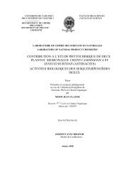
![SYNTHESIS AND ANTI-HIV ACTIVITY OF [d4U]-SPACER-[HI-236 ...](https://img.yumpu.com/30883288/1/190x245/synthesis-and-anti-hiv-activity-of-d4u-spacer-hi-236-.jpg?quality=85)