
Create successful ePaper yourself
Turn your PDF publications into a flip-book with our unique Google optimized e-Paper software.
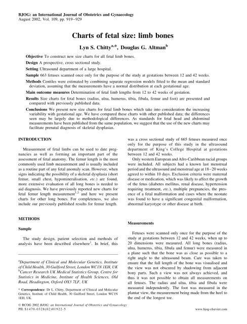
922 L.S. CHITTY & D.G. ALTMANFig. 5. Fitted 3rd, 10th, 50th, 90th and 97th centiles and raw data for tibia length.Fig. 6. Fitted 3rd, 10th, 50th, 90th and 97th centiles and raw data for fibula length.D RCOG 2002 Br J Obstet Gynaecol 109, pp. 919–929
SIZE CHART FOR FETAL LIMB BONES 923Fig. 7. Fitted 3rd, 10th, 50th, 90th and 97th centiles and raw data for foot length.Statistical methodsThe statistical methods used to analyse the data aredescribed in detail in Altman and Chitty 3 and Roystonand Wright 4 . In brief, for each measurement, fractionalpolynomial regression models were fitted separately toestimate the mean and standard deviation (SD) as functions<strong>of</strong> gestational age. The SD was modelled via theabsolute residuals from the regression to estimate themean. The centiles were obtained by combining thesetwo regression models, making the assumption that ateach gestational age, the measurements had a normaldistribution. The assumption <strong>of</strong> normality and the goodness<strong>of</strong> fit <strong>of</strong> each regression model were carefullyassessed 3 . All analyses used individual <strong>fetal</strong> measurementsand exact gestational age. No observations were excludedfrom the analyses.RESULTSOnly two <strong>of</strong> the sample <strong>of</strong> 665 fetuses were excluded,both because <strong>of</strong> congenital abnormalities. The number <strong>of</strong>measurements obtained varied for the different <strong>limb</strong>sbecause we only measured <strong>bones</strong> which were clearlyviewed in the appropriate plane.For each <strong>limb</strong> measurement, in turn, Figs. 1–7 show theraw data with the 3rd, 10th, 50th, 90th and 97th centilesTable 1. Fitted 3rd, 10th, 50th, 90th and 97th centiles <strong>of</strong> humerus length at 12 to 42 exact weeks <strong>of</strong> gestation, with number <strong>of</strong> fetuses for completed weeks <strong>of</strong>gestation.Weeks <strong>of</strong> gestation n Fitted centiles SD3rd 10th 50th 90th 97th12 8 3.7 4.8 7.1 9.5 10.6 1.813 18 7.2 8.3 10.7 13.1 14.2 1.914 18 10.5 11.7 14.1 16.5 17.7 1.915 14 13.7 14.8 17.3 19.8 21.0 2.016 15 16.7 17.9 20.4 23.0 24.2 2.017 22 19.6 20.8 23.4 26.0 27.2 2.018 18 22.3 23.6 26.2 28.9 30.1 2.119 22 24.9 26.2 28.9 31.6 32.9 2.120 21 27.4 28.7 31.5 34.2 35.5 2.221 22 29.8 31.2 34.0 36.8 38.1 2.222 20 32.1 33.5 36.3 39.2 40.5 2.2(continued on next page)D RCOG 2002 Br J Obstet Gynaecol 109, pp. 919–929
924 L.S. CHITTY & D.G. ALTMANTable 1 (continued)Weeks <strong>of</strong> gestation n Fitted centiles SD3rd 10th 50th 90th 97th23 22 34.3 35.7 38.6 41.5 42.9 2.324 24 36.4 37.8 40.7 43.7 45.1 2.325 20 38.4 39.8 42.8 45.8 47.2 2.426 19 40.3 41.7 44.8 47.9 49.3 2.427 24 42.1 43.6 46.7 49.8 51.3 2.428 20 43.9 45.3 48.5 51.7 53.2 2.529 21 45.5 47.0 50.2 53.5 55.0 2.530 19 47.1 48.6 51.9 55.2 56.7 2.631 26 48.6 50.2 53.5 56.8 58.4 2.632 25 50.0 51.6 55.0 58.4 59.9 2.633 23 51.4 53.0 56.4 59.8 61.5 2.734 20 52.7 54.3 57.8 61.3 62.9 2.735 20 53.9 55.6 59.1 62.6 64.3 2.836 24 55.1 56.8 60.3 63.9 65.6 2.837 19 56.2 57.9 61.5 65.1 66.8 2.838 20 57.2 58.9 62.6 66.3 68.0 2.939 14 58.2 60.0 63.7 67.4 69.2 2.940 13 59.1 60.9 64.7 68.5 70.3 3.041 25 60.0 61.8 65.6 69.5 71.3 3.042 17 60.8 62.6 66.5 70.4 72.2 3.0Total 613Table 2. Fitted 3rd, 10th, 50th, 90th and 97th centiles <strong>of</strong> radius length at 12 to 42 exact weeks <strong>of</strong> gestation, with number <strong>of</strong> fetuses for completed weeks <strong>of</strong>gestation.Weeks <strong>of</strong> gestation n Fitted centiles SD3rd 10th 50th 90th 97th12 6 2.2 3.3 5.5 7.8 8.8 1.713 8 4.8 5.9 8.2 10.5 11.6 1.814 16 7.6 8.7 11.0 13.4 14.5 1.815 11 10.3 11.5 13.9 16.3 17.4 1.916 12 13.0 14.2 16.7 19.1 20.3 1.917 16 15.6 16.8 19.3 21.9 23.1 2.018 15 18.1 19.3 21.9 24.5 25.7 2.019 24 20.4 21.7 24.4 27.0 28.3 2.120 22 22.7 23.9 26.7 29.4 30.7 2.121 21 24.8 26.1 28.9 31.6 32.9 2.222 19 26.8 28.1 30.9 33.8 35.1 2.223 21 28.6 30.0 32.9 35.8 37.1 2.324 20 30.4 31.8 34.7 37.7 39.1 2.325 22 32.0 33.5 36.5 39.5 40.9 2.426 20 33.6 35.0 38.1 41.2 42.6 2.427 24 35.1 36.5 39.7 42.8 44.3 2.428 21 36.5 38.0 41.2 44.3 45.8 2.529 21 37.8 39.3 42.6 45.8 47.3 2.530 19 39.0 40.6 43.9 47.2 48.7 2.631 26 40.2 41.8 45.1 48.5 50.1 2.632 25 41.3 42.9 46.4 49.8 51.4 2.733 23 42.4 44.0 47.5 51.0 52.6 2.734 19 43.4 45.0 48.6 52.1 53.8 2.835 22 44.3 46.0 49.6 53.2 54.9 2.836 22 45.2 46.9 50.6 54.3 56.0 2.937 18 46.1 47.8 51.6 55.3 57.0 2.938 18 46.9 48.7 52.5 56.2 58.0 3.039 11 47.7 49.5 53.3 57.2 59.0 3.040 14 48.4 50.3 54.2 58.1 59.9 3.041 21 49.1 51.0 55.0 58.9 60.8 3.142 15 49.8 51.7 55.7 59.7 61.6 3.1Total 572D RCOG 2002 Br J Obstet Gynaecol 109, pp. 919–929
SIZE CHART FOR FETAL LIMB BONES 925Table 3. Fitted 3rd, 10th, 50th, 90th and 97th centiles <strong>of</strong> ulna length at 12 to 42 exact weeks <strong>of</strong> gestation, with number <strong>of</strong> fetuses for completed weeks <strong>of</strong>gestation.Weeks <strong>of</strong> gestation n Fitted centiles SD3rd 10th 50th 90th 97th12 6 3.9 5.0 7.3 9.6 10.7 1.813 13 6.2 7.3 9.6 12.0 13.1 1.814 15 8.8 9.9 12.4 14.8 15.9 1.915 12 11.6 12.8 15.3 17.8 18.9 1.916 11 14.5 15.7 18.2 20.8 22.0 2.017 18 17.3 18.6 21.2 23.8 25.0 2.018 16 20.1 21.4 24.0 26.7 28.0 2.119 24 22.8 24.0 26.8 29.5 30.8 2.120 22 25.3 26.6 29.4 32.2 33.5 2.221 20 27.8 29.1 32.0 34.8 36.2 2.222 20 30.1 31.4 34.4 37.3 38.7 2.323 21 32.3 33.7 36.6 39.6 41.0 2.324 21 34.3 35.8 38.8 41.9 43.3 2.425 22 36.3 37.8 40.9 44.0 45.5 2.426 20 38.2 39.7 42.8 46.0 47.5 2.527 24 39.9 41.5 44.7 47.9 49.5 2.528 20 41.6 43.2 46.5 49.8 51.3 2.629 21 43.2 44.8 48.2 51.5 53.1 2.630 20 44.7 46.3 49.8 53.2 54.8 2.731 27 46.2 47.8 51.3 54.8 56.4 2.732 25 47.5 49.2 52.7 56.3 58.0 2.833 23 48.8 50.5 54.1 57.7 59.4 2.834 17 50.0 51.8 55.4 59.1 60.8 2.935 21 51.2 53.0 56.7 60.4 62.2 2.936 20 52.3 54.1 57.9 61.7 63.5 3.037 19 53.4 55.2 59.1 62.9 64.7 3.038 17 54.4 56.2 60.2 64.1 65.9 3.139 12 55.4 57.2 61.2 65.2 67.1 3.140 11 56.3 58.2 62.2 66.3 68.2 3.241 20 57.2 59.1 63.2 67.3 69.3 3.242 14 58.0 60.0 64.1 68.3 70.3 3.3Total 572Table 4. Fitted 3rd, 10th, 50th, 90th and 97th centiles <strong>of</strong> femur length at 12 to 42 exact weeks <strong>of</strong> gestation, with number <strong>of</strong> fetuses for completed weeks <strong>of</strong>gestation.Weeks <strong>of</strong> gestation n Fitted centiles SD3rd 10th 50th 90th 97th12 10 4.4 5.5 7.7 10.0 11.1 1.813 18 7.5 8.6 10.9 13.3 14.4 1.814 18 10.6 11.7 14.1 16.5 17.6 1.915 15 13.6 14.7 17.2 19.7 20.8 1.916 20 16.5 17.7 20.3 22.8 24.0 2.017 23 19.4 20.7 23.3 25.9 27.2 2.118 20 22.3 23.6 26.3 29.0 30.2 2.119 25 25.1 26.4 29.2 32.0 33.3 2.220 22 27.9 29.2 32.1 34.9 36.3 2.221 23 30.6 32.0 34.9 37.8 39.2 2.322 22 33.2 34.6 37.6 40.6 42.0 2.323 22 35.8 37.2 40.3 43.4 44.8 2.424 25 38.3 39.8 42.9 46.1 47.6 2.525 22 40.8 42.3 45.5 48.7 50.2 2.526 22 43.1 44.7 48.0 51.3 52.8 2.627 24 45.4 47.0 50.4 53.8 55.3 2.6(continued on next page)D RCOG 2002 Br J Obstet Gynaecol 109, pp. 919–929
926 L.S. CHITTY & D.G. ALTMANTable 4 (continued)Weeks <strong>of</strong> gestation n Fitted centiles SD3rd 10th 50th 90th 97th28 20 47.6 49.3 52.7 56.2 57.8 2.729 22 49.8 51.4 55.0 58.5 60.1 2.830 21 51.8 53.5 57.1 60.7 62.4 2.831 27 53.8 55.5 59.2 62.9 64.6 2.932 26 55.7 57.4 61.2 64.9 66.7 2.933 23 57.5 59.3 63.1 66.9 68.7 3.034 20 59.2 61.0 64.9 68.8 70.6 3.035 22 60.8 62.6 66.6 70.6 72.4 3.136 25 62.3 64.2 68.2 72.3 74.1 3.237 19 63.7 65.6 69.7 73.8 75.8 3.238 21 64.9 66.9 71.1 75.3 77.3 3.339 14 66.1 68.1 72.4 76.7 78.7 3.340 15 67.2 69.2 73.6 77.9 79.9 3.441 26 68.1 70.2 74.6 79.0 81.1 3.542 17 69.0 71.1 75.6 80.1 82.2 3.5Total 649Table 5. Fitted 3rd, 10th, 50th, 90th and 97th centiles <strong>of</strong> tibia length at 12 to 42 exact weeks <strong>of</strong> gestation, with number <strong>of</strong> fetuses for completed weeks <strong>of</strong>gestation.Weeks <strong>of</strong> gestation n Fitted centiles SD3rd 10th 50th 90th 97th12 7 4.4 5.4 7.6 9.8 10.8 1.713 9 5.8 6.9 9.2 11.4 12.5 1.814 14 8.0 9.1 11.4 13.7 14.8 1.815 11 10.6 11.7 14.1 16.4 17.6 1.916 16 13.3 14.5 16.9 19.4 20.5 1.917 20 16.2 17.4 19.9 22.4 23.5 2.018 15 19.0 20.2 22.8 25.4 26.6 2.019 22 21.8 23.1 25.7 28.3 29.6 2.120 20 24.5 25.8 28.5 31.2 32.5 2.121 21 27.2 28.5 31.2 34.0 35.3 2.222 16 29.7 31.0 33.8 36.7 38.0 2.223 18 32.1 33.5 36.4 39.2 40.6 2.324 22 34.4 35.8 38.8 41.7 43.1 2.325 21 36.6 38.0 41.0 44.1 45.5 2.426 20 38.7 40.1 43.2 46.3 47.8 2.427 21 40.7 42.2 45.3 48.5 49.9 2.528 17 42.6 44.1 47.3 50.5 52.0 2.529 21 44.4 45.9 49.2 52.5 54.0 2.630 17 46.1 47.7 51.0 54.3 55.9 2.631 23 47.7 49.3 52.7 56.1 57.7 2.732 21 49.3 50.9 54.4 57.8 59.5 2.733 21 50.8 52.4 55.9 59.5 61.1 2.834 19 52.2 53.9 57.5 61.0 62.7 2.835 19 53.5 55.2 58.9 62.6 64.3 2.936 20 54.8 56.6 60.3 64.0 65.7 2.937 14 56.0 57.8 61.6 65.4 67.2 3.038 18 57.2 59.0 62.9 66.7 68.5 3.039 11 58.3 60.2 64.1 68.0 69.8 3.140 12 59.4 61.3 65.2 69.2 71.1 3.141 22 60.4 62.3 66.4 70.4 72.3 3.242 16 61.4 63.3 67.4 71.6 73.5 3.2Total 544D RCOG 2002 Br J Obstet Gynaecol 109, pp. 919–929
SIZE CHART FOR FETAL LIMB BONES 927Table 6. Fitted 3rd, 10th, 50th, 90th and 97th centiles <strong>of</strong> fibula length at 12 to 42 exact weeks <strong>of</strong> gestation, with number <strong>of</strong> fetuses for completed weeks <strong>of</strong>gestation.Weeks <strong>of</strong> gestation n Fitted centiles SD3rd 10th 50th 90th 97th12 5 3.6 4.6 6.8 9.0 10.0 1.713 5 5.2 6.2 8.5 10.7 11.8 1.714 11 7.4 8.5 10.8 13.1 14.2 1.815 10 10.0 11.1 13.5 15.9 17.0 1.916 15 12.8 14.0 16.4 18.8 20.0 1.917 19 15.6 16.8 19.3 21.8 23.0 2.018 14 18.4 19.7 22.2 24.8 26.0 2.019 21 21.2 22.4 25.1 27.7 29.0 2.120 19 23.9 25.1 27.9 30.6 31.8 2.121 21 26.4 27.7 30.5 33.3 34.6 2.222 17 28.9 30.2 33.1 35.9 37.3 2.223 19 31.2 32.6 35.5 38.5 39.8 2.324 24 33.5 34.9 37.9 40.9 42.3 2.325 19 35.6 37.0 40.1 43.2 44.6 2.426 21 37.6 39.1 42.2 45.4 46.8 2.427 21 39.6 41.1 44.3 47.5 49.0 2.528 17 41.4 42.9 46.2 49.5 51.0 2.629 21 43.1 44.7 48.0 51.4 52.9 2.630 20 44.8 46.4 49.8 53.2 54.8 2.731 23 46.4 48.0 51.5 54.9 56.6 2.732 21 47.9 49.5 53.1 56.6 58.3 2.833 20 49.3 51.0 54.6 58.2 59.9 2.834 20 50.7 52.4 56.1 59.7 61.5 2.935 20 52.0 53.7 57.5 61.2 63.0 2.936 20 53.2 55.0 58.8 62.6 64.4 3.037 14 54.4 56.2 60.1 64.0 65.8 3.038 18 55.5 57.4 61.3 65.3 67.1 3.139 10 56.6 58.5 62.5 66.5 68.4 3.140 12 57.6 59.5 63.6 67.7 69.6 3.241 22 58.6 60.5 64.7 68.9 70.8 3.342 16 59.5 61.5 65.8 70.0 72.0 3.3Total 535Table 7. Fitted 3rd, 10th, 50th, 90th and 97th centiles <strong>of</strong> foot length at 12 to 42 exact weeks <strong>of</strong> gestation, with number <strong>of</strong> fetuses for completed weeks <strong>of</strong>gestation.Weeks <strong>of</strong> gestation n Fitted centiles SD3rd 10th 50th 90th 97th12 3 5.9 6.8 8.9 10.9 11.8 1.613 3 8.5 9.6 11.7 13.9 14.9 1.714 7 11.3 12.3 14.6 16.9 18.0 1.815 7 14.0 15.2 17.6 20.0 21.2 1.916 12 16.8 18.0 20.6 23.2 24.4 2.017 16 19.6 20.9 23.6 26.3 27.6 2.118 11 22.4 23.7 26.6 29.5 30.8 2.219 16 25.2 26.6 29.6 32.6 34.0 2.320 15 28.0 29.5 32.6 35.8 37.2 2.521 21 30.8 32.3 35.6 38.9 40.4 2.622 18 33.5 35.1 38.6 42.0 43.6 2.723 18 36.2 37.9 41.5 45.0 46.7 2.824 22 38.9 40.7 44.4 48.0 49.8 2.925 17 41.5 43.3 47.2 51.0 52.8 3.026 20 44.1 46.0 50.0 53.9 55.8 3.127 22 46.6 48.6 52.7 56.8 58.7 3.228 20 49.1 51.1 55.3 59.6 61.6 3.3(continued on next page)D RCOG 2002 Br J Obstet Gynaecol 109, pp. 919–929
928 L.S. CHITTY & D.G. ALTMANTable 7 (continued)Weeks <strong>of</strong> gestation n Fitted centiles SD3rd 10th 50th 90th 97th29 20 51.4 53.5 57.9 62.3 64.3 3.430 18 53.7 55.8 60.4 64.9 67.0 3.531 24 55.9 58.1 62.8 67.4 69.6 3.632 22 58.0 60.3 65.1 69.9 72.1 3.833 19 60.0 62.3 67.3 72.2 74.5 3.934 11 61.9 64.3 69.4 74.5 76.8 4.035 15 63.7 66.1 71.4 76.6 79.0 4.136 16 65.4 67.9 73.3 78.6 81.1 4.237 11 66.9 69.5 75.0 80.5 83.1 4.338 12 68.4 71.0 76.7 82.3 85.0 4.439 6 69.7 72.4 78.2 84.0 86.7 4.540 10 70.9 73.7 79.6 85.5 88.3 4.641 10 71.9 74.8 80.8 86.9 89.7 4.742 8 72.8 75.7 81.9 88.1 91.0 4.8Total 450superimposed. The estimated centiles for exact weeks <strong>of</strong>gestation are shown in Tables 1–7 together with thenumber <strong>of</strong> observations and fitted standard deviation.The regression equations are given in the Appendix.The assumption <strong>of</strong> a normal distribution for each measurementat each gestational age was found to be reasonable.Figure 8 compares our centiles for tibia and radius withthose <strong>of</strong> two other studies. There is quite good agreementFig. 8. Comparison <strong>of</strong> 5th, 50th and 95th centiles for tibia and radius obtained in this study (solid lines) with those <strong>of</strong> Merz et al. 5 (panels a and c) andExacoustos et al. 6 (panels b and d) (short dashed lines). Also shown in panels (b) and (d) are the fitted curves <strong>of</strong> Exacoustos et al. 6 (long dashed lines). Thedata <strong>of</strong> Merz et al. 5 relate to completed weeks <strong>of</strong> gestation. Exacoustos et al. 6 do not specify whether their centiles refer to exact or completed weeks <strong>of</strong>gestation.D RCOG 2002 Br J Obstet Gynaecol 109, pp. 919–929
SIZE CHART FOR FETAL LIMB BONES 929with the centiles <strong>of</strong> Merz et al. 5 but rather more discrepancyfrom the findings <strong>of</strong> Exacoustos et al. 6 , especially inearly gestation. Their centiles are much closer together,although the medians are similar.DISCUSSIONWe have constructed new <strong>size</strong> charts for <strong>fetal</strong> long boneand foot length from 12 until 42 weeks <strong>of</strong> gestation. Aswith other measurements 2,7 – 9 , we have shown the need totake into account the increasing variability <strong>of</strong> the measurementswith increasing gestational age in the construction <strong>of</strong>the centiles. Our study was designed to overcome many <strong>of</strong>the methodological weaknesses that have been noted inmany published <strong>fetal</strong> <strong>size</strong> centiles 10,11 . In particular, fetuseswere measured on only one occasion for the purposes <strong>of</strong>this study, all observations were collected prospectivelyand expressly for the development <strong>of</strong> centile charts, weexcluded only two fetuses (for congenital abnormalities),we used statistical methods which give proper attentionto the changing variability with increasing gestation andwe carefully assessed the goodness <strong>of</strong> fit <strong>of</strong> the modelsobtained 3 .Our charts <strong>of</strong> <strong>fetal</strong> <strong>size</strong> were derived from cross sectionaldata. They are appropriate for comparing the <strong>size</strong> <strong>of</strong> a fetusat a known gestational age with reference data. They arenot suitable for judging the appropriateness <strong>of</strong> the growth<strong>of</strong> a fetus between two occasions 3 . Our centiles werederived from a population <strong>of</strong> western Europeans (75%)and Afro-Caribbeans (25%). The charts might not beentirely applicable to other ethnic groups.We have compared our new charts with those <strong>of</strong> someother researchers using the radius and tibia as illustrativeexamples. Comparison with other studies is only possible ifthey present either means and outer centiles, or a fullspecification <strong>of</strong> a statistical model. Many studies fail todo so. Our centiles are quite close to those <strong>of</strong> Merz et al. 5 ,who used a broadly similar approach to statistical analysisbut did not give full detail <strong>of</strong> their sampling strategy. Bycontrast, our data are rather different from the centiles <strong>of</strong>Exacoustos et al. 6 , especially in early gestation. Thoseauthors presented a model for the mean (see Fig. 8), butthe outer centiles appear not to be derived from a statisticalmodel. Both <strong>of</strong> these groups studied ‘normal’ pregnancies,but neither explained how their sample was selected.In order to diagnose skeletal dysplasias as accurately aspossible, it is necessary to compare the length <strong>of</strong> the <strong>bones</strong>with head and body <strong>size</strong> as well as determining the pattern<strong>of</strong> shortening (mesomelic, acromelic or rhizomelic). Anadvantage <strong>of</strong> our study is that all the measurementsreported in this paper and our other papers 8,9 were obtainedon the same sample <strong>of</strong> fetuses, thus making comparisonbetween the observed centiles <strong>of</strong> different body measurementsmore valid.References1. Altman DG, Chitty LS. <strong>Charts</strong> <strong>of</strong> <strong>fetal</strong> <strong>size</strong>. 1: Methodology. Br JObstet Gynaecol 1994;101:29–34.2. Chitty LS, Altman DG, Henderson A, Campbell S. <strong>Charts</strong> <strong>of</strong> <strong>fetal</strong><strong>size</strong>. 4: Femur length. Br J Obstet Gynaecol 1994;101:132–135.3. Altman DG, Chitty LS. Design and analysis <strong>of</strong> studies to derive charts<strong>of</strong> <strong>fetal</strong> <strong>size</strong>. Ultrasound Obstet Gynecol 1993;3:378–384.4. Royston P, Wright EM. How to construct ‘normal ranges’ for <strong>fetal</strong>variables. Ultrasound Obstet Gynecol 1998;11:30–38.5. Merz E, Grüssner A, Kern F. Mathematical modeling <strong>of</strong> <strong>fetal</strong> <strong>limb</strong>growth. J Clin Ultrasound 1989;17:179–185.6. Exacoustos C, Rosati P, Rizzo G, Arduini D. Ultrasound measurements<strong>of</strong> <strong>fetal</strong> <strong>limb</strong> <strong>bones</strong>. Ultrasound Obstet Gynecol 1991;1:325–330.7. Chitty LS, Campbell S, Altman DG. Measurement <strong>of</strong> the <strong>fetal</strong> mandible—feasibilityand construction <strong>of</strong> a centile chart. Prenat Diagn1993;13:749–756.8. Chitty LS, Altman DG, Henderson A, Campbell S. <strong>Charts</strong> <strong>of</strong> <strong>fetal</strong><strong>size</strong>. 2: Head measurements. Br J Obstet Gynaecol 1994;101:35–43.9. Chitty LS, Altman DG, Henderson A, Campbell S. <strong>Charts</strong> <strong>of</strong> <strong>fetal</strong><strong>size</strong>. 3: Abdominal circumference. Br J Obstet Gynaecol 1994;101:125–131.10. Deter RL, Harrist RB, Birnholz JC, Hadlock FP. Quantitative ObstetricalUltrasonography. New York: Wiley, 1986.11. British Medical Ultrasound Society Fetal Measurements WorkingParty. Clinical Applications <strong>of</strong> Ultrasonic Fetal Measurements.London: British Institute <strong>of</strong> Radiology, 1990.Appendix. The regression equations used to generate thecentiles in the figures and tables are as follows, where w isexact gestational age in weeks.HumerusMean: 11.459w 2.2362w log(w) 63.704SD: 0.040292w þ 1.3464RadiusMean: 7983/w 2 1698.6/w þ 91.634SD: 0.046386w þ 1.1933UlnaMean: 11120/w 2 2146.3/w þ 108.94SD: 0.049218w þ 1.2021FemurMean: 3.4162w 0.0004791w 3 32.425SD: 0.058328w þ 1.0605TibiaMean: 14451/w 2 2553.2/w þ 120.05SD: 0.049978w þ 1.1102FibulaMean: 13697/w 2 2458.0/w þ 116.51SD: 0.053841w þ 1.0451FootMean: 0.36909w 2 0.084175w 2 log(w) 14.158SD: 0.10865w þ 0.27971Accepted 21 March 2002D RCOG 2002 Br J Obstet Gynaecol 109, pp. 919–929