, Diagnosis an-&& of Shrimp Diseases - Central Institute of ...
, Diagnosis an-&& of Shrimp Diseases - Central Institute of ...
, Diagnosis an-&& of Shrimp Diseases - Central Institute of ...
- No tags were found...

You also want an ePaper? Increase the reach of your titles
YUMPU automatically turns print PDFs into web optimized ePapers that Google loves.
OVERVIEW OF STRATEGIES FOR PREVENTION AND CONTROL OF DISEASESIN SHRIMP AQUACULTURET.C. S<strong>an</strong>tiagoThe global aquaculture activity has been recognized as the fastest growing enterprisewith <strong>an</strong> estimated <strong>an</strong>nual growth rate <strong>of</strong> 11 %. The aquaculture activity in India has beencontributing signific<strong>an</strong>tly in the various spheres <strong>of</strong> people's development, in terms <strong>of</strong> providinglivelihood, food security, employment <strong>an</strong>d trade. The aquaculture sector in our country ismultifaceted. India is blessed with a long coastline <strong>of</strong> over 8000 km providing immense scopefor brackishwater <strong>an</strong>d marine aquaculture. The l<strong>an</strong>d base aquaculture systems include vastpotential for freshwater <strong>an</strong>d coid-water ecosystems. Although the aquaculture activity in Indiais not very new, modern practices with intensification on scientific bases using hatcheryproduced seeds, formulated feeds <strong>an</strong>d pond <strong>an</strong>d water m<strong>an</strong>agement methods have been initiatedover the last 20 years, <strong>an</strong>d species cultured include diverse aquatic fauna such as finfish,shrimps, crabs, lobsters, prawns, oysters <strong>an</strong>d mussels. However, this intensification <strong>of</strong>aquaculture has resulted in increased incidence <strong>of</strong> the disease problems in cultured stock. TheWhite Spot Syndrome Virus (WSSV) alone is causing <strong>an</strong> <strong>an</strong>nual loss to the tune <strong>of</strong> Rs.300crores, since 1994 to the shrimp culture industry, according to conservative estimates.Similarly, the epizootic ulcerative syndrome in freshwater <strong>an</strong>d brackishwater fish has beenrecognized as one <strong>of</strong> the major cause <strong>of</strong> decline in finfish production. The disease problems<strong>an</strong>d related crop losses have become a major limiting factor in the growth <strong>of</strong> aquacultureactivity. As a result <strong>of</strong> such continued setback, the scientific community <strong>an</strong>d policy makershave initiated steps to overcome the disease problems in order to ensure sustainabledevelopment <strong>of</strong> aquaculture in the country. The objective <strong>of</strong> this article is to provide <strong>an</strong>overview <strong>of</strong> some import<strong>an</strong>t diseases that are responsible for production losses to commerciallyimport<strong>an</strong>t species <strong>of</strong> aquaculture in the country. Further, a brief account on the preventive <strong>an</strong>dcontrol methods available <strong>an</strong>d strategies for disease m<strong>an</strong>agement are also provided.DISEASESViral diseases constitute the most serious problems for shrimp culture due to highinfectivity, pathogenicity <strong>an</strong>d noi~avaliIability<strong>of</strong> curative measures. Nearly 20 viruses arereported to affect penaeid shrimp throughout the world, among which, four viruses areimport<strong>an</strong>t as far as their impact on the production is concerned (Table 1). The Monodon BaculoVirus (MBV) outbreak in Taiw<strong>an</strong> in 1988, followed by Yellow Head Virus (YHV) disease in
1992 in Thail<strong>an</strong>d, Taura Syndrome Virus (TSV) in 1992 in Ecuador, White Spot SyndroineVirus (WSSV) in 1993 in China <strong>an</strong>d Thail<strong>an</strong>d, <strong>an</strong>d the same virus in a number <strong>of</strong> other Asi<strong>an</strong>countries including India have lead to production losses <strong>of</strong> cultured stock <strong>of</strong> shrimp. Amongthese the most devastating one is the WSSV in India. Although MBV has been also frequentlyfound in our shrimp aquaculture systems, its impact on production is not reported to be asdevastating as that caused by WSSV. TSV <strong>an</strong>d YHV assume inlport<strong>an</strong>ce in view <strong>of</strong> theattempts <strong>of</strong> introduction <strong>of</strong> exotic shrimps for culture purposes in the country. The bacterialinfections caused by Vibrio species are the second import<strong>an</strong>t cause <strong>of</strong> mortality <strong>of</strong> culturedshrimp both in hatcheries <strong>an</strong>d grow-out systems.The freshwater prawn,M. rosenbewgii is reported to be relatively less susceptible todiseases th<strong>an</strong> penaeid shrimps, possibly due to lower stocking densities. However, during thepast several years, these species are also suffering mortality due to white tail disease, whichresembles idiopathic muscle necrosis reported about 16 years back by Nash et 81 (1987).Recent reports suggest that <strong>an</strong> RNA virus <strong>of</strong> the family Nodaviridae causes the disease.Fish Health M<strong>an</strong>agement StrategiesAn underst<strong>an</strong>ding about the environment, biota <strong>an</strong>d biology <strong>of</strong> the target species alongwith the in depth knowledge <strong>of</strong> the disease, pathogen, disease development, diagnostics,epidemiology <strong>an</strong>d control measures are essential factors in m<strong>an</strong>agement <strong>of</strong> a disease problem.Hence, Fish health m<strong>an</strong>agement requires a holistic approach, addressing all aspects thatcontribute to the development <strong>of</strong> disease. Disease out break is <strong>an</strong> end result <strong>of</strong> negativeinteraction between pathogen, host <strong>an</strong>d the environment. Hence, m<strong>an</strong>agement <strong>of</strong> diseaseproblems must be aimed towards broader ecosystem m<strong>an</strong>agement with a view to control farm--level environmental deterioration <strong>an</strong>d to take preventative measures against the introduction <strong>of</strong>pathogens into the aquaculture system. The emphasis should be on better m<strong>an</strong>agement forprevention, which is likely to be more cost effective th<strong>an</strong> treatment, involving both on-farmm<strong>an</strong>agement <strong>an</strong>d the m<strong>an</strong>agement <strong>of</strong> the environment. Steps must include reducing the use <strong>of</strong>chemicals <strong>an</strong>d drugs. Regulations with respect to l<strong>an</strong>d <strong>an</strong>d water usage, environmentalprotective measures, inputs that go into the aquaculture systeps, farm-wise <strong>an</strong>d region-wisemust be put in place by the Government for disease m<strong>an</strong>agement <strong>of</strong> aquatic <strong>an</strong>imals <strong>an</strong>dsustainable development <strong>of</strong> aquaculture at large. In addition, research <strong>an</strong>d development,training programs, extension, <strong>an</strong>d information exch<strong>an</strong>ge would help achieve the objective <strong>of</strong>disease prevention <strong>an</strong>d control in aquaculture effective. The FAO's Code <strong>of</strong> Conduct forResponsible Fisheries would provide a good base for the national <strong>an</strong>d international cooperation
in harmonizing aquatic <strong>an</strong>imal health m<strong>an</strong>agement activities.The ultimate goal <strong>of</strong> most aquaculture operations is to produce maximum possiblebiomass per culture unit area in a sustainable m<strong>an</strong>ner, regardless <strong>of</strong> the type <strong>of</strong> operation <strong>an</strong>dthe species cultured. However, the production depends upon a number <strong>of</strong> factors includingenvironmental conditions, availability <strong>of</strong> good quality water, nutrition <strong>an</strong>d disease <strong>an</strong>dmortality <strong>of</strong> cultured stock. Ipcidence <strong>an</strong>d severity <strong>of</strong> infectious disease outbreaks very <strong>of</strong>tendepend on the quality <strong>of</strong> environment. Hence the foremost import<strong>an</strong>t step in aquaculture healthm<strong>an</strong>agement is to provide the best quality environment within the culture unit.Coastal aquaculture in general, <strong>an</strong>d shrimp farming in particular, heavily relies uponwild brood stock for seed production. The health status <strong>of</strong> broodstock population has generallybeen neglected. The asymptomatic wild broostock population plays a major role in the verticaltr<strong>an</strong>smission <strong>of</strong> the pathogen. There is a need to evaluate the health status <strong>of</strong> different stocks<strong>an</strong>d to develop me<strong>an</strong>s <strong>of</strong> controlling the entry <strong>of</strong> the pathogen into the breeding <strong>an</strong>d farmedpopulations.Development <strong>of</strong> Specific Pathogen Free (SPF) stock is a must to produce disease freeseeds. However these facilities need to be created <strong>an</strong>d implemented adopting internationallyaccepted norms <strong>an</strong>d with proper scientific evaluation.Timely <strong>an</strong>d correct diagnosis <strong>of</strong> the disease using the right diagnostic tool is one <strong>of</strong> themost import<strong>an</strong>t components in the aquatic health m<strong>an</strong>agement. Present disease diagnosticsapabilities depend largely llpon the availability <strong>of</strong> sophisticated laboratories. Hence it isnecessary to take procedures out <strong>of</strong> the laboratory <strong>an</strong>d explores ways in which they c<strong>an</strong> bebetter applied under farm/field conditions.These are crucial component <strong>of</strong> <strong>an</strong> effective health m<strong>an</strong>agement programme.Quar<strong>an</strong>tine does not only me<strong>an</strong> that exotic species should be subjected to rigorous checks toavoid introduction <strong>of</strong> pathogens into a country or state, but it is also imperative that thebroodstocldspawners/seeds arriving at a culture facility are screened for the presence <strong>of</strong>pathogens prior to their introduction to the system. Establishing effective quar<strong>an</strong>tine guidelines<strong>an</strong>d health certification procedures could help minimize the risk <strong>of</strong> introduction <strong>of</strong> harmfulpathogens. Hence to provide a mech<strong>an</strong>ism to facilitate trade in aquatic species, a proper healthm<strong>an</strong>age~nent mech<strong>an</strong>ism such as quar<strong>an</strong>tine <strong>an</strong>d health certification is necessary for the tr<strong>an</strong>s--boundary movement <strong>of</strong> aquatic <strong>an</strong>imals on the pre-border (exporter), border <strong>an</strong>d post-border(importer), to minimize the risk <strong>of</strong> pathogen tr<strong>an</strong>sfer <strong>an</strong>d associated risk <strong>of</strong> disease outbreaks.New generation approaches such as Surveilla~~ce techniques, Contingency pl<strong>an</strong>ning <strong>an</strong>dImport Risk Analysis (IRA) ire gaining import<strong>an</strong>ce as critical tools in the health m<strong>an</strong>agement
strategies <strong>of</strong> aquatic <strong>an</strong>imals for quick <strong>an</strong>d effective response to new disease outbreaks.Trained In<strong>an</strong>power <strong>an</strong>d capacity building are the import<strong>an</strong>t steps towards <strong>an</strong> effectiveextension system. A working extension system for awareness building <strong>an</strong>d effectivecommunication among farmers / aquaculturists, Govt. agencies <strong>an</strong>d pl<strong>an</strong>ners is pivotal for thesuccessful implementation <strong>of</strong> <strong>an</strong>y aquatic health m<strong>an</strong>agement programme.One <strong>of</strong> the most import<strong>an</strong>t factors dealing with a di'sease outbreak is information.Correct information is the key element in deciding upon the best me<strong>an</strong>s <strong>of</strong> dealing with adisease. To meet this objective a scientific <strong>an</strong>d functional disease reporting system applicableat local (farm level), national <strong>an</strong>d regional level <strong>an</strong>d aquatic <strong>an</strong>imal health information syslelnat national <strong>an</strong>d regional level is necessary. Policies <strong>an</strong>d legislature governing resources (soil<strong>an</strong>d water) allocation <strong>an</strong>d quality assur<strong>an</strong>ce in aquaculture, related to the physicochemicalcomponents <strong>an</strong>d biological components should be in place in all the coi~ntries practicingaquaculture. Functioning <strong>of</strong> a national level (each country) body with necessary responsibility<strong>an</strong>d m<strong>an</strong>date to implement a 'national health m<strong>an</strong>agement strategy' or 'health m<strong>an</strong>agementregulation' on the basis <strong>of</strong> existing international st<strong>an</strong>dards, guidelines or recom~nendation fromFAO, 01E <strong>an</strong>d NACA <strong>an</strong>d WTO must be there in issues related to aquaculture <strong>an</strong>d aquatic<strong>an</strong>imal health m<strong>an</strong>agement for the region.
SI-IRXMP DISEASES: GENEIL4L ASPECTSS. V. Alav<strong>an</strong>di <strong>an</strong>d T.C. S<strong>an</strong>tiagoIntroductionCoastal aquaculture, especially shrimp aquaculture has undergone a fast growth inrecent times in India. While traditional type <strong>of</strong> shrimp farms were being improved, newextensive <strong>an</strong>d semi-intensive farms were being established at rapid pace. Majority <strong>of</strong> theinvestors ventured into aquaculture by initially familiarising themselves with technical aspects<strong>of</strong> site selection, pond design, feeding techniques, intensive stocking etc. More <strong>of</strong>ten, thesignific<strong>an</strong>t impact <strong>of</strong> disease was overlooked. However, concomit<strong>an</strong>t with the rapid exp<strong>an</strong>sion<strong>an</strong>d intensification <strong>of</strong> shrimp farining activities serious disease outbreaks were <strong>of</strong> frequentoccurrence.Attention to disease problems was paid only when widespread outbreak <strong>of</strong> diseasealarmingly reduced the pr<strong>of</strong>it from shrimp farming projects. It has become essential for shrimpfarmers to underst<strong>an</strong>d the biological <strong>an</strong>d environmental factors that lead to diseasedevelopment, the maladies fiat c<strong>an</strong> cause considerable loss to cultured shrimp, the earlydetection <strong>of</strong> incidence <strong>of</strong> diseases <strong>an</strong>d drawing up farming strategy that would minimise orprevent the onset <strong>of</strong> diseases.What is disease <strong>an</strong>d how diseases develop?As <strong>an</strong>y other living org<strong>an</strong>isms, shrimp also have specific physiological functions forgrowth <strong>an</strong>d development, which is greatly influenced by various factors <strong>of</strong> the environment inwhich they are living. Any impairment in the physiological functioning may lead to abnormalcondition <strong>of</strong> <strong>an</strong> org<strong>an</strong>ism, <strong>an</strong>d this phenomenon is known as disease. However, m<strong>an</strong>y expertsconsider that there are 3 factors, which interact with each other <strong>an</strong>d result in the occurrence <strong>of</strong>disease. These factors are the host (shrimp), the environment <strong>an</strong>d disease-causing org<strong>an</strong>ism(pathogen). Therefore, disease c<strong>an</strong> be described as <strong>an</strong> expression <strong>of</strong> complex interaction <strong>of</strong>host, pathogen <strong>an</strong>d environment (Fig. ).A decline in llost's immunity is the main cause <strong>of</strong> disease. A lot <strong>of</strong> factors will impairshrimp health <strong>an</strong>d the most import<strong>an</strong>t pre-disposing factors leading to diseases in shrimpculture are:I. Adverse environment11. High stoclting density with limited water exch<strong>an</strong>ge facilities111. Nutritional deficiencyipoor nourishment
IV. Accum~ilation <strong>of</strong> unused feedV. Inadequate aerationVI. Sub-optimal or heavy algal blooms in the pondVII. Physical injury <strong>an</strong>dVIII. Presence <strong>of</strong> virulent pathogens in high count.In these, ch<strong>an</strong>ges in the physicaI or chernicaI factors will be obvious, but the biological factorswill be subtle <strong>an</strong>d complicated. This c<strong>an</strong> be explained by micro ecology. This refers to theinteraction <strong>of</strong> biological factors <strong>an</strong>d it explains the interaction between normal microorg<strong>an</strong>isms<strong>an</strong>d its environment.HostLike <strong>an</strong>y other crustace<strong>an</strong>s, shrimp host's body is covered by exoskeleton, which isregularly replaced by a new one during moulting. The moulting process exerts energyrequirement on the shrimp <strong>an</strong>d renders the shrimp susceptible to disease agents or c<strong>an</strong>nibalism.In addition, the shrimp's nutritional well being, size <strong>an</strong>d immune response determine its degree<strong>of</strong> resist<strong>an</strong>ce to disease agents. Behavioural characteristic such as burrowing at the pondbottom also exposes the shrimp condition prevailing in the pond.EnvironmentThe term environment in aquaculture comprises the pond soil, rearing water <strong>an</strong>d thevarious living org<strong>an</strong>isms in it. The living org<strong>an</strong>isms include not only shrimp but also otheraquatic fauna <strong>an</strong>d flora including pathogenic org<strong>an</strong>isms. The survival <strong>an</strong>d growth <strong>of</strong> theorg<strong>an</strong>isms is largely influenced by various physico-chemical parameters such pH, dissolvedoxygen, temperature, light etc. Any abnormal ch<strong>an</strong>ge in these factors will adversely affectshrimp in the culture system. For example, high ammonia level, low dissolved oxygen etc. arestressful <strong>an</strong>d may affect the survival <strong>of</strong> shrimp.PathogenVarious pathogenic org<strong>an</strong>isms may be present in the aquaculture system. They may bethe part <strong>of</strong> the natural flora <strong>an</strong>d fauna <strong>of</strong> the rearing water or pond soil. Various disease causingorg<strong>an</strong>ism <strong>of</strong> shrimp have been reported. Mere presence <strong>of</strong> these org<strong>an</strong>isms may not cause <strong>an</strong>ydisease condition. However, when present in large numbers these may readily invade theinjured tissues get established <strong>an</strong>d multiply resulting in disease <strong>an</strong>d death. Nevertheless, the
qu<strong>an</strong>titative level <strong>of</strong> pathogen is influenced largely by prevailing culture condition such asavailability <strong>of</strong> food source, temperature, dissolved osygen, pH etc.Fig. 1. Interaction between host pathogen <strong>an</strong>d environmentNo diseaseEnvironment
VIRAL DISEASES WITH SPECIAL REFERENCE TO INDIAN SHRIMP FARMINGT.C.S<strong>an</strong>tiago, K.K. Vijay<strong>an</strong>, S.V. Alav<strong>an</strong>di <strong>an</strong>d N. Kalaim<strong>an</strong>iViruses are ~~ltramicroscopic, infective agents capable <strong>of</strong> multiplying in the host livingcells causing improper cell function or cell destruction leading to the death <strong>of</strong> the host. Viraldiseases constitute the most serious problems <strong>of</strong> shrimp culture due to the high infectivity,pathogenicity <strong>an</strong>d total lack <strong>of</strong> curative measures. Worldwide, shrimp aquaculture hassuffered subst<strong>an</strong>tial economic losses due to pathogenic viruses, <strong>an</strong>d the Indi<strong>an</strong> shrimp farmingis no exception. So far, 15 viruses infecting cultured shrimps have been recorded across theshrimp farming countries <strong>of</strong> the world (Table). Till today, only five viruses have beenrecorded from Indi<strong>an</strong> farms.Monodon baculovirus (MBV)Nrrture <strong>of</strong> infection:Monodon baculovirus (MBV) is the first viral pathogen to be recorded from thecultured penaeids <strong>of</strong> India. Presently the virus is enzootic in Indi<strong>an</strong> hatcheries <strong>an</strong>d farms,infecting both P. monodon <strong>an</strong>d P. indicus. MBV infections have been observed in thehepatop<strong>an</strong>creatic cells <strong>of</strong> all life stages <strong>of</strong> the prawn except egg, nauplius <strong>an</strong>d protzoea 1 <strong>an</strong>d2 stages. Postlarvae <strong>an</strong>d farmed shrimps <strong>of</strong> all sizes with severe MBV infections appearnormal <strong>an</strong>d healthy. The virus, widely distributed in the cultured populations is well toleratedby the shrimps, as long as rearing conditions are optimal. Hence, under good culture practicesthe impact <strong>of</strong> the MBV infection c<strong>an</strong> be minimal. However, under adverse environn~entalconditions, MBV may predispose infected shrimp to infection by other pathogens, causingpoor growth, secondary infections <strong>an</strong>d mortality.Pathogenisis <strong>an</strong>d diagnosis:MBV is a single-enveloped, rod shaped, occluded double str<strong>an</strong>ded DNA virusbelonging to the group baculovirus. The virus occurs freely or within proteinaceouspolyhedral occlusion bodies in the nucleus, with virions measuring 75-300nm. The presence<strong>of</strong> MBV in the prawn c<strong>an</strong> be detected by direct microscopic examination <strong>of</strong> iinpressionsmears <strong>of</strong> infected hepatop<strong>an</strong>creas (HP) or midgut tissue, stained with 0.05 to 0.1% <strong>of</strong>malachite green by demonstrating the usually multiple spherical intr<strong>an</strong>uclear incl~~sion bodies.Histoiogical preparations <strong>of</strong> the infected HP c<strong>an</strong> be used for further confirmation due to the
presence <strong>of</strong> prominent eosinophiiic single to multiple spherical bodies within thehypertrophied nuclei <strong>of</strong> the hepatop<strong>an</strong>creatic tubule or midgut epithelial cells. Tr<strong>an</strong>smissionelectron microscopy (TEM) c<strong>an</strong> also used to show the presence <strong>of</strong> MBV virions. DNA basedrapid diagnostic tools? polymerase chain reaction (PCR) <strong>an</strong>d DIG-labelled DNA probes arealso available for the early diagnosis <strong>of</strong> MBV.Prevention nnd control:MBV infection may be prevented only through avoid<strong>an</strong>ce by quar<strong>an</strong>tine methods,destruction <strong>of</strong> contaminated stocks, <strong>an</strong>d disinfection <strong>of</strong> contaminated faciiities. There is notreatment for MBV, however good farm m<strong>an</strong>agement c<strong>an</strong> minimize this disease.Infectious Hepatop<strong>an</strong>creatic <strong>an</strong>d Lymphoid org<strong>an</strong> Necrosis Disease (IHLN)Nature <strong>of</strong> infection:The first ever shrimp epizootic reported from India is the IHLN disease, from shrimpfarms located along the K<strong>an</strong>daleru Creek, Nellore, Andhra Pradesh during July 1994. Thiswas a localized epizootic confined to the watershed areas <strong>of</strong> the K<strong>an</strong>daleru Creek. The IHLNaffected the crops <strong>of</strong> culture duration r<strong>an</strong>ging from 60 to 100 days weighing 3-28 g. The onset<strong>of</strong> the disease was sudden <strong>an</strong>d within 3-5 days post infection, more th<strong>an</strong> 90 % <strong>of</strong> the stock inthe farms was lost. The disease prevailed in a virulent form for about three months. Grosssigns <strong>of</strong> the diseased shrimp were: light yellow or pinkish cephalothorax, reddishdiscoloration <strong>of</strong> the body <strong>an</strong>d-appendages, empty gut, lethargy, poor escape reflex, secondarybacterial infection <strong>an</strong>d mortality. Dead shrimps were found scattered all over the pondbottom. Only P. rnonodon was affected, P.indicus was found refractory to the disease.Prttlzogenisis <strong>an</strong>d diagnosis:The most prominent feature <strong>of</strong> the disease was the highly mel<strong>an</strong>ized <strong>an</strong>d shrunken HP.Acute damages were observed in the HP, m<strong>an</strong>ifested by multi-focal necrosis <strong>of</strong> the tubuleepithelium marked by hemocytic infiltration <strong>an</strong>d encapsulation resulting in mel<strong>an</strong>ization.Densely stained, globular, basophilic bodies were observed in the HP cells <strong>an</strong>d lymphoidorg<strong>an</strong> (LO). The mortality pattern <strong>an</strong>d external signs <strong>of</strong> infection (yellow cephalothorax) <strong>of</strong>the disease suggested resembl<strong>an</strong>ce to yellow head virus (YHD) in the P. monodon. However,the prominent necrotic ch<strong>an</strong>ges in the HP <strong>an</strong>d LO <strong>an</strong>d absence <strong>of</strong> pathological ch<strong>an</strong>ges in thegills, indicated that the disease clearly differed from YHD, however, the etiology <strong>of</strong> thedisease was <strong>of</strong> viral nature. The peculiar host specificity to P. rnonodon, presence <strong>of</strong>
asophilic globular structures resembling viral inclusion bodies in the HP <strong>an</strong>d LO, sudden<strong>an</strong>d mass mortality, are the diagnostic features <strong>of</strong> THLN disease.Prevention rind control:NoneHepatop<strong>an</strong>creatic Parvo Virus (HPV)Nolure <strong>of</strong> infection:The HPV has been observed in the heptop<strong>an</strong>creas <strong>of</strong> cultured P. monodon <strong>an</strong>d P.itldiczrs. However, the infected shrimps did not show <strong>an</strong>y external signs <strong>of</strong> the disease.Further, the virus was not associated with <strong>an</strong>y mortality. Gross signs <strong>of</strong> HPV may not bespecific, but in severe infections may include <strong>an</strong> atrophied HP, poor growth rate, <strong>an</strong>orexia <strong>an</strong>dsecondary infections by pathogenic Vibrios. Only few samples (3-5 samples) <strong>of</strong> shrimpscollected during a 12 months period showed the presence <strong>of</strong> HPV, indicating the lowincidence <strong>of</strong> this virus in Indi<strong>an</strong> shrimp farms.Patlzogenisis <strong>an</strong>d diagnosis:Histopathologically, basophilic inclusion bodies <strong>of</strong> HPV c<strong>an</strong> be seen in necrotic <strong>an</strong>datrophied hepatop<strong>an</strong>creatocytes. The HPV is a single str<strong>an</strong>ded-DNA virus <strong>of</strong> 22-24 nm size.DIG-labeled HPV gene probes are also available for the sensiti;ediagnosis <strong>of</strong> HPV.Prevention <strong>an</strong>d control:Avoid the occurrence <strong>of</strong> the disease by quar<strong>an</strong>tine methods <strong>an</strong>d destruction <strong>of</strong> theinfected stoclcs. There is no treatment for HPV.White Spot Disease (WSD)The nature if infection:The first incidence <strong>of</strong> White Spot Disease (WSD) in India was noticed in December1992 in P. monodon <strong>an</strong>d P.indicus from a few seawater based farms near Tuticorin,Tamilnadu. Infected shrimps with prominent white spots on the cephalothorax region <strong>of</strong>exoskeleton succumbed to death. The incidence was a localised one <strong>an</strong>d did not cause alarmdue to the limited impact <strong>an</strong>d localised nature. Since then for about one-<strong>an</strong>d-half-years, therewas a temporary reprieve fiom the disease. However, during November 1994, the diseasestaged a comeback in the shrimp farming belts <strong>of</strong> Andhra .?radesh <strong>an</strong>d Tamilnadu. Thevirulence <strong>of</strong> the disease was such that the cumulative mortality reached 100% after theappear<strong>an</strong>ce <strong>of</strong> clinical signs in most <strong>of</strong> the infected farms, within a period <strong>of</strong> 3-10 days.
Disease affected the shrimps <strong>of</strong> all ages <strong>an</strong>d sizes, extensive to intensive farming conditions<strong>an</strong>d all r<strong>an</strong>ge <strong>of</strong> salinities. 'The most i~nport<strong>an</strong>t fact about the WSD is its wide r<strong>an</strong>ge <strong>of</strong> hosts,i.e. it infects all cultured penaeids, crabs, lobsters <strong>an</strong>d other crustace<strong>an</strong>s like copepods <strong>an</strong>damphipods. Acutely affected shrimps showed lethargy <strong>an</strong>d <strong>an</strong>orexia. The moribund shrimpshowed up on the water surface <strong>an</strong>d gathered on the edges <strong>of</strong> the pond. By September 1995,the disease spread to the shrimp farms in Kerala, Karnataka, Goa, Maharashtra, Gujarat,Orissa <strong>an</strong>d West Bengal. The impact was so severe that it forced the closure <strong>of</strong> m<strong>an</strong>y farmscreating a total chaos in the Indi<strong>an</strong> shrimp aquaculture industry. During the period 1994-1995alone, the shrimp loss due to the disease was about 15000 tonnes valued at Ks 500 crores.Even now, the shrimp farnls in the country are under the grip <strong>of</strong> this epizootic with ch<strong>an</strong>gingvirulence.Patitogenesis <strong>an</strong>d diagrzosis:The causative agent <strong>of</strong> the WSD was found to be a rod-shaped virus, the white spotvirus (WSV). This non-occluded, enveloped, nuclear virus infects shrimp tissues <strong>of</strong>ectoderamal <strong>an</strong>d mesodermal origin.Typical clinical sign in infected shrimp is theappear<strong>an</strong>ce <strong>of</strong> white spots or0patches <strong>of</strong> 0.5 to 3mm in diameter on the inner surface <strong>of</strong> theexoslteleton.In m<strong>an</strong>y cases, moribund shrimps displayed reddish to pinkish colorationwitliout <strong>an</strong>y white spots. Histologically, the infection is characterized by eosinophilic toprogressively more basophilic inclusion bodies in the hypertrophied nuclei <strong>of</strong> infected cells,due to the development <strong>an</strong>d accumulation <strong>of</strong> intr<strong>an</strong>uclear virions. Histopathological studydemonstrates that WSV targets various tissues originating from mesoderin <strong>an</strong>d ectoder~n,particularly cuticular epidermis, gills, stoamch, lymphoid org<strong>an</strong>s, he~natopoietic <strong>an</strong>d <strong>an</strong>tenna1gl<strong>an</strong>d. The disease could be diagnosed by histology <strong>an</strong>d confirmed by TEM. New generationDNA based diagnostic tools like gene probes <strong>an</strong>d PCR are also available for theasymptomatic detection <strong>of</strong> WSD.Though morphological features, histopathological response, <strong>an</strong>d mode <strong>of</strong> infection aresimilar, the white spot disease has been named differently by various authors from differentcountries: rod-shaped nuclear virus <strong>of</strong> Penaeus japonicus (RV-PJ) <strong>an</strong>d penaeid rod shapedDNA virus (PRDV) in Jap<strong>an</strong>; systemic ectodermal <strong>an</strong>d mesodermal baculovirus (SEMBV)<strong>an</strong>d white spot syndrome virus (WSSV) in Thail<strong>an</strong>d; hypodermal hematopoietic necrosisbaculovirus (WSBV) in China <strong>an</strong>d white spot baculovirus (WSBV) <strong>an</strong>d white spot disease(WSD) in Taiw<strong>an</strong>. The name white spot disease (WSD) for the disease, <strong>an</strong>d white spot virus(WSV) for the pathogen has been used for the Indi<strong>an</strong> strain.
Preventiotz und control:There is no treatment for WSD. Preventive measures include avoid<strong>an</strong>ce <strong>of</strong> thedisease by quar<strong>an</strong>tine methods, destruction <strong>of</strong> known contaminated stocks, <strong>an</strong>d disinfection <strong>of</strong>the culture facility c<strong>an</strong> help to remove the possibility <strong>of</strong> infection. Use <strong>of</strong> UV radiation <strong>an</strong>dOzone (physical disinfect<strong>an</strong>ts) <strong>an</strong>d sodium hypochlorite, Benzalitonium chloride <strong>an</strong>dpovidone iodine (chemical disinfect<strong>an</strong>ts) at proper doses has been found useful in inactivatingthe WSV from the rearing systems.Another interesting aspect <strong>of</strong> WSV infection in 1ndi<strong>an</strong>.shrimp farms is the ch<strong>an</strong>gingvirulence status. During the last two years, m<strong>an</strong>y farmers were able to make a reasonableharvest <strong>of</strong> 1-2 tonnes <strong>of</strong> 15-30g prawns, inspite <strong>of</strong> observing a few specimens with grosssigns <strong>of</strong> WSV infection present in their ponds during the initial phase <strong>of</strong> culture <strong>an</strong>d then laterthroughout the cultivation cycle. Similar observations were reported from other shrimpfarming countries <strong>of</strong> Asia like Thail<strong>an</strong>d. This st<strong>an</strong>ds out against the situation <strong>of</strong> massive <strong>an</strong>dtotal mortality during the initial phase <strong>of</strong> the epizootic. It appears that either the shrimp islearning to live with the virus (viral accommodation) or the virus itself is ch<strong>an</strong>ging itsvirulence to a less lethal level. However this phenomenon is not uniform across the country<strong>an</strong>d the incidence <strong>of</strong> WSD mortality is still common. It is essential to resolve the scientificdetails <strong>of</strong> this phenomenon through research, which may be useful in the control <strong>of</strong> the whitespot epizootic.
BACTERIAL AND FUNGAL DISEASES OF SHRIMPS.V. Alav<strong>an</strong>diThe bacteria causing diseases <strong>of</strong> penaeid shrimp constitute part <strong>of</strong> the natural microbialflora <strong>of</strong> seawater. Accumulation <strong>of</strong> un-utilized feed <strong>an</strong>d metabolites <strong>of</strong> shrimp in the culturet<strong>an</strong>ks1 ponds enrich the water with org<strong>an</strong>ic matter that supports the growth <strong>an</strong>d multiplication<strong>of</strong> bacteria <strong>an</strong>d other microorg<strong>an</strong>isms. Bacterial infections <strong>of</strong> shrimp are primarily stressrelated. Adverse environmental conditions or mech<strong>an</strong>ical injuries are import<strong>an</strong>t predisposingfactors <strong>of</strong> bacterial infection; <strong>an</strong>d disease. The most common shrimp pathogenic bacteriabelong to the genus Yibrio. Other Gram-negative bacteria such as Aeromonas spp.,Pseudomonas spp., <strong>an</strong>d Flavobacterium spp., are also occasionally implicated in shrimpdiseases.Bacterial Septicaemia (Vibrio disease)Signs <strong>an</strong>d Symptoms: This is one <strong>of</strong> the severe systemic diseases caused by bacteria. Theaffected shrimps are lethargic <strong>an</strong>d show abnormal swimming behaviour. The periopods <strong>an</strong>dpleopods may appear reddish due to exp<strong>an</strong>sion <strong>of</strong> chromatophores <strong>an</strong>d the shrimps may showslight flexure <strong>of</strong> the abdominal musculature. In severely affected shrimps the gill coversappear flared up <strong>an</strong>d eroded. In more severe cases extensively mel<strong>an</strong>ised black blisters c<strong>an</strong> beseen on the carapace <strong>an</strong>d abdomen.Cause: Bacteria such as Yibrio alginolyficus, V. <strong>an</strong>guillarunz, K parahaemolyticws, VibrioSPP-<strong>Diagnosis</strong>: The bacterial septicaemia or systemic vibriosis is diagnosed based on the grosssigns <strong>an</strong>d syinptoms, <strong>an</strong>d confirmed by isolation <strong>of</strong> pathogen fiom haemolymph by st<strong>an</strong>dard~nicrobiological methods <strong>an</strong>d histopathology.Preverztion: Maintain good water quality <strong>an</strong>d reduce the org<strong>an</strong>ic load by increased waterexch<strong>an</strong>ge.Control: Increase water exch<strong>an</strong>ge with good quality seawater. Feed shrimps with <strong>an</strong>tibioticfortified feeds (only after ascertaining in-vitro sensitivity <strong>of</strong> the pathogen). e.g., feedscontaining oxytetracycline @ 1.5g /Kg, fed at 2-10% <strong>of</strong> body weight for 10-14 days alongwith proper water <strong>an</strong>d pond m<strong>an</strong>agement. Sufficient withdrawal period (about 25 - 30 days)should be aIlowed for the <strong>an</strong>tibiotic to become inactive or harmless.
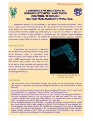
Luminescent Bacterial DiseaseThe luminescent bacterial disease is a serious problem in the hatcheries. Occasionally,the jriveniles <strong>an</strong>d adult shrimp may also be affected in the growqut farms.Signs <strong>an</strong>d Symptoms: The infected larvae appear luminescent in darkness, <strong>an</strong>d suffer heavymortality.Cause: Luminescent bacteria, viz., Vibrio harveyi.<strong>Diagnosis</strong>: Goss signs <strong>an</strong>d symptoms <strong>an</strong>d microscopic demonstration <strong>of</strong> swarming bacteriawithin the haemocoel <strong>of</strong> moribund shrimp larvae would confirm luminescent bacterialdisease. The luminescent bacteria c<strong>an</strong> be readily isolated on Zobell's Marine Agar or aselective medium. Identity <strong>of</strong> the isolates could be confirmed based on their morphological<strong>an</strong>d biochemical characteristics.Prevention: Use ultraviolet irradiated <strong>an</strong>d chlorinated (calcium hypochlorite 200ppm for 1h.) water. Cle<strong>an</strong> the debris collected at the bottom <strong>of</strong> the culture t<strong>an</strong>ks daily.Controt Exch<strong>an</strong>ge 80% <strong>of</strong> water daily with UV sterilised 1 s<strong>an</strong>d filtered seawater.Brown spot disease (Shell disease or Rust disease)Signs nnd Symptoms: The affected <strong>an</strong>imals show presence <strong>of</strong> brownish to black eroded areason the body surface <strong>an</strong>d appendages.Cause: Bacteria such as Vibrio spp., Aeromonas spp., <strong>an</strong>d Flavobacterium spp., withchitinolytic activity.Dingnosis: <strong>Diagnosis</strong> <strong>of</strong> brown spot disease is achieved by simple observations on the grosssigns <strong>an</strong>d symptoms <strong>an</strong>d confirmed by isoIation <strong>of</strong> the bacteria from the site <strong>of</strong> infection onZobelI's Marine Agar <strong>an</strong>d identification <strong>of</strong> the pathogen.Prevention: Reduce org<strong>an</strong>ic load in water by increased water exch<strong>an</strong>ge. Avoid unnecessaryh<strong>an</strong>dling <strong>an</strong>d overcrowding to minimise ch<strong>an</strong>ces <strong>of</strong> injury <strong>an</strong>d infection.Control: Induction <strong>of</strong> moulting by applying tea seed cake may be useful. Improve waterquality by increasing water excl~<strong>an</strong>ge. Although <strong>an</strong>tibiotics may be useful their use in theculture sytem is not recommended.Necrosis <strong>of</strong> appendagesSigns <strong>an</strong>d symptoms: The tips <strong>of</strong> walking legs, swimmerets <strong>an</strong>d uropods <strong>of</strong> affected shrimpundergo necrosis <strong>an</strong>d become brownish <strong>an</strong>d black. The setae, <strong>an</strong>tennae <strong>an</strong>d appendages maybe broken <strong>an</strong>d mel<strong>an</strong>ised.
Cause: The epibiotic bacteria stich as P'ibrio spp., Pseudomonas spp., Aeromonas spp. <strong>an</strong>dFlavobacrerium spp.Dingnosis: Based on gross signs <strong>an</strong>d symptoms.Prevention: Maintain good. water quality. Stock shrimp at optimum density. Avoidunnecessary h<strong>an</strong>dling <strong>of</strong> the shrimp, which may lead to injuries, leading to infection <strong>an</strong>dnecrosis.Control: Induction <strong>of</strong> moulting by applying 0.5 - 1 ppm tea seed cake may be <strong>of</strong> help.Vibriosis in larvaeSigns <strong>an</strong>d Symptoms: The affected larvae show necrosis <strong>of</strong> appendages, exp<strong>an</strong>dedchrornatopl~ores, empty gut, absence <strong>of</strong> faecal str<strong>an</strong>ds <strong>an</strong>d poor feeding. Cumulativemortalities may be very high reaching up to 80% within few days.Cause: Bacteria, viz., Vibrio alginolyticus, V.parahaernolyticus, <strong>an</strong>d V.<strong>an</strong>guillarun~.<strong>Diagnosis</strong>: Microscopic demonstration <strong>of</strong> motile bacteria in the body cavity <strong>of</strong> moribundshrimp larvae, <strong>an</strong>d isolation <strong>an</strong>d identification <strong>of</strong> pathogenic bacteria would help in thediagnosis <strong>of</strong> the disease.Prevention: Maintain good water quality <strong>an</strong>d reduce org<strong>an</strong>ic load in the water by increasedwater exch<strong>an</strong>ge.Control: 10-15 ppln EDTA to the rearing water.Filamentous Bacterial DiseaseSigns <strong>an</strong>d Symptonzs: The affected shrimp larvae show fouling <strong>of</strong> gills, setae, appendages<strong>an</strong>d body surface. Moulting <strong>of</strong> affected shrimps is impaired <strong>an</strong>d may die due to hypoxia.Cause: Filamentous bacteria, such as Lezjcothrix nzucor.<strong>Diagnosis</strong>: <strong>Diagnosis</strong> <strong>of</strong> filamentous bacterial disease could be achieved based on gross signs<strong>an</strong>d symptoms <strong>an</strong>d by microscopically demonstrating filamentous bacterial fouling <strong>of</strong> bodysurface <strong>an</strong>d appendages <strong>of</strong> shrimp larvae.Prevention: Maintain good water quality with optimal physico-chemical parameters.Control: 0.25 - 1 ppm Copper sulphate bath treatment for 4-6 hrs.FUNGAL DISEASES
Larval MycosesIt is one <strong>of</strong> the most devastating diseases in shrimp hatcheries. However, larval mycoseshave been successfi~lly controlled during the recent years with better m<strong>an</strong>agement practices.Signs <strong>an</strong>d symplorns: Affected larvae appear opaque followed by sudden mortality.protozoeal <strong>an</strong>d mysis stages are highiy susceptible. Within 1-2 day's, whole stock <strong>of</strong> shrimplarvae may suffer mortality.Cause: Oomycetous fungi, Lagenidilrm spp, Sirolpidium spp,'<strong>an</strong>d Haliphthoros spp. Thesefungi are filamentous, non-septate <strong>an</strong>d coenocytic. Upon infection, the fungal myceliumreplaces the larval tissues <strong>an</strong>d ramifies into various parts <strong>of</strong> the body. Vegetative propagation<strong>of</strong> these fungi is through production <strong>of</strong> bi-flagellate zoospores, which are released into therearing medium. These zoospores further infect fresh shrimp larvae. These fungi c<strong>an</strong> beisolated on peptone yeast extract glucose (PYG) agar or Saboraud's dextrose agar.Dingnosis: Microscopic demonstration <strong>of</strong> presence <strong>of</strong> extensively br<strong>an</strong>ched non-septate,fungal hyphae within the body cavity <strong>of</strong> the shrimp larvae.Prevention: Remove bottom sediments <strong>an</strong>d dead larvae periodically. Disinfect the t<strong>an</strong>ks <strong>an</strong>dother equipment in the hatchery from time to time. Treat spawners with 5 ppm trefl<strong>an</strong> bath for1 h.Control: When the disease is detected in early stages, Trefl<strong>an</strong> (Trifluralin) 0.1 -0.2 pp~n bathfor I day may help in reducing Inass mortality.TheOther fungi such as Fusarium spp. cause infections in nauplii, protozoea, juveniles<strong>an</strong>d adults. Black gill disease is <strong>of</strong>ten caused by this fungus. The fungus c<strong>an</strong> be identified bymicroscopic examination <strong>of</strong> its characteristic c<strong>an</strong>oe shaped micro-conidia. Other oomycetousfungi such as Saprolegnia spp. <strong>an</strong>d Leptolegnia spp. are also known to affect shell <strong>of</strong> shrimp<strong>an</strong>d produce dark necrotic lesions causing gradual mortality.
PARASiTIC AND NON-INFECTIOUS IIISEASESK. P. Jithendr<strong>an</strong>PARASITIC DISEASESAmong the disease causing org<strong>an</strong>isms <strong>of</strong> shrimp, parasites, especially protozo<strong>an</strong>parasites form <strong>an</strong> import<strong>an</strong>t group. Although, several diseases caused by parasites have beennoticed in shrimp, <strong>of</strong>ten, chronic conditions caused by protozo<strong>an</strong>s play a crucial role inshrimp production. The protozoa, affecting shrimp c<strong>an</strong> be grouped as parasites <strong>an</strong>dcommensals. Following are the major disease problems caused by the protozoa:Protozo<strong>an</strong> foulingCotton shrimp diseaseEnterozoic cephaline gregarine infectionInvasive protozo<strong>an</strong> infection"'Protozo<strong>an</strong> FoulingThis is a serious disease problem commonly encountered both in hatchery <strong>an</strong>d farm.Signs <strong>an</strong>d symptoms: Affected shrimps are restless <strong>an</strong>d their locomotion <strong>an</strong>d respiratoryfunctions are hampered. Heavily infected, larger shrimp <strong>of</strong>ten have fuzzy-mat like appear<strong>an</strong>ceon the body surface, appendages <strong>an</strong>d gills. Animals also show brownish discoloration due toalgal filaments or debris ent<strong>an</strong>gled with the epibiont.Causative org<strong>an</strong>ism: Peritrichous ciliates such as Zoothamniwm, Epistylis, Vorticella,Acinata etc.<strong>Diagnosis</strong>: Based on gross signs <strong>an</strong>d symptoms. Fresh smear preparation <strong>of</strong> the surfacescrapping or gill or appendages will reveal the morphology <strong>of</strong> the protozo<strong>an</strong>.Prevention: Maintain good water quality, reduce the org<strong>an</strong>ic subst<strong>an</strong>ce, silt <strong>an</strong>d sediment onthe pond bottom, maintaining optimum dissolved oxygen level (5-6 ppm) <strong>an</strong>d frequentexch<strong>an</strong>ge <strong>of</strong> water.Control: Formalin is the chemo therapeut<strong>an</strong>t <strong>of</strong> choice. Treatment with formalin 15-25 ppmconcentration (single treatment) for ponds or dip treatment <strong>of</strong> affected <strong>an</strong>imals in 50-100 ppmfor 30 min is useful. Good aeration during treatment is essential.
Cotton <strong>Shrimp</strong> Disease or Milk <strong>Shrimp</strong> DiseaseSigns rrnd symptoms: The pathogenic protozo<strong>an</strong> infect <strong>an</strong>d replace striated muscle, causing itto become opaque <strong>an</strong>d white. The muscle <strong>of</strong> such shrimp appears cooked. In severelyaffected shrimps, the exoskeleton appears bluish black, <strong>an</strong>d white tumour-like swelling maybe found on the gills <strong>an</strong>d subcuticle. A few species infect gonads, heart, haemolymph vessel,hepatop<strong>an</strong>creas <strong>an</strong>d produce enlarged gonads.Carisative org<strong>an</strong>isms: Microspore<strong>an</strong>s such as Agmusorna, Arneson <strong>an</strong>d Pleistophora.<strong>Diagnosis</strong>: Based on gross signs <strong>an</strong>d symptoms, the disease c<strong>an</strong> be tentatively diagnosed.Microscopic examination <strong>of</strong> squash preparation or impression smears stained with Giernsawill reveal large number <strong>of</strong> microspore<strong>an</strong> spores. Spore characteristics may vary from speciesto species.Prevention: Affected <strong>an</strong>imals should be destroyed <strong>an</strong>d burried away from the farm. Beforestocking, the possible conditioning host / intermediate host should be eliminated.Control: No treatment has been reported for Penaeids.Enterozoic Cephaline Gregarine InfectionSigns <strong>an</strong>d symptoms: Affected shrimp show loss <strong>of</strong> appetite, lethargy <strong>an</strong>d weakness. Often,low levels <strong>of</strong> mortalities.Causative org<strong>an</strong>isms: Cephaline gregarines such as Nernutopsis <strong>an</strong>d Cephalolobz~s.<strong>Diagnosis</strong>: Microscopic observation <strong>of</strong> the digestive system reveals the developmental stages<strong>of</strong> the parasites. Rectal portion show white, spherical gametocysts attached to the wall.Prevention: Infection has been generally observed in culture system, which uses wild seeds.So the best preventive measure is avoid<strong>an</strong>ce <strong>of</strong> wild seed. Elimination <strong>of</strong> intermediate hostsfrom the culture system also prevents the disease occurrence. -.Control: No treatment is reported.Invasive Protozo<strong>an</strong> InfectionThis has been noticed in a few cases in hatcheries. Often, heavy mortalities also havebeen recorded. Causative org<strong>an</strong>isms include ciliate protozoa, Par<strong>an</strong>ophrys <strong>an</strong>d Paraoronemu,leptomonad-like org<strong>an</strong>isms. Control <strong>an</strong>d preventive measures are not reported.
XOS-INFECTIOUS DISEASESNon-infectious disease are common in the grow-out farms. as influences <strong>of</strong> nutritionalfactors, environmental factors such as temperature extremes <strong>an</strong>d oxygen depletion, toxicityfrom biotic <strong>an</strong>d abiotic origins, become critical during the lengthy culture period.S<strong>of</strong>t shell syndromeS<strong>of</strong>t shell syndrome is a condition in which shrimp exoskeleton becomes s<strong>of</strong>t. Cuticle<strong>of</strong> the affected shrimps is persistently s<strong>of</strong>t, loose <strong>an</strong>d papery for several weeks. Affectedshrimps are weak <strong>an</strong>d show poor escape reflex, <strong>an</strong>d these <strong>an</strong>imals are susceptible toc<strong>an</strong>nibalism. Severely affected P. indicus <strong>of</strong>ten show uilduIating gut in the first threeabdominal segments. Several factors are implicated as causative agent for this condition as:sudden fluctuation in water salinity, high soil pH, highly reducing conditions in soil, loworg<strong>an</strong>ic matter in soil, low phosphate content <strong>an</strong>d pesticide pollution in water, nutritionaldeficiency <strong>an</strong>d insufficient \xiater exch<strong>an</strong>ge. The disease may be prevented or controlledthrough environmental <strong>an</strong>d dietary ln<strong>an</strong>ipulations by providing favourable water <strong>an</strong>d soilconditions in the pond <strong>an</strong>d feeding adequately with bal<strong>an</strong>ced diets.Black gill diseaseA number <strong>of</strong> abiotic <strong>an</strong>d biotic reasons have been attributed to the black gill inshrimps. Presence <strong>of</strong> excessive levels <strong>of</strong> toxic subst<strong>an</strong>ces such as nitrite, ammonia, heavymetals, crude oils etc. in the culture water may lead to black gill disease. High org<strong>an</strong>ic load,heavy siltation <strong>an</strong>d reducing conditions in rearing pond c<strong>an</strong> also cause this disease in shrimps.Attack <strong>of</strong> certain bacterial, fungal <strong>an</strong>d protozo<strong>an</strong> pathogens c<strong>an</strong> also cause black gill conditionin shrimp. Affected shrimps have gills with black to brown discoloration, in acute casesnecrosis <strong>an</strong>d atrophy <strong>of</strong> the gill lainellae may be apparent. The blackening is due to thedeposition <strong>of</strong> mel<strong>an</strong>in at sites <strong>of</strong> massive haemocyte accumulation, followed by dysfunction<strong>an</strong>d destruction <strong>of</strong> whole gill processes.Treatment <strong>of</strong> the black gill disease depends upon the cause <strong>of</strong> the disease. Preventiveor corrective measure may be adopted to avoid or reduce the biotic / abiotic factors in therearing pond to co~ztrol the disease condition.
Red diseaseThe juveniles <strong>an</strong>d adult shrimps / broodstock affected with red disease have reddishdiscoloration in body, pleopods <strong>an</strong>d gills. Definite causative agent is not known. One <strong>of</strong> thereasons believed to be the cause <strong>of</strong> disease is a microbial toxin in r<strong>an</strong>cid or spoiled diets or indetritus <strong>of</strong> ponds rich in org<strong>an</strong>ic matter. Extreme conditions <strong>of</strong> pH or salinity in pond watermay also cause the disease. Healthy m<strong>an</strong>agement <strong>of</strong> ponds along with the use <strong>of</strong> good qualityfeed may help in the avoid<strong>an</strong>ce <strong>of</strong> red disease.Cramped tail diseaseAffected shrimps have rigid dorsal flexure <strong>of</strong> the abdomen, which c<strong>an</strong>not bestraightened. These shrimps lie on their sides at the bottom <strong>of</strong> the pond <strong>an</strong>d are susceptible toc<strong>an</strong>nibalism. Exact cause for this disease is not known, but environmental <strong>an</strong>d nutritionalcauses have been suggested. Mainten<strong>an</strong>ce <strong>of</strong> healthy conditions in the pond with properfeeding with bal<strong>an</strong>ced diet may be helpful in the prevention I control <strong>of</strong> this disease.Gas-bubble diseaseSuper saturation <strong>of</strong> atmospheric gases <strong>an</strong>d oxygen in the pond c<strong>an</strong> result in the gasbubbledisease, which affects the shrimps <strong>of</strong> all sizes. Presence <strong>of</strong> gas bubble in the gills orunder the cuticle is the characteristic <strong>of</strong> this disease. Gas bubble disease due to oxygen is notlethal, while that <strong>of</strong> nitrogen c<strong>an</strong> be lethal. The threshold saturation level to cause the gasbubble,in the case <strong>of</strong> nitrogen is 118 % while that <strong>of</strong> oxygen is 250 % <strong>of</strong> normal saturation.The severely affected or dead shrimp due to this disease may float near the water surface.Super saturation <strong>of</strong> the gases must be avoided to prevent the disease.Muscle necrosis<strong>Shrimp</strong>s <strong>of</strong> all life stages are affected with muscle necrosis. Affected shrimps arecharacterised by the presence <strong>of</strong> white opaque areas in body musculature, usually in the lowerabdomen or some times in the appendages. The condition is reversible in the early stages ifthe corrective measures are taken, but in severe cases sloughing <strong>of</strong> the affected areas occursdue to secondary bacterial infection leading to death. This disease is associated with poorenvironmental conditions such as low oxygen levels, <strong>an</strong>d salinity or temperature shock.Overcrowding <strong>an</strong>d poor h<strong>an</strong>dling also c<strong>an</strong> cause muscle necrosis. Avoid<strong>an</strong>ce <strong>of</strong>overcrowding, proper h<strong>an</strong>dling <strong>an</strong>d mainten<strong>an</strong>ce <strong>of</strong> favourabIe environmental factors mayhelp to contain the disease.
INVESTIGATING SHRIMP DISEASES: OBSEliVATION ON POhZ) gL SHRIMPAND SAMPLINGS.V.Alav<strong>an</strong>di, K.K. Vijay<strong>an</strong> T.C. S<strong>an</strong>tiago <strong>an</strong>d N. Kalaim<strong>an</strong>iProper <strong>an</strong>d accurate diagnosis <strong>of</strong> diseases forms the cardinal step in <strong>an</strong>y diseasecontrol <strong>an</strong>d prevention programme.' To diagnose a shrimp disease problem, history <strong>of</strong> thefarms, the soil <strong>an</strong>d water conditions <strong>of</strong> the ponds, incidence <strong>of</strong> <strong>an</strong>y disease problem in theadjoining areas, possibility <strong>of</strong> disseminating disease through birds or other carriers, are <strong>of</strong>great import<strong>an</strong>ce.These are the signific<strong>an</strong>t information related to the epidemiology <strong>of</strong> thedisease. This section stresses the need to examine general information on the farming activityC<strong>an</strong>d the information regarding disease on site <strong>an</strong>d some points on collection <strong>of</strong> samples forlaboratory investigation.1. Background information about the farming practicesA. Exnrnirzatio?~ <strong>of</strong> pondsThis involves the various parameters <strong>of</strong> ponds such as methods followed in thepreparation <strong>of</strong> ponds, depth, nature <strong>of</strong> the bottom, water treatment methods, nature <strong>of</strong> waterinlet <strong>an</strong>d outlet procedure etc. Apart from these, colour <strong>of</strong> water, algal blooms, turbidity <strong>of</strong>water <strong>an</strong>d presence <strong>of</strong> bioluminescence during night are also import<strong>an</strong>t criteria, whichdetermine the health <strong>of</strong> the shrimp.B. Stocking parametersThese parameters include origin <strong>an</strong>d source <strong>of</strong> seeds, health status <strong>of</strong> spawners <strong>an</strong>d thelarvae, survival rate <strong>of</strong> larvae within the hatchery <strong>an</strong>d nursery, whether <strong>an</strong>y <strong>an</strong>tibiotic1disinfect<strong>an</strong>t used for larval rearing; stocking density, time <strong>of</strong> stocking etc. These parametersare very much import<strong>an</strong>t in assuring the healthy or resist<strong>an</strong>t nature <strong>of</strong> the larvae <strong>an</strong>d in thediagnosis <strong>of</strong> <strong>an</strong>y possible disease problem.C. M<strong>an</strong>agentent practicesThese include data generated out <strong>of</strong> the close monitoring <strong>of</strong> the system for the growth,survival <strong>an</strong>d occurrence <strong>of</strong> diseases. These also include the quality <strong>of</strong> the feed, feedingregime, consumption <strong>of</strong> feed by the shrimp, time <strong>an</strong>d rate <strong>of</strong> water exch<strong>an</strong>ge <strong>an</strong>d the use <strong>of</strong>chemicals, immullostimul<strong>an</strong>ts or bioremedial measures also to be recorded.D. Environmental parameters<strong>Diseases</strong> may be <strong>of</strong> infectious <strong>an</strong>d non-infectious eteologies. Majority <strong>of</strong> the noninfectiousdiseases are due to nutritional deficiency or due to abnormal environmental
conditions. Hence, a close examination <strong>an</strong>d recording <strong>of</strong> the various parameters <strong>of</strong> water <strong>an</strong>dsoil quality should be done periodically.2. Field observation for signs <strong>an</strong>d synlptomsA. Observation <strong>of</strong> beltaviorar <strong>of</strong> shrimpCritical observation on the behaviour <strong>of</strong> shrimp will give <strong>an</strong> indication <strong>of</strong> the health <strong>of</strong>the <strong>an</strong>imals. Signific<strong>an</strong>t behaviour includes escape reflex, swimming at the surface, moultingbehaviour, feeding behaviour etc.Healthy shrimp will have quick reflexes i.e., shrimp will respond inst<strong>an</strong>t<strong>an</strong>eously to<strong>an</strong>y outside disturb<strong>an</strong>ces or artificial stimulation; <strong>Shrimp</strong> showing poor escape reflex may notbe in a healthy condition.Swimming at the surface is <strong>an</strong> indication <strong>of</strong> either inadequate oxygen level, orrespiratory impairment. Disease conditions, such as fouling, white spot disease etc alwaysshow such behavioural ch<strong>an</strong>ges.Moulting behaviour is <strong>an</strong>other import<strong>an</strong>t factor, which has to be observed. Regular<strong>an</strong>d continued moulting indicates continuous growth. Abnormal moulting indicates a diseasedcondition. Chronic condition due to hepatop<strong>an</strong>creatic infection may cause abnormal moulting.Feeding behaviour is also <strong>an</strong> import<strong>an</strong>t indicator <strong>of</strong> health <strong>of</strong> the shrimp. Diseasedshrimp normally will show reduced appetite (e.g. white spot disease, protozo<strong>an</strong> fouling).However, it has been reported that juveniles <strong>an</strong>d sub-adult affected with yellow head diseaseshow <strong>an</strong> abrupt abnormal increase in feeding rate for several days.B. Observation <strong>of</strong> external signs <strong>an</strong>d symptomsTo assess the health status <strong>of</strong> a shrimp, following gross signs should be examined.i. Colour <strong>an</strong>d nature <strong>of</strong> exoskeleton: Healthy shrimp will have pale blue coloured,bright, smooth <strong>an</strong>d clear cuticle with proper hardness. Exoskeleton will showbrownish discoloration <strong>an</strong>d occasional mat-like appear<strong>an</strong>ce (muddy cuticle) inprotozo<strong>an</strong> fouling. Moulted shrimp <strong>an</strong>d shrimp with s<strong>of</strong>t shell syndrome will shows<strong>of</strong>t exoskeleton. However, the shrimp with s<strong>of</strong>t shell syndrome will have a hardrostra1 spine.ii. Apart from these, visible blisters or brown or black eroded areas on the exoskeletonmay indicate possible bacterial infection. Presence <strong>of</strong> white spots or patches on thecarapace indicates the visual disease, white spot disease.iii. Appendages: Tips <strong>of</strong> walking legs, swimmerets <strong>an</strong>d uropods may show necrosis <strong>an</strong>dbecome brownish black indicating a possible bacterial infection. Often, physical injury
lnay be a predisposing factor for bacterial infection <strong>an</strong>d result<strong>an</strong>t necrosis <strong>an</strong>dmel<strong>an</strong>isation.iv.Muscula~ure: White muscular opacity may indicate muscle necrosis due toenvironmental stress or a microspore<strong>an</strong> infection.C. Examination <strong>of</strong> internal org<strong>an</strong>s for pathological sigtzsi. W: Gills are normally cle<strong>an</strong>, semi-tr<strong>an</strong>sparent <strong>an</strong>d colourless. External fouling dueto epicommensals will cause dark yellow discoloration. Bacterial infection c<strong>an</strong> causeblackening <strong>of</strong> gills. Vibriosis may cause yellow discolouration <strong>of</strong> br<strong>an</strong>chiostegites.ii. Hepato~<strong>an</strong>creas: Hepatop<strong>an</strong>creas <strong>of</strong> normal shrimp will be obvious, with proper size<strong>an</strong>d shape. Colour <strong>of</strong> top half is brown <strong>an</strong>d the bottom has a white membr<strong>an</strong>e cover.Abnormal colour, enlargement or atrophy etc. are indication <strong>of</strong> bacterial or viralinfection, or the presence <strong>of</strong> toxic subst<strong>an</strong>ces or nutritional deficiency.iii. Haemolvnzph: Haelnolymph <strong>of</strong> normal shrimp have slight blue colour. It will easilycoagulate in 1 min. after taking it out from shrimp. Some <strong>of</strong> the bacterial <strong>an</strong>d viralinfections cause the haemolymph non-coagulable, colourless or light reddish or muddyin nature.3. Collection <strong>of</strong> samplesFor accurate diagnosis <strong>of</strong> the disease, typical <strong>an</strong>d representative sample <strong>of</strong> infected<strong>an</strong>imals should be collected. Very <strong>of</strong>ten, in one pond itself there will be multiple infections.All the dead <strong>an</strong>imals [nay not be a representative sample. Instead <strong>of</strong> dead <strong>an</strong>imals, moribund<strong>an</strong>imals will be suitable for <strong>an</strong>alysing the symptoms, for pathological studies <strong>an</strong>d for isolation<strong>of</strong> pathogens. Moribund shrimp may have secondary infection also, <strong>an</strong>d more <strong>of</strong>ten, shrimpwith disease in the initial stage may not exhibit the real symptoms. All these factors should betaken into account while collecting the required sample.According to m<strong>an</strong>y experts, there are four methods to collect the samples <strong>of</strong> shrimp:i. Picking from the sides around the pondsii. Catching from the middle <strong>of</strong> the pondiii. Using cast netiv. From the feed trays.These samples will really reflect the actual disease status in pond. Samples <strong>of</strong>moribund shrimp, which are collected from the sides around the ponds, will be mostly at theterminal stage <strong>of</strong> infection. Ths salnples collected from the middle may be in <strong>an</strong> intermediate
stage. Cast net will give a r<strong>an</strong>dom sample <strong>an</strong>d is preferable, while the samples froin the feedtray will be usually healthy.
BACTERIOLOGICAL METHODSS.V. Alav<strong>an</strong>diThe methods <strong>of</strong> microbiological examination <strong>of</strong> shrimp are essentially similar to thosefollowed for the higher <strong>an</strong>imals. The first step in shrimp diagnostic bacteriology is to isolatethe pathogen froin the diseased shrimp <strong>an</strong>d then identify the same based on its cultural,morphological, physiological, biochemical <strong>an</strong>d serological characteristics. The methodsrequired for isolation <strong>an</strong>d identification <strong>of</strong> shrimp pathogens are described here. However,for more details, the readers are advised to refer Austin (1988). A method for determination<strong>of</strong> total viable counts <strong>of</strong> bacteria in water samples is also included.Aseptic techniquesMainten<strong>an</strong>ce <strong>of</strong> aseptic codditions during all stages <strong>of</strong> microbiology work is the first stepfor successful microbiological investigation. It is essential to take suitable measures to ensurethe recovery <strong>of</strong> bacteria <strong>of</strong> interest. There are several sources <strong>of</strong> contamination. The air maycontain dust particles <strong>an</strong>d aerosols (micro droplets <strong>of</strong> water). Other materials like glassware,buffers, culture media, <strong>an</strong>d equipment used <strong>an</strong>d even the careless personnel carrying out thework c<strong>an</strong> be sources <strong>of</strong> contamination. Hence, it is very import<strong>an</strong>t to take suitable measuresto get rid <strong>of</strong> the contaminating microorg<strong>an</strong>isms. In order to achieve this, several methods havebeen evolved. The methods <strong>of</strong> sterilisation <strong>of</strong> different kinds <strong>of</strong> materials used in thelaboratory are given in Table I.Pure culture techniqueGrowth <strong>of</strong> pure culture is necessary before <strong>an</strong>y cultural, biochemical or sensitivity tests arerun to identify <strong>an</strong>d characteri~e the suspected bacterial pathogen. A number <strong>of</strong> methods areused for this purpose. These are:- Streak plate technique (streaking onto solid media)- Pour plate technique (Incorporation into molten semi-solid media)- Dilution in liquid media
Among all these, the streak plate method is the most commonly method, which allowsob.taining pure culture <strong>of</strong> specific bacterium from mixed bacterial populations. Bacteria froma mixed culture are streaked over the agar surface in a pattern that deposits them further <strong>an</strong>dfurther apart. Towards the end <strong>of</strong> the pattern, the resulting colonies <strong>of</strong> different bacteria areseparated from each other. The single individual colonies <strong>of</strong> different types are then picked upwith the help <strong>of</strong> a bacteriological loop <strong>an</strong>d streaked on <strong>an</strong>other plate to obtain pure cultures.Purity <strong>of</strong> the culture should be confirmed by periodic streaking on plates <strong>an</strong>d observing theircultural, morphoiogical <strong>an</strong>d biochemical characteristics.- Table 1. Sterilization methodsMaterials -- sterilised .--- Methods <strong>of</strong> sterilisation -...All types <strong>of</strong> glassware like pipettes, tubes, Dry heat methodflasks, petri dishes etc.Hot air oven160°C for ------- 2 hr or 180°C for 1 hr ---Destruction <strong>of</strong> used, contaminated material,dead <strong>an</strong>imals, tissues etc.Most bacteriological media, glassware /containers, (decontamination <strong>of</strong> used media),steel items, corlts, rubber materials, filterpads, filter assembly, distilled water, buffers,solutions etc.IncinerationMoist heatAutoclaving 121°C for 15 minMost tissue culture media, <strong>an</strong>tibiotics, sera, Filtrationsolutions containing heat sensitive materials Membr<strong>an</strong>e filters <strong>of</strong> 0.22 pm 1 0.45 p~n porelike amino ,acids, carbohydrates, biological sizematerials etc.-Cultural <strong>an</strong>d morphological characters <strong>of</strong> bacteriaFor identification <strong>of</strong> bacteria, some general cultural <strong>an</strong>d morphological characteristics likesize, shape, pigmentation, opacity <strong>of</strong> the bacterial colonies on the solid media; cell shape(rods cocci, coccobacilli, comma), sporulation, etc are import<strong>an</strong>t <strong>an</strong>d aid in preliminarygrouping <strong>of</strong> the bacteria. Some <strong>of</strong> the cultural <strong>an</strong>d morphological characteristics useful fordistinguishing bacteria ari given in Tables 2 <strong>an</strong>d 3.
In place <strong>of</strong> ZMA, nutrient agar prepared in aged seawater would suffice. The ZMA favoursthe growth <strong>of</strong> all the heterotrophic bacteria occurring in the brackish <strong>an</strong>d marineenvironments, whereas, the TCBS medium is selective for isolation <strong>of</strong> shrimp pathogenssuch as Vibrio spp. Mycological agar/ Sabouraud's dextrose agar is used for isolation <strong>of</strong>fungi. A selective medium for isolation <strong>of</strong> l~iminescent bacteria is used wheneverrequired.ii. Incubate the inoculated agar plates at optimal temperature (30°C) for 24-48 h <strong>an</strong>d observefor development <strong>of</strong> bacterial colonies.iii.Examine cultural characteristics <strong>of</strong> the bacterial colonies as given in the subsequentsections <strong>an</strong>d record.iv. Obtain pure culture <strong>of</strong> bacteria by piciting up morphologically distinct colonies with thehelp <strong>of</strong> a sterile bacteriological loop <strong>an</strong>d subculture on ZMAYor further characterization. ,Gram Staining <strong>of</strong> BacteriaPriizciple: Staining bacteria by Gram's method is widely used for classification <strong>of</strong> bacteriainto Gram positive <strong>an</strong>d Gram negative bacteria. The bacterial cell walls containpeptidoglyc<strong>an</strong>s, which is a thick layer in the Gram-positive bacteria. The pararos<strong>an</strong>iline dyesuch as crystal violet treated with iodine mord<strong>an</strong>t remains trapped in the cell wall <strong>an</strong>d hence,the cells are not de-stained upon treatment with alcohol.Procedure:i. Prepare shears <strong>of</strong> bacteria on cle<strong>an</strong> glass slide using sterile nichrome loop by mixing witha drop <strong>of</strong> sterile normal saline.ii. Fix the smears by air-drying or by gently passing the slide over the Bunsen flame.iii. Stain the smears with Crystal Violet solution for 1 minute. "iv. Wash in tap water for few seconds.v. Flood the smears with Iodine solution for 30 seconds.vi. Wash in tap water for 15 seconds.vii.De-colorize with 95% ethyl alcohol for 30 seconds.viii.Wash with tap water.
ix. Counter-stain with Safr<strong>an</strong>in solution for 10 seconds.x. Wash in tap water. Blot dry <strong>an</strong>d examine under oil immersion objective <strong>of</strong> the microscope.Itzterpretntiun: Violet coloured bacteria: Gram positive; Red / pink coloured bacteria: Gramnegative.Record size, shape arr<strong>an</strong>gement <strong>an</strong>d other morphological characteristics.Motility test (H<strong>an</strong>ging drop method)i. Place a very small drop <strong>of</strong> log phase broth culture <strong>of</strong> bacteria with the help <strong>of</strong> sterileinoculating loop (2 mm dia) at the centre <strong>of</strong> a cover glass.ii. Place small drops <strong>of</strong> water on the corners <strong>of</strong> the cover glass....111.Invert the cover glass over the cavity <strong>of</strong> slide, so that the drop <strong>of</strong> culture is h<strong>an</strong>ging at thecentre <strong>of</strong> the cavity slide.iv.0bserve the h<strong>an</strong>ging drop <strong>of</strong> bacterial culture under the microscope for tnortility <strong>of</strong>bacteria.v. Darting or zig-zag motility indicates that the bacteria may have polar flagellation, while,slow motility or vibratory motility indicates peritrichous flagellation.Oxidase testPrinciple: Some bacteria possess cytochrome oxidase or indophenol oxidase, which catalysestr<strong>an</strong>sport <strong>of</strong> electrons from donor compounds to oxygen. In this test, the N N N'N'tetramethyl p-phenelene diamine dihydrochloride, a colourless dye serves <strong>an</strong> artificialelectron acceptor. The oxidase enzyme produced by bacteria oxidises the dye producingcoloured indophenol blue.Procedure:i. Place a strip <strong>of</strong> whatm<strong>an</strong> No.1 filter paper in a petri dish.ii. Add 2-3 drops <strong>of</strong> freshly prepared 1% solution <strong>of</strong> N,N,N',N'- tetramethyl paraphenylenediamine dihydrochloride.iii.S~near the test colony <strong>of</strong> bacteria on the filter paper using a sterile capillary.
Interpretation: Positive reaction is indicated by development <strong>of</strong> a deep purple coloi~r <strong>of</strong> thesmear.Catalase testPrinciple: Bacteria possess <strong>an</strong> enzyme called catalase which cataIyses breakdown <strong>of</strong> toxichydrogen peroxide (H2O2) formed during the cell's metabolism into water <strong>an</strong>d oxygen. Whena solution <strong>of</strong> H202 is added to bacterial cell suspension, the catalase enzyme is activated,resulting in the release <strong>of</strong> 02, which is observed as effervescence.Procedure:i. Make a drop <strong>of</strong> heavy srispension <strong>of</strong> test culture <strong>of</strong> bacteria on a slide.ii. Place a drop <strong>of</strong> 10% hydrogen peroxide solution over the bacterial si~spension.iii.0bserve for small air bubbles.Inferpretation: Production <strong>of</strong> gas bubbles (effervescence) indicates a positive reaction.*Carbohydrate Fermentation testPrinciple: When the bacteria are grown in basal media containing specific carbohydrates suchas glucose, sucrose, lactose, m<strong>an</strong>nitol efc, in the presence <strong>of</strong> a pH indicator, m<strong>an</strong>ifest intocolour ch<strong>an</strong>ge depending on the metabolic pathway used by the bacteria.Procedure:i. Inoculate the bacterial isolate in duplicate into phenol red broth base incorporated withsugars such as glucose, lactose, m<strong>an</strong>nitol e6c. in sugar fermentation tubes.ii. Overlay one tube with sterile mineral oil (e.g. liquid paraffin) about 1-2 cm. Incubate thetubes at 37OC.iii. Observe the tubes for colour ch<strong>an</strong>ge at 24,48 <strong>an</strong>d 72 h intervals.Interpretation: Acid production in open tube indicates oxidative metabolism <strong>an</strong>d acidproduction in the tube overlaid with mineral oil indicates fermentative metabolism <strong>of</strong> thebacteria.Lysine decarboxylase, Orinithine decarboxylase <strong>an</strong>d Arginine dihydrolase test
Pritzciple: Some bacteria are able to produce enzymes that attack carboxyl group <strong>of</strong> aminoacids. The reaction is <strong>an</strong>aerobic. Bacteria are inoculated into tubes containing the amino acidalong with a control tube, which contains only the basal medium without the amino acid. Thetube with amino acid is made <strong>an</strong>aerobic by overlaying with sterile mineral oil over themedium. If the test org<strong>an</strong>ism does not produce decarboxylase, both the control <strong>an</strong>d test tubesturn yellow due to fermentation <strong>of</strong> small amount <strong>of</strong> glucose present in the medium yieldingacidic products, lowering the pH <strong>of</strong> the medium. If the amino acid is decarboxylated, thetubes revert back to original purple colour, because <strong>of</strong> the alkaline amines produced duringthe reaction, which increase the pH <strong>of</strong> the medium.Procedure:i. Inoculate the tubes <strong>of</strong> Moeller's decarboxylase medium containing appropriate amino acid(lysine 1 arginine 1 ornithine) along with a control tube without amino acid with bacterialisolates.ii. Overlay the tube containing amino acid with 2-3 cm mineral oil (liquid paraffin).iii. Incubate the tubes at 37OC <strong>an</strong>d observe daily for 4 days for ch<strong>an</strong>ge <strong>of</strong> colour.Interpretation: Purple colour (original coiour <strong>of</strong> the medium, alkaline reaction) indicatesdecarboxylation <strong>of</strong> lysine <strong>an</strong>d ornithine <strong>an</strong>d positive reaction for arginine dihydrolase.Yellow colour indicates fermentation <strong>of</strong> glucose only <strong>an</strong>d negative reaction for decarboxylase<strong>an</strong>d dihydrolase.O-Nitrophenyl-P-D-galactopyr<strong>an</strong>oside (ONPG) hydrolysis testPrinciple: The test demonstrates the ability <strong>of</strong> bacteria to ferment lactose. Two enzymes areinvolved in this activity. The permease permits the lactose molecule into the cell, while the P-galactosidase hydrolyses lactose to galactose <strong>an</strong>d glucose. Some bacteria lack the ability toproduce permease <strong>an</strong>d possess the enzyme P-galactosidase. The ONPG, a compound similarto lactose molecule is hydrolysed by the enzyme P-galactosidase into galactose <strong>an</strong>d o-nitrophenyl, which is a yellow compound.Procedure:Inoculate heavily, a tube containing ONPG broth with the bacterial culture
Incubate at 37°C for 1-2 h.Examine the colour ch<strong>an</strong>ge <strong>of</strong> the broth from colourless to yellow.Inferprelation: Development <strong>of</strong> yellow colour indicates positive activity for P-galactosidaseactivity, (fermentation <strong>of</strong> lactose).Nitrate reductionPrinciple: Bacteria c<strong>an</strong> assimilate inorg<strong>an</strong>ic nitrate into their proteins by virtue <strong>of</strong> one <strong>of</strong> theenzymes in a complex process called nitrate reductase, which converts nitrate to nitrite(N03+N02). NO2 is detected by <strong>an</strong> inorg<strong>an</strong>ic assay using a-naphthylamine <strong>an</strong>d sulf<strong>an</strong>ilicacid.Procedure:i. Inoculate test culti~re <strong>of</strong> bacteria to tubes containing about 2 ml <strong>of</strong> nutrient brothsupplemented with 0.1% KN03 <strong>an</strong>d 0.2% agar.ii. Incubate at 37°C for 24 11.iii.Add 1 ml each <strong>of</strong> a-napathylami~ie solution <strong>an</strong>d Sulf<strong>an</strong>ilic acid solution.Inierpretrrtion: Positive reaction (conversion <strong>of</strong> NO3-+NOz) is indicated by development <strong>of</strong>pink colour.Note: When there is no development <strong>of</strong> pink colouration, add a pinch <strong>of</strong> 7inc dust. Absencc<strong>of</strong> colouration indicates positive result for the reaction N03-+N02.Indole testPrinciple: Indole is produced upon degradation <strong>of</strong> tryptoph<strong>an</strong> by some bacteria. Production <strong>of</strong>indole is detected by formation <strong>of</strong> pink coloured compound when it reacts with <strong>an</strong> aldehydesucl~ as p-dimethyl amino benzaldehyde.Procedure:i. Grow bacteria in 1% peptone water broth for 24 h at 37°C.ii. Observe for turbidity indicative <strong>of</strong> growth.iii.Add 0.5 ml <strong>of</strong> Kovac's reagent to the broth culture <strong>of</strong> bacteria <strong>an</strong>d shake gently.iv. Observe for development <strong>of</strong> pink colour.
Iizterpretation: Development <strong>of</strong> pink colour indicates a positive reaction.Voges Proscauer? TestPrinciple: The test detects acetoin or acetyl methyl carbinol, <strong>an</strong> intermediate product in theformation <strong>of</strong> butylene glycol during the metabolism <strong>of</strong> glucose. Acetoin is oxidised todiacetyl in the presence <strong>of</strong> oxygen by potassium or sodium hydroxide, which is a red colouredcomplex. Sensitivity <strong>of</strong> the test is further improved by addition <strong>of</strong> a-naphthol prior toaddition <strong>of</strong> KOH.Procedure:i. Grow pure culture <strong>of</strong> the bicteria in 5 ml <strong>of</strong> MRVP broth at 37°C for 48 h.ii. Tr<strong>an</strong>sfer about 2.5 ml <strong>of</strong> culture to <strong>an</strong>other tube.iii.Add 0.3 ml <strong>of</strong>.alcoholic a-naphthol <strong>an</strong>d 0.1 ml <strong>of</strong> 40% KOH solution, gently agitate thetube <strong>an</strong>d allow to st<strong>an</strong>d for 10-15'.iv. Observe for formation <strong>of</strong> red colour.I~zterpretation: Developinent <strong>of</strong> red / crimson colour indicates that bacteria produce acetylmethyl carbinol.Salt toler<strong>an</strong>cePrinciple: Various species <strong>of</strong> Vibrio <strong>an</strong>d related bacteria c<strong>an</strong> be differentiated based on theirability to grow in the presence <strong>of</strong> different levels <strong>of</strong> sodium chloride.Procedure:Inoculate bacterial culture into nutrient broth tubes containing 0, 3, 6, 8 <strong>an</strong>d 10% NaCI..I'Incubate at 37'C overnight <strong>an</strong>d observe for growth, which is indicated by turbidity <strong>of</strong> thebroth, compared to uninoculated control.Sensitivity to 01129Prirzciple: Vibrio species are sensitive to 150 pg <strong>of</strong> 01129 (2,4 di amino, 6-7 di isopropylpteridine), while the other related genera like Pseudomonas, Aeromonas, Plesiomonas,
Alkaligens: etc. are resist<strong>an</strong>t. The test is done by using 0/129 impregnated discs e~nployingconventional disc diff~ision method <strong>of</strong> Bauer et a1 (1966).Prepnrtrtion <strong>of</strong> 01'129 discs: Prepare 7500 <strong>an</strong>d 500 pgIm1 (7.5 rng <strong>an</strong>d 0.5 mg respectively) insrerile glass double distilled water. Spot 20 p1 <strong>of</strong> these stock solutions onto sterile <strong>an</strong>tibioticdiscs to obtain discs containing 150 or 10 pg 01129. Dry the discs in a desiccator underaseptic conditions at room temperature. Store at 4OC till use.Procedure: Test sensitivity <strong>of</strong> the bacterial isolates as the protocol given for <strong>an</strong>tibioticsensitivity testing.Interpretation: Zone <strong>of</strong> inhibition <strong>of</strong> growth around the 01129 impregnated discs indicatessusceptibility <strong>of</strong> bacterial isolate to 01129.Antibiotic / Drug sensitivity testingThe <strong>an</strong>tibiotic I drug sensitivity testing method employed is Kirby-Bauer's discdiffusion technique. Co~nmercially available <strong>an</strong>tibiotic discs are used for this purpose. Theculture medium used for <strong>an</strong>tibiotic sensitivity testing is Mueller-Hinton agar supplementedwith 1 % sodium chloride.Preparation <strong>of</strong> the Irloculum:Inoculate pure culture <strong>of</strong> bacteria into 5 ml Zobell's marine broth tubes with the help<strong>of</strong> sterile inoculation loop. Incubate for 2 to 8 h at 30°C till moderate growth is obtained.Note: Obtain turbidity <strong>of</strong> broth culture (by diluting the culture using sterile sea water or sterilephosphate buffered saline, pH 7.4) equivalent to 0.5 ml <strong>of</strong> 1.175% BaC12.2 Hz0 solutionadded to 99.5 ml <strong>of</strong> 0.36 N sulphuric acid.Inoculation:9Dip a sterile swab into the inoculum <strong>an</strong>d squeeze <strong>of</strong>f the excess fluid by pressing theswab against the inside wall <strong>of</strong> the tube. Streak the entire agar plate thoroughly on the surface.Application <strong>of</strong> <strong>an</strong>tibiotic discs:Apply discs onto the plates aseptically using sterile forceps. Press the discs firmly onthe agar to enable smooth diffusion <strong>of</strong> <strong>an</strong>tibiotic. Place the <strong>an</strong>tibiotic discs at least 20 mmapart. Incubate the plates at 30°C.
Examine the plates after 24 h. Measure the zone <strong>of</strong> inhibition <strong>an</strong>d record. See thezone interpretative chart given by the supplier <strong>of</strong> <strong>an</strong>tibiotic discs <strong>an</strong>d record as sensitive orresist<strong>an</strong>t.ESTIMATION OF TOTAL VIABLE BACTERIAL COlJNT IN WATEII SAMPLEEstimation <strong>of</strong> total v!able count (TVC) is used to determine the density <strong>of</strong> livingbacteria in a sample. A simple serial dilution <strong>of</strong> the water sample followed by spread plate orpour plate method is employed to achieve this objective. The sample is diluted scrially insterile normal saline solution. 'She serial dilution helps to reduce the nu~nbcr <strong>of</strong> bacteria in themcdiun~ to ~n<strong>an</strong>ageable limit as the plates showing 30-300 colonies are considercd countableWhen a diluted sample is plated, the number <strong>of</strong> colonies produccd c<strong>an</strong> be used to calculatethe original cell density in the sample using the formula as follows:Bacterial count (TVC) = No. <strong>of</strong>colr~nics observed XImI <strong>of</strong> sample plated Dilution factorSince it is virtually irnpossiblc to know ifthe colon~es produccd on plates or~ginated with asinglc cell or a cluster <strong>of</strong> cells the tcrm Colony forming unit (cfi~)/ml is used instead <strong>of</strong>ExampleSay the number <strong>of</strong> colonies counted on the plate = 55Volume <strong>of</strong> diluted sample plated = 0.1 mlDilution factor in the tube from which the sample was taken for plating - 10'I-lence total no. <strong>of</strong> bacteria in the sample = 55 x 1 = 550 x 105 --0.1
Culture Media, Reagents <strong>an</strong>d StainsCulture Mediai. Zobell's Marine Agarii. Aero~?onas selective mediumiii. Psezrdornonas selective mediumiv. Zobell's Marine Brothv. Nutrient broth.vi. Thiosulfate Citrate Bile Salts Sucrose (TCBS) Agarvii. Mycological agarviii. Sabauraud's dextrose agarix. Marine Oxidation Fermentation medium (MOF)x. Decarboxylase base (for testing decarboxylation <strong>of</strong> amino acids)xi. MRVP mediumxii. Phenol red broth basexiii. Amino acids: lysine, arginine <strong>an</strong>d ornithine; NaCl, NaOH, <strong>an</strong>d other required chemicalsThese items c<strong>an</strong> be obtained as dehydrated powders from commercial sources.Medium for isolation <strong>of</strong> bioluminescent bacteria:Peptone: 5.0 gYeast extract: 3.0 gGlycerol: 3.0 mlAgar: 15.0 gDistilled water: 250 nilAged sea water: 750 mlDissolve the ingredients <strong>an</strong>d adjust pH to 7.8. Autoclave at 15 Ib for 15 min, cool to48*~, pour plates.Peptone water:Peptone: I .Og*NaCI: 0.5gDistilled water: 1001x1(*Increase the NaCl concentration to 1.0 to 1.5% when working with bacterial isolates <strong>of</strong>braclcishwater or marine environment)ONPG Broth:I. ONPG Solution:ONPG:*Sodium phosphate buffer: 100 ml.0.6 g(*Dissolve 13.8 g Sodium phosphate (Na2HP04) in 50 ml <strong>of</strong> warm distilled water in avolumetric flask. Add distilled water to make up to abdut 80 ml. Adjust the pH to 7.0with 5N NaOH. Malce the volume up to 100 ml. Filter sterilize the solution. Store inthe refrigerator in a dark brown bottle)
Reagents2. Add 25 ml <strong>of</strong> ONPG soIution to 75 rnl peptone water. Dispense 0.5 ml volumes insterile tubes.1. Reagents for nitrate reduttion test:Solution A : alpha-napathylamine: 1 gDistilled water:20 mlDissolve, filter <strong>an</strong>d add 180 ml <strong>of</strong> 5 N acetic acid.Solution B : Sulfalinic acid:0.5 g5 N acetic acid: 150 ml.2. Kovac's reagent for Indole test:n-amyl or n-butyl alcohol: 150 mlpara dirnethyl amino benzaldehyde: 10 gConc. HC1:50 mlDissolve the aldehyde in alcohol <strong>an</strong>d Slowly add acid.3. Reagent for Voges Proscauer's test:A : 5% alpha-napthol in absolute alcoholB : 40% KOH or NaOH.StainsFor Gram's staining <strong>of</strong> bacteria1. Solution A : Crystal violet: 2 gEthyl alcohol (95%) : 20 mlSolution B : Ammonium Oxallate: 0.8 gDistilled water: 90 miMix both solutions <strong>an</strong>d filter.2. Gram's iodine:Iodine:1 gPotassium Iodide: 2 gDistilled water: 300 ml.3. Safr<strong>an</strong>in solution:Safr<strong>an</strong>in (2.5% solution in 95% ethyl alcohol): 10 ml.Distilled water:100 ml.
Differential characteristics <strong>of</strong> Vibrio, Ae~omonas <strong>an</strong>d Plesinrt~onas" G E3 i: 3 % z'L.4 YCharacteristic 5 .% & . pa t c g % zE 0- Q . r o o g3 r g . D3-Q Q 4 Q s & 2K L S S L a,Lysine decarboxylase P P P P P P N N D PArginine dihydrolase P P P P P P P P P POrnithine decarboxylase P P N P P P N N N PGas from glucose N N N N N N D N D NAcid from L-Arabinose N D N D N N P d D NAcid from Inositol N N N N N N N N N PAcid from Salicin N d N N N P P . N D NAcid from sucrose 6' P d N N P N P P P NVoges Proskauer testd N d N P N N P D NONPG hydrolysisP d N N N P P P P PGrowth at 43 C P d N P P P d N D PSusceptibility to 01129: IOpg S d S R R S R S R dSusceptibility to 01129: 15Opg S S S S S S S S R SGrowth in 0% NaCl P N N N N N d d P PGrowth in 3% NaClGrowth in 6%NaC1P P P P P P P P P PN P P P P P P d N NGrowth in 8%NaC1N d N P P N d N N NGrowth in 10% NaCl N d N N P N d N N NEsculin Hydrolysis N N N N N N d N D NNitrate reduction P P P P P P P P P PGrowth in TCBS Y Y I G G G Y G Y Y N NP:positive; N:negative; D: different reactions; d:delayed positive; Y:yellow; G:green; S:sensitive; R:resist<strong>an</strong>t
HISTOLOGICAL TECHNIQUES AS DIAGNOSTIC TOOL IN SHRIMP DISEASESX. K. Vijay<strong>an</strong> <strong>an</strong>d S.V. Alav<strong>an</strong>diHistology, the study <strong>of</strong> the micro<strong>an</strong>atomy <strong>of</strong> specific tissue, has been successfullyemployed as a diagnostic tool in fish diseases, as is the case in medical <strong>an</strong>d veterinarysciences. Knowledge about the pathological m<strong>an</strong>ifestations plays a very import<strong>an</strong>t role in thediagnosis <strong>of</strong> shrimp diseases. M<strong>an</strong>y <strong>of</strong> the recognised diseases <strong>of</strong> penaeid shrimp, especiallyviral diseases, were first recognised <strong>an</strong>d diagnosed by routine histological procedures. Eventoday, pathological m<strong>an</strong>ifestations based on histological sections stained with hematoxylin &eosin forin the most import<strong>an</strong>t tool in shrimp disease diagnosis.9Collection <strong>an</strong>d preparation <strong>of</strong> rnaterials/samplesFor histological studies, the tissue samples should be removed from living,<strong>an</strong>aesthetised <strong>an</strong>imal by biopsy. For the study <strong>of</strong> diseased shrimp select those, which aremoribund, discoloured <strong>an</strong>d displaying abnormal behaviour, except in case <strong>of</strong> intentionalr<strong>an</strong>dom sampling for estimation <strong>of</strong> disease prevalence.Fixation <strong>of</strong> samplesThe term 'fixation' me<strong>an</strong>s to immobilize. This is one <strong>of</strong> the crucial steps inhistological procedure. The main objective <strong>of</strong> fixation is to preserve the cellular config~lration<strong>of</strong> the tissue by preventing self-destruction <strong>of</strong> tissues through autolysis <strong>an</strong>d bacterialdegradation (putrefaction) besides denaturation <strong>of</strong> the proteins in the tissues. The tissue isfixed after being talcen out from the specimens, fresh or dead, to avoid rapidly setting postmortemch<strong>an</strong>ges. Fixation <strong>of</strong> shrimp tissues c<strong>an</strong> be done in m<strong>an</strong>y ways. The whole specimenc<strong>an</strong> be fixed live by immersion or injection <strong>of</strong> the fixatives into vital areas before immersionwith proper fixative. Generally, 5-10 times the volume <strong>of</strong> fixatives should be used for eachspecimen. Various fixatives have been used for the preservation <strong>of</strong> shrimp <strong>an</strong>d othercrustace<strong>an</strong>s with varying success. Among these are simple fixatives (eg. formalin, inetli<strong>an</strong>ol,eth<strong>an</strong>ol etc.) or compound fixatives in which mixtures <strong>of</strong> several fixing agents in liquid formare used. Most routine histological studies <strong>of</strong> shrimp employ Davidson's Alcohol FormalinAcetic acid (AFA) as the fixative. However, Neutral Buffered Formalin (NBF) is alsorecommended. The fixatives c<strong>an</strong> be prepared as follows.
Davidson's Alcohol Formatin Acetic Acid fixative (AFA)95 % Ethyl alcohol 330 n~lForrnalin200 mlGlacial Acetic acid115 miDistilled Water335 ml* Fixation time 24 - 72 h at room temperature. Then tr<strong>an</strong>sfer to 50-70 %ethyl alcohol for storage.Neutral Buffered Formalin (NBF)FormalinDistilled waterSodium dihydrogen orthophosphate 4 !2Di-sodium hydrogen orthophosphate 6 gFixation time 24 h to indefinite.Out <strong>of</strong> these two fixatives, Davidson's fixative is the best for shrimp histology. Larvae<strong>an</strong>d early post larvae c<strong>an</strong> be directly immersed in the fixative. Juveniles <strong>an</strong>d adult shrimpsshould be injected with 1-10 ml (depending on the size <strong>of</strong> shrimp) <strong>of</strong> fixative intohepatop<strong>an</strong>creas, region <strong>an</strong>terior to hepatop<strong>an</strong>creas, <strong>an</strong>terior abdominal <strong>an</strong>d posteriorabdominal regions. A large share <strong>of</strong> fixatives should be injected into the cephalothorasicregion <strong>an</strong>d posterior abdominal region. The amount <strong>of</strong> fixative c<strong>an</strong> vary approximately 5-10% <strong>of</strong> body weight. After the injection, cut open the cuticle from sixth abdominal segment tothe rostrum with a sharp scissors, without damaging the internal org<strong>an</strong>s. The specimensshould be immersed in 5-10'volumes <strong>of</strong> fixative (i.e. tissue <strong>of</strong> 1 ml volume require 10 mlfixative) for 24 h. For large <strong>an</strong>imals, fixation c<strong>an</strong> be done even up to 72 h. After that, thespecimen is tr<strong>an</strong>sferred to 50 % ethyl alcohol for storage.Complete history <strong>of</strong> the specimens such as gross observation, species, age, weight,source etc. <strong>an</strong>d <strong>an</strong>y other pertinent information helpful in diagnosis should be recorded.DecalcificationA specimen may contain a mixture <strong>of</strong> hard <strong>an</strong>d s<strong>of</strong>t tissues. The s<strong>of</strong>t tissues c<strong>an</strong> beprocessed for histological examination without <strong>an</strong>y special treatment. However, hardcalcifiedtissues such as cuticle may require special treatment like decalcification. Thisprocess will s<strong>of</strong>ten the calcified tissues by removing calcium ions from bony components,
sufficiently to allow s~nooth sectioning. Tissues fixed in Davidspn's fixative or NBF has to beplaced in decalcifying solution for 24 -72 h depending upon the nature <strong>an</strong>d size <strong>of</strong> the tissues.Decalcifying solution70 % ethyl alcohol 98n11Conc. Nitric acid2mlAfier proper decalcification, wash the tissue in 70 % ethyl alcohol for 2-3times <strong>an</strong>d store in fresh 70 % ethyl alcohol.Further processing <strong>of</strong> the fixed tissue involves dehydration through ascending grades<strong>of</strong> aIco11ol (or cellosolve, diox<strong>an</strong>e, isopropl~yl alcohol etc.), clearing <strong>of</strong> tissue using a paraffin-miscible solvent such as xylene, chlor<strong>of</strong>orm or methyl benzoate <strong>an</strong>d finally impregnation /infiltration with paraffin wax <strong>an</strong>d embedding.DehydrationIn order to infiltrate with paraffin wax it is first necessary to remove all water from thefixed tissues by dehydration. Dehydration is a process <strong>of</strong> gradual or stepwise replacement <strong>of</strong>water by a graded dehydrating agents <strong>an</strong>d it is usual to begi~ with 50-70 % ethyl alcohol,through progressive higher grades <strong>of</strong> alcohol to saturate the tissue with absolute alcohol tocomplete the dehydration, as shown below.Clearing50 % alcohol lh70 % alcohol lh90 % alcohol Ih100 % alcohol 30min. X 2As alcohol is not miscible with paraffin wax, it is first necessary to treat the tissuewith <strong>an</strong> agent, which is miscible with both the subst<strong>an</strong>ces. There are several such reagents ingeneral use <strong>of</strong> which xylene (or chlor<strong>of</strong>orm, toluene, benzene, methyl benzoate, clove oil,cedar wood oil etc.) is the most favoured. The optimum time, for which the tissue shoi~ld bekept in a clearing agent, is indicated by the shine or tr<strong>an</strong>sparency <strong>of</strong> the tissue (1-2 h).Absolute alcohol + Xylene (I : 1)XyleneInfiltration / Impregnationmicrotome.lhlh X2The aim <strong>of</strong> impregnation is to make the tissue firm for the purpose <strong>of</strong> sectioning with
Paraffin cold impregnationXylene <strong>an</strong>d paraffin shavings (1: 1) I hHot impregnationTr<strong>an</strong>sfer the tissue in the cavity blocks or other small tray containing molten paraffinkept at 58 - 60 "C.Infiltration time depen'hs on tile size <strong>an</strong>d nature <strong>of</strong> the tissues.EmbeddingThe method <strong>of</strong> embedding or reinforcement <strong>of</strong> tissue is done using paraffin wax (orcelloidin, gelatin etc.). After proper paraffin infiltration, the tissues c<strong>an</strong> be tr<strong>an</strong>sferred toappropriate blocks (depending on the size <strong>of</strong>the tissues) containing molten paraffin. We use ahistoembedder (Leica, Germ<strong>an</strong>y) in our lab for impregnation in molten paraffin wax,dispensing molten wax for block preparation. Extreme care should be talcen to get the correctorientation <strong>of</strong> the tissue <strong>an</strong>d to avoid air bubbles. Allow the paraffin to solidify <strong>an</strong>d removethe paraffin block containing tissue.Labelling <strong>an</strong>d storageLabels with concise information on a small paper in lead pencil, is generally insertedon one side <strong>of</strong> the block during casting. Store the blocks in thick ziplock polybags or woodenboxes with cloth lining or alternatively in a mixture <strong>of</strong> equal volume <strong>of</strong> 70 % alcohol <strong>an</strong>dglycerine in well stoppered bottles.SectioningSections <strong>of</strong> the tissues c<strong>an</strong> be talcen using a microtome. Before sectioning, the tissueembeddedparaffin blocks should be trimmed to suitable size. Care should be taken to see theproper orientation <strong>of</strong> the tissue. Fix the trimmed block on to a holder <strong>an</strong>d take the sections inthe form <strong>of</strong> a ribbon <strong>of</strong> appropriate thickness. Sections <strong>of</strong> 5 - 7 pm thickness are good forroutine histopathological studies. Two main features govern satisfactory sectioning <strong>of</strong> tissues,a cle<strong>an</strong> <strong>an</strong>d sharp knife <strong>an</strong>d reduction <strong>of</strong> the temperature <strong>of</strong> the block by keeping in freezer forfew hours, which increase its hardness.SpreadingThe resulting ribbons containing tissue sections c<strong>an</strong> be cut into smaller pieces, put ona cle<strong>an</strong> glass slide, which is coated with egg albumin. One slide c<strong>an</strong> hold one or more ribbonsaccording to the size <strong>of</strong> the tissue or the width <strong>of</strong> the ribbon. Proper spreading <strong>of</strong> the ribbonc<strong>an</strong> be done in two ways
1. Sn~all pieces <strong>of</strong> ribbon c<strong>an</strong> be put in a water bath containing warm water (or a tissuefloating bath). When the ribbon gets spread due to the high temperature <strong>of</strong> water, put acle<strong>an</strong> albumin coated slide underneath the ribbon <strong>an</strong>d just lift the slide in such a way thatthe ribbon sticks to the surface <strong>of</strong> the slide. Drain the water <strong>an</strong>d keep the slide in a sl<strong>an</strong>tingposition on a slide rack free from dust.2. Cut the ribbon into small pieces; keep one or more ribbons over the slide coated withadhesive, put a few drops <strong>of</strong> water on the slide so as to float the ribbon on the watersurface. Place the slide on a slide-warming table or pass it over a flame <strong>of</strong> spirit lamp. Twoneedles c<strong>an</strong> be used to spread the ribbon to the maximum, drain the water <strong>an</strong>d keep theslide in a sl<strong>an</strong>ting position.Whichever the method we follow, proper care should be talcen to avoid wrinltles in thesection. Improper spreading will interfere with staining <strong>an</strong>d also microscopic observation.After proper drying, slides c<strong>an</strong> be kept in a dust-pro<strong>of</strong> box for some time for adequateadhesion. These slides c<strong>an</strong> also be stored indefinitely.StainingBefore the tissue sections are subjected to staining, the sections should bedeparaffinized thoroughly <strong>an</strong>d dehydrated. Hematoxylin <strong>an</strong>d eosin staining c<strong>an</strong> be employedfor routine histological preparation <strong>an</strong>d this is the best method for histological diagnosis <strong>of</strong>viral diseases. Steps involved in staining with H & E are as follows.A. Harris' haematoxylinHaematoxylin crystal5.0 g100% alcohol 50.0 mlAmmonium / potassium alum I00 gDist. Water1000 mlMercuric oxide2.5 gGlacial acetic acid (after cooling) 8 mlDissolve the haematoxylin in absolute alcohol; add the alum, previouslydissolved in hot distilled water. Heat the mixture to boiling points <strong>an</strong>d add themercuric oxide, cool rapidly <strong>an</strong>d filter. This stain is ready for use when cool. Stainingtime 2-3 min.B. Eosin1% aqueous eosin or alcoholic eosin
ProcedureDe-paraffinise the slid'c in sylene 1 h X 2Absolute alcohol15 min X290% alcoholI5 min70% alcohol15 min50% alcohol15 rninWater10 rnin X2Stain in hematoxylin2-5 minWash in wateradequatelyDe-stain in acid alcohol (if needed) adequatelyWash in tap water1 min50 % alcohol 30 mill70 % alcohol 15 minx2Stain in Eosin1 min90 % alcohol15 minx2Absolute alcohol20 min X 2Alcohol + Xylene (I : 1)15 lninXvlene 30 min X 2Mount in DPX <strong>an</strong>d labelObserve under microscopeHistopathological characteristics <strong>of</strong> viral diseases <strong>of</strong> shrimpAlthough numerous viral diseases reported form penaeid shrimps, only three areprevalent in India. Following are the account <strong>of</strong> histopathological characteristics <strong>of</strong> theseiinport<strong>an</strong>t viral diseases.Monodon baculovirus (MBV) infection<strong>Shrimp</strong> infected with MBV <strong>of</strong>ten appear clinically normal. Hepatop<strong>an</strong>creas is thetarget org<strong>an</strong> <strong>of</strong> this virus. The pathological ch<strong>an</strong>ges include focaI to extensive necrosis <strong>of</strong>tubular epithelium <strong>of</strong> hepatop<strong>an</strong>creas. Single to multiple intra-nuclear, eosinophilic occlusionEbodies c<strong>an</strong> be detected in the epithelial cells. In case <strong>of</strong> severe infection, necrotic tubularlumen may be seen filled with the sloughed epithelial cell debris <strong>an</strong>d viral occlusion. Due tothe enlarged multiple occlusion bodies, the nuclei will appear hypertrophied withfragmentation <strong>an</strong>d margination <strong>of</strong> chromatin material. Very <strong>of</strong>ten, hypertrophied nuclei mayappear as 'signet ring' with peripherally displaced <strong>an</strong>d compressed nuclei. Thus, hypertrophy
<strong>of</strong> the nuclei with s~ibsequent cellular destruction <strong>an</strong>d desquamation <strong>of</strong> epithelial cells will bethe obvious histological ch<strong>an</strong>ges.Histopathology <strong>of</strong> white spot diseaseHistopathologically, WSV is characterized by wide spread <strong>an</strong>d severe nuclearhypertrophy, chromatin margination <strong>an</strong>d eosinophilic (in the early stages the inclusion will beeosinophilic with a hyaline space between the inclusion <strong>an</strong>d the nuclear wall <strong>an</strong>d is known asCowdry A-type inclusion) to large basophilic (in the later stage, the inclusions stain deeplybasophilic <strong>an</strong>d fills the entire hypertrophied nuclei) intra-nuclear inclusions. Similar butvariable multifocal necrosis will be observed in all the tissues originated from ectoderm <strong>an</strong>dmesoderm. The import<strong>an</strong>t target org<strong>an</strong>s are: connective tissues, sub-cuticular epidermis,stomach, foregut <strong>an</strong>d hindgut epithelium, heart, striated muscle, midgudt, ovary walls,<strong>an</strong>tenna1 gl<strong>an</strong>d <strong>an</strong>d nervous tissues.Histopathology <strong>of</strong> Infectious Hepatop<strong>an</strong>creas <strong>an</strong>d Lymphoid org<strong>an</strong> necrosis (IHLN)The causative agent has been not identified conclusively. However, preliminarystudies indicate the involvement <strong>of</strong> a viral pathogen <strong>an</strong>d the disease is presulnptivelydiagnosed as IHLN based on the characteristic histopathological ch<strong>an</strong>ges. Histologically themost prominent feature <strong>of</strong> the disease is the acute damage observed in the hepatop<strong>an</strong>creas.They include multifocal necrosis in the tubular epithelium marked by he~nocytic infiltration<strong>an</strong>d encapsulation resulting in mel<strong>an</strong>ization. Lymphoid org<strong>an</strong> also show marked necrosisassociated with the degeneration <strong>of</strong> stromal matrix cells. In the case <strong>of</strong> secondary bacterialinfection histological sections <strong>of</strong> lymphoid org<strong>an</strong> may show bacterial colonies in the necroticarea.
Biochemistry <strong>of</strong> Nucleic Acids with reference to polymerase chain reactior~T.C. S<strong>an</strong>tiago, K.K. Vijay<strong>an</strong>, S.V. Alav<strong>an</strong>di <strong>an</strong>d N. Kalaim<strong>an</strong>lThe primary aim <strong>of</strong> this lecture is to give a brief introduction to the basic structure <strong>an</strong>dphysical <strong>an</strong>d chemical properties <strong>of</strong> nucleic acids so as to easily underst<strong>an</strong>d the in vitroamplification <strong>of</strong> DNA through Polymerase Chain Reaction (PCR). The knowledge on thesebasic principles will help to m<strong>an</strong>ipulate the reaction to suit ones need <strong>an</strong>d effectively use thePCR technology in shrimp disease diagnosis. This will also help in underst<strong>an</strong>ding <strong>an</strong>d solvingthe variot~s problems one may face during the application <strong>of</strong> this technology. Therefore, Ihave avoided the c~11nberson-16 chemistry <strong>of</strong> the nucleic acids, but the most reiev<strong>an</strong>t principlesthat are required for the underst<strong>an</strong>ding the PCR are discussed <strong>an</strong>d explained.DNA was isolated way back in 1869DNA was first isolated from pus cells <strong>an</strong>d from salmon sperm by Friedrich Meiescher in1869. Since it was isolated from nuclei it was called nuclein. DNA from different cells <strong>an</strong>dviruses vary in their nucleotide sequence, nucleotide ratio <strong>an</strong>d molecular weight. In factnucleic acids are the major component <strong>of</strong> the cell. The genomic content <strong>of</strong> the cell varies from0.01 pg in prokaryotes to 0.3 to 10 pg in higher pl<strong>an</strong>ts <strong>an</strong>d <strong>an</strong>imals. However, the vastmajority <strong>of</strong> the nucleic acid in cells is present as complexes with proteins. Prokaryotic DNAforms complexes with polyami~les <strong>an</strong>d proteins while eukaryotic DNA is associated withhistones <strong>an</strong>d various non-histone proteins. The amount <strong>of</strong> DNA in <strong>an</strong>y given species <strong>of</strong> cell ororg<strong>an</strong>ism is const<strong>an</strong>t <strong>an</strong>d c<strong>an</strong> not be altered either by environmental or nutritional ormetabolic conditions. The germ cells (spermfegg) <strong>of</strong> higher <strong>an</strong>imals possess only one half <strong>of</strong>the amount <strong>of</strong> DNA found in somatic cells <strong>of</strong> the same species.Nucleotides are the building blocks <strong>of</strong> Nucleic acidsJust as the amino acids are building blocks <strong>of</strong> peptides (proteins), the nucleotides are thebuilding blocks for nucleic acids. The monomeric units <strong>of</strong> DNA are calleddeoxyribonucleotides. Each <strong>of</strong> nucleotide contains three characteristic components. (a) aheterocyclic nitrogenous base, derivative <strong>of</strong> either a pyrimidine or purine. (b) a pentose sugar~nolecule <strong>an</strong>d (c) a molecule <strong>of</strong> phosphate. There are four different deoxyribonucleotides
which serve as the inajor building blocks <strong>of</strong> DNA macromolecule. They are all similar exceptfor the nitrogenous base. Each nucleotide is named after the base. The purine derivatives areadenine (A) <strong>an</strong>d gu<strong>an</strong>ine (G) while the pyrimidine derivatives are cytosine (C) <strong>an</strong>d thymine(T). Similarly, four different ribonucleotides are the building blocks for the RNA. They arethe purine bases adenine <strong>an</strong>d gu<strong>an</strong>ine. The pyrimidine bases are cytosine <strong>an</strong>d uracil (U). Thepentose sugar is different in DNA <strong>an</strong>d RNA, DNA contains 2-deoxy ribose sugar, while RNAcontains ribose sugar.Nucleic Acids exist in different formsNucleic acids exist in two major types, nameiy DNA <strong>an</strong>d RNA. Though DNA exists in onetype they exist in different forms, as linear, circular, single str<strong>an</strong>ded <strong>an</strong>d double str<strong>an</strong>dedforms. On the contrary mostly RNA exists as single str<strong>an</strong>ded form. There are three types <strong>of</strong>RNA present in a living cell- messenger RNA (mRNA), Ribosomal RNA (rRNA) <strong>an</strong>dtr<strong>an</strong>sfer RNA (tRNA). Even though RNAs are single str<strong>an</strong>ded, they forin extensive secondarystructures as in the case <strong>of</strong> tRNAs <strong>an</strong>d rRNAs. Messenger RNAs <strong>of</strong> eukaryotic cells areunique that they contain long stretches <strong>of</strong> poly (A) sequences at the 3' ends (the carbon atomsare numbered by adding prime to the number, for ego 2nd carbon atom in the sugar is writtenas 2', 3rd as 3' etc). In general DNA contains the genetic information; however, in certainviruses RNA contains the genetic information.DNX4ZNA are formed by covalent links <strong>of</strong> Deoxy/Oxy ribonucleotidesA nucleic acid is polynucleotide - that is a polymer consisting <strong>of</strong> nucleotides. The pentosesugar is a cyclic five carbon ribose sugar in case <strong>of</strong> RNA <strong>an</strong>d 2'deoxyribose sugar in the case<strong>of</strong> DNA. A purine or pyrimidine base is attached to the 1' carbon atom <strong>of</strong> the pentose sugar by<strong>an</strong> N-glycosidic bond. A phosphate is attached to the 5' carbon <strong>of</strong> the sugar by phosphoesterbond. It is this phosphate which gives the strong negative charge for the nucleotides <strong>an</strong>d thenucleic acids. This property is used in agarose gel electrophoresis <strong>of</strong> nucleic acids. Thenucleotides in nucleic acids are covalently linked by a second phosphoester bond that joinsthe 5' phosphate <strong>of</strong> one nucleotide <strong>an</strong>d the 3' OH group <strong>of</strong> adjacent nucleotide. This phosphateplus its bonds to the 3' <strong>an</strong>d 5' carbon atoms is called a phosphodiester bonds.
DNA e.rl1i6it.v base equivuleizcc!Edwin Chargaff <strong>an</strong>d his colleagues tising qu<strong>an</strong>titative chro~natographic separation methods<strong>an</strong>alyzed the base composition <strong>of</strong> nucleic acids <strong>an</strong>d proposed the Chargaffs rule for DNA. ( I)The base composition <strong>of</strong> DNA varies corn species to species. (2) DNA specimen isolateddifferent tissues <strong>of</strong> the same species have the same base composition. (3) The basecomposition <strong>of</strong> DNA in a given species does not ch<strong>an</strong>ge with age, nutritional state or ch<strong>an</strong>gesin environme~~t. (4) The number <strong>of</strong> adenine residues is always equal to the number <strong>of</strong> thymineresidues, A=T. Similarly Gu<strong>an</strong>ine residues is always equal to cytosine residues, G=C. (5)DNA extracted from closely related species have similar base composition whereas those <strong>of</strong>widely different species have widely different base composition.Watson attd Crick model <strong>of</strong> DNAIn the year 1953 Watson <strong>an</strong>d Crick proposed a structure for the DNA based on thecrystallographic structure. It was <strong>an</strong> epoch making proposal, which revolutionized the world,for which they got the Nobel Prize. The structure proposed by Watson <strong>an</strong>d Crick explainedthe long known Chargaffs rule. The DNA exists in a double helix state. The two str<strong>an</strong>ds runin opposite direction (<strong>an</strong>ti parallel). The polarity <strong>of</strong> the DNA str<strong>an</strong>d is 5' to 3'. These twostr<strong>an</strong>ds are held together by Hbonding between complementary N- bases. A bonds with T <strong>an</strong>dG bonds with C. Phosphate <strong>an</strong>d sugar form backbones on the outside <strong>an</strong>d hydrophobic N-bases are inside, stacking on'top <strong>of</strong> one <strong>an</strong>other. The double helix is 20A in diameter. Thehelix makes a coinplete turn within 10 nucleotides at a dist<strong>an</strong>ce <strong>of</strong> 34A. The raise pernucleotide is 3.4A. It is import<strong>an</strong>t to note that nucleic acids have poIarity. This me<strong>an</strong>s thattheir ends (termini) are not the same. One end <strong>of</strong> the polynucleotide chain bears a 5'phosphate group whereas the other end bears a 3' hydroxyl group. By convention the sequence<strong>of</strong> the nucleic acid is written in the 5'-3' direction, the 5' terminus always being to the left.DNA c<strong>an</strong> exist in different fornzsThe geometry <strong>of</strong> the DNA double helix was deduced from X-ray diffraction studies usingDNA fibers. Analysis <strong>of</strong> such patterns revealed the existence <strong>of</strong> three different conformations<strong>of</strong> the DNA. They are called as A-DNA, B-DNA <strong>an</strong>d C-DNA. The Watson <strong>an</strong>d Crick modelis nothing but the B-DNA. A-DNA differs from the B structure in the following respects. Thebase pairs, although parallel to one <strong>an</strong>other <strong>an</strong>d spaced by the same amount (0.30nm) as in the
B form, are inclined by about 20 degrees to the pl<strong>an</strong>es perpendicular to the helix axis. Thenumber <strong>of</strong> bases per turn is 12 <strong>an</strong>d hence the structure is fatter, but longitudinally morecompact. The two grooves that run around the outside helix are approximately the same sizein A-DNA. But the grooves vary in their size in BONA, called ,major <strong>an</strong>d minor grooves. Thebase pairs overlap in B-DNA such that the top view <strong>of</strong> the helix appears to be full <strong>of</strong> basepairs but A-DNA has a hole down the middle. C-DNA is a distorted B structure with a nonintegralnumber <strong>of</strong> bases per full turn <strong>an</strong>d the base pairs are somewhat more inclined to pl<strong>an</strong>esparallel to the axis. There is also <strong>an</strong>other form <strong>of</strong> (synthetic) DNA the Z-DNA. It is lefth<strong>an</strong>ded. The base pairs are inclined to 7degrees <strong>an</strong>d there are 12 base pairs per full turn. Thebiological signific<strong>an</strong>ce <strong>of</strong> 2-DNA is still uncertain.Secondary structure <strong>of</strong> DNA is drastically different from the primary structureLinear nature <strong>of</strong> a DNA sequence is regarded as the primary structure. In the case <strong>of</strong> DNA,secondary structure consists <strong>of</strong> two independent, covalently linked chains coiled around acommon axis <strong>an</strong>d forms double helix. In the case <strong>of</strong> RNA molecule which is primarily singlestr<strong>an</strong>ded, a number <strong>of</strong> intra-str<strong>an</strong>d H-bonding c<strong>an</strong> be found. This intra str<strong>an</strong>d bending <strong>of</strong> thehelix axis in different directions is called the secondary structure. They are termed differentlyas hairpin loops, hairpin bends, stem loops cruciform etc. Secondary structures <strong>of</strong> RNA arebelieved to play import<strong>an</strong>t biologicaI roles such as recognition regions for certain enzymes.DNA is stabilized by various forcesWhat holds the DNA double helix together? One <strong>of</strong> the factors is the hydrogen bonding. TheWatson-Crick Model is thermodynamically correct. AT base pairing has two hydrogenbonding, while G:C pairing has three hydrogen bonding which is'more stable th<strong>an</strong> the former.Hydrogen bonding is not the only stabilizing factor. Hydrophobic interactions in basestacking interactions between aromatic rings inside the helix are the major stabilizing forcesagainst repulsion by negatively charged phosphates. The presence <strong>of</strong> counter ions such as~g2' <strong>an</strong>d K+ also play a role in stabilization <strong>of</strong> DNA double helix.DNA is very stable conzpared to RNACompared to DNA, RNA is less stable. This is primarily due to the single str<strong>an</strong>ded nature.The nature <strong>of</strong> the pentose sugar also plays a role in the stability. RNA c<strong>an</strong> be easily
hydrolyzed by dilute alkali. The sugar moiety contains 2' hydraxj4 group. Dilute sodiumhydroxide produces a mixture <strong>of</strong> nucleosides, 2' <strong>an</strong>d 3"phsphates. Cyclic 2', 3'lnonophosphates are the first products <strong>of</strong> the action <strong>of</strong> alkali on RNA. They are furtherhydrolyzed by ailtali, which attacks either one <strong>of</strong> tile two P-0-C linkages to yield a mixture <strong>of</strong>2' <strong>an</strong>d 3' nucleoside phosphates. Since DXA has no 2' OH group it c<strong>an</strong> not be hydrolyzed byalkali.DNA c<strong>an</strong> be denatured by heat a~zd acids but not by alkaliGentle acid hydrolysis <strong>of</strong> DNA at pH 3.0 causes selective hydrolytic removal <strong>of</strong> all its purinebases without affecting the pyrimidine deoxyribose bonds or the phosphodiester bonds <strong>of</strong> thebackbone. The resulting DNA derivative, which is devoid <strong>of</strong> purine bases is called apurinicacid. Selective removal <strong>of</strong> the pyrimidine baes, accomplished by some what differentchemical conditions produces apyrimidinic acid. As mentioned above alkali c<strong>an</strong>not hydrolyzethe DNA. Nucleases also hydrolyze the nucleic acids. Nucleic acids c<strong>an</strong> be denatured byheating. This process is called melting. When nucleic acid is denatured the stacking is lost<strong>an</strong>d hence the UV-absorb<strong>an</strong>ce increases. This increase is called hyperchromic shift. For ,total6denaturation <strong>of</strong> the double str<strong>an</strong>ded DNA, the hyper chromic effect is <strong>of</strong> the order <strong>of</strong> 30%.The temperature at which the solution contains 50% denatured <strong>an</strong>d 50% double str<strong>an</strong>dedDNA is called the melting temperature (Tm) .The value <strong>of</strong> Tm is the function <strong>of</strong> the nature <strong>of</strong>the DNA, ions in the solutions <strong>an</strong>d the ionic strength. Renaturation is not simply the reversal<strong>of</strong> denaturation. If the denatured DNA solutions are maintained 5-10 "C below the meltingtemperature, the comple~nentary str<strong>an</strong>ds will slowIy reassociate <strong>an</strong>d the double helicalstructures will reform. The whole process is concentration dependent. Thermal denaturation isone <strong>of</strong> the properties exploited in polymerase chain reaction (PCR).DNA replicates by semi-conservative mech<strong>an</strong>isntThe most striking feature <strong>of</strong> the Watson-Crick model <strong>of</strong> DNA,from the genetic point <strong>of</strong> viewis, that the two str<strong>an</strong>ds <strong>of</strong> double helical DNA are complementary. The replication <strong>of</strong> each t<strong>of</strong>orm new complementary str<strong>an</strong>ds results in formation <strong>of</strong> two daughter duplex DNAZmolecules, each <strong>of</strong> which. contains one str<strong>an</strong>dfiom the parental DNA. This process is calledsemi conservative replication. This model was conclusively proved by Meselson <strong>an</strong>d Stahl in1957 by ingenious experiment using bacteria. The same is true in other dividing cells.
DNA polymerase is tlze key enzyme in DNA replicationThe enzymatic mech<strong>an</strong>ism by which the DNA is replicated was elucidated by A. Kornberg<strong>an</strong>d his colleagues in 1956. The enzyme involved in this process is DNA polymerase I. Laterit was found out that, other enzymes, (Pol I1 <strong>an</strong>d Pol 111) were also involved in replication.Now it is shown that Pol I11 is the major enzyme concerned in the replication process,although Pol I participates. This also functions in repair <strong>of</strong> DNA. It also has the 3'-5' <strong>an</strong>d 5'-3'exonuclease activity. The most striking <strong>an</strong>d the characteristic piopefly <strong>of</strong> DNA polymerase isthat it requires the presence <strong>of</strong> some pre-existing DNA called primer, in the absence <strong>of</strong> whichthe purified enzyme will not be able to inalte <strong>an</strong>y DNA at all.Pre existing DNA primer <strong>an</strong>d template is essential for DNA replicationOkazaki <strong>an</strong>d his colleagues discovered that nascent DNA occurs in short pieces, calledOkazaki fragments. These fragments are found in viral, bacterial <strong>an</strong>d eukaryotic cells d~~ringDNA replication. ReplicEAtion <strong>of</strong> DNA in slzort steps is a device that permits replication <strong>of</strong>both str<strong>an</strong>ds <strong>of</strong> DNA by DNA polymerase that replicates only in 5l.3' direction. These shortpieces are quickly joined by covalent bonds. It was also shown that the purified DNApoIymerase c<strong>an</strong> not utilize the native DNA str<strong>an</strong>d as a primer. Therefore, DNA replication ispreceded by the formation <strong>of</strong> a short str<strong>an</strong>d <strong>of</strong> RNA complementary to a section <strong>of</strong> doublestr<strong>an</strong>d DNA. This priming RNA is generated by a DNA directed RNA polymerase. Once thepriming RNA str<strong>an</strong>d has been made, DNA polymerase beg& to add nucleotides to formDNA from 5l.3' direction. This is the principle that is involved in the necessity <strong>of</strong> primers forthe PCR. .RNA loo cnn act as a template for DNA synthesis.There are m<strong>an</strong>y viruses whose genetic material is made <strong>of</strong> RNA. These viruses replicate theirgenome via synthesis <strong>of</strong> DNA. This is mediated by <strong>an</strong> enzyme called reverse tr<strong>an</strong>scriptase(RT). This enzyme was first isolated by Temin <strong>an</strong>d Baltimore. It is primarily <strong>an</strong> RNAdependent DNA polymerase. Such enzymes are purified from RNA tumor viruses. Thisenzyme is used in molecular biology to synthesize complimentary DNA (cDNA) frommRNAs. They are also used to amplify RNA viral genes through RT-PCR. This technology isused for detecting the presence <strong>of</strong> RNA viruses.
Polymerase Chain Reaction (PCR) is the result <strong>of</strong> successful exploitation <strong>of</strong> the properties<strong>of</strong> DNA <strong>an</strong>d its replication.DNA polymerase uses single str<strong>an</strong>ded DNA as a template for the synthesis <strong>of</strong> acompleinentary new str<strong>an</strong>d. These single str<strong>an</strong>ded DNA templates c<strong>an</strong> be produced by simplyheating double str<strong>an</strong>ded DNA to temperatures near boiling. DNA polymerase also requires asmall section <strong>of</strong> double str<strong>an</strong>ded DNA to initiate (prime) synthesis. Therefore the startingpoint for DNA synthesis c<strong>an</strong> be specified by supplying <strong>an</strong> oligonucIeotide primer (a smallpiece <strong>of</strong> DNA with 15-40 nucleotides) that <strong>an</strong>neals to the template-DNA at that point. This isthe first import<strong>an</strong>t feature <strong>of</strong> the PCR- that DNA polymerase c<strong>an</strong> be directed to synthesize a6'specific region <strong>of</strong> DNA. Both DNA str<strong>an</strong>ds c<strong>an</strong> seNe as templates for synthesis, provided <strong>an</strong>oligonucleotide primer is supplied for each str<strong>an</strong>d. For a PCR, the primers are chosen to fl<strong>an</strong>kthe region <strong>of</strong> DNA that is to be amplified so that the newly synthesized str<strong>an</strong>ds <strong>of</strong> DNA,starting at each primer, extend beyond the position <strong>of</strong> the primer on the opposite str<strong>an</strong>d.Therefore, new primer binding sites are generated on each newly synthesized DNA str<strong>an</strong>d.The reaction mixture is again heated to separate the original <strong>an</strong>d newly synthesized str<strong>an</strong>ds,which are then available for further cycles <strong>of</strong> primer hybridization, DNA synthesis <strong>an</strong>d str<strong>an</strong>dseparation. The net result <strong>of</strong> a PCR is the amplification <strong>of</strong> the DNA-molecules in geometricproportion. This is all possible due the DNA polymerase from a heat stable bacteriumThermzis aquaticus (Taq). Using this technology it is now possible to synthesize DNA in thepurest form, which is the basic requirement for <strong>an</strong>y genetic m<strong>an</strong>ipulations. PCR technologynow forms the most preferred, rapid, sensitive <strong>an</strong>d specific diagnostic tool both in hum<strong>an</strong>s<strong>an</strong>d <strong>an</strong>imals.IConclusionThe basic knowledge on the structure <strong>an</strong>d function <strong>of</strong> nucleic acid is the basis <strong>of</strong> m<strong>an</strong>yrevolutionary developments in biology. PCR is one such technology that has revolutionizedmolecular biology. The deeper underst<strong>an</strong>ding <strong>of</strong> the chemistry <strong>of</strong> nucleic acids will help notonly to underst<strong>an</strong>d the novel technologies but also to develop new technologies in future.
IntroductionMolecular diagnosis in shrimp disease with special reference to PCR <strong>of</strong>Indi<strong>an</strong> white spot virusT.C. S<strong>an</strong>tiago, K.K. Vijay<strong>an</strong>, S.V. Alav<strong>an</strong>di <strong>an</strong>d N. Kalaim<strong>an</strong>iIncreasing disease problems mars the growth <strong>of</strong> promising aquaculture industry bothnationally <strong>an</strong>d internationally. Vulnerability <strong>of</strong> the aquaculture production system to disease isdue to the co-existence <strong>an</strong>d close interaction <strong>of</strong> host, pathogen <strong>an</strong>d environment. A small shift<strong>of</strong> equilibrium between these three c<strong>an</strong> trigger a disease outbreak leading to mortalityresulting in crop failure. The latest viral disease problem in shrimp farming arena due to thewhite spot virus exposed the vulnerability <strong>of</strong> aqua-business.Disease problems are inevitable, as aquaculture has to look forward to produce more<strong>an</strong>imal protein, more jobs <strong>an</strong>d more revenues for the people. To tackle the menace <strong>of</strong> diseaseproblems, a scientific health m<strong>an</strong>agement approach has to be developed emphasizing theconventional wisdom - 'prevention is better th<strong>an</strong> cure'. An integral part <strong>of</strong> such a program isthe use <strong>of</strong> diagnostic tests at the strategic point <strong>of</strong> production cycle to eliminate or control thedisease causing pathogens.Conventional diagnostic methodsThe conventional diagnostic methods practiced in aquaculture are mostly adaptedfrom the field <strong>of</strong> hum<strong>an</strong> health <strong>an</strong>d veterinary sciences. Among the diagnostics mentioned(Table I),visual examination, microscopic, histological examination <strong>an</strong>d bacterialexamination are the most widely used <strong>an</strong>d still form the essential part <strong>of</strong> disease diagnosis.But these methods <strong>of</strong>ten fail to deliver data in time to support a decision making in thehealth m<strong>an</strong>agement to salvage the crop. This is mainly due to the time consuming <strong>an</strong>dlaborious methodologies <strong>an</strong>d the inability <strong>of</strong> these tests to detect sub-clinical I latent I carrier,state <strong>of</strong> infection.DNA - based diagnosticsDevelopments in molecular biology enabled researchers to collect information on thegenetic material that serves as the blueprint for all living org<strong>an</strong>isms. The most recentdevelopment in diagnostics have utilized molecular biology to design new generation <strong>of</strong>diagnostics tools, the Polymerase Chain Reaction (PCR) <strong>an</strong>d Gene Probes. These DNA-baseddiagnostic tools st<strong>an</strong>d out among other conventional diagnostic methods with its speed,sensitivity <strong>an</strong>d simplicity. Polymerase Chain Reaction <strong>an</strong>d Gene probes capable <strong>of</strong> identifying
a number <strong>of</strong> viral, bacterial <strong>an</strong>d parasitic pathogens are finding their way into the area <strong>of</strong>infectious disease diagnosis in aquatic species.Table 1: Methods available for disease diagnosis <strong>an</strong>d pathogen detectionMethodHistoryDirect microscopyHistopathologyElectron microscopyCulture <strong>an</strong>dbiochemicalcharacterizationEnh<strong>an</strong>cementBioassaySerological methodsTissue cultureDNA probesPCRTests <strong>an</strong>d data obtainedHistory <strong>of</strong> disease at facility or region, facility design, source <strong>of</strong>seed, type <strong>of</strong> feed used, environmental conditions etc.Gross, clinical signs, lesions visible, behaviour, abnormalgrowth, feeding or food conversion efficiency, etc.Bright-field, phase contrast, or dark field examination <strong>of</strong> stainedor unstained tissue smears, whole-mounts, etc. <strong>of</strong> diseased orabnormal specimensRoutine histological or histochemical <strong>an</strong>alysis <strong>of</strong> tissue sectionsUltrastructural examination <strong>of</strong> tissue sections, negatively stainedvirus preparations, or sample surfacesRoutine culture <strong>an</strong>d isolation <strong>of</strong> bacteria <strong>an</strong>d identification usingbiochemical reactionsRearing samples <strong>of</strong> the appropriate life stages under controlledconditions to enh<strong>an</strong>ce expression <strong>of</strong> latent or low gradeinfectionsExposure to potential pathogensUse <strong>of</strong> specific <strong>an</strong>tibodies as diagnostic reagents in immunoblot,agglutination, ELISA, IFAT, or other tests.In vitro culture <strong>of</strong> pathogens in cell linesDetection <strong>of</strong> unique portion <strong>of</strong> a pathogen's nucleic acid using alabelled DNA probeAmplification <strong>of</strong> unique portion <strong>of</strong> a pathogen's genome toreadily detectable concentrations using specific primer pairsThe key to the DNA based diagnostics is the generation <strong>of</strong> unique geneticinformation <strong>of</strong> the target pathogen through recombin<strong>an</strong>t DNA technology. This is done bypurifying the infectious agent <strong>of</strong> interest (Fig. 1) <strong>an</strong>d isolating the nucleic acid. Isolated DNAis then subjected to restriction digestion <strong>an</strong>d cloning. From the selected clones, desired DNAfragment has to be sequenced. Once the adequate genetic information (sequence information)is generated, the information c<strong>an</strong> be used in PCR or gene probes.Polymerase Chain Reaction (PCR)PCR is relatively a simple technique by which a DNA or cDNA template is amplifiedm<strong>an</strong>y thous<strong>an</strong>d or a lnillion fold quickly <strong>an</strong>d reliably in a short period <strong>of</strong> 3-4 hours. So far noother technique has equalled PCR in sensitivity, which is about one DNA target molecule.
A typical amplification reaction includesI. The sample <strong>of</strong> the target DNA11. Two oligonucleotide primers111. Four Deoxynucleotide triphosphates (dNTPs)IV. Reaction bufferV. Magnesium <strong>an</strong>d optional additivesVI. Taq-DNA polymeraseVII. dd Hz0The components <strong>of</strong> the reaction are mixed <strong>an</strong>d placed in <strong>an</strong> automated instrumentcalled thermocycler that takes the reaction to a series <strong>of</strong> different temperatures for varyingamounts <strong>of</strong> time. This series <strong>of</strong> temperature <strong>an</strong>d time is referred to as one cycle <strong>of</strong>amplification. In each cycle <strong>of</strong> amplification the qu<strong>an</strong>tity <strong>of</strong> target DNA doubles, <strong>an</strong>d as fewas 20 cycles would generate approximately a million times the amount <strong>of</strong> target DNA whichwas present initially.The first step <strong>of</strong> PCR involves thermal denaturation <strong>of</strong> the double-str<strong>an</strong>ded targetDNA molecules. The next step is the <strong>an</strong>nealing <strong>of</strong> oligonucleotide primers to thecolnplementary target sequences by temperature reduction. Th,~reafter, prilner directed DNAsynthesis reaction will follow with the help <strong>of</strong> thermostable DNA taq polymerase, resulting inthe doubling <strong>of</strong> the amount <strong>of</strong> target sequence in the sample. By repeating the cycle <strong>of</strong>denaturation, primer <strong>an</strong>nealing <strong>an</strong>d DNA synthesis (primer extension), the copy number <strong>of</strong>the target DNA is increased exponentially.Once a product is obtained, it c<strong>an</strong> be <strong>an</strong>alyzed in a number <strong>of</strong> ways like agarose gelelectrophoresis. The products will be readily visible by UV tr<strong>an</strong>sillumination <strong>of</strong> <strong>an</strong> ethidiumbromide stained gel.Nested PCRNested PCR or two-step PCR is useful in reducing or eliminating unw<strong>an</strong>ted productssilnulta~leously increasing the sensitivity signific<strong>an</strong>tly. An aliquot <strong>of</strong> the first PCR product isthen subjected to <strong>an</strong> additional round <strong>of</strong> amplification using primers complementary to thesequences internal to the first set <strong>of</strong> primers. Only the legitimate product is amplified in thesecond round. This approach <strong>of</strong> two-step amplification is <strong>of</strong>ten successful even if the.designed product is initially below the level <strong>of</strong> detection by ethidium bromide staining.Optimization <strong>of</strong> PCRDesigning <strong>of</strong> <strong>an</strong> ideal primer pair, optimisation <strong>of</strong> the concentration <strong>of</strong> Mg, primer <strong>an</strong>dtemplate DNA, buffer pH <strong>an</strong>d cycling conditions are import<strong>an</strong>t for the success <strong>of</strong> a PCR.Ideally PCR primers should have 40 - 60 % G+C content, which generally r<strong>an</strong>ge in length
from 15-30 bases. Negative controls are m<strong>an</strong>datory in each PCR run to rule out <strong>an</strong>y false-positive results caused by contamination. However, every step should be taken to avoid thepossible contamination during the setting up <strong>of</strong> a PCR.Nucleic Acid ProbesNucleic acid probes are segments <strong>of</strong> DNA or RNA that have been labelled withenzymes, <strong>an</strong>tigenic subst<strong>an</strong>ces, chemiluminescent subst<strong>an</strong>ces or radioisotopes. Probes c<strong>an</strong> bedirected to either DNA or RNA targets. Probes c<strong>an</strong> bind with complimentary sequences <strong>of</strong>pathogenic DNA during the detection process providing a signal (like colour ch<strong>an</strong>ge) that c<strong>an</strong>be identified or measured. Nowadays, non-radioactive probes (eg: digoxigenin (DIG) labelledprobes) are gaining import<strong>an</strong>ce due to their high level <strong>of</strong> sensitivity <strong>an</strong>d safety as compared tothe radioactive probes. The in-situ hybridization <strong>an</strong>d dot blot hybridization are examples <strong>of</strong>gene probes, which are finding its use in aquatic disease diagnostics. However, PCR hasadv<strong>an</strong>tages over the gene probes in its sensitivity to be used for direct detection in clinicalspecimens.PCR - based diagnosis <strong>of</strong> Indi<strong>an</strong> white spot virusIn the aquaculture <strong>of</strong> penaeid shrimps, White Spot Disease (WSD) caused by WhiteSpot Virus (WSV) is the major cause <strong>of</strong> morbidity <strong>an</strong>d mortality today in Asia, resulting inhuge economic losses for shrimp farmers. Among all the recorded viral diseases in culturedpenaeids, WSD is the most widespread. The rapid onset <strong>an</strong>d lethality <strong>of</strong> WSV has put Asi<strong>an</strong>shrimp farming as a wliole at the breaking point. The WSV was first reported in 1992 fromTaiw<strong>an</strong>, subsequently from most shrimp farming countries in Asia <strong>an</strong>d recently from thewestern hemisphere.White spot disease has diminished the prospects <strong>of</strong> shrimp farming in India. This isthe most virulent virus ltnown to affect cultured shrimps. Till date, no treatment is known tocontrol the White Spot Disease. Hence, early diagnosis followed by suitable m<strong>an</strong>agementpractices is the only alternative in tackling this disease. <strong>Diagnosis</strong> <strong>of</strong> WSD c<strong>an</strong> be done bymethods such as histopathological techniques. A presumptive diagnosis c<strong>an</strong> also be done byobserving clinical symptoms such as the presence <strong>of</strong> white spots. These methods c<strong>an</strong> detectthe WSD only in the late stage <strong>of</strong> infection. The PCR is a powerful <strong>an</strong>d sensitive diagnostictools for identification <strong>of</strong> viral pathogens even at a very early stage (asymptomatic / carrierstage) <strong>of</strong> infection.
Purification <strong>of</strong> Virus, DNA extraction <strong>an</strong>d sequencingIn the case <strong>of</strong> white spot virus, ectodermal or mesodermal tissues <strong>of</strong> the shrimpinfected with WSV c<strong>an</strong> be used for viral purification. The purity <strong>of</strong> the virus is checked using2 % PTA stained TEM. Viral DNA isolation is done using proteinase K <strong>an</strong>d CTAB treatmentfollowed by phenol-chlor<strong>of</strong>orm extraction <strong>an</strong>d eth<strong>an</strong>ol precipitation. After checking the purity<strong>of</strong> the DNA using electrophoresis, sequence information <strong>of</strong> WSV is generated followingcloning <strong>an</strong>d sequencing <strong>of</strong> WSV genome.Simplified schemec<strong>of</strong> diagnosis <strong>of</strong> the Indi<strong>an</strong> white-spot disease by polymerase chainreaction in 4-6 h.Broodstockl post-larvael field sampleEye-stalk/ Pleopodl haemolymphfI Homogenise with buffer IBoil for I 0 min i.tCool, Centrifuge <strong>an</strong>d CollectSupernat<strong>an</strong>t (Template DNA)vPCR (3 hours)Electrophoresis (45 min.)
PCR Primers for Indi<strong>an</strong> WSVTwo sets <strong>of</strong> PCR primers have been designed from the sequencing information <strong>of</strong> afragment <strong>of</strong> Indi<strong>an</strong> White Spot Virus, for the 1' step <strong>an</strong>d Second step (nested) PCRamplification by scientists <strong>of</strong> CIBA, with products <strong>of</strong> 600 bp <strong>an</strong>d 300 bp respectively.PCR - ProtocolPCR <strong>an</strong>d agarose gel electrophoresis are used in conjunction to determine thepresence or absence <strong>of</strong> WSV virus in shrimp. The st<strong>an</strong>dard operation procedure consists <strong>of</strong>:I. DNA -template preparation / nucleic acid extractionGenomic DNA extracted from the infected <strong>an</strong>imal tissue or DNA-templatepreparations using simple methods like boiling <strong>of</strong> the sample with a suitable buffer c<strong>an</strong> beused as a template for WSV PCR. Samples used for template preparations c<strong>an</strong> be storedfrozen or preserved in 70-95 % eth<strong>an</strong>ol. The template c<strong>an</strong> be stored frozen at - 20 to -70 OC.11. PCR preparation <strong>an</strong>d reaction1. Prepare a master mix <strong>of</strong> the following components aliquot into individual 25 p1PCR reaction vials.dd-HzO12 p1 X number <strong>of</strong> samples10 X buffer 2.5 pl X number <strong>of</strong> samplesdNTP solution*4 p1 X number <strong>of</strong> samplesPrepare solution by adding 10 pM stock solutions <strong>of</strong> dNTPs into the ratio <strong>of</strong>One part dATP: 1 part dGTP: 1 part dCTP: lpart dTTP: 4 parts ddH20Primer A (pM)1 pl X number <strong>of</strong> samplesPrimer B (pM)1 pI X number <strong>of</strong> samples25 mM MgClz 1.51-1122 pl master mix per vialTo each PCR reaction vial containing 22 p1 master mix, add template DNA (5ngipl): 2p12. Program thermal cycler as follows:1 Cycle <strong>of</strong>: 95 OC for 3 minutes30 Cycles <strong>of</strong>: 95 OC for 0.5 minute58 OC for lminute
72 OC for lminute1 cycle <strong>of</strong> 72 OC 5 minutes3. Place the reaction tubes in thermal cycler, heat to 95 OC <strong>an</strong>d hold at that temperature.4. Add lpl(1 p1 <strong>of</strong> a 0.5 U solution) Taq polymerase to each sample. Resume program.When program is complete, remove samples, add 5p1 <strong>of</strong> loading buffer to each samplemix well <strong>an</strong>d spin briefly.Agarose gel electrophoresis1. Assemble large gel electrophoresis box to run a maximum <strong>of</strong> 20 samples.2. Prepare a 2.0 % agarose gel by adding 1.46 grams <strong>of</strong> agarose to 73 ml <strong>of</strong> 1 X TBE buffer*(*1 X TBE buffer is prepared by diluting 5 X TBE. 5 X TBE is prepared by adding54.0 grams Tris base <strong>an</strong>d 27.5 grams boric acid to 980 ml <strong>of</strong> dHzO. Add 20 ml <strong>of</strong> 0.5 MEDTA (pH 8.0). Filter solution through a 0.45-micron filter).3. Cover gel with I X TBE <strong>an</strong>d load samples. Load 5 p1 <strong>of</strong> PCR marker into one l<strong>an</strong>e <strong>of</strong> thegel.Run the gel at 80 volts for about 1.5 hours, or until the loading dye l<strong>an</strong>e runs <strong>of</strong>f the <strong>an</strong>odeside <strong>of</strong> the gel.4. Place gel in a 0.5 pglml ethidium bromide dye solution for 15 to 20 minutes.(A 0.5 pglml ethidium bromide solution is prepared by adding 10 pl <strong>of</strong> a 1% stock solution to200 in1 <strong>of</strong> 1 X TBE buffer).5. Remove the gel from ethidium bromide solution <strong>an</strong>d rinse TBE buffer. Place gel on a UVtr<strong>an</strong>silluminator with filter 312 nm to visualize PCR product <strong>an</strong>d photograph.PConclusionMolecular diagnostic technique like PCR provides a fast, sensitive <strong>an</strong>d specific toolfor disease diagnostics in aquaculture. The selective amplification <strong>of</strong> a small segment <strong>of</strong>pathogenic DNA from the mass <strong>of</strong> unrelated host DNA sequences to the detectable level, <strong>an</strong>dthat too from <strong>an</strong> extremely small qu<strong>an</strong>tity <strong>of</strong> the starting material, makes PCR a revolutionarytechnology. The PCR has a wide <strong>an</strong>d signific<strong>an</strong>t use in checki& the entry <strong>of</strong> lethal pathogenslike virus to the hatchery <strong>an</strong>d growout system by helping to select healthy broodstock <strong>an</strong>dseeds. This c<strong>an</strong> also be used in the epizootiological study <strong>of</strong> the pathogen in <strong>an</strong> effort to drawdisease control measures. In the coming days, DNA based diagnostic tools are going to play amajor role in the newly evolving aquatic <strong>an</strong>imal healthcare.
ANTIOXIDANTS IN MANAGEMENT OF OXIDATIVE STRESS AND HEALTHN. Kalaim<strong>an</strong>iOxidative stress has been implied, in the case <strong>of</strong> diseases that include immune injury,drug <strong>an</strong>d toxin induced reactions, ischaemia <strong>an</strong>d subsequent reperfusion injury, nutritionaldeficiencies, radiation injury, aging, hemolytic diseases, lung disorders, heart <strong>an</strong>dcardiovascular system, kidney <strong>an</strong>d gastro intestinal disorders <strong>an</strong>d diseases affecting the brain,nervous system, neuromuscular disorders, cataract <strong>an</strong>d retinal damages <strong>an</strong>d a variety <strong>of</strong> skindiseases. In most diseases <strong>an</strong>d as well as in several conditions <strong>of</strong> toxicity, increased oxid<strong>an</strong>t<strong>an</strong>d free radical formation is a consequence <strong>of</strong> the disease. Such pathological conditions havebeen implicated as resulting from damage caused by active oxygen/free radicals (oxy-rad)such as superoxide, hydrogen peroxide, hydroxyl radical, lipid radicals <strong>an</strong>d nitrogen oxide.Free Radicals, Reactive Oxygen Species in Cell <strong>an</strong>d Tissue DamageMolecular oxygen takes up electrons during respiration in the living cells in asequential <strong>an</strong>d orderly m<strong>an</strong>ner. Intermediates produced during this process are oxid<strong>an</strong>ts like-superoxide <strong>an</strong>d hydroxyl radical <strong>an</strong>d hydrogen peroxide. (Saugstad, 1989).0, +e- 0,- (Superoxide <strong>an</strong>ion)0, + e- + 2~+____,-H202 (Hydrogen Peroxide)Hz02+ e-OH- + OH ' (Hydroxyl radical)OH-+H+ -----+ H20 (Water)Superoxide <strong>an</strong>d hydroxyl free radicals together with H202 <strong>an</strong>d singlet oxygen arecalled the Reactive Oxygen Species (ROS) or peroxid<strong>an</strong>ts, <strong>an</strong>d are endogenous in origin.They interact with exogenous free radical inducers <strong>an</strong>d with nitric oxide (NO). Oxygenradicals are formed (albeit in small amounts) during cellular respiration <strong>an</strong>d may leak out <strong>of</strong>the mitochondria <strong>an</strong>d interact with endoplasmic reticulum, plasma, membr<strong>an</strong>es <strong>an</strong>d otherstructures. A second source <strong>of</strong> ROS is the x<strong>an</strong>thine oxidase enzyme which also catalyses theunivalent oxidation <strong>of</strong> purines with concomit<strong>an</strong>t formation <strong>of</strong> superoxide radical, Hz02 <strong>an</strong>dperhaps singlet oxygen .
Another source <strong>of</strong> ROS is the activated neutrophils, undergoing 'respiratory burst' as asequel to the detection <strong>of</strong> foreign body in the system. Other sources <strong>of</strong> FR generation are bythe action <strong>of</strong> epinephrine, prostagl<strong>an</strong>din synthesis <strong>an</strong>d calcium overload. Exogenous agents toprod~lce active oxygen include photochemical smog <strong>an</strong>d <strong>an</strong>tic<strong>an</strong>cer drugs. Added to this,tr<strong>an</strong>sition metals, iron <strong>an</strong>d copper contained in our body promote the generation <strong>of</strong> the mosthighly reactive class <strong>of</strong> active oxygen lcnown as hydroxyl radicaj.With the discovery <strong>of</strong> the biology <strong>of</strong> NO, the role <strong>of</strong> oxid<strong>an</strong>ts in cell damage hasbecome better understood more th<strong>an</strong> ever since most cells c<strong>an</strong> produce not one but tworadicals, 0Y <strong>an</strong>d NO. When NO <strong>an</strong>d Oz- coexist, these c<strong>an</strong> react to give ONOO- , a potentoxid<strong>an</strong>t. It has been shown that ONOO-rather th<strong>an</strong> Oz- or NO is the most likely c<strong>an</strong>didate forthe actual cytotoxic molecule in reperfusion injury.Oxid<strong>an</strong>ts are also encountered in the living cells formed by the nitroso derivatives <strong>of</strong>proteins or amino compounds. Nitrates <strong>an</strong>d nitrites present in food <strong>an</strong>d water c<strong>an</strong> react t<strong>of</strong>orm nitroso proteins in the stomach <strong>an</strong>d other parts <strong>of</strong> alimentary tract. The nitroso group isa free radical inducer. Formation <strong>of</strong> Nitrates <strong>an</strong>d nitrites due to environmental pollution byresidual feed may result in the formation <strong>of</strong> nitroso coinpounds which induce free radicalformation which affects the health <strong>of</strong> the <strong>an</strong>imal.The metal chelating agents such as tr<strong>an</strong>sferrin, lact<strong>of</strong>errin <strong>an</strong>d ceruloplasmin to bindharmful metal ions are also present to minimise the detrimenhl effects <strong>of</strong> oxy-rad. Cellulardamage occurs in the condition in which the rate <strong>of</strong> oxygen radicals (oxy-rad) formation isincreased <strong>an</strong>d/or the activity <strong>of</strong> the defense system is impaired. Oxidatively damaged cellconstituents c<strong>an</strong> be removed <strong>an</strong>d repaired by a restoration system that the cells possess. Thepathways for the generation <strong>of</strong> reactive oxygen species <strong>an</strong>d <strong>of</strong> the actions <strong>of</strong> some <strong>of</strong> theenzymes involved in <strong>an</strong>tioxid<strong>an</strong>t defenses in the cell is depicted in Figure 1.Prevention <strong>an</strong>d control <strong>of</strong> diseases using <strong>an</strong>tioxid<strong>an</strong>ts:Successful prevention or control <strong>of</strong> some <strong>of</strong> the diseases <strong>an</strong>d toxicities using<strong>an</strong>tioxid<strong>an</strong>ts <strong>an</strong>d free radical scavengers has confirmed the role <strong>of</strong> free radicals <strong>an</strong>d oxid<strong>an</strong>tsin toxicology <strong>an</strong>d diseases. This is further confirmed from a variety <strong>of</strong> reports in which freeradical scavengers <strong>an</strong>d <strong>an</strong>tioxid<strong>an</strong>t enzymes are able to prevent or partially inhibit thepathological ch<strong>an</strong>ges. Experimental production <strong>of</strong> colorectal c<strong>an</strong>cer in mice was inhibitedwith Vitamin E (a free radical scavenger) in the diet. This was one <strong>of</strong> the earliest findings inthis direction.
M<strong>an</strong>y potent chemical carcinogens are metabolised into free radicals <strong>an</strong>d potentiatedthe generation <strong>of</strong> Superoxide <strong>an</strong>d Hydroperoxide In studies made with naphthyl amines <strong>an</strong>dazodyes, it has bee demonstrated that a correlation existed between carcinogenesis <strong>an</strong>d theformation <strong>of</strong> free radicais <strong>an</strong>d H202. Aflatoxin B1 is also found to have the toxic function bythe generation <strong>of</strong> free radicals <strong>an</strong>d H202. AFBl induced killing <strong>of</strong> rat hepatocytes c<strong>an</strong> becountered successfully by catalase <strong>an</strong>d SOD (which destroy Hz02 <strong>an</strong>d Superoxide radicalsrespectively), <strong>an</strong>d m<strong>an</strong>nitol, deferoxamine <strong>an</strong>d other free radical scavengers. Most <strong>of</strong> theltnown chemical carcinogens are capable <strong>of</strong> forming free radicals in the host cells <strong>an</strong>d alsoreact with endogenous H202 to form superoxide <strong>an</strong>d or hydroxyl radicals.Mech<strong>an</strong>ism <strong>of</strong> Damage to CeIlular Architecture by Free RadicalsReaction with lipidsFree radicals attack at various levels in the cellular architecture <strong>an</strong>d a few <strong>of</strong> these aredescribed below. They damage cells through lipid peroxidation (LPO) on membr<strong>an</strong>eproducing structural <strong>an</strong>d functional ch<strong>an</strong>ges in the cell membr<strong>an</strong>e. Membr<strong>an</strong>es are dynamicfluid structures where lipids <strong>an</strong>d proteins are held together <strong>an</strong>d the fluidity is closely reIated tothe presence <strong>of</strong> polyunsaturated fatty acids (PUFA) side chains. Oxidative deterioration <strong>of</strong>PUFA is triggered by <strong>an</strong> abstraction <strong>of</strong> hydrogen either by free radicals or ROS <strong>an</strong>d the lipidperoxidation is different from the cycloxygenase action. The reaction is outlined below:Lipid -H + R*+ RH + Lipid' (R' being the free radical)Molecular rearr<strong>an</strong>gement on the fatty acyl chain forms a conjugated diene which takes up amolecule <strong>of</strong> oxygen.Lipid -H + R' -+ RH + Lipid' (Hydrogen abstraction step)Lipid* + O2 -+ Lipid 00' (lipid peroxide radical)This free radical (Lipid 00') c<strong>an</strong> abstract hydrogen from <strong>an</strong>other molecule <strong>of</strong> lipid to formlipid hydro peroxide.Lipid 00' + Lipid -H -+ Lipid OOH + Lipid'And the chain reaction continyes.Lipid hydroperoxide c<strong>an</strong> also be formed by the action <strong>of</strong> singlet oxygen on a lipid.LH + ' 02 -+ L*OOHLipid hydroperoxides are unstable <strong>an</strong>d breakdown in biological systems giving rise toa variety <strong>of</strong> compounds including malondialdehyde (MDA) <strong>an</strong>d 4-Hydroxy nonenal (HNE).Smaller components <strong>of</strong> LPO are 2-alkenals, <strong>an</strong>d proteins <strong>an</strong>d phospholipid bound aldehydes
which may also be toxic. LPO products are measured by their reaction with thiobarbituricacid, <strong>an</strong>d have been generally used as <strong>an</strong> index <strong>of</strong> oxid<strong>an</strong>t <strong>an</strong>d free radical damage.Reaction with proteinsFree radicals <strong>an</strong>d oxid<strong>an</strong>ts react with proteins mainly in two ways. By abstractinghydrogen from thiol groups, proteins are oxidised, leading to disulphide linkages <strong>an</strong>d theresult<strong>an</strong>t conformational <strong>an</strong>d functional ch<strong>an</strong>ges. Such reactions occur in the cells due to thesimult<strong>an</strong>eous presence <strong>of</strong> molecular oxygen, tr<strong>an</strong>sition metal ions <strong>of</strong> iron <strong>an</strong>d copper <strong>an</strong>d freeradicals or free radical inducers. These are named 'Mixed Function Oxidation' <strong>an</strong>d have beenrenamed 'Metal Catalysed Oxidation' by Stadtm<strong>an</strong> <strong>an</strong>d his group (Levine et al., 1990).Oxidative stress c<strong>an</strong> result from exogenous sources i.e., redzox active xenobiotics or freeradical generators. The possible fate <strong>of</strong> oxidised cellular proteins c<strong>an</strong> be depicted as,Proteins -+ DenaturedIHydrophobic proteinsThis leads to cross-linking <strong>an</strong>d formation <strong>of</strong> insoluble aggregates, fragmentation <strong>an</strong>dincreased susceptibility to proteolysis. Denaturation occurs due to variety <strong>of</strong> possibilities suchas(a) The interconversion <strong>of</strong> aminoacids, exemplified by the oxidation <strong>of</strong> cysteine (SH) tocystine(-S-S-).(b) Other alterations in the aminoacid side chain reported include Carbonyl oxidation <strong>of</strong>the lysine residue forming y glutamyl semialdehyde <strong>an</strong>d(c) hydroxylation <strong>of</strong> phenylal<strong>an</strong>ine to tyrosine.These alterations ch<strong>an</strong>ge the primary structure <strong>of</strong> the proteins, the isoelectric point,folding <strong>an</strong>d hydrophobicity. Hydrophobic patches on proteins contribute to proteinaggregation. These authors have also observed good corr~lation between increases inhydrophobicity <strong>an</strong>d proteolysis.Reaction with DNAExposure to free radicals <strong>an</strong>d oxid<strong>an</strong>ts <strong>an</strong>d increased cellular generation <strong>of</strong> superoxide <strong>an</strong>dhydroxide radicals <strong>an</strong>d H202 lead to DNA str<strong>an</strong>d breakage. The str<strong>an</strong>d break may occur dueto activation <strong>of</strong> some specific DNA-cleaving mech<strong>an</strong>ism. Both purine <strong>an</strong>d pyrimidine basesare modified by free radicals, especially the hydroxy radicals.Exposure <strong>of</strong> E.coli cells exposed to H202 at concentrations in the r<strong>an</strong>ge <strong>of</strong> 1-3mM isfound to be mediated through superoxide <strong>an</strong>d hydroxyl radicals, which leads to DNA damage.This c<strong>an</strong> be blocked by iron chelating agents which prevent the Fenton-reaction within the
cells. It has been shown that possibly some c<strong>an</strong>cers may originate as a result <strong>of</strong> faulty repair<strong>of</strong> DNA damage produced by free radicals.Lipid peroxidation, (LPO) in biological membr<strong>an</strong>es causes alterations in fluidity, fallin membr<strong>an</strong>e potential, increased permeability to H+ <strong>an</strong>d other ions <strong>an</strong>d eventual ruptureleading to release <strong>of</strong> cell <strong>an</strong>d org<strong>an</strong>elle contents such as lysosomal hydrolytic enzymes.Earlier it was shown that the disrupted or damaged tissues undergo lipid peroxidation at afaster rate th<strong>an</strong> their healthy counterparts. Hence the sequence <strong>of</strong> events may be conjured upas:Disease or Toxin + cell damage or death + increased LPO which will well expIainthe elevated lipid peroxidation products in disease <strong>an</strong>d toxicology.Cellular Defense by Antioxid<strong>an</strong>t Enzymes <strong>an</strong>d free Radical ScavengersAvoid<strong>an</strong>ce <strong>of</strong> free radical formationPotentially injurious effects <strong>of</strong> oxid<strong>an</strong>ts <strong>an</strong>d free radicals on the living org<strong>an</strong>ism areprevented by a well-org<strong>an</strong>ised defense system <strong>an</strong>d they function at four levels. The first <strong>an</strong>dbest effort is avoid<strong>an</strong>ce. This is achieved by cytochrome oxidase <strong>an</strong>d other metalloenzymes. They help the cells in carrying out the tetravalent reduction <strong>of</strong> oxygen to waterwithout releasing the toxic intermediates in a free state. Further, metal ions, which couldparticipate in the oxid<strong>an</strong>t producing reactions, are generally carried or sequestered by proteinslike tr<strong>an</strong>sferrin <strong>an</strong>d ferritin, so as to minimise the amount <strong>of</strong> free iron in the cells. Similarlycopper is bound to ceruloplasmin <strong>an</strong>d the ionic form is not generally available in the freestate. Albumin binds copper tightly <strong>an</strong>d iron weakly. Haptoglobin/hemopexin binds freehemoglobinlheme. Hemoglobin <strong>an</strong>d methemoglobin are powerful peroxides <strong>an</strong>d c<strong>an</strong>accelerate lipid peroxidation while haptoglobin inhibits the reaction by binding tohaemoglobin. The <strong>an</strong>tioxid<strong>an</strong>f role <strong>of</strong> urate is due to its ability to tightly bind iron <strong>an</strong>d copperions.Prevention <strong>of</strong> free radicals (FRs) acting on the cellThe second line <strong>of</strong> defense is prevention, by providing a continuous supply <strong>of</strong> GSH.The oxidised <strong>an</strong>d reduced glutathiones are interconvertible <strong>an</strong>d the cell maintains the bulk inthe active (reduced) form. This reaction-is catalysed by the enzyme glutathione reductase.GlutathioneGSSG + NADPH2GSH + NADPreductase
The liver is the major site <strong>of</strong> GSH synthesis in hum<strong>an</strong>s <strong>an</strong>d <strong>an</strong>imals.In the liver, itdetoxifies endogenous metabolic peroxides through glutathione peroxidase <strong>an</strong>d <strong>of</strong> exogenoussubst<strong>an</strong>ces such as drugs <strong>an</strong>d other xenobiotics through glutathione-S-tr<strong>an</strong>sferase. GSHsynthesised in the hepatic cells is either tr<strong>an</strong>slocated to plasma or excreted into the bilethrough carrier mediated tr<strong>an</strong>sport. During infection <strong>an</strong>d inflammatory processesGSH ismobilised from the liver to the pathological site.Decrease <strong>of</strong> hepatic thiols is due toincreased efflux <strong>of</strong> glutathione in shock induced inflammatory reaction.Regulation <strong>of</strong> cellular levels <strong>of</strong> GSH c<strong>an</strong> be broadly divided into four areas (1) Uptake<strong>of</strong> precursor aminoacids <strong>an</strong>d intact GSH, (2) the regulation <strong>of</strong> the enzymes necessary forGSH synthesis, (3) alteration in the cellular redox system due to increased lipid peroxidation<strong>an</strong>d cross-linking <strong>of</strong> glutathione with aldehyde, (4) direct oxidation <strong>of</strong> glutathione by ROS.Uptake <strong>of</strong> cysteine is the rate-limiting step for GSH synthesis.GSH also forms conjugated products with ingested toxins <strong>an</strong>d provide a detoxifyingstep, the GSH conjugates show less toxicity <strong>an</strong>d are easily excreted in the faeces <strong>an</strong>d urine.Glutathione-S-tr<strong>an</strong>sferase (GSTs) is inducible <strong>an</strong>d has been found to be induced in rat liverwhen AFBl is ingested.Combating free radicals <strong>an</strong>d their inactivationThe third line <strong>of</strong> defense is damage control which is achieved by providing freeradical scavengers <strong>an</strong>d <strong>an</strong>tioxid<strong>an</strong>t enzymes.The former arrests the initiation <strong>an</strong>dpropagation <strong>of</strong> the free radical chain reactions. They react rapidly with the free radicals,inactivate them <strong>an</strong>d control the damage. While doing so the scavengers themselves areconverted to radicals, which are m<strong>an</strong>y times less toxic th<strong>an</strong> the original free radical.Vitamin E ( a - tocopherol), Vitamin C (ascorbic acid) Vitamin A, !.3 - carotene <strong>an</strong>d reducedglutathione are free radical scavengers <strong>an</strong>d are considered as <strong>an</strong>tioxid<strong>an</strong>ts. These free radicalscavengers interact as synergists <strong>an</strong>d such interaction takes place at different levels, asdetailed below :-aAntioxid<strong>an</strong>t regeneration - e.g. Vitamin E is regenerated by Vitamin Cb Protective mech<strong>an</strong>ism - e.g. Vitamin E protects p-carotene fromautooxidationcCompensatory mech<strong>an</strong>ism - e.g. Vitamin E ameliorates selenium deficiency<strong>an</strong>d vice versad. Complementary mech<strong>an</strong>ism - e.g. p-Carotene may complement Vitamin E <strong>an</strong>dprevent lipid peroxidation
AFB, induced ~nutagenesis in Salmonella is partially inhibited by the free radicalscavengers, vitamin <strong>an</strong>alogues, Ascorbic acid <strong>an</strong>d vitamin E. Vitamin E was more potentth<strong>an</strong> vitamin C in AFBl induced mutagenesis. Dietary selenium supplementation was foundto provide protection against AFB, induced neoplastic foci in rat liver.Antioxid<strong>an</strong>t enzyme activitiesAntioxid<strong>an</strong>t enzymes qe universally distributed in all the cells <strong>an</strong>d combat ROS <strong>an</strong>ddestroy them to prevent their interaction with cellular compounds. Superoxide dismutase(SOD) protects cells <strong>an</strong>d tissues-from inflammatory damage by inactivating the superoxidefree radical.SOD20,-'H2022~'Catalase <strong>an</strong>d glutathione peroxidase (GPx) act upon Hz02 <strong>an</strong>d inactivate it. Both theenzymes have been detected in all hum<strong>an</strong> cells <strong>an</strong>d org<strong>an</strong>s with the highest activity inerythrocytes, liver <strong>an</strong>d kidney.Catalase2H202 2H,O + 02GPxH202 + 2GSH 2H20 + GSSG (Oxidised glutathione)GPx is a selenium containing protein <strong>an</strong>d c<strong>an</strong> act not only on hydrogen peroxide butalso on lipid hydroperoxide <strong>an</strong>d arrest lipid peroxidation process.
Figure I - Summary <strong>of</strong> the pathways for the generation <strong>of</strong> reactive oxygen species <strong>an</strong>d <strong>of</strong> theactions <strong>of</strong> some <strong>of</strong> the enzymes involved in <strong>an</strong>tioxid<strong>an</strong>t defenses in the cell. Storey K B,1996)Repair <strong>of</strong> free radical induced damageThe fourth <strong>an</strong>d final line <strong>of</strong> defense is to repair the damage which has occurred to themacromolecules in the cell structure, function <strong>an</strong>d viability <strong>of</strong> the cells.presuinably by the accelerated removal <strong>of</strong> damaged molecules.Repairs are doneFor e.g. lipid peroxidativeproduct in the membr<strong>an</strong>e are removed by the action <strong>of</strong> specific phospholipases, followed byreplacement with new molecules. Damaged proteins are degraded into smaller fragments byproteases <strong>an</strong>d damaged parts <strong>of</strong> the DNA are cleared <strong>an</strong>d new segments formed at the site byDNA polymerase-I.Inefficiency at <strong>an</strong>y <strong>of</strong> the four levels <strong>of</strong> defense against free radicals <strong>an</strong>d oxid<strong>an</strong>ts maylead to susceptibility to tissue damage, m<strong>an</strong>ifesting as infections, inflammatory ordegenerative diseases. The process is schematically represented in figure 2.Active Oxygen sourcescell org<strong>an</strong>elleleukocytes ¯ophagesx<strong>an</strong>thine oxidasechemicalsNO synthase *vCell ComponentAntioxid<strong>an</strong>tsEnzymes* SOD* catalaseGSH peroxidaseG-6PDCompoundstr<strong>an</strong>sferrinferritinceruloplasmin* vitamin Evitamin C* uric acid+fSynthesis or + Degradation <strong>of</strong> DamagedReconstitution -4 Molecules <strong>an</strong>d Removalprotein synthesis -/phospholipaselipid acylationmacroxyproteinasej DNA nnlvmerase, DNA glycosylase/'
FIGURE 2. An overview <strong>of</strong> oxygen radical damage <strong>an</strong>d repair (Nakazawa et al., 1996)Challenge tests with luminous bacteria <strong>an</strong>d other causative agents frequentlyencountered in shrimp hatchery were conducted on the post larvae (p12) <strong>of</strong> P. monodon. Theywere classified as low, moderate <strong>an</strong>d high mortality producing group based on the observation<strong>of</strong> mortality rate during the course <strong>of</strong> the experiment. Oxid<strong>an</strong>t stress studies were carried outin the three groups after pooling out the samples after the conclusion <strong>of</strong> the experiment.Theparameters studied werethe enzymes such as SOD, Catalase, Glutathione peroxidase,Glutathione reductase <strong>an</strong>d <strong>an</strong>tioxid<strong>an</strong>ts, GSH <strong>an</strong>d Vit C <strong>an</strong>d lipid peroxidation (LPO).Initially it was observed that the levels <strong>of</strong> <strong>an</strong>tioxid<strong>an</strong>t enzyme activities were high in the highmortality group <strong>an</strong>d gradually reduced in moderates followed by low mortality group. Thiswas due to the effect <strong>of</strong> combating the free radicals produced during the initial stage <strong>of</strong>infection.
SHRIMP HEALTH: MANAGEMENTT.C. S<strong>an</strong>tigo, S.V. Alav<strong>an</strong>di <strong>an</strong>d N. Kalaim<strong>an</strong>iIntroductionAquaculture has become the fastest growing activity contributing signific<strong>an</strong>tly tonational economic development through export <strong>of</strong> fish <strong>an</strong>d fishery produce <strong>an</strong>d providingfood security to the country. Indi<strong>an</strong> aquaculture sector has achieved remarkable growthduring the past 15 years, especially with respect to shrimp production through aquaculture.LDisease problems in culture systems have become a signific<strong>an</strong>t constraint to production fromaquaculture sector affecting economic development <strong>of</strong> the farming communities in m<strong>an</strong>ycountries around the world including India. It has been estimated that economic losses due todisease <strong>an</strong>d environment-related problems account to <strong>an</strong>nual losses to aquaculture productionto the tune <strong>of</strong> more th<strong>an</strong> US$3 thous<strong>an</strong>d million per year in the Asi<strong>an</strong> countries aloneaccording to the estimates made in the year 1995 (Subasinghe, 1996). Despite goodm<strong>an</strong>agement practices adopted by aquaculturists, disease <strong>an</strong>d mortality problems continue toconfront shrimp aquaculture sector including emergence <strong>of</strong> new disease problems. In thiscontext, it is essential to underst<strong>an</strong>d aspects <strong>of</strong> disease m<strong>an</strong>agement in aquaculture systemsboth at macro-level, i.e., global level, regional level, including national levels, <strong>an</strong>d also atmicro-level, i.e., on farm 1 in hatcheries, <strong>an</strong>d explore possibility <strong>of</strong> available avenues fordisease prevention <strong>an</strong>d control.Epizootics <strong>of</strong> shrimpDisease problems in farmed shrimp are caused by infectious <strong>an</strong>d non-infectiousagents. Infectious diseases are caused by pathogenic org<strong>an</strong>isins such as viruses, bacteriafungi <strong>an</strong>d parasites. Viral diseases constitute the most serious problems for shrimp culturedue to high infectivity, pathogenicity <strong>an</strong>d nonavalilability <strong>of</strong> curative measures. Worldshrimp culture has suffered economic loss due to pathogenic viruses. So far nearly 20 viruseshave been recorded <strong>an</strong>d four <strong>of</strong> them are <strong>of</strong> great concern. For example, Monodon baculovirus (MBV) outbreak in Taiw<strong>an</strong> Province <strong>of</strong> China in 1988, followed by a series <strong>of</strong> shrimpviral disease outbreaks due to Yellow-head virus (YHV) in 1992 in Thail<strong>an</strong>d, TauraSyndrome virus (TSV) in 1992 in Ecuador, white spot syndrome virus (WSSV) in 1993 inChina <strong>an</strong>d Thail<strong>an</strong>d, <strong>an</strong>d the same virus in a number <strong>of</strong> other Asi<strong>an</strong> countries including India
were responsible for production losses <strong>of</strong> cultured shrimp. Among these the most devastatingone is the WSV in India as well as in Asia <strong>an</strong>d TSV in the Americas.Approaches to aquatic <strong>an</strong>imal health m<strong>an</strong>agementSeveral factors may be involved in the occurrence <strong>of</strong> epizootics <strong>of</strong> cultured stock <strong>an</strong>dare <strong>of</strong>ten complex <strong>an</strong>d difficult to pinpoint. Therefore, disease m<strong>an</strong>agement must be viewedwith a holistic <strong>an</strong>gle, considering the host, pathogen <strong>an</strong>d environment <strong>an</strong>d their inter-relationships. Health m<strong>an</strong>agement in aquaculture is defined as a process encornpassingpre-border (exporter), border, <strong>an</strong>dpost-border (importer) activities, as well as relev<strong>an</strong>t national<strong>an</strong>d regional capacity-bzrilding requirements (infastrructu <strong>an</strong>d specialized expertise) foraddressing aquatic <strong>an</strong>imal h5aZth related activities, <strong>an</strong>d developnzent <strong>an</strong>d implementation <strong>of</strong>efective national <strong>an</strong>d regional policies <strong>an</strong>d regulato y fr<strong>an</strong>zeworks to reduce the risk <strong>of</strong>disease spread through nzovements (intra- <strong>an</strong>d international) <strong>of</strong> live aquatic <strong>an</strong>imals. Hence,treatment <strong>of</strong> disease should not consider the pathogen alone. M<strong>an</strong>agement <strong>of</strong> diseaseproblems must comprise <strong>of</strong> broader ecosystem m<strong>an</strong>agement with a view to control farm-levelenvironmental deterioration <strong>an</strong>d to take preventative measures against the introduction <strong>of</strong>pathogens to aquatic <strong>an</strong>imals. The emphasis should be on better m<strong>an</strong>agement for prevention,which is likely to be more cost effective th<strong>an</strong> treatment, involving both on-farm m<strong>an</strong>agement<strong>an</strong>d the m<strong>an</strong>agement <strong>of</strong> the environment. Steps must be initiated towards reducing the use <strong>of</strong>chemicals <strong>an</strong>d drugs. Regulations with respect to water usage, environmental protectivemeasures, inputs that go into the aquaculture systems, farm-wise <strong>an</strong>d region-wise must be putin place by the Government for disease m<strong>an</strong>agement <strong>of</strong> aquatic <strong>an</strong>imals <strong>an</strong>d sustainabledevelopment <strong>of</strong> aquaculture at large. In addition, research, training programs, extension, <strong>an</strong>dinformation exch<strong>an</strong>ge would be more effective <strong>an</strong>d responsive to farmers' needs if based onSystem M<strong>an</strong>agement ~~~rdach (SMA). The FAO's Code <strong>of</strong> Conduct for ResponsibleFisheries would provide a good base for the national <strong>an</strong>d international cooperation inharmonizing aquatic <strong>an</strong>imal health m<strong>an</strong>agement activities.Aquatic <strong>an</strong>imal Quar<strong>an</strong>tineTr<strong>an</strong>sport <strong>of</strong> shrimp (larvae, adult <strong>an</strong>d broodstock) across different countries <strong>an</strong>dwithin the country is a practice among aqua-farming countries to meet the specific needs.Lack <strong>of</strong> regulatory frameworks in most countries, till recently, to monitor movement <strong>of</strong>aquatic <strong>an</strong>imals across the borders led to spread <strong>of</strong> pathogens <strong>an</strong>d outbreak <strong>of</strong> infectious
diseases. Although India is one <strong>of</strong> the leading countries in aquaculture production <strong>an</strong>d export,<strong>an</strong> aquatic <strong>an</strong>imal quar<strong>an</strong>tine policy is yet to be formulated. Quar<strong>an</strong>tine procedures help inavoiding or greatly reducing introduction <strong>of</strong> unknown etiologic agents <strong>of</strong> disease. Further, it isimport<strong>an</strong>t to note here that future exports <strong>of</strong> fishery produce would depend on <strong>an</strong> effectivequar<strong>an</strong>tine policy.The Australi<strong>an</strong> Quar<strong>an</strong>tine policy is a sort <strong>of</strong> role model for those countries seeking toestablish fish quar<strong>an</strong>tine policy, which has been based on <strong>an</strong> exhaustive studies such asimport risk <strong>an</strong>alysis, pre-border, border, post-border quar<strong>an</strong>tine protocols, communityresponsibility, inputs required for quar<strong>an</strong>tine policy <strong>an</strong>d capacity building to achieve thequar<strong>an</strong>tine objective. The C<strong>an</strong>adi<strong>an</strong> shellfish s<strong>an</strong>itation programme (CSSP) through foodinspection agency (CFLA) regulates import <strong>an</strong>d export, certification, depuration, <strong>an</strong>d evaluateslaboratories. In addition, the CSSP co-ordinates (i) disease reporting, provide advice ondisease surveill<strong>an</strong>ce, <strong>an</strong>d monitoring projects <strong>an</strong>d aquatic <strong>an</strong>imal disease information systems,(ii) assists prioritization <strong>of</strong> research needs in aquatic <strong>an</strong>imal health, <strong>an</strong>d (iii) undertake policy,co-ordination <strong>of</strong> functions related to aquatic <strong>an</strong>imal disease m<strong>an</strong>agement under the CCEAD(Consultative Committee on Exotic Animal <strong>Diseases</strong>).One <strong>of</strong> the import<strong>an</strong>t aspects <strong>of</strong> quar<strong>an</strong>tine is the disease surveill<strong>an</strong>ce <strong>an</strong>d reportingsystem. Disease surveill<strong>an</strong>ce is a process by which information on the disease status,import<strong>an</strong>t diseases <strong>an</strong>d pathogen <strong>of</strong> a farm, zone, country or region is gathered. This will giveinformation required for export /import health certification <strong>an</strong>d providing evidence for thepresence or absence <strong>of</strong> a disease in that locale. A national surveill<strong>an</strong>ce programme is astructured pl<strong>an</strong> for the detection <strong>of</strong> specified diseases <strong>an</strong>d disease causing agents in a country.This requires input from personnel trained in the diagnosis <strong>of</strong> diseases in the field that reportsthese facts to the health m<strong>an</strong>agement personnel. For success this procedure should besupported by accurate <strong>an</strong>d rapid diagnostic procedures to identify the pathogen. The datashould be entered into a database to help routine monitoring. These surveill<strong>an</strong>ce programmesgreatly help in import risk <strong>an</strong>alysis <strong>an</strong>d zoning programmes <strong>of</strong> a country. Further, <strong>an</strong>y newdisease or outbreaks c<strong>an</strong> be contained <strong>an</strong>d eradicated in <strong>an</strong> area, if suitable surveill<strong>an</strong>cepractice is in place.The design, structure <strong>an</strong>d methodologies employed in a surveill<strong>an</strong>ce programmedepend on the purpose, list <strong>of</strong> problems to be addressed, diseases <strong>an</strong>d pathogens <strong>of</strong> concern,resources available <strong>an</strong>d the m<strong>an</strong>power. This is <strong>of</strong> regional interest, where all participatingcountries follow a uniform code <strong>of</strong> reporting. FA0 has developed suitable reporting system
for Asiatic region. The participating countries should maintain clear records, which will helpin tracing the source material for taking corrective measures in containing the occurrence <strong>of</strong>diseases. In this regard, disease-reporting system developed for Asiatic region by NACA hasto be taken seriously <strong>an</strong>d implemented.Import risk <strong>an</strong>alysis: Prior to introduction <strong>of</strong> <strong>an</strong>y exotic species, it is essential to conductimport risk <strong>an</strong>alysis that is based on the best available scientific information. Risk assessmentmust include both qualitative.<strong>an</strong>d qu<strong>an</strong>titative risk assessment, associated with a variety <strong>of</strong>aquatic <strong>an</strong>imals <strong>an</strong>d likely hazards associated with a specific import. The quar<strong>an</strong>tine policyhas to address a number <strong>of</strong> factors including safe-guarding hum<strong>an</strong>s, <strong>an</strong>imals, pl<strong>an</strong>ts <strong>an</strong>d theenvironment. Import <strong>of</strong> aquatic <strong>an</strong>imals involves a high degree <strong>of</strong> risk either to the nativeaquatic fauna or even to hum<strong>an</strong>s, due to possible introduction <strong>of</strong> new pathogens, which arenot present in the importing country. Hence it is essential that the importing country conductsimport risk <strong>an</strong>alysis prior to importation <strong>of</strong> aquatic <strong>an</strong>imals in to the country.Epidemiological information: Countries have to make available to other countries, throughthe OIE, the information that is necessary to reduce spreading <strong>of</strong> import<strong>an</strong>t aquatic <strong>an</strong>imaldiseases <strong>an</strong>d their aetiological agents <strong>an</strong>d to assist in achieving better world-wide control <strong>of</strong>these diseases. In addition, information on the measures taken to prevent the spread <strong>of</strong>diseases, including possible quar<strong>an</strong>tine measures <strong>an</strong>d restrictions on the movement <strong>of</strong> aquatic<strong>an</strong>imals, aquatic <strong>an</strong>imal products, biological products <strong>an</strong>d other miscell<strong>an</strong>eous objects thatmay be responsible for tr<strong>an</strong>smission <strong>of</strong> disease must be also made available to <strong>an</strong> importingcountry.<strong>Diseases</strong> notifiable to the World org<strong>an</strong>isation for <strong>an</strong>imal lzealtlz (OIE): It is the list <strong>of</strong>communicable diseases that are considered to be <strong>of</strong> socio-economic or public healthimport<strong>an</strong>ce within countries <strong>an</strong>d that are signific<strong>an</strong>t in the international trade in aquatic<strong>an</strong>imals <strong>an</strong>d aquatic <strong>an</strong>imal products. Reports <strong>of</strong> these diseases are normally submitted once ayear. Taura Syndrome, white spot disease <strong>an</strong>d yellow head disease are notifiable shrimpdiseases listed by the OIE.Aquatic Animal Qur<strong>an</strong>fine Policy in India: India is yet to have a aquatic <strong>an</strong>imal quar<strong>an</strong>tinepolicy. However, <strong>an</strong> effort towards establishment <strong>of</strong> a quar<strong>an</strong>tine policy has been initiated bythe Indi<strong>an</strong> Council <strong>of</strong> Agricultural Research involving National Bureau <strong>of</strong> Fish GeneticResources, Lucltnow, <strong>Central</strong> <strong>Institute</strong> <strong>of</strong> Brackishwater Aquaculture, Chennai, <strong>Central</strong>Marine Fisheries Research <strong>Institute</strong>, Kochi, <strong>Central</strong> <strong>Institute</strong> <strong>of</strong> Freshwater Aquaculture,Bhub<strong>an</strong>eshwar <strong>an</strong>d other fisheries <strong>Institute</strong>s in the country. In view <strong>of</strong> the recent decisionsken'
to introduce exotic shrimp like P. v<strong>an</strong>namei, the immediate need is to expedite steps to evolve<strong>an</strong>d put in place a national aquatic <strong>an</strong>imal quar<strong>an</strong>tine policy to help sustainable developmellt<strong>of</strong> Fisheries <strong>an</strong>d Aquaculture in the country,Disease <strong>Diagnosis</strong>Successful aquatic <strong>an</strong>imal health m<strong>an</strong>agement relies on the accurate <strong>an</strong>d rapiddiagnosis <strong>of</strong> various diseases. New assays from genetic engineering using nucleic acid probeshave come as boon to the aquaculture industry. Techniques such as Polymerase ChainReaction (PCR) c<strong>an</strong> detect pathogens in the org<strong>an</strong>ism even in the sub-clinical stages <strong>of</strong>infection, when a very few copies <strong>of</strong> the pathogen would be present. Recently, the CIBAdeveloped a simplified, cost effective, user-friendly <strong>an</strong>d rapid PCR diagnostic kit for thedetection <strong>of</strong> WSSV in shrimp, which is marketed by B<strong>an</strong>galore Genei to cater to the needs <strong>of</strong>shrimp farmers. This diagnostic lcit c<strong>an</strong> diagnose WSSV infection in shrimp at <strong>an</strong>y stage <strong>of</strong>life cycle within few hours. This cost-effective diagnostic kit has become very successfi~l <strong>an</strong>dpopular in the aquaculture industry in the country. The development <strong>of</strong> PCR for the detection<strong>of</strong> WSSV is a major breakthrough in combating the shrimp viral epizootic in India.Diagnostic Itits based on similar molecular approache5 using DNA probes, are alsoeffective in detecting the presence <strong>of</strong> nucleic acid sequences <strong>of</strong> pathogens from infectedtissues. However, these techniques are relatively time consuming th<strong>an</strong> the PCR assay.Immunological assays, including fluorescent <strong>an</strong>tibody techniques (FAT) <strong>an</strong>d enzyme linltedimmunosorbent assays (ELISA) using monoclonal <strong>an</strong>tibodies developed against WSSV <strong>an</strong>dother pathogens are also presently available the diagnosis. However, the sensitivity <strong>an</strong>dspecificity <strong>of</strong> these assays needs to be coinpared with that <strong>of</strong> PCR, which is regarded as themost sensitive <strong>an</strong>d rapid diagnostic technique available so far.One <strong>of</strong> the latest diagnostic techniques is the development <strong>of</strong> peptide nucleic acidprobes combining the use <strong>of</strong> a light-emitting polymer with peptide nucleic acid (PNA). Thisis so sensitive that the DNA amplification steps c<strong>an</strong> be reduced <strong>an</strong>d possibly eliminated.PNA is a synthetic <strong>an</strong>alogue <strong>of</strong> DNA, which replaces the phosphate sugar backbone <strong>of</strong> DNAwith a peptide backbone. For the purposes <strong>of</strong> the testing method, the PNA is tailor-made tocome together with the DNA being sought, say, WSSV DNA. The PNA is bound to smallfluorescent molecules. If the DNA has the complementary code <strong>of</strong> the PNA, the two str<strong>an</strong>dscome together. The result<strong>an</strong>t pairing will be negatively charged overall due to the negative
charge <strong>of</strong> the DNA fragment. The negative PNA-DNA pairing will be attracted to thepositively charged conjugated polymer. When that happens, the little light attached to thePNA tail goes on, <strong>an</strong>d the test is positive for the DNA being sought. If the light doesn't go on,the DNA is not present, me<strong>an</strong>ing that the infectious agent is not present. Such diagnostic testshave been developed for the diagnosis <strong>of</strong> <strong>an</strong>thrax, tuberculosis <strong>an</strong>d infectious diseases inhum<strong>an</strong>s. Si~nilar approaches may be adopted for developing rapid diagnostic techniques forshrimp <strong>an</strong>d fish pathogens.Use <strong>of</strong> <strong>an</strong>tibiotics for disease prevention <strong>an</strong>d control<strong>Diseases</strong> caused by various etiologic agents followed by mortality <strong>of</strong> cultured stockhave become limiting factors in the production <strong>of</strong> aquaculture sector. The farm <strong>an</strong>d hatcherypersonnel in their distress to save the stock <strong>of</strong>ten resort to the use <strong>of</strong> various remedialmeasures, including the <strong>an</strong>timicrobials for disease control. Antibiotics are also <strong>of</strong>tenemployed as prophylactic agents in shrimp hatcheries. The frequency <strong>of</strong> use <strong>of</strong> <strong>an</strong>timicrobialagents is higher in the hatcheries <strong>an</strong>d semi-intensive farms compared to the traditionalsystems <strong>of</strong> aquaculture. A wide r<strong>an</strong>ge <strong>of</strong> broad-spectrum <strong>an</strong>tibiotics, the tetracyclines,chloramphenicol, macrolides like erythromycin, amino-glycoside <strong>an</strong>tibiotics such asstreptomycin, k<strong>an</strong>amycin, gentamycin, nitr<strong>of</strong>ur<strong>an</strong>s like nitr<strong>of</strong>urazone, furazolidone, <strong>an</strong>d eventhe third generation quinolones such as cipr<strong>of</strong>loxacin, norfloxacin <strong>an</strong>d nalidixic acid arebeing used in aquaculture.Oxytetracycline <strong>an</strong>d oxolinic acid are the most common<strong>an</strong>tibiotics used in the shrimp hatcheries <strong>an</strong>d farms. Chloramphenicol, although not approvedby the FDA <strong>an</strong>d b<strong>an</strong>ned by the Ministry <strong>of</strong> Commerce, Govt <strong>of</strong> India, has been extensivelyused in shrimp hatcheries as a prophylactic. The <strong>an</strong>timicrobials are usually administered t<strong>of</strong>infish <strong>an</strong>d crustacea through feed or as bath. However, a strong scientific database isrequired for the use <strong>of</strong> <strong>an</strong>tibiotics either as prophylactics or therapeutics for various lifestages <strong>of</strong> cultured stock, <strong>an</strong>d their pharmacological aspects in the prevailing agro-climaticregions.Emergence <strong>of</strong> drug resist<strong>an</strong>t strains: Widespread use <strong>an</strong>d abuse <strong>of</strong> <strong>an</strong>tibiotics in thehatcheries <strong>an</strong>d farms has also lead to the development <strong>of</strong> multiple drug resist<strong>an</strong>ce among thebacterial flora. Antibiotic resist<strong>an</strong>ce in bacteria is <strong>of</strong>ten mediated by the drug resist<strong>an</strong>tplasmids (R-factors) although, chromosomal mediated drug resist<strong>an</strong>ce is not uncommon. Thedrug resist<strong>an</strong>t plasmids are tr<strong>an</strong>sferable among the bacteria <strong>an</strong>d hence the drug resist<strong>an</strong>t genes
c<strong>an</strong> spread to pathogenic strains. Inst<strong>an</strong>ces <strong>of</strong> drug resist<strong>an</strong>ce determin<strong>an</strong>ts from fishpathogens to hum<strong>an</strong> pathogens have been also documented <strong>an</strong>d hence c<strong>an</strong> pose a seriousproblem in the treatment <strong>of</strong> infectious diseases in hum<strong>an</strong>s.Antibiotic residues in tissues: The <strong>an</strong>tibiotics used in aquaculture either for prophylactic ortherapeutic purposes <strong>of</strong>ten accumulate in the tissues <strong>of</strong> aquatic <strong>an</strong>imals. Presence <strong>of</strong><strong>an</strong>timicrobial drug residues in the edible tissues <strong>of</strong> aquaculture products c<strong>an</strong> cause allergies,toxic effects, ch<strong>an</strong>ges in the intestinal microbial flora <strong>an</strong>d acquisition <strong>of</strong> drug resist<strong>an</strong>ce by thebacteria <strong>of</strong> hum<strong>an</strong>s. The acceptable levels <strong>of</strong> drug residues in the fishery products are basedon the maximum residue limit (MRL). Hence, from the context <strong>of</strong> food safety, usage <strong>of</strong><strong>an</strong>timicrobials for prophylactic or therapeutic purposes in fish <strong>an</strong>d shrimp prior to harvestingmust be restricted or harvesting must wait till <strong>an</strong>tibiotic residues are withdrawn from thetissues by natural metabolic processes. e.g., MRL for oxytetracycline in the shrimp tissuesmust be
Europe<strong>an</strong> countries have in place the regulatory policy for use <strong>of</strong> <strong>an</strong>tibiotics <strong>an</strong>d other drugsin aquaculture. The drug approval process <strong>an</strong>d regulations differ in each <strong>of</strong> these countries.e.g., oxytetracycline is approved for use in aquaculture in the USA, C<strong>an</strong>ada, Europe<strong>an</strong>countries <strong>an</strong>d Jap<strong>an</strong>, whereas, its usage in Australia is not approved. Similarly, oxolinic acidis approved in Jap<strong>an</strong> <strong>an</strong>d Europe<strong>an</strong> countries, while in USA, C<strong>an</strong>ada <strong>an</strong>d Australia its use inaquaculture is not approved. Chloramphenicol, despite its b<strong>an</strong> by the regulatory bodies inseveral countries including the FAO, has been widely used in our country until its recent b<strong>an</strong>in shrimp aquaculture by the MPEDA along with other <strong>an</strong>tibiotics, such as furazolidone,neomycin, nalidixic acid <strong>an</strong>d sulphamethoxazole.In view <strong>of</strong> the marketing problems associated with <strong>an</strong>tibiotic residues in the shrimptissues, it is prudent for the aquaculture m<strong>an</strong>agers to refrain from using <strong>an</strong>tibiotics until thescientific basis <strong>of</strong> their usage <strong>an</strong>d withdrawal periods are available The need <strong>of</strong> the hour is toput in place a regulatory policy <strong>an</strong>d provide guidelines on the use <strong>of</strong> <strong>an</strong>tibiotics in aquaculture<strong>an</strong>d bring about enforcement on the aquafarming community to comply with the regulationsin order to protect environmental problems associated with their use <strong>an</strong>d sustainability <strong>of</strong>aquaculture industry.ProbioticsMicrobes play both direct <strong>an</strong>d indirect roles in aquaculture. They not only causediseases but also are beneficial. As a soil flora, they too influence the aquatic environment.Their beneficial role had been recognized more th<strong>an</strong> fifty years ago <strong>an</strong>d the beneficial bacteriahad been defined as probioti& in <strong>an</strong>imal husb<strong>an</strong>dry. They have been used to raise healthy <strong>an</strong>ddisease resist<strong>an</strong>t farm <strong>an</strong>imals. They have been accepted as better, cheaper <strong>an</strong>d more effectivein promoting <strong>an</strong>imal health th<strong>an</strong> <strong>an</strong>tibiotics, in farm <strong>an</strong>imals. Of late researchers tried to lookfor beneficial bacteria for use in aquaculture. Large number <strong>of</strong> isolates have been isolated thatare useful in imparting resist<strong>an</strong>ce to disease <strong>an</strong>d improving soil <strong>an</strong>d water quality. However,the results are not always encouraging. This may probably be due to the selection <strong>of</strong>inappropriate microorg<strong>an</strong>isms. Before recommending <strong>an</strong> org<strong>an</strong>isin as a probiotc it isnecessary that the bacteria have to be evaluated whether it is able to impart resist<strong>an</strong>ce <strong>an</strong>dcompete with the potential pathogenic bacteria. It should be able to colonize <strong>an</strong>d prevent theestablishment <strong>of</strong> the pathogenic bacteria. This involves the viability <strong>of</strong> the strain <strong>an</strong>d itsability to live within the larvae <strong>an</strong>d in the environment. The pathogenesis <strong>of</strong> the org<strong>an</strong>ism tothe target <strong>an</strong>imal has also to be tested in actual farm condition apart from the laboratoryexperiments. If these characteristics are positive it may be <strong>an</strong> efficient probiotics. The next
step would be to determine the route <strong>of</strong> administering the strain either by bath or by 111ixillgwith the feed. Finally economic evaluation has to be done to see whether it is worth theinvestment.ImnzunostimulffntsThe Invertebrates encounter all the usual sorts <strong>of</strong> challenges to self-integrity. Theirhabitats are laden with infectious agents: viruses, bacteria, fungi, protists, <strong>an</strong>d other <strong>an</strong>imals.<strong>Shrimp</strong>s have innate defense systems, including the phagocytic cells, production <strong>of</strong> toxicoxygen <strong>an</strong>d nitrogen metabolites, <strong>an</strong>d mel<strong>an</strong>ization pathways, use <strong>of</strong> RNA interference(RNAi), pattern-recognition receptors (PRRs), <strong>an</strong>ti-microbial peptides (AMPS), whicheffectively help in combating infection. However, with the adv<strong>an</strong>cement in aquacultilretechnology, application <strong>of</strong> extr<strong>an</strong>eous subst<strong>an</strong>ces to boost immune system <strong>of</strong> shrimps hascome into practice. These are usually chemical subst<strong>an</strong>ces, which aid <strong>an</strong>imals (shrimp) indefending themselves against disease outbreaks <strong>an</strong>d are called immunostimul<strong>an</strong>ts. P-1, 3 1 1,6- gluc<strong>an</strong>s have been reported to be most promising subst<strong>an</strong>ces with i~n~nunosti~nulatoryproperlies. A number <strong>of</strong> other subst<strong>an</strong>ces, such as laminarin, chitos<strong>an</strong>, saponins, barleygluc<strong>an</strong>s, lact<strong>of</strong>errin, zymos<strong>an</strong>, dextr<strong>an</strong>, peptidoglyc<strong>an</strong>s (PG), lipopolysaccharides (LPS),inulin, levamisole, herbal extracts, etc. are also reported to he useful in enh<strong>an</strong>cing shrimpimmune system.Plz age Tlz empyWith the developn~ents in aquaculture <strong>an</strong>d associated disease outbreaks, use <strong>of</strong> <strong>an</strong>tibiotics<strong>an</strong>d therapeut<strong>an</strong>ts has also subst<strong>an</strong>tially increased over the years. Bacteria resist<strong>an</strong>t to most orall available <strong>an</strong>tibiotics are increasingly causing serious problems, raising widespread fears<strong>of</strong> returning to a pre-<strong>an</strong>tibiotic era <strong>of</strong> untreatable infections <strong>an</strong>d epidemics. As <strong>an</strong> alternativeto the use <strong>of</strong> <strong>an</strong>timicrobial agents against the pathogenic bacteria, the possibilities <strong>of</strong> using aspecific kind <strong>of</strong> virus that attack only pathogenic microorg<strong>an</strong>isms (phage therapy) appears tobe promising. Almost a century ago, H<strong>an</strong>kin had reported that the waters <strong>of</strong> the G<strong>an</strong>ges <strong>an</strong>dJamuna rivers in India had marked <strong>an</strong>tibacterial action, which could pass through a very fineporcelain filter. He also had reported that boiling destroyed this activity. He partic~~larlystudied the effects 011Vibrio cholerae <strong>an</strong>d suggested that the s~tbst<strong>an</strong>ce responsible keptcholera epidemics from spreading by ingestion <strong>of</strong> the water <strong>of</strong> these rivers. This filterablesitbst<strong>an</strong>ce was nothing but the viruses (called as bacteriopl~ages) that specifically affectedYibrio cholerae <strong>an</strong>d brought about their lysis <strong>an</strong>d killing. This <strong>an</strong>d several initial studies gave
irth to a new concept to treatment <strong>of</strong> disease: "Phage therapy". Phage therapy was triedextensively <strong>an</strong>d m<strong>an</strong>y successes were reported for a variety <strong>of</strong> diseases, including dysentery,typhoid <strong>an</strong>d paratyphoid fevers, cholera, <strong>an</strong>d pyogenic (pus-producing) <strong>an</strong>d urinary-tractinfections. Phages were poured directly into lesions, given orally or applied as aerosols orenemas. Similar approach could be incorporated in the therapy <strong>of</strong> bacterial diseases in theaquaculture systems. We need to create phage b<strong>an</strong>ks by isolating <strong>an</strong>d characterisingbacteriophages specific to bacterial pathogens that are encountered in the aquaculti~resystems <strong>an</strong>d evaluate their efficacy. Phages <strong>of</strong> some fish pathogenic bacteria, such asAeronzonas salmonicida, A. hydrophila, Edwardsiella tarda <strong>an</strong>d Yersinia ruckeri, have beenreported. However, application <strong>of</strong> phage therapy in aquaculture requires a great deal <strong>of</strong>research.Specific pathogen free (SPQIhigh health stockThe concept <strong>of</strong> specific pathogen free / high health stock has been developed in orderto tackle the growing disease problenls in penaeid aquaculture. U.S. Marine shrimp farmingprogram has adopted this cincept. The specific pathogen free stock is the one which isdeveloped as free from certain pathogens, such as viruses <strong>an</strong>d protozo<strong>an</strong> parasites. Forpractical purposes, captive populatioiis <strong>of</strong> selected L. v<strong>an</strong>namei maintained in the oce<strong>an</strong>ic<strong>Institute</strong> nucleus- breeding center is referred to as SPF. However, the putative adv<strong>an</strong>tages <strong>of</strong>stock could be useful in conjunction with proper husb<strong>an</strong>dry practices to tackle the diseaseproblems. Most <strong>of</strong> the shrimp aquaculturists consider the SPF stock as p<strong>an</strong>acea for diseasecontrol. There is no specific evidence to show the adv<strong>an</strong>tage <strong>of</strong> SPF stock in the control <strong>of</strong>major disease such as viral disease. According to Lightner, there is no criltured penaeidwhich shows <strong>an</strong>y type <strong>of</strong> disease resist<strong>an</strong>ce to the most dreaded viral disease, the white spotvirus syndrome (WSSV). However, the SPF L. v<strong>an</strong>namei is projected as a disease resist<strong>an</strong>tstock to tide over the problem <strong>of</strong> WSSV. This approach <strong>of</strong> introduction <strong>of</strong> nonnative specieshas to be viewed with caution, especially in countries such as India, where native speciessuch as Fenneropenaeus indicz~s, the Indi<strong>an</strong> white shrimp is already existing <strong>an</strong>d consideredas c<strong>an</strong>didate species along with P. monodon.