Halimeda - Phycology Research Group, Ghent University
Halimeda - Phycology Research Group, Ghent University
Halimeda - Phycology Research Group, Ghent University
- No tags were found...

Create successful ePaper yourself
Turn your PDF publications into a flip-book with our unique Google optimized e-Paper software.
Universiteit GentFaculteit WetenschappenVakgroep BiologieOnderzoeksgroep AlgologieKrijgslaan 281 (S8)9000 Gent, Belgiëhttp://allserv.ugent.be/phycology<strong>Ghent</strong> <strong>University</strong>Faculty of SciencesBiology Department<strong>Phycology</strong> <strong>Research</strong> <strong>Group</strong>Krijgslaan 281 (S8)9000 <strong>Ghent</strong>, Belgiumhttp://allserv.ugent.be/phycologyResegmenting <strong>Halimeda</strong>Molecular and morphometric studies ofspecies boundaries within a green algal genusHeroen VerbruggenProefschrift ingediend tot het behalen vande graad van Doctor in de Wetenschappen(Biologie)Promotoren:Prof. Dr. Eric CoppejansDr. Olivier De ClerckDissertation submitted for the degree ofDoctor of Sciences (Biology)Supervisors:Prof. Dr. Eric CoppejansDr. Olivier De ClerckApril 22nd, 2005 22 april 2005
SupervisorsProf. Dr. Eric CoppejansDr. Olivier De ClerckMembers of the reading committee:Prof. Dr. Eric CoppejansProf. Dr. Wytze StamDr. Olivier De ClerckDr. Wiebe KooistraMembers of the examination committee:Prof. Dr. Wim Vyverman (chairman)Prof. Dr. Dominique AdriaensProf. Dr. Eric CoppejansProf. Dr. Koen SabbeProf. Dr. Erik SmetsProf. Dr. Wytze StamDr. Olivier De ClerckDr. Wiebe KooistraNotes:1. This dissertation is not to be considered as printed matter in the sense of International Code ofBotanical Nomenclature 1 article 29.2. The author and the supervisors give authorization to consult and copy parts of this work forpersonal use. Any other use is limited by laws of copyright. Permission to reproduce anymaterial contained in this work should be obtained from the author.1 Greuter, W., McNeill J., Barbie F.R., Burdet H.M., Demoulin V., Filgueiras T.S., Nicolson D.H., Silva P.C.,Skog J.E., Trehane P., Turland N.J. & Hawksworth D.L. (eds.): International Code of Botanical Nomenclature(Saint Louis Code) adopted by the Sixteenth International Botanical Congress St. Louis, Missouri, July – August1999. Published in 2000. Regnum Vegetabile 138. Koeltz Scientific Books, Königstein.
3Table of contentsDankwoord – Acknowledgements 5Part 1: Introduction and objectivesChapter 1: Taxonomic difficulties and objectives 9Chapter 2: Morphology of the green algal genus <strong>Halimeda</strong> 13Chapter 3: A synopsis of <strong>Halimeda</strong> morpho-taxonomic history 27Chapter 4: The molecular revolution in <strong>Halimeda</strong> systematics 33Part 2: Basic subdivision of the genusChapter 5: Morphological characterization of lineages within the calcified tropical seaweed genus<strong>Halimeda</strong> 41Part 3: Phylogenetics and biogeographyChapter 6: Genetic patterns in the calcified tropical seaweed species <strong>Halimeda</strong> opuntia, H. distorta,H. hederacea and H. minima provide insights in species boundaries and inter-oceanicdispersal 63Chapter 7: Evolution and phylogeography of the <strong>Halimeda</strong> cuneata–discoidea–tuna cryptic speciescomplex 79Part 4: Morphometric tools for <strong>Halimeda</strong> taxonomyChapter 8: Morphometric taxonomy of siphonous green algae: a methodological study within thegenus <strong>Halimeda</strong> 99Chapter 9: Deviant segments hamper a morphometric approach towards <strong>Halimeda</strong> taxonomy 119Part 5: DNA barcoding and morphometrics pinpoint species boundariesChapter 10: Molecular and morphometric data pinpoint species boundaries in <strong>Halimeda</strong> sectionRhipsalis 133Chapter 11: Phylogeny and taxonomy of <strong>Halimeda</strong> incrassata, including the description ofH. kanaloana and H. heteromorpha spp. nov. 155Part 6: Summary and prospects for <strong>Halimeda</strong> evolutionary studiesChapter 12: Summary 187Chapter 13: Prospects for <strong>Halimeda</strong> evolutionary studies 191Chapter 14: Samenvatting 199Chapter 15: Vooruitzichten voor evolutionaire studies van <strong>Halimeda</strong> 205Personal contribution 213
5Dankwoord – AcknowledgementsVerschillende mensen zeggen mij dat het toch wel sterk is dat ik er in slaag mijn doctoraatsproefschriftaf te hebben op slechts drie en een half jaar. Ik kan dat alleen bevestigen, maar moet er dadelijk aantoevoegen dat dit meer te maken heeft met de ondersteuning die ik van alle kanten heb gekregen danwel met mijn ijver. Ik zou dan ook iedereen die heeft bijgedragen aan dit proefschrift uitgebreid willendanken.Eric, het is mede door jouw publicaties dat gedurende mijn studententijd mijn interesse voor zeewierenis gegroeid. Jij hebt me aangespoord om een doctoraat te maken over <strong>Halimeda</strong>, en het is mededankzij jou dat ik over de fondsen en faciliteiten kon beschikken die dit alles hebben mogelijkgemaakt. Jij hebt me bijgestaan met oneindig veel kleine administratieve en praktische dingetjes,waarvan ik waarschijnlijk al de helft vergeten ben, maar zonder dewelke het lang niet zo gemakkelijkzou zijn geweest. Bovendien is jouw uitgebreide wiercollectie, al kon ik ze niet gebruiken voor mijnmoleculair en morfometrisch werk, een bron van inspiratie en informatie geweest. Ten slotte wil ik jedanken voor het nauwlettend nalezen van mijn vaak nonchalante teksten.Oli, alleen een overtreffende trap van dank kan uitdrukken hoeveel jij hebt betekend voor dit doctoraat.Jij hebt me begeleid door de alledaagse beslommeringen, me geïnspireerd door jouw bewonderingvoor de wieren en me richting gegeven door eenvoudige suggesties en vragen. Jouw inzichten,praktische hulp bij de uitvoering van het onderzoek en het schrijven van de teksten, en inzet voor hetcentrum voor moleculaire fylogenie en evolutie hebben er voor gezorgd dat dit werk is wat het is. Iksta er telkens weer van versteld hoe je erin slaagt je geduld te bewaren wanneer ik voor de 523ste keerop één dag je lokaal kom binnengestormd met een vraag of een tekst om na te lezen. Bedankt ook voorde casual praatjes en activiteiten buiten het werk, en om me bij momenten met beide voeten op degrond te houden. Bedankt voor alles. Zonder jou had ik dit niet gekund.Wiebe, dankzij jou heeft mijn doctoraat een vliegende start gehad. Nog voor ik er goed en wel aanbegon heb jij me voorzien van een collectie specimens, een set DNA sequenties en een berg ideeën,zonder dewelke dit doctoraat er helemaal anders had uitgezien. Door jouw inzichten en door mij tebetrekken in twee van jouw publicaties ben ik dadelijk op het goede spoor terecht gekomen. Verderwas jij er altijd om de zwakke punten van mijn publicaties aan te wijzen en voor te stellen hoe hetbeter kon. Ook veel dank voor de gastvrijheid tijdens mijn twee bezoeken in Napels en om gids-adinterimte spelen die me de stad toonde.Ellen, al kom jij minder op de voorgrond in mijn publicaties, zonder jou had ik dit nooit klaargespeeld.Jij was verantwoordelijk voor het grootste deel van het praktisch moleculair werk zodat ik me hebkunnen concentreren op microscopie, gegevensanalyse en schrijven. Respect! Ik wens je heel veelsucces met je nieuwe project en hoop dat we nog verder zullen kunnen samenwerken aan allerleigroenwier-histories. Tom, jij hebt me gedurende de volledige rit vergezeld als bureaugenoot van debovenste plank. Ik heb jou mogen terroriseren met de meest uiteenlopende muzikale uitspattingen ende laatste remixes van god-weet-wat, zonder daarvoor agressie te moeten incasseren. Verder kon ik bijjou altijd terecht voor een praatje over het duiken, de onderwaterfotografie, de wetenschap, of hetleven zoals het is. Hopelijk kunnen we in de nabije toekomst samen nog meer inzichten opdoen overde wieren van de Arabieren. In sha' Allah! Frederik, de andere man die er bijna de hele tijd bij was,heel erg bedankt voor de gesprekken over allerlei wetenschappelijke en andere onderwerpen. Je benteen fantastische kerel die veel kleur geeft aan de onderzoeksgroep. Veel plezier met je nieuwe project!Enrico, il mio amico dall'Africa del Sud, many thanks for your company during various excursions in<strong>Ghent</strong> and for your total insight in many aspects of life, not in the least 007 movies. It was a pleasurehanging out with you. Henry, jij bent de man waarmee over zo ongeveer alles kan gebabbeld worden.Grandioze verteller! Geweldig bedankt daarvoor en spijtig dat je er niet meer bij bent. Klaas, de
6nieuwe man op het lab die met enkele klikken, een paar kaarten en een satellietbeeld of twee een computerkan doen kreunen, je bent een fantastische nieuwe bureaugenoot. Veel succes met je onderzoek!Caroline, onze nieuwe aanwinst op de Ledeganck die met gemak centrifuges en thermocyclers de baasblijft, ik wens je veel plezier in je werk bij ons. Christelle, jij slaagt er telkens in mij te verbazen methoe simpel SAP kan zijn. Ongelooflijk bedankt om mij tal van administratieve taken uit de handen tenemen. Het is dankzij zulke dingetjes dat ik veel gemakkelijker kon werken aan dit proefschrift. Verdernog een dikke merci om altijd voor de vrolijke noot te zorgen op de gang. Cathy, de vrouw die bijtijdsde hongerigen spijst en de dorstigen laaft, heel erg bedankt voor de dagelijkse koffietafel, om deherbariumspecimens te monteren, de databases zorgvuldig bij te houden, en voor zoveel andere kleinedingen.Ook de protistologen en aquatische ecologen hebben bijgedragen tot de goede sfeer op de werkvloer.Jullie zijn met te veel om op te noemen, maar toch stevig bedankt allemaal! Een speciaal woordje vandank en respect aan Mieke, die gedurende de laatste weken een uitstekende stresscopiloot is geweest.Griet, bedankt om de Astrid-gesprekken altijd van de nodige spice te voorzien en voor de praatjes overITS en meer down-to-earth dingen. Hara, bedankt om me gedurende de laatste maanden regelmatigeens een hart onder de riem te steken. Ook de mariene biologen van het centrum voor moleculairefylogenie en evolutie wil ik bedanken voor de leuke werksfeer. Danny, Sofie, Thomas, het wordtgeapprecieerd! Verder nog een stevige merci aan Renata en Andy, twee andere werkpaarden op hetcentrum voor moleculaire fylogenie en evolutie. Renaat, bedankt om me te helpen met de SEM.Dit werk werd mogelijk gemaakt door de medewerking van tal van fondsen, waaronder het Bijzonderonderzoeksfonds van de Universiteit Gent, het Fonds voor Wetenschappelijk Onderzoek–Vlaanderenen het Leopold III–Fonds voor Natuuronderzoek en Natuurbehoud. Je remercie Paino Vanai defaciliter mes travaux sur le terrain sur l'île de Wallis. I thank Claude Payri, Antoine N'Yeurt, AndréPham, and Francesca Benzoni for their friendship and help during my fieldwork in the South Pacific.Courtney and Tom, my two dive buddies in Jamaica, many thanks for all the help during my two staysat HUML. I had a great time with you guys and hope to see you again soon! Cristine, Dinky,Lawrence, Roxie and Filip, many thanks for your help and company during my phycologicalexploration of the Philippines. Another word of thanks goes out to Else, Yves, Bruno, Kerry, John andRob for their company and help during an expedition to Kwazulu-Natal. The following people areacknowledged for sending herbarium specimens and/or pictures hereof: Willem Prud'homme vanReine and Pieter Baas (L), Mike Wynne and Stuart Lyndsey (MICH), Edith Bury and Bruno deReviers (PC), and Jenny Bryant (BM). I thank Antoine N'Yeurt, Peter Vroom, Heather Spalding,Kimberly Page, Peter Wirtz, Manfred Kaufmann and Oliver Gussmann, and all the other people whohave sent me specimens. I would like to thank all the reviewers of the individual papers and of mythesis for the constructive remarks and discussions.Los amigos, te veel om allemaal bij naam te noemen, maar zeker niet minder belangrijk en daaromallemaal heel hard bedankt. Een speciaal woordje van dank aan Kurt, Katrien, Debbie en Liesbeth, dieer in slagen mijn verstand op bijna wekelijkse basis te resetten met de nodige Flatergesprekken en bijhorendehumor. Een dikke merci ook aan alle mensen van Poseidon Leuven.En last but not least, mijn familie... Er zijn geen woorden om uit te drukken hoe dankbaar ik jullie ben.Zonder jullie steun van morele en andere aard had ik dit nooit gekund. Moeder, vader, Lu en Caroline,en alle grootouders, bedankt voor alle zorgen! Het is aan jullie dat ik dit werk opdraag.Heroen VerbruggenGent, 7 april 2005
7Part 1Introduction and objectives
Chapter 1 Taxonomic difficulties and objectives 9Taxonomic difficulties and objectivesThe genus <strong>Halimeda</strong> Lamouroux (1812) belongs to the chlorophyte algae and is characterized by thallicomposed of calcified green segments. Like all members of the bryopsidalean algae, <strong>Halimeda</strong> thalliconsist of a single, multinucleate, tubular cell. The branches of this cell are called siphons and arespatially organized to form the segments and string them together. Chapter 2 provides an elaboratetreatise of the external morphological and anatomical features of <strong>Halimeda</strong>. Appendix 1 lists the 36species recognized at present, the current sectional subdivision, and the taxonomy of the most recentmonograph (Hillis-Colinvaux 1980).Owing to its importance in tropical ecosystems, <strong>Halimeda</strong> has received a great deal of taxonomicattention. The monographs of Barton (1901), Hillis (1959) and Hillis-Colinvaux (1980) are threemilestones of <strong>Halimeda</strong> taxonomy. In the course of time research methods and focal charactersevolved, resulting in considerable differences between the abovementioned taxonomic treatises.Chapter 3 provides an historical account of <strong>Halimeda</strong> taxonomy, stressing the characters used bydifferent authors and outlining how this affected classification.The application of molecular phylogenetic techniques to the genus has perturbed previous taxonomicinsights (Kooistra et al. 2002). Firstly, the molecular phylogenetic subdivision of the genus disaccordswith the classic sectional subdivision. Secondly, several species comprise two unrelated, cognate taxa.These molecular phylogenetic findings are elaborated in Chapter 4. In conclusion, the work ofKooistra et al. (2002) pinpoints a number of shortcomings of the antecedent morphological studies,providing rationale for new taxonomic attention.The present study has three principal goals:1. to elaborate the molecular phylogenetic and phylogeographic structure within the species-richsections Rhipsalis and <strong>Halimeda</strong> of the genus <strong>Halimeda</strong>2. to delineate species boundaries within problematic morpho-complexes on the basis of DNAsequences3. to reconcile morphological and molecular insights in species- and section-level taxonomyusing a series of morphometric toolsTo address these broadly defined goals, a number of narrower goals, which form the basis of theresearch chapters of the thesis, can be delineated.1. A first aim is to search for congruence between morphology and the molecular phylogeny ofKooistra et al. (2002) at a sectional level. Kooistra et al. (2002) demonstrated that the sectionsof Hillis-Colinvaux (1980) did not correspond with molecular phylogenies. The aim is todelineate sections on the basis of groups recognized in the phylogram of Kooistra et al. (2002)and to reinvestigate morphologically the samples of the phylogenetic analysis as well asadditional samples to yield morphological definitions of the sections. This goal will beaddressed in Chapter 5.2. A second specific aim of this study is to reassess species boundaries in a group of speciesresembling H. distorta, H. hederacea, and H. opuntia using molecular and morphological dataand to investigate the phylogeography and inter-oceanic dispersal of H. opuntia usingmolecular data. This goal will be addressed in Chapter 6.3. The disjunct distribution of H. cuneata in the subtropical regions of the Indian Ocean (Hillis-Colinvaux 1980) entices speculation about the patterns of relatedness of these disjunctpopulations. A third aim of this study is to assess the phylogeographic study of this species in
10Verbruggena framework of related species using molecular phylogenetic tools. A first subgoal within thisresearch topic is to investigate species boundaries between H. cuneata and the related speciesH. discoidea and H. tuna on the basis of plastid and nuclear DNA sequence data. A secondsubgoal within this research topic is to compare the possible phylogeographic pattern withinH. cuneata with the scenarios that Hommersand (1986) put forward to explain the affinitiesbetween subtropical floras in the Indian Ocean. These goals will be addressed in Chapter 7.4. Another aim of this study is to develop a set of morphometric techniques that can help reconcilemorphological and molecular insights in species-level taxonomy. A first subgoalwithin this research topic is to assess the taxonomic utility of a number of morphometricmethods and morphological characters. This subgoal will be addressed in Chapter 8. A secondsubgoal is to investigate whether morphological data gathered from segments with aberrantproperties (e.g. not calcified, in the basal thallus zone) have an impact on the taxonomic powerof the data. This subgoal will be addressed in Chapter 9.5. The last aim is to apply the developed morphometric methods to a comprehensive set ofspecimens of section Rhipsalis, which is known to comprise several ill-defined species and aspecies within which cryptic diversity is apparent (Noble et al. 1987, Kooistra et al. 2002).More specifically, a first subgoal is to delineate species on the basis of concordant genotypicclusters in a set of plastid and nuclear DNA sequences. A second subgoal is to pinpointmorphological species boundaries by applying discriminant analysis to a set of morphometricdata gathered from the same specimens used for genotypic species delineation, using thegenotypic clusters as a priori groups. A third subgoal is to adapt the taxonomy of sectionRhipsalis to conform to the new insights. The first and second subgoals will be addressed inChapter 10, the third in Chapter 11.ReferencesBarton E.S. (1901) The genus <strong>Halimeda</strong>. Monographs of the Siboga Expedition 60. Brill, Leiden.32pp.Hillis L. (1959) A revision of the genus <strong>Halimeda</strong> (order Siphonales). Publications of the Institute ofMarine Science 6: 321–403.Hillis-Colinvaux L. (1980) Ecology and taxonomy of <strong>Halimeda</strong>: primary producer of coral reefs. Advancesin Marine Biology 17: 1–327.Hommersand M.H. (1986) The biogeography of the South African marine red algae: a model.Botanica Marina 24: 257–270.Kooistra W.H.C.F., Coppejans E.G.G. & Payri C. (2002) Molecular systematics, historical ecology,and phylogeography of <strong>Halimeda</strong> (Bryopsidales). Molecular Phylogenetics and Evolution 24:121–138.Lamouroux J.V.F. (1812) Extrait d'une mémoire sur la classification des polypiers coralligères nonentièrement pierreux. Nouveau Bulletin des Sciences par la Societe Philomatique de Paris 3: 181–188.Noble J.M. (1987) A taxonomic study of the genus <strong>Halimeda</strong> Lamouroux (Chlorophyta, Caulerpales)from the Heron Island region of the southern Great Barrier Reef, Australia. Masters degree thesis,<strong>University</strong> of Melbourne, Melbourne, Australia.
Chapter 1 Taxonomic difficulties and objectives 11Appendix 1: Taxonomy of the genus <strong>Halimeda</strong> and species authorities. The first column representscontemporary insights (including results from this work), the second those of Hillis-Colinvaux (1980). Species whose taxonomic position has changed are in boldface; changes arespecified with colors. Species published since Hillis-Colinvaux (1980) are underlined.contemporary insightssection Rhipsalis J. Agardh ex De ToniH. borneensis TaylorH. cylindracea DecaisneH. favulosa HoweH. incrassata (Ellis) LamourouxH. heteromorpha N'YeurtH. kanaloana VroomH. macroloba DecaisneH. melanesica ValetH. monile (Ellis & Solander) LamourouxH. simulans HoweH. stuposa Taylorsection Micronesicae Hillis-ColinvauxH. cryptica Colinvaux & GrahamH. fragilis TaylorH. micronesica Yamadasection <strong>Halimeda</strong>H. cuneata HeringH. discoidea DecaisneH. gigas TaylorH. hummii BallantineH. lacunalis TaylorH. macrophysa AskenasyH. magnidisca NobleH. scabra HoweH. taenicola TaylorH. tuna (Ellis & Solander) LamourouxH. xishaensis Tseng & Dongsection Pseudo-opuntia J. Agardh ex De ToniH. gracilis Harvey ex J. AgardhH. lacrimosa Howesection Opuntia J. Agardh ex De ToniH. copiosa Goreau & GrahamH. distorta (Yamada) ColinvauxH. goreauii TaylorH. howensis Kraft & NobleH. minima (Taylor) ColinvauxH. opuntia (Linnaeus) LamourouxH. renschii HauckH. velasquezii Taylorspecies of uncertain affinityH. bikinensis Taylorspecies of unconfirmed statusH. irregularis LamourouxH. nervata ZanardiniH. papyracea ZanardiniH. rectangularis J. AgardhHillis-Colinvaux (1980)section Rhipsalis J. Agardh ex De ToniH. borneensis TaylorH. cylindracea DecaisneH. favulosa HoweH. incrassata (Ellis) LamourouxH. macroloba DecaisneH. monile (Ellis & Solander) LamourouxH. simulans HoweH. stuposa Taylorsection Crypticae Hillis-ColinvauxH. cryptica Colinvaux & Grahamsection Micronesicae Hillis-ColinvauxH. fragilis TaylorH. melanesica ValetH. micronesica Yamadasection <strong>Halimeda</strong> J. Agardh ex De ToniH. bikinensis TaylorH. cuneata HeringH. discoidea DecaisneH. gigas TaylorH. gracilis Harvey ex J. AgardhH. lacrimosa HoweH. lacunalis TaylorH. macrophysa AskenasyH. scabra HoweH. taenicola TaylorH. tuna (Ellis & Solander) Lamourouxsection Opuntia J. Agardh ex De ToniH. copiosa Goreau & GrahamH. distorta (Yamada) ColinvauxH. goreauii TaylorH. minima (Taylor) ColinvauxH. opuntia (Linnaeus) LamourouxH. renschii HauckH. velasquezii Taylorspecies of unconfirmed statusH. irregularis LamourouxH. nervata ZanardiniH. papyracea ZanardiniH. rectangularis J. Agardh
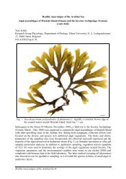
Chapter 2 <strong>Halimeda</strong> morphology 13Morphology of the green algal genus <strong>Halimeda</strong> 1Heroen VerbruggenThe aim of this chapter is to give an overview of the morphology of the segmented, calcified greenalgal genus <strong>Halimeda</strong>. Generalities and particularities of the appearance of mature thalli, segmentmorphology and anatomical features are described and illustrated. Sexual reproduction andcalcification are also shortly mentioned.General appearanceThalli of the green algal genus <strong>Halimeda</strong> are characterized by a segmented habit (Figs 1–6; Lamouroux1812). From its holdfast, the alga grows in pulses, at each time giving rise to a new, flat, greensegment (Colinvaux et al. 1965, Hillis-Colinvaux 1980; Hay et al. 1988). New segments grow fromcertain points on the distal edge of the mother segment (Verbruggen & Kooistra 2004). This progressivegrowth of one segment on top of the former gives rise to a catenate appearance (Hillis-Colinvaux1980).In essence, thalli consist of rows of segments. At certain points along the thallus these rows branch.This happens when two or more daughter segments sprout from the distal edge of a segment andsubsequently each give rise to a branch. Substantial variation in the number and location of ramificationsexists among and within species (Figs 1–6; Barton 1901, Gilmartin 1960, Hillis-Colinvaux1980).<strong>Halimeda</strong> occupies many habitats of the tropical and subtropical marine environment (Goreau & Graham1967, Hillis-Colinvaux 1974, 1977, 1980, Noble 1987, Littler & Littler 2000, 2003). Their habitatreflects in their morphology in a variety of ways (Gilmartin 1960, Colinvaux et al. 1965, Hillis-Colinvaux 1980, Kooistra et al. 2002, Verbruggen et al. submitted). The most apparent adaptation toenvironmental factors is the holdfast of the algal body (Hillis-Colinvaux 1980, Verbruggen & Kooistra2004). In essence, <strong>Halimeda</strong> holdfasts are composed of a more or less organized mass of branchingrhizoids (Hillis 1959, Hillis-Colinvaux 1980).Species growing on hard substrate are attached by means of a felt-like holdfast (Fig. 7; Verbruggen &Kooistra 2004). In a felt-like holdfast, rhizoids are compacted into a dense mass (Hillis-Colinvaux1980). Near the substrate, the rhizoid mass spreads outwards to some extent, resulting in a relativelylarge contact area between holdfast and substrate (Fig. 7).The second type of holdfast is present in sand-dwelling species, where a mass of rhizoids penetratesinto the sand. Rhizoids adhere to adjacent sand grains. As such, a bulbous structure of sand and interwovenrhizoids is formed (Fig. 8; Barton 1901, Hillis 1959, Hillis-Colinvaux 1980). This bulbousholdfast provides stability in the sandy substratum much like the root of a land plant does.A third holdfast type occurs in species sprawling over rubble. In these species, the holdfast consists ofa few branched, loose rhizoids attaching to morsels of rock and sand (Fig. 9; Hillis-Colinvaux 1980,Verbruggen & Kooistra 2004). In contrast to the other two holdfast types, where a single holdfastattaches the thallus at its base, this type of holdfast occurs at multiple points along the sprawling1 This chapter was adapted from a manuscript of an invited review article in preparation. As a consequence, itanticipates a few results presented in the research chapters of this thesis.
14Verbruggenthallus, usually at the nodes between segments (Hillis-Colinvaux 1980). The occurrence of multipleholdfasts strongly improves the strength of attachment to a substrate that is relatively unstable bynature.Figs 1–6. General appearance of <strong>Halimeda</strong> thalli. Fig. 1. H. simulans, specimenH.0030. Fig. 2. H. macroloba, specimen HV47c. Fig. 3. H. hummii, specimenH.0233. Fig. 4. H. tuna, specimen HV55. Fig. 5. H. micronesica, specimen HV295.Fig. 6. H. opuntia, specimen HV940. All specimens deposited in the <strong>Ghent</strong> <strong>University</strong>Herbarium (GENT).
Chapter 2 <strong>Halimeda</strong> morphology 15Figs 7–9. Holdfast types. Fig. 7. Felt-like holdfastconsisting of a dense mat of rhizoids; H. cuneata f.undulata, specimen HV742. Fig. 8. Bulbous holdfastconsisting of rhizoids attaching to sand particles; H.cylindracea, specimen HV558. Fig. 9. Secondaryholdfast of a few branched, loose rhizoids attached tosand; H. gracilis, specimen KZNb2243. All specimensdeposited in GENT.External features of segments and nodesSegments range in size from 0.2 mm in H. hummii (Ballantine 1982) to over five centimeters in H.magnidisca (Noble 1986 – measurements of segment width; species authorities listed in Appendix 1).Segment shape also varies considerably. Among the most common shapes are reniform, ovate, elliptical,obovate and cuneate (Figs 10–14; Hillis-Colinvaux 1980, Verbruggen et al. 2005a). Within mostof these categories, the relative width (e.g. measured as width over length ratio) can change considerably.Although many species have segments with entire margins, shallow to deep lobes along the marginof the segment are present in a variety of species (Figs 15–18; Hillis-Colinvaux 1980, Noble 1987,Verbruggen et al. submitted). The shape of the segment base varies from auriculate to acute and featuresa small stalk in some species (e.g. Fig. 10).Although there is definitely a genetic component to segment size and shape (Verbruggen & Kooistra2004, Verbruggen et al. 2005b), environmental conditions are also important (Hillis-Colinvaux 1980,Vroom et al. 2003, Smith et al. 2004, Verbruggen et al. submitted). The best known and most strikingexample is the segment form variability of H. opuntia (Barton 1901, Kooistra & Verbruggen 2005).This sprawling species occurs in a relatively broad range of environmental conditions (Taylor 1950,1960, Hillis-Colinvaux 1980) and its segments range from broad and reniform in exposed habitats toelongate and trilobed in sheltered habitats. Not only do such differences exist between segments ofspecimens from different habitats; they can also be situated within individual specimens. Cushions ofH. opuntia occurring in lagoonal and back-reef habitats throughout the tropics (Taylor 1950, 1960,Hillis-Colinvaux 1980, Coppejans et al. 1992, Silva et al. 1996, Littler & Littler 2000, Payri et al.2000, Littler & Littler 2003) feature horizontal runner branches ramifying infrequently and consistingof long, trilobed segments on the one hand and densely branching clumps with segments that are usuallybroad and reniform (pers. obs.). Culture experiments have confirmed that segment shape varia-Figs 10–18. Segment shape. Fig. 10. Reniform segment; H. opuntia, specimenHV61. Fig. 11. Broad ovate segment; H. distorta, specimen HV767. Fig. 12. Elliptical–discoidsegment; H. heteromorpha, specimen HV629. Fig. 13. Broad obovatesegment; H. heteromorpha, specimen HV629. Fig. 14. Cuneate segment; H. discoidea,specimen HV605. Fig 15. Segment with entire distal margin; H. heteromorpha,specimen HV763. Fig 16. Shallowly lobed segment; H. heteromorpha, specimenHV763. Fig 17. Medium deeply lobed segment; H. heteromorpha, specimenHV763. Fig 18. Deeply lobed segment; H. heteromorpha, specimen HV763. Allspecimens deposited in GENT.
16VerbruggenFigs 19–21. Three-dimensional segment properties. Fig. 19. Undulate segment; H.cuneata f. undulata, specimen HV742. Fig. 20. Keeled segment; H. distorta, specimenHV82. Fig. 21. Ribbed segment; H. distorta, specimen HV767. All specimensdeposited in GENT.bility of <strong>Halimeda</strong> species can be caused by environmental factors such as light intensity (Colinvauxet al. 1965, Hillis-Colinvaux 1980).So far, attention has been paid only to size and shape of segments, in other words two-dimensionalcharacters. Inclusion of the third dimension adds another couple of characters of potential taxonomicsignificance. Segment thickness varies considerably within and among species, ranging from 0.4 mmin H. hummii to almost 10 mm in basal segments of H. cylindracea. Further three-dimensional charactersinclude the planarity and ribbedness of segments. While in most species segments are flat, incertain species (or forms) they are not. In H. cuneata f. undulata, for example, segments are markedlyundulated (Fig. 19; Barton 1901, Littler & Littler 2003), and in H. distorta, segments are often keeledor distorted (Fig. 20; Hillis-Colinvaux 1980, Kooistra & Verbruggen 2005). In a few species, ribbedsegments occur (Fig. 21; Barton 1901, Hillis-Colinvaux 1980). These ribs are longitudinal zones of thesegment that are thicker than the surrounding segment. Ribs start at the segment base and run longitudinallyacross the segment towards the daughter segments.The segment surface can have different appearances. In some species, it is rugged as a consequence ofthe presence of pits or projections (e.g. H. favulosa; Hillis-Colinvaux 1980, Verbruggen et al. submitted).At the other extreme, segments can be smooth and glossy (e.g. H. hederacea; Hillis 1959, Hillis-Colinvaux 1980, Kooistra & Verbruggen 2005). Most species are situated in between.As mentioned above, new segments sprout from the distal margin of their parent segment. Due toanatomical constraints (see below), daughter segments can only grow from certain zones along themargin of the parent segment. The extent of these zones varies strongly. In certain species, daughtersegments can grow from the entire distal margin of the segment. On the other edge of the spectrum,there are species in which segments can only grow from a single or a limited number of pits situatedalong the distal margin.The contact zone between <strong>Halimeda</strong> segments is usually called node. The appearance and flexibilityof nodes vary considerably (Kooistra et al. 2002, Verbruggen & Kooistra 2004). While in most speciesdaughter segments are sessile at the margin of the mother segment, in H. cuneata the daughter segmentsare separated from the mother segment by a stalk zone (Hillis 1959, Hillis-Colinvaux 1980).The extent of development of these stalk zones varies (Hillis 1959). In essence, two types can be distinguished.In the first type, the stalks consist of a naked bundle of siphons (Fig. 22). In the secondtype, the stalk zone is developed into a so-called cushion segment, which is basically a mini-segmentintercalated between the mother- and daughter segments (Fig. 23).
Chapter 2 <strong>Halimeda</strong> morphology 17Figs 22, 23. Stalked H. cuneata nodes.Fig. 22. Stalk consisting of decorticatefilaments; specimen KZNb2249. Fig. 23.Stalk consisting of corticated filaments(cushion segment); specimen KZNb2352.All specimens deposited in GENT.Anatomical structuresLike all Caulerpalean algae, <strong>Halimeda</strong> consists of a single, multinucleate cell (van den Hoek et al.1995, Vroom et al. 1998, Vroom & Smith 2001). The tubular cell branches and anastomoses to formthe thallus. Within each segment, two zones of differently organized siphons (branches of the tubularcell) can be discerned: a central medulla and its surrounding cortex (Fig. 24). In order to observe thedifferent anatomical features, segments have to be appropriately dissected and sectioned. Proceduresfor doing so have been described in Barton (1901), Hillis-Colinvaux (1980) and Verbruggen et al.(2005a).Features of the medullaIn the medulla, siphons are arranged parallel to the thallus axis (Fig. 24). The bundle of filamentscomprising the medulla forms the organic skeleton of the thallus, proceeding through the nodes andstringing segments together (Fig. 24). Siphons branch at more or less regular intervals (Fig. 25). Atramifications, the main branch proceeds towards the distal edge of the segment while the majority ofside branches gives rise to the cortex (Fig. 26). The size of medullar siphon segments between ramificationsvaries considerably between species, within species and even within individual segments(Verbruggen et al. 2005a). The shape of medullar siphon segments shows some variability as well. InH. gracilis, for example, medullar siphons are markedly more slender than in other species (Verbruggen& Kooistra 2004). Ramifications in themselves show some features too. In most species, they aretrichotomous (Fig. 27), with the central branch continuing upwards and the two side branches proceedinginto the cortex. However, dichotomous and quadrichotomous ramifications occur (Figs 28,29) and even dominate in certain species (unpublished results). Above the ramifications, the main andFigs 24–30. Segment anatomy – medulla. Fig. 24. Cross section through two subsequentsegments showing medulla (m) and cortex (c); arrows indicate the position ofthe node; H. incrassata, specimen H.0667. Fig. 25. Medullar siphon ramifying atmore or less constant distance, with smaller side branches; H. incrassata, specimenH.0668. Fig. 26. Side branch of medullar siphon branching into the cortex (c); H.micronesica, specimen H.0014. Fig. 27. Trichotomous ramification of medullar siphon;H. monile, specimen HV344. Fig. 28. Dichotomous ramification of medullarsiphon; H. tuna, specimen HV319. Fig. 29. Quadrichotomous ramification of medullarsiphon; H. lacunalis, specimen HV306-1. Fig. 30. Medullar siphon showingconstriction but no ramification; H. taenicola, specimen HV285-1. All specimensdeposited in GENT.
18Verbruggenside branches are constricted (Figs 27–29). In a few species, some siphons are constricted at relativelyconstant intervals without branching (Fig. 30, pers. obs.) and the intervals between subsequent constrictionscorrespond to the length of medullar siphon segments of the more common, trichotomouslybranching siphons in the same species.In ribbed segments, medullar siphons are unevenly distributed within the segment. Anatomically, theribs consist of thick bundles of medullar siphons surrounded by a cortex (Kooistra & Verbruggen2005). The remainder of the segment consists of a much thinner layer of medullar siphons envelopedwith a cortex.Features of the nodal zoneIn most species, medullar siphons fuse at the nodes. The presence or absence and the type of anastomosishave been treated before in Barton (1901), Hillis (1959) and Hillis-Colinvaux (1980). In a firstfusion type, all siphons anastomose into a single unit. Siphons keep their normal appearance below thenode, and, at the node, fuse sidewise with their adjacent neighbors. This results in a pattern of largepores visible in properly prepared slides (Figs 31, 32). Above the node, all siphons reappear in linewith their subnodal counterparts. In other words, for each siphon arriving at the node, there is one thatdeparts into the subsequent segment. Two adaptations of this fusion pattern occur (Hillis 1959, Hillis-Colinvaux 1980). The first adaptation involves the number of siphons participating in the fusion. Inthe species where this type of adaptation occurs, siphons fuse into a small number of groups (withnumerous siphons each). A second type of adaptation is observed in H. melanesica and H.heteromorpha, where the large pores have been reduced to small ones or are absent altogether (Figs33, 34, Verbruggen et al. submitted).A second common pattern of nodal siphon anatomy is present in many rock-growing species of waveaffectedhabitats. Here, the siphons get narrower and start branching irregularly shortly below thenode. At the node, pairs or triplets of these narrow siphons anastomose. Unlike the former and subsequentfusion patterns, a lower number of siphons reappears above the node. For all siphons that anastomoseinto a unit, only a single siphon re-emerges above the node (Figs 35, 36). Owing to the irregularbranching just below the node and the anastomosis of the resulting branches in different fusionunits, this type of anastomosis leads to strong entanglement of siphons below the node (Fig. 35).Anastomosis of several siphons into a single unit that continues in the subsequent segment as a singlesiphon is called complete fusion.A third and similar fusion pattern, which is much less common than the former, was described byVerbruggen & Kooistra (2004). The difference with the former pattern is that the appearance of thesubnodal siphons is similar to that of the medullar siphons throughout the segments. In other words,siphons do not get narrower below the node, and there is neither dense branching nor siphon entanglementjust below the node (Figs 37, 38).A fourth pattern is one of incomplete fusion of a small number of siphons (usually 2–4). At the node,siphons fuse with one or two neighbors over a short distance (Fig. 39, 40). To put it differently, thenode is composed of many pairs or small groups of siphons that are connected sideways with largepores. In this group species too, reduction of the pattern of anastomosis (in the sense of smaller pores)occurs in a few species.Alternatively to all patterns described above, in a few species, siphons proceed through the node withoutany form of fusion (Fig. 41). In most species in which this type of nodal siphon behavior occurs,several parallel siphons pass through the node. However, in the species H. cryptica, the number of siphonsgoing through the node is reduced to one (Colinvaux & Graham 1964, Verbruggen & Kooistra2004).
Chapter 2 <strong>Halimeda</strong> morphology 19The former patterns are fairly constant within species. Nevertheless, a few exceptions have beennoted. For example, in H. hummii some siphons go through the node without fusing, but most showcomplete or incomplete fusion in pairs or triplets (Ballantine 1982, Wysor & Kooistra 2003). A secondexample is H. lacrimosa, in which complete and incomplete fusion can occur in combination (Hillis-Colinvaux 1980). The nodes of H. cuneata are a class on their own (see above). In this species, siphonfusion is of the second type. As in other species, fusion occurs at the distal edge of the parent segment(Fig. 42). In the stalk zone, siphons feature thick cell walls and irregular constrictions (Fig. 42;Bandeira-Pedrosa et al. 2004). From the base of the daughter segment onwards, siphons return to theirnormal, medullar form (Fig. 42). In case a cushion segment is present (Fig. 23), the central strand ofsiphons is surrounded by a cortex, much as in regular segments (see below).As mentioned above, new segments can originate along the entire distal margin of the parent segmentor at a limited number of pits along the distal segment edge. Medullar siphons fuse just below the pitsin question and the fused medullar siphons reach the segment surface at the height of the pit. It seemsthat new segments can only grow from such fused medullar siphons. As mentioned above, pits canbecome more elongated and cover the entire distal segment margin.Figs 31–42. Segment anatomy – node. Figs 31, 32. Fusion into a single unit with obviouspores connecting adjacent siphons; H. incrassata, specimen HV448. Figs 33, 34. Fusion into asingle unit with diminutive pores connecting some adjacent siphons; H. heteromorpha, specimenHV629. Figs 35, 36. Complete fusion of siphons in pairs or triplets, with chaoticbranching of siphons beneath the node; arrows in Fig. 35 pinpoint fusions; H. tuna, specimenHV319. Figs 37, 38. Complete fusion of a siphon pair, without chaotic branching of siphonsbeneath the node; H. lacrimosa, specimen H.0308. Figs 39, 40. Incomplete fusion of siphonsin triplets or pairs; H. copiosa, specimen H.0265. Fig. 41. Siphon going through the nodewithout any kind of fusion; arrow indicates position of node; H. micronesica, specimenH.0014. Fig. 42. Nodal anatomy of H. cuneata, showing complete siphon fusion in pairs at thedistal margin of the parent segment (indicated with arrows), irregularly constricted siphonsgoing through the stalk zone (s) and resumption of regular medullar branching above the stalkzone; specimen KZNb2263. All specimens deposited in GENT.
20VerbruggenFeatures of the cortexThe cortex originates from side branches of the medullar siphons (Fig. 26). In most cases, sidebranches of medullar siphons have a similar shape, but are somewhat smaller (Fig. 25); and the closerto the segment periphery, the shorter the siphon segments get (Fig. 43, Barton 1901). The segmentsbetween closely spaced siphon ramifications are called utricles.The pattern described above is not fixed. In certain species, especially those with thin segments, thegradual change from medullar siphon to utricle is replaced by a more sudden change, in which sidebranches of medullar siphons take the form of utricles (Fig. 44). In heavily calcified segments, there isusually only one layer of siphons connecting the medullar siphons and the utricles. In this case, theFigs 43–54. Segment anatomy – cortex. Fig. 43. Cortical utricles gradually decreasingin size from the medulla (below) to the periphery (above) of the segment;H. monile, specimen HV333. Fig. 44. Giant subperipheral utricles arising directlyfrom medullar siphons; H. taenicola, specimen H.0037. Fig. 45. Single layer ofcylindrical utricles spanning most of the distance between the medulla and the peripheryof the segment; H. distorta, specimen HV572. Fig. 46. Cross-sectionthrough a H. macrophysa segment showing the medullar strand of siphons and large,detached, peripheral utricles; specimen HV8. Fig 47. Detail of large, detached, peripheralutricles and minute subperipheral utricles of H. macrophysa, specimenHV8. Fig. 48. Cylindrical secondary utricle suddenly widening at its distal end andgiving rise to seven peripheral utricles; H. gracilis, specimen HV312. Fig. 49.Spined peripheral utricle; H. scabra, specimen L.0351084. Fig. 50. Schematic diagramof differential branching rates; p – peripheral utricle, s – secondary utricle, t –tertiary utricle, q – quaternary utricle. Fig. 51. Lateral adhesion of peripheral utricles(adh); H. discoidea, specimen OMII118. Fig. 52. Firmly attached peripheral utriclesin an irregular polygonal pattern (surface view); H. melanesica, specimen HV818.Fig. 53. Detached peripheral utricles in surface view; H. macroloba, specimenHV206. Fig. 54. Surface view showing fusion between adjacent peripheral utricles(arrows); H. discoidea, specimen H.0204. All specimens deposited in GENT exceptL.0351084 (H. scabra) from the National Herbarium of the Netherlands, Leiden<strong>University</strong> Branch (L).
Chapter 2 <strong>Halimeda</strong> morphology 21side branches of medullar siphons are elongate and pass through the most heavily calcified segmentzone towards the periphery of the segments where they branch several times over a short distance toform the cortical utricles (Fig. 45).Utricles often occur in more or less definable layers, which are sometimes numbered from the segmentsurface inwards (Hillis 1959; Hillis-Colinvaux 1980). Utricles from the peripheral layer are referred toas primary utricles, those from the layer inwards as secondary utricles, and so on. Numbering layers issometimes problematic due to differential branching rates. A situation that often occurs is that tertiaryutricles have different numbers of subsequent utricle generations. Imagine a tertiary utricle with threedaughter utricles of which the first one bears three peripheral utricles (Fig. 50, left box). Imagine anotherone of the daughter utricles of the tertiary utricle bearing daughter utricles which, in turn, eachbear peripheral daughter utricles (Fig. 50, right box). In this situation, the original tertiary utricle becomesa quaternary utricle. This situation suggests that the developmental process of utricle formationis not a definite one but can adapt to circumstances at a very small scale.Considerable size and shape variation can be observed in utricles. In certain species, utricles arestrongly inflated (Figs 44, 51). Opposite to that, utricles can be non-inflated, cylindrical tubes (Fig.45). The whole spectrum of intermediates occurs. Utricle shape is diagnostic for a few species andsections. For example, peripheral utricles of H. scabra bear a terminal spine (Fig. 49, Howe 1905),peripheral utricles of H. macrophysa are markedly inflated (Figs 46, 47, Askenasy 1888), peripheralutricles of Brazilian H. cuneata are characterized by lenticular thickenings on the inside of their distalcell wall (Bandeira-Pedrosa et al. 2004), and secondary utricles of section Pseudo-opuntia widen suddenlyat their distal end (Fig. 48; Verbruggen & Kooistra 2004). Most species, however, have highlysimilar cortex features and are usually diagnosed on the basis of size characteristics of the utricles.Peripheral utricles usually attach to one another laterally due to the presence of a common cuticle-likestructure (Bandeira-Pedrosa et al. 2003). The degree of attachment varies from attachment at the verydistal girdle of the utricles, in which case only the cuticle-like layer provides adhesion, to attachmentalong two-thirds of the utricle length, in which case the lateral cell walls fuse (e.g. H. discoidea, Fig.51). In some species, utricles are unattached (e.g. H. macrophysa, Fig. 47). In surface view, the appearanceof peripheral utricles depends mostly on their degree of attachment. When they are fully attached,the surface view is one of polygons (mostly hexagons) closely fitted together (Fig. 52). Whenthe attachment is less strict, polygons become rounded in the corners (Fig. 53) or get reduced to loosecircles when the utricles are not attached at all. In a few species with fully attached peripheral utricles,some adjacent primary utricles fuse with one another (Fig. 54, Howe 1907).Reproductive structures<strong>Halimeda</strong> gametes are formed in stalked gametangial clusters. Such clusters vary strongly in size,shape, structural origin and position on the segment, both among and within species (Hillis-Colinvaux1980, Vroom & Smith 2003). Gametangial clusters are situated mostly along the outer rim of segments(Fig. 55). Nonetheless, several species also have gametangial clusters arising from the segmentsurface (Fig. 56). In H. cryptica, all gametangial clusters are situated on one side of the segment (Graham1975). Gametophores, the stalks that carry gametangial clusters, have been observed to originatein three ways (Hillis 1959, Hillis-Colinvaux 1980). First, they can arise at the distal segment rim orpits, as continuations of the main medullary filaments subsequent to nodal fusion (Fig. 57). Second,they can originate as lateral outgrowths from medullary filaments without fusion (i.e. not at pores ordistal segment rim – not depicted here). Third, they can originate as extensions from peripheral or secondaryutricles (Figs 58, 59). In general, the gametophore is a long, unbranched siphon, whereasbranches within the gametangial cluster are short (Figs 57, 58). However, in several species, the gametophoreramifies once or twice along its length, giving rise to multiple gametangial clusters (Fig.
22Verbruggen60). Each gametangial group comprises a number of spherical to pyriform gametangia and a moreelongate, club-shaped discharge papilla (Figs 57, 58; Drew & Abel 1988, Vroom & Smith 2003).Reproduction in <strong>Halimeda</strong> is holocarpic: after the cell content of the whole thallus has been transformedinto and released as gametes, the thallus dies (Meinesz 1980 and references therein). Reproductiveevents show seasonal and lunar periodicity (Beth 1962, Drew & Abel 1988, Clifton 1997,Clifton & Clifton 1999), but certain species are found reproductive in different seasons at differentlocalities (Drew & Abel 1988) and low fractions of populations of certain species are reproductivethroughout the year (Verbruggen et al. submitted). Where observed in detail, gametangial clusters areformed during the night, grow darker during the first day and second night, and discharge arounddawn of the second day (Hillis-Colinvaux 1980, Drew & Abel 1988, Clifton 1997, Clifton & Clifton1999). <strong>Halimeda</strong> is dioecious and thalli carrying macrogametangia can be discerned from thalli carryingmicrogametangia because macrogametangia are large and brown to dark green while microgametangiaare smaller and yellow to light green (Feldmann 1951, Meinesz 1980). Broadcast spawning(simultaneous release of gametes in the water column) generally occurs around dawn, in speciesspecifictime-frames (Clifton 1997). Just prior to release, the gametes from all gametangia in agametangial cluster migrate downwards through the gametangial stalks to be released through thecluster's discharge papilla (Hillis-Colinvaux 1980, Drew & Abel 1988).<strong>Halimeda</strong> gametes are biflagellate, pyriform cells that vary in size from about 2 to about 20 µm in size(Meinesz 1980, Hillis-Colinvaux 1980, Clifton & Clifton 1999 and references in these). Macrogametes,characterized by an eyespot, are generally larger than the microgametes lacking eyespots.Development of the H. tuna zygote into a filamentous life-stage, similar to Pseudochlorodesmis thalli,is described in Meinesz (1972, 1980). It is has not been ascertained how the filamentous form is linkedto the segmented thallus.Figs 55–60. Reproductive structures. Fig 55. Gametangial clusters originating fromdistal edges of segments; H. macroloba, specimen HV206. Fig. 56. Gametangialclusters originating from the segment surface; H. cuneata, specimen KZNb2263.Fig. 57. Gametophores originating from the main medullary filaments subsequent tonodal fusion at the distal segment edge; dp – discharge pores, ss – segment surface;H. macroloba, specimen HV206. Fig. 58. Gametophore originating from secondaryutricle; H. incrassata, specimen H.0143. Fig. 59. Gametophore originating fromsecondary utricle; gp – gametophore, p – peripheral utricle, s – secondary utricle, t –tertiary utricle; H. incrassata, specimen H.0143. Fig. 60. Gametophore originatingfrom the main medullary filaments subsequent to nodal fusion at the distal segmentedge and separating into three clusters by a central ramification (ram); H.cylindracea, specimen HV590. All specimens deposited in GENT.
Chapter 2 <strong>Halimeda</strong> morphology 23CalcificationSeveral bryopsidalean algae are characterized by intra- and/or extracellular calcium carbonate deposition(Böhm et al. 1978). The mechanisms of calcification have been reviewed by Borowitzka (1982).In <strong>Halimeda</strong>, calcium carbonate is deposited extracellularly, in between cortical and (to a lesser extent)medullar siphons (Figs 61, 62; Askenasy 1888, Wilbur et al. 1969, Borowitzka & Larkum 1977,Böhm et al. 1978). The needle-shaped crystals are orthorhombic (aragonite; Fig. 63; McConnell &Colinvaux 1967). The degree of calcification is an important determinant of color, flexibility and brittlenessof <strong>Halimeda</strong> segments. Segment color varies from dark green in barely calcified segments togreenish white in segments of strongly calcified species. In addition, segments can become brownishby cell wall thickening, in particular near the thallus base. Flexible thalli allow specimens to grow inhabitats characterized by high water movement by aligning the thallus with the current, thus reducingdrag. There are two major components to thallus flexibility. First, flexibility of nodes plays a role.Second, segment pliability is of importance, especially in species with rigid nodes. Several speciesfeature highly swollen secondary utricles (Figs 44, 51), leaving little space for calcification and renderingsegments flexible (Verbruggen & Kooistra 2004). An important drawback of reduced calcificationin tropical reef ecosystems characterized by high grazing pressure is increased palatability (Duffy& Hay 1990, Hay et al. 1994, Schupp & Paul 1994, Paul 1997). Initial segment growth occurs at night(Hay et al. 1988) and calcification, which is dependent upon photosynthesis, starts on the second dayof segment development (Wilbur et al. 1969). Segments are fully calcified after about 2–3 days. Whencalcified segments are shed from the thallus after reproduction, they form an important fraction of thesediment and are responsible for much of the sand formation in tropical lagoons (e.g. Chapman &Mawson 1906; Drew 1983; Freile et al. 1995).Figs 61–63. Calcification. Fig. 61. SEM micrograph of fractured segment showing acentral zone of medullary filaments and relatively little carbonate and a heavilycalcified cortical zone on either side; H. gracilis, specimen HV824. Fig. 62. SEMmicrograph of cortical zone of fractured segment showing the calcium carbonatedepositions in between cortical siphons; H. gracilis, specimen HV824. Fig. 63. SEMmicrograph of needle-shaped aragonite crystals; H. gracilis, specimen HV824. Allspecimens deposited in GENT.ReferencesAskenasy E. (1888) Algen. In: Die Forschungsreise S.M.S. Gazelle Th. 4, Bot., Berlin.Ballantine D.L. (1982) <strong>Halimeda</strong> hummii sp. nov., <strong>Halimeda</strong> cryptica v. acerifolia var. nov.(Caulerpales, Chlorophyta), and additional records of <strong>Halimeda</strong> species from Puerto Rico.Journal of <strong>Phycology</strong> 18: 86–91.Bandeira-Pedrosa M.E., Bouzon Z.L., Pereira S.M. & Oliveira E.C. (2003) Ultrastructure of somespecies of <strong>Halimeda</strong> (Bryopsidales, Chlorophyta) from Brazil. Cryptogamie Algologie 24: 219–231.Bandeira-Pedrosa M.E., Pereira S.M.B., Bouzon Z.L. & Oliveira E.C. (2004) <strong>Halimeda</strong> cuneata(Bryopsidales, Chlorophyta), a new record for the Atlantic Ocean. Phycologia 43: 50–57.
24VerbruggenBarton E.S. (1901) The genus <strong>Halimeda</strong>. Monographs of the Siboga Expedition 60. Brill, Leiden.32pp.Beth K. (1962). Reproductive phases in populations of <strong>Halimeda</strong> tuna in the Bay of Naples.Publicazione della Stazione Zoologica di Napoli 32: 515–534.Böhm L., Fütterer D., Kaminski E. (1978) Algal calcification in some Codiaceae (Chlorophyta):ultrastructure and location of skeletal deposits. Journal of <strong>Phycology</strong> 14: 486–493.Borowitzka M.A. (1982) Mechanisms in algal calcification. In: Round F.E. & Chapman D.J. (eds.)Progress in Phycological <strong>Research</strong>. Vol. 1. Elsevier, Amsterdam, The Netherlands. pp. 137–178.Borowitzka M.A. & Larkum A.D.W. (1977) Calcification in the green alga <strong>Halimeda</strong>. I. Anultrastructure study of the thallus development. Journal of <strong>Phycology</strong> 13: 6–16.Chapman F. & Mawson D. (1906) On the importance of <strong>Halimeda</strong> as a reef-forming organism: with adescription of the <strong>Halimeda</strong>-limestones of the New Hebrides. Journal of the Geological Society ofLondon 62: 702–711.Clifton K.E. (1997) Mass spawning by green algae on coral reefs. Science 275: 1116–1118.Clifton K.E. & Clifton L.M. 1999. The phenology of sexual reproduction by green algae(Bryopsidales) on Caribbean coral reefs. Journal of <strong>Phycology</strong> 35: 24–34.Colinvaux L.H. & Graham E.A. (1964) A new species of <strong>Halimeda</strong>. Nova Hedwigia 7: 5–10.Colinvaux L.H., Wilbur K.M. & Watabe N. (1965) Tropical marine algae: growth in laboratoryculture. Journal of <strong>Phycology</strong> 1: 69–78.Coppejans E., Beeckman H. & De Wit M. (1992) The seagrass and associated macroalgal vegetationof Gazi Bay (Kenya). Hydrobiologia 247: 59–75.Drew E.A. (1983) <strong>Halimeda</strong> biomass, growth rates and sediment generation on reefs in the centralGreat Barrier Reef province. Coral Reefs 2: 101–110.Drew E.A. & Abel K.M. (1988) Studies on <strong>Halimeda</strong> I. The distribution and species composition of<strong>Halimeda</strong> meadows throughout the Great Barrier Reef Province. Coral Reefs 6: 195–205.Duffy J.E. & Hay M.E. (1990) Seaweed adaptations to herbivory. Bioscience 40: 368–375.Feldmann J. (1951) Sur la reproduction sexuée de l'<strong>Halimeda</strong> tuna (Ell. et Sol.) Lamour. f. platydisca(Decaisne) Barton. Comptes Rendus Hebdomadaires des Séances de l'Académie des Sciences,Série D 233: 1309–1310.Freile D., Milliman J.D. & Hillis L. (1995) Leeward bank margin <strong>Halimeda</strong> meadows and draperiesand their sedimentary importance on the western Great Bahama bank slope. Coral Reefs 14: 27–33.Gilmartin M. (1960) The ecological distribution of the deep water algae of Eniwetok. Ecology 41:210–221.Goreau T.F. & Graham E.A. (1967) A new species of <strong>Halimeda</strong> from Jamaica. Bulletin of MarineScience 17: 432–441.Graham E.A. (1975) Fruiting in <strong>Halimeda</strong> (order Siphonales) 1. <strong>Halimeda</strong> cryptica Colinvaux andGraham. Bulletin of Marine Science 25: 130–133.Hay M.E., Kappel Q.E. & Fenical W. (1994) Synergisms in plant defenses against herbivores:interactions of chemistry, calcification, and plant quality. Ecology 75: 1714–1726.Hay M.E., Paul V.J., Lewis S.M., Gustafson K., Tucker J. & Trindell R.N. (1988) Can tropicalseaweeds reduce herbivory by growing at night? Diel patterns of growth, nitrogen content,herbivory, and chemical versus morphological defenses. Oecologia 75: 233–245.Hillis L. (1959) A revision of the genus <strong>Halimeda</strong> (order Siphonales). Publications of the Institute ofMarine Science 6: 321–403.Hillis-Colinvaux L. (1974) Productivity of the coral reef alga <strong>Halimeda</strong> (Siphonales). Proceedings ofthe 2nd International Coral Reef Symposium 1: 35–42.Hillis-Colinvaux L. (1977) <strong>Halimeda</strong> and Tydemania: distribution, diversity and productivity atEnewetak. Proceedings of the 3rd International Coral Reef Symposium 1: 365–370.
Chapter 2 <strong>Halimeda</strong> morphology 25Hillis-Colinvaux L. (1980) Ecology and taxonomy of <strong>Halimeda</strong>: primary producer of coral reefs.Advances in Marine Biology 17: 1–327.Howe M.A. (1905) Phycological studies. II. New Chlorophyceae from Florida and the Bahamas.Bulletin of the Torrey Botanical Club 32: 241–252.Howe M.A. (1907) Phycological studies. III. Further notes on <strong>Halimeda</strong> and Avrainvillea. Bulletin ofthe Torrey Botanical Club 34: 491–515.Kooistra W.H.C.F., Coppejans E.G.G. & Payri C. (2002). Molecular systematics, historical ecologyand phylogeography of <strong>Halimeda</strong> (Bryopsidales). Molecular Phylogenetics and Evolution 24:121–138.Kooistra W.H.C.F. & Verbruggen H. (2005) Genetic patterns in the calcified tropical seaweeds<strong>Halimeda</strong> opuntia, H. distorta, H. hederacea and H. minima (Bryopsidales, Chlorophyta) provideinsights in species boundaries and inter-oceanic dispersal. Journal of <strong>Phycology</strong> 41: 177–187.Lamouroux J.V.F. (1812) Extrait d'une mémoire sur la classification des polypiers coralligères nonentièrement pierreux. Nouveau Bulletin des Sciences par la Societe Philomatique de Paris 3: 181–188.Littler D.S. & Littler M.M. (2000) Caribbean reef plants. An identification guide to the reef plants ofthe Caribbean, Bahamas, Florida and Gulf of Mexico. OffShore Graphics, Washington. 542 pp.Littler D.S. & Littler M.M. (2003) South Pacific Reef Plants. A diver's guide to the plant life of SouthPacific coral reefs. OffShore Graphics, Washington. 331 pp.McConnell D. & Colinvaux L.H. (1967) Aragonite in <strong>Halimeda</strong> and Tydemania (order Siphonales).Journal of <strong>Phycology</strong> 3: 198–200.Meinesz M.A. (1972) Sur le cycle de l'<strong>Halimeda</strong> tuna (Ellis et Solander) Lamouroux (Udoteaceae,Caulerpale). Comptes Rendus Hebdomadaires des Séances de l'Académie des Sciences, Série D275: 1363–1365.Meinesz A. (1980) Connaissances actuelles et contribution a l'etude de la reproduction et du cycle desUdoteacees (Caulerpales, Chlorophytes). Phycologia 19: 110–138.Noble J.M. (1986) <strong>Halimeda</strong> magnidisca (Caulerpales, Chlorophyta), a new species from the GreatBarrier Reef, Australia. Phycologia 25: 331–339.Noble J.M. (1987) A taxonomic study of the genus <strong>Halimeda</strong> Lamouroux (Chlorophyta, Caulerpales)from the Heron Island region of the southern Great Barrier Reef, Australia. MSc thesis, <strong>University</strong>of Melbourne, Melbourne, Australia. 200pp.Paul V.J. (1997) Secondary metabolites and calcium carbonate as defenses of calcareous algae oncoral reefs. Proceedings of the 8th International Coral Reef Symposium: 707–712.Payri C., N'Yeurt A.D.R. & Orempuller J. (2000) Algae of French Polynesia. Au Vent des Iles, Tahiti,French Polynesia. 320 pp.Schupp P.J. & Paul V.J. (1994) Calcium carbonate and secondary metabolites in tropical seaweeds:variable effects on herbivorous fish. Ecology 75: 1172–1185.Silva P.C., Basson P.W. & Moe R.L. (1996) Catalogue of Indian Ocean Algae. <strong>University</strong> ofCalifornia Publications in Botany (vol. 79), Berkeley, CA.Smith J.E., Smith C.M., Vroom P.S., Beach K.L. & Miller S. (2004) Nutrient and growth dynamics of<strong>Halimeda</strong> tuna on Conch Reef, Florida Keys: Possible influence of internal tides on nutrient statusand physiology. Limnology and Oceanography 49: 1923–1936.Taylor W.R. (1950) Plants of Bikini and other northern Marshall Islands. Algae: Chlorophyceae.<strong>University</strong> of Michigan Press, Ann Arbor, Michigan. 218 pp.Taylor W. R. (1960) Marine algae of the Eastern tropical and sub-tropical coasts of the Americas.<strong>University</strong> of Michigan Press, Ann Arbor, Michigan. 870 pp.van den Hoek C., Mann D.G. & Jahns H.M. (1995) Algae: an introduction to phycology. PressSyndicate, <strong>University</strong> of Cambridge, Cambridge, England.
26VerbruggenVerbruggen H., De Clerck O., Cocquyt E., Kooistra W.H.C.F. & Coppejans E. (2005) Morphometrictaxonomy of siphonous green algae: a methodological study within the genus <strong>Halimeda</strong>(Bryopsidales). Journal of <strong>Phycology</strong> 41: 126–139.Verbruggen H., De Clerck O., Kooistra W.H.C.F. & Coppejans E. (2005b) Molecular and morphometricdata pinpoint species boundaries in <strong>Halimeda</strong> section Rhipsalis (Bryopsidales, Chlorophyta).Journal of <strong>Phycology</strong>: accepted.Verbruggen H. & Kooistra W.H.C.F. (2004) Morphological characterization of lineages within thecalcified tropical seaweed genus <strong>Halimeda</strong> (Bryopsidales, Chlorophyta). European Journal of<strong>Phycology</strong> 39: 213–228.Verbruggen H., N'Yeurt A.D.R., Spalding H.I. & Vroom P.S. (submitted) Phylogeny and taxonomy of<strong>Halimeda</strong> incrassata, including the description of H. kanaloana and H. heteromorpha spp. nov.(Bryopsidales, Chlorophyta). Submitted to European Journal of <strong>Phycology</strong>.Vroom P.S. & Smith C.M. (2001) The challenge of siphonous green algae. American Scientist 89:524–531.Vroom P.S. & Smith C.M. (2003) Reproductive features of Hawaiian <strong>Halimeda</strong> velasquezii(Bryopsidales, Chlorophyta), and an evolutionary assessment of reproductive characters in<strong>Halimeda</strong>. Cryptogamie Algologie 24: 355–370.Vroom P.S., Smith C.M., Coyer J.A., Walters L.J., Hunter C.L., Beach K.S. & Smith J.E. (2003) Fieldbiology of <strong>Halimeda</strong> tuna (Bryopsidales, Chlorophyta) across a depth gradient: comparativegrowth, survivorship, recruitment, and reproduction. Hydrobiologia 501: 149–166.Vroom P.S., Smith C.M. & Keeley S.C. (1998) Cladistics of the Bryopsidales: a preliminary analysis.Journal of <strong>Phycology</strong> 34: 351–360.Wilbur K.M., Colinvaux L.H., Watabe N. (1969) Electron microscope study of calcification in thealga <strong>Halimeda</strong> (order Siphonales). Phycologia 8: 27–35.Wysor B. & Kooistra W.H.C.F. (2003) An annotated list of marine Chlorophyta from the Caribbeancoast of the Republic of Panama. Nova Hedwigia 77: 487–523.
Chapter 3 Synopsis of morpho-taxonomic history 27A synopsis of <strong>Halimeda</strong> morpho-taxonomic historyHeroen VerbruggenThe aim of this chapter is to give an overview of <strong>Halimeda</strong> taxonomic history, focusing on trends ofcharacter use. Throughout the history of <strong>Halimeda</strong> systematics, taxonomists have stressed differentcharacters, resulting in significantly different species delineations and classifications. The generaltrend is towards expansion of the arsenal of characters. After an initial period in which segment morphologyand thallus habit were the main diagnostic features, an increasing number of anatomical characterswere examined and stressed in species descriptions. Recently, electron microscopy and statisticalanalysis of morphological data have been added to the miscellany of techniques used.FoundationsThe foundations of <strong>Halimeda</strong> taxonomy were reviewed in great detail in the monograph of Hillis-Colinvaux (1980). Therefore, only a selection of interesting topics and the characters used for speciesdelineation are discussed here. The earliest historical record of <strong>Halimeda</strong> tuna (as Sertolara; authoritiesof <strong>Halimeda</strong> species and sections in Appendix 1 of Chapter 1), the type species of the genus, datesback to 1599 (Hillis-Colinvaux 1980), and it was not until the 18th century that additional specieswere described. The first taxonomic studies of the genus were by John Ellis, who diagnosed severalspecies of the genus (Ellis & Solander 1786). Even though Ellis studied the anatomy of H. incrassata(as Corallina incrassata; Ellis 1767, cited after Hillis-Colinvaux 1980), no anatomical characters wereused to distinguish between the species described in Ellis & Solander (1786). Ellis' anatomical observationsenticed him to consider <strong>Halimeda</strong> (as Corallina) as an animal (Hillis-Colinvaux 1980). Thegenus <strong>Halimeda</strong> was separated from Corallina only in 1812 by Jean Vincent Felix Lamouroux 1 .The description of <strong>Halimeda</strong> species solely based on segment morphological differences persistedthroughout most of the 19th century (Lamouroux 1816, Decaisne 1842, Zanardini 1851, Kützing 1858,Agardh 1887), bringing the total number of species to 26 in 1887. Jacob Georg Agardh (1887) partitionedthe genus into four species groups (Tunae, Pseudo-opuntiae, Opuntiae, and Rhipsales) of implicitrank on the basis of differences in segment morphology and thallus appearance. Giovanni BatistaDe Toni (1889) cited Agardh's descriptions as diagnoses for sections.Eugen Askenasy (1888) studied a collection of Pacific specimens of <strong>Halimeda</strong> from the S.M.S. Gazelleexpedition. He pioneered the use of anatomical features for taxonomic purposes. He found significantdifferences in peripheral utricle size and, from these differences, described a new species (H.macrophysa) and a new variety (H. opuntia var. macropus Askenasy). Furthermore, Askenasy discoveredthat adjacent medullar siphons of H. incrassata and H. macroloba were interconnected at nodesby means of lateral pores.1 <strong>Halimeda</strong> is a conserved name (Silva 1952). The name Sertularia was proposed for this genus in 1760 (Ludwig1760, cited after Silva 1952), 52 years before the establishment of the <strong>Halimeda</strong> denomination by Lamouroux(1812).
28VerbruggenBarton monographEthel Sarel Barton was the first to provide a comprehensive monograph of the genus (Barton 1901).She studied large collections from the coral-boring expedition to Funafuti (Barton 1900) and the Sibogaexpedition to Indonesia (Barton 1901). Additionally, she had the collections of the British Museumand Kew Herbarium at hands.Barton meticulously studied a variety of anatomical characters and asserted that such characters weretaxonomically more informative than thallus habit and segment shape. She stated about segment morphologythat "the connecting links [between species] were so complete that it would have been necessary,using these characters alone, to reduce the number of species to two" (Barton 1901, p. 9). A firstanatomical character studied in a taxonomic context was the pattern of nodal siphon fusion. Bartonrecognized three fusion types, namely fusion into a single unit by pores, complete fusion into pairs ortriplets, and incomplete fusion into pairs or triplets, and she used these types to discern betweengroups of species and some individual species. Following Askenasy's example, Barton also carefullynoted the size of peripheral utricles, and introduced the degree of lateral adhesion between adjacentperipheral utricles as a character. Size and adhesion of peripheral utricles were exclusively used fordistinguishing H. cuneata, H. macrophysa and H. tuna (as interpreted by her). Three-dimensionalsegment structure (ribbedness, undulateness) was another character introduced by her.Barton's taxonomy was characterized by extensive lumping of existing species into only a fewmorpho-types that did not show overlap in the characters used. The number of species was reducedfrom 27 to seven 2 . Within most species, several (often distinct) forms were described.With hindsight, the monograph of Barton has been a significant advancement of <strong>Halimeda</strong> taxonomy.First, it reported on the systematic variability in nodal fusions, which could be used for delineatingspecies groups. Second, through the study of the majority of type specimens, Barton tidied up thetaxonomic confusion created by many species descriptions based on segment morphology alone.Third, she carefully described intraspecific plasticity of external morphological and anatomical characters.Lastly, the illustrations of Barton were of exceptional quality, and, together with her cleardescriptions, pinpoint differences between entities that are informative to this day.Post-Barton taxonomyIn the period after Barton's monograph, many new collections from the Caribbean region becameavailable. These were studied mainly by Marshall Avery Howe (1905a, b, 1907, 1909) and FrederikBørgesen (1911, 1913). These authors used the whole gamut of external morphological and anatomicalcharacters used by Barton (1901) and added a few of their own. Howe used utricular spines todistinguish the new species H. scabra from H. tuna (Howe 1905a) and thoroughly examined the utilityof secondary utricle size and shape for distinguishing H. tuna from H. discoidea (Howe 1907) as wellas for the description of H. lacrimosa (Howe 1909). Furthermore, he used primary utricle size and theabsence of utricle adherence to recognize a new species H. favulosa (Howe 1905b). The presence oflateral fusions between adjacent peripheral utricles and the number of peripheral utricles borne onsecondary utricles were also introduced as taxonomic characters (Howe 1907, 1909). Some of thebroad species described by Barton (1901) were split up. Børgesen (1911, 1913) more closely hungonto Barton's taxonomy, not recognizing H. monile and H. simulans as separate taxa from H. incrassata.However, Børgesen did agree with Howe on the distinctness of H. tuna and H. discoidea. Børgesen'staxonomy is characterized by a few broad species with many well-described and illustrated formsand varieties recognized within each of them.2 With hindsight, several of the species of Barton (1901) were poly- and paraphyletic.
Chapter 3 Synopsis of morpho-taxonomic history 29The first major collections from the Pacific Ocean were made in the framework of the Bikini bombingproject. These collections contained many new species described by William Randolph Taylor (1950),using the broad range of external and anatomical characters used by Barton, Howe and Børgesen.Another species from the Pacific Ocean (H. micronesica) was described by Yukio Yamada (1941,1944). Both Yamada and Taylor recognized a nodal siphon pattern not discussed by any of the formerauthors, namely the proceeding of medullar siphons from one segment into the next without any kindof fusion.Hillis monographsLlewellya Hillis monographed the genus twice (Hillis 1959, Hillis-Colinvaux 1980) and, together withcolleagues, published numerous papers on the taxonomy, ecology, culturing, calcification and sedimentologyof <strong>Halimeda</strong>. In her first monograph, all known species were described and illustrated. Shefollowed Howe (1907) in the recognition of H. simulans and H. monile at the specific rank. The heightof nodal fusions appeared in some of the descriptions and was considered useful for identification ofH. simulans. Although initially introduced by Howe (1907, 1909), Hillis (1959) provided the firstcomprehensive overview of the number of peripheral utricles borne by secondary utricles. She did notrecognize many subspecific entities, even though she elaborated intraspecific morphological variabilitywithin H. tuna, H. discoidea, H. opuntia, and a few other species. The monograph by Hillis(1959) contained illustrations of the thallus habit and cortex of all species. In sharp contrast to Barton's(1901) monograph, intraspecific plasticity in external morphological features was barely illustrated.The scarce information on reproductive structures available at that time was summarized and illustrated,but not used in the taxonomic treatise.During the two decennia following Hillis' (1959) monograph, several new <strong>Halimeda</strong> species were described(Taylor 1962, Colinvaux & Graham 1964, Valet 1966, Goreau & Graham 1967, Colinvaux1968, Taylor 1975, Hillis-Colinvaux 1975). The most explosive expansion of species was in sectionOpuntia, which was enlarged from one to seven species by description of new species (Taylor 1962,Goreau & Graham 1967) and recognition of entities formerly described as forms within other speciesat the specific rank (Colinvaux 1968, Hillis-Colinvaux 1975). With these studies a new type of nodalsiphon pattern was revealed: in H. cryptica only a single siphon passes through the node (Colinvaux &Graham 1964). Apart from this, no new characters were revealed and the recognition of new specieswas based on combinations of external morphological and anatomical characters.Hillis' second monograph (Hillis-Colinvaux 1980) was a comprehensive review of the knowledge on<strong>Halimeda</strong>, including chapters on morphology, taxonomic history, contemporary taxonomy, culture,growth and calcification, reproduction, biogeography, productivity and ecology. Hillis-Colinvaux paida lot of attention to the historical account, in particular to the earliest taxonomic works. The taxonomypresented in this monograph was very close to that of Hillis (1959), with incorporation of the new speciesor changes in rank established in the meanwhile. No new taxonomically informative characterswere added and, as in her 1959 monograph, intraspecific morphological variability was barelyillustrated. For the majority of species a photograph of a single specimen, representing the morphologyof the type, was presented. Hillis-Colinvaux (1980) brought the sectional subdivision of the genusback into prominence. This subdivision, which had remained unchanged and unused since De Toni(1889), had lost credibility because it was based only on segment shape and thallus habit, which wereregarded as exceedingly variable characters. Hillis-Colinvaux (1980) based her sectional division onthe anatomical character that had dominated the division of the genus into species groups since Barton(1901), namely the nodal siphon pattern. She recognized five patterns, among which the three ofBarton (1901), continuation of several siphons without any form of fusion, and the node consisting ofa single siphon.
30VerbruggenRecent developmentsIn the period since Hillis-Colinvaux' (1980) monograph, four additional species have been described(H. xishaensis: Dong & Tseng 1980, H. hummii: Ballantine 1982, Wysor & Kooistra 2003; H.magnidisca: Noble 1986; H. howensis: Kraft 2000). All these descriptions were based on thoroughexaminations of external and anatomical characters used in the monographs of Hillis. Except Noble(1986), who introduced the use of cortical anatomy of basal segments to support the identity of H.magnidisca, none of these authors added new taxonomic characters.Joanne Margaret Noble monographed the <strong>Halimeda</strong> species from Heron Island in the southern GreatBarrier Reef in an unpublished MSc thesis (Noble 1987). She used the same species concept as Hillis-Colinvaux (1980) and provided excellent descriptions and illustrations of the Great Barrier Reefspecimens and several type collections. She thoroughly discussed morphological differences betweenspecies and the vagueness of certain species boundaries. No new characters were introduced in Noble'sthesis.Dragastan et al. (2002) investigated the morphology of recent collections of extant species and ofsegments in limestone drilling cores from the western Pacific. They gathered a dataset of measurementsof segments and several anatomical structures. Segments from near the base, center, and apex ofthalli of extant species were examined. The obtained dataset was used to identify Pleistocene–Pliocenesegments from the cores. Many extant species were identified from the deposits and were photographedwith remarkable detail. Additionally, Dragastan et al. (2002) synonymized a number of fossilspecies with extant species because the fossil species, which were often described only on the basis ofanatomical structures as observed in thin sections, conformed to basal segments of extant species.Vroom & Smith (2003) assessed the putative taxonomic information contained in reproductive structures.They gathered morphometric information on gametophores, gametangia and gametes from theliterature and carried out statistical analysis of these data in a taxonomic context. Although it must betaken into account that their study was based on low numbers of observations, Vroom & Smith's(2003) analyses indicated that certain reproductive features, in particular gametophore length, aretaxonomically informative. However, the absence of reproductive structures from the majority of collectionslimits the taxonomic utility of such structures.Very recently, Bandeira-Pedrosa et al. (2003, 2004) studied the ultrastructure of six <strong>Halimeda</strong> speciesfrom Brazil in search for new taxonomic characters. However, they found great ultrastructural uniformityamong species. Nonetheless, they did reveal lenticular thickenings on the inside of cell wallsof peripheral utricles of Brazilian H. cuneata. This character could, however, be easily observed usingregular light microscopy after toluidine blue staining (Bandeira-Pedrosa 2004). Its presence in otherspecies remains to be determined.In conclusion, the <strong>Halimeda</strong> studies published since the monograph of Hillis-Colinvaux (1980) haveadded a few new characters, some of which could be widely applicable (e.g. basal segment anatomy,reproductive features). Additionally, morphometric data are more often compiled and analyzed statistically.ReferencesAgardh J.G. (1887). Till algernes systematik. Nya bidrag. (Femte afdelningen). Acta UniversitatisLundensis 23 (2): 1-174.Askenasy E. (1888) Algen. In: Die Forschungsreise S.M.S. Gazelle Th. 4, Bot., Berlin.Ballantine D.L. (1982) <strong>Halimeda</strong> hummii sp. nov., <strong>Halimeda</strong> cryptica v. acerifolia var. nov. (Caulerpales,Chlorophyta), and additional records of <strong>Halimeda</strong> species from Puerto Rico. Journal of<strong>Phycology</strong> 18: 86–91.
Chapter 3 Synopsis of morpho-taxonomic history 31Bandeira-Pedrosa M.E., Bouzon Z.L., Pereira S.M. & Oliveira E.C. (2003) Ultrastructure of somespecies of <strong>Halimeda</strong> (Bryopsidales, Chlorophyta) from Brazil. Cryptogamie Algologie 24: 219–231.Bandeira-Pedrosa M.E., Pereira S.M.B., Bouzon Z.L. & Oliveira E.C. (2004) <strong>Halimeda</strong> cuneata (Bryopsidales,Chlorophyta), a new record for the Atlantic Ocean. Phycologia 43: 50–57.Barton E.S. (1900) On the forms, with a new species, of <strong>Halimeda</strong> from Funafuti. Bot. J. Linnean Soc.London 34: 479–482.Barton E.S. (1901) The genus <strong>Halimeda</strong>. Monographs of the Siboga Expedition 60. Brill, Leiden.32pp.Børgesen F. (1911) Some Chlorophyceae from the Danish West Indies. Botanisk Tidsskrift 31: 127–152.Børgesen F. (1913) The marine algae of the Danish West Indies. Part 1. Chlorophyceae. DanskBotanisk Arkiv 1 (4): 1–160. Reprinted in 1985 by Koeltz, Koenigstein, Germany.Colinvaux L.H. (1968) New species of <strong>Halimeda</strong>: A taxonomic reappraisal. Journal of <strong>Phycology</strong> 4:30–35.Colinvaux L.H. & Graham E.A. (1964) A new species of <strong>Halimeda</strong>. Nova Hedwigia 7: 5–10.Decaisne J. (1842) Mémoires sur les corallines ou polypiers calcifères. Annales des Sciences Naturelles,Botanique Ser. 2, vol 16: 85–120.De Toni G.B. (1889) Sylloge algarum omnium hucusque cognitarum. Vol. I. Chlorophyceae. Patavii,privately published.Dong M.L. & Tseng C.K. (1980) Studies on some marine green algae from the Xisha Islands,Guangdong Province, China. II. Studia Marina Sinica 17: 1–10.Dragastan O.N., Littler D.S. & Littler M.M. (2002) Recent vs. fossil <strong>Halimeda</strong> species of Angaur Island,Palau and adjacent western Pacific areas. Acta Palaeontologica Romaniae Special PublicationNo. 1. Cartea Universitara, <strong>University</strong> of Bucharest. Bucharest, Romania.Ellis J. (1767) Extract of a letter from John Ellis, Esq. F.R.S. to Dr. Linnaeus, of Upsal F.R.S., on theanimal nature of the genus of zoophytes called Corallina. Philosophical Transactions 57: 404–427.Ellis J. & Solander D. (1786) The natural history of many curious and uncommon zoophytes, collectedfrom various parts of the globe by the late John Ellis ... Systematically arranged and described bythe late Daniel Solander. B. White & son, London.Goreau T.F. & Graham E.A. (1967) A new species of <strong>Halimeda</strong> from Jamaica. Bulletin of MarineScience 17: 432–441.Hillis L. (1959) A revision of the genus <strong>Halimeda</strong> (order Siphonales). Publications of the Institute ofMarine Science 6: 321–403.Hillis-Colinvaux L. (1975) <strong>Halimeda</strong> renschii revived, and <strong>Halimeda</strong> batanensisi (Chlorophyta,Siphonales). Phycologia 14: 93–96.Hillis-Colinvaux L. (1980) Ecology and taxonomy of <strong>Halimeda</strong>: primary producer of coral reefs. Advancesin Marine Biology 17: 1–327.Howe M.A. (1905a) Phycological studies. I. New Chlorophyceae from Florida and the Bahamas. Bulletinof the Torrey Botanical Club 32: 241–252.Howe M.A. (1905b) Phycological studies. II. New Chlorophyceae, new Rhodophyceae and miscellaneousnotes. Bulletin of the Torrey Botanical Club 32: 563–586.Howe M.A. (1907) Phycological studies. III. Further notes on <strong>Halimeda</strong> and Avrainvillea. Bulletin ofthe Torrey Botanical Club 34: 491–515.Howe M.A. (1909) Phycological studies. IV. The genus Neomeris and notes on other Siphonales.Bulletin of the Torrey Botanical Club 36: 75–104.Kraft G.T. (2000) Marine and estuarine benthic algae (Chlorophyta) of Lord Howe Island, SouthwesternPacific. Australian Systematic Botany 13: 509–648.Kützing F.T. (1858) Tabulae phycologicae. Vol. 8. Nordhausen, Germany.
32VerbruggenLamouroux J.V.F. (1812) Extrait d'un memoire sur la classification de polypes coralligenes non entierementpierreux. Nouveau Bulletin Scientifique de la Societe Philomatique 3: 181–188.Lamouroux J.V.F. (1816) Histoire des polypiers coralligenes flexibles, vulgairement nommés geophytes.Poisson, Caen, France.Ludwig C.G. (1760) D. Christiano Gottlieb Ludwig ... Definitiones generum plantarum ... nucn auctaset emendatas edidit D. Georgius Rudolphus Boehmer ... Leipzig.Noble J.M. (1986) <strong>Halimeda</strong> magnidisca (Caulerpales, Chlorophyta), a new species from the GreatBarrier Reef, Australia. Phycologia 25: 331–339.Noble J.M. (1987) A taxonomic study of the genus <strong>Halimeda</strong> Lamouroux (Chlorophyta, Caulerpales)from the Heron Island region of the southern Great Barrier Reef, Australia. Masters degree thesis,<strong>University</strong> of Melbourne. Melbourne, Australia.Silva P.C. (1952) A review of nomenclatural conservation in the algae from the point of view of thetype method. <strong>University</strong> of California Publications in Botany 25 (4): 241–324.Taylor W.R. (1950) Plants of Bikini and other northern Marshall Islands. <strong>University</strong> of MichiganPress, Ann Arbor, MI.Taylor W.R. (1962) Two undescribed species of <strong>Halimeda</strong>. Bulletin of the Torrey Botanical Club 89:172–177.Taylor W.R. (1975) A new species of <strong>Halimeda</strong> from Malaysia. Contributions from the <strong>University</strong> ofMichigan Herbarium 11: 81-83.Valet G. (1966) Sur une espèce rare et une nouvelle espèce d'<strong>Halimeda</strong> de Mélanésie. Revue Généralede Botanique 73: 680–685.Vroom P.S. & Smith C.M. (2003) Reproductive features of Hawaiian <strong>Halimeda</strong> velasquezii (Bryopsidales,Chlorophyta), and an evolutionary assessment of reproductive characters in <strong>Halimeda</strong>.Cryptogamie Algologie 24: 355–370.Yamada Y. (1941) [On the species of <strong>Halimeda</strong> from Micronesia]. Kagaku Nanyõ 4: 108–121.Yamada Y. (1944) New Caulerpas and <strong>Halimeda</strong>s from Micronesia. Scientific Paper of the Institute ofAlgological <strong>Research</strong> of Hokhaido <strong>University</strong> 3: 27–29.Wysor B. & Kooistra W.H.C.F. (2003) An annotated list of marine Chlorophyta from the Caribbeancoast of the Republic of Panama. Nova Hedwigia 77: 487–523.Zanardini G. (1851) Algae novae vel minus cognitae in mari Rubro a Portiero collectae. Flora 34: 33–38.
Chapter 4 Molecular revolution in <strong>Halimeda</strong> systematics 33The molecular revolution in <strong>Halimeda</strong> systematicsHeroen VerbruggenThe aim of this chapter is to describe the molecular phylogenetic studies that had been carried out on<strong>Halimeda</strong> before the onset of my PhD research. Before that, however, the employed molecularmarkers are introduced. This section is not limited to the markers used before my research; it also introducesthe DNA regions used in my own work. After a short introduction to some preliminarystudies, the most comprehensive study by Kooistra et al. (2002) is expanded on. Their phylogenetictree is interpreted in terms of ecological divergence and further diversification of the genus. Furthermore,the historical biogeography and taxonomic consequences of this study are stressed.Molecular markersNuclear ribosomal DNAMolecular phylogenetic studies of <strong>Halimeda</strong> have been mainly based on nuclear ribosomal DNA sequences.Nuclear ribosomal DNA is organized into arrays at one or more chromosomal locations(Rogers & Bendich 1987, Wendel et al. 1995). Each array contains hundreds to thousands repeats of abasic element (Figure 1). This basic element, the rDNA cistron, is composed of the 18S ribosomalsubunit, the internal transcribed spacer 1, the 5.8S ribosomal subunit, the internal transcribed spacer 2,the 28S ribosomal subunit, and the intergenic spacer (IGS) (Figure 1). The intergenic spacer comprisesa nontranscribed part at the 5' end and a transcribed part at the 3' end (Figure 1). In most circumstances,all subsequent elements in the repeatare identical as a result of concertedevolution. Concerted evolution is the molecularprocess of DNA sequence homogenizationamong different loci withinmultigene families and thus also affects thenrDNA (Arnheim et al. 1980). Sequencehomogenization via concerted evolution isdriven by the molecular processes of geneconversion and unequal crossing over(Liao 1999). Nuclear ribosomal DNA isinherited biparentally and crossing overbetween maternal and paternal copies isknown to occur.Both small (18S) and large (28S) ribosomalsubunit sequences are commonly used inplant phylogenetic studies at highertaxonomic levels (Small et al. 2004). Ingeneral, their rate of evolution is too slowto provide resolution at lower taxonomiclevels. For phylogenetic inference at theseFigure 1. Nuclear ribosomal genes are organized in arrays(above). Each repeat unit contains DNA coding for the 18S(small) rRNA subunit, a first internal transcribed spacer (ITS1),the 5.8S rRNA subunit, a second internal transcribed spacer(ITS2), and the 28S (large) rRNA subunit. The intergenic spacer(IGS) separates such gene clusters from one another in the array.It consists of a nontranscribed spacer (NTS), at the end of whichthe transcription initiation sequence (TIS) is situated, and anexternal transcribed spacer (ETS). Individual arrays, of whichseveral can be present in a single genome, contain hundreds tothousands of repeat units. After Wendel et al. (1995) and Linderet al. (2000).
34Verbruggenlevels, the more variable ITS region is routinely employed (Small et al. 2004). ETS, which is alsomore variable than the rRNA coding sequences, has also been employed for low-level phylogenetics(Baldwin & Markos 1998, Linder et al. 2000). ITS and ETS sequences are excised from the precursorRNA resulting from transcription of the ribosomal RNA cistron. Highly conserved secondary andhigher order structural features play a vital role in the excision process (van Nues et al. 1995).Between the conserved regions, more variable regions are present (van Nues et al. 1995).Plastid DNAEach plastid contains several nucleoids, which each contain multiple plastid genomes (Armbrust1998). Individual plastid DNA molecules come in various structures, among which circular, linear andbranched (Oldenburg & Bendich 2004). Individual DNA molecules generally comprise several genomecopies (Oldenburg & Bendich 2004). Normally, plastid DNA inheritance is uniparental and nonrecombinational,which is beneficial for phylogenetic inference. Yet these generalizations are notwithout exceptions (Peters et al. 2004, briefly reviewed in Small et al. 2004).Two types of plastid markers have been used for phylogenetic inference in <strong>Halimeda</strong>. The first is tufA,which codes one part of the peptide chain elongation factor Tu (van der Meide et al. 1982). This geneof prokaryotic origin (Delwiche et al. 1995) is present in chlorophytes plastids, and was transferredfrom the chloroplast to the nucleus during streptophyte evolution (Baldauf & Palmer 1990). The use oftufA in chlorophyte phylogenetics has been limited. Results from Delwiche et al. (1995) suggest that itdoes not provide much resolution among chlorophyte lineages. On the other hand, tufA has proven auseful marker for phylogenetic inference within the genus Caulerpa (Famà et al. 2002, de Senerpont-Domis et al. 2003).Provan et al. (2004) developed eight universalprimer pairs for amplification of greenalgal plastid DNA. They studied the plastidgenome of four chlorophytes and found thatthe region between rps11 and rpl2 containsmostly ribosomal proteins in a constant order.Primers were designed from conservativeregions within genes, each primer pairspanning an intergenic spacer (Figure 2).Molecular phylogeneticsFigure 2. Graphical representation of the syntenic regionbetween the genes for ribosomal protein s11 (rps11) and rpl2.These genes occur in a constant sequence in the four chlorophyteplastid genomes studied by Provan et al. (2004). The UCPstretches indicated above and below indicate regions amplifiedby the universal chlorophyte primers of Provan et al. (2004). Notshown to scale. After Provan et al. (2004)Preliminary assaysHillis et al. (1998) conducted the first phylogenetic study of <strong>Halimeda</strong>. They analyzed sequences ofthe latter half of the nuclear 18S ribosomal DNA (723 bp), including an insert of approximately 102bp. This preliminary study included only 10 species out of 33 recognized at that time, but allowedprudent confirmation of four out of the five sections defined by Hillis-Colinvaux (1980). Furthermore,Hillis et al. (1998) demonstrated that phylogenetic analysis of molecular and qualititative morphoanatomicaldata yielded the same basic generic subdivision.A first expansion of the sequence set appeared in Kooistra et al. (1999). Their phylogeny was based onpartial 18S sequences of 48 specimens belonging to 15 species. The traditional sectional subdivision,which was based on nodal fusion patterns (Hillis-Colinvaux 1980), was further confirmed with the inclusionof H. cryptica. Nonetheless, the sections <strong>Halimeda</strong> and Opuntia appeared non-monophyletic.Next to this finding, Kooistra et al. (1999) stressed the historical biogeographic patterns in the phylogeny.The H. discoidea and section Rhipsalis clades split up into distinct Indo-Pacific and Caribbean
Chapter 4 Molecular revolution in <strong>Halimeda</strong> systematics 35clusters. The distinctness of entities from Atlantic and Indo-Pacific ocean basins was linked to the interruptionof the marine tropical belt by the land bridges of Suez and Panama.Hillis (1999, 2001) put the phylogenetic results of Hillis et al. (1998) and Kooistra et al. (1999) in apaleontological perspective. These papers provied no new data and very few new insights.A comprehensive phylogenetic studyThe most comprehensive study on <strong>Halimeda</strong> evolution is Kooistra et al. (2002). This study expandedthe dataset to include 28 species and sequences of ITS1, 5.8S, and ITS2. Inclusion of these more variablemarkers yielded a well-resolved phylogeny (Figure 3).Basic subdivision of the genusKooistra et al. (2002) recognized five clear-cut clades within the genus (Figure 3), of which only one(lineage 5) corresponded to a traditional section (Opuntia). The other four lineages were subsets ofpreviously described sections or included species from different sections. Morphological characterevolution was inferred by plotting morphological data over the phylogeny. This allowed morphologicalcharacterization of certain lineages and identification of morphological convergence (Kooistra etal. 2002). Relationships between lineageswere fairly well resolved, but atree featuring swapped lineages 2 and 3was not significantly worse accordingto the Kishino-Hasegawa test.Historical ecologyKooistra et al. (2002) plotted species'ecological characteristics, such as attachmentto unconsolidated substrateand growth in sheltered localities, ontotheir nrDNA tree. The ecological featureswere clearly linked to majorclades in the phylogeny. Nonetheless,in all lineages featuring a certain ecologicalcharacter, there were one or afew species which didn't feature thecharacter in question. It was hypothesizedthat ecological niche shifts wereat the base of the intial radiation of thegenus into its major lineages and that,within these principal lineages, similarniche shifts were responsible for theincomplete corellation between ecologyand topology. Kooistra et al. (2002)also showed that certain morphologicalcharacters corellated with ecologicalfeatures, suggesting that these traitshave an adaptive value. For example,large, bulbous holdfasts can anchor<strong>Halimeda</strong> in unconsolidated substrata,and strong calcification keeps herbivoresaway. Pliable, barely calcifiedFigure 3. Phylogenetic tree of 28 <strong>Halimeda</strong> species inferred bymaximum likelihood from nuclear ribsomal partial SSU, ITS1, 5.8S,ITS2 and partial LSU sequences. Maximum parsimony bootstrappingproportions are indicated at nodes. Adapted from Kooistra et al. (2002).Note: The species H. hederacea, which was not included in Appendix 1of Chapter 1, has recently been synonymized with H. distorta (Kooistra& Verbruggen 2005)
36Verbruggensegments probably evolved as an adaptation to drag reduction in herbivore-poor habitats.Historical biogeographyThe three most species-rich lineages show a basal partitioning of the species into Atlantic and Indo-Pacific clusters (Figure 3), although some exceptions are present (e.g. H. lacunalis). Near-perfectseparation of lineages into Atlantic and Indo-Pacific sister clades suggests allopatric divergence followinga vicariance event. Kooistra et al. (2002) listed the closure of the Tethys Seaway in the MiddleEast (ca. 13 Ma), the shoaling of the Central American Isthmus (ca. 3.5 Ma), and the intensification ofthe Benguela upwelling (ca. 2 Ma) as possible events causing vicariance between the Atlantic andIndo-Pacific ocean basins. Of these alternatives, vicariance caused by the rise of the Panamanian Isthmuswas considered most probable.Cryptic species diversityOne of the most significant results from Kooistra et al. (2002) was the discovery of cryptic diversitywithin species. All but one amphitropical species of the genus were shown to consist of two, only distantlyrelated, genetic entities (Figure 3). Given the distant relatedness of the genetic entities withinpresumed morpho-species, and the fact that they occur in highly similar habitats, morphological convergencetriggered by comparable selective pressures was put forward as the presumed cause of theobserved cryptic diversity.ReferencesArmbrust E.V. (1998) Uniparental inheritance of chloroplast genomes. In Merchant S. (ed.) TheMolecular Biology of Chloroplasts and Mitochondria in Chlamydomonas. Kluwer, Amsterdam,The Netherlands. pp. 93–113.Arnheim N., Krystal M., Schmickel R., Wilson G., Ryder O. & Zimmer E. (1980) Molecular evidencefor genetic exchanges among ribosomal genes on nonhomologous chromosomes in man and apes.Proceedings of the National Academy of Sciences of the USA 77: 7323–7327.Baldauf S.L. & Palmer J.D. (1990) Evolutionary transfer of the chloroplast tufA gene to the nucleus.Nature 344: 262–265.Baldwin B.G. & Markos S. (1998) Phylogenetic utility of the external transcribed spacer (ETS) of18S-26S rDNA: congruence of ETS and ITS trees of Calycadenia (Compositae). MolecularPhylogenetics and Evolution 10: 449–463.Delwiche C.F., Kuhsel M. & Palmer J.D. (1995) Phylogenetic analysis of tufA sequences indicates acyanobacterial origin of all plastids. Molecular Phylogenetics and Evolution 4: 110–128.de Senerpont-Domis L.N., Fama P., Bartlett A.J., Prud'homme van Reine W.F., Espinosa C.A. &Trono G.C. (2003) Defining taxon boundaries in members of the morphologically and geneticallyplastic genus Caulerpa (Caulerpales, Chlorophyta). Journal of <strong>Phycology</strong> 39: 1019–1037.Famà P., Wysor B., Kooistra W.H.C.F. & Zuccarello G.C. (2002) Molecular phylogeny of the genusCaulerpa (Caulerpales, Chlorophyta) inferred from chloroplast tufA gene. Journal of <strong>Phycology</strong>38: 1040–1050.Hillis L.W. (1999) Phylogeny of <strong>Halimeda</strong> (Bryopsidales): linking paleontological, morphological andmolecular data. Acta Palaeontologica Romaniae 2: 183–189.Hillis L.W. (2001) The calcareous reef alga <strong>Halimeda</strong> (Chlorophyta, Bryopsidales): a Cretaceousgenus that diversified in the Cenozoic. Palaeogeography, Palaeoclimatology, Palaeoecology 166:89–100.Hillis L.W., Engman J.A. & Kooistra W.H.C.F. (1998) Morphological and molecular phylogenies of<strong>Halimeda</strong> (Chlorophyta, Bryopsidales) identify three evolutionary lineages. Journal of <strong>Phycology</strong>34: 669–681.Hillis-Colinvaux L. (1980) Ecology and taxonomy of <strong>Halimeda</strong>: primary producer of coral reefs.Advances in Marine Biology 17: 1–327.
Chapter 4 Molecular revolution in <strong>Halimeda</strong> systematics 37Kooistra W.H.C.F., Caldéron M. & Hillis L.W. (1999) Development of the extant diversity in<strong>Halimeda</strong> is linked to vicariant events. Hydrobiologia 398: 39–45.Kooistra W.H.C.F., Coppejans E.G.G. & Payri C. (2002) Molecular systematics, historical ecology,and phylogeography of <strong>Halimeda</strong> (Bryopsidales). Molecular Phylogenetics and Evolution 24:121–138.Kooistra W.H.C.F. & Verbruggen H. (2005) Genetic patterns in the calcified tropical seaweeds<strong>Halimeda</strong> opuntia, H. distorta, H. hederacea and H. minima (Bryopsidales, Chlorophyta) provideinsights in species boundaries and inter-oceanic dispersal. Journal of <strong>Phycology</strong> 41: 177–187.Liao D.Q. (1999) Concerted evolution: Molecular mechanism and biological implications. AmericanJournal of Human Genetics 64: 24–30.Linder C.R., Goertzen L.R., Heuvel B.V., Francisco-Ortega J. & Jansen R.K. (2000) The completeexternal transcribed spacer of 18S-26S rDNA: Amplification and phylogenetic utility at lowtaxonomic levels in Asteraceae and closely allied families. Molecular Phylogenetics andEvolution 14: 285–303.Oldenburg D.J. & Bendich A.J. (2004) Most chloroplast DNA of maize seedlings in linear moleculeswith defined ends and branched forms. Journal of Molecular Biology 335: 953–970.Peters A.F., Scornet D., Müller D.G., Kloareg B. & Cock J.M. (2004) Inheritance of organelles inartificial hybrids of the isogamous multicellular chromist alga Ectocarpus siliculosus(Phaeophyceae). European Journal of <strong>Phycology</strong> 39: 235–242.Provan J., Murphy S. & Maggs C.A. (2004) Universal plastid primers for Chlorophyta andRhodophyta. European Journal of <strong>Phycology</strong> 39: 43–50.Rogers S.O. & Bendich A.J. (1987) Ribosomal RNA genes in plants: variability in copy number andin the intergenic spacer. Plant Molecular Biology 9: 509–520.Small R.L., Cronn R.C. & Wendel J.F. (2004) Use of nuclear genes for phylogeny reconstruction inplants. Australian Systematic Botany 17: 145–170.van der Meide P.H., Vijgenboom E., Dicke M. & Bosch L. (1982) Regulation of the expression oftufA and tufB, the two genes coding for the elongation factor EF-Tu in Escherichia coli. FEBSletters 139: 325–330.van Nues R.W., Rientjes J.M., Morre S.A., Mollee E., Planta R.J., Venema J. & Raue H.A. (1995)Evolutionarily conserved structural elements are critical for processing of Internal TranscribedSpacer 2 from Saccharomyces cerevisiae precursor ribosomal RNA. Journal of MolecularBiology 250: 24–36.Wendel J.F., Schnabel A. & Seelanan T. (1995) Bidirectional interlocus concerted evolution followingallopolyploid speciation in cotton (Gossypium). Proceedings of the National Academy of Sciencesof the USA 92: 280–284.
39Part 2Basic subdivision of the genus
Chapter 5 Morphological characterization of <strong>Halimeda</strong> lineages 41Morphological characterization of lineages within thecalcified tropical seaweed genus <strong>Halimeda</strong>Heroen Verbruggen & Wiebe H.C.F. KooistraEuropean Journal of <strong>Phycology</strong> 39: 213–228 (2004)Abstract<strong>Halimeda</strong> Lamouroux constitutes a genus of calcified and segmented green seaweeds within the Bryopsidales.Molecular phylogenetic assessments have uncovered five principal monophyletic lineageswithin the genus. In the present study we define these lineages morphologically. We gathered morphologicaldata from specimens used in the molecular analyses as well as from collections having asimilar morphology and originating from the same geographical region. Starting from the lineages andtheir morphological synapomorphies, we define and illustrate five natural sections within <strong>Halimeda</strong>.All or most medullary siphons traversing the nodes between segments fuse into a single unit in specimensof lineage 1 (section Rhipsalis), and segments at the thallus base fuse with one another. Medullarysiphons of specimens in lineage 2 (section Micronesicae) traverse the node without fusing.Medullary siphons of specimens in lineage 3 (section <strong>Halimeda</strong>) divide frequently below the nodesand become entangled among one another. The segments of specimens in this lineage possess a continuousuncorticated band along the distal perimeter instead of three or more pits as encountered insegments of specimens in all other lineages. Members of lineage 4 (section Pseudo-opuntia) possessclub-shaped subperipheral utricles in their cortical region. Medullary siphons of specimens in lineage5 (section Opuntia) fuse over only a short distance at the nodes and retain their identity. Apart fromthese synapomorphies, the lineages can be delimited further by a characteristic combination of symplesiomorphiesand homoplasies. In addition we examined the morphology of H. bikinensis Taylor, aspecies not included in the molecular analyses, and discuss its ambiguous position in our sectionalsystem.IntroductionThe green calcified seaweed genus <strong>Halimeda</strong> Lamouroux (1812) (Bryopsidales, Chlorophyta) occursin reefs and lagoons across the tropics and subtropics (Barton 1901, Taylor 1950, Tsuda & Wray 1977,Dong & Tseng 1980, Hillis-Colinvaux 1980, 1988, Drew & Abel 1988, Tsuda & Kamura 1991, Drew1995, Littler & Littler 2000, Bandeira-Pedrosa et al. 2001). The characteristically segmented thalli arecomposed of ramifying siphons forming a medulla and a surrounding cortex (Barton 1901; Hillis-Colinvaux 1980). The siphons in the medulla string segments together and ramify into the cortex.There they rebranch frequently and terminate in a layer of inflated peripheral utricles. The latter adhereto one another and so enclose the segment's intersiphonal spaces (Barton 1901, Hillis-Colinvaux1980). There, calcium carbonate precipitates as aragonite (Borowitzka & Larkum 1977). Some medullarysiphons surface in weakly calcified regions along the segment's distal perimeter where theyadhere and may fuse. New segments (Hay et al. 1988), secondary holdfasts (Hillis-Colinvaux 1980,Walters & Smith 1994) or gametophores bearing bladder-like gametangia (Gepp 1904, Kamura 1966,Graham 1975, Drew & Abel 1988) develop from their tips. Thalli propagate clonally by means of
42Verbruggen & Kooistra'runner' rhizoids (Hillis-Colinvaux 1980) orfragmentation (Walters & Smith 1994, Walterset al. 2002). Sexual reproduction occursperiodically; the gametes are released inconcert in species-specific short intervals(Meinesz 1980, Drew & Abel 1988, Clifton1997, Clifton & Clifton 1999).The genus currently comprises 34 describedextant species and several fossil taxa (Braga etal. 1996, Schlagintweit & Ebli 1998, Hillis2000). All extant species 1 and their taxonomicauthorities are listed in Table 1. Hillis-Colinvaux(1980) proposed five sections within theextant diversity based predominantly on patternsof medullary siphon anatomy at nodesbetween segments (Askenasy 1888, Barton1901). These patterns often conflict with distributionsof character states associated withutricle morphology and branching modes aswell as with thallus habit across the taxa(Kooistra et al. 2002). Results of molecularphylogenetic studies in Kooistra et al. (2002)indicate that most sections sensu Hillis-Colinvauxare not monophyletic.The principal goals of this study are to demarcatemonophyletic sections within <strong>Halimeda</strong>and to uncover their defining morphologicaltraits. A morphological definition of thesenatural groups not only provides a helpful tooltowards accurate identification of species butalso allows, at least tentatively, placement ofrelatively recent fossil specimens in these sections.To achieve our goals, we inferred a maximum likelihood phylogeny from nuclear rDNAsequences of specimens across the taxonomic diversity and demarcated principal lineages therein. Wethen examined morphology and anatomy of the specimens included in the phylogeny in search ofthose traits whose states define one or more of these lineages. In addition, we included specimens inthe morphological analyses for which no sequences were available but we used the latter specimensonly to ascertain their fit into sections, not to redefine the sections.Materials and methodsTable 1. List of currently 1 recognized <strong>Halimeda</strong> species andtheir taxonomic authorities. Species indicated with an asteriskwere not examined in this study.SpeciesauthorityH. bikinensis TaylorH. borneensis TaylorH. copiosa Goreau & GrahamH. cryptica Colinvaux & GrahamH. cuneata HeringH. cylindracea DecaisneH. discoidea DecaisneH. distorta (Yamada) ColinvauxH. favulosa HoweH. fragilis TaylorH. gigas TaylorH. goreauii TaylorH. gracilis Harvey ex J. AgardhH. howensis Kraft & Noble*H. hummii BallantineH. incrassata (Ellis) LamourouxH. lacrimosa HoweH. lacunalis TaylorH. macroloba DecaisneH. macrophysa AskenasyH. magnidisca NobleH. melanesica ValetH. micronesica YamadaH. minima (Taylor) ColinvauxH. monile (Ellis & Solander) LamourouxH. opuntia (Linnaeus) LamourouxH. renschii HauckH. scabra HoweH. simulans HoweH. stuposa TaylorH. taenicola TaylorH. tuna (Ellis & Solander) LamourouxH. velasquezii TaylorH. xishaensis Dong & Tseng*A list of specimens, together with their taxonomic identifications, herbarium codes and the GenBankaccession numbers for their partial nuclear rDNA sequences is presented in Appendix 1. Details ofpreservation, taxonomic identification, DNA extraction, PCR and sequencing protocols can be foundin Kooistra et al. (2002). The 155 specimens of <strong>Halimeda</strong> used in this study were attributable to 32 of34 currently recognized species (Table 1). All 49 specimens used for molecular analyses in this study1 This refers to all extant species recognized at the time this paper was published (i.e. May 2004).
Chapter 5 Morphological characterization of <strong>Halimeda</strong> lineages 43as well as those used in previous publications on <strong>Halimeda</strong> by Kooistra and co-workers (Kooistra etal. 2002) are deposited in the GENT herbarium.Phylogenetic analyses of the alignment were carried out using PAUP* version 4.0.b10 (Swofford2002). In all analyses, ambiguities were treated as uncertainties and gaps as missing data. Sequencesof Udotea flabellum (Ellis & Solander) Howe and Penicillus capitatus Lamarck were used as the outgroup(Kooistra 2002, Kooistra et al. 2002). Hierarchical likelihood ratio tests (hLRT's) were performedusing Modeltest v3.06 (Posada & Crandall 1998). Resulting optimal parameters were thenused to constrain maximum likelihood (ML) analysis. The ML analysis was carried out under the heuristicsearch option and tree bisection/reconnection branch swapping and was constrained using optimalhLRT parameter settings. Weighted (K=2; Goloboff 1993) maximum parsimony (MP) analysiswas carried out under the heuristic search option and tree bisection/reconnection branch swapping.Bootstrap analyses (1000 replicates) were performed in weighted MP under the same settings.Morphological analysis was also carried out onspecimens used in the molecular analysis unless, ina few cases, not enough material was available. Inthat case, specimens unambiguously belonging tothe same species and coming from the samegeographical region were used. Additionalspecimens, for which no sequences were available,have also been examined (Appendix 1). Thallus andsegment characteristics were noted. Anatomicaldetails were gathered by dissection of segments asdescribed in Hillis-Colinvaux (1980) with thefollowing modifications. The cortex was sectionedfollowing decalcification. The medullary and nodalregions therein were examined after decalcificationand removal of the surrounding cortical parts. Inthose cases where all nodal siphons fused into asingle aggregate, nodal structures were alsosectioned lengthwise. Scraped-off cortex fragmentswere used to examine segment surface.Observations on cortical structures were done usinga slide with a cavity, allowing a better 3Dimpression. Camera lucida drawings were madeusing an Olympus BX51 microscope (Olympus,Tokyo, Japan).ResultsHierarchical likelihood ratio tests performed on thesequence data set favored a general-time-reversiblebase substitution model with estimated values forthe following parameters: base frequencies: A =0.206, C = 0.271, G = 0.305, T = 0.218; substitutionrates: A ↔ C = 1.211, A ↔ G = 1.890, A ↔ T =1.524, C ↔ G = 0.653, C ↔ T = 3.525 relative to G↔ T = 1.000; proportion of invariable sites = 0.550;gamma shape parameter = 0.455. The tree resultingfrom our ML analysis constrained with these pa-Figure 1. Maximum likelihood phylogram inferred frompartial SSU nrDNA, ITS1, 5.8S rDNA and ITS2 of 47specimens of <strong>Halimeda</strong> species and two outgroupspecies (see Appendix 1). – Ln likelihood = 9895.44751,tree-length = 1390 steps. The phylogram presented herehas been redrawn with the outgroup taxa pruned away.MP bootstrap values >50% are indicated belowinternodes. Lineages 1–5 are explained in text.
44Verbruggen & Kooistrarameters is presented in Figure 1. The topology is highly similar to those in Kooistra et al. (2002). Thetree-topology resulting from weighted MP analysis (not shown) differed only in a single aspect fromthat in Figure 1: H. hummii and H. lacunalis did not form a clade. Five principal lineages marked inFigure 1 obtained high bootstrap support as did the clade containing lineages 4 and 5. Yet, the basalclades grouping these lineages obtained poor or insufficient support as in Kooistra et al. (2002). Allsequence pairs belonging to the same morphologically defined species obtained high bootstrap support.Figures 2 to 42 illustrate the general morphology and anatomical characters of specimens in eachof the five lineages.Lineage 1, Figures 2–10.Western Atlantic (Caribbean region):H. favulosa, H. incrassata, H. monile, H. simulansIndo-Pacific basin: H. borneensis, H. cylindracea, H. incrassata, H. macroloba, H. melanesicaMost specimens were anchored in sandy substrata by means of a bulbous holdfast (Figures 2, 3).Lower segments were large and barrel-shaped and the walls of their cortical siphons were stronglythickened thus giving rise to a stiff, stipe-like structure. In many species, segments on top of this socalledpseudo-stipe were moderately calcified, enlarged and partially fused in a fan- or squat-pillarlikestructure (Figure 2). <strong>Halimeda</strong> melanesica was also recovered in lineage 1, yet it lacked a bulbousholdfast and a pseudo-stipe. Nonetheless, the lowermost segments were also considerably larger thanthose in the upper region of the thallus. This species was encountered on wave-affected rock and rubble.Nodes connecting segments in the middle thallus region possessed relatively thick-walled medullarysiphons connecting with all their immediate neighbors by means of pores (Figures 4, 5, 6) thus givingrise to a single pack of interconnected medullary siphons. Notably, siphons did not fuse at the nodes inpartially fused (basal) segments of H. borneensis and H. macroloba.The cortex was dense and, depending on the species and the location of the examined segment in thethallus, consisted of three to many layers of moderately inflated utricles (Figures 9, 10). In general,peripheral utricles were irregularly polygonal in surface view (Figures 7, 8).Lineage 2, Figures 11–17.Western Atlantic (Caribbean region): H. crypticaIndo-Pacific basin: H. fragilis, H. micronesicaThe specimens of Indo-Pacific species were found in wave-affected biotopes (mostly H. micronesica),on shallow reef slopes and in channels with strong tidal currents (both H. fragilis and H. micronesica).Our specimens of H. cryptica originated from deep (> 25m) cliffs facing the open sea. Segments oflineage 2 specimens appeared strongly calcified and brittle with flexible nodes. The specimens belongingto H. fragilis and H. micronesica were dull grayish green whereas those of H. cryptica weregrass green on the segment side facing the light and white on the opposite side.Specimens from this lineage possessed a single huge nodal siphon (H. cryptica) or several smallerones passing through the nodes without fusion (H. fragilis and H. micronesica, Figures 13, 14).Figures 2–17 (facing page). Morphology of <strong>Halimeda</strong> sections. Figs 2–10. Section Rhipsalis, Lineage 1. Figs 2, 3. Generalmorphology. Fig. 2. H. simulans, H.0032. Fig. 3. H. cylindracea, HEC7612. Figs 4, 5. Medullary and nodal fusion. Fig. 4. H.cylindracea, H.0018. Fig. 5. H. simulans, H.0071. Fig. 6. Detail of nodal fusion. H. simulans, H.0071. Figs 7, 8. Surfaceview. Fig. 7. H. simulans, H.0071. Fig. 8. H. cylindracea, H.0018. Figs 9, 10. Cortical structures. Fig. 9. H. simulans,H.0071. Fig. 10. H. cylindracea, H.0018. Arrows indicate the location of the node. Figs 11–17. Section Micronesicae,Lineage 2. Figs 11, 12. General morphology. Fig. 11. H. fragilis, HEC14230. Fig. 12. H. micronesica, WLS184-02. Fig. 13.Medulla going through the nodal region. H. micronesica, H.0014. Fig. 14. Detail of a siphon at the node. H. fragilis, HV53.
Chapter 5 Morphological characterization of <strong>Halimeda</strong> lineages 45Figs 15, 16. Surface view. Fig. 15. H. cryptica, H.0237. Fig. 16. H. micronesica, WLS184-02. Fig. 17. Cortical structures. H.micronesica, WLS184-02. The cortical structures of H. micronesica are drawn from a slide prepared differently from those ofall other species, because of the total lack of adhesion between utricles. Arrows indicate the location of the node. Scale barsrepresent: 25 mm for thalli, 500 mm for medulla, 250 mm for details of nodal structure, 60 mm for cortical structures andsurface view.
46Verbruggen & KooistraThe cortex was relatively thin and consisted of a series of cylindrical utricles gradually becominglonger and broader from the periphery inwards (Figure 17). Both Indo-Pacific species possessed primaryutricles separating completely on decalcification of the segment and being round in surface view(Figure 16). In contrast, the peripheral utricles of H. cryptica adhered to each other and were irregularlypolygonal in surface view (Figure 15).Lineage 3, Figures 18–27.Atlantic: H. discoidea, H. hummii, H. scabra, Mediterranean H. tuna, Western Atlantic H. tunaIndo-Pacific basin: H. discoidea, H. gigas, H. lacunalis, H. macrophysa, H. magnidisca,H. taenicolaIndo-Pacific and possibly Brazil: H. cuneata (Bandeira-Pedrosa et al. 2001)Specimens of this lineage were found in semi-sheltered to exposed biotopes. In general, the thallusattached to hard substrata by means of a felt-like, discoid holdfast. Two major thallus morphologieswere encountered: <strong>Halimeda</strong> lacunalis, H. hummii and H. cuneata possessed smooth, small and moderatelycalcified segments with flexible nodes whereas others such as H. discoidea, H. gigas and H.macrophysa had pliable, weakly calcified and large segments with broad but rather inflexible nodes.Yet, the division is not strict because H. magnidisca possessed large, pliable and weakly calcifiedsegments with narrow and flexible nodes and thalli of H. taenicola were composed of small, moderatelycalcified segments with broad, inflexible nodes. These distinct thallus morphologies did notcluster in the phylogenetic tree. Our specimens of H. magnidisca deviated from the type material inthat their holdfasts, though sand-encrusted, were not perfectly bulbous; the thalli grew on hard substratacovered with a thin layer of sand. On the other hand, we occasionally observed specimens ofother lineage 3 species anchoring in unconsolidated substrata by a minute bulbous holdfast.Medullary siphons branched frequently and entangled strongly below the distal perimeter of the segmentto fuse in a single band in the segment's upper rim (Figures 20–23). New segments emerged fromanywhere along this band (Figures 18, 19). The nodal fusions were complete: the fused units continuedinto the subsequent segment as single, broad siphons (Figures 20–23) until they ramified. Inspecies with large pliable segments, the cortex consisted of a single or double layer of large and swollensub-peripheral utricles leaving little space for calcification (H. discoidea, H. gigas, H. macrophysa,H. magnidisca) whereas in species with small segments and flexible nodes, the cortex contained one toseveral layers of variously formed subperipheral utricles (H. cuneata, H. lacunalis, see Hillis-Colinvaux1980, Figure 20; see also H. hummii in Ballantine 1982). In all but one species (H. macrophysa),peripheral utricles adhered firmly, did not separate after we decalcified the segment, and showed anirregularly polygonal surface pattern (Figures 24, 25).Lineage 4, Figures 28–33.Atlantic (Caribbean region): H. gracilis, H. lacrimosaIndo-Pacific basin: H. gracilisThe specimens of this lineage were collected from relatively deep sites; they sprawled over rocky orpartially unconsolidated substrata on reef slopes. Their fairly small segments were strongly calcified.In H. gracilis, three to several uncorticated pits were distributed along the segment's apical rim. In H.lacrimosa these pits appeared reduced and scattered over the upper part of the segment.Medullary siphons fused completely at the nodes (Figures 30, 31) though Hillis-Colinvaux (1980) reportedoccasional occurrence of incomplete fusions in H. lacrimosa. The distance between subsequentramifications in the subnodal region was larger than in other lineages and the siphons did not entangleamong one another.Secondary cortical utricles expanded only at their apex, the expanded areas forming a distinct layer.Numerous peripheral utricles sprouted from the broadened distal end of each secondary utricle (Figure
Chapter 5 Morphological characterization of <strong>Halimeda</strong> lineages 4733). Similarly, peripheral utricles broadened only slightly at their base and more strongly towards theirdistal end (Figure 33). Peripheral utricles were round in cross-section. Around their tips, they formedlateral cell wall extensions that adhered to those of adjacent utricles in a hexagonal pattern. In the resultingsurface view, the utricles appeared rounded as well as hexagonal, the prominence of each dependingon the focal plane (Figure 32). The peripheral utricles adhered to each other, although notstrongly.Figures 18–27. Section <strong>Halimeda</strong>, Lineage 3. Figs. 18, 19. General morphology. Fig. 18. H. tuna, HV55. Fig. 19. H. lacunalis,HV306. Figs. 20, 21. Medullary and nodal fusion. Fig. 20. H. tuna, HV54. 21. H. lacunalis, HV306. Figs. 22, 23.Detail of nodal fusion. Fig. 22. H. lacunalis, H.0118. Fig. 23. H. tuna, H.0113. Figs. 24, 25. Surface view. Fig. 24. H. tuna,HV54. Fig. 25. H. lacunalis, HV306. Figs. 26, 27. Cortical structures. Fig. 26. H. tuna, H.0113. Fig. 27. H. taenicola,H.0037. The cortical structures of H. taenicola are highly variable between specimens and can be quite different from what isdrawn in Fig. 27. Arrows indicate the location of the node. Scale bar represents: 25 mm for thalli, 500 mm for medullary, 250mm for details of nodal structure, 60 mm for cortical structures and surface view.
48Verbruggen & KooistraLineage 5, Figures 34–42.Atlantic (Caribbean region): H. copiosa, H. goreauiiIndo-Pacific basin: H. distorta-hederacea species complex, H. minima, H. renschii,H. velasqueziiPan-tropical: H. opuntiaThe specimens of this lineage were collected from various reef habitats. Most species showed a preferencefor a single habitat type: our specimens of H. renschii were found in moderately wave-exposedlocalities whereas those of H. copiosa, H. goreauii, H. minima and H. distorta always came fromsheltered localities. <strong>Halimeda</strong> opuntia was ecologically plastic, abounding in a range of habitats fromshaded sheltered lagoons and deep fore reefs to moderately exposed reef crests. Thallus shape was alsostrongly linked with habitat type: H. renschii thalli were erect, whereas those of specimens found inmore sheltered habitats were pendant or sprawling (Figures 34, 35). The segments of specimens belongingto this lineage were relatively small and heavily calcified.Nodal medullary siphons fused briefly (in pairs and threes) without losing their identity (Figures 36–38).The cortex appeared thin: the few siphons emerging from the medulla usually did not ramify untilclose to the segment's periphery (Figures 41, 42).Opuntioid lineagesThe clade with lineages 4 and 5 possessed morphological synapomorphies as well. Specimensabounded in habitats under moderate to high grazing pressure and often revealed a sprawling mode ofgrowth. The primary holdfast of full-grown thalli was often difficult to locate or was altogether absent.In the latter case, numerous secondary holdfasts attached the thallus to the substratum. Specimens ofthe sister species H. goreauii and (Atlantic) H. copiosa lacked such holdfasts.The medullary siphons were generally narrower and smaller than those in the three other lineages.The cortical siphons emerging from the medulla usually did not ramify until close to the segment's periphery.The large intersiphonal space was filled with aragonite, rendering the segments rigid and generallybrittle. As in lineage 2, subperipheral utricles were cylindrical or widened only slightly towardstheir distal end.Morphological observations on the type specimen of H. bikinensisWe have re-examined the type material of H. bikinensis (WRT46-156, MICH). The cortex was thin,the utricles were club-shaped, and the peripheral utricles possessed lateral cell wall extensions at theirtips. In the subnodal medullary, siphon ramifications were widely spaced and trichotomous and did notbecome entangled with one another. Complete and incomplete fusions occurred together at the node.These character states were all typical for lineage 4 taxa. Furthermore, the segments were heavily calcifiedand brittle and possessed rhizoid tufts emerging from the uncorticated rim adjacent to theFigures 28–42 (facing page). Morphology of <strong>Halimeda</strong> lineages. Figs. 28–33. Section Pseudo-opuntia, Lineage 4. Fig. 28.General morphology. H. gracilis, C&PvR13865B. Fig. 29. General morphology. H. lacrimosa, redrawn from Hillis-Colinvaux(1980). Fig. 30. Medullary and nodal fusion. H. gracilis, HV317. Fig. 31. Detail of nodal fusion. H. gracilis, H.0259.Fig. 32. Surface view. H. gracilis, HV317. Fig. 33. Cortical structures. H. gracilis, HV317. Figs. 34–42. Section Opuntia,Lineage 5. Fig. 34. General morphology. H. hederacea, HV1 Fig. 35. General morphology. H. distorta, HV199. Figs. 36, 37.Medullary and nodal fusion. Fig. 36. H. opuntia, HV19. Fig. 37. H. hederacea, HV9. Fig. 38. Detail of nodal fusion. H.copiosa, H.0265. Figs. 39, 40. Surface view. Fig. 39. H. distorta, HV199. Fig. 40. H. hederacea, HV9. Figs. 41, 42. Corticalstructures. Fig. 41. H. distorta, HV199. Fig. 42. H. hederacea, HV9. Arrows indicate the location of the node. Scale barsrepresent: 25 mm for thalli, 500 mm for medullary, 250 mm for details of nodal structure, 60 mm for cortical structures andsurface view.
Chapter 5 Morphological characterization of <strong>Halimeda</strong> lineages 49
50Verbruggen & Kooistraattachment region of daughter segments. These character states are also encountered in H. gracilis(lineage 4). What was peculiar, however, is that the type specimen of H. bikinensis also possessed anuncorticated rim along the distal segment perimeter, a character state typical for species in lineage 3.We attempted a molecular examination of the type specimen but unfortunately, its DNA was totallydegraded.DiscussionThis study reveals that the five natural lineages from phylogenies based on partial nuclear rDNA sequences(Kooistra et al. 2002) possess readily recognizable morphological characters. Using thegroups' morphological character states, we redefine sections established by Hillis-Colinvaux (1980).Her sections based solely on medullary siphon patterns at the nodes between segments are alreadysurprisingly close to the ones we establish here indicating that Barton (1901) and Hillis-Colinvaux(1980) were right in their notion that these patterns delimited natural groups. We define our sectionsonly through synapomorphies, though we note the symplesiomorphies since each section is defined bya particular combination. Although we provide some ecological information with the sections, we referto Kooistra et al. (2002) for detailed historic ecological patterns in the evolution of the lineages andfor habitat descriptions of species to Hillis-Colinvaux (1980) and references therein.History of subdivisions in <strong>Halimeda</strong>De Toni (1889) introduced sections in <strong>Halimeda</strong> taxonomy. He cited the descriptions of Agardh's(1887) subgeneric groupings of implicit hierarchy as diagnoses for his sections Tunae, Pseudoopuntiae,Opuntia and Rhipsales. His division is based mainly on thallus appearance, a notoriouslyunreliable feature (Hillis-Colinvaux 1980, Kooistra et al. 2002). Moreover, several species in his sectionsare of uncertain status (Hillis-Colinvaux 1980). Almost a century later, Hillis-Colinvaux (1980)revised the generic subdivision. She redescribed three out of De Toni's four sections (Tunae, Opuntiae,Rhipsales) and diagnosed two novel sections (Micronesicae and Crypticae). She further altered thespelling of De Toni's section names to conform to the ICBN. Section Tunae was renamed <strong>Halimeda</strong>because it contains H. tuna, the type species of the genus. She based her sectional descriptions solelyon nodal fusion patterns, although she noted that other characters accompanied these patterns.A new sectional divisionSection Rhipsalis J. Agardh ex De Toni, Lineage 1Type species: Caribbean H. incrassataThe defining characters of this lineage are the interconnecting pores of the nodal siphons and the segmentagglutination in the basal thallus region in H. melanesica and in the thallus region above thepseudo-stipe in all other species. The bulbous holdfast and the pseudo-stipe are not diagnostic for thissection because H. melanesica (lineage 1) lacks these traits whereas H. magnidisca, which is a memberof lineage 3, does possess a bulbous holdfast and a stipe-like basal zone when growing on sand(Noble 1986). Both the bulbous holdfast and the pseudo-stipe are adaptations to growth in unconsolidatedsubstrata (Hillis-Colinvaux 1980; Kooistra et al. 2002).Hillis-Colinvaux (1980) assigned H. melanesica to her section Micronesicae (our lineage 2) becausethe description (Valet 1966) mentions only sparse siphon fusion if any at all. Yet placement in lineage1 corroborates its morphology because minute pores connect the nodal medullary siphons (Kooistra etal. 2002).
Chapter 5 Morphological characterization of <strong>Halimeda</strong> lineages 51Section Micronesicae Hillis-Colinvaux, Lineage 2Type species: H. micronesicaA single character defines this lineage: broadened siphons pass unfused through the nodes. Unfusednodal siphons appear to render nodes flexible minimizing drag in wave-affected environments (H.micronesica), and habitats with strong tidal currents (H. fragilis and H. micronesica). The single nodalmedullary siphon observed in H. cryptica may result from secondary reduction related to the species'adaptation to deep sites. Yet such environments are not necessarily sheltered. There, thalli are exposedto current, swell from long surface waves and high-amplitude internal waves (Pinkel 1983, personalobservations).The single siphon traversing the node between segments of H. cryptica enticed Hillis-Colinvaux(1980) to propose a monotypic section Crypticae because it sets the species apart from all other<strong>Halimeda</strong> species. However, recovery of H. cryptica in lineage 2 indicates that section CrypticaeHillis-Colinvaux is obsolete.Section <strong>Halimeda</strong> Lineage 3The defining traits of this lineage are the subnodal entanglement of medullary siphons and the presenceof an uncorticated band along the distal part of the segment perimeter. New segments emergefrom anywhere along this band. De Toni (1889) validly described this section as Tunae based on thedescription of a grouping of implicit hierarchy by Agardh (1887). Hillis-Colinvaux (1980) renamedthe section <strong>Halimeda</strong> because a section containing the type species of the genus must have the samename as the genus (ICBN). However, she retained the original authorities, meaning that the full nameof the section was <strong>Halimeda</strong> J. Agardh ex De Toni. The Saint Louis ICBN states that the name of asection containing the type species of the genus should not be followed by an author citation. This ishere corrected.Noble (1986) did not allocate H. magnidisca to any of Hillis-Colinvaux' sections because the specimensexamined by her possess segments and siphon fusion patterns typical for section <strong>Halimeda</strong> butbulbous holdfasts and stipitate lower segments typical of section Rhipsalis sensu Hillis-Colinvaux(1980). Kooistra et al. (2002) showed that this species is a member of lineage 3 (section <strong>Halimeda</strong>).Apparently, bulbous holdfasts have been acquired multiple times independently as an adaptation togrowth on soft substrata.<strong>Halimeda</strong> hummii was placed in section Opuntia sensu Hillis-Colinvaux (1980) by Hillis et al. (1998),but according to the molecular phylogeny in Kooistra et al. (2002) this species belongs within lineage3 (section <strong>Halimeda</strong>). Apparently, many characters have evolved in this species to states similar tothose in lineage 5 (section Opuntia). Incidentally, some medullary siphons may fuse incompletely, butif present, they are always accompanied by completely fused siphons. The non-entangling behavior ofsiphons in the subnodal region, too, is reminiscent of lineages 4 or 5 rather than of lineage 3. The differencefrom lineages 4 and 5 lies in the way new segments arise. In H. hummii, as in all other membersof the <strong>Halimeda</strong> section, segments can emerge anywhere along the uncorticated band in the distalpart of the segment perimeter whereas in lineages 4 and 5, new segments emerge only from uncorticatedpits.Section Pseudo-opuntia J. Agardh ex De Toni, Lineage 4Type species: Indo-Pacific H. gracilisThe defining character of this lineage is encountered in the cortical structure: secondary cortical utriclesexpand only at their apex and have a large and fairly constant number of peripheral utriclesarranged around their distal end (Kooistra et al. 2002). The section was first validly described by DeToni (1889) but later rendered obsolete by Hillis-Colinvaux (1980). She moved H. gracilis, the only
52Verbruggen & Kooistraunambiguous species in it, to her section <strong>Halimeda</strong>. Complete nodal siphon fusion is the defining traitof Hillis-Colinvaux' (1980) section <strong>Halimeda</strong>. Yet, her section is paraphyletic and the trait is a symplesiomorphyshared between specimens in lineages 3 and 4. Therefore we propose to re-establish DeToni's (1889) section Pseudo-opuntia, with H. lacrimosa and H. gracilis as its members. This treatmentrenders both sections <strong>Halimeda</strong> and Pseudo-opuntia natural units.The Pseudo-opuntia lineage shares its main nodal fusion pattern with the <strong>Halimeda</strong> lineage. The distinctionlies in the behavior of medullary siphons just below the nodes and in the way new segmentsarise. In members of section <strong>Halimeda</strong>, the medullary siphons ramify frequently below the nodes andconsequently, become entangled with one another. In members of Pseudo-opuntia, the medullary siphonsshow no sign of entanglement in the subnodal zone because the distance between subsequentramifications is relatively large. Moreover, in members of section <strong>Halimeda</strong>, segments arise fromanywhere in the uncorticated band that spans the distal part of the segment perimeter. In sectionPseudo-opuntia, segments arise from round to slightly elongated pits.Section Opuntia J. Agardh ex De Toni, Lineage 5Type species: H. opuntiaThis section has only a single defining character: the nodal medullary siphons fuse briefly in pairs orthrees (and rarely in small groups) without losing their identity. Yet, H. lacrimosa (Hillis-Colinvaux1980), H. hummii (Ballantine 1982) and H. borneensis can occasionally show similar patterns in thenodes, alongside the typical patterns in these species. Section Opuntia was erected by De Toni (1889),and drastically expanded by Hillis-Colinvaux (1980). The species composition of this section as inHillis-Colinvaux (1980) is maintained unaltered.Opuntioid lineagesLineages 4 and 5 could also be merged into a single section. The defining traits would then be a thinsubperipheral cortex and heavily calcified segments. All but two species (H. lacrimosa, H. renschii)show a sprawling, or pendant, habit and live in sheltered to semi-exposed habitats. Nonetheless, webelieve that the dominant patterns of nodal fusion and the shape of the peripheral and secondary utriclesdiffer sufficiently between the two lineages to maintain them as different sections.Morphological symplesiomorphies and homoplasiesMany characters are present in two or more lineages that are not sister clades. For example, the presenceof uncorticated pits from which daughter segments can emerge is an ancestral trait. This characteronly changed state in the common ancestry of lineage 3. The central uncorticated pit appears to havestretched out laterally to occupy most of the upper segment rim (Kooistra et al. 2002). Unlike in allother lineages where new segments emerge exclusively from the uncorticated pits in the segment rim,new segments emerge from anywhere along this band.Lineages 3 and 4 share patterns of nodal siphon fusion while all other lineages possess their own nodalpattern of siphon behavior. In Figure 1, complete fusion is a symplesiomorphy of these lineages.Lineage 2 and the opuntioid clade (lineages 4 and 5) share generally well-calcified and often brittlesegments. In addition, the utricles in the subperipheral cortex are generally not notably swollen. Yet, itshould be noted that calcification and utricle shape are related because the more swollen the utricles,the less intersiphonal space remains to be filled with aragonite. Morphologically, it would be moreparsimonious to let lineages 2 and 3 switch positions. Such a switch is not improbable because bootstrapsupport for the clade uniting lineages 3, 4 and 5 is below 50% and Kooistra et al. (2002) showedthat this alternative topology is not significantly worse using the Kishino-Hasegawa test option in
Chapter 5 Morphological characterization of <strong>Halimeda</strong> lineages 53PAUP*. It should be noted however, that even in this alternative topology strong calcification does notbecome a synapomorphy because several species in lineage 3 also possess strongly calcified segments.Species not included in the phylogenyAlthough we did not have access to specimens of the following taxa for molecular analyses, we expectthat H. favulosa will be recovered in lineage 1 (section Rhipsalis), H. scabra and H. xishaensis willfall within lineage 3 (section <strong>Halimeda</strong>), and H. howensis will be recovered in lineage 5 (sectionOpuntia) because according to their descriptions in Hillis-Colinvaux (1980), Dong & Tseng (1980)and Kraft (2001) these taxa share all synapomorphies and the proper combination of symplesiomorphieswith these lineages. However, whether these species constitute genetically and biologicallyvalid taxa or just plastic extremes within other species remains to be resolved.Placement of H. bikinensis in our system remains puzzling. Its original description (Taylor 1950) andthose in Hillis (1959) and Hillis-Colinvaux (1980) as well as the anatomical characteristics we observedin the type permit placement in either lineage 3 or 4. The anatomy and general morphology ofthe type specimen indicate that it belongs to section Pseudo-opuntia rather than section <strong>Halimeda</strong>. Thethin cortex, the club-shaped secondary utricles and the lateral cell wall extensions at the tips of peripheralutricles as well as the widely spaced, trichotomous ramifications of the subnodal medullary andthe complete and incomplete nodal fusions occurring side by side are all typical for species in our sectionPseudo-opuntia (lineage 4). However, the presence of the uncorticated rim along the distal segmentperimeter of H. bikinensis is a synapomorphy of section <strong>Halimeda</strong> (lineage 3) in our molecularphylogeny. In section Pseudo-opuntia, the uncorticated region is limited to a series of round to slightlyelongate pits along the perimeter (H. gracilis) or a number of reduced pits scattered over the upper partof the segment (H. lacrimosa).If H. bikinensis groups within lineage 4 then the uncorticated rim is not a synapomorphy of lineage 3whereas if it goes with lineage 3 then the apically inflated secondary utricles in the cortex do not definesection Pseudo-opuntia. If the species forms a lineage on its own behalf, then only the entanglingsiphons below the nodes remain a synapomorphy of lineages 3, and taxa in lineage 4 do not possess asingle synapomorphy.Given the problems H. bikinensis creates in our sectional division, it is understandable that Hillis-Colinvaux (1980) grouped species in lineages 3 and 4 in a single section <strong>Halimeda</strong> defined by completefusion of medullary siphon in pairs and triplets at the nodes. According to the molecular phylogenies,however, her section is paraphyletic. It should be stressed also that Hillis-Colinvaux' (1980)concept of H. bikinensis differs from that of Taylor (1950). The rhizoidal tufts emerging from the uncorticatedrim suggest a sprawling habit of the type specimen Taylor collected. This sprawling behaviourgoes unnoticed in Hillis-Colinvaux (1980). Instead, she states that thalli are erect. The generalmorphology of the specimen depicted by her corresponds very well to that of some Indo-Pacific<strong>Halimeda</strong> discoidea. Also, re-examination of specimens from the National History Museum (London)identified by her as H. bikinensis revealed only thoroughly swollen, albeit smallish, secondary utriclesas encountered in some Indo-Pacific H. discoidea.Key to the sectionsIn order to facilitate assignment of specimens to our sections, we present a key based on morphologicalcharacters. The two most problematic cases of morphological convergence (H. hummii and H.melanesica) key out even if a misstep occurs. <strong>Halimeda</strong> bikinensis also keys out separately.1 a Siphons not fusing at the nodes between segments .......................................................................21 b Siphons fusing at the nodes between segments, either over a short or a long distance..................3
54Verbruggen & Kooistra2 a Subperipheral cortex dense, consisting of moderately swollen utricles with a constriction at theirbase. Nodal siphons adhering in groups and usually communicating with each other throughminute pores. Thalli erect......................................................... H. melanesica of section Rhipsalis2 b Subperipheral cortex thin, consisting of cylindrical utricles (neither swollen, nor constricted).Diameter and length of subperipheral utricles increasing towards the medulla. Adherence ofnodal siphons absent or weak. No pores connecting the neighbouring nodal siphons. Thalli erector pending ......................................................................................................section Micronesicae3 a Complete fusion of siphons at the node: fused siphons continue into the subsequent segment asa single, thicker siphon...................................................................................................................43 b Nodal siphon fusion over a short distance (once to twice the siphon diameter) ............................84 a Siphons below the node relatively narrow and frequently branching. Subnodal branches numerous,entangling and fusing towards the node, resulting in difficult observation of the fusion pattern.Segments weakly to moderately calcified. Uncorticated rim present along the distal perimeterof the segment. Daughter segments arising from any point along this rim. Thalli erect ...................................................................................................................................section <strong>Halimeda</strong>4 b Siphons not branching more frequently below the node than elsewhere in the medulla. Entanglementof subnodal siphons absent or weak, resulting in easy observation of the fusion pattern.....55 a Uncorticated belt extending along the distal part of the segment perimeter. Daughter segmentsemerging anywhere along this belt. Thalli erect or partially sprawling................................................................................................................................................................................H. bikinensis5 b No uncorticated belt along the distal part of the segment perimeter. Daughter segments emergingfrom isolated pits or from small, slightly elongate uncorticated regions along the segmentperimeter. Thalli erect, sprawling or pending ................................................................................66 a Segments flattened, paper-thin, moderately calcified. Segments emerging from anywhere alongthe distal part of the perimeter of the parent segment................... H. hummii of section <strong>Halimeda</strong>6 b Segments thicker than 0.6 mm, either flattened or globose to tear-shaped, and strongly calcified .........................................................................................................................................................77 a Segments flattened or globose to tear-shaped. Daughter segments of flattened segments emergingfrom large pits along the distal segment perimeter; daughter segments of globose to tearshapedsegments arising from (generally three) reduced pits spread over the top region of thesegment ...................................................................................................... section Pseudo-opuntia7 b Segments flattened. Daughter segments emerging anywhere along the uncorticated belt alongthe distal part of the segment perimeter ......................................................................H. bikinensis8 a Nodal siphons generally fusing into a single unit. Nodal siphons adhering to all their neighboursand communicating with them by means of large pores. Occasionally, siphons fusing in largegroups (more than five siphons) rather than into a single unit. Thallus erect and often possessinga bulbous holdfast. Segments in the basal zone of the thallus usually agglutinating into stipeand/orfan-like structures. Segments moderately to strongly calcified. Pores occasionally minuterather than large; in this case nodal siphons adhering in groups and thallus holdfast felt-like...................................................................................................................................... section Rhipsalis8 b Siphons fusing in twos, threes, or occasionally in small groups (less than five siphons). Thallierect, pending or sprawling. Attachment to the substrate by means of rhizoid tufts or a non-bulbous,felty holdfast. Segments in the basal thallus region not agglutinating to a stipe- and/or fanlikestructure...................................................................................................................................99 a Incomplete (short) fusions and complete fusions co-occur. Mature thalli smaller than 5 cm................................................................................................................. H. hummii of section <strong>Halimeda</strong>9 b All fusions incomplete. Mature thalli larger than 5 cm.......................................... section Opuntia
Chapter 5 Morphological characterization of <strong>Halimeda</strong> lineages 55PerspectivesA revision of <strong>Halimeda</strong> species is needed, given the existence of paraphyletic species 2 , of cryptic diversityhidden within a single perceived species (e.g. H. minima) and of cognate pairs, geneticallydistant species that have converged morphologically (Kooistra et al. 2002). Hillis-Colinvaux (1980)had access only to morphological character states; she had no analysis of independent data to determinewhich morphological features were homologous. For instance, she could not identify all the cognatesreported by Kooistra et al. (2002). Although she noticed slight morphological differences betweenwhat she perceived as the same species in different geographical regions [see Colinvaux (1969)on H. copiosa – H. hederacea and Hillis-Colinvaux (1980) on Mediterranean and western Atlantic H.tuna], she could not evaluate the meaning of these differences because the characters also variedwithin geographical regions. Many species in her most recent monograph (Hillis-Colinvaux 1980) arethus biphyletic entities. They may, in fact, differ between one another morphologically, either in as yetunexplored characters or in characters that are now considered variable. A new monograph ought,apart from proper illustrations of anatomical details, to incorporate a thorough evaluation of measurablefeatures associated with medullary, cortical and gametangial siphon anatomy and segment morphology.Such an approach may uncover currently overlooked differences among species and sections;differences that will not only facilitate distinction of cognates and other look-alikes but will also permitmore sound comparison with the morphology of fossil <strong>Halimeda</strong>.AcknowledgementsWe thank M. Coffroth, P. Colinvaux, E. Coppejans, O. Dargent, O. De Clerck, G. De Smedt, R.Haroun, I. Hendriks, L. Hillis, L. Kirkendale, F. Leliaert, L. Liao, J. Maté , A. N'Yeurt, C. Payri, G.Procaccini, W.F. Prud'homme van Reine, T. Schils and B. Wysor for sample collection,E. Coppejans, P. Baas, W.F. Prud'homme van Reine and M.G. Wynne for facilitating access to herbariumspecimens from the GENT, L and MICH herbaria, P. Vanai (Environmental Service of Wallisand Futuna) for facilitating fieldwork on Uvea Island, and P. Goetghebeur for his help on nomenclature.We thank E. Coppejans for his valuable comments on the manuscript. H.V. is indebted to theBijzonder Onderzoeksfonds (<strong>Ghent</strong> <strong>University</strong>) for grant 011D9101 and to the Fund for Scientific<strong>Research</strong> Flanders for research project grant 3G002496.ReferencesAgardh, J.G. (1887). Till Algernes Systematik, nya bidrag, femte afdelningen. Lunds Universitets års-Skrift, Afdeln. Mathem. Naturvet.Askenasy, E. (1888). Algen. In: Die Forschungsreise S.M.S. Gazelle Th. 4, Bot., Berlin.Ballantine, D.L. (1982). <strong>Halimeda</strong> hummii sp. nov., <strong>Halimeda</strong> cryptica v. acerifolia var. nov. (Caulerpales,Chlorophyta), and additional records of <strong>Halimeda</strong> species from Puerto Rico. J. Phycol., 18:86–91.Bandeira-Pedrosa, M.E., Pereira, S.M.B. & Oliveira, E.C. (2001). Taxonomy and distribution of thegenus <strong>Halimeda</strong> Lamouroux (Bryopsidales, Chlorophyta) in the Brazilian coast. In Supplementalabstracts in Programme 7 th Int. Phycol Congress, 72. Aristotle Univ. of Thessaloniki, Greece.Barton, E.S. (1901). The genus <strong>Halimeda</strong>. Monographs of the Siboga Expedition 60. Leiden.Borowitzka, M.A. & Larkum, A.D.W. (1977). Calcification in the green alga <strong>Halimeda</strong>. V. An ultrastructurestudy of the thallus development. J. Phycol., 13: 6–16.Braga, J.C., Martin, J.M. & Riding, R. (1996). Internal structure of segment reefs: <strong>Halimeda</strong> algalmounds in the Mediterranean Miocene. Geology, 24: 35–38.Clifton, K.E. (1997). Mass spawning by green algae on coral reefs. Science, 275: 1116–1118.2 Clarification: morphologically defined species that are paraphyletic on the basis of molecular data
56Verbruggen & KooistraClifton, K.E. & Clifton, L.M. (1999). The phenology of sexual reproduction by green algae (Bryopsidales)on Caribbean coral reefs. J. Phycol., 35: 24–34.Colinvaux, L. Hillis. (1969). <strong>Halimeda</strong> copiosa and <strong>Halimeda</strong> hederacea. J. Phycol., 5: 88.De Toni, J.B. (1889). Sylloge Algarum. Vol. I. Sect. I. Chlorophyceae.Dong, M. & Tseng, C.K. (1980). Studies on some marine green algae from the Xisha Islands, GuandongProvince, China. II. Stud. Mar. Sinica, 17: 1–10.Drew, E.A. (1995). Diversity of the green algal genus <strong>Halimeda</strong> in the Chagos Archipelago, centralIndian Ocean. Aquat. Bot., 52: 143–150.Drew, E.A. & Abel, K.M. (1988). Studies on <strong>Halimeda</strong>. II: reproduction, particularly the seasonalityof gametangia formation, in a number of species from the Great Barrier Reef Province. CoralReefs, 6: 207–218.Gepp, E.S. (1904). The sporangia of <strong>Halimeda</strong>. J. Bot., Lond., 42: 193–197.Goloboff, P.A. (1993). Estimating character weights during tree search. Cladistics, 9: 83-91.Graham, E.A. (1975). Fruiting in <strong>Halimeda</strong> (order Siphonales) 1. <strong>Halimeda</strong> cryptica Colinvaux andGraham. Bull. Mar. Sci., 25: 130–133.Hay, M.E., Paul, V.J., Lewis, S.M., Gustafson, K., Tucker, J. & Trindell, R.N. (1988). Can tropicalseaweeds reduce herbivory by growing at night? Diel patterns of growth, nitrogen content, herbivory,and chemical versus morphological defenses. Oecologia, 75: 233–245Hillis, L.W. (1959). A revision of the genus <strong>Halimeda</strong> (order Siphonales). Publications of the Instituteof Marine Science 6: 321–403Hillis, L.W. (2000). Phylogeny of <strong>Halimeda</strong> (Bryopsidales): linking paleontological, morphologicaland molecular data. Acta Palaeontologica Romaniae, Cluj-Napoca, 2: 183–189.Hillis, L.W., Engman, J.A. & Kooistra, W.H.C.F. (1998). Morphological and molecular phylogeniesof <strong>Halimeda</strong> (Chlorophyta, Bryopsidales) identify three evolutionary lineages. J. Phycol., 34:669–681.Hillis-Colinvaux, L. (1980). Ecology and taxonomy of <strong>Halimeda</strong>: primary producer of coral reefs.Adv. Mar. Biol., 17: 1–327.Hillis-Colinvaux, L. (1988). Calcareous green algae in the reefs of the Indian Ocean. Biol. Soc. Wash.Bull., 8: 14–18.Kamura, S. (1966). On the sexual reproduction of two species of <strong>Halimeda</strong> (Chlorophyta) Bulletin ofArts and Science, <strong>University</strong> of the Ryukyus, Mathematics and Natural Sciences, 9: 302–313.Kooistra, W.H.C.F. (2002). Molecular phylogenies of Udoteaceae (Bryopsidales, Chlorophyta) revealnon-monophyly for Udotea, Penicillus and Chlorodesmis. Phycologia, 41: 453–462.Kooistra, W.H.C.F., Coppejans, E.G.G. & Payri, C. (2002). Molecular systematics, historical ecologyand phylogeography of <strong>Halimeda</strong> (Bryopsidales). Mol. Phylogen. Evol., 24: 121–138.Kraft, G.T. (2001). Marine and estuarine benthic algae (Chlorophyta) of Lord Howe Island, SouthwesternPacific. Aust. Syst. Bot., 13: 509–648.Lamouroux, J.V.F. (1812). Extrait d'un mémoire sur la classification des polypes coralligènes nonentièrement pierreux. Nouveau Bulletin Scientifique de la Société Philomatique, 3: 181–188.Littler, D.S. & Littler, M.M. (2000). Caribbean reef plants: an identification guide to the reef plants ofthe Caribbean, Bahamas, Florida and Gulf of Mexico. Offshore graphics Inc., Washington D.C.Meinesz, A. (1980). Connaissances actuelles et contribution à l'étude de la reproduction et du cycledes Udotéacées (Caulerpales, Chlorophytes). Phycologia, 19: 110–138.Noble, J.N. (1986). <strong>Halimeda</strong> magnidisca (Caulerpales, Chlorophyta), a new species from the GreatBarrier Reef, Australia. Phycologia, 25: 331–339.Pinkel, R. (1983). Doppler sonar observations of internal waves: wave-field structure. J. Phys.Oceanogr., 13: 804–815.Posada, D. & Crandall, K.A. (1998). Modeltest: testing the model of DNA substitution. Bioinformatics,14: 817–818.
Chapter 5 Morphological characterization of <strong>Halimeda</strong> lineages 57Schlagintweit, F. & Ebli, O. (1998). <strong>Halimeda</strong> paucimedullaris n. sp. and Oroseina pletzachensis n.sp., two new calcareous algae from the Upper Cretaceous of the Northern Calcareous Alps (Gosau<strong>Group</strong>, Austria), followed by remarks on Dissocladella ? pyriformis Schlagintweit, 1991. RevuePaléobiol., Genève, 17: 361–371.Swofford, D.L. (2002). "PAUP* Phylogenetic Analysis Using Parsimony (* and other methods)Version 4.0b10," Sinauer Associates Inc., Sunderland, Massachusetts.Taylor, W.R. (1950). Plants of Bikini and other northern Marshall Islands. Univ. of Michigan Press,Ann Arbor, Michigan.Tsuda, R.T. & Kamura, S. (1991). Floristics and geographic distribution of <strong>Halimeda</strong> (Chlorophyta) inthe Ryukyu Islands. Jpn. J. Phycol., 39: 57–76.Tsuda, R.T. & Wray, R.O. (1977). Bibliography of marine benthic algae in Micronesia. Micronesica,13: 85–120.Valet, G. (1966). Sur une espèce rare et une nouvelle espèce d'<strong>Halimeda</strong> de Mélanésie. Rev. Gén. Bot.,73: 680–685.Walters, L.J. & Smith, C.M. (1994). Rapid rhizoid production in <strong>Halimeda</strong> discoidea Decaisne(Chlorophyta, Caulerpales) fragments: a mechanism for survival after separation from adult thalli.J. Exp. Mar. Biol. Ecol., 175: 105–120.Walters, L.J., Smith, C.M., Coyer, J.A., Hunter, C.L., Beach, K.S. & Vroom, P.S. (2002). Asexualpropagation in the coral reef macroalga <strong>Halimeda</strong> (Chlorophyta, Bryopsidales): production, dispersaland attachment of small fragments. J. Exp. Mar. Biol. Ecol., 278: 47–65
58Verbruggen & KooistraAppendix 1. List of specimens used in this study.The second column lists the accession number of the specimens as they are lodged in the GENT herbarium. Codes inbrackets after the specimen number indicate specimen from other herbaria; L denotes Leiden, MICH denotes Michigan. Thethird column lists the specimen numbers as used in Fig. 1 and in Kooistra et al. (2002). Geographical location: CAR:Caribbean Sea; MED: Mediterranean Sea; RS: Red Sea; C: central; E: eastern; W: western; A: Atlantic Ocean; I: IndianOcean; P: Pacific Ocean.Species Specimen number Voucher Geographical location GenBankH. borneensis HV18 Zanzibar, Tanzania (WI)HEC12603aZanzibar, Tanzania (WI)HEC12603b 99-128 Zanzibar, Tanzania (WI) AF525559Snellius-II 10101Maisel Islands, Indonesia (WP)PH534Mindanao, Philippines (WP)H.0267 99-138 New Caledonia (WP) AF525550H. stuposa L0238148 (L) Marshall Islands (CP)L0238149 (L)Marshall Islands (CP)WRT46-591 (MICH)Marshall Islands (CP)H. melanesica HV22 Zanzibar, Tanzania (WI)L0238145 (L) L0238145 Taka Garlarang, Indonesia (WP) AF407237H. incrassata IP PH194 Cebu, Philippines (WP)PH197 99-073 Cebu, Philippines (WP) AF407241HV146Moorea, French Polynesia (CP)H.0019 98-117 Great Barrier Reef, Australia (WP)H.0035 99-001 Tahiti, French Polynesia (CP)H.0040 99-009 Rangiroa, French Polynesia (CP)H.0045 99-021 Rangiroa, French Polynesia (CP) AF525573H. macroloba HV5 Zanzibar, Tanzania (WI)HEC12583Zanzibar, Tanzania (WI)H.0157 98-017 Pangasinan, Philippines (WP) AF525560H.0228 97-486 Exmouth, W Australia (EI)H.0038 99-006 Tahiti, French Polynesia (CP) AF525563HV183Tahiti, French Polynesia (CP)HV206Tahiti, French Polynesia (CP)H. cylindracea HV323 East Sinai, Egypt (RS)SOC364 99-030 Socotra, Yemen (WI) AF525546HEC7612Madang, Papua New Guinea (WP)H.0018 98-105 Great Barrier Reef, Australia (WP) AF525548H. simulans H.0032 Galeta, Panama (CAR)H.0071 97-071 Bocas del Toro, Panama (CAR)H.0367 97-089 Panama (CAR) AF407235H. incrassata CAR H.0179 99-087 Bahamas (CAR) AF407233H.0180 99-084 Florida, U.S.A. (CAR)H.0181 99-083 Florida, U.S.A. (CAR) AF525537H. favulosa L0351088 (L) Bahamas (CAR)H. monile H.0145 98-100 Florida, U.S.A. (CAR)HOD-RD2.02-65Dominican Republic (CAR)HOD-RD2.02-50Dominican Republic (CAR)H.0228 98-034 Yucatan, Mexico (CAR) AF407234H. cryptica H.0237 97-482 Discovery Bay, Jamaica (CAR) AF407244HV401St. Ann's Bay, Jamaica (CAR)HV483Priory Bay, Jamaica (CAR)H. micronesica HV4 Zanzibar, Tanzania (WI)SEY484Poivre Atoll, Seychelles (CI)no voucher 99-050 Great Barrier Reef, Australia (WP) AF407243H.0014 98-110 Great Barrier Reef, Australia (WP)WLS184-02Wallis Island (CP)
Chapter 5 Morphological characterization of <strong>Halimeda</strong> lineages 59WLS420-02Wallis Island (CP)H. fragilis HEC14230 Mnazi Bay, Tanzania (WI)HV53Mnazi Bay, Tanzania (WI)PH316Luzon, Philippines (WP)HEC10230Motupore, Papua New Guinea (WP)H.0125 99-092 Bile Bay, Guam (WP) AF407245WRT46-394 (MICH)Marshall Islands (CP)H. hummii H.0002 Galeta, Panama (CAR)H.0232 99-052 Portobelo, Panama (CAR)H.0235 99-107 Isla Mamey, Panama (CAR) AF525582H.0251 98-164 Portobelo, Panama (CAR)H.0253 98-053 San Blas, Panama (CAR) AF525581H. discoidea ATL H.0138 99-187 Isla Grande, Panama (CAR)H.0144 99-105 Florida, U.S.A. (CAR)H.0207 97-547 Gran Canaria, Canary Islands (EA) AF407249H.0209 98-052 Sao Vicente, Cape Verde (CA)H. tuna MED H.0113 99-043 Naples, Italy (MED) AF407250HV55Ischia Island, Italy (MED)HV319Rosas, Spain (MED)H. tuna ATL H.0074 97-069 Panama (CAR) AF525589H.0140 99-189 Panama (CAR)H.0231 98-038 Puerto Morelos, Mexico (CAR) AF407248H. scabra L0351081 (L) Florida, U.S.A. (CAR)L0351084 (L)Florida, U.S.A. (CAR)H. lacunalis H.0118 99-095 Bile Bay, Guam (WP)H.0121 99-101 Agat Bay, Guam (WP) AF525579WRT46-21 (MICH)Marshall Islands (CP)WRT46-424 (MICH)Marshall Islands (CP)H. magnidisca SOC252 99-031 Socotra, Yemen (WI) AF525595SOC348 99-028 Socotra, Yemen (WI) AF525596SOC385Socotra, Yemen (WI)H. discoidea IP HV3 Zanzibar, Tanzania (WI)SOC299 99-032 Socotra, Yemen (WI) AF407254H.0041 99-014 Moorea, French Polynesia (CP) AF525604H.0203 98-068 Huatulco, Mexico (EP)H.0204 98-161 Bahia Banderas, Mexico (EP)HV215Tahiti, French Polynesia (CP)HV216Tahiti, French Polynesia (CP)H. taenicola WRT46-551 (MICH) Marshall Islands (CP)H.0037 99-004 Tahiti, French Polynesia (CP) AF407255HV285Rangiroa, French Polynesia (CP)HV306-1Rangiroa, French Polynesia (CP)H. cuneata no voucher 96-AU-3 W Australia (EI) AF525606WA102Rottnest Island, W Australia (EI)WA182Rottnest Island, W Australia (EI)WA206Carnac Island, W Australia (EI)KZN352KwaZulu Natal, South Africa (WI)KZN703KwaZulu Natal, South Africa (WI)KZN2048KwaZulu Natal, South Africa (WI)HEC15194Fort Dauphin, Madagascar (WI)H. macrophysa HV8 Zanzibar, Tanzania (WI)HEC15023Tuléar, Madagascar (WI)H.0024 98-125 Great Barrier Reef, Australia (WP)H.0271 99-142 New Caledonia (WP) AF525590H. gigas HV48 Mnazi Bay, Tanzania (WI)H.0122 99-102 Cocos Island, Guam (WP)WRT46-419 (MICH)Marshall Islands (CP)L0238136 (L)Marshall Islands (CP)
60Verbruggen & KooistraH. gracilis IP HEC11839 HEC-11839 Beruwala, Sri Lanka (CI)HEC12045 Zanzibar, Tanzania (WI) AF407257C&PvR13087BMadang, Papua New Guinea (WP)C&PvR13255BMadang, Papua New Guinea (WP)C&PvR13346BMadang, Papua New Guinea (WP)HV317Rangiroa, French Polynesia (CP)H. lacrimosa H.0308 95-BA-010 Bahamas (CAR) AF407258L0351077 (L)Mariguana, Bahamas (CAR)H. gracilis CAR H.0259 99-109 Galeta, Panama (CAR) AF407259H.0266 99-112 Galeta, Panama (CAR)H.0405 98-093 Isla Grande, Panama (CAR) AF525609H. minima SOC251 99-025 Socotra, Yemen (WI) AF407264SOC384 99-026 Socotra, Yemen (WI) AF407263Snellius-II 10184Tukang Besi, Indonesia (WP)Snellius-II 10229Tukang Besi, Indonesia (WP)no voucher 98-031 Tukang Besi, Indonesia (WP) AF525621PH526 99-075 Mindanao, Philippines (WP) AF525618HOD-PH99-46Mindanao, Philippines (WP)H.0380 99-093 Bile Bay, Guam (WP) AF525622H.0382 99-098 Apra Harbor, Guam (WP) AF407265WRT46-108 (MICH)Marshall Islands (CP)HV67Moorea, French Polynesia (CP)H. renschii HV7 Zanzibar, Tanzania (WI)HEC15079Tuléar, Madagascar (WI)SOC384Socotra, Yemen (WI)Snellius-II 10943Komodo Island, Indonesia (EI)C&PvR13855B 99-114 Madang, Papua New Guinea (WP) AF407262no voucher 95-Guam8A Double Reef, Guam (WP) AF525614H. opuntia ATL HOD-RD2.02-1 Dominican Republic (CAR)HOD-RD2.02-41Dominican Republic (CAR)H.0263 97-083 Bocas del Toro, Panama (CAR)H.0262 98-189 Tamandare, Brazil (EA) AF525639H. opuntia IP HV19 Zanzibar, Tanzania (WI)HV5Zanzibar, Tanzania (WI)HEC12584 99-131 Zanzibar, Tanzania (WI) AF525629HV162Tahiti, French Polynesia (CP)H. distorta no voucher 98-143 Cebu, Philippines (WP) AF525652H.0280 99-151 New Caledonia (WP) AF525641H.0475 99-045 Great Barrier Reef, Australia (WP) AF407269HV199Tahiti, French Polynesia (CP)H. hederacea IP HV1 Kunduchi, Tanzania (WI)HV9Zanzibar, Tanzania (WI)H. copiosa ATL H.0264 97-085 Bocas del Toro, Panama (CAR)H.0265 97-095 Bocas del Toro, Panama (CAR)H.0330 97-481 Discovery Bay, Jamaica (CAR) AF525612H. goreauii A.3336 (MICH) Jamaica (CAR)A.3337 (MICH) Jamaica (CAR)H.0258 99-108 Isla Galeta, Panama (CAR) AF525610H. velasquezii HV28 Zanzibar, Tanzania (WI)GV2379 (MICH)Luzon, Philippines (WP)H. bikinensis WRT46-156 (MICH) Marshall Islands (CP)Penicillus capitatus H.0349 98-181 San Blas, Panama (CAR) AF407271Udotea flabellum H.0415 98-196 Portobelo, Panama (CAR) AF407270
61Part 3Phylogenetics and biogeography
Chapter 6 <strong>Halimeda</strong> systematics and phylogeography 63Genetic patterns in the calcified tropical seaweeds<strong>Halimeda</strong> opuntia, H. distorta, H. hederacea and H. minimaprovide insights in species boundaries and inter-oceanic dispersalWiebe H.C.F. Kooistra & Heroen VerbruggenJournal of <strong>Phycology</strong> 41: 177–187 (2005)AbstractThe section Opuntia within the green seaweed genus <strong>Halimeda</strong> includes sprawling and pendant thallicomposed of strongly calcified segments. Within this section, identification of Atlantic material isstraightforward, but Indo-Pacific material is often difficult to key out. This is particularly true forspecimens resembling H. opuntia, H. distorta, and H. hederacea; many specimens do not fit any typeor are morphologically intermediate. The goals of the present study are to define morphologically andgenetically distinct groups among such specimens and to assess phylogeographic patterns within thesegroups. Specimens were collected throughout the geographical and morphological range. Sequences ofH. minima and H. gracilis were included as outgroups. Two morphological groups were discernedwithin the ingroup; the first fit H. opuntia, whereas most specimens in the second group, referred to asthe distorta–hederacea complex, did not fit any species description unambiguously. The latter weresubdivided into two subgroups corresponding more or less to H. hederacea and H. distorta, yet intermediatesbetween these morphs existed. A phylogeny inferred from partial nuclear rDNA sequencesshowed one lineage with H. opuntia and a second one containing the distorta–hederacea complex,thus corroborating the two major morphological groups. The distorta–hederacea complex containedtwo clades that show only partial correspondence with the morphological subgroups. Therefore, H.hederacea is synonymized with H. distorta. Phylogeographic structure within H. opuntia indicatedthat this species dispersed from the Indo-Pacific into the Atlantic. Fossil records of the species alsoshow occurrence at Pacific sites throughout the last 10 5 years and a sudden appearance in the Caribbeanand Bahamas during the last millennium.IntroductionThe seaweed genus <strong>Halimeda</strong> (Bryopsidales, Chlorophyta) is easily recognized by its calcified andsegmented habit (Hillis-Colinvaux 1980). Yet identification at the species level is, at times, more demanding(Hillis-Colinvaux 1980), especially within section Opuntia (Verbruggen and Kooistra 2004).This section currently includes nine described species: western Atlantic H. copiosa Goreau & Grahamand H. goreauii Taylor; Indo-western Pacific H. distorta (Yamada) Colinvaux, H. hederacea Colinvaux,H. howensis Noble & Kraft, H. minima (Taylor) Colinvaux, H. renschii Hauck, and H. velasqueziiTaylor; and pantropical H. opuntia (Linnaeus) Lamouroux (Hillis-Colinvaux 1980, Kraft 2000,Bandeira-Pedrosa et al. 2003).Our first goal is defining species boundaries within a particularly problematic group of specimenswithin the section Opuntia, namely, specimens resembling H. opuntia, H. distorta, and H. hederacea(Barton 1901, Yamada 1941, 1944, Colinvaux 1968, 1969, Hillis-Colinvaux 1980). The group is monophyleticaccording to phylogenies in Kooistra et al. (2002) and Verbruggen and Kooistra (2004),
64Kooistra & Verbruggenbut it is unclear whether the species are monophyletic. Type specimens and species descriptions in thisgroup often poorly reflect the morphological diversity encountered and many intermediate morphologiesoccur.Morphological plasticity could present one reason for identification problems. <strong>Halimeda</strong> opuntia, forexample, forms brittle networks composed of almost tripartite segments in shaded sheltered lagoonsand compact cushions of reniform segments on wave-exposed rocky surfaces (Littler and Littler 2000,p. 406, personal observations). Plasticity can obscure species boundaries, resulting in description ofoddities on the fringes of the plasticity range as new species. Unjust lumping is also possible becausediscrimination between species may fail if shared morphological trends along environmental gradientsobscure species-specific differences. Moreover, genetic, biological, and morphological boundariesbetween some species may become fuzzy through hybridization. Recent work in the related genusCaulerpa (Famà et al. 2000, Durand et al. 2002, de Senerpont Domis et al. 2003) and in several coralgenera (Veron 1995, Van Oppen 2001) also shows considerable intraspecific genetic diversity and illdefinedspecies boundaries.Our second goal is to address phylogeographic patterns and in particular dispersal directionality withinthe aforementioned group. Kooistra et al. (2002) revealed phylogeographic structure within the genus<strong>Halimeda</strong>, but their sample coverage did not permit addressing intraspecific patterns. Their data suggestinteroceanic dispersal within H. opuntia, but more specimens are needed to confirm directionalityand to assess whether it has been a unique event or happened multiple times independently.To address these issues, we collected specimens across the morphological diversity and distributionrange of the group and sorted them based on shared morphological traits. A phylogeny was inferredfrom a region of their nuclear rDNA sequence also used in Kooistra et al. (2002). Obtained morphologicalgroups were then compared with clades in the phylogeny to define monophyletic taxa andidentify their boundaries. Collection sites of the specimens were used to construct an area phylogramto identify dispersal events and to determine their directionality. Morphological information fromspecimens belonging to H. minima was included to ascertain morphological distinctness of this speciesfrom the group in the focus of this study. Sequences of H. minima and H. gracilis Harvey ex J. Agardhwere included as nearby and more distant outgroups, respectively. The latter species belongs to sectionPseudo-opuntia, the sister of section Opuntia (Verbruggen and Kooistra 2004).Materials and methodsCollections of specimens and their preservation were carried out as described in Kooistra et al. (2002).The specimens in Table 1, which are lodged in the <strong>Ghent</strong> <strong>University</strong> Herbarium, were assigned togroups on the basis of their gross morphology. When possible, these groups were given species namesby comparison with descriptions and illustrations in Barton (1901), Taylor (1950, 1960), Colinvaux(1968, 1969), and Hillis-Colinvaux (1980).The DNA purification, PCR amplification, and sequencing of the target region were as described inKooistra et al. (2002). The region comprised the small subunit (SSU, from approximately position 500onward), an insert in the SSU (Hillis et al. 1998, Durand et al. 2002), the internal transcribed spacerregions (ITS1, ITS2), and the 5.8S rDNA. Alignment was done by eye using Sequence AlignmentEditor Se-Al, version 2.0a11 (http://evolve.zoo.ox.ac.uk/). The alignment is available at the TreeBASEserver (http://www.treebase.org) under study accession number S1208, matrix accession numberM2087.Phylogenetic analyses of the alignment were carried out using PAUP*, version 4.0.b10 (Swofford2002). Hierarchical likelihood ratio tests (alpha = 0.01) were performed on a sequence data set includingall <strong>Halimeda</strong> rDNA sequences available to us on 1 January 2004 using Modeltest v3.06(Posada and Crandall 1998) to find the most appropriate model and settings for maximum likelihood
Chapter 6 <strong>Halimeda</strong> systematics and phylogeography 65(ML) analyses. The ML analysis was carried out under the heuristic search option and tree bisection/reconnection-branchswapping and was constrained using aforementioned Modeltest parametersettings. The ML-bootstrap analysis (1000 replicates) was carried out using the fast stepwise additionoption and other settings as with the heuristic search in ML analysis.The ML-bootstrap analyses were carried out first among the sequences within the H. opuntia cladeonly and among those within the distorta–hederacea complex only. Then, two sequences were selectedfrom the H. opuntia clade and two from the distorta–hederacea clade, each on both sides of theTable 1. Taxa used in analyses with voucher number of specimen, collection site, and the GenBank accession code of theobtained sequences. An "A" behind the voucher code indicates that the sample contained several specimens from which morethan one has been sequenced. Sequences indicated in bold have been included in the global ML analysis. Vouchers with an Lnumber are located in the Leiden herbarium. All other specimens have been lodged in the <strong>Ghent</strong> <strong>University</strong> Herbarium,Belgium (GENT). CP, central Pacific; EA, eastern Atlantic; EP, eastern Pacific; I, island; IO, Indian Ocean; MED,Mediterranean Sea; WA, western Atlantic; WP, western Pacific. a Also used in Kooistra et al. (2002). b ITS2 of this specimencould not be amplified.
66Kooistra & Verbruggenbasal dichotomies of these clades in the ML tree. All other sequences in the H. opuntia clade and thedistorta–hederacea clade were then deleted before a global bootstrap analysis.Maximum parsimony (MP) trees were generated under the heuristic search procedure with the treebisection/reconnection-branch swapping algorithm. Ambiguities were treated as polymorphisms andgaps as missing data. Branches were collapsed if their minimum length was zero. The resulting treeswere filtered to retain only the shortest ones. MP-bootstrap values were obtained using 1000 replicatesunder the same settings as with MP analysis but with maximum number of trees retained per cycle (setMaxTrees) limited to 200.ResultsMorphological observationsThe specimens in Table 1 could be divided into three major morphological groups. In the first group,thalli consisted of small segments; the basal ones were thick and tripartite and the upper segmentswere thin and flat, most often broad ellipsoidal to ovate (Fig. 1a) but sometimes broad trilobed (Fig.1b). Segments were often markedly ribbed; the ribs resulted from bundles of medullary siphons connectingthe segment's basal node with the nodes along the segments upper rim. Thalli attached at theirbasal segment only (Fig. 1, a and b), pending from the side of rocks. This morphology conforms to thedescriptions of H. minima in Taylor (1950), Colinvaux (1968), and Hillis-Colinvaux (1980).Specimens in the second group possessed thalli of which the upper segments have a reniform outlineand a clearly lobed outer edge (Fig. 1d). In most cases, segments were markedly ribbed. The three tofive ribs ran from the segments' base to the nodes along the distal perimeter. The nodes were clearlyvisible and often protruded above the segment perimeter. The segment surface appeared dull and feltrough. Considerable plasticity was observed in segment shape. Segments from cushion-shaped specimensencountered on wave-affected reef crests were reniform (Fig. 1, d and e), whereas those in thebrittle networks found in shaded lagoonal environments possessed an almost tripartite outline (Fig.1c). This morphology conforms to the descriptions of H. opuntia in the literature (Barton 1901, Taylor1950, 1960, Hillis-Colinvaux 1980).Specimens in the third group possessed segments of different morphology at the base and the apex ofthe thallus (Fig. 2, a–j). Near the base, segments were often tripartite (Fig. 2, g–j). These segmentsconsisted basically of three bundles of medullary siphons and their surrounding cortex, each proceedingfrom the segment base to the daughter segments or uncorticated pits. Segments higher up the thalluswere flat to contorted, often in one and the same thallus. They usually showed a clear ribbing, andtheir nodes were not elevated markedly above the segment perimeter. The outline of the upper segmentswas highly variable and led us to define morphological subgroups with "varieties" in each.These different morphologies were quite distinct, but a few intermediates were observed. Thalli weresprawling (Fig. 2, a, f, and i), and rhizoid tufts for secondary attachment were usually present throughoutthe thallus (Fig. 2e). As a reference, the third group is cited as the distorta–hederacea complex.The first subgroup (Fig. 2, a–e) included stiff sprawling thalli forming mats about 10–15 cm across. Inall specimens, segments of the middle and distal regions were relatively large; those near the basewere smaller, thick, and tripartite. The segment surface appeared shiny and felt smooth in some driedspecimens. This subgroup is referred to as H. hederacea. Two varieties were present within this subgroup.
Chapter 6 <strong>Halimeda</strong> systematics and phylogeography 67Fig. 1. <strong>Halimeda</strong> minima and <strong>Halimeda</strong> opuntia. Arrows indicate holdfasts. Scale bars, 1 cm. (a–b) <strong>Halimeda</strong> minima. (a)Habit of a plant with broad ovate segments, HV746. (b) Habit of a plant with relatively narrow segments, HV765. (c–e)<strong>Halimeda</strong> opuntia. (c) Tripartite segments of a plant growing in the shade, SEY735. (d) Reniform segments of a plant growingin sunny conditions, PH163. (e) Habit of a plant growing in sunny conditions, HV6.(1a) Distal segments large, hederifoliate, broader than high, relatively thick, usually ribbed, and, attimes, keeled (Fig. 2, ac). These thalli are conform H. opuntia forma hederacea as described inBarton (1901).(1b) Distal segments large, reniform, broader than high, relatively thin, flat (not keeled), and usuallynot ribbed (Fig. 2d). This morph is not described in the <strong>Halimeda</strong> monographs.The second subgroup included lax sprawling thalli with relatively small upper segments. Segmentsnear the base were thick and tripartite. The segment surface was dull and coarse in dried specimens.This subgroup is referred to as H. distorta and contained three varieties.(2a) Distal segments small, about as broad as high, relatively thin, distorted. This morph is depicted inHillis-Colinvaux' (1980) description of H. distorta. It is not depicted here.
68Kooistra & VerbruggenFig. 2. <strong>Halimeda</strong> distorta–hederacea complex. Arrows indicate holdfasts. Scale bars, 1 cm. (a–e) <strong>Halimeda</strong> cf. hederacea(morphs 1a and 1b). (a) Thallus habit (morph 1a), HV199. (b) Segment morphology (morph 1a), HV600. (c) Segment morphology(morph 1a), HV572. (d) Segment morphology (morph 1b), HV127. (e) Detail of a tuft of holdfast rhizoids, HV600.(f–j) <strong>Halimeda</strong> cf. distorta (morphs 2b and 2c). (f) Part of a chimerical thallus (morph 2b), HV767. (g) Segment morphologyof proximal segments (morph 2b), HV767. (h) Segment morphology of distal segments (morph 2b), HV767. (i) Thallus habit(morph 2c), HV275b. (j) Segment morphology (morph 2c), HV275b.(2b) Distal segments relatively small, broad ovate to reniform, broader than high, flat, usually ribbed(Fig. 2, f and h). This morph is not depicted in the <strong>Halimeda</strong> monographs unless maybe infigure 20 in Barton (1901, as H. opuntia forma triloba).(2c) Distal segments generally as those of the basal region: tripartite over trilobed to rhomboidal,relatively small, flat to slightly distorted (Fig. 2, i and j). This morph is depicted in Taylor(1950) as H. opuntia forma elongata (plate 41).
Chapter 6 <strong>Halimeda</strong> systematics and phylogeography 69The specimens used for molecular phylogenetic analyses have been assigned to the above-mentionedsubgroups and varieties (Fig. 3a). Note that the specimens depicted in Figures 1 and 2 do not alwayscorrespond to sequenced specimens (Table 1). This is because the latter were often clippings, toosmall to give a complete image of the thallus morphology as seen in Figures 1 and 2.Sequence analyses and phylogeniesThe alignment of the 53 sequences was straightforward; only 24 of 1758 positions needed introductionof gaps in some of the sequences, and no ambiguous alignment possibilities were encountered. Onehundred forty-nine positions were parsimony informative and were located predominantly in the ITSregions. Virtually none of the sequences obtained from conspecific specimens were identical, not evenif the specimens originated from proximal sites. The least rejected sequence evolution model resultingfrom the hierarchical likelihood ratio test was a general time reversible one with estimated values forthe following parameters: base frequencies: A = 0.206, C = 0.271, G = 0.305, T = 0.218; substitutionrates: A ↔ C = 1.211, A ↔ G = 1.890, A ↔ T = 1.524, C ↔ G = 0.653, CT = 3.525 relative to G ↔T = 1.000; proportion of invariable sites = 0.550; gamma shape parameter = 0.455. An ML tree inferredfrom all 54 sequences is shown in Figure 3a. The tree is 619 steps long (rescaled consistencyindex = 0.689, homoplasy index = 0.456; –ln L = 5073.4327). MP analysis resulted in 205 equally MPtrees of which the 66 shortest were 628 steps long (rescaled consistency index = 0.699; homoplasy index= 0.463; –ln L between 5427.6007 and 5438.4730; MP trees not shown). These MP trees werenine steps longer than the ML tree, but the topology was essentially the same as that of the ML tree.The ML tree shows a well-supported (94%) clade with all sequences from specimens with a morphologyconforming that of H. opuntia. The H. opuntia clade revealed considerable intraspecific variation,but little phylogenetic structure was recovered among the sequences; only two internal clades obtainedany support above the 50% threshold.All sequences from specimens belonging to the distorta–hederacea complex grouped in a clade with76% ML-bootstrap support as nearest sister to the H. opuntia clade. Sequence variation within thedistorta–hederacea clade was comparable with that within the H. opuntia clade as illustrated by similaroverall branch lengths in the two clades, but phylogenetic structure was better resolved in the former;several internal clades obtained support. Sequences from lax thalli with small segments (morphologies2a–2c) were all but one recovered in the first major clade of the distorta–hederacea complex(Fig. 3a), whereas those obtained from stiffer thalli with relatively large segments (morphologies1a–1b) were found at the basal polytomy of the complex (Fig. 3a). Phylogenetic separation of morphswas incomplete: Specimens 98–135 and 98–142 showed an intermediate morphology, and specimen98–152 with its lax thallus and small segments was recovered among the stiff thalli with large segmentsin the lower polytomy. The previously described further division (indicated with a, b, or c)within the two major morphological groups did not reveal clear grouping in the phylogeny.Sequences of H. minima were recovered in a clade as sister to the clade with H. opuntia and the distorta–hederaceacomplex. Differences among sequences in this clade were more pronounced thanwithin the H. opuntia clade or within the distorta–hederacea complex. In fact, three genetically distinctgroups separated by long branches and obtaining high bootstrap support were discernible withinthe H. minima clade.Table 2 lists the distribution of ambiguities over the sequences of H. minima, H. opuntia, and the distorta–hederaceacomplex. Although about the same number of sequences has been included for H.opuntia and the distorta–hederacea complex, the former showed about twice as many ambiguitiesthan the latter. Y was the most frequent ambiguity encountered across all sequences, followed by R,M, and W. Table 2 also lists the distribution of ambiguities over the different sequence regions;
70Kooistra & VerbruggenFig. 3. (a) Maximum likelihood tree inferred from partial nuclear rDNA cistron data of <strong>Halimeda</strong> minima, H. opuntia, thedistorta–hederacea group, and H. gracilis. The latter have been treated as outgroup (see Table 2). Car, Caribbean; Fr.,French. Bootstrap values greater than or equal to 50% are indicated to the left of the clade. The ML and MP bootstrap valuesare indicated above and below branches, respectively. The ML bootstrap values not encased in a box have been generated byanalyses of sequences within the H. opuntia clade or within the distorta–hederacea clade; values encased in a box have beengenerated in the global ML analysis (see text). Specimen numbers are explained in Table 1. Identification of specimenswithin the distorta–hederacea complex into morphological subgroups is indicated behind the specimen numbers (meaning of1a–2c, see text). An asterisk denotes that only a few segments were available for study. The topology is available from Tree-BASE under the same accession number as the alignment. (b) Area phylogram representation of a. Collection regions areindicated at end nodes. For more detailed information on collection sites, see Table 1. Car, Caribbean; Fr., French. The mostparsimonious explanation for the occurrence of Atlantic specimens has been marked as thickened branches.ambiguities were encountered mainly in theITS2 region of the distorta–hederacea complexand H. opuntia; they were far less frequentin the ITS2 of H. minima, and theywere rare in the other sequence regions. Ambiguitieswere encountered at positions in thealignment where the remaining sequencesbelonging to the same species containedeither one or the other base of which the particularambiguity was composed. As an example,positions showing ambiguity Y insome H. opuntia sequences contained C or Tin the remaining sequences of this species.Table 2. Distribution of ambiguities over the alignment.
Chapter 6 <strong>Halimeda</strong> systematics and phylogeography 71PhylogeographyIn the area phylogram in Figure 3b, sample sites (Table 1) have been indicated at the end nodes of thetree in Figure 3a to infer the most parsimonious phylogeographic explanation of their distribution. Allsequences of H. minima and the distorta–hederacea complex were obtained from Indo-Pacific specimens.Within the distorta–hederacea complex, Philippine specimens grouped together and so didmany but not all French Polynesian ones. Sequences of H. opuntia showed a basal Pacific grade witha well-supported (84%ML, 81%MP) clade with specimens from the Caribbean and Indian Ocean. Althoughthe Caribbean sequences form a grade with one from the Indian Ocean among them, monophylyfor the Caribbean sequences is not rejected because of the total lack of bootstrap support withinthis clade. The sequence of a Brazilian specimen grouped with Pacific neighbors and not with theCaribbean ones, though monophyly for Atlantic sequences is not strictly rejected because all cladesseparating the well-supported clade with Caribbean sequences from the Brazilian one did not obtainsufficient support.DiscussionClassical versus genealogical interpretation of species boundariesOur major morphological subdivision of the specimens corresponds with the phylogenetic one; all sequencesof the specimens belonging to our H. opuntia morph form a clade and so do those in the distorta–hederaceacomplex and those in the H. minima morphological group. Nonetheless, conflict occursbetween the classical and phylogenetic interpretation of species in the studied group for two reasons.First, well-supported lineages exists within the H. minima clade and the distorta–hederaceacomplex, suggesting the existence of genetically separated populations or even biologically distincttaxa therein. Second, species as perceived and delineated by classical taxonomy (Hillis-Colinvaux1980) appear to conflict with our phylogenetic clades and morphological groups.Specimens assignable to H. minima are genetically and morphologically distinct from the group ofspecimens assignable to H. opuntia and the H. distorta–hederacea complex. Results support those inVerbruggen and Kooistra (2004) that H. minima consists of at least two genetically distinct lineages,that variation among the sequences within the clades is comparable to that within other <strong>Halimeda</strong> species,and that each lineage seems to be widely distributed. Therefore, we believe that there exist atleast two biologically distinct species in H. minima. The H. minima group is morphologically diverse,and morphological patterns within this group should be addressed with a more sizable set of specimens,including H. howensis, H. renschii, and H. velasquezii.<strong>Halimeda</strong> opuntia constitutes a monophyletic taxon. The tightly packed cushions composed of reniformsegments encountered on wave-exposed sites and brittle networks of tripartite segments found inmangles (see illustrations in Littler and Littler 2000 and Fig. 1, c–e) do not show any grouping in thetree and probably result entirely from plasticity. Moreover, if cushions found on exposed reef flats aremaintained in shaded aquaria, they sprout brittle thalli like those encountered in mangles. Similarmorphological plasticity under influence of environmental factors has been demonstrated in H. cylindraceaDecaisne (Gilmartin 1960).Hillis-Colinvaux (1980) merged all forms described in H. opuntia under a single species, and at firstsight our results prove her right. Nonetheless, Hillis-Colinvaux' interpretation of H. opuntia may havebeen too broad. <strong>Halimeda</strong> distorta (our morph 2a) is described very narrowly in her monographs,whereas our morphs 2b and 2c do not seem to be represented in her work. The segments of thesemorphs are often tripartite like those of H. opuntia growing in low light conditions. Furthermore, distalsegments of morph 2b resemble those of typical H. opuntia cushions. Hillis-Colinvaux (1980) stressedthat very few specimens of H. distorta were at her disposal. Given the fact that our morphs 2b and 2care relatively common in Pacific atolls, a region well studied by Hillis-Colinvaux, we suspect that she
72Kooistra & Verbruggenwas misguided by the resemblance between both species and included our morphs 2b and 2c in H.opuntia.In the distorta–hederacea complex, morphotypes seem to correlate with evolutionary history becausethe clade, including lax specimens with relatively small segments (including H. cf. distorta), is distinctfrom the remaining grade with stiff specimens and large segments (including H. cf. hederacea). Thespecimens resembling the types of H. distorta (WLS060-02) and H. hederacea (98–143) are recoveredin the clade and the basal grade, respectively, but not all specimens in the clade resemble the type ofH. distorta and neither do all those in the basal group resemble that of H. hederacea. Apparently, thetype specimens are just morphs within one or two morphologically more broadly defined species insidethe distorta–hederacea complex. The absence of any monophyly among the observed varietiessuggests that these are part of plasticity ranges like those observed in H. opuntia; in fact, many in-betweencases were noted already.The taxonomic history of the entity H. hederacea has been a dynamic one. Barton (1901) recognizedH. opuntia forma hederacea as a distinct form within H. opuntia. Colinvaux (1968) recognized it as H.hederacea, but merged it with Atlantic H. copiosa one year thereafter (Colinvaux 1969). Recently,Kooistra et al. (2002) demonstrated that Atlantic and Indo-Pacific H. copiosa are cognates, distantlyrelated entities that have converged upon comparable thallus habit and anatomy; therefore, they proposedto reestablish H. hederacea provisionally. The taxonomic history of <strong>Halimeda</strong> distorta issomewhat more straightforward. First, Yamada (1941, 1944) described it as a form of H. incrassata(Ellis) Lamouroux, and subsequently Colinvaux (1968) elevated this form to the species level.The absence of a tight correlation between morphology and phylogeny in the H. distorta–hederaceacomplex calls for a re-taxonomization of the group. Even though morphotypes correlate with cladesand grades, the relatively low support of the different subgroupings and the existence of specimensshowing intermediate morphologies leave us with a lack of evidence to sustain the distinction betweenH. hederacea and H. distorta. Therefore, we propose the provisional merger of both species. Becausethe formae H. incrassata forma distorta and H. opuntia forma hederacea were elevated to the specificrank in the same study, either name can be used. We are of the opinion that distorta is the more appropriateone for two reasons. First, the epithet distorta means distorted, indicating that segments are notentirely flat. This is the case in a majority of our specimens, either in the basal or the distal part. Onlya small part of the specimens has ivy leaf-like segments, making the hederacea epithet less appropriate.Second, the denomination hederacea could bring about confusion with H. copiosa. The latterspecies occurs exclusively in the Atlantic Ocean, whereas the hederacea–distorta complex is restrictedto the Indo-Pacific. Yet the species H. hederacea was described by Colinvaux (1968) based on a combinationof Indo-Pacific and Atlantic specimens. The Atlantic specimens belong to H. copiosa sensuKooistra et al. (2002). In our proposal, H. copiosa refers to Atlantic specimens corresponding to thedescription in Goreau and Graham (1967) and H. distorta refers to all Indo-Pacific specimens correspondingto our morphs 1a, 1b, 2a, 2b, and 2c and their intermediates (Fig. 2, a–j).Discordances between classical and genealogical perceptions of species need to be stressed. Althoughfor H. opuntia both perceptions seem to correspond well, classical H. minima appears to be under-taxonomized,whereas the H. distorta–hederacea group either has been over-taxonomized or has beentaxonomized using the wrong morphological characters. The existence of several genetically distincttaxa within morphologically perceived species has also been observed in other <strong>Halimeda</strong> lineages(Kooistra et al. 2002). All these cases represented genetically distinct Atlantic and Indo-Pacific taxathat had converged upon one another morphologically, possibly because they grow in highly similarenvironments. Cryptic and pseudocryptic species have been noted also in other green algal groups(Angeler et al. 1999, Coleman 2001, Durand et al. 2002, O'Kelly et al. 2004).
Chapter 6 <strong>Halimeda</strong> systematics and phylogeography 73Additional information on species boundaries could be acquired by studying reproductive events andanatomical characters. In a group of closely related <strong>Halimeda</strong> species (Atlantic H. incrassata, H.monile (Ellis et Solander) Lamouroux, and H. simulans Howe), the timing of concerted spawning differedbetween species (Clifton 1997, Clifton and Clifton 1999). A similar approach combined withinterfertility studies could provide insights into the nature of subgroups uncovered in the present study.Furthermore, studies analyzing both morphometric and DNA sequence data (Verbruggen et al. 2005)can reveal which morphological characters can be used to diagnose different species and their internalclades.Intraspecific and intraindividual sequence variationIf the well-supported clades within the H. distorta–hederacea complex and within H. minima representbiologically distinct species, then considerable sequence variation still abounds within each ofthese species. Such high intraspecific variation is also encountered within monophyletic morphologicallydefined species in other <strong>Halimeda</strong> lineages (Verbruggen and Kooistra 2004, unpublished data)and within their distant bryopsidalean relative Caulerpa racemosa (Forsskål) C. Agardh (Famà et al.2000, Durand et al. 2002). Possible explanations for such high sequence variation are a high mutationrate, poorly performing concerted evolution (Dover 1982, Arnheim 1983, Jorgensen and Cluster1988), ancestral polymorphism in arising species (Durand et al. 2002), and hybridization and polyploidizationevents (Scholin et al. 1995, Wendel et al. 1995).Lineage-specific high mutation rates on the rDNA sequences could explain the high intraspecific sequencevariation observed in both <strong>Halimeda</strong> and Caulerpa (Famà et al. 2000, Durand et al. 2002, Verbruggenand Kooistra 2004, this study) as well as a highly elevated substitution rate in nuclear SSUrDNA sequences of Bryopsidalean algae in general (Zechman et al. 1999). If elevated rates affect theSSU, then the same may be true for the far less conserved ITS sequences.Concerted evolution generally performs poorly if organisms reproduce exclusively clonally or if therDNA sequences occur scattered throughout the genome. <strong>Halimeda</strong> species, and particularly membersof the section Opuntia, can grow clonally for extended periods (Hillis-Colinvaux 1980, Walters et al.2002), but generally populations also experience frequent reproductive events during which a largepart of the biomass is shed as gametes (Hillis-Colinvaux 1980, Drew and Abel 1988, Clifton andClifton 1999). Given such frequent and massive sexual reproduction, one would expect rapid reshufflingand homogenization of all copies along a chain of rDNA sequences (Hillis and Dixon 1991).Hybridization events can explain the observed intraindividual sequence variation (Table 2) and theapparent lack of intraspecific phylogenetic structure among sequences within <strong>Halimeda</strong>. Hybridizationmay have been more extensive in H. opuntia than in the H. distorta–hederacea complex because despitecomparable sequence variation (as illustrated by similar branch lengths in the H. opuntia cladeand the clade with the distorta complex), phylogenetic resolution is worse among the sequences of H.opuntia, and these sequences show a markedly higher number of intraindividual polymorphisms (Table2). Similarly, high polymorphism and ill-defined species boundaries have been assigned to hybridizationin corals (Van Oppen et al. 2001).Phylogeographic structure within the distorta–hederacea and opuntia cladesAlthough the results of this study support the distinctness of the H. opuntia clade from the one with H.distorta–hederacea complex, it has not improved phylogeographic resolution within these two clades.Both the huge distribution ranges and the apparent paucity of phylogeographic patterning within theclades of H. opuntia and the H. distorta–hederacea complex suggest that <strong>Halimeda</strong> disperses overlarge distances and that dispersion is apparently ongoing and frequent. Mature <strong>Halimeda</strong> thalli areunlikely long-distance travelers because they are calcified and therefore sink rapidly if they become
74Kooistra & Verbruggendislodged (Walters et al. 2002). Yet it might be the propagules and the juvenile uncalcified thalli(Meinesz 1980) that do most of the traveling (Kooistra et al. 2002).Interoceanic dispersalOur data suggest that H. opuntia is of Indo-Pacific origin because its nearest neighbor taxa (the distorta–hederaceacomplex and H. minima) are all strictly Indo-Pacific. Moreover, within the H. opuntiaclade in Figure 3b, all sequences from Caribbean specimens and two from Indian Ocean specimensform a well-supported clade within a grade of Pacific sequences. <strong>Halimeda</strong> opuntia appears to havesettled the Brazilian coast independently from the region comprising the Caribbean and Bahamas becausethe sequence of the Brazilian specimen is distinct from those of Caribbean specimens and appearsin a different part of the phylogeny. However, bootstrap support for the various clades in the H.opuntia clade is insufficient to confirm independent dispersal. The data suggest that the Caribbeanfounders arrived from the Indian Ocean because sequences of specimens from that region are recoveredin the basal part of the Indo-Caribbean clade. Yet, this inference is weak because only two specimensfrom the Indian Ocean were included.<strong>Halimeda</strong> opuntia may have arrived in the Caribbean, Bahamas, and Brazil as a hitchhiker in thefouling biota on ship hulls. It is now a common constituent of western Atlantic vegetations, formingnascent biohermal structures in lagoons and dense sprawls over leeward reef slopes (Hillis-Colinvaux1980). The very first collection of H. opuntia, from Jamaica, at the close of the 17th century (Sloane1707) proves its presence in the Caribbean three centuries ago. Yet interoceanic shipping commencedabout another two centuries earlier, at the end of the 15th century. The possibility of an arrival in thewestern Atlantic before the closure of the Panama Isthmus at about 3.1 Ma B.P. (Coates and Obando1996) is unlikely because that event could be linked to the separation of Atlantic and Indo-Pacificclades deeper down in the <strong>Halimeda</strong> phylogeny (Kooistra et al. 2002), and one of these Indo-Pacificclades gave rise to H. opuntia.Evidence from the fossil stratigraphy also suggests recent settlement of H. opuntia in the Caribbeanand Bahamas. Calcified segments of this species accumulate between 1 and 2 m per millennium (Drewand Abel 1985, Hudson 1985, Freile 2004). At Pacific undisturbed sites, extensive deposits of segmentsbelonging to H. opuntia and its sister, H. distorta (Finckh 1904, Drew and Abel 1985), musthave accrued over several 100,000 years. Instead, layers of H. opuntia segments at Atlantic locations(Hudson 1985, Andersen and Boardman 1989) are no more than 1 m thick, suggesting occurrence fora millennium at the most. As an example, modern day channel sands of Pigeon Creek (San Salvador,Bahamas) consist of up to 50% <strong>Halimeda</strong> segments with a considerable portion of those representedby H. opuntia (Mitchell 1986). In contrast, a nearby and ecologically comparable Sangamon interglacialunit contains only small amounts of <strong>Halimeda</strong> segments, but there is no trace of H. opuntia(Thalman and Teeter 1983).The notion that extant H. opuntia is not a western Atlantic native is relevant for paleontologists andecologists alike. Undisturbed layers of fossil segments may allow more precise dating of the arrival aswell as reconstruction of how local biota adapted to the newcomer over a period spanning severalcenturies. Generally, species are considered to be nonindigenous if scientific records exist from thetimes predating their arrival. Unfortunately, however, human meddling with the global marine biodiversitydates further back than keen scientific interest in this biodiversity and "important constituentsof the local seaweed flora" may well be historic invaders.AcknowledgementsLlewellya W. Hillis is acknowledged for stimulating W. K. to study <strong>Halimeda</strong> and for funding (A. W.Mellon Foundation and Scholarly Studies). W. K. thanks STRI for providing research infrastructureand additional grants. H. V. is indebted to the Bijzonder Onderzoeksfonds (<strong>Ghent</strong> <strong>University</strong> grant
Chapter 6 <strong>Halimeda</strong> systematics and phylogeography 75011D9101), the Fund for Scientific <strong>Research</strong> Flanders (grant 3G002496), C. Payri, P. Vanai, and theEnvironmental Service of Wallis and Futuna for facilitating fieldwork on Uvea Island, and C. & T.Leigh for facilitating fieldwork in Jamaica. We thank D. Freile for providing information on fossilsand P. Goetghebeur for comments on nomenclature. The data set would not have been obtained withoutthe collections of specimens by K. Arano, E. Coppejans, O. Dargent, E. Drew, F. Gurgel, O.Gussmann, L. Hillis, L. Kirkendale, F. Leliaert, P. Marshall, J. Maté, C. Payri, R. Robertson, P. Skelton,M. Waycott, S. Williams, B. Wysor, and G. Zuccarello.ReferencesAndersen, C. B. & Boardman, M. R. 1989. The depositional evolution of Snow Bay, San Salvador. InMylroie, J. E. [Ed.] Proc. of the 4 th Symp. on the geology of the Bahamas. Bahamian Field Station,San Salvador, Bahamas, pp. 7-22.Angeler, D. G., Schagerl, M. & Coleman, A. W. 1999. Phylogenetic relationships among isolates ofEudorina species (Volvocales, Chlorophyta) inferred from molecular and biochemical data. J.Phycol. 35:815-23.Arnheim, N. 1983. Concerted evolution of multigene families. In Nei, M. & Koehn, M. [Eds.]Evolution of genes and proteins. Sinauer Associates, Sunderland, MA, pp. 38–61.Bandeira-Pedrosa, M. E., Bouzon, Z. L., Pereira, S. M. B. & Oliveira, E. 2003. Ultrastructure of somespecies of <strong>Halimeda</strong> (Bryopsidales, Chlorophyta) from Brazil. Cryptogamie, Algologie 24:219-31.Barton, E. S. 1901. The genus <strong>Halimeda</strong>. Monograph LX of: Uitkomsten op zoologisch botanischoceanographisch en geologisch gebied verzameld in Nederlandsch Oost-Indie 1988-1900 aanboord H. M. Siboga onder commando van luitenant ter zee 1e kl. G. F. Tydeman, uitgegeven doorDr. Max Weber, Prof. In Amsterdam, Leider der Expeditie. E. J. Brill, Publishers and Printers,Leiden. 32 pp + 4 pls.Clifton K.E. 1997. Mass spawning by green algae on coral reefs. Science 275:1116–8Clifton, K. E. & Clifton, L. M. 1999. The phenology of sexual reproduction by green algae(Bryopsidales) on Caribbean coral reefs. J. Phycol. 35:24-34.Coates, A. G. & Obando, J. A. 1996. The geologic evolution of the Central American Isthmus. InJackson, J. B. C., Budd, A. F. & Coates, A. G. [Eds.] Evolution and environment in TropicalAmerica. <strong>University</strong> of Chicago Press, Chicago, pp. 21-56.Coleman, A. W. 2001. Biogeography and speciation in the Pandorina/Volvulina (Chlorophyta)superclade. J. Phycol. 37:836-51.Colinvaux, L. Hillis. 1968. New species of <strong>Halimeda</strong>: a taxonomic reappraisal. J. Phycol. 4:30-5.Colinvaux, L. Hillis. 1969. <strong>Halimeda</strong> copiosa and <strong>Halimeda</strong> hederacea. J. Phycol. 5:88.de Senerpont Domis, L.N., Famà, P., Bartlett, A.J., Prud'homme van Reine, W.F., Armenta-Espinosa,C., Trono Jr., G.C. 2003. Defining taxon boundaries in members of the morphologically andgenetically plastic genus Caulerpa (Caulerpales, Chlorophyta). J. Phycol. 39:1019–37Dover, G. 1982. Molecular drive: a cohesive mode of species evolution. Nature 299:111-7.Drew, E. A. & Abel, K. M. 1985. Biology, sedimentology and geography of the fast inter-reefal<strong>Halimeda</strong> meadows within the Great Barrier Reef Province. Proc. 5th Int. Coral Reef Symp. 5:15-20Drew, E. A. & Abel, K. M. 1988. Studies on <strong>Halimeda</strong>. II: reproduction, particularly the seasonality ofgametangia formation, in a number of species from the Great Barrier Reef Province. Coral Reefs6:207-18.Durand, C., Manuel, M., Boudouresque, C. F., Meinesz, A., Verlaque, M. & LeParqo, Y. 2002.Molecular data suggest a hybrid origin for the invasive Caulerpa racemosa (Caulerpales,Chlorophyta) in the Mediterranean Sea. J. Evol. Biol. 15:122-33.Famà, P., Olsen, J. L., Stam, W. T. & Procaccini, G. 2000. High levels of intra- and inter-individualpolymorphism in the rDNA ITS1 of Caulerpa racemosa (Chlorophyta). Eur. J. Phycol. 35:349-56.
76Kooistra & VerbruggenFinckh, A. E. 1904. Biology of the reef-forming organisms at Funafuti Atoll. In Anonymous [Ed.]Royal Society of London, Coral reef committee report. The Atoll of Funafuti, borings into a coralreef and the results. The Royal Society of London, London, UK, pp. 125-50.Freile, D. 2004. Carbonate productivity rates of <strong>Halimeda</strong> at two different environments, San SalvadorIsland, Bahamas. Proceedings of the 11 th Symposium on the Geology of the Bahamas and OtherCarbonate Regions. pp. 95-106. Gerace <strong>Research</strong> Center, San Salvador, Bahamas.Gilmartin, M. 1960. The ecological distribution of the deep water algae of Eniwetok Atoll. Ecology41:210-21.Goreau, T.F. & Graham, E.A. 1967. A new species of <strong>Halimeda</strong> from Jamaica. Bull. Mar. Sci. 17:432– 441.Hillis, D. M. & Dixon, M. T. 1991. Ribosomal DNA: molecular evolution and phylogenetic inference.Q. Rev. Biol. 66:411-53.Hillis, L. W., Engman, J. A. & Kooistra, W. H. C. F. 1998. Morphological and molecular phylogeniesof <strong>Halimeda</strong> (Chlorophyta, Bryopsidales) identify three evolutionary lineages. J. Phycol. 34:669-81.Hillis-Colinvaux, L. 1980. Ecology and taxonomy of <strong>Halimeda</strong>: primary producer of coral reefs. Adv.Mar. Biol. 17:1-327.Hudson, J. H. 1985. Growth rate and carbonate production in <strong>Halimeda</strong> opuntia: Marquesas Keys,Florida. In Toomey, D. F. & Nitecki, M. H. [Eds.] Paleoalgology: contemporary research andApplications. Springer Verlag, Heidelberg, Germany, pp. 257-63.Jorgensen, R. A. & Cluster, P. D. 1988. Modes and tempos in the evolution of nuclear ribosomal DNA:new characters for evolutionary studies and new markers for genetic and population studies. Ann.Missouri Bot. Gard. 75:1238-47.Kooistra, W. H. C. F., Coppejans, E. G. G. & Payri, C. 2002. Molecular systematics, historical ecologyand phylogeography of <strong>Halimeda</strong> (Bryopsidales). Mol. Phylogen. Evol. 24:121-38.Kraft, G.T. 2000. Marine and estuarine benthic algae (Chlorophyta) of Lord Howe Island, SouthwesternPacific. Austr. Syst. Bot. 13: 509–648.Littler, D. S. & Littler, M. M. 2000. Caribbean reef plants: an identification guide to the reef plants ofthe Caribbean, Bahamas, Florida and Gulf of Mexico, Offshore graphics Inc., Washington D.C,542 pp.Meinesz, A. 1980. Connaissances actuelles et contribution à l’étude de la reproduction et du cycle desUdotéacées (Caulerpales, Chlorophytes). Phycologia 19:110-38.Mitchell, S. W. 1986. Sedimentology of Pigeon Creek, San Salvador Island, Bahamas In Curran, H.A. [Ed.] Proc. of the 3 rd Symp. on the Geol. of the Bahamas. Bahamian Field Station, SanSalvador, Bahamas, pp. 215-30.O’Kelly, C. J., Wysor, B. & Bellows, W. K. 2004. Gene sequence diversity and the phylogeneticposition of algae assigned to the genera Phaeophila and Ochlochaete (Ulvophyceae, Chlorophyta).J. Phycol. 40:789-99.Posada, D. & Crandall, K. A. 1998. Modeltest: testing the model of DNA substitution. Bioinformatics14:817–8.Scholin, C.A., Hallegraeff, G. M. & Anderson, D.M. 1995. Molecular evolution of the Alexandriumtamarense 'species complex' (Dinophyceae): Dispersal in the North American and West Pacificregions. Phycologia 34:472-85.Sloane, H. 1707. A Voyage to the Islands Madera, Barbados, Nieves, S. Christophers and Jamaica,with the Natural History of the Herbs and Trees, Four-footed Beasts, Fishes, Birds, Insects,Reptiles, &c. of the last of those Islands. Vol. 1. Printed by the British Museum for the author,London, U.K. 264 pp.Swofford, D. L. 2002. PAUP* Phylogenetic Analysis Using Parsimony (* and other methods) Version4.0b10, Sinauer Associates Inc., Sunderland, Massachusetts.
Chapter 6 <strong>Halimeda</strong> systematics and phylogeography 77Taylor, W.R. 1950. Plants of Bikini and other northern Marshall Islands. Algae: Chlorophyceae. Univ.of Michigan Press, Ann Arbor, Michigan, 218 pp.Taylor, W. R. 1960. Marine algae of the Eastern tropical and sub-tropical coasts of the Americas.Univ. of Michigan Press, Ann Arbor, Michigan, 870 pp.Thalman, K. L. & Teeter, J. W. 1983. A Pleistocene estuary and its modern analogue, San Salvador,Bahamas. In Gerace, D. T. (Ed.) Proc. of the 1 st Symp. on the Geol. of the Bahamas. BahamianField Station, San Salvador, Bahamas, pp. 18-21.Van Oppen, M. J. H., McDonald, B. J., Willis, B. & Miller, D. J. 2001. The evolutionary history of thecoral genus Acropora (Scleractinia, Cnidaria) based on a mitochondrial and a nuclear marker:reticulation, incomplete lineage sorting, or morphological convergence? Mol. Biol. Evol. 18:1315-29.Verbruggen, H. & Kooistra, W.H.C.F. 2004. Morphological characterization of lineages within thecalcified tropical seaweed genus <strong>Halimeda</strong> (Bryopsidales, Chlorophyta). Eur. J. Phycol. 39:213-28.Verbruggen, H., De Clerck, O., Cocquyt, E., Kooistra, W.H.C.F. & Coppejans, E. (2005)Morphometric taxonomy of siphonous green algae: a methodological study within the genus<strong>Halimeda</strong> (Bryopsidales). J. Phycol.: 126–139.Veron, J. E. N. 1995. Corals in space and time: the biogeography and evolution of the Scleractinia,Cornell <strong>University</strong> Press, Ithaca, 321 pp.Walters, L. J., Smith, C. M., Coyer, J. A., Hunter, C. L., Beach, K. S. & Vroom, P. S. 2002. Asexualpropagation in the coral reef macroalga <strong>Halimeda</strong> (Chlorophyta, Bryopsidales): production,dispersal and attachment of small fragments. J. Exp. Mar. Biol. Ecol. 278:47-65.Wendel, J.F., Schnabel, A. & Seelanan, T. 1995. Bidirectional interlocus concerted evolutionfollowing allopolyploid speciation in cotton (Gossypium). PNAS 92: 280–284.Yamada, Y. 1941. Species of <strong>Halimeda</strong> in the South Sea. Kagaku Nanyo 4:108-21.Yamada, Y. 1944. New Caulerpas and <strong>Halimeda</strong>s from Micronesica. Scientific papers of the Instituteof Algological <strong>Research</strong> of Hokkaido <strong>University</strong>. 3:27-9.Zechman, F. W., Kooistra, W. H. C. F., Olsen, J. L. & Stam, W. T. 1999. Current perspectives on thephylogeny of ulvophycean green algae. In Anonymous [Ed.] Abstracts of the XVI InternationalBotanical Congress. St. Louis, Missouri. p. 144.
Chapter 7 The <strong>Halimeda</strong> cuneata–discoidea–tuna cryptic species complex 79Evolution and phylogeography of the<strong>Halimeda</strong> cuneata–discoidea–tuna cryptic species complexHeroen Verbruggen, Olivier De Clerck, Tom Schils,Wiebe H.C.F. Kooistra & Eric CoppejansManuscript submittedAbstractNuclear ribosomal and plastid DNA sequences of specimens belonging to section <strong>Halimeda</strong> of thepantropical green seaweed genus <strong>Halimeda</strong> show that the group under scrutiny contains many moregenetically delineable species than those recognized by classical taxonomy. Discordances betweenphylograms inferred from nuclear and plastid DNA sequences suggest that reticulate evolution hasbeen involved in speciation within the clade. Nonetheless, our data do not allow ruling out certain alternativeexplanations for the discordances. Several pseudo-cryptic species are restricted to the marginsof the generic distribution range, indicating that gene flow at distributional extremities is lowenough to allow genetic differentiation and speciation. In a clade of H. cuneata sibling species fromwidely separated subtropical localities in the Indian Ocean, the South African sibling branches offfirst, leaving the Arabian and West Australian species as closest relatives. We hypothesize that geographicisolation of the siblings may have taken place following Pleistocene or Pliocene periods ofclimatic cooling during which subtropical species occupied larger distribution ranges. A more basalseparation of Atlantic, Indo-Pacific, and Mediterranean species indicates vicariance. The alternativeevents that could have caused this vicariance are discussed.IntroductionIn the tropical marine realm, patterns and processes of speciation are seldom obvious. A striking contradictionin this context is that while marine populations are presumed to be more open than their terrestrialcounterparts as a consequence of genetic remixing brought about by ocean currents, many speciesshow large genetic differences between geographically separated populations (e.g. McMillan &Palumbi, 1995; Duke et al., 1998; Lessios et al., 2003), sometimes to such a degree that geographicentities deserve species status (e.g. Pakker et al., 1996; Muss et al., 2001; De Clerck et al., 2005).Additionally, several marine species have been shown to contain cryptic diversity unlinked with geography(Knowlton 1993).Marine macroalgae abound in almost all coastal habitats. Despite their high diversity and abundance,the patterns of their evolution and processes involved in their speciation have not yet been intensivelystudied. The green algal genus <strong>Halimeda</strong>, the focus of this paper, is among the better studied (Kooistraet al., 2002; Verbruggen & Kooistra, 2004; Kooistra & Verbruggen, 2005; Verbruggen et al., 2005c).<strong>Halimeda</strong> is a common inhabitant of tropical and warm-temperate marine environments and a prominentprimary producer, source of food and habitat, and carbonate sand producer (Hillis-Colinvaux,1980). The algal body of <strong>Halimeda</strong> is composed of flattened, green, calcified segments interconnected
80Verbruggen, De Clerck, Schils, Kooistra & Coppejansby uncalcified nodes (Lamouroux, 1812; Hillis-Colinvaux, 1980). From the anatomical point of view,the entire algal body consists of a single, tubular cell that branches to form an organized network ofsiphons (Barton, 1901; Hillis-Colinvaux, 1980). Sexual reproduction occurs periodically (Drew &Abel, 1988) and gametes are released into the water column during mass spawning events (Clifton,1997). Sympatric species have been shown to spawn in slightly different timeframes (Clifton, 1997;Clifton & Clifton, 1999). Reproduction is followed by death of the alga, after which the calcified segmentsdisconnect from one another and contribute to the sediment (Meinesz, 1980; Freile et al., 1995).These segments endure in the fossil record and often make up the bulk of the carbonate structure oftropical reefs (Bassoullet et al., 1983; Hillis-Colinvaux, 1986).Kooistra et al. (2002) examined the phylogeny, biogeography and historical ecology of the genus onthe basis of partial nuclear ribosomal cistron sequences of 28 out of the 33 species recognized at thattime. Sequences grouped in five clear-cut clades, which subsequently formed the foundation of a newsectional subdivision (Verbruggen & Kooistra, 2004). Kooistra et al. (2002) also showed that the genericsubdivision was correlated with ecological properties such as growth on unconsolidated substratesand in sheltered localities. Finally, it was shown that each of the five lineages featured distinctAtlantic and Indo-Pacific subgroups, indicating that vicariance has been at play.The focus of this study is on section <strong>Halimeda</strong> of the genus, and more particularly on the species H.cuneata, H. discoidea, and H. tuna (species authorities in Appendix 1). Within this section, elevenspecies are currently recognized, the majority of which are epilithic and occur in wave-affected habitats(Verbruggen & Kooistra, 2004). <strong>Halimeda</strong> cuneata, H. discoidea and H. tuna are morphologicallyplastic species and the boundaries between these species are not always clear. Several species of<strong>Halimeda</strong> section <strong>Halimeda</strong> range throughout either the tropical Indo-Pacific or the Caribbean.<strong>Halimeda</strong> discoidea and H. tuna are pantropical and the latter species also occurs in the MediterraneanSea (Hillis-Colinvaux, 1980). <strong>Halimeda</strong> cuneata has a disjunct distribution in the subtropical parts ofthe Indian Ocean, with populations in Australia, SE Africa, SW Madagascar and the Arabian Sea(Hillis-Colinvaux, 1980) and has recently been reported from Brazil (Bandeira-Pedrosa et al., 2004).In former studies, the molecular phylograms of section <strong>Halimeda</strong> were ill-resolved (Kooistra et al.,2002; Verbruggen & Kooistra, 2004). Cryptic diversity was revealed within H. discoidea and H. tuna:the former consisted of separate Atlantic and Indo-Pacific units and the latter showed strong divergencebetween specimens from the Caribbean and Mediterranean Seas (Kooistra et al., 2002;Verbruggen & Kooistra, 2004).The goals of this study are (1) to evaluate monophyly of species within <strong>Halimeda</strong> section <strong>Halimeda</strong>using molecular tools, (2) to elucidate the phylogenetic history of the section using plastid DNA markers(tufA, rps19–rps3 and rpl5–rps8–infA) and nuclear ribosomal sequences (18S–ITS1–5.8S–ITS2),and (3) to examine biogeographic patterns within the section as a whole and phylogeographic patternswithin H. discoidea and H. cuneata.Materials and methodsTaxa of <strong>Halimeda</strong> section <strong>Halimeda</strong> were collected throughout most of their distribution ranges.Vouchers were deposited in the <strong>Ghent</strong> <strong>University</strong> Herbarium (GENT). Identifications followed Hillis-Colinvaux (1980), Ballantine (1982) and Noble (1986) and, for H. cuneata forms, Barton (1901) andLittler & Littler (2003). We were unable to obtain specimens suitable for DNA analysis of three speciesof section <strong>Halimeda</strong> (H. gigas, H. scabra, and H. xishaensis). <strong>Halimeda</strong> gigas was represented inthe studies of Kooistra et al. (2002) and Verbruggen & Kooistra (2004), but these specimens belong toH. cuneata f. undulata, an entity not recognized in the monographic work these authors used for theiridentifications (Hillis-Colinvaux 1980). Extraction of DNA followed Kooistra et al. (2002). The nuclearITS1–5.8S–ITS2 region was amplified according to Kooistra et al. (2002) and plastid partialrps19–rps3 (UCP7) according to Provan et al. (2004). Amplified products were sequenced with an
Chapter 7 The <strong>Halimeda</strong> cuneata–discoidea–tuna cryptic species complex 81ABI Prism 3100 automated sequencer (Applied Biosystems, Foster City, CA) and submitted to Genbank(accession numbers in Appendix 1). For a subset of specimens, additional sequences were obtained(plastid partial tufA: Famà et al. [2002]; plastid rpl5–rps8–infA [UCP3]: Provan et al. [2004];nuclear 18S rDNA: Kooistra et al. [2002]).Partial rps19–rps3 and tufA DNA sequences were aligned on the basis of a blueprint created byalignment of these regions' amino acid sequences using ClustalW 1.82 at the EBI (European BioinformaticsInstitute) server, with default settings. Alignment of the rpl5–rps8–infA plastid region wasmore complex. Open reading frames were assessed using ORF Finder at the NCBI (National Centerfor Biotechnology Information) server. The initiation codon of rps8 was situated within the rpl5 genein all ingroup sequences, so that a few (4–17) base pairs were used for both genes. The region ofoverlap was duplicated for alignment. Between rps8 and infA, a small (5–32 base pairs) spacer waspresent. Amino acid sequences corresponding to the different genes were aligned using ClustalW 1.82at the EBI server, with default settings. The obtained amino acid alignment was used as a blueprint forDNA sequence alignment. The rps8–infA spacer was excluded from the alignment. After alignment,sequence blocks of all plastid regions (tufA, rps19–rps3 and rpl5–rps8–infA) were concatenated. Nuclearribosomal DNA sequences were aligned by eye. Alignments are available from the first authorupon request (will be submitted to Treebase after acceptance).Neighbor joining analysis was applied to the rps19–rps3 and ITS1–5.8S–ITS2 alignments to aid identificationof groups of specimens at the species level. The NJ analyses were carried out in PAUP*4.0b10 (Swofford, 2003), using the ML distance measure. Likelihood parameter settings were determinedusing Modeltest 3.5 (Posada & Crandall, 1998). Other search options were: starting treesobtained by stepwise random sequence addition, TBR branch swapping, maximum 10 3 rearrangementsper addition-sequence replicate, and 250 addition-sequence replicates. NJ bootstrapping analysis (500replicates) was carried out with five addition-sequence replicates per bootstrap replicate. Maximumparsimony (MP) and maximum likelihood (ML) analyses were carried out using the same softwareand settings except (1) maximum 10 6 rearrangements per addition-sequence replicate for MP analysis,(2) a NJ starting tree for ML analysis, and (3) 25 addition-sequence replicates for MP and ML analysis.Bootstrapping was not carried out under the MP and ML criteria. The combined plastid and 18S–ITS1–5.8S–ITS2 sequence alignments were subjected to heuristic ML analysis in PAUP* 4.0b10, withbase-substitution models determined by Modeltest 3.5. Settings were as mentioned above for NJanalysis. Heuristic ML bootstrapping (100 replicates) was carried out with five addition-sequence replicatesper bootstrap replicate. Bayesian posterior probabilities to indicate statistical support for interiorbranches were calculated using MrBayes v3.0B4 (Ronquist & Huelsenbeck, 2003). The differentgenes and regions (plastid tufA, rps19, rps3, rpl5, rps8 and infA; and nuclear 18S, ITS1, 5.8S andITS2) were subjected to MrModeltest 2.0 (Nylander, 2004) independently and optimal substitutionmodels were specified to MrBayes for each gene separately (Ronquist & Huelsenbeck, 2003). Analyseswere run with four Markov chains for 10 6 generations, with a tree saved every 100th generation.The first 1000 trees were discarded as burn-in. Identical ML and BI analyses were carried out on analignment of combined plastid sequences after exclusion of all positions showing gaps in the ingroup.A single H. gracilis sequence was used as outgroup in all above analyses (Appendix 1). Sequences ofIndo-Pacific H. discoidea were subjected to heuristic ML analysis in PAUP* 4.0b10 and Bayesiananalysis in MrBayes v3.0B4, with base-substitution models determined by MrModeltest 2.0. All settingsfor ML and BI were as mentioned above, but no outgroup was specified.Significance of length differences between trees obtained by the analyses described above and userdefinedtrees was assessed using the Shimodaira-Hasegawa (SH) test (Shimodaira & Hasegawa, 1999;Goldman et al., 2000) implemented in PAUP* 4.0b10, with resampling-estimated log-likelihood(RELL) optimization and 1000 bootstrap replicates.
82Verbruggen, De Clerck, Schils, Kooistra & CoppejansTable 1. Length, variability, base composition, and selected substitution models of the molecular markers a .rps19–rps3 c ITS1–5.8S–ITS2 c 18S– ITS1–5.8S–ITS2 d tufA rpl5-rps8-infAsequence length b 549–603 443–476 1671–1700 858 623–677alignment length 660 528 1756 858 735constant positions 385 350 1515 589 425variable positions 275 178 241 249 310parsimony informative positions 210 145 119 153 153A 40.37% 22.83% 20.85% 36.89% 31.24%C 14.89% 31.05% 27.38% 11.39% 17.21%G 14.62% 28.95% 31.44% 20.55% 17.65%T 30.12% 17.17% 20.33% 31.17% 33.90%selected substitution model TVM+G GTR+I+G 18S: GTR+I+G GTR+I+G rpl5: GTR+GITS1: K80+Grps8: GTR+G5.8S: K80+I+GinfA: JC+GITS2: HKY+Ga the selected model for the concatenated plastid dataset as a whole was GTR+I+Gb ingroup sequences onlyc statistics of the dataset with all specimens; Modeltest ran without distinction between regionsd the selected model for the 18S–ITS1–5.8S–ITS2 dataset as a whole was GTR+I+GResultsIdentifications and DNA sequencesSpecimen identifications are presented in Appendix 1. Information on length, variability, and basecomposition of the molecular markers can be found in Table 1. Different species exhibited markedlydivergent ITS1–5.8S–ITS2 and partial rps19–rps3 sequences. Within each of the species H. cuneata,H. discoidea and H. tuna, two or more genotypic groups were present. Sequences within such genotypicgroups differed in only one or a few positions (or not at all) while sequences among genotypicgroups differed more substantially (Figure 1). Modeltest revealed differences in optimal base substitutionmodels among regions (Table 1).Cryptic species diversityThe phylograms resulting from NJ analysis of partial rps19–rps3 and ITS1–5.8S–ITS2 sequences arepresented in Figures 2 and 3, respectively. The specimens clustered in a number of dense cladesFigure 1. Pairwise corrected distances of rps19–rps3 (A) and ITS1–5.8S–ITS2 (B) sequence data. Top panels show the distributionof p-distances between specimens belonging to the same genotypic cluster; lower panels represent the distribution ofdistances between specimens belonging to different genotypic clusters. The secondary peak marked "H. discoidea 1" in thetop panel of B consists of divergences between Omanese and Indo-Pacific specimens of genotypic cluster H. discoidea 1.
Chapter 7 The <strong>Halimeda</strong> cuneata–discoidea–tuna cryptic species complex 83(boxed in figures) with little or no internal structure. These clades corresponded to genotypic groupsrecognized in the sequence alignments. Branches leading to different genotypic clusters were of variablelength and obtained high support (bootstrap proportion generally > 90 and often 100). The MLand MP topologies were highly similar to the NJ trees, showing dense clusters of specimens identicalto those recognized in the NJ trees and branch lengths highly comparable to those of the NJ trees. Allclades receiving bootstrap support in the NJ tree were present in the obtained ML tree and the majorityrule consensus tree of all obtained equally MP trees. These patterns of similarity were observed for theplastid and nuclear datasets.Several species turned out to comprise two or more genotypic clusters. <strong>Halimeda</strong> tuna sequences separatedin two distinct clades and H. discoidea consisted of three divergent genotypic clusters. Withinthe genetic diversity of H. cuneata, seven distinct clusters were disclosed. Sequences of H. cuneata f.digitata were recovered within the H. discoidea 1 clade.Genotypic clusters within morphologically perceived species generally accorded with geographic originof the samples. Of the two H. tuna clades, one contained specimens collected in the Caribbean Sea(H. tuna 1) and the second contained specimens collected in the Mediterranean Sea (H. tuna 2).Specimens belonging to H. cuneata 1 and 2 were all collected in SE Africa; those of H. cuneata 3Figure 2. Neighbor joining phylogram inferred from rps19–rps3 sequences of 60 <strong>Halimeda</strong> specimens (score = 1.13797).The outgroup was removed from the tree. Bootstrap proportions exceeding 50% are indicated at branches.
84Verbruggen, De Clerck, Schils, Kooistra & Coppejanscame from SW Australia, and specimens of H. cuneata 4 originated from the Arabian Sea (Socotra).Specimens of H. cuneata f. undulata were collected from several places in the Indo-Pacific, andspecimens belonging to H. cuneata 7 came from Brazilian populations. Within H. discoidea, genotypiccluster 1 contained specimens from throughout the Indo-Pacific. Specimens of the second genotypiccluster were collected throughout the Atlantic, and specimens of genotypic cluster 3 originatedfrom the Caribbean Sea. <strong>Halimeda</strong> discoidea 1 split up into two widely divergent clades in the ITS1–5.8S–ITS2 phylogram. These clades were not regarded separate genotypic clusters because they didnot show strong divergence in the rps19–rps3 data.The phylogeographic structure of H. discoidea 1 (including H. cuneata f. digitata) is presented in Figure4. Sequences of the ITS1–5.8S–ITS2 region formed three main groups (Figure 4A). The first, mostwidely ranging group contained specimens from the tropical parts of the Indian and Pacific Oceans.The second and third group contained two specimens from Socotra and three specimens from Oman,respectively. The rps19–rps3 tree (Figure 4B) contained less sequences but also showed distinctnessof Omanese and Socotran sequences. Philippine and French Polynesian sequences formed a cladewidely divergent from the other sequences. The Hawaiian specimen, which was contained in theFigure 3. Neighbor joining phylogram inferred from ITS1–5.8S–ITS2 sequences of 59 <strong>Halimeda</strong> specimens (score =0.90205). The outgroup was removed from the tree. Bootstrap proportions exceeding 50% are indicated at branches.
Chapter 7 The <strong>Halimeda</strong> cuneata–discoidea–tuna cryptic species complex 85Figure 4. Internal phylogeny of H. discoidea. (A) Single ML tree inferred from ITS1–5.8S–ITS2 sequences (–ln L =817.15664). (B) Single ML tree inferred from rps19–rps3 sequences (–ln L = 795.24718). Bayesian posterior probabilitiesexceeding 50% are indicated at branches.tropical group of the ITS1–5.8S–ITS2 tree, was not contained in the clade with the Philippine andFrench Polynesian specimens in the rps19–rps3 phylogram.Phylogenies, topological discordances and historical biogeographyFigure 5 shows the phylograms inferred from concatenated plastid and 18S–ITS1–5.8S–ITS2 sequences.Exclusion of gap sites from the concatenated plastid data did not influence topology and hadlittle impact on support, short branches generally obtaining slightly less support. Sequences grouped intwo major clades in both phylograms. Whereas the upper clade was strictly Indo-Pacific, the lowerclade contained a Mediterranean species, an Indo-Pacific species, and all Atlantic–Caribbean species.The lower clade consisted of 4 lineages among which relationships remained unresolved. <strong>Halimeda</strong>hummii and H. tuna formed a well-supported group in both phylograms, as did H. discoidea 2 and 3.<strong>Halimeda</strong> cuneata 7 was recovered as closest sister to the H. discoidea 2–3 group in the plastid treebut as closest sister of the H. hummii–tuna clade in the nuclear phylogram.Within the upper, Indo-Pacific lineage, phylogenetic structure was well-resolved in the plastid phylogram.There were three main species clusters. The first comprised H cuneata 1, H. magnidisca, and H.discoidea 1; the second comprised three subtropical H. cuneata haplotypes (2, 3 and 4 – in gray box);and the third comprised H. taenicola, H. cuneata f. undulata and H. macrophysa. In the clade comprisingthe three subtropical H. cuneata entities (gray box), the SE African entity (2) branched offfirst, leaving the SW Australian (3) and Arabian (4) entities as closest sisters. In the nuclear tree, theIndo-Pacific lineage was relatively poorly resolved, and structured differently than in the plastid phylogram.<strong>Halimeda</strong> cuneata 1 and 2 clustered, and so did H. cuneata 3 and 4 together with H. magnidisca.As was the case in the plastid tree, H. cuneata f. undulata and H. macrophysa were sisters.Topological discordances among trees were tested for significance using the Shimodaira-Hasegawatest. In two of six cases, clades obtained by ML analysis of one dataset were rejected by the otherdataset (Figure 6). Additionally, for the strictly subtropical H. cuneata clade in the plastid tree, the alternativetopology with the Australian entity taking a basal position and the SE African and Arabianentities as closest sisters was tested against the original topology. This alternative was significantlyworse than the original tree (length difference = 14.14948; p = 0.017).
86Verbruggen, De Clerck, Schils, Kooistra & CoppejansFigure 5. ML phylograms inferred from chloroplast and nuclear ribosomal DNA sequences. (A) ML tree of combinedchloroplast data of 17 <strong>Halimeda</strong> taxa. –ln L = 11579.11. (B) ML tree inferred from nuclear ribosomal DNA data of 17<strong>Halimeda</strong> taxa. –ln L = 5113.61. Outgroup was pruned from the trees. ML bootstrap proportions (L...) and Bayesian posteriorprobabilities (B...) for clades (expressed in %) that exceeded 50 are indicated at the appropriate branches. Scalebars are insubstitutions per site.
Chapter 7 The <strong>Halimeda</strong> cuneata–discoidea–tuna cryptic species complex 87DiscussionThe obtained molecular phylogenetic data broach several issues about the evolutionary history of<strong>Halimeda</strong> section <strong>Halimeda</strong>. First, the rps19–rps3 and ITS1–5.8S–ITS2 phylograms show that thegroup under scrutiny contains many more genetically delineable species than those recognized byclassical taxonomy. Second, the topological discordances between nuclear and plastid phylograms aresuggestive of reticulate speciation. Third, the fact that some cryptic species are restricted to the marginsof the generic distribution range alludes to the importance of peripatric isolation. Fourth, the separationof Indo-Pacific from Atlantic species in the phylograms confirms the idea of vicariance. Finally,the topology of H. cuneata reveals information about the historical biogeography of subtropical locationsin the Indian Ocean. In what follows, these five topics will be addressed in more detail.Cryptic speciesOur data leave no doubt about the inadequacy of current species delineations in <strong>Halimeda</strong> section<strong>Halimeda</strong>. Assuming that each densely packed genotypic cluster in Figures 2 and 3 constitutes a speciesimplies the existence of sixteen rather than eight species in the examined group. Kooistra et al.(2002) and Verbruggen & Kooistra (2004) first revealed the existence of widely divergent genealogicalspecies within morphological <strong>Halimeda</strong> species. In their analyses, Atlantic and Indo-Pacificspecimens of certain species were recovered in different clades. This inter-oceanic cryptic diversity iscorroborated by our data. Additionally, we reveal the existence of a second level of formerly unrecognizedspecies diversity in the species H. discoidea and H. cuneata, situated within ocean basins. Asimilar pattern was found previously for H. minima (Kooistra et al., 2002; Kooistra & Verbruggen,2005) and Indo-Pacific H. incrassata (Verbruggen et al. 2005c).Our findings suggest that morphology-based taxonomical practices have not provided the resolution todetect differences between certain genealogical species. Yet morphological differences betweenpopulations which now turn out to be distinct genealogical species were noted before but consideredinsufficient for recognition as separate species. For example, small morphological differences betweenMediterranean and Caribbean populations of H. tuna have been reported but the species was not splitbecause of high levels of intra-regional morphological plasticity (Hillis, 1959; Hillis-Colinvaux,1980). Molecular analyses reveal that Mediterranean and Atlantic populations are distinct and justifyrecognition of both entities at the specific rank (Kooistra et al., 2002; this study). Similarly, Bandeira-Pedrosa et al. (2004) considered the option that Brazilian H. cuneata (clade 7 of the present study)Figure 6. Discordances between chloroplast and nuclear markers: Shimodaira-Hasegawa test results. Panel A shows speciesrelationships suggested by the 18S–ITS1–5.8S–ITS2 data that did not conform to the plastid topology. These relationshipswere used as constraints for ML analysis of the concatenated plastid data. The significance of length difference between suchconstrained trees and the original plastid topology of Fig. 5A were tested using the SH-test. Length differences and levels ofsignificance are listed. Similarly, clades species relationships suggested by the plastid dataset were tested against nuclear data(panel B).
88Verbruggen, De Clerck, Schils, Kooistra & Coppejansevolved independently from the Indo-Pacific diversity of this species but nevertheless refrained fromdescribing it as a new species. Our sequence data show that the Brazilian population has Atlantic rootsdemonstrating yet another case of parallel evolution in the Atlantic and Indo-Pacific ocean basins.Taxonomists are now facing the challenge of elucidating whether and how morphology and molecularphylogenetics can be reconciled. Within <strong>Halimeda</strong> and its relative Caulerpa, the combined action ofmolecular and morphometric tools has been successful at defining morphological boundaries betweenpseudo-cryptic species (de Senerpont-Domis et al., 2003; Verbruggen et al., 2005a, b, c).Topological discordances: possible reticulate speciationReticulate speciation causes the genome of the daughter species to contain traces from both parentalspecies. The nuclear DNA of the daughter species will be a mixture of the genomes of both parenttaxa. Nuclear ribosomal DNA, however, will in many cases be homogenized by interlocus concertedevolution during the next couple of generations (Liao, 1999; Small et al., 2004). Plastid inheritance isclonal, and in normal circumstances only one parent contributes the entire plastid genome. As a consequenceof these different modes of nrDNA and plastid DNA inheritance, reticulate evolutionaryevents can cause topological differences among trees inferred from nrDNA and plastid DNA sequences.In contrast to higher plants, where reticulate evolution is well-studied (Linder & Rieseberg,2004), the roles of hybrid speciation and introgression in green algal diversification have not beenthoroughly assessed. However, karyological (Kapraun, 1993, 1994; Kapraun & Buratti, 1998), andmolecular (Durand et al., 2002) studies suggest that they may be at play. Artificial hybrids between thegreen microalgae Eudorina and Pleodorina, which have been kept in culture for decades and are capableof reproduction, have been studied (Coleman, 2002, and references therein). However, suchhybrids are not known to occur in the wild.Of the topological discordances between nuclear and plastid phylograms in the present study, only theposition of H. cuneata 2 was truly conflicting according to Shimodaira-Hasegawa tests. The fact that<strong>Halimeda</strong> is a broadcast spawner (Clifton, 1997) could promote hybridization between sympatric speciesin the absence of intrinsic reproductive barriers. In the H. incrassata–monile–simulans speciesgroup, gametes are released in species-specific time intervals, which is suggestive of hybridizationavoidance (Clifton, 1997; Clifton & Clifton, 1999; Kooistra et al., 2002). Despite these arguments infavor of reticulate evolution, several other possibilities cannot be excluded. Incomplete lineage sorting,the complexity of ITS sequence alignment, and amplification and sequencing of paralogoussequences or pseudogenes are all known to limit the phylogenetic utility of rDNA–ITS sequence data(Alvarez & Wendel, 2003), and are valid and non-refutable alternative explanations for the observedpattern. The limited data at hand do not allow us to unequivocally single out the causes of topologicaldiscordances (Holder et al., 2001). In order to discern between the possibilities, karyological, genomic,and single-copy nuclear sequence data could be utilized (Hegarty & Hiscock, 2005).Geographic speciation modesSeveral geographic modes of speciation have been put forward (reviewed in e.g. Coyne & Orr, 2004).Allopatric or vicariant speciation is caused by divergent evolution of geographically isolated populations.Parapatric speciation is a variant hereof, in which speciation of adjacent populations takes placein the presence of (a limited amount of) gene flow. In peripatric speciation a small, peripheral populationdiverges from the main population. In sympatric speciation, species formation occurs within therange of the parental species.The <strong>Halimeda</strong> section under study may represent examples of several of these geographic modes ofspeciation. The cryptic species contained within H. cuneata, H. discoidea and H. tuna often appear tooccupy non-overlapping distribution ranges. The most marked examples are H. tuna, of which thepseudo-cryptic species in the Mediterranean Sea and Atlantic Ocean are distinct, and H. cuneata,
Chapter 7 The <strong>Halimeda</strong> cuneata–discoidea–tuna cryptic species complex 89which has isolated cryptic species in Brazil, SE Africa, the Arabian Sea, and Western Australia. Eventhough our sampling sizes and density may be on the low side to be entirely convincing of non-overlappingranges, our current data strongly suggest this. For what follows, we will assume that the differentcryptic species are limited to the distribution ranges outlined above. Under this assumption, itcan be concluded that allopatric speciation is be the most common mechanism of cryptic species formationwithin the section under study. Cryptic endemicity, the partitioning of cryptic species amonggeographical locations, is fairly common in marine algae (e.g. Pakker et al., 1996; Gurgel et al., 2003;De Clerck et al., 2005) and sedentary marine animals (e.g. Muss et al., 2001; Carlin et al., 2003).The phylogeographic structure of H. discoidea 1 is suggestive of incipient peripatric speciation of anOmanese population. Seasonal coastal upwelling of cold water along the southern coast of the Arabianpeninsula, from which H. discoidea is absent except at the mouth of the Gulf of Oman, may contributeto genetic isolation of warm water populations from the tropical Indian Ocean and the Gulf of Oman.H. discoidea 2 and 3 are sympatric in much of the Caribbean basin (HV, unpublished results).Whereas specimens from genotypic cluster 2 are most commonly found in shallow bays and lagoons,our specimens from genotypic cluster 3 all originated from deeper waters (15–50 m) along outer reefslopes, suggesting that these species originated sympatrically by habitat shift. <strong>Halimeda</strong> cuneata 1 and2 are also sympatric but do not show obvious ecological differences.Interestingly, in cases of cryptic species diversity within the Indo-Pacific and within the AtlanticOcean, cryptic species with restricted distribution ranges are confined to the edges of the generic distributionrange, often in regions characterized by colder temperatures (Arabian Sea, SE Africa, SWAustralia). The distribution range of marine green algae is known to be strongly governed by temperature(van den Hoek, 1982). Species intolerant of tropical temperatures may thus show antitropicalpopulations, and genetic isolation of such populations may be promoted through the absence of suitablestepping stones in the tropics and the small average dispersal distances of most marine algae (cf.Kinlan & Gaines, 2003).Global biogeographyThe basal division of sequences in an Indo-Pacific and a (mainly) Atlantic clade conforms to the resultsof previous studies (Kooistra et al., 1999; Kooistra et al., 2002; Verbruggen & Kooistra, 2004;Verbruggen et al. submitted). A number of vicariance events are commonly invoked to explain Atlantic–Indo-Pacificsister relationships. The earliest event is the widening of the central Atlantic Ocean(Jurassic – Smith et al., 1994). The second is the closure of the Tethys Sea in the Middle East (Miocene– Rögl & Steininger, 1984). The third event, situated in the Pliocene, is the rise of the CentralAmerican Isthmus (Coates & Obando, 1996). The fourth and most recent barrier between the Atlanticand Indo-Pacific oceans for tropical organisms was the intensification of the Benguela upwelling inSouth Africa (late Pliocene – Marlow et al., 2000).The first scenario can be falsified with the fossil record: there have been no reports of the genus in theCaribbean before the Miocene except for one (Beckmann & Beckmann, 1966; cited after Bassoullet etal., 1983), the age of which was questioned by Bassoullet et al. (1983). Similarly, the third and fourthscenarios can be considered unlikely because the occurrence of such recent events relatively deep inthe phylogeny disaccord with the presence of extant species in Plio- and Miocene deposits (Bassoulletet al., 1983; Dragastan et al., 2002). However, given the potential of <strong>Halimeda</strong> species to convergeonto the same morphology (Kooistra et al., 2002; Verbruggen et al., 2005c), reports of extant speciesfrom the fossil record should be treated with extreme caution (Kooistra et al., 2002).Besides the fossil record, the evolutionary position of Mediterranean H. tuna 2 can contribute to thediscussion. The relatively basal position of this species in the lower clade suggests that this species is apaleo-endemic from the time that the Mediterranean Sea was formed rather than a recent invader fromthe Atlantic. This would imply that the vicariance event that caused the split in our trees was asso-
90Verbruggen, De Clerck, Schils, Kooistra & Coppejansciated with the closure of the Tethys Sea in the Middle East. This scenario has two flaws. First, it requiresassuming that <strong>Halimeda</strong> persisted in the Mediterranean Sea during the Messinian crisis (Duggenet al., 2003), during which the Mediterranean Sea almost completely dried up. Second, a more derivedposition of Mediterranean H. tuna may be concealed by extinction. In the latter case, it is notunthinkable that the vicariance at the base of the observed pattern was the rise of the Central AmericanIsthmus as suggested by Kooistra et al. (2002), and that the Mediterranean Sea was recolonized fromthe Atlantic Ocean after the Messinian crisis.In conclusion, each scenario has its pros and cons, and only more accurate information from the fossilrecord can discriminate between the events that could be at the base of the observed vicariance.Biogeography of the subtropical Indian OceanThe subtropical regions of the Indian Ocean (Arabian Sea, SE Africa, SW Australia) foster rich seaweedfloras and high endemism (Phillips, 2001; Schils, 2002; Bolton et al., 2004). Moreover, biogeographiclinks between these regions have been described on the basis of shared taxa (Norris & Aken,1984; Joosten & van den Hoek, 1986; Schils & Coppejans, 2003). Hommersand (1986) put forwardthree evolutionary-biogeographic scenarios that could explain the affinities between these distant floras.First, convergent evolution as a response to similar environments could cause the pattern. Second,common taxa could represent relics of a continuous distribution along the Cretaceous coast of Gondwanalandthat was fragmented by the northward migration of Africa, Australia, and the Indian subcontinent.Third, the links could have come about through dispersal of species from their origin in SWAustralia to SE Africa and the Arabian Sea through the Indian Ocean during Plio- or Pleistocene periodsof global cooling (Figure 7).Interpretation of the origin and topology of the subtropical H. cuneata 2–4 clade of Figure 5A mayprovide additional information on the subtropical floristic similarities. That this clade is scattered inthe nuclear tree (Figure 5B) should not hinder biogeographic interpretation. If the discordances werecaused by reticulate speciation, biogeographic inference is a matter of reconciling information fromboth trees, taking into account that both parental species of species with discordant positions musthave been sympatric. Alternatively, if the discordances between our trees were caused by incompletelineage sorting, rDNA paralogues–pseudogenes or ITS alignment errors, one would expect the plastidtree to provide the more accurate representationof evolutionary history because(1) the clonal inheritance of plastid DNAshows faster coalescence through asmaller effective population size (Small etal., 2004), (2) no paralogues or pseudogenesare known for plastid genes in theulvophycean algae, and (3) alignment ofplastid genes is trouble-free and leavingout gaps did not influence the obtainedtopology.Morphological convergence, the first possiblecause put forward by Hommersand(1986), is not an issue for biogeographicinterpretation within the H. cuneata 2–4species group because this group is monophyletic(with relatively high support) inthe plastid phylogram. Neither does H.cuneata 2–4 fit the second scenarioFigure 7. Map of the Indian Ocean showing present-day surfacecurrents (dashed lines) and the migration path from SW Australia toSE Africa (bold solid lines) proposed by Hommersand (1986). Theopposing surface currents in the Arabian Sea represent the SWmonsoon current (Somali current – W to E) and the NE monsooncurrent (E to W). Map outline from ReefBase (www.reefbase.org);current patterns after Gordon & Fine (1996), Kemp (1998) andPidwirny (2004); migration path after Hommersand (1986).
Chapter 7 The <strong>Halimeda</strong> cuneata–discoidea–tuna cryptic species complex 91because it would imply that the 18S region of <strong>Halimeda</strong> evolves much slower than that of other greenalgal lineages (e.g. Olsen et al., 1994) while in reality <strong>Halimeda</strong> and its allies show higher mutationrates (Zechman et al., 1999; Kooistra et al., 2002). Furthermore, because the fossil record shows verylittle species diversification until the Late Cretaceous – Paleocene (Hillis, 2001), it is against all probabilitythat a derived clade such as H. cuneata 2–4 would date back to the Early Cretaceous coasts ofGondwanaland.Hommersand's third hypothesis, migration of species across low latitudes during periods of globalcooling in the Plio- or Pleistocene thus seems the most probable and has been corroborated by molecularclock analyses of antitropical fish, echinoderm and mollusk species (Hilbish et al., 2000; Burridge,2002; Waters & Roy, 2003). However, the basal position of the SE African sequence countersHommersand's proposed migration path from SW Australia to the Arabian Sea and SE Africa. Analternative interpretation could be that migration acted the other way around or that the populations wesee now are relics of a population with a much wider distribution at times that the sea surface at lowlatitudes was colder.AcknowledgementsWe are grateful to E. Cocquyt, C. De maire, C. VanKerckhove and A. Vierstraete for laboratory andadministrative assistance. We thank K. Arano, C. Battelli, P. Colinvaux, Y. De Jong, R. Diaz, C.Galanza, S. Guimaraes, O. Gussmann, R. Haroun, I. Hendriks, L. Hillis, L. Kirkendale, T. & C. Leigh,F. Leliaert, J. Maté, A. Maypo, A. N'Yeurt, D. Olandesca, F. Parrish, K. Pauly, C. Payri, G.Procaccini, W. Prud'homme van Reine, E. Tronchin, S. Wilson and B. Wysor for providing specimensand/or assistance during field work. We thank Jenny Bryant (British Museum – Natural History) forproviding pictures of the type material of H. cuneata. Funding was provided by BOF (<strong>Ghent</strong><strong>University</strong>, grant 011D9101), FWO-Flanders (grants G.0136.01 and G.0142.05; postdoctoralfellowship grants to ODC and TS), the Flemish Government (BIL 01/46) and the King Leopold IIIFund for Nature Exploration and Conservation.ReferencesAlvarez, I., Wendel, J.F., 2003. Ribosomal ITS sequences and plant phylogenetic inference. Mol.Phylogen. Evol. 29, 417–434.Ballantine, D.L., 1982. <strong>Halimeda</strong> hummii sp.nov., <strong>Halimeda</strong> cryptica v. acerifolia var.nov.(Caulerpales, Chlorophyta), and additional records of <strong>Halimeda</strong> species from Puerto Rico. J.Phycol. 18, 86–91.Bandeira-Pedrosa, M.E., Pereira, S.M.B., Bouzon, Z.L., Oliveira, E.C., 2004. <strong>Halimeda</strong> cuneata(Bryopsidales, Chlorophyta), a new record for the Atlantic Ocean. Phycologia 43, 50–57.Barton, E.S., 1901. The genus <strong>Halimeda</strong>. Siboga Expedition Monograph 60. Brill, Leiden.Bassoullet, J.P., Bernier, P., Deloffre, R., Genot, P., Poncet, J., Roux, A., 1983. Les algues Udoteacéesdu Paléozoique au Cénozoique. Bulletin Des Centres De Recherches Exploration-Production Elf-Aquitaine 7, 449–621.Beckmann, J.-P., Beckmann, R. (1966) Calcareous algae from the Cretaceous and Tertiary of Cuba.Schweizerische Paläontologische Abhandlungen 85, 45 pp.Bolton, J.J., Leliaert, F., De Clerck, O., Anderson, R.J., Stegenga, H., Engledow, H.E., Coppejans, E.,2004. Where is the western limit of the tropical Indian Ocean seaweed flora? An analysis ofintertidal seaweed biogeography on the east coast of South Africa. Mar. Biol. 144, 51–59.Burridge, C.P., 2002. Antitropicality of Pacific fishes: molecular insights. Environ. Biol. Fish. 65,151–164.Carlin, J.L., Robertson, D.R., Bowen, B.W., 2003. Ancient divergences and recent connections in twotropical Atlantic reef fishes Epinephelus adscensionis and Rypticus saponaceous (Percoidei :Serranidae). Mar. Biol. 143, 1057–1069.
92Verbruggen, De Clerck, Schils, Kooistra & CoppejansClifton, K.E., 1997. Mass spawning by green algae on coral reefs. Science 275, 1116–1118.Clifton, K.E., Clifton, L.M., 1999. The phenology of sexual reproduction by green algae(Bryopsidales) on Caribbean coral reefs. J. Phycol. 35, 24–34.Coates, A.G., Obando, J.A., 1996. The geological evolution of the Central American Isthmus. In:Jackson, J.B.C., Budd, A.F., Coates, A.G. (Eds.) Evolution and environment in tropical America.<strong>University</strong> of Chicago Press, Chicago, pp. 21–56.Coleman, A.W., 2002. Comparison of Eudorina/Pleodorina ITS sequences of isolates from naturewith those from experimental hybrids. Am. J. Bot. 89, 1523–1530.Coyne, J.A., Orr, H.A., 2004. Speciation. Sinauer, Sunderland.De Clerck, O., Gavio, B., Fredericq, S., Bárbara, I., Coppejans, E., 2005. Systematics of Grateloupiafilicina (Halymeniaceae, Rhodophyta), based on rbcL sequence analyses and morphologicalevidence, including the reinstatement of G. minima and the description of G. capensis spec. nov.J. Phycol. 41, in press.de Senerpont-Domis, L.N., Famà, P., Bartlett, A.J., Prud'homme van Reine, W.F., Espinosa, C.A.,Trono, G.C., 2003. Defining taxon boundaries in members of the morphologically and geneticallyplastic genus Caulerpa (Caulerpales, Chlorophyta). J. Phycol. 39, 1019–1037.Dragastan, O.N., Littler, D.S., Littler, M.M., 2002. Recent vs. fossil <strong>Halimeda</strong> species of AngaurIsland, Palau and adjacent western Pacific areas. Acta Palaeontologica Romaniae, Specialpublication no. 1. Cartea Universitara, <strong>University</strong> of Bucharest, Bucharest.Drew, E.A., Abel, K.M., 1988. Studies on <strong>Halimeda</strong>. II: reproduction, particularly the seasonality ofgametangia formation, in a number of species from the Great Barrier Reef Province. Coral Reefs6, 207–218.Duggen, S., Hoernle, K., Van Den Bogaard, P., Rüpke, L., Phipps Morgan, J., 2003. Deep roots of theMessinian salinity crisis. Nature 422, 602–606.Duke, N.C., Benzie, J.A.H., Goodall, J.A., Ballment, E.R., 1998. Genetic structure and evolution ofspecies in the mangrove genus Avicennia (Avicenniaceae) in the Indo-West Pacific. Evolution 52,1612–1626.Durand, C., Manuel, M., Boudouresque, C.F., Meinesz, A., Verlaque, M., Le Parco, Y., 2002.Molecular data suggest a hybrid origin for the invasive Caulerpa racemosa (Caulerpales,Chlorophyta) in the Mediterranean Sea. J. Evol. Biol. 15, 122–133.Famà, P., Wysor, B., Kooistra, W.H.C.F., Zuccarello, G.C., 2002. Molecular phylogeny of the genusCaulerpa (Caulerpales, Chlorophyta) inferred from chloroplast tufA gene. J. Phycol. 38, 1040–1050.Freile, D., Milliman, J.D., Hillis, L. (1995) Leeward bank margin <strong>Halimeda</strong> meadows and draperiesand their sedimentary importance on the western Great Bahama bank slope. Coral Reefs 14, 27–33.Goldman, N., Anderson, J.P., Rodrigo, A.G., 2000. Likelihood-based tests of topologies inphylogenetics. Syst. Biol. 49, 652– 670.Gurgel, C.F.D., Liao, L.M., Fredericq, S., Hommersand, M.H., 2003. Systematics of Gracilariopsis(Gracilariales, Rhodophyta) based on rbcL sequence analyses and morphological evidence. J.Phycol. 39, 154–171.Hegarty, M.J., Hiscock, S.J., 2005. Hybrid speciation in plants: new insights from molecular studies.New Phytol. 165, 411– 423.Hilbish, T.J., Mullinax, A., Dolven, S.I., Meyer, A., Koehn, R.K., Rawson, P.D., 2000. Origin of theantitropical distribution pattern in marine mussels (Mytilus spp.): routes and timing oftransequatorial migration. Mar. Biol. 136, 69–77.Hillis, L., 1959. A revision of the genus <strong>Halimeda</strong> (order Siphonales). Pub. Inst. Mar. Sci. 6, 321–403.Hillis, L.W., 2001. The calcareous reef alga <strong>Halimeda</strong> (Chlorophyta, Bryopsidales): a cretaceousgenus that diversified in the Cenozoic. Palaeogeogr. Palaeocl. 166, 89–100.
Chapter 7 The <strong>Halimeda</strong> cuneata–discoidea–tuna cryptic species complex 93Hillis-Colinvaux, L., 1980. Ecology and taxonomy of <strong>Halimeda</strong>: primary producer of coral reefs. Adv.Mar. Biol. 17, 1–327.Hillis-Colinvaux, L., 1986. Historical perspectives on algae and reefs: Have reefs been misnamed?Oceanus 29, 43–49.Holder, M.T., Anderson, J.A., Holloway, A.K., 2001. Difficulties in detecting hybridization. Syst.Biol. 50, 978–982.Hommersand, M.H., 1986. The biogeography of the South African marine red algae: a model. Bot.Mar. 24, 257–270.Joosten, A.M.T., van den Hoek, C., 1986. World-wide relationships between red seaweed floras: amultivariate approach. Bot. Mar. 29, 195–214.Kapraun, D.F., 1993. Karyology of marine green algae. Phycologia 32, 1–21.Kapraun, D.F., 1994 Cytophotometric estimation of nuclear DNA contents in thirteen species of theCaulerpales (Chlorophyta). Cryptogamic Botany 4, 410–418.Kapraun, D.F., Buratti, J.R., 1998. Evolution of genome size in the Dasycladales (Chlorophyta) asdetermined by DAPI cytophotometry. Phycologia 37, 176–183.Kinlan, B.P., Gaines, S.D., 2003. Propagule dispersal in marine and terrestrial environments: Acommunity perspective. Ecology 84, 2007–2020.Knowlton, N., 1993. Sibling species in the sea. Annu. Rev. Ecol. Syst. 24, 189–216.Kooistra, W.H.C.F., Caldéron, M., Hillis, L.W., 1999. Development of the extant diversity in<strong>Halimeda</strong> is linked to vicariant events. Hydrobiologia 398, 39–45.Kooistra, W.H.C.F., Coppejans, E.G.G., Payri, C., 2002. Molecular systematics, historical ecology,and phylogeography of <strong>Halimeda</strong> (Bryopsidales). Mol. Phylogen. Evol. 24, 121–138.Kooistra, W.H.C.F., Verbruggen, H., 2005. Genetic patterns in the calcified tropical seaweeds<strong>Halimeda</strong> opuntia, H. distorta, H. hederacea and H. minima (Bryopsidales, Chlorophyta) provideinsights in species boundaries and inter-oceanic dispersal. J. Phycol. 41, 177–187.Lamouroux, J.V.F., 1812. Extrait d'un mémoire sur la classification des Polypiers coralligènes nonentièrement pierreux. Nouveaux Bulletin des Sciences, par la Société Philomathique de Paris 3,181–188.Lessios, H.A., Kane, J., Robertson, D.R. 2003. Phylogeography of the pantropical sea urchinTripneustes: Contrasting patterns of population structure between oceans. Evolution 57, 2026–2036.Liao, D.Q., 1999. Concerted evolution: Molecular mechanism and biological implications. Am. J.Hum. Genet. 64, 24–30.Linder, C.R., Rieseberg, L.H. 2004. Reconstructing patterns of reticulate evolution in plants. Am. J.Bot. 91, 1700–1708.Littler, D.S., Littler, M.M., 2003. South Pacific reef plants. OffShore Graphics, Washington DC.Marlow, J.R., Lange, C.B., Wefer, G., Rosell-Mele, A., 2000. Upwelling intensification as part of thePliocene-Pleistocene climate transition. Science 290, 2288–2291.McMillan, W.O., Palumbi, S.R., 1995. Concordant evolutionary patterns among Indo-West Pacificbutterflyfishes. Proc. Roy. Soc. London, Ser. B – Biol. Sci. 260, 229–236.Meinesz, A., 1980. Connaissances actuelles et contribution a l'etude de la reproduction et du cycle desUdoteacees (Caulerpales, Chlorophytes). Phycologia 19, 110–138.Muss, A., Robertson, D.R., Stepien, C.A., Wirtz, P., Bowen, B.W., 2001. Phylogeography ofOphioblennius: The role of ocean currents and geography in reef fish evolution. Evolution 55,561–572.Noble, J.M., 1986. <strong>Halimeda</strong> magnidisca (Caulerpales, Chlorophyta), a new species from the GreatBarrier Reef, Australia. Phycologia 25, 331–339.Norris, R.E., Aken, M.E., 1984. Marine benthic algae new to South Africa. S. Afr. J. Bot. 51, 55–65.Nylander, J.A.A., 2004. MrModeltest v2.0. Department of Systematic Zoology, Uppsala <strong>University</strong>,Sweden.
94Verbruggen, De Clerck, Schils, Kooistra & CoppejansOlsen, J.L., Stam, W.T., Berger, S., Menzel, D., 1994. 18S rDNA and evolution in the Dasycladales(Chlorophyta): modern living fossils. J. Phycol. 30, 729–744.Pakker, H., Klerk, H., Hein van Campen, J., Olsen, J.L., Breeman, A.M., 1996. Evolutionary andecological differentiation in the pantropical to warm-temperate seaweed Digenea simplex(Rhodophyta). J. Phycol. 32, 250–257.Phillips, J.A., 2001. Marine macroalgal biodiversity hotspots: why is there high species richness andendemism in southern Australian marine benthic flora? Biodivers. Conserv. 10, 1555–1577.Posada, D., Crandall, K.A., 1998. Modeltest: testing the model of DNA substitution. Bioinformatics14, 817–818.Provan, J., Murphy, S., Maggs, C.A., 2004. Universal plastid primers for Chlorophyta andRhodophyta. Eur. J. Phycol. 39, 43–50.Rögl, F., Steininger, F.F., 1984. Neogene Paratethys, Mediterranean and Indo-Pacific seaways.Implications for the paleobiogeography of marine and terestrial biotas. In: Brenchley, P.J. (Ed.)Fossils and Climate. John Wiley & sons, New York, pp. 171–200.Ronquist, F., Huelsenbeck, J.P., 2003. MrBayes 3: Bayesian phylogenetic inference under mixedmodels. Bioinformatics 19, 1572–1574.Schils, T., 2002 Marine plant communities of upwelling areas within the Arabian Sea: A taxonomic,ecological and biogeographic case study on the marine flora of the Socotra Archipelago (Yemen)and Masirah Island (Oman). Doctoral thesis, <strong>Ghent</strong> <strong>University</strong>, <strong>Ghent</strong>.Schils, T., Coppejans, E., 2003. Spatial variation in subtidal plant communities around the SocotraArchipelago and their biogeographic affinities within the Indian Ocean. Mar. Ecol. Prog. Ser. 251,103–114.Shimodaira, H., Hasegawa, M., 1999. Multiple comparisons of log-likelihoods with applications tophylogenetic inference. Mol. Biol. Evol. 16, 1114–1116.Small, R.L., Cronn, R.C., Wendel, J.F., 2004, Use of nuclear genes for phylogeny reconstruction inplants. Aust. Syst. Bot. 17, 145–170.Smith, A.G., Smith, D.G., Funnell, B.M., 1994. Atlas of Mesozoic and Cenozoic coastlines.Cambridge <strong>University</strong> Press, Cambridge.Swofford, D.L., 2003. PAUP*. Phylogenetic Analysis Using Parsimony (*and other methods).Version 4. Sinauer Associates, Sunderland.van den Hoek, C., 1982. The distribution of benthic marine algae in relation to the temperatureregulation of their life histories. Biol. J. Linnean Soc. 18, 81–144.Verbruggen, H. De Clerck, O., Cocquyt, E., Kooistra, W.H.C.F., Coppejans, E. 2005a. Morphometrictaxonomy of siphonous green algae: a methodological study within the genus <strong>Halimeda</strong>(Bryopsidales). J. Phycol. 41, 126–139.Verbruggen, H., De Clerck, O., Coppejans, E., 2005b. Deviant segments hamper a morphometricapproach towards <strong>Halimeda</strong> taxonomy. Cryptogamie Algol. 26, in press.Verbruggen, H., De Clerck, O., Kooistra, W.H.C.F., Coppejans, E., 2005c. Molecular andmorphometric data pinpoint species boundaries in <strong>Halimeda</strong> section Rhipsalis (Bryopsidales,Chlorophyta). J. Phycol., accepted.Verbruggen, H., Kooistra, W.H.C.F., 2004. Morphological characterization of lineages within thecalcified tropical seaweed genus <strong>Halimeda</strong> (Bryopsidales, Chlorophyta). Eur. J. Phycol. 39, 213–228.Verbruggen, H., N'Yeurt, A., Spalding, H., Vroom, P.S., submitted. Phylogeny and taxonomy of<strong>Halimeda</strong> incrassata, including the description of H. kanaloana and H. heteromorpha spp. nov.(Bryopsidales, Chlorophyta).Waters, J.M., Roy, M.S., 2003. Global phylogeography of the fissiparous sea-star genusCoscinasterias. Mar. Biol. 142, 185–191.
Chapter 7 The <strong>Halimeda</strong> cuneata–discoidea–tuna cryptic species complex 95Zechman, F.W., Kooistra, W.H.C.F., Olsen, J.L., Stam, W.T., 1999. Current perspectives on thephylogeny of ulvophycean green algae. Abstracts of the 16th International Botanical Congress, St.Louis, MI, p. 144.Appendix 1. Specimen list.Specimen numbers correspond to accession numbers in <strong>Ghent</strong> <strong>University</strong> Herbarium (GENT). Species authorities are H.cuneata Hering, H. cuneata f. digitata Barton, H. cuneata f. undulata Barton, H. discoidea Decaisne, H. gigas Taylor, H.gracilis Harvey ex J. Agardh, H. hummii Ballantine, H. incrassata (Ellis) Lamouroux, H. lacunalis Taylor, H. macrophysaAskenasy, H. magnidisca Noble, H. minima (Taylor) Colinvaux, H. monile (Ellis & Solander) Lamouroux, H. scabra Howe,H. simulans Howe, H. taenicola Taylor, H. tuna (Ellis & Solander) Lamouroux, H. xishaensis Dong & Tseng. Note thatKooistra et al. (2002) and Verbruggen & Kooistra (2004) identified H.0122 (their 99-102) as H. gigas Taylor. However, thespecimen belongs to H. cuneata f. undulata, an entity not recognized in the taxonomic work used for species identification inthese previous studies (Hillis-Colinvaux, 1980). The current identification of this material is based on Barton (1901) andLittler & Littler (2003). Specimens identified here as H. magnidisca and H. cuneata 4 deviated from the type material asdescribed in Kooistra et al. (2002) and Verbruggen & Kooistra (2004).genotypic cluster number geographical origin rps19-rps3 SSU-ITS tufA rpl5-rps8-infAH. cuneata 1 H.0195-1 Scottsburgh, South Africa AF407256KZN2K4-21 Palm Beach, KwaZulu Natal, South Africa AY823860 AY823840 AY826353 AY826369KZN2K4-41 Marina Beach, KwaZulu Natal, South Africa AY823861 AY823842KZNb2263 Jesser Point, Sodwana, South Africa AY823862H. cuneata 2 H.0195-2 Scottsburgh, South Africa AY823863KZN2K4-20 Palm Beach, KwaZulu Natal, South Africa AY823864 AY823843KZNb2352 Palm Beach, KwaZulu Natal, South Africa AY823865 AY823841 AY826354 AY826370H. cuneata 3 H.0210 Rottnest Island, Western Australia AY823866 AY823844H.0550 Rottnest Island, Western Australia AY823867H.0551 Rottnest Island, Western Australia AY823868WA182 Rottnest Island, Western Australia AY823869 AY823838 AY826355 AY826371WA206 Carnac Island, Western Australia AY823870H. cuneata 4 SOC212b Socotra (Yemen) AY823871 AY823845SOC252 Socotra (Yemen) AY823872 AF525595 AY826356 AY826372SOC348 Socotra (Yemen) AY823873 AF525596H. cuneata 5 (f. digitata) HV605 Mactan Island, Philippines AY823874 AY823846HV732 Uson Island, Philippines AY823875 AY823847HV827 Dancalan, Luzon, Philippines AY823876H. cuneata 6 (f. undulata) H.0122 Cocos Island, Guam AY823877 AF407252 AY826357 AY826373HV33 Pongwe, Zanzibar, Tanzania xxxHV41 Ras Ruvula, Mnazi Bay area, Tanzania xxxHV560 Malapascua Island, Philippines AY823878HV586 Malapascua Island, Philippines AY823879 AY823848HV683 Catagbacan, Bohol, Philippines AY823880 AY823849H. cuneata 7 H.0500 Espirito Santo State, Brazil AY823881 AY823837 AY826358 AY826374H.0501 Espirito Santo State, Brazil AY823882 AY823850H.0502 Espirito Santo State, Brazil AY823883 AY823851H. discoidea 1 no voucher Rhiq di Katanan, Socotra (Yemen) AF525605H.0041 Moorea, French Polynesia AY823887 AF525604H.0053 Nukubiko Reef, Fiji AF525600H.0117 Cocos Island, Guam AF525597H.0153 Eastern Island, Midway, Hawaii (U.S.A.) AY823888 AF525602H.0172 Bolinao, Pangasinan, Philippines AF525601H.0191 Lapu Lapu, Mactan Island, Philippines AF525599H.0204 Puerto Vallarta Bay, Pacific Mexico AF525598HEC12634 Cairo, Zanzibar, Tanzania AF525603HV58 Paea, Tahiti, French Polynesia xxxMAS153 Masirah, Oman AY823884 AY823852MAS356 Masirah, Oman AY823885 AY823853OMII118 Sur, Oman AY823886 AY823839 AY826359 AY826375SOC212a Socotra (Yemen) AY823889 AY823854SOC299 Socotra (Yemen) AY823890 AF407254 AY826360 AY826376H. discoidea 2 H.0144 Florida, U.S.A. AY823891 AF525585H.0207 Gran Canaria, Canary Islands (Spain) AY823892 AF407249 AY826361 AY826377H.0209 Sao Vicente, Cape Verde AY823893 AF525587H.0410 Isla Grande, Panama AF525586HV495 Discovery Bay, Jamaica AY823894 AY823855H. discoidea 3 HV396 St. Ann's Bay, Jamaica AY823895 AY826362 AY826378HV431 St. Ann's Bay, Jamaica AY823896H. hummii H.0233 Galeta, Panama xxx xxxH.0251 Portobelo, Panama AF525583H.0253 House Reef, San Blas, Panama xxxH. lacunalis H.0118 Bile Bay, Guam AY823897 AF407246H.0121 Agat, Guam AY823898 AF525579 AY826363 AY826379H.0212 Guam AF525580HV306-1 Rangiroa, French Polynesia AY823899 AY786519HV308-1 Rangiroa, French Polynesia AY823900 AY786520
96Verbruggen, De Clerck, Schils, Kooistra & CoppejansH. macrophysa H.0024 Lizard Island, Great Barrier Reef, Australia AF407251H.0271 New Caledonia xxx xxx xxxH. magnidisca HEC12910 Mnazi Bay, Tanzania AY823901 AY826364 AY826380SOC385 Socotra (Yemen) AY823902 AF407253H. taenicola H.0037 Tahiti, French Polynesia AY823903 AF407255HV285-1 Rangiroa, French Polynesia AY823904 AY786521 AY826365 AY826381HV285-2 Rangiroa, French Polynesia AY823905 AY786522HV306-3 Rangiroa, French Polynesia AY823906 AY786523H. tuna 1 H.0074 Isla Colon, Panama AY823909 AF525589H.0231 Puerto Morelos, Mexico AY823910 AF407248 AY826367 AY826383HV447 Discovery Bay, Jamaica AY823911 AY823857HV458 Lee Reef, St. Ann's Bay, Jamaica AY823912 AY823858HV459 Lee Reef, St. Ann's Bay, Jamaica AY823913 AY823859H. tuna 2 H.0113 Naples, Italy AY823907 AF407250 AY826366 AY826382H.0202 Malta AF525588H.0493 Piran, Slovenia AY823908 AY823856HV319 Rosas, Spain AY786524outgroup: H. gracilis HV317 Rangiroa, French Polynesia AY823914 AY786526 AY826368 AY826384HEC11839 Beruwela, Sri Lanka AF407257
97Part 4Morphometric tools for<strong>Halimeda</strong> taxonomy
Chapter 8 Morphometric taxonomy of <strong>Halimeda</strong> 99Morphometric taxonomy of siphonous green algae:a methodological study within the genus <strong>Halimeda</strong>Heroen Verbruggen, Olivier De Clerck, Ellen Cocquyt,Wiebe H.C.F. Kooistra & Eric CoppejansJournal of <strong>Phycology</strong> 41: 126–139 (2005)AbstractSpecies-level taxonomy of Bryopsidalean genera is often based on quantifiable morphological characters.Yet there are relatively few examples of statistically founded morphometric studies within thisgroup of siphonous algae and macroalgae in general. Molecular phylogenetic studies have revealedcases of cryptic diversity in several Bryopsidalean genera and call for new approaches toward taxonomy.We present a combined molecular and morphometric approach toward <strong>Halimeda</strong> taxonomyusing a selection of specimens representing the five natural lineages within the genus. A phylogenywas inferred from partial nuclear rDNA sequences (3' end of small subunit, internal transcribed spacerregion 1, 5.8S, internal transcribed spacer region 2, and 5' end of large subunit) from our and previouslystudied specimens. Segment size and shape descriptors were acquired using different techniques,including landmark analysis and elliptic Fourier analysis. A broad range of anatomical structures wasmeasured. Taxonomic utility of the different methods and characters was assessed using predictivediscriminant analysis. Molecular data were used to delimit species groups. Segment morphologicalcharacters proved fairly good predictors for species membership, but anatomical variables yielded thebest results. The good performance of morphometric taxon predictors offers perspectives, not only forfuture taxonomic case studies within problematic species complexes, but also for thorough examinationsof the rich fossil record of <strong>Halimeda</strong>. Statistically founded morphometric studies can probablyhelp elucidate taxonomic issues within other Bryopsidalean genera as well.IntroductionMorphometric analysis, the mathematical investigation of shape, allows objective and statisticallysound evaluation of morphological variation to answer a broad spectrum of biological questions.Macroalgae are at first sight less suited for morphometric investigation because their structures andbranching patterns are marked by considerable stochastic variation and plasticity. Contrary to botanistsand zoologists, phycologists do not have the habit of embracing morphometrics to answer their taxonomicand ecological questions. Nonetheless, during the last 5 years, a raise in interest for morphometricscould be observed (Sherwood and Sheath 1999, Kraan et al. 2001, Krellwitz et al. 2001, Collado-Vides 2002, Hubbard and Garbary 2002, Vieira and Necchi 2002, de Senerpont Domis et al. 2003,Kamiya et al. 2003, Vroom and Smith 2003, Vroom et al. 2003, Haywood et al. 2004, Murray et al.2004). Achievements of these studies include reports of morphological differences between taxa andthe description of taxon boundaries. In the present study, we explore the taxonomic utility of morphometricmethods in the genus <strong>Halimeda</strong>. This genus belongs to the Bryopsidales, a group of algaecharacterized by siphonous thalli, each specimen essentially comprising a single giant multinucleatecell. The siphons branch, anastomose, and adhere to form very simple (e.g. Derbesia, Bryopsis) tohighly complex (e.g. Avrainvillea, <strong>Halimeda</strong>) thalli. Within genera, direct observations of thallusshape and a limited number of anatomical characters and measurements are supposed to lead to accu-
100Verbruggen, De Clerck, Cocquyt, Kooistra & Coppejansrate taxonomic assignment. However, recent molecular studies showed morphological convergenceand cryptic diversity (Kooistra 2002, Kooistra et al. 2002); that is, genetically distinct species cannotbe keyed out with current procedures and insights in morphology. Yet these classical approaches maynot use the anatomical and morphological information to the fullest.The genus <strong>Halimeda</strong> is a particularly interesting target for morphometric studies because its thalli arecomposed of calcified segments with a particular shape (Fig. 1, a and b), and many anatomical structurescan be quantified. Within segments, an inner medulla and an outer cortex can be discerned (Fig.1, e–h). Medullar siphons are oriented along the thallus axis and string the segments together. Thesesiphons branch laxly, with the main branch continuing toward the thallus apex and side branches givingrise to the cortex (Fig. 1, g and h). In the cortex, siphon branching is denser. This results in severallayers of short, often inflated siphons called utricles. Peripheral utricles adhere (Fig. 1, c, d, g, and h)and enclose an intersiphonal space where aragonite precipitation occurs (Borowitzka and Larkum1977).Classical morphotaxonomy discerned 35 species in five sections (Dong and Tseng 1980, Hillis-Colinvaux1980, Ballantine 1982, Noble 1986, Kraft 2000). Although sections were defined on the basis ofpatterns of siphon fusion at nodes between segments, species recognition within sections was primarilybased on segment shape and cortical patterns. This taxonomy has been questioned by molecularphylogenetic studies based on partial rDNA sequence analysis. The evolutionary partitioning of thegenus proved different from Hillis-Colinvaux' (1980) sectional subdivision (Verbruggen and Kooistra2004), and several nonmonophyletic species and cases of cryptic diversity within species were revealed(Kooistra et al. 2002). The phylogenetic insights resulted in a revision of the sectional subdivision,which is now in accordance with major evolutionary directions within the genus. Each of the newsections can easily be recognized by unique combinations of synapo- and symplesiomorphies (Kooistraet al. 2002, Verbruggen and Kooistra 2004). Within lineages, some species can be easily recognized,whereas other species lump into morphocomplexes with or without internal molecular phylogeneticstructure. In addition to the previously mentioned cases of nonmonophyly and hidden diversity,additional taxonomic problems occur (Noble 1987, Dargent 1997). Specimens are often difficultto identify with existing identification keys and taxonomic descriptions. This is probably due to acombination of three factors. First, traditional qualitative characters generally are not diagnostic forclosely related species. Second, intraspecific morphological plasticity appears to be underestimated.Finally, there seems to exist a historical bias of taxonomy toward Caribbean species, with Indo-Pacificrepresentatives fitted in without full consideration of morphological variation. A new approach towardthe taxonomy of the genus is sorely needed.Here we explored the utility of a combined molecular and morphometric approach toward speciesseparation in <strong>Halimeda</strong>, using a limited number of specimens representing the five natural lineageswithin the genus. We aimed 1) to estimate the feasibility of morphometric taxonomy within the genus,2) to gain insight in the taxonomic utility of different morphometric methods and characters at differenttaxonomic levels, 3) to narrow the range of techniques and measurements for future case studieswithin morphological species complexes, and 4) to discuss the potential of morphometric taxonomy inBryopsidalean algae.The first two questions were tackled by comparing the adequacy of clade membership prediction by aseries of morphometrics (quantitative morphological characters) taken from segments and anatomicalstructures. Molecular information was used as an objective allocator of specimens to species-levelgroups; in other words, the species groups used were delimited on the basis of objective sequence data.Segment size and shape were digitized using landmark methods (Rohlf and Marcus 1993) and outlineanalysis (elliptic Fourier analysis [EFA]; Kuhl and Giardina 1982). The third question was raised
Chapter 8 Morphometric taxonomy of <strong>Halimeda</strong> 101Fig. 1. Thallus morphology and anatomical details of a few <strong>Halimeda</strong> species used in this study. (a–b) Thallus morphology.(a) H. lacunalis, HV306-1. (b) H. gracilis, C&PvR13865B. (c–d) Surface view. (c) H. tuna, HV54. (d) H. gracilis, HV317.(e–f) Medulla and nodal region. (e) H. opuntia, HV19. (f ) H. tuna, HV54. (g–h) Cortical region in cross-section. (g) H. tuna,HV54. (h) H. micronesica, WLS184-02. Arrows indicate the location of the node. Scale bars: 25 mm for thalli, 400 mm formedulla, 60 mm for cortical structures and surface views. (Adapted from Verbruggen and Kooistra 2004). All specimensdeposited in GENT.when dissecting segments and measuring their anatomical structures proved to be extremely time consuming.To approach this issue, the behavior of central and deviance measures of anatomical variableswere examined in the light of specimen and segment sampling approaches. In a parallel study (Verbruggenet al. 2005), we reported on the exploratory statistics and so-called deviant segments and theirinfluence on group membership prediction.
102Verbruggen, De Clerck, Cocquyt, Kooistra & CoppejansMaterials and methodsSpecies samplingNine species were sampled covering the phylogenetic spectrum of the genus. From each of the largesections (Rhipsalis, <strong>Halimeda</strong>, and Opuntia), two or three species were chosen; from smaller sections(Micronesicae and Pseudo-opuntia), a single species was selected. Morphological species identificationswere based on Hillis-Colinvaux (1980). For each species, two or three specimens were examined(Table 1). All specimens are deposited in the <strong>Ghent</strong> <strong>University</strong> Herbarium, Belgium (GENT).Sequence analysisDNA was extracted and amplified as described in Kooistra (2002). Sequences of the nuclear rDNAregion were obtained (3' end of 18S, internal transcribed spacer [ITS] region 1, 5.8S, ITS2, and 5' endof 28S) following procedures in Kooistra (2002). Of specimens HV46b and H.0257, only partial sequencescould be obtained. For HV46b it concerns ITS1, 5.8S, ITS2, and the 5' end of 28S; for H.0257it concerns the 3' end of 5.8S, ITS2, and the 5' end of 28S. For specimen HV45, we were unable toobtain a sequence. Forward and reverse sequences were merged using Autoassembler, version 1.4.0(Applied Biosystems, Foster City, CA, USA). Obtained sequences were added to the alignment ofKooistra (Kooistra et al. 2002, Verbruggen and Kooistra 2004) and were aligned manually in BioEdit,version 5.0.9 (Hall 1999). Unweighted maximum parsimony trees were inferred from the obtainedalignment of specimens in Tables 1 and 2 using PAUP*, version 4.0.b10 (Swofford 2001) using theTBR branch-swapping algorithm. Gaps were treated as missing data, and starting trees were obtainedvia stepwise addition. Sequences of Udotea flabellum (Ellis and Solander) Howe and Penicillus capitatusLamarck were used as outgroup (Kooistra et al. 2002, Verbruggen and Kooistra 2004). Bootstrapsupport (100 replicates) was obtained under the same criteria as the heuristic maximum parsimonyanalysis.MorphometricsSegment samplingFor morphometric examination of small specimens (less than about 100 segments) all segments wereinvestigated. Of larger thalli, between 48 and 89 segments were sampled in series spanning the entireTable 1. Specimens in the morphometric study. Given are their accession number in the GENT herbarium, geographic originand Genbank accession number of their nuclear rDNA sequences.section species GENT geographic origin ocean GenbankRhipsalis H. borneensis HV18-1 Zanzibar Island (Tanzania) Western Indian AY786512HV183b Tahiti, French Polynesia Central Pacific AY786513H. macroloba HV38 Zanzibar Island (Tanzania) Western Indian AY786514HV45 Mnazi Bay, Tanzania Western Indian –––HV206 Tahiti, French Polynesia Central Pacific AY786515Micronesicae H. micronesica H.0014-1 Great Barrier Reef, Australia Western Pacific AY786516WLS184-02 Wallis Island (France) Central Pacific AY786517WLS420-02 Wallis Island (France) Central Pacific AY786518<strong>Halimeda</strong> H. lacunalis HV306-1 Rangiroa, French Polynesia Central Pacific AY786519HV308-1 Rangiroa, French Polynesia Central Pacific AY786520H. taenicola HV285-1 Rangiroa, French Polynesia Central Pacific AY786521HV285-2 Rangiroa, French Polynesia Central Pacific AY786522HV306-3 Rangiroa, French Polynesia Central Pacific AY786523H. tuna H.0113-1 Naples, Italy Mediterranean Sea AF407248HV319 Rosas, Spain Mediterranean Sea AY786524Pseudo-Opuntia H. gracilis HV312-1 Rangiroa, French Polynesia Central Pacific AY786525HV317-1 Rangiroa, French Polynesia Central Pacific AY786526Opuntia H. goreauii H.0257 Bocas del Toro, Panama Caribbean Sea AY786527H.0258-1 Galeta, Panama Caribbean Sea AF525610H. opuntia HV46b Mnazi Bay, Tanzania Western Indian AY786528HV61 Moorea, French Polynesia Central Pacific AY649380
Chapter 8 Morphometric taxonomy of <strong>Halimeda</strong> 103thallus length. For sprawling species (H. gracilis and H. opuntia), the basal reference segment wasarbitrarily chosen.Thallus structureThe position of segments within the thallus was determined as the distance of the segment to the basalsegment (number of intermediate nodes). Segments were classified into three groups according to theirlocation along the thallus axis: the lowermost 25% (thal_prt = 1), the central 50% (thal_prt = 2), andthe uppermost 25% (thal_prt = 3). The binary variable apical was set to 1 for apical segments or 0 fornonapical segments. Noted was also whether segments were calcified or not. The local branching patternwas characterized by counting the number of sister segments and the number of daughter segments.Segment morphologySegment form was digitized from calibrated digitalpictures of various thallus parts. After being numbered,individual segments were aligned horizontallywith their base pointing downward (Fig. 2, aand b).Categorical shape variablesFrom the segment pictures, a first set of six variables(Table 3: s7–s12) corresponding to characterstraditionally used in taxonomic treatises of<strong>Halimeda</strong>, was gathered for specimens HV18-1,HV206, WLS184-02, HV285-1, HV319, HV312-1, H.0257, and HV46b.Conventional measurementsFive landmarks were digitized using tpsDIG (version1.31, Rohlf 2001a). Two landmarks wereplaced on the right and left sides of the segment’sattachment zone (Fig. 2a); three more were placedat the left, top, and right extremities of the segment.The following conventional size variables (segmentlength and width, the height where the widthis maximal, and width of the attachment zone,variables s13–s17) were calculated from the landmarkcoordinate files using the following equations(visualized in Fig. 2b).y1+ y2length = y4−2width = x5− x3y 3 + y5y1+ y2homw = −2 2attach =( y1− y2) 2 + ( x1− x2) 2Segment thickness was measured using calipers.The five conventional variables presented a log-Fig. 2. Explanation of methods. (a) Digitized landmarks1–5. (b) Size properties of segments as calculated fromthe landmark coordinates. The fifth segment size variable,thickness, is not shown. (c) Dissection of a segment. Thegray lines illustrate the part cut out for node dissection (1)and the slices made for observations of the cortex (2). (d)Measurements made on medullar structures. (e) Measurementsmade on cortical structures. In the case shownhere, utr_len and utr_diam (general notation used duringdata acquisition) correspond to sec_len and sec_diam,respectively (because it concerns a secondary utricle).The variable per_surf is not shown.
104Verbruggen, De Clerck, Cocquyt, Kooistra & Coppejansnormal distribution and were transformed using the neperian logarithm (ln) for all analyses that requiredit.Ratio shape variablesUsing untransformed size variables, five ratios expected to provide useful segment shape informationwere calculated: segment length over segment width, width of attachment zone over segment width,height of maximal segment width over segment length, segment thickness over segment length, andsegment thickness over width of attachment zone (Table 3, s18–s22).Landmark analysisWe carried out a geometric landmark analysis (Bookstein 1989, 1991, Rohlf and Marcus 1993) on thegroup of landmarks described above. This type of analysis extracts the variation in geometric configu-Table 2. List of previously published sequences used in our phylogeny. For specimens housed in the <strong>Ghent</strong> <strong>University</strong> herbarium,their accession number in this herbarium is given. For the biphyletic species (see Kooistra et al. 2002), the geographicorigin is indicated after the species epiteth with CAR for Caribbean, ATL for Atlantic, IP for Indo-Pacific, and MEDfor Mediterranean.species GENT geographic location ocean GenbankH. borneensis HEC12603b Zanzibar, Tanzania Western Indian AF525559H. borneensis H.0267 New Caledonia Western Pacific AF525550H. melanesica no voucher Taka Garlarang, Indonesia Western Pacific AF407237H. incrassata IP PH197 Cebu, Philippines Western Pacific AF407241H. incrassata IP H.0045 Rangiroa, French Polynesia Central Pacific AF525573H. macroloba H.0157 Pangasinan, Philippines Western Pacific AF525560H. macroloba H.0038 Tahiti, French Polynesia Central Pacific AF525563H. cylindracea SOC364 Socotra, Yemen Western Indian AF525546H. cylindracea H.0018 Great Barrier Reef, Australia Western Pacific AF525548H. simulans H.0367 Panama Caribbean Sea AF407235H. incrassata CAR H.0181 Florida, U.S.A. Caribbean Sea AF525537H. incrassata CAR H.0179 Bahamas Caribbean Sea AF407233H. monile H.0228 Yucatan, Mexico Caribbean Sea AF407234H. cryptica H.0237 Discovery Bay, Jamaica Caribbean Sea AF407244H. micronesica no voucher Great Barrier Reef, Australia Western Pacific AF407243H. fragilis H.0125 Bile Bay, Guam Western Pacific AF407245H. hummii H.0253 San Blas, Panama Caribbean Sea AF525581H. discoidea ATL H.0207 Gran Canaria, Canary Islands Eastern Altantic AF407249H. tuna MED H.0113 Naples, Italy Mediterranean Sea AF407250H. tuna ATL H.0231 Puerto Morelos, Mexico Caribbean Sea AF407248H. lacunalis H.0121 Agat Bay, Guam Western Pacific AF525579H. magnidisca SOC252 Socotra, Yemen Western Indian AF525595H. magnidisca SOC348 Socotra, Yemen Western Indian AF525596H. discoidea IP SOC299 Socotra, Yemen Western Indian AF407254H. discoidea IP H.0041 Moorea, French Polynesia Central Pacific AF525604H. taenicola H.0037 Tahiti, French Polynesia Central Pacific AF407255H. cuneata no voucher W Australia Eastern Indian AF525606H. macrophysa H.0271 New Caledonia Western Pacific AF525590H. gigas H.0122 Cocos Island, Guam Western Pacific AF407252H. gracilis IP HEC11839 Beruwala, Sri Lanka Central Indian AF407257H. lacrimosa H.0308 Bahamas Caribbean Sea AF407258H. gracilis CAR H.0405 Isla Grande, Panama Caribbean Sea AF525609H. minima SOC384 Socotra, Yemen Western Indian AF407263H. minima PH526 Mindanao, Philippines Western Pacific AF525618H. minima H.0382 Apra Harbor, Guam Western Pacific AF407265H. renschii C&PvR13855B Madang, Papua New Guinea Western Pacific AF407262H. opuntia H.0262 Tamandare, Brazil Western Atlantic AF525639H. opuntia HEC12584 Zanzibar, Tanzania Western Indian AF525629H. distorta no voucher Cebu, Philippines Western Pacific AF525652H. distorta H.0280 New Caledonia Western Pacific AF525641H. hederacea IP H.0475 Great Barrier Reef, Australia Western Pacific AF407269Penicillus capitatus H.0349 San Blas, Panama Caribbean Sea AF407271Udotea flabellum H.0415 Portobelo, Panama Caribbean Sea AF407270
Chapter 8 Morphometric taxonomy of <strong>Halimeda</strong> 105ration of a set of landmarks that are superimposed on each of the specimens and codes this variation ina series of continuous variables (partial warps). The whole analysis up until the extraction of partialwarp scores was performed with tpsRELW (version 1.31, Rohlf 2001b). Landmark configurationswere aligned using the general orthogonal least-squares method (Rohlf and Slice 1990). The configurationof averaged landmarks was chosen to be the point of tangency between Kendall’s shape spaceand tangent space (Rohlf et al. 1996). The relation between Procrustes distances and the linear distancesin tangent space was evaluated for linearity using tpsSMALL (version 1.19, Rohlf 1998). Partialwarp scores were computed with alpha set to zero so as not to weigh the partial warps (Rohlf1993). Uniform shape changes (s23 and s24) were computed following Bookstein (1996). Nonuniform,localizable shape changes are coded in variables s25–s28.Elliptic Fourier analysisIn EFA (Kuhl and Giardina 1982), shape characteristics of the outline of the studied object are translatedinto mathematical variables. The resulting number of variables is proportional to the desired degreeof precision between the original outline and the closed contour captured in the variables.Table 3. List of segment morphological variables. The variable group to which they belong are indicated on the right handside.variable descriptions1 dis_base location of the segment in the thallus: measured in # of nodes from the basal segments2 thal_prt thallus part the segment is located ins3 apical is the segment apical? (binary variable)s4 calcif is the segment fully calcified? (binary variable)s5 no_sist number of sister segmentss6 no_daugh number of daughter segmentss7 form_seg categorical segment form: reniform, ovate, elliptical, obovate, cuneate, rectangulars8 seg_widt categorical variable for relative segment width: narrow, medium, broads9 stalk categorical variable describing the proximal stalk zone: absent, intermediate, presents10 form_bas categorical variable for the form of the segment base: auriculate to acute in five stepss11 lobedne categorical variable describing the segment's lobedness: absent, shallow, medium, deeps12 numlobes number of lobes: 1 to 6 (six meaning many)s13 length segment length (mm); L_length is neperian logarithm hereofs14 width segment width (mm); L_width is neperian logarithm hereofs15 attach width of attachment zone (mm); L_attach is neperian logarithm hereofs16 homw height of maximal segment width (mm); L_homw is neperian logarithm hereofs17 thick segment thickness (mm); L_thick is neperian logarithm hereofs18 len_wd relative segment width: length over width ratios19 att_wd relative width of attachment zone: attach over width ratios20 homw_len relative height of maximal width: homw over length ratios21 thk_len relative segment thickness: thickness over length ratios22 thk_att ratio of segment thickness over the width of the attachment zones23 pw_uniX uniform shape change score Xs24 pw_uniY uniform shape change score Ys25 pw_1X partial warp score 1Xs26 pw_1Y partial warp score 1Ys27 pw_2X partial warp score 2Xs28 pw_2Y partial warp score 2Ys29-s68 fc1a-10d set of 40 fourier coefficients (10 harmonics, 4 series) fourierthallus structurevariablescategorical shapevariablesconventionalmeasurementsratio shapevariablespartial warpscores
106Verbruggen, De Clerck, Cocquyt, Kooistra & CoppejansSegment outlines were traced manually from the aligned segment images. The outlines were then rotated90 degrees clockwise to facilitate their digitization using the center of the attachment zone as afixed starting point (tpsDIG version 1.31, Rohlf 2001a). All points along the segment outlines weresaved. Visual inspection of the degree to which outlines reconstructed from Fourier coefficients correspondedto the original outlines (EFAWin, Isaev 1995), suggested that 10 harmonics captured themajor shape properties of the segments. Coefficients for 10 harmonics were extracted with Morpheuset al. (Slice 2000). The Morpheus et al. software was set to remove size differences. Rotational correctionwas omitted because the visual alignment already present in the outline pictures was more appropriate.The program was set to use the first coordinate couple as a fixed starting point. All 40 variablesresulting from the analysis (four per harmonic, fc1a–fc10d, s29–s68) were added to the data set.Dissection procedures for siphonal structuresFive to eight randomly chosen segments per specimen were decalcified in a 20% HCl solution andrinsed in tap water. From the central-axial region of the segment, a long and relatively narrow piece,extending into the preceding segment over the node connecting both segments, was excised (Fig. 2, c,1). The cortex was scraped away from the excised fragment with a scalpel. The cortex piece was thenTable 4. List of anatomical variables. The variable group to which they belong are indicated on the right hand side.variable descriptiona1 len_ir distance between two subsequent ramifications (µm)a2 diam_ir medullary siphon diameter (µm)a3 l_d_ir length over diameter ratio of the siphon segment: len_ir / dia_ira4 constr_m constriction of main branch diameter (µm)a5 constr_s constriction of side branch diameter (µm)a6 frac_di fraction dichotomous ramificationsa7 frac_tri fraction trichotomous ramificationsa8 frac_qua fraction quadrichotomous ramificationsa9 len_supr distance from below node to supranodal ramification (µm)a10 diam_supr thickness of the supranodal interramification (µm)a11 node_act actual pore size or node height (µm)a12 per_surf surface diameter peripheral utricle (µm)a13 per_len length (height) of peripheral utricle (µm)a14 per_diam diameter of peripheral utricle (µm)a15 per_l_d relative length of the peripheral utricle: per_len over per_diam ratioa16 per_adh distance over which peripheral utricles adhere (µm)a17 sec_len length (µm) of the secondary utriclea18 sec_diam maximal diameter (µm) of the secondary utriclea19 sec_l_d relative length of secondary utricle: sec_len over sec_diam ratioa20 sec_2_succ fraction of measured secondary utricles carrying two peripheral utriclesa21 sec_3_succ fraction of measured secondary utricles carrying three peripheral utriclesa22 sec_4_succ fraction of measured secondary utricles carrying four peripheral utriclesa23 sec_5p_succ fraction of measured secondary utricles carrying five or more peripheral utriclesa24 ter_len length (µm) of the tertiary utriclea25 ter_diam maximal diameter (µm) of the tertiary utriclea26 ter_l_d relative length of tertiary utricle: ter_len over ter_diam ratioa27 ter_2_succ fraction of measured tertiary utricles carrying two secondary utriclesa28 ter_3_succ fraction of measured tertiary utricles carrying three secondary utriclesa29 ter_4_succ fraction of measured tertiary utricles carrying four secondary utriclesa30 ter_5p_succ fraction of measured tertiary utricles carrying five or more secondary utriclesmedullarpropertiesnodalprop'speripheralutriclessecondaryutriclestertiaryutricles
Chapter 8 Morphometric taxonomy of <strong>Halimeda</strong> 107placed on a microscope slide to yield a surface view. The medullar part that remained after removal ofthe cortex was prepared for examination as follows. For specimens belonging to section Rhipsalis(sensu Verbruggen and Kooistra 2004) the nodal region was sectioned longitudinally. Sections wereplaced on a microscope slide in clear Karo corn syrup diluted with water, and the siphons above andbelow the node were spread open using dissecting needles. For specimens belonging to the other sections,siphons were spread open so that nodal structure became clear. The cortex was examined incavity slides using thick transverse cuts (Fig. 2, c, 2).Medulla and nodeFrom the medulla, seven types of measurements were taken (see Fig. 2d for a visualization and Table4 for a list). Cell walls were included in the measurements. When connecting siphon segments weremeasured, the sequence in which they occurred was noted as well. The whole slide was screened formedullar ramifications; the fraction of dichotomous, trichotomous, and quadrichotomous ramificationswas calculated from their counts.CortexThe surface diameter of peripheral utricles was measured from the scraped-off cortex part that wasfixed in surface view. Another set of peripheral utricles was drawn in side view (camera lucida), andthe utricles' length, width, and length-over-width ratio were computed from scanned images of thesedrawings (Fig. 2e). If peripheral utricles adhered to their neighbors, the height of the adhesion zonewas recorded. For subperipheral utricles, we recorded length, diameter, the ratio between the formertwo, and the number of daughter utricles (Fig. 2e). Only the data for secondary and tertiary utricleswas retained for analysis (variables a17–a23 for secondary utricles and a24–a30 for tertiary utricles).If measured utricles were connected to each other, their sequence in the series was noted.Where possible, at least 10 replicate measurements (per segment) were made (e.g. measurements on10 random peripheral utricles). These replicate measurements were averaged to yield a single value foreach segment. All anatomical observations were made with a BX51 light microscope (OlympusEurope, Hamburg, Germany).Statistical analysesTaxonomic utility of the dataTo test the taxonomic utility of the data set as a whole and its separate components, discriminantanalyses (DAs) were performed using the GDA module of Statistica 6.0 (StatSoft Inc., Tulsa, OK,USA) with a priori groups corresponding to species clusters found in the molecular phylogeny. Thepercentage of correctly classified segments in DA was used as a measure of taxonomic power. Assumptionsfor DA were met except for some cases of heteroscedasticity. Because DA is fairly robustagainst violation of the homoscedasticity assumption (Lachenbruch 1975, Klecka 1980), in particularwhen violation is not due to outliers (which was not the case in our data), we further disregarded thisassumption. Multicollinearity issues are touched on in Results. Segments from the basal thallus regionand apical and noncalcified segments were excluded from all DAs (Verbruggen et al. 2005).Segment morphologyClassification success of six DA models was compared. These models differed in the variable sets theyincluded (conventional size, ratio shape sensu stricto + partial warp scores, Fourier coefficients, andcombinations of these; see Fig. 4 for a complete list of combinations used). All effects were entered atonce, prior probabilities were set to equal, and cross-validation was set so that 50% randomly chosensegments were used to build the model and the remaining segments were used to evaluate it. This procedurewas repeated 20 times with different randomizations of segments used for model building andfor testing. First, models were built to yield maximal separation of all nine species in the study (here-
108Verbruggen, De Clerck, Cocquyt, Kooistra & Coppejansafter called nine-species models/analyses). Afterward, models were built for maximal separation of(closely related) taxa within individual sections (H. macroloba vs. H. borneensis, H. tuna vs. H. taenicolavs. H. lacunalis, and H. opuntia vs. H. goreauii; hereafter called within-section models/analyses).Because the data set of categorical shape variables was incomplete, we tested its taxonomic utilityseparately, on a subset of the data. Classification success of DA models based on categorical variableswas compared with that of models based on ratio shape variables, partial warp scores, and Fourier coefficients.Additional models, embodying categorical variables and ratio shape variables or Fouriercoefficients, were built and compared with models in which a single variable set was used.AnatomySimilarly for the anatomical data, a DA approach was used to determine the utility of the differentvariables. Anatomical structures not present in all nine species were omitted from nine-species analyses(basic models). For these analyses, we used four variable groups and different combinations ofthese groups: 1) the medullar, a1–a6; 2) the nodal zone, a9 and a10; 3) the peripheral utricles, a12–a15; and 4) the subperipheral utricles, a17–a20. Discriminant analyses were performed in a non-stepwisemanner with equal prior probabilities and without cross-validation. For some within-sectionmodels, additional variables could be included (extended models). For DAs within sections Rhipsalisand Opuntia, actual node height (a11) was added to the models with nodal variables. Peripheral utriclemodels were expanded with the distance over which utricles adhere (a16). Finally, within-sectionmodels were expanded with the tertiary utricle variables a24–a27 for sections Rhipsalis and Opuntia.Redundancy between anatomical variables within variable groups was traced using principal componentsanalysis biplots and r 2 values of their linear regressions. One of both variables in the well-fittinglinear regressions was subsequently left out of the discriminant models described above to evaluate itsinfluence on classification success. Between-variable-group redundancy was examined by runningcanonical correlation analyses of all pairs of variables sets.Sampling segments for anatomical investigationOf all three representatives of H. taenicola in this study, five segments were dissected and 10 withinsegmentreplicate measurements of each of the anatomical variables were taken. The data set comprisingall 150 measurements per structure was considered to represent the statistical population. Fourtypes of randomized subsets were created from the original data by retaining different numbers ofspecimens, numbers of segments per species, and/or numbers of replicate measurements per segment.The properties of the four subset types, together with their abbreviations, are listed in Table 5. Per subsettype, 50 random subsets were created using self-written algorithms. In choosing which types ofsubsets to compare, we strived for equaltotal measuring effort (Table 5, fourthcolumn). The anatomical variables examinedwere a1–a5, a9, and a12–a15.After calculating the Euclidean distancesbetween the means and SDs of the differentsubsets and those of the statisticalpopulation, patterns in these distanceswere visualized with the aid of mean andwhisker plots. To test for differences betweendifferent segment sampling strategies,one-way analysis of variance testsand Tukey HSD post-hoc tests (Zar 1999)were carried out.Table 5. Summary of the different subset-types created to assesshow their means and standard deviations approximate that of thestatistical population. The first column lists the number of (randomlychosen) specimens retained in the subset. The second columnspecifies how many segments were randomly chosen from each ofthe specimens. The third column lists the number of replicatemeasurements taken from each of the segments. The fourth and fifthcolumns list the total sample size (total number of replicates) and theabbreviation of the subset type used throughout the paper. Thisabbreviation consists of a series of three numbers representing (inthis order) the content of column 1, 2 and 3.specimens segments replicates total abbreviationreplicates2 1 10 20 2-1-101 2 10 20 1-2-102 2 5 20 2-2-53 1 7 21 3-1-7
Chapter 8 Morphometric taxonomy of <strong>Halimeda</strong> 109ResultsMolecular phylogeny, species identification,and the morphometric data setFigure 3 presents 1 of 105 equally most parsimonioustrees. Bootstrap values larger than50 are indicated below branches. The phylogenysupports the five sections of the genussensu Verbruggen and Kooistra (2004) andillustrates the representation of each of thesefive sections in our morphometric study (grayboxes). All specimens used in our morphometricanalyses fell within existing species cladesor were closely associated sisters to them. Forall but two specimens, morphology- and sequence-basedidentifications corresponded.Only specimens HV183b and HV18-1 keyedout as H. simulans but clustered within the H.borneensis clade. We followed Kooistra et al.(2002) in their opinion that these look-alikespecies are geographically restricted and clingto the H. borneensis denomination for theseIndo-Pacific specimens.Of the 21 specimens listed in Table 1, 1346segments were digitized. For each of thesesegments, values for 62 variables related tomorphology and local thallus structure weregenerated (6 thallus structure variables, 5 sizevariables, 5 ratio variables, 6 partial warpscores, 40 Fourier coefficients). An additionalsix categorical variables were coded for a subsetof 536 segments. In total, 104 segmentswere dissected. This resulted in measurementsbeing taken of 2193 utricles (of which 1008were peripheral), 982 medullar siphonbranches, and 827 nodal structures.H. macroloba H.0157H. macroloba H.0038939454H. macroloba HV206H. macroloba HV38H. incrassata H.004587100 H. incrassata PH197H. melanesicaH. borneensis HEC12603bH. borneensis HV18-199 H. borneensis H.0267H. borneensis HV183bRhipsalisH. cylindracea SOC36410099 H. cylindracea H.0018H. monile H.02287153H. simulans H.0367H. incrassata H.0179100 H. incrassata H.0181Micronesicae1008710080H. cryptica H.0237H. fragilis H.0125H. micronesica AF407243H. micronesica WLS184-02H. micronesica WLS420-0281 H. micronesica H.0014H. lacunalis H.0121100 H. lacunalis HV306-195 H. lacunalis HV308-176H. hummii H.0253H. tuna H.0231H. discoidea H.020761 H. tuna HV31997 H. tuna H.011357H. discoidea H.0041100 H. discoidea SOC299<strong>Halimeda</strong>9459100H. cuneataH. magnidisca SOC252H. magnidisca SOC348H. taenicola H.0037100H. taenicola HV285-296H. taenicola HV306-3H. taenicola HV285-1H. macrophysa H.027156 H. gigas H.0122H. gracilis HEC1183995H. gracilis HV312Pseudo-opuntia95 H. gracilis HV31799H. gracilis H.040567 H. lacrimosa H.03087895H. copiosa H.0330H. goreauii H.0261H. goreauii H.025866 H. goreauii H.0455H. goreauii H.0257Opuntia9988H. renschii C&PvR13855H. minima SOC38475H. minima PH52696 H. minima H.0382Taxonomic utility: matrix ill-conditioning100H. opuntia H.0262H. opuntia HV46bDiscriminant analysis reported matrix ill-conditioning95 H. opuntia HEC12584(problematic levels of multicollin-H. distorta AF525652H. opuntia HV61earity) when multiple variable groups wereH. hederacea H.047550 changes7673 H. distorta H.0280combined in one predictive model. InitialFig. 3. Phylogeny of <strong>Halimeda</strong> taxa used in this study. One ofanalyses pointed out that a number of variablescould be discarded without loss of dismonybootstrap values 450 are indicated below and section105 most parsimonious trees (1506 steps). Maximum parsicriminativepower. Segment size variability names above internodes. The outgroup taxa were removedfrom the tree. Taxa used in the morphometric study are inwas efficiently captured by two variables, thegray boxes.log-transforms of segment length and thickness.Furthermore, partial warp scores proved to be closely related to ratio variables; many of themshowed nearly perfect correlations (Verbruggen et al. 2005: Fig. 1). Therefore, these two variable sets100
110Verbruggen, De Clerck, Cocquyt, Kooistra & Coppejanswere merged and a number of ratios (s18–s20) were excluded from all further analyses. Their notationin Figures 4 and 5 is "ratio s.s. + landmark." For DAs based on anatomical data, multicollinearity wasnot an issue of importance.Segment morphologyClassification success in DA varied strongly between species and methods. The models' performancewas poor for species of section one (H. macroloba and H. borneensis). Rather than being misclassifiedwithin the lineage, their segments were most often mistaken for segments of section <strong>Halimeda</strong> species.For the other species, classification success was considerably higher. The upper graph of Figure 4shows the overall classification success for the six combinations of morphometric variables used. Thepercentage correctly classified segments variedbetween 50% and 70%. Of the individual variablesets, Fourier coefficients performed best.The combination of ratio shape variables andpartial warp scores had the poorest differentiatingpower. All models with more than onevariable group showed significantly more correctallocations. Nevertheless, results were farfrom additive, indicating considerable redundancybetween variable sets. Including Fouriercoefficients in any of these nine-species modelssignificantly increased its taxonomic power.When the success of classification into speciesgroups was examined within each of the sectionsseparately, patterns somewhat differentfrom those observed previously were obtained.These within-section patterns were highly uniformfor the three sections examined. The lowergraph of Figure 4 shows the taxonomic powerof the different models for section <strong>Halimeda</strong>(H. lacunalis, H. taenicola, and H. tuna). Comparedwith nine-species models, overall classificationsuccess was higher, and the contributionof the different variable sets to the taxonomicpower of the model differed. The mostremarkable difference is that adding Fouriercoefficients to a model now decreased its taxonomicpower, whereas in nine-species modelsthis always led to an increase in classificationsuccess.Fig. 4. Classification success of DA models constructed usingdifferent combinations of segment morphological variables(abscissa). The graphs depict the percentage of segmentsthat were assigned to the species to which they belong.In the upper graph, the DA models were built for maximaldiscrimination between species within the whole genus; inthe lower graph, only species belonging to section <strong>Halimeda</strong>were retained for DA. Models containing Fourier coefficientsare in gray. The central squares indicate median values, theboxes are the 25 percentiles, the whiskers refer to nonoutlierrange, and the outer dots represent outliers.We also assessed the taxonomic utility of the categorical shape variables, using the subset of data forwhich these variables were available (Fig. 5). On average, categorical variables scored 47.5% correctclassifications, significantly less than any other model tested. The combination of ratio variables(sensu stricto) and partial warps scored somewhat higher, but it was Fourier coefficients and modelsincorporating several variable sets that performed best. When categorical shape predictors were combinedwith ratio shape variables and partial warps or with Fourier coefficients (last two cases in Fig.5), it became clear that categorical variables added power to the DA model based on ratio shape variablesbut not to that based on Fourier coefficients. The significance levels of the differences between
Chapter 8 Morphometric taxonomy of <strong>Halimeda</strong> 111simple models and models enriched with categoricalshape predictors were < 0.001 and 0.999for the ratio and landmark model and the Fouriermodel, respectively (Tukey HSD test).AnatomyTable 6 summarizes the results of the anatomybasedpredictive discriminant models. Overallclassification success was high: Models basedon more than one variable group always yieldedmore than 90% correct classifications. In ninespeciesmodels, segments were most often misclassifiedas a species belonging to another sectionrather than to the same section. An examinationof the factor structures of the models indicatedthat the different diameter measures of different combinations of segment morphological variablesFig. 5. DA with categorical segment shape descriptors. Themedullar and cortical siphons had a high differentiatingpower. The anatomy model (7) had to the species group to which they belong. The central dotsused in the DA models are indicated along the abscissa. Thegraphs depict the percentage of segments that were assignedprimary and secondary roots with high loadings indicate median values, the boxes are the 25 percentiles, theof the cortical variables, indicating their relativewhiskers refer to nonoutlier range, and the outer dots representoutliers.importance for discriminating between speciesat this taxonomic level. Within-section predictive DA always exceeded 90% classification success;models with more than one variable group even resulted in 100% correct allocations (except for onemodel in section <strong>Halimeda</strong>).The results of the reruns of DAs without some of the strongly correlated variables were not uniformthroughout the genus but generally resulted in a decrease of discriminatory power. As an example weelaborate the results for the set of medullar variables. Internal redundancy in this variable set was high.The first two principal components of a PCA carried out on this variable set represented 80% of thetotal variability in the data. Variables a4, a5, and a6 clustered close together in the biplot. For ninespeciesanalyses, the DA model with all medullar (s.s.) variables included yielded a classificationsuccess of 87.3%. Leaving out variable a5 reduced this value to 84.1%, whereas omitting variables a4or a6 had stronger effects (reduction to 76.2% and 79.4%, respectively). With both variables a5 and a6Table 6. Classification success of the DA models based on anatomical data, given as the percentage of segmentsassigned to the species group they actually belong to. For certain individual sections, different b and e scores aregiven. These letters stand for basic models (including only variables measurable throughout the whole genus)and extended models (including variables measurable in this section but not throughout the genus), respectively.nine species section Rhipsalis section <strong>Halimeda</strong> section Opuntia1 – medullar structures 87.3 100 87.0 1002 – nodal structures 58.7 b: 91.7 82.6 b: 90.9e: 100 e: 1003 – peripheral utricles 84.1 b: 100 b: 91.3 b: 100e: 100 e: 91.3 e: 1004 – subperipheral utricles 69.8 b: 91.7 87.0 b: 90.9e: 91.7 e: 1005 – medulla s.l. (1–2) 90.5 b: 100 100 b: 100e: 100 e: 1006 – cortex (3–4) 93.7 b: 100 95.6 b: 100e: 100 e: 1007 – anatomy (1–4) 100 b: 100 100 b: 100e: 100 e: 100
112Verbruggen, De Clerck, Cocquyt, Kooistra & Coppejansleft out, classification success was 79.4%. For analyses within sections Opuntia or Rhipsalis, therewere no observable effects of leaving out any of the variables a4, a5, and a6 or any of their combinations.In section <strong>Halimeda</strong>, however, classification success was left unaffected when omitting variablesa4 or a6, whereas leaving out a5 resulted in a slight increase from 87.0% to 91.3%. Removal ofvariable a3, which had low loadings on the roots of the different models, reduced classification successfrom 87.3% to 81.0% for nine-species models and did not alter the performance of within-sectionmodels.Redundancy between variable groups was evident as well. Canonical analyses run between each twosets of anatomical variables revealed that between28% and 70% of the variability within onevariable set was accounted for by the other variableset. Mutual redundancy of variable groupsbased on similar structures and on locally adjacentstructures was higher.Sampling segments for anatomical investigationThe upper graph of Figure 6 depicts the distancesbetween the mean values of the differentsubsets and the statistical population. The lowergraph of Figure 6 shows the distances for theSDs. The patterns shown in these graphs wererepresentative of those found for most variablesstudied. Both for means (upper graph) and SDs(lower graph), the y values of the 2-1-10 and 1-2-10 sampling strategies were higher than thoseof the 2-2-5 and 3-1-7 sampling methods. Thesedifferences were, however, generally nonsignificant(Tukey HSD tests). For the most part, thedistances from the population score were significantlydifferent (higher) only for the secondsampling method (1-2-10).Fig. 6. The upper graph shows the Euclidean distancesbetween the mean values of the variable diam_ir for thedifferent types of subsets described in Table 3 and the meanof the statistical population. The lower graph depicts theEuclidean distances between the SDs of the variable diam_irfor the different types of subsets and the SD of the statisticalpopulation. The filled square and whiskers represent themean distance ± 1 SD for the population of 50 randomsubsets of each given type.DiscussionThe days that <strong>Halimeda</strong> species were described solely on the basis of what their segments looked likelie far behind us. Soon after taxonomists started examining the anatomy of the thalli (Askenasy 1888),they realized that this was the key to a firm taxonomy, and the first measurements of anatomicalstructures appeared in literature (Barton 1901, Howe 1907). With both revisions of Hillis (1959, Hillis-Colinvaux1980), even more emphasis was placed on anatomical characters. Molecular studieshave now unveiled that these anatomical characters have not engendered an evolutionary correct classification(Kooistra et al. 2002, Verbruggen and Kooistra 2004). For example, the species H. incrassata,H. copiosa, and H. discoidea each consist of two unrelated species (Kooistra et al. 2002). Furthermore,within the species H. minima, H. tuna, and the H. distorta complex, significant phylogeneticstructure indicates cryptic species diversity (Kooistra et al. 2002, Kooistra and Verbruggen 2005, Verbruggenand Kooistra 2004). Verbruggen and Kooistra (2004) advocated the use of quantitative morphologicalcharacters in taxonomical analyses. The method presented in the present study, whichunites molecular information and morphometric data of segments and anatomy, permits distinction ofthe different species with high accuracy.
Chapter 8 Morphometric taxonomy of <strong>Halimeda</strong> 113Segment morphology was regarded by Hillis-Colinvaux (1980) as an "exceedingly variable character"of limited utility in <strong>Halimeda</strong> systematics. Where used in taxonomy, segments are typified only bysimple linear measurements and a descriptive expression of shape. Our results show that linear measurementsare good predictors of species membership (Fig. 4). Shape descriptors, whether based on ratiosof linear measurements or on geometric morphometric analyses, are in themselves of slightlylower taxonomic utility. When shape and size information are combined, DA models can pinpoint optimallyto which species a specimen belongs.Categorical shape variables, coding characters used in traditional taxonomy, are no match for morphometricvariable sets. But here again, models incorporating both types of data performed best. Aninteresting result in this context was that adding categorical variables to different discriminant modelshad discrepant outcomes. For the ratio- and landmark-based discriminant model, higher classificationsuccess was achieved, whereas no effect was observed for the DA model based on Fourier coefficients.This is not surprising: the five landmarks on which ratio shape variables are based (Fig. 2a)were unable to capture certain segment properties that are embodied by the categorical shape variables(e.g. auriculate base, presence of a stalk). Conversely, Fourier coefficients are based on complete outlinesof segments and are able to capture all properties coded in the categorical shape variables.Comparing nine-species and within-section analyses yields information on the contrasts in performanceof discriminant models in broad- versus narrow-scale taxonomy. The most important result inthis context was that nine-species models reacted positively to the addition of Fourier coefficients,whereas within-section designs reacted negatively. Yet it is obvious that models solely based on Fouriercoefficients achieved higher levels of classification success than those based solely on ratio shapevariables and partial warps on both taxonomic scales. The latter finding implies that Fourier coefficientshold taxonomically useful information that is not present in the ratio shape variables or partialwarps. But then why do within-section models enriched with Fourier coefficients perform worse thantheir more basic counterparts? In part, this is because those shape attributes coded in Fourier coefficientsand not in the other shape data sets are largely uninformative at this taxonomic level. Furthermore,variables inherently incorporate a certain amount of noise and, in our case, lead to enhancedlevels of multicollinearity. Therefore, adding a substantial amount of variables to discriminant modelscan lead to a reduction of their performance by lowering the accuracy and/or precision of regressioncoefficient estimation (Grapentine 1997).In the progression of <strong>Halimeda</strong> studies, it was soon conceived that ecological factors can have a stronginfluence on segment morphology and that, for solving taxonomic issues, emphasis could better beplaced on anatomical characters. At first, attention was paid to nodal fusion patterns (Barton 1901).Cortex structure and utricle dimensions soon followed as prime characters for species delineation(Barton 1901, Howe 1907). We did not include structural patterns in our analyses; instead, we concentratedon the dimensions of the different anatomical structures. Our results support the notion thatanatomical information is a valuable species delineator. Classification success increased with expansionof the predictor variable content of the models, often reaching 100% in within-section analyses.The achievements and the buildup of the models show that cortical variables (particularly utriclelength and diameter) have a high discriminative potential. This result justifies the importance traditionaltaxonomists attached to these measurements.Optimization of the proceduresOne of the missions of this study was to attain a time-efficient protocol for future case studies by reducingthe number of methods applied and measurements taken while maintaining the achievement ofpredictive DAs. Our results provide clear information on which methods and measurements to abandonfor narrow-scale investigations. The reduction of within-section classification success after additionof Fourier coefficients suggests that outline analysis can be dropped without devaluating the tax-
114Verbruggen, De Clerck, Cocquyt, Kooistra & Coppejansonomy-beneficial information content of the data. In addition to this, EFA is labor intensive becausesegment outlines have to be traced manually. Automatic detection is virtually impossible becausesegments are often superimposed and cannot be distinguished by a brightness threshold. Furthermore,the attachment zone between mother and daughter segments is hard to detect automatically. Isolatingsegments would solve these problems but is, because of its destructive nature, unjustifiable when usinghistoric or rare herbarium materials.The process of excluding variables from the anatomical data was not as straightforward as it was forthe morphological data. In spite of the fact that redundancy levels were high and that discarding certainvariables did not substantially alter the classification success, results were not homogenousthroughout the phylogenetic spectrum of the genus. Only the width of medullar side branches at theirconstriction appears superfluous throughout all lineages within the genus and is thus eligible for exclusionfrom future studies.Despite not being able to eliminate a subset of anatomical measurements, time-efficiency of anatomicaldata gathering can be substantially improved. Our analysis of the behavior of central and deviancemeasures of a number of representative anatomical variables as a function of specimen and segmentsampling approaches, although possibly impeded by small sampling size, indicates that dissectingfewer segments per specimen and more specimens per population better approximates the variabilityfound within that population than dissecting more segments per specimen from a small number ofspecimens. Because dissecting and preparing a segment is approximately equally time consuming asmeasuring 10 replicates of all anatomical structures in the slide, reducing the number of segments perspecimen is a time saver. Of course, when picking out a limited amount of segments per specimen, thisshould be done minimizing the risk of those segments being deviant. Consequently, apical segments,noncalcified segments, and segments from the basal thallus region should be precluded (Verbruggen etal. 2005).PerspectivesThe molecular phylogenetic studies of Kooistra point out several groups of species that require additionaltaxonomic attention (Kooistra et al. 1999, 2002). It mostly concerns phylogenetically distantlook-alikes with fuzzy species delineations (e.g. H. borneensis–simulans, H. copiosa–hederacea) andmorphospecies that are nonmonophyletic (e.g. H. incrassata, H. discoidea, H. tuna, H. gracilis) orembody cryptic diversity (e.g. H. minima, H. hederacea–distorta complex). Traditional taxonomicpractices have provided insufficient arguments to lead to an evolutionary correct classification of thephyletic entities within each of these species groups. It is self-evident that this does not imply the absenceof morphological differences between the phyletic entities. On the contrary, subtle anatomicaldifferences between genetically distinct entities of H. tuna and H. discoidea were observed (Hillis1959, Hillis-Colinvaux 1980). These findings strengthen our belief that a morphometric approach embodyingprecise measurements and thorough statistical analyses can aid the taxonomy of each of theproblematic morphocomplexes.In this context, the application of DAs on morphometric data, with a priori groups defined on the basisof molecular data, opens a variety of perspectives. First, it permits evaluating whether species differencesactually exist. This can be evaluated by means of multivariate analysis of variance (or nonparametricalternatives) or by checking whether the number of accurate designations in DA exceeds thatexpected by chance alone. Second, it allows pinpointing morphometric variables that provide strongdifferentiating power. These variables can then be translated into diagnostic characters by whichspecimens can be identified. Alternatively, a computer-assisted identification system could be established.This system could be trained (calibrated) using the existing database of segment pictures. Viaautomated contour extraction of digitized isolated segments, new specimens could then be identifiedusing an automated classification procedure (e.g. neural networks). Our results indicate that for such
Chapter 8 Morphometric taxonomy of <strong>Halimeda</strong> 115an approach to yield high levels of correct classification, several segments per specimen must be included.The present study used within-section DAs to estimate the feasibility of morphometric taxonomy ofclosely related species. The high levels of adequate group membership prediction are promising foractual case studies. Nonetheless, two things must be stressed here. First, the sample size of this studywas far too small to draw firm taxonomic conclusions. Adding specimens will undoubtedly increasethe ranges of most variables and consequently alter the DA models. Second, species sampling in thisstudy did not include actual problem clades. To some extent this limits the value of the predictive extrapolationtoward puzzling morphocomplexes, but, on the other hand, the broad-scale sampling presentedhere is essential for the development of a sound morphometric modus operandi.Not only does the finding that morphometrics allows species identification in our data set appear to bepromising for future taxonomic case studies in extant representatives, it also pleads for a more standardizedapproach of the <strong>Halimeda</strong> fossil record. Of particular interest are the ambiguous species ages.Kooistra et al. (2002) estimated these to be less than 3 million years, whereas several paleontologicalstudies report extant species from Miocene (Dragastan et al. 2002), Eocene (Dragastan and Soliman2002), and Cretaceous (Dragastan et al. 2002, and references therein) deposits. Meticulous morphometriccomparisons of extant and fossil <strong>Halimeda</strong> specimens could be used as additional informationin this matter.Finally, we wish to broaden the discussion toward Bryopsidalean algae. Within individual genera ofthis group, basic anatomical architecture is fairly homogeneous, whereas finer structural elements exhibitvariation that is considered to be taxonomically useful (Silva 1959a,b, Ducker 1967, Farghalyand Denizot 1979, Olsen-Stojkovich 1985, Kraft 1986, Littler and Littler 1990, 1992, Krellwitz et al.2001, de Senerpont Domis et al. 2003). On the other hand, traditional morphotaxonomic insights havebeen refuted by phylogenetic studies (Kooistra 2002, Kooistra et al. 2002). Recent morphometricstudies on Bryopsidalean algae (Krellwitz et al. 2001, Hubbard and Garbary 2002, de SenerpontDomis et al. 2003, this study, unpublished data) prove that the combination of architectural constancyand fine-structural variability within genera permits establishing a morphometric method that is applicableto a variety of species yet provides accurate taxonomic resolution. In conclusion, most currentevidence agrees that the shaky species-level taxonomy of Bryopsidalean algae benefits from studiesfounded on a combination of molecular phylogenetics and statistically founded morphometrics.AcknowledgementsH.V. is indebted to the Bijzonder Onderzoeksfonds (<strong>Ghent</strong> <strong>University</strong>, grant 011D9101) and O.D.C. tothe Fund for Scientific <strong>Research</strong> Flanders (grant 3G002496 and postdoctoral research grant). P. Colinvaux,L. Hillis, A. N’Yeurt, and G. Procaccini are acknowledged for specimen collection. H. V. thanksC. Payri, P. Vanai, and the Environmental Service of Wallis and Futuna for facilitating fieldwork onUvea Island. The comments from the associate editor M. Kamiya and two anonymous referees aregreatly appreciated.ReferencesAskenasy, E. 1888. Algen. In: Die Forschungsreise S.M.S. Gazelle Th. 4, Bot., Berlin.Ballantine, D.L. 1982. <strong>Halimeda</strong> hummii sp.nov., <strong>Halimeda</strong> cryptica v. acerifolia var.nov.(Caulerpales, Chlorophyta), and additional records of <strong>Halimeda</strong> species from Puerto Rico. J.Phycol. 18: 86–91Barton, E.S. 1901. The genus <strong>Halimeda</strong>. Monographs of the Siboga Expedition 60. Brill, Leiden, TheNetherlands. 32 pp.Bookstein, F.L. 1989. Principal warps: thin-plate splines and the decomposition of deformations. IEEET. Pattern Anal. 11: 567–85
116Verbruggen, De Clerck, Cocquyt, Kooistra & CoppejansBookstein, F.L. 1991. Morphometric tools for landmark data. Cambridge <strong>University</strong> Press. 435 pp.Bookstein, F.L. 1996. Standard formula for the uniform shape component in landmark data. In MarcusL.F., Corti M., Loy A., Naylor G.J.P. and Slice D.E. (eds.) Advances in Morphometrics. Nato ASIseries A: Life sciences vol 284. Plenum Press, New York, U.S.A. and London, U.K. pp. 153–68Borowitzka, M.A. and Larkum, A.D.W. 1977. Calcification in the green alga <strong>Halimeda</strong>. V. Anultrastructure study of the thallus development. J. Phycol. 13: 6–16.Collado-Vides, L. 2002. Morphological plasticity of Caulerpa prolifera (Caulerpales, Chlorophyta) inrelation to growth form in a coral reef lagoon. Bot. Mar. 45: 123–9Dargent, O. 1997. Etude systématique du genre <strong>Halimeda</strong> (Caulerpales, Bryopsidophycées), dans lazone de contact entre l'Océan Indien et l'Océan Pacifique. MSc thesis, Université Pierre et MarieCurie (Paris VI), Paris, France. 30 p.de Senerpont Domis, L.N., Famà, P., Bartlett, A.J., Prud'homme van Reine, W.F., Armenta-Espinosa,C., Trono Jr. G.C. 2003. Defining taxon boundaries in members of the morphologically andgenetically plastic genus Caulerpa (Caulerpales, Chlorophyta). J. Phycol. 39: 1019–37Dong, M. & Tseng, C.K. 1980. Studies on some marine green algae from the Xisha Islands, GuandongProvince, China. II. Stud. Mar. Sin. 17: 1–10.Dragastan, O.N. and Soliman, H.A. 2002. Paleogene calcareous algae from Egypt. Micropaleontology48: 1–30Dragastan, O.N., Littler, D.S. and Littler, M.M. 2002. Recent vs. fossil <strong>Halimeda</strong> species of AngaurIsland, Palau and adjacent western Pacific areas. Acta Palaeontologica Romaniae. Specialpublication no. 1. Cartea Universitara, <strong>University</strong> of Bucharest, Bucharest, Romania. 20 pp.Ducker, S.C. 1967. The genus Chlorodesmis (Chlorophyta) in the Indo-Pacific region. Nova Hedwigia13: 145–82Farghaly, M.S. and Denizot, M. 1979. Le genre Rhipiliopsis. Définition et place dans les Caulerpales(Chlorophycées). Rev. Algol. 14: 169–84Grapentine, T. 1997. Managing multicollinearity: Real-world survey data can create surrealisticanalytic models. Grapentine Company On-line Publications. http://www.grapentine.com/articles/multicol.pdfHall, T.A. 1999. BioEdit: a user-friendly biological sequence alignment editor and analysis programfor Windows 95/98/NT. Version 5.0.9. Nucl. Acids. Symp. Ser. 41: 95–8Haywood, A.J., Steidinger, K.A., Truby, E.W., Bergquist, P.R., Bergquist, P.L., Adamson, J. andMacKenzie, L. 2004. Comparative morphology and molecular phylogenetic analysis of three newspecies of the genus Karenia (Dinophyceae) from New Zealand. J. Phycol. 40: 165–79Hillis, L.W. 1959. A revision of the genus <strong>Halimeda</strong> (order Siphonales). Publ. Inst. Mar. Sci. 6: 321–403Hillis-Colinvaux, L. 1980. Ecology and taxonomy of <strong>Halimeda</strong>: primary producer of coral reefs. Adv.Mar. Biol. 17: 1–327Howe, M.A. 1907. Phycological studies - III. Further notes on <strong>Halimeda</strong> and Avrainvillea (pro parte:<strong>Halimeda</strong>). Bull. Torrey Bot. Club 34: 491–516Hubbard, C.B. and Garbary, D.J. 2002. Morphological variation of Codium fragile (Chlorophyta) inEastern Canada. Bot. Mar. 45: 476–85Isaev, M. 1995. EFAWin. Windows shell for EFA. Free for download from http://life.bio.sunysb.edu/morph/Kamiya, M., Zuccarello, G.C. and West, J.A. 2003. Evolutionary relationships of the genus Caloglossa(Delesseriaceae, Rhodophyta) inferred from large-subunit ribosomal RNA gene sequences,morphological evidence and reproductive compatibility, with description of a new species fromGuatemala. Phycologia 42: 478–97Klecka, W.R. 1980. Discriminant analysis. Sage, Beverly Hills, CA. 71 pp.Kooistra, W.H.C.F. 2002. Molecular phylogenies of Udoteaceae (Bryopsidales, Chlorophyta) revealnonmonophyly for Udotea, Penicillus and Chlorodesmis. Phycologia 41: 453–62
Chapter 8 Morphometric taxonomy of <strong>Halimeda</strong> 117Kooistra, W.H.C.F., Caldéron, M. and Hillis, L.W. 1999. Development of the extant diversity in<strong>Halimeda</strong> is linked to vicariant events. Hydrobiologia 398: 39–45Kooistra, W.H.C.F., Coppejans, E.G.G. and Payri, C. 2002. Molecular systematics, historical ecologyand phylogeography of <strong>Halimeda</strong> (Bryopsidales). Mol. Phylogen. Evol., 24: 121–38Kooistra, W.H.C.F. and Verbruggen, H. 2005. Genetic patterns in the calcified tropical seaweedspecies <strong>Halimeda</strong> opuntia, H. distorta, H. hederacea, and H. minima (Bryopsidales, Chlorophyta)provide new views on species boundaries and inter-oceanic dispersal. J. Phycol.: 177–187.Kraan, S., Rueness, J. and Guiry, M.D. 2001. Are North Atlantic Alaria esculenta and A. grandifolia(Alariaceae, Phaeophyceae) conspecific? Eur. J. Phycol. 36: 35–42Kraft, G.T. 1986. The green algal genera Rhipiliopsis A. and E.S. Gepp and Rhipiliella gen. nov.(Udoteaceae, Bryopsidales) in Australia and the Philippines. Phycologia 25: 47–72Kraft, G.T. 2000. Marine and estuarine benthic algae (Chlorophyta) of Lord Howe Island, SouthwesternPacific. Austral. Syst. Bot. 13: 509–648Krellwitz, E.C., Kowallik, K.V. and Manos, P.S. 2001. Molecular and morphological analyses ofBryopsis (Bryopsidales, Chlorophyta) from the western North Atlantic and Caribbean. Phycologia40: 330–9Kuhl, F.P. and Giardina, C.R. 1982. Elliptic Fourier features of a closed contour. Computer Graphicsand Image Processing 18: 236–58Lachenbruch, P.A. 1975. Discriminant Analysis. Hafner Press, New York. 128 pp.Littler, D.S. and Littler, M.M. 1990. Systematics of Udotea species (Bryopsidales, Chlorophyta) in thetropical western Atlantic. Phycologia 29: 206–52Littler, D.S. and Littler, M.M. 1992. Systematics of Avrainvillea (Bryopsidales, Chlorophyta) in thetropical western Atlantic. Phycologia 31: 375–418Murray, S., Jorgensen, M.F., Daugbjerg, N. & Rhodes, L. 2004. Amphidinium revisited. II. Resolvingspecies boundaries in the Amphidinium operculatum species complex (Dinophyceae), includingthe descriptions of Amphidinium trulla sp nov and Amphidinium gibbosum comb. nov. J. Phycol.40: 366–82Noble, J.M. 1986. <strong>Halimeda</strong> magnidisca (Caulerpales, Chlorophyta), a new species from the GreatBarrier Reef, Australia. Phycologia 25: 331–9Noble, J.M. 1987. A taxonomic study of the genus <strong>Halimeda</strong> Lamouroux (Chlorophyta, Caulerpales)from the Heron Island region of the southern Great Barrier Reef, Australia. Masters degree thesis,<strong>University</strong> of Melbourne, Melbourne, Australia. 200 pp.Olsen-Stojkovich, J. 1985. A systematic study of the genus Avrainvillea Decaisne (Chlorophyta,Udoteaceae). Nova Hedwigia 41: 1–68Rohlf, F.J. 1993. Relative warp analysis and an example of its application to mosquito wings. In:Marcus L.F., Bello E. Garcia-Valdecasas A. (eds.). Contributions to Morphometrics. MuseoNacional de Ciencias Naturales (CSIC) Vol. 8, Madrid, Spain. pp. 131–59Rohlf, F.J. 1998. tpsSMALL version 1.19. State <strong>University</strong> of New York at Stony Brook, Stony Brook,NY. Downloadable from http://life.bio.sunysb.edu/morph/Rohlf, F.J. 2001a. tpsDIG version 1.31. State <strong>University</strong> of New York at Stony Brook, Stony Brook,NY. Free for download from http://life.bio.sunysb.edu/morph/Rohlf, F.J. 2001b. tpsRELW version 1.31. State <strong>University</strong> of New York at Stony Brook, Stony Brook,NY. Free for download from http://life.bio.sunysb.edu/morph/Rohlf, F.J. and Marcus, L.M. 1993. A revolution in morphometrics. Trends Ecol. Evol. 8: 129–32Rohlf, F.J. and Slice, D.E. 1990. Extensions of the Procrustes method for the optimal superimpositionof landmarks. Syst. Zool. 39: 40–59Rohlf, F.J., Loy, A. and Corti, M. 1996. Morphometric analysis of old world Talpidae (Mammalia,Insectivora) using partial-warp scores. Syst. Biol. 45: 344–62
118Verbruggen, De Clerck, Cocquyt, Kooistra & CoppejansSherwood, A.R. and Sheath, R.G. 1999. Biogeography and systematics of Hildenbrandia(Rhodophyta, Hildenbrandiales) in North America: inferences from morphometrics and rbcL and18S rRNA gene sequence analyses. Eur. J. Phycol. 34: 523–32Slice, D.E. 2000. Morpheus et al. Software for morphometric research. Beta version of 01/31/2000.Free for download from http://life.bio.sunysb.edu/morph/morpheus/Silva, P.C. 1959a. The genus Codium (Chlorophyta) in South Africa. The Journal of South AfricanBotany 15: 103–65Silva, P.C. 1959b. Codium (Chlorophyta) in the tropical Western Atlantic. Nova Hedwigia 1: 497–536Swofford, D.L. 2001. PAUP* Phylogenetic Analysis Using Parsimony (*and other methods) Version4.0.b10. Sinauer Associates Inc., Sunderland, MA.Verbruggen, H. and Kooistra, W.H.C.F. 2004. Morphological characterization of lineages within thecalcified tropical seaweed genus <strong>Halimeda</strong> (Bryopsidales, Chlorophyta). Eur. J. Phycol. 39: 213–28Verbruggen, H., De Clerck, O. and Coppejans, E. 2005. Deviant segments hamper a morphometricapproach towards <strong>Halimeda</strong> taxonomy. Cryptogamie Algologie 26: in press.Vieira, J. and Necchi, O. 2002. Microhabitat and plant structure of Characeae (Chlorophyta) populationsin streams from Sao Paulo State, southeastern Brazil. Cryptogamie Algologie 23: 51–63Vroom, P.S. and Smith, C.M. 2003. Reproductive features of Hawaiian <strong>Halimeda</strong> velasquezii(Bryopsidales, Chlorophyta), and an evolutionary assessment of reproductive characters in<strong>Halimeda</strong>. Cryptogamie Algologie 24: 355–70Vroom, P.S., Smith, C.M., Coyer, J.A., Walters, L.J., Hunter, C.L., Beach, K.S. and Smith, J.E. 2003.Field biology of <strong>Halimeda</strong> tuna (Bryopsidales, Chlorophyta) across a depth gradient: comparativegrowth, survivorship, recruitment, and reproduction. Hydrobiologia 501: 149–66Zar, J.H. 1999 Biostatistical Analysis. Fourth edition. Prentice-Hall, Upper Saddle River, NJ. 663 pp.
Chapter 9 Deviants segments hamper <strong>Halimeda</strong> morphometrics 119Deviant segments hamper a morphometric approachtowards <strong>Halimeda</strong> taxonomyHeroen Verbruggen, Olivier De Clerck & Eric CoppejansCryptogamie Algologie 29: accepted (2005)AbstractTraditional taxonomy of the segmented green algal genus <strong>Halimeda</strong> is largely based on descriptiveexpressions of thallus habit, segment shape and anatomical structures. In the course of the last decade,molecular phylogenetic studies have revealed non-monophyly and cryptic diversity in several species.In an attempt to tackle the taxonomic problems that were raised by these molecular studies, a combinedmolecular and morphometric method was developed. In this study, a morphometric pilot data setis explored. This resulted in the discovery of segments aberrant in morphology and/or anatomy. Theseare primarily apical and non-calcified segments, and segments from the basal part of the algal body.To answer the question whether incorporation of deviant segments into a morphometric dataset has anegative effect on its taxonomic value, a discriminant analysis approach was used. The a priori groupsfor discriminant analysis were determined by molecular methods, i.e. independent from morphology.Comparison of discriminant analyses that included and excluded deviant segments demonstrated thenegative influence of such segments on the taxonomic power of the data. Omitting non-calcified andapical segments and segments from the basal thallus region yielded the same results as the exclusionof all deviant segments, irrespective of their location in the algal body. This result permits a simplerecommendation towards precluding these types of segments from further studies.IntroductionThe chlorophyte genus <strong>Halimeda</strong> is ubiquitous throughout the tropics (Taylor, 1960; Hillis-Colinvaux,1980; Littler & Littler, 2000, 2003). Its segmented thalli consist of a single giant tubular cell (siphon)that branches and anastomoses to form one of the most architecturally complex algal bodies within theorder Bryopsidales (Vroom et al., 1998). The medulla consists of lengthwise siphons that branch offinto the cortex, where they form series of short, inflated branches called utricles.In current <strong>Halimeda</strong> taxonomy, sections and species are defined using descriptive expressions of thallushabit, segment shape and anatomical structures (Hillis-Colinvaux, 1980; Verbruggen & Kooistra,2004). In addition, measurements of segment size and a limited number of anatomical structures areusually specified, and aid to distinguish between certain species. Although the major evolutionarylineages within the genus can be recognized with relative ease (Verbruggen & Kooistra, 2004), withinthese lineages species are often difficult to identify with existing identification keys and morphologicalinsights. Additionally, molecular phylogenetic studies have shown that several species arenonmonophyletic or incorporate hidden diversity (Kooistra et al., 1999; 2002; Kooistra & Verbruggen,2005). It is clear that classical approaches do not provide systematicians with the acumen needed tocome to an evolutionarily correct species-level taxonomy. This appears not to be the case for<strong>Halimeda</strong> alone; more and more cases of cryptic diversity and erroneous species boundaries within allthree major marine macroalgal groups have become evident (e.g. Siemer et al., 1998; van der Strate etal., 2002; Zuccarello et al., 2002; Zuccarello & West, 2003; Saunders & Lehmkuhl, 2003). If we strive
120Verbruggen, De Clerck & Coppejansfor a correct morphological interpretation of species boundaries, new methods of examining and interpretingmorphology are needed.In this and a parallel paper (Verbruggen et al., 2005), we introduce a combined molecular and morphometricapproach towards <strong>Halimeda</strong> taxonomy. While Verbruggen et al. (2005) concentrate on thetaxonomic utility of the different methods and variables; the focus of this study is on exploration of themorphometric data and on the study of deviant segments within the thallus. More specifically, weidentify and characterize deviant segments. Thereafter, their influence on the taxonomic power of themorphometric data is assessed and suggestions towards the exclusion of certain groups of segmentsfrom further studies are put forward.Materials and methodsThe procedures for segment dissection and the morphometric methods employed are explained inmore detail in Verbruggen et al. (2005). We will here restrict ourselves to a short overview of the molecularand morphometric dataset and expand on the statistical analyses used to address this paper'squestions. For the tables listing the morphometric variables, we refer to Verbruggen et al. (2005: tables3 & 4). Notations of the type s?? or a?? refer to variable numbers in these tables (in this notation ?? arenumbers, while s and a stand for segment morphological and anatomical variables, respectively).Species sampling, sequence analysis and morphometricsTwenty-one specimens from nine species spanning the phylogenetic range of the genus were examined(Table 1). All specimens were deposited in the <strong>Ghent</strong> <strong>University</strong> Herbarium, Belgium (GENT).Specimens were assigned to species-level groups by sequencing the nuclear ribosomal DNA (SSU,ITS1, 5.8S, ITS2 and partial LSU) and determining the species-level clade to which they belong in aphylogenetic tree (Verbruggen et al., 2005 and references therein).The morphometric study involved gathering segment morphological and anatomical variables. Foranalyses of segment morphology, small plants (< 100 segments) were studied in their entirety, whereasseries of segments spanning the entire thallus length were studied for larger specimens (totallingbetween 48 and 89 segments studied per specimen).The position of segments within the thallus was determined as the distance of the segment to the basalsegment (number of intermediate nodes; variable s1). Segments were classified in three groups accordingto their location alongthe thallus axis: the lower-most25% (s2 = 1), the central 50%(s2 = 2), and the apical-most25% (s2 = 3). The binary variableapical (s3) was set to 1 forapical segments or 0 for non-apicalsegments. It was also notedwhether the segments were calcifiedor not (s4). The localbranching pattern was characterizedby counting the numberof sister segments (s5) and thenumber of daughter segments(s6).All segments were digitallyphotographed, numbered, andaligned with their base pointingTable 1. Specimens in the morphometric study. Given are their accessionnumber in the GENT herbarium and geographic origin.section species GENT geographic originRhipsalis H. borneensis HV18-1 Zanzibar Island (Tanzania)HV183b Tahiti, French PolynesiaH. macroloba HV38 Zanzibar Island (Tanzania)HV45 Mnazi Bay, TanzaniaHV206 Tahiti, French PolynesiaMicronesicae H. micronesica H.0014-1 Great Barrier Reef, AustraliaWLS184-02 Wallis Island (France)WLS420-02 Wallis Island (France)<strong>Halimeda</strong> H. lacunalis HV306-1 Rangiroa, French PolynesiaHV308-1 Rangiroa, French PolynesiaH. taenicola HV285-1 Rangiroa, French PolynesiaHV285-2 Rangiroa, French PolynesiaHV306-3 Rangiroa, French PolynesiaH. tuna H.0113-1 Naples, ItalyHV319 Rosas, SpainPseudo-Opuntia H. gracilis HV312-1 Rangiroa, French PolynesiaHV317-1 Rangiroa, French PolynesiaOpuntia H. goreauii H.0257 Bocas del Toro, PanamaH.0258-1 Galeta, PanamaH. opuntia HV46b Mnazi Bay, TanzaniaHV61 Moorea, French Polynesia
Chapter 9 Deviants segments hamper <strong>Halimeda</strong> morphometrics 121downwards. Six qualitative characters used in traditional taxonomy were gathered for a subset of thespecimens (Verbruggen et al., 2005: table 3: s7–s12). These variables are referred to as categoricalshape variables. Using landmarks superimposed on the digital segment pictures, a series of size propertiesof the segments were calculated (Verbruggen et al., 2005: figure 2a, 2b and table 3: s13–s17).These variables are called conventional measurements; they showed a log-normal distribution andwere transformed using the neperian logarithm (= logarithm with e as base) when necessary. The ratioshape variables were calculated from the conventional measurements; they are ratios of conventionalmeasurements (Verbruggen et al., 2005: table 3: s18–s22). A geometric landmark analysis (Bookstein,1989, 1991; Rohlf & Marcus, 1993) was carried out on the landmarks under the conditions specified inVerbruggen et al. (2005). Partial warp scores were extracted and saved to variables s23–s28. Anelliptic Fourier analysis (Kuhl & Giardina, 1982) was carried out on the digitized segment outlines inMorpheus et al. (Slice, 2000) with settings as in Verbruggen et al. (2005). The ten extracted harmonicsyielded a total of 40 fourier coefficients (Verbruggen et al., 2005: table 3: s29–s68).From each specimen, between five and eight segments spanning the thallus from the basal to the apicalregion dissected. In total, data was gathered for 30 variables (Verbruggen et al., 2005: table 4: a1–a30). Ten replicate measurements were made for all structures. The different measurements are visualizedin Verbruggen et al. (2005: figures 2d & 2e). Measurements were taken from the medullar filamentsthroughout the segment (a1–a8) and at the node (a9–a11). Utricular properties were recordedfrom the outer three layers (a12–a16, a17–a23 and a24–a30 for the peripheral, secondary and tertiaryutricles, respectively).Exploratory analysesA number of correlation-matrix based principal components analyses (PCA) were performed on a subsetof segments and variables. The first analysis was carried out on the conventional measurements,ratio shape variables and partial warp scores (s13–s28). All segments were included in this analysis.The second PCA included only anatomical variables. It concerns medullar and nodal characters a1–a6,a9 and a10, the variables associated with the peripheral utricles except a16, and some associated withthe secondary utricles (a17–a20). Variables a11 and a16 were omitted because they were not applicablethroughout the genus. The tertiary utricles were left out for the same reason. Variables a7 and a8were left out because they are dependent on a6 (a6 + a7 + a8 = 1). In this analysis, only dissected segmentswere included. The third ordination is a combined analysis of segment morphological and anatomicaldata (s13–s28, a1–a6, a9, a10, a17–a20). Only dissected segments were included. A fourthordination was carried out on the combined data set plus the thallus structure variables (s1–s6). In thisanalysis too, only dissected segments were included. All ordinations were carried out with thePCA&CA module of Statistica 6.0 (Statsoft Inc., Tulsa, OK).Calculation of segment deviation and identification of deviant segmentsThe deviation of a segment within the specimen to which it belonged was estimated as the Euclidiandistance between the segment and its specimen mean in the space spanned by the axes of ordinationsof different subsets of variables.Segment morphologyDeviation in segment size was calculated for each segment as the Euclidian distance between the positionof the segment in question and the specimen mean in the multivariate space spanned by all fourprincipal components resulting from a PCA of the log-transformed s13–s15 and s17. To calculate deviationin segment shape, the same procedure was used in the multivariate space spanned by the 23principal components resulting from a PCA of ratio shape variables, partial warp scores, and fouriercoefficients (first three harmonics only). A single measure of deviation of segment morphology wascalculated by assessing the multivariate distances between the segments in question and the specimen
122Verbruggen, De Clerck & Coppejansmean in the space spanned by the four principal components from the segment size PCA above andthe first four principal components from the segment shape PCA above. The three measures of segmentmorphology deviation explained above were calculated for all 1346 segments in the study.AnatomyThe Euclidian distance between the location of the specimen mean and the segment in the multivariatespace spanned by all seven principal components resulting from a PCA of variables a12–a15 and a17–a19 was calculated as a measure of deviation in cortical structure. The deviation in medullar structureswas based on the multivariate distances in the space spanned by the axes of the PCA of a1–a5 and a9–a10. The general measure of anatomical deviation was calculated for the space spanned by the firstfour principal components of both anatomy-based PCAs described above. The three measures of anatomicaldeviation were calculated for all 104 dissected segments.For all six types of deviation, segments with the 10% highest Euclidean distances were considered seriouslydeviant.Typification of the seriously deviant segmentsIn order to typify the seriously deviant segments, their frequency patterns against thallus part (s2), apicality(s3), and calcification state (s4) were analysed. We used chi-square tests to test for significantdeviations from the expected frequencies. Since for some cells in some tables the expected frequencyexceeded the absolute value of the difference between the observed and expected frequency, log-likelihoodratio tests were used in addition to the chi-square tests. The G-values listed in Table 2 are equalto the double of the sum of all log-likelihood ratios in the table. For contingency tables containingcells with counts less than five, chi-square and G-statistics were computed using Yates' correction. Tocomplete the picture, ANOVA was used to test whether or not the deviation measures differed significantlybetween segments with different states of thallus part (s2), apicality (s3) and calcification state(s4). The effect of thallus part was tested using single-factor ANOVAs and differences were pointedout using Tukey HSD post-hoc comparisons. The effects of apicality and presence of calcificationwere tested together in main-effects ANOVAs (no interactions). Segments from the basal thallus regionwere excluded from this analysis since apical and non-calcified segments usually don't occur inthis region.Influence of deviant segments on taxonomic powerTaxonomic power of the morphometric data was estimated by comparing the adequacy of groupmembership prediction of segments in discriminant analysis (DA). Species-level clades from the molecularphylogeny were used as the a priori groups for the DA. To assess the influence of deviant segments,three different DA models for discriminating between the nine species in the study were builtfrom a selection of segment morphological variables (s13, s17, s21–s28). The three models differed inthe segments that they included. The first model included all segments; the second model includedonly segments that were not seriously deviant; and the third model excluded segments from the basalthallus region, apical segments and non-calcified segments. Henceforth, these latter three segmenttypes will be abbreviated as B-segments (basal zone), A-segments (apical) and N-segments (non-calcified),and the union of these segment types as BAN-segments. All effects were entered at once, priorprobabilities were set to equal and cross-validation was set so that 50% randomly chosen segmentswere used to build the model and the remaining segments were used to evaluate it. This procedure wasrepeated 20 times with different randomizations of which segments to use for model building andwhich for testing.For the anatomy, a similar approach was used. The same three DA models were built using variablesa1–a5, a9, a10, a12–a15, a17–a19. Again, we performed DA in a non-stepwise manner with equalprior probabilities. Owing to the relatively low number of segments after the removal of BAN-
Chapter 9 Deviants segments hamper <strong>Halimeda</strong> morphometrics 123segments, we chose not to use cross-validation to test the anatomy-based model. So in all three cases,all available segments (except those excluded by the model of course) were used to build and test themodel.Although most assumptions for DA were met, heteroscedasticity (nonconstant variance) was an issue.Because DA is robust against violation of the heteroscedasticity assumption (Lachenbruch, 1975;Klecka, 1980), we further disregarded this assumption (see also Verbruggen et al., 2005). Owing to themeticulous selection of variables for our models, multicollinearity was not an issue (see alsoVerbruggen et al., 2005). All DAs were performed using the GDA module of Statistica 6.0 (StatsoftInc., Tulsa, OK).The classification successes of the three discriminant models based on morphological data were comparedusing a one-way ANOVA and a Tukey HSD post-hoc test. Both these analyses were carried outin the ANOVA module of Statistica 6.0 (Statsoft Inc., Tulsa, OK).ResultsMolecular phylogenyFigure 3 from Verbruggen et al. (2005) shows the placement of specimens used in our morphometricanalyses within a phylogeny of the genus. All our specimens clustered within or as the closest sister toexisting species clades.Basic statisticsThe dataset consisted of 21 specimens (Table 1). From a total of 1346 segments, data for 62 variablesrelating to the morphology of the segment and local thallus structure were gathered (6 thallus structurevariables, 5 size variables, 5 ratio variables, 6 partial warp scores, 40 fourier coefficients). In addition,six categorical variables were coded for a subset of 536 segments. Within the 104 segments that weredissected, a total of 2193 utricles were measured. Of these, 1008 were peripheral. From the slides withthe medullar siphons, data was gathered from 827 nodal structures and 982 medullar siphon branches.OrdinationsFig. 1 shows the biplots of the PCAs performed on the segment morphological (Fig. 1a), anatomical(Fig. 1b), and combined (Fig. 1c) data sets. In all three PCAs the first two principal components accountedfor about 50% of the total variance. The biplot of the segment morphological data (Fig. 1a)showed no separation between species. Although species clusters were clearly present, they showedlarge overlap, both between closely and distantly related taxa. As for the variables, the distinction betweenthe size descriptors (fourth quadrant; e.g. L_length, L_attach) and the shape descriptors (firstand third quadrant; e.g. att_wd, pw_2X) was clear. Nonetheless, certain shape aspects were correlatedwith size (e.g. pw_uniY and L_width).In the PCA biplot of anatomical data (Fig. 1b) species clusters showed substantially less overlap thanwas the case for the segment morphological data. In general, closely related species clustered together.<strong>Halimeda</strong> gracilis fell out of the bunch because of its high loadings on the second principal component.The third graph (Fig. 1c) was based on segment morphological and anatomical data. Segment size andshape variables were located primarily in the first and third quadrants whereas anatomical variablesconcentrated in the second quadrant. The first axis was strongly correlated with variables measuringthe size of segments and anatomical structures. Many segment shape descriptors (ratio variables, partialwarp scores) had intermediate loadings on the first axis while shape characters of anatomicalstructures had low loadings. The highest loadings on the second principal component came from a
124Verbruggen, De Clerck & Coppejanssubset of the segment shape variables. Owing to the limited number of cases, species clusters couldhardly be recognized.A fourth ordination (not shown) including the location of the segment along the thallus axis and thenumber of sister and daughter segments made clear that some of the morphometric variables correlatewith these properties. For example, segments further away from the base were smaller and their medullarsiphons were narrower. Also, the number of daughter segments was positively correlated withthe width of the mother segment.Fig. 1. Ordinations of morphometric data. a. PCA biplot of segment morphologicaldata. b. PCA biplot of anatomical data. c. Biplot of the PCA based on segment morphologicaland anatomical data. Case coordinates were divided by the maximum absolutevalue to fit within the [-1,1] range.
Chapter 9 Deviants segments hamper <strong>Halimeda</strong> morphometrics 125Deviant segmentsFig. 2 shows representative examples of the patterns that segments' deviations follow along the thallusaxis. Patterns tended to change according to the thallus habit of the species. For erect species, segmentmorphology was strongly deviant in the basal thallus region and less but variably deviant higher up thethallus. Sprawling species, on the other hand, showed no such pattern. With regards to anatomicaldata, no consistent patterns were observed. Nonetheless, the correlation between segment morphologicaldeviation and anatomical deviation was significant (Fig. 3; Spearman rank test:R = 0.3, p = 0.001).One-way ANOVA tests showed that Euclidean distances between segments and their specimen meanwere significantly different in the different thallus parts, both for segment morphology (F = 65.2, p
126Verbruggen, De Clerck & CoppejansTable 2. Frequency tables showing patterns of deviance. Observed frequencies of non-deviantand deviant segments are given in each first and second data column, respectively. Each thirdcolumn gives the probability of deviance for the segment property in question. When largerthan 0.25, this value is in boldface. Frequency tables are given for each of six variable groups(columns) and three segment characteristics (rows). The results of the chi-square and log-likelihoodratio tests for deviances from the expected frequencies are also listed: the chi-squareand G-values are followed by the level of significance (*: 0.05 > p > 0.01; **: 0.01 > p >0.001; ***: 0.001 > p). Subscripts c denote that Yates' correction was used.segment size segment shape segment morphologyno yes no yes no yesthallus part base 85 59 0.41 89 55 0.38 90 54 0.38center 763 47 0.06 758 52 0.06 755 55 0.07apex 361 31 0.08 362 30 0.08 364 28 0.07χ 2 = 169 (***) χ 2 = 139 (***) χ 2 = 132 (***)G = 115 (***) G = 96.1 (***) G = 91.3 (***)apical no 886 92 0.09 870 108 0.11 875 103 0.11yes 323 45 0.12 339 29 0.08 334 34 0.09χ 2 = 2.33 χ 2 = 2.93 χ 2 = 0.49G = 2.25 G = 3.07 G = 0.50calcified no 177 29 0.14 187 19 0.09 191 15 0.07yes 1032 108 0.09 1022 118 0.1 1018 122 0.11χ 2 = 4.05 (*) χ 2 = 0.24 χ 2 = 2.23G = 3.73 G = 0.25 G = 2.41medulla cortex anatomyno yes no yes no yesthallus part base 14 7 0.33 15 6 0.29 15 6 0.29center 55 3 0.05 53 5 0.09 55 3 0.05apex 21 3 0.13 22 2 0.08 20 4 0.17χ 2 c = 10 (**)G c = 7.7 (**)χ 2 c = 5.8G c = 3.5χ 2 c = 7.5 (**)G c = 5.6 (*)apical no 78 8 0.09 74 12 0.14 78 8 0.09yes 12 5 0.29 16 1 0.06 12 5 0.29χ 2 = 5.2 (*) χ 2 c = 0.27 χ 2 = 5.2 (*)G = 4.3 (*) G c = 0.29 G = 4.3 (*)calcified no 9 5 0.36 13 1 0.07 9 5 0.36yes 81 8 0.09 77 12 0.13 81 8 0.09χ 2 = 7.8 (**) χ 2 c = 0.05 χ 2 = 7.8 (**)G = 6.1 (*) G c = 0.06 G = 6.1 (*)Fig. 3. (above) Scatterplot of deviation in segment morphologicalversus deviation in anatomical characters.Fig. 4. (right) Adequacy of species group membershipprediction of different groups of segments. The left-handside of the graph depicts classification success of segmentmorphology-based DA models; the right-hand sideis based on DA models built using anatomical variables.The segments that were included/excluded from theanalyses are indicated along the horizontal axis. Boxesindicate the 25–75 percentile range and whiskers depictthe non-outlier range.
Chapter 9 Deviants segments hamper <strong>Halimeda</strong> morphometrics 127Whereas the graphs for the anatomical data (Fig. 2, lower two graphs) did not show recurrent patternsand the ANOVAs left doubt for some characters, the contingency tables indicated that deviations ofthe observed frequencies from the expected frequencies were significant in the majority of cases. Significantdeviation was observed for all the examined segment properties (thallus part, apicality andcalcification). In all cases, the chi-square and log-likelihood ratio tests were significant for counts ofsegments seriously deviant in their medullar and general anatomical characters. When only corticalcharacters were examined, the observed frequencies of seriously deviant segments corresponded to thefrequencies that could be expected by chance.Influence of deviant segments on taxonomic powerFig. 4 shows the classification success of individual segments in species groups, according to the sixDA models built. Models incorporating all segments performed worst. For segment morphology, theyachieved an average of 58% successful allocations. Both other models reached classification successesof about 10% higher. A one-way ANOVA showed that the differences were significant (F = 178.15, p< 0.001). A post-hoc comparison found significant differences between the model with all segments(model 1) and both other (2 & 3) models (Tukey HSD: p < 0.001 for both cases). The models that excludedall deviant segments (model 2) and BAN-segments (model 3) did not differ significantly.Models based on anatomical variables yielded very good (> 95%) separation between species. Themodel built using all segments performed worse than both other models, which achieved very comparablemembership predictions (96.1% versus 98.8% and 98.4%).DiscussionQualitative characters and measurements of a very limited number of structures have dominated<strong>Halimeda</strong> taxonomy for many years. Over the last few decades, however, it has become apparent thatquite a few taxa are non-monophyletic, embodied considerable cryptic diversity, or are, perhaps as aconsequence of regional bias, ill-defined (Noble, 1987; South, 1992; Dargent, 1997; Kooistra et al.,1999, 2002; Verbruggen & Kooistra, 2004). This and a parallel study (Verbruggen et al., 2005) aim tolay the foundation for a new approach towards taxonomy that encompasses molecular and morphometricinformation.In the course of explorative analyses of our data, segments showing considerable morphometric deviationfrom the remainder of the segments in the specimen were detected. Deviations in both anatomicalfeatures and segment morphology were observed. The revelation that segments towards thebase of the algal body showed larger deviation in erect species while they did not in sprawling speciesdoes not require much explanation. The basal segments of erect species are adapted for attachment tothe substratum. Sprawling species, on the other hand, depend on secondary holdfasts for attachment.Their primary base is often difficult to track down or becomes lost altogether when the plants spreadout and, with time and incidents, lose connection with their base. In our specimens of H. gracilis andH. opuntia, no primary holdfasts were present and the basal reference segment was arbitrarily chosen.The observation that deviations at the base exceeded those of other thallus parts was confirmed statisticallyin subsequent analyses. It was also demonstrated that apical and non-calcified segments had atendency towards aberrance. Although the latter had been previously indicated by the ANOVAs, thecontingency tables provided conclusive evidence for the divergent proportions of seriously deviantsegments in these segment types. Notwithstanding the fact that chi-square analyses prohibit drawingconclusions in terms of cause and consequence, in combination with the high probabilities of encounteringseriously deviant segments in the non-calcified and apical segment classes and the results of theANOVAs, little doubt remains about the anomaly of these segments. Here again, the explanation isobvious from the biology of <strong>Halimeda</strong> thalli. Branches grow one segment at a time (Hillis-Colinvauxet al., 1965, 1980; Drew & Abel, 1988; Hay et al., 1988). New segments develop at night (Hay et al.,
128Verbruggen, De Clerck & Coppejans1988) and, during the following 2–3 days, grow to their full size and calcify (Hillis-Colinvaux et al.,1965, 1980; Hay et al., 1988). This probably explains why segment size was more often deviant innon-calcified segments. The number of segments with seriously deviant shape, on the other hand, didnot appear to be related to the degree of calcification of the segment, indicating that segments take ona shape similar to that of adult segments before completion of the calcification process. This is alsosuggested in Figure 1b from Hay et al. (1988).From an anatomical point of view, segment formation starts from uncorticated regions on the distalrim of the mother segment (Hillis-Colinvaux, 1980). From these regions, a tuft of filaments growsoutwards. This tuft becomes increasingly organized and starts showing differentiation betweenmedulla and cortex between day 1 and 2. Utricles attach to one another near the end of this period.After this, substantial growth of filaments and utricles still has to occur to attain the final segment sizearound day 2–3. This explains why the non-calcified segments in our study were often seriouslydeviant in anatomical characters. These non-calcified segments typically had attached utricles and hadalready taken the final segment form, but had not yet attained the final segment size. As aconsequence, utricles and medullary filaments were smaller than those of adult segments.The question remains: does the incorporation of deviant segments into a morphometric dataset hampertaxonomic studies based on this dataset? Using the discriminant analysis methods introduced in Verbruggenet al. (2005), the effect of exclusion of certain segment types on the taxonomic power of themorphometric data was verified. The statistical experiment was designed to allow three comparisons.Firstly, the effect of introducing deviant segments to the analysis was verified. Secondly, the effect ofintroducing BAN-segments was examined. Lastly, the taxonomic power of models with deviant segmentswas compared to that of models with BAN-segments.The juxtaposition of the results with and without deviant segments leaves no doubt about the significantdeterioration of discriminating power caused by the deviant segments (Fig. 4, left panel). Foranatomical data, results cannot be compared statistically, but a rise of taxonomic power was observedwhen deviant segments were omitted (Fig. 4, right panel). Similarly, excluding BAN-segments yieldedresults comparable to analyses that excluded deviant segments. This result, in combination with theobservation that deviant segments are primarily found in BAN-regions, suggests that BAN-segmentsare primarily responsible for the lowered taxonomic power in our analyses that included all segments.In earlier taxonomic works, the use of mature segments from the central thallus region for anatomicalinvestigation was proposed (Taylor, 1950; Hillis-Colinvaux, 1980), however without any rationalebeing given. In more recent literature it was shown that aberrant basal segments can actually aid taxonomy.Noble (1986) was the first to illustrate the difference in anatomy of segments from the baseand center of thalli and used this information as supplementary evidence to erect H. magnidisca. Dragastanet al. (2002) investigated the anatomy of segments throughout the thalli of recent species andcompared these to the anatomy of fossil segments. They provided convincing evidence that severalfossil segments were mistakenly described as new species while they actually concerned aberrant(basal) segments of recent species.Deviant segments from the basal thallus region have thus on the one hand generated substantial taxonomicconfusion in the past, but on the other hand integration of their morphological properties has ledto a firmer taxonomy. Our data clearly confirm the first statement: we demonstrated that basal segmentsblur group boundaries. Unfortunately, in its current form and extent, our dataset prohibits testingthe second statement of increased taxonomic adeptness by incorporating information of basal segmentsas a set of additional characters.The present study explored a combined molecular and morphometric approach towards <strong>Halimeda</strong> taxonomy.Our aim with this combined approach was to tackle taxonomic issues within complexes ofclosely related or morphologically similar species. With the present sampling and analysis methods,
Chapter 9 Deviants segments hamper <strong>Halimeda</strong> morphometrics 129deviant segments significantly lowered the taxonomic power of the data. Exclusion of all seriouslydeviant segments or only BAN-segments yielded nearly-identical results. Therefore, we suggest exclusionof BAN segments from future studies. However, if the present morphometric method fails in certaincases, the answer could lie in the incorporation of deviant segments. In this case, data from thebasal segments must not be merged with that of segments from the central region but must be coded inseparate variables. In other words, they must be treated as a supplementary source of data.ReferencesH.V. is indebted to the Bijzonder Onderzoeksfonds (<strong>Ghent</strong> <strong>University</strong>, grant 011D9101) and O.D.C. tothe Fund for Scientific <strong>Research</strong> Flanders (grant 3G002496 and postdoctoral research grant). We thankWiebe Kooistra for providing many of the specimens used in this study and Ellen Cocquyt for laboratoryassistance and extensive fine-tuning of the molecular methods. H.V. acknowledges Claude Payri,Paino Vanai and the Environmental Service of Wallis and Futuna for facilitating fieldwork on UveaIsland. We thank two reviewers and the communicating editor for their valuable suggestions and improvementsto the manuscript.ReferencesBookstein F.L., 1989. Principal warps: thin-plate splines and the decomposition of deformations. IEEETransaction on Pattern Analysis and Machine Intelligence 11: 567–585.Bookstein F.L., 1991. Morphometric tools for landmark data. Cambridge, Cambridge <strong>University</strong>Press, 435 pp.Dargent O., 1997. Etude systématique du genre <strong>Halimeda</strong> (Caulerpales, Bryopsidophycées), dans lazone de contact entre l'Océan Indien et l'Océan Pacifique. MSc thesis, Université Pierre et MarieCurie (Paris VI), Paris, France, 30 pp.Dragastan O.N., Littler D.S. & Littler M.M., 2002. Recent vs. fossil <strong>Halimeda</strong> species of AngaurIsland, Palau and adjacent western Pacific areas. Acta Palaeontologica Romaniae. Special publicationno. 1. Cartea Universitara, <strong>University</strong> of Bucharest, Bucharest, Romania, 20 pp.Drew E.A. & Abel K.M., 1988. Studies on <strong>Halimeda</strong>. II: Reproduction, particularly the seasonality ofgametangia formation, in a number of species from the Great Barrier Reef Province. Coral Reefs6: 207–218Hay M.E., Paul V.J., Lewis S.M., Gustafson K., Tucker J. & Trindell R.N., 1988. Can tropical seaweedsreduce herbivory by growing at night? Diel patterns of growth, nitrogen content, herbivory,and chemical versus morphological defenses. Oecologia 75: 233–245Hillis-Colinvaux L., Wilbur K.M. & Watabe N., 1965. Tropical marine algae: growth in laboratoryculture. Journal of <strong>Phycology</strong> 1: 69–78.Hillis-Colinvaux L., 1980. Ecology and taxonomy of <strong>Halimeda</strong>: primary producer of coral reefs. Advancesin Marine Biology 17: 1–327.Klecka W.R., 1980. Discriminant analysis. Sage, Beverly Hills, CA.Kooistra W.H.C.F. & Verbruggen H., 2005. Genetic patterns in the calcified tropical seaweeds<strong>Halimeda</strong> opuntia, H. distorta, H. hederacea and H. minima (Bryopsidales, Chlorophyta) provideinsights in species boundaries and inter-oceanic dispersal. Journal of <strong>Phycology</strong> 41: 177–187.Kooistra W.H.C.F., Caldéron M. & Hillis L.W., 1999. Development of the extant diversity in<strong>Halimeda</strong> is linked to vicariant events. Hydrobiologia 398: 39–45.Kooistra W.H.C.F., Coppejans E.G.G. & Payri C., 2002. Molecular systematics, historical ecology andphylogeography of <strong>Halimeda</strong> (Bryopsidales). Molecular Phylogenetics and Evolution 24: 121–138.Kuhl F.P. & Giardina C.R., 1982. Elliptic Fourier features of a closed contour. Computer Graphicsand Image Processing 18: 236–258.Lachenbruch P.A., 1975. Discriminant Analysis. Hafner Press, New York.Littler D.S. & Littler M.M., 2000. Caribbean reef plants. An identification guide to the reef plants ofthe Caribbean, Bahamas, Florida and Gulf of Mexico. OffShore Graphics, Washington, 542 p.Littler D.S. & Littler M.M., 2003. South Pacific reef plants. A divers' guide to the plant life of SouthPacific coral reefs. OffShore Graphics, Washington, 331 pp.
130Verbruggen, De Clerck & CoppejansNoble J.M., 1986. <strong>Halimeda</strong> magnidisca (Caulerpales, Chlorophyta), a new species from the GreatBarrier Reef, Australia. Phycologia 25: 331–339.Noble J.M., 1987. A taxonomic study of the genus <strong>Halimeda</strong> Lamouroux (Chlorophyta, Caulerpales)from the Heron Island region of the southern Great Barrier Reef, Australia. MSc thesis, <strong>University</strong>of Melbourne, Melbourne, Australia. 200p.Rohlf F.J. & Marcus L.M., 1993. A revolution in morphometrics. Trends in Ecology and Evolution 8:129–132.Saunders G.W. & Lehmkuhl K.V., 2003. Molecular phylogeny and the biodiversity of algae: a chroniccascade of cryptic taxa. Book of Abstracts. Third European Phycological Congress, Queen's <strong>University</strong>Belfast, p 38.Siemer B.L., Stam W.T., Olsen J.L. & Pedersen P.M., 1998. Phylogenetic relationships of the brownalgal orders Ectocarpales, Chordariales, Dictyosiphonales, and Tilopteridales (Phaeophyceae)based on RUBISCO large subunit and spacer sequences. Journal of <strong>Phycology</strong> 34: 1038–1048.Slice D.E., 2000. Morpheus et al. Software for morphometric research. Beta version of 01/31/2000.Free for download from http://life.bio.sunysb.edu/morph/morpheus/South G.R., 1992. Contributions to a catalogue of the Marine Algae of Fiji. 1. <strong>Halimeda</strong> (Chlorophyceae).Marine Studies Program, volume 2. Fiji, The <strong>University</strong> of the South Pacific, 16 pp.Taylor W.R., 1950. Plants of Bikini and other Northern Marshall Islands. Ann Arbor, <strong>University</strong> ofMichigan Press, 227 p.Taylor W.R., 1960. Marine algae of the eastern tropical and subtropical coasts of the Americas. AnnArbor, <strong>University</strong> of Michigan Press, 870 p.van der Strate H.J., Boele-Bos S.A., Olsen J.L., van de Zande L. & Stam W.T., 2002. Phylogeographicstudies in the tropical seaweed Cladophoropsis membranacea (Chlorophyta, Ulvophyceae) reveala cryptic species complex. Journal of <strong>Phycology</strong> 38: 572–582.Verbruggen H. & Kooistra W.H.C.F., 2004. Morphological characterization of lineages within the calcifiedtropical seaweed genus <strong>Halimeda</strong> (Bryopsidales, Chlorophyta). European Journal of <strong>Phycology</strong>39: 213–228.Verbruggen H., De Clerck O., Cocquyt E., Kooistra W.H.C.F. & Coppejans E., 2005. Morphometrictaxonomy of siphonous green algae: a methodological study within the genus <strong>Halimeda</strong> (Bryopsidales).Journal of <strong>Phycology</strong> 41: 126–139.Vroom P.S., Smith C.M. & Keeley S.C., 1998. Cladistics of the Bryopsidales: a preliminary analysis.Journal of <strong>Phycology</strong> 34: 351–360.Zuccarello G.C. & West J.A., 2003. Multiple cryptic species: Molecular diversity and reproductiveisolation in the Bostrychia radicans/B. moritziana complex (Rhodomelaceae, Rhodophyta) withfocus on North American isolates. Journal of <strong>Phycology</strong> 39: 948–959.Zuccarello G.C., Sandercock B. & West J.A., 2002. Diversity within red algal species: variation inworld-wide samples of Spyridia filamentosa (Ceramiaceae) and Murrayella periclados(Rhodomelaceae) using DNA markers and breeding studies. European Journal of <strong>Phycology</strong> 37:403–417.
131Part 5DNA barcoding and morphometricspinpoint species boundaries
Chapter 10 Species boundaries in <strong>Halimeda</strong> section Rhipsalis 133Molecular and morphometric data pinpointspecies boundaries in <strong>Halimeda</strong> section RhipsalisHeroen Verbruggen, Olivier De Clerck, Wiebe H.C.F. Kooistra & Eric CoppejansJournal of <strong>Phycology</strong> 41: in press (2005)AbstractMolecular systematic studies have changed the face of algal taxonomy. Particularly at the specieslevel, molecular phylogenetic research has revealed the inaccuracy of morphology-based taxonomy:cryptic and pseudo-cryptic species were shown to exist within many morphologically conceived species.This study focused on section Rhipsalis of the green algal genus <strong>Halimeda</strong>. This section wasknown to contain cryptic diversity and to comprise species with overlapping morphological boundaries.In the present study, species diversity within the section and identity of individual specimenswere assessed using ITS1–5.8S–ITS2 (nrDNA) and rps3 (cpDNA) sequence data. The sequencesgrouped in a number of clear-cut genotypic clusters that were considered species. The same specimenswere subjected to morphometric analysis of external morphological and anatomical structures. Morphologicaldifferences between the genotypic cluster species were assessed using discriminant analysis.It was shown that significant morphological differences exist between genetically delineated speciesand that allocation of specimens to species on the basis of morphometric variables is nearly perfect.Anatomical characters yielded better results than external morphological characters. Two approacheswere offered to allow future morphological identifications: a probabilistic approach based onclassification functions of discriminant analyses, and the classical approach of an identification key.IntroductionThe last two decades have seen the incorporation of molecular phylogenetic methods in algal systematicresearch. Several studies have shown that morphological taxonomic insights did not correspondwith the evolutionary history inferred from DNA sequences. This has been especially true for specieslevelstudies, in which many cases of cryptic and pseudo-cryptic diversity were revealed (e.g. van derStrate et al. 2002, Zuccarello and West 2003, Gurgel et al. 2003, Cohen et al. 2004, De Clerck et al.2005). Cryptic species are species that are morphologically indistinguishable whereas pseudo-crypticentities are distinguishable morphologically once the appropriate characters are considered (Knowlton1993). Such key-traits may not immediately catch the attention of the observer because they are oftenmore subtle than trends in environmentally induced phenotypic plasticity shared among the entities.Morphological plasticity in its own right has also lead to erroneous taxonomy; several molecular phylogeneticstudies have demonstrated that morphological oddities at the fringes of the plasticity spectrumhave been described as new species (e.g. Zuccarello and West 2002, Yano et al. 2004, Kooistraand Verbruggen 2005).Thalli of the tropical green algal genus <strong>Halimeda</strong> are composed of green, calcified segments (Lamouroux1812, Hillis-Colinvaux 1980). Anatomically, the thalli consist of a single, branched, siphonouscell. The highly organized siphonous branches form the segments and string them together (Barton1901, Hillis-Colinvaux 1980). <strong>Halimeda</strong> is a well-studied example of a genus in which species diver-
134Verbruggen, De Clerck, Kooistra & Coppejanssity was underestimated by morphology-based taxonomy. First, all but one of the pantropical specieswere shown to consist of two unrelated species, one inhabiting the Caribbean and a second populatingIndo-Pacific coasts (Kooistra et al. 2002). Second, a considerable number of additional cryptic specieswere found within both ocean basins (Verbruggen and Kooistra 2004, Verbruggen et al. submitted).Systematists are now facing the challenge of distinguishing among species that have not been recognizedby many generations of alpha-taxonomists. In an attempt to provide a tool for this purpose, Verbruggenet al. (2005a, b) applied a series of morphometric techniques to nine <strong>Halimeda</strong> species representingthe five sections of the genus. The present study puts the morphometric techniques explored inVerbruggen et al. (2005a) into practice within <strong>Halimeda</strong> section Rhipsalis. In this section, medullarsiphons that go through the nodes between segments fuse with their neighbors laterally, resulting in ameshwork of pores interconnecting the siphons at the height of the node (Kooistra et al. 2002, Verbruggenand Kooistra 2004). The section is further characterized by segment agglutination in the basalthallus region (Kooistra et al. 2002, Verbruggen and Kooistra 2004). Most species belonging to sectionRhipsalis grow on sandy or muddy substrates of tropical lagoons and mangroves. Their holdfast ismodified into a large bulbous structure to allow attachment in loose substratum. However, this holdfasttype is not a defining trait for the section because bulbous holdfasts can be found, at times, inother sections (Verbruggen and Kooistra 2004) and one species in the section (H. melanesica, speciesauthorities listed in Appendix 1) has lost the bulbous holdfast secondarily (Kooistra et al. 2002, Verbruggenand Kooistra 2004).The section features several taxonomic problems. First, Noble (1987) noticed that the absence of nodalfusions, which sets H. melanesica apart from other species, was not constant within the species. Shenoted considerable blurring of the boundary between H. melanesica and H. incrassata because of thisvariability. Second, H. incrassata turned out to consist of two unrelated species, one in the Atlanticand one in the Indo-Pacific (Kooistra et al. 2002). The morphological boundaries between the entitiesremained a mystery. Third, current species boundaries contradict genetic patterns in the species pair H.simulans–borneensis. On a morphological basis, H. borneensis was thought to be restricted to SW Pacificwaters. <strong>Halimeda</strong> simulans was reported from the Caribbean and several locations in the Indo-Pacific (Hillis-Colinvaux 1980). Verbruggen et al. (2005a) showed that Indo-Pacific specimens identifiedas H. simulans did not belong to the clade of Atlantic H. simulans but instead clustered with H.borneensis. Fourth, a similar situation occurs with the H. monile–cylindracea species pair. <strong>Halimeda</strong>cylindracea is an Indo-Pacific species, and Indo-Pacific specimens identified as H. monile belong toH. cylindracea. Fifth, the status of H. stuposa, which had never been questioned in traditional taxonomicresearch, was doubted by Kooistra et al. (2002) because the SSU sequence obtained from anisotype specimen was nearly identical to that of H. borneensis.This study aims (1) to identify genotypic clusters in a set of ITS1–5.8S–ITS2 and rps3 sequences andto redefine species on the basis of these clusters, (2) to assess whether it is possible to distinguish betweengenotypic cluster species on the basis of morphometric variables, (3) to pinpoint speciesboundaries using morphometric variables, (4) to present a probabilistic approach toward species identificationbased on measurements of anatomical structures, and (5) to present a more classical identificationmethod (i.e. a dichotomous key).Materials and methodsSpecimen collection, DNA sequencing and phylogenetic inferenceSpecimens were collected from natural populations throughout the species' ranges (Appendix 1). Partof the thallus was preserved in ethanol 95% or silica-gel for DNA extraction; the remainder of thespecimen was preserved in liquid preservative (ethanol 95% or formalin 5%) for morphometric analyses.Specimens were identified using Hillis-Colinvaux (1980).
Chapter 10 Species boundaries in <strong>Halimeda</strong> section Rhipsalis 135Extraction of total genomic DNA followed Kooistra et al. (2002), but for a few specimens, a standardcetyl trimethyl ammonium bromide (CTAB) procedure was used. The nuclear ribosomal ITS1–5.8S–ITS2 region and the plastid UCP7 region (rps19–rps3) were amplified according to Kooistra et al.(2002) and Provan et al. (2004), respectively. Sequences were determined with forward and reverseprimers, using an ABI Prism 3100 automated sequencer (Applied Biosystems, Foster City, CA). Ofthe rps19–rps3 sequences, partial rps19 and the intergenic spacer were discarded, leaving only partialrps3 sequences. The rps3 sequences were aligned on the basis of their amino acid sequences usingClustalW 1.82 with default settings (European Bioinformatics Institute [EBI] server, www.ebi.ac.uk/clustalw). The ITS1–5.8S–ITS2 sequences were aligned using ClustalW 1.82 (EBI server, defaultsettings). Sequences and alignments were submitted to Genbank (see Appendix 1 for accession numbers)and Treebase (preliminary accession number SN2128).Both alignments were subjected to MP analysis in PAUP* 4.0b10 (Swofford 2003). Starting treeswere obtained by random stepwise addition. A single tree was retained at each step. Branch swappingwas achieved by tree bisection-reconnection. Gaps were treated as missing data. The number of rearrangementswas limited to 100 million per addition-sequence replicate. The analysis performed 50 addition-sequencereplicates and was carried out without outgroup (midpoint rooting). MP bootstrapping(1000 replicates) was performed using the same MP settings (Felsenstein 1985). Genotypic clusters inthe DNA data were identified by eye from the obtained phylograms.MorphometricsMeasurements and morphometric analyses were carried out as detailed in Verbruggen et al. (2005a),with a number of modifications. Per specimen, ten segments were photographed. These segments werepicked at random, after exclusion of apical and non-calcified segments, and segments from the basalthallus zone, as recommended by Verbruggen et al. (2005b). From the aligned digital images, categoricalshape variables were scored. Landmarks were placed on the images as described in Figure 2a ofVerbruggen et al. (2005a) and served for landmark analysis and calculation of conventional measurementsand ratio shape variables. In the light of the conclusions of Verbruggen et al. (2005a), ellipticFourier analysis of segment outlines was omitted. Table 1 lists the segment variables and their abbreviations.Two data sets were constructed from the data: a first one with data of all segments (10 perspecimen), and a second one with a single entry per specimen (median values of segments belongingto the specimen in question).Measurements of anatomical structures were made according to Verbruggen et al. (2005a), with someslight modifications. Anatomical investigation was limited to a single segment from the central part ofthe thallus, following the recommendations of Verbruggen et al. (2005b). All anatomical observationswere made with an Olympus BX51 microscope (Olympus Europe, Hamburg, Germany). The diameterof side branches of medullar siphons at their constriction was not measured. Peripheral utricles weredrawn and digitized as described in Verbruggen et al. (2005a). Images were aligned to have the upperplane of utricles horizontal, and were overlain with a pattern of horizontal lines (Fig. 1A). The patternconsisted of five equidistant horizontal lines and was superimposed on the utricle in such a way thatthe upper line touched the top side of the utricle and the lower line went through the base of the utricle.Ten landmarks (see Fig. 1A) were digitized on the pictures using tpsDig 1.40 (Rohlf 2004). Fromthe landmark files, several size and shape variables were calculated (Fig. 1B, C): utricle height andwidth, their ratio (formula 1), the relative width at 75, 50 and 25% of the utricle's height (formulas 2, 3and 4). Table 2 lists the anatomical variables and their abbreviations. Ten replicate measurements persegment were made (e.g. measurements of ten random peripheral utricles within a single segment).Two data sets were created: a first one with data of all replicates (10 per specimen), and a second onewith a single entry per specimen (median values of replicates). All data sets are available from the correspondingauthor upon request.
136Verbruggen, De Clerck, Kooistra & CoppejansTable 1. Variables describing segment morphology.categorical shape variabless01 form_seg categorical segment form: reniform, ovate, elliptical, obovate, cuneate, rectangulars02 seg_widt categorical variable for relative segment width: narrow, medium, broads03 stalk categorical variable describing the proximal stalk zone: absent, intermediate, presents04 form_bas categorical variable for the form of the segment base: auriculate to acute in five stepss05 lobedne categorical variable describing the segment's lobedness: absent, shallow, medium, deeps06 numlobes number of lobes: 1 to 6 (six meaning many)conventional measurementss07 length segment length (mm)s08 width segment width (mm)s09 attach width of attachment zone (mm)s10 homw height of maximal segment width (mm)s11 thick segment thickness (mm)ratio shape variabless12 thk_len relative segment thickness: thickness over length ratios13 thk_att ratio of segment thickness over the width of the attachment zonepartial warp scores (landmark analysis)s14 pw_UniX uniform shape change score Xs15 pw_UniY uniform shape change score Ys16 pw_1X partial warp score 1Xs17 pw_1Y partial warp score 1Ys18 pw_2X partial warp score 2Xs19 pw_2Y partial warp score 2YStatistical analysis of morphometricsData explorationExplorative data analysis included visual examination of univariate histograms. Measurement datawere log-transformed for analyses requiring so (neperian logarithm; indicated with prefix L_ added tothe variable name). Principal component analyses (PCA) were carried out to explore the multivariatedata sets in more detail. All PCA were carried out in Statistica 6.0 (Statsoft, Tulsa, OK).Initial discriminant analysesThe four data sets were subjected to discriminantanalysis (DA) using the General DiscriminantAnalysis module of Statistica. Genotypic clustersfound in the molecular phylogenies were used as apriori groups in DA. Classifications were carriedout with equal prior probabilities and withoutcross-validation. All effects were entered at once.DA of degenerate data setsAfter initial DA, further DA were carried out onpartial data sets, with the aim of singling out charactersor character combinations that allow goodseparation between species. Structure coefficientsof the canonical roots of previous DA were usedas a guide for further DA: variables uncorrelatedwith major canonical roots were omitted. Furthermore,we closed in on specific species groups byincluding only those species in DA.Fig. 1. Peripheral utricle measurements. Panel A showshow the utricles were overlain with a line pattern and theresulting ten digitized positions. Panel B illustrates themeasurements calculated from the landmark files. Panel Cshows the ratios calculated from the measurements.
Chapter 10 Species boundaries in <strong>Halimeda</strong> section Rhipsalis 137Table 2. Variables describing anatomical structures.medullar charactersa01 diam_ir distance between two subsequent ramifications (µm)a02 constr_m medullary siphon diameter (µm)a03 len_ir length over diameter ratio of the siphon segment: len_ir / dia_ira04 ir_rel_len constriction of main branch diameter (µm)a05 dichotomy fraction dichotomous ramificationsa06 trichotomy fraction trichotomous ramificationsa07 quadrichotomy fraction quadrichotomous ramificationsnodal propertiesa08 node_act distance from below node to supranodal ramification (µm)a09 len_supr thickness of the supranodal interramification (µm)a10 diam_supr actual pore size or node height (µm)peripheral utriclesa11 p_surf surface diameter peripheral utricle (µm)a12 p_height height of peripheral utricle (µm)a13 p_width diameter of peripheral utricle (µm)a14 p_rel_w_75 relative width of peripheral utricle at 3/4 of its heighta15 p_rel_w_50 relative width of peripheral utricle at 1/2 of its heighta16 p_rel_w_25 relative width of peripheral utricle at 1/4 of its heighta17 p_rel_h relative height of utricle: p_height over p_width ratiosecondary utriclesa18 s_height height (µm) of the secondary utriclea19 s_width maximal diameter (µm) of the secondary utriclea20 s_rel_h relative height of secondary utricle: s_height over s_width ratioa21 s_succ number of peripheral utricles carried by the secondary utricletertiary utriclesa22 t_height height (µm) of the tertiary utriclea23 t_width maximal diameter (µm) of the tertiary utriclea24 t_rel_h relative height of tertiary utricle: t_height over t_width ratioa25 t_succ number of secondary utricles carried by the tertiary utricleResultsSequence data, genotypic clusters and identificationsInformation on length, base composition, and variability of sequence data are listed in Table 3. Figures2 and 3 depict the phylograms obtained by MP analysis of ITS1–5.8S–ITS2 and rps3 sequence data,respectively. The trees featured a number of genotypic clusters of closely related specimens separatedfrom other such clusters by long branches with high bootstrap support. Specimens forming a genotypiccluster in the ITS–5.8S–ITS2 tree, also grouped in the rps3 tree and vice versa.Species names were assigned to the genotypic clusters on the basis of correspondence with morphology-basedidentifications of specimens belonging to the clusters. In a few cases, genotypic clustersand morphological identifications did not match. Several specimens with a H. simulans morphologywere recovered in the H. borneensis cluster, and the H. incrassata 1a genotypic cluster contained multiplespecimens that stood midway betweenH. incrassata and H. melanesica Table 3. Length, variability and composition of DNA data.morphologies.There was a discrepancy in branch lenghtsbetween the H. monile–simulans–incrassata2 group and the remainder of the speciesin the rps3 tree, branches betweenspecies being much longer within thegroup in question. Furthermore, withinspeciessequence divergence was largewithin H. monile and H. incrassata 2.ITS1–5.8S–ITS2 rps3sequence length 436–472 660–876alignment length 485 1014constant positions 338 422variable positions 147 592parsimony informative positions 116 497T 19.1% 26.0%C 29.3% 18.7%A 20.8% 34.6%G 30.8% 20.7%indels 5.6% 26.6%
138Verbruggen, De Clerck, Kooistra & CoppejansFig. 2. Maximum Parsimony tree inferred from nuclear ribosomal ITS1–5.8S–ITS2 DNA sequences. One of 19 MP trees of309 steps. Maximum Parsimony bootstrap values are indicated at branches.
Chapter 10 Species boundaries in <strong>Halimeda</strong> section Rhipsalis 139These discrepancies were caused by codon indels.Within the Indo-Pacific H. incrassata diversity (named H. incrassata 1 in Figs 2 and 3), two genotypicclusters were present. The first cluster (1a) represented the bulk of the specimens and occurs throughoutthe Indo-Pacific. The second cluster (1b) contained five specimens from Honolua Bay, Maui,Hawaii. In the ITS1–5.8S–ITS2 tree (Fig. 2), cluster 1b branched off from within cluster 1a, whichwas left paraphyletic. The branch leading towards cluster b was very long and obtained 100% bootstrapsupport. In the rps3 tree, clusters 1a and 1b were both monophyletic and received high bootstrapsupport. Cluster 1a was closest sister to H. macroloba; cluster 1b was sister to the H. macroloba–incrassata 1a clade. Clusters 1a and 1b were retained as distinct entities for further analyses.Fig. 3. Maximum Parsimony tree inferred from plastid rps3 DNA sequences. One of 49 MP trees of 1178 steps. MaximumParsimony bootstrap values are indicated at branches.
140Verbruggen, De Clerck, Kooistra & CoppejansWe were unable to obtain H. stuposa specimens suitable for DNA analysis. Amplification of DNAfrom the specimen sequenced by Kooistra et al. (2002) failed on several attempts. As a consequence,this species was not represented in the trees. Nonetheless, H. stuposa was retained as a separate entityin further analyses.Exploration of morphometric dataSegment morphological variables were scored from 90 specimens and anatomical variables from 86specimens belonging to ten species (genotypic clusters). This resulted in data for 900 segments, 860nodal and medullar structures, 860 peripheral utricles, 1030 secondary and 536 tertiary utricles, addingup to a total of 14312 anatomical measurements.Figure 4 shows the biplots of PCA carried out on segment morphological and anatomical data sets(single entry per specimen). In the biplot of segment morphological data (panel A), certain genotypicclusters occupied non-overlapping regions (e.g. H. monile vs. H. simulans – encircled in figure). Mostof the genotypic cluster species, however, showed partial or complete overlap in the first two dimensionsof principal component space. All species involved in taxonomic problems (see introduction)showed mutual overlap except H. stuposa, the two specimens of which fell outside of the H. borneensisrange. Species within the look-alike species pairs H. simulans–borneensis and H. monile–cylindracea showed considerable overlap. The three H. incrassata genotypic cluster species and H.melanesica occupied partially overlapping areas.Principal component analysis of anatomical data resulted in the biplot shown in panel B (Fig. 4).Genotypic cluster species were far from randomly dispersed on the graph. The left hand side of thegraph (second and third quadrant) contained H. incrassata 1a, H. incrassata 1b, H. incrassata 2 and H.macroloba. The first and fourth quadrant (right hand side of graph) contained the other species. Apartfrom this basic subdivision, most genotypic cluster species occupied overlapping regions in the biplot.Initial discriminant analysesThe DA carried out on the complete sets of medians demonstrated differences between all species.Figure 5 depicts canonical biplots for segment morphological and anatomical data. The biplot of segmentmorphological data (panel A) did not show obvious species separation in the first two roots. Theanatomical data, on the other hand, separated several species using only the first two canonical roots.For the segment morphological data, all interspecific distances (squared Mahalanobis distances) weresignificantly different from zero, except for H. simulans–borneensis (p = 0.2989), H. monile–cylindracea (p = 0.4036) and H. melanesica–incrassata 1a (p = 0.2729). Classification tests based onsegment morphology achieved between 58% and 100% success (average 74%), meaning that specimensbelonging to a species were allocated to that species in 58% to 100% of the cases tested. Theworst classification results were obtained for H. borneensis, which was often mistaken for H. simulans(4/17). <strong>Halimeda</strong> incrassata 2 was casually misclassified as H. simulans (2/23), H. incrassata 1a(2/23), or H. borneensis (2/23). <strong>Halimeda</strong> incrassata 1a also obtained relatively low classification success.Its specimens were occasionally allocated to various other species. Adding categorical shapevariables increased classification success by about 10% (average 83%).The anatomical data set achieved higher classification success (average 97%). For most species allspecimens were correctly classified. Only H. borneensis and H. monile were mistaken for each other;one specimen was misclassified in each direction. All interspecific squared Mahalanobis distanceswere significantly different from zero at the 5% significance level.When the original data (10 replicates per specimen) were used instead of the median values, there wasconsiderably more overlap in the canonical biplots (not shown). Even for the anatomical data, noclear-cut clusters were obvious in the first two canonical dimensions. Nonetheless, classification successwas only slightly less; for anatomical data it was rarely lower than 90%.
Chapter 10 Species boundaries in <strong>Halimeda</strong> section Rhipsalis 141Fig. 4. Principal Component Analysis biplots of segment morphological (A) and anatomical (B) data (one entry perspecimen). In panel A, the areas occupied by H. monile and H. simulans have been encircled. Variables included in theanalysis were the log-transformed s07, s08, s11–s19 for segment morphology and the log-transformed a01–a05, a08–a20,a22–a24 for anatomy.Fig. 5. Discrimination of ten <strong>Halimeda</strong> species based on segment morphology (A) and anatomy (B). The variables includedin DA were s07, s11–s19 for segment morphology and a01–a05, a08–a12, a14–a20, a22–24 for anatomy.
142Verbruggen, De Clerck, Kooistra & CoppejansProbabilistic identification approachTable 4 presents the classification functions of anatomical variables for the ten studied species. Theseclassification functions resulted from DA of the anatomical data set (median values; excluding tertiaryutricles). The functions allowed 96% correct identifications. Misidentifications only occurred for thespecies H. monile and H. borneensis (87% correct allocations). All other species obtained 100% classificationsuccess.Additional discriminant analysesFurther discriminant analyses, containing only subsets of characters and taxa, were carried out to singleout characters with diagnostic value. These results are not presented in full because they are not ofgeneral interest. Below, we will expand on the distinction between the three H. incrassata entities asan example. Instead of reporting the results in full, they were interpreted and used to set up an identificationkey. This key, presented in Table 5, incorporates traditional and morphometric data and led to100% correct identifications of the specimens incorporated in this study.Table 4. Classification functions for anatomical variables. Specimens can be identified by filling in the values obtained forthe different variables. The species that receives the highest score is the species to which the specimen belongs with the highestprobability. Probability values can be calculated by dividing the scores for each species by the sum of all scores.<strong>Halimeda</strong> favulosa is not included; this species can be easily recognized by its exceptionally large peripheral utricles (see line1 of identification key – Table 5).speciesscoreH. incrassata 2 81.8 · L_diam_ir – 107.5 · L_constr_m + 146.9 · L_len_ir + 115.9 · L_node_act – 2.13 · L_len_supr+ 207.9 · L_diam_supr + 385.8 · L_p_surf + 122.7 · L_p_height + 187.6 · L_p_width+ 21.9 · L_p_rel_w_75 + 112.5 · L_p_rel_w_50 – 30.4 · L_p_rel_w_25 + 239.0 · L_p_rel_h– 103.2 · L_s_height + 323.1 · L_s_width – 3028H. simulans 80.6 · L_diam_ir – 97.1 · L_constr_m + 142.2 · L_len_ir + 113.9 · L_node_act + 0.77 · L_len_supr+ 219.9 · L_diam_supr + 333.7 · L_p_surf + 72.0 · L_p_height + 183.5 · L_p_width– 23.5 · L_p_rel_w_75 + 112.4 · L_p_rel_w_50 – 32.6 · L_p_rel_w_25 + 221.0 · L_p_rel_h– 91.4 · L_s_height + 313.2 · L_s_width – 2697H. monile 66.8 · L_diam_ir – 102.6 · L_constr_m + 143.9 · L_len_ir + 121.2 · L_node_act + 7.09 · L_len_supr+ 212.8 · L_diam_supr + 365.3 · L_p_surf + 89.2 · L_p_height + 203.8 · L_p_width+ 2.23 · L_p_rel_w_75 + 107.8 · L_p_rel_w_50 – 17.6 · L_p_rel_w_25 + 255.9 · L_p_rel_h– 93.5 · L_s_height + 297.6 · L_s_width – 2843H. incrassata 1b 72.9 · L_diam_ir – 104.4 · L_constr_m + 147.4 · L_len_ir + 123.8 · L_node_act + 4.87 · L_len_supr+ 226.4 · L_diam_supr + 401.4 · L_p_surf + 108.0 · L_p_height + 225.6 · L_p_width+ 7.46 · L_p_rel_w_75 + 110.9 · L_p_rel_w_50 – 3.01 · L_p_rel_w_25 + 252.3 · L_p_rel_h– 105.5 · L_s_height + 320.3 · L_s_width – 3279H. incrassata 1a 62.8 · L_diam_ir – 110.9 · L_constr_m + 151.7 · L_len_ir + 107.0 · L_node_act + 2.47 · L_len_supr+ 210.8 · L_diam_supr + 405.5 · L_p_surf + 181.7 · L_p_height + 169.1 · L_p_width+ 80.6 · L_p_rel_w_75 + 112.9 · L_p_rel_w_50 – 46.3 · L_p_rel_w_25 + 224.9 · L_p_rel_h– 106.9 · L_s_height + 313.6 · L_s_width – 3179H. macroloba 83.2 · L_diam_ir – 108.9 · L_constr_m + 151.8 · L_len_ir + 115.1 · L_node_act + 6.09 · L_len_supr+ 213.9 · L_diam_supr + 384.3 · L_p_surf + 128.6 · L_p_height + 146.4 · L_p_width+ 24.4 · L_p_rel_w_75 + 147.1 · L_p_rel_w_50 – 67.6 · L_p_rel_w_25 + 252.9 · L_p_rel_h– 88.0 · L_s_height + 317.6 · L_s_width – 3073H. borneensis 60.9 · L_diam_ir – 105.9 · L_constr_m + 153.2 · L_len_ir + 109.5 · L_node_act + 2.42 · L_len_supr+ 209.8 · L_diam_supr + 369.1 · L_p_surf + 100.2 · L_p_height + 202.8 · L_p_width+ 24.2 · L_p_rel_w_75 + 114.1 · L_p_rel_w_50 – 10.4 · L_p_rel_w_25 + 249.2 · L_p_rel_h– 96.0 · L_s_height + 287.9 · L_s_width – 2777H. melanesica 54.8 · L_diam_ir – 102.1 · L_constr_m + 140.4 · L_len_ir + 99.3 · L_node_act + 11.24 · L_len_supr+ 211.7 · L_diam_supr + 368.8 · L_p_surf + 145.8 · L_p_height + 159.4 · L_p_width– 11.6 · L_p_rel_w_75 + 134.0 · L_p_rel_w_50 – 49.4 · L_p_rel_w_25 + 218.9 · L_p_rel_h– 90.9 · L_s_height + 289.5 · L_s_width – 2769H. cylindracea 80.6 · L_diam_ir – 84.7 · L_constr_m + 132.8 · L_len_ir + 108.2 · L_node_act + 3.99 · L_len_supr+ 222.9 · L_diam_supr + 289.6 · L_p_surf + 70.4 · L_p_height + 136.7 · L_p_width– 35.8 · L_p_rel_w_75 + 114.8 · L_p_rel_w_50 – 34.2 · L_p_rel_w_25 + 184.8 · L_p_rel_h– 77.2 · L_s_height + 302.3 · L_s_width – 2395H. stuposa 76.8 · L_diam_ir – 104.8 · L_constr_m + 146.7 · L_len_ir + 103.2 · L_node_act – 3.52 · L_len_supr+ 218.7 · L_diam_supr + 330.7 · L_p_surf + 91.8 · L_p_height + 174.3 · L_p_width– 95.0 · L_p_rel_w_75 + 140.4 · L_p_rel_w_50 – 54.7 · L_p_rel_w_25 + 251.6 · L_p_rel_h– 76.0 · L_s_height + 289.4 · L_s_width – 2656
Chapter 10 Species boundaries in <strong>Halimeda</strong> section Rhipsalis 143Figure 6A depicts the canonical biplot of the DA carriedout on segment morphological data of H. incrassataspecimens. Entity 1b was distinct; entities 1a and2 showed considerable overlap. Segment size (representedby L_length) was highly correlated with theprincipal root and allowed distinction between entity1b and the other two entities (Fig. 6B). None of theindividual segment morphological characters allowedunambiguous distinction between entities 1a and 2.The canonical biplot based on anatomical variables(Fig. 7A) showed perfect separation between all threeH. incrassata entities. Entities 1a and 2 separatedalong the first root; entity 1b separated from the restalong the second root. Root 1 had the highest correlationwith variables associated with peripheral utricles(L_p_height and L_p_surf). Nonetheless, neither ofthese characters allowed unambiguous separation betweenentities 1a and 2 (e.g. L_p_surf: Fig. 7B). Thesecond root showed high correlation with charactersassociated with nodal anatomy (L_node_act,L_diam_supr and L_len_supr). Length of the supranodalsiphon could be used to distinguish between en-Fig. 6. Discrimination between H. incrassata entitiesusing segment morphological variables. (A) Canonicalbiplot of DA with variables s7, s11–s19 (log-transformedwhen necessary). (B) Estimated distribution of viariableL_length for the three H. incrassata entities. All based ondataset of median values. Symbols as in Figures 5 and 6.Fig. 7. Discrimination between H. incrassata entitiesusing anatomical variables. (A) Canonical biplot of DAwith (log-transformed) variables a1–a12, a14–a20. (B)Estimated distribution of variable L_p_surf for the threeH. incrassata entities. (C) Estimated distribution ofvariable L_len_supr for the three entities. (D) H.incrassata 1a and 2 observations separate perfectlyusing two variables associated with peripheral utricles.All based on dataset of median values.
144Verbruggen, De Clerck, Kooistra & CoppejansTable 5. Key to <strong>Halimeda</strong> species of section Rhipsalis.The correctness of identifications based on this key is expected to decrease if segment selection and measurements do notfollow the following guidelines. Juvenile specimens must be avoided. Ten segments must be chosen at random from thecentral region of the specimen (see Verbruggen et al. 2005b). All segment measurements must be expressed in mm, and themedian values over the ten segments must be used in the key. One segment from the central region must be dissectedaccording to Verbruggen et al. (2005a) and guidelines in this study. Anatomical measurements must be taken in tenfold (seeMaterials and Methods) and expressed in µm. The median values of these ten measurements must be used in the key. If thesedirections are not strictly followed, the use of the presented key is fundamentally faulty.1 a Segment surface rugose, appearing pitted. Peripheral utricles exceeding 110 µm in surface diameter and 170 µm inheight............................................................................................................................................................... H. favulosa1 b Segment surface smooth to somewhat rugose, very rarely appearing pitted. Peripheral utricles smaller .........................22 a Thallus with extensive (no less than 2 cm, often more than 4 cm high) basal zone made up of massive, stipitate,cylindrical to slightly flattened segments..........................................................................................................................32 b Basal zone different ..........................................................................................................................................................73 a Cylindrical segments restricted to basal zone. Segments higher up the thallus flattened and broader than long.Peripheral utricles exceeding 40 µm in surface diameter and 45 µm in height.................................................................43 b Majority of segments higher up the thallus also cylindrical, never broader than high. Peripheral utricles smaller................................................................................................................................................................................. H. cylindracea4 a Supranodal siphons longer than 350 µm...........................................................................................................................54 b Supranodal siphons shorter...............................................................................................................................................65 a Segment length exceeding 8.5 mm. Nodal fusions obvious; height of nodal fusions (including cell walls) exceeding 42µm. Diameter of supranodal siphons exceeding 140 µm. Peripheral utricles exceeding 42% of their maximal width at1/4th from their base. Subperipheral utricles markedly swollen, almost round ....................................... H. incrassata 1b5 b Segment length less than 8.5 mm. Nodal fusions not always obvious; height of nodal fusions not exceeding 42 µm.Diameter of supranodal siphons generally less than 140 µm. Peripheral utricles not generally reaching 42% of theirmaximal width at 1/4th from their base. Subperipheral utricles not markedly swollen, elongate.............H. incrassata 1a6 a The result of [–3.4 · (width of peripheral utricles) + 283 µm] exceeds the height of the peripheral utricles. Nodalfusions always obvious; fusion height (including cell walls) exceeding 32 µm. Holdfast generally bulbous........................................................................................................................................................................................ H. incrassata 26 b The result of the equation is less than the height of the peripheral utricles. Nodal fusions not always obvious; fusionheight less than 38 µm. Holdfast generally matted ...................................................................................H. incrassata 1a7 a Segment width exceeding 12.5 mm ..............................................................................................................H. macroloba7 b Segment width smaller than 12.5 mm...............................................................................................................................88 a Peripheral utricles exceeding 56 µm in width and 72 µm in height..................................................................................68 b Peripheral utricles smaller ................................................................................................................................................99 a Width of peripheral utricles exceeding the result of [–1.67 · (width of secondary utricles) + 124 µm]..........................109 b Width of peripheral utricles smaller than the result of the equation................................................................................1110 a Nodal fusions obvious; height of nodal fusion (including cell walls) exceeding 30 µm. Length of supranodal siphonnot exceeding 335 µm. Width of secondary utricles exceeding 42 µm...................................................... H. incrassata 210 b Nodal fusions not always obvious; height of nodal fusion less than 30 µm. Length of supranodal siphon exceeding 335µm. Width of secondary utricles smaller than 45 µm ..................................................................................H. melanesica
Chapter 10 Species boundaries in <strong>Halimeda</strong> section Rhipsalis 14511 a Height over width ratio of peripheral utricles exceeding 1.6 ..........................................................................................1211 b Height over width ratio of peripheral utricles not exceeding 1.6 ....................................................................................1312 a Length of supranodal siphons exceeding 300 µm. Height of nodal fusions (including cell walls) exceeding 45 µm.Width of peripheral utricles exceeding 30 µm....................................................................................................H. monile12 b Supranodal siphons shorter than 300 µm. Height of nodal fusions less than 45 µm. Width of peripheral utricles lessthan 35 µm.........................................................................................................................................................H. stuposa13 a Numerous cylindrical segments; longer than broad. Segment width smaller than the result of [10.83 · (segmentthickness) – 15.33]..............................................................................................................................................H. monile13 b Segments rarely cylindrical; of variable shape, often about as broad as long or broader than long. Segment widthbigger than the result of the equation..............................................................................................................................1414 a Peripheral utricles exceeding 55 µm in height and 40 µm in width. Segment length over width ratio exceeding 0.9.Segment width less than 5.7 mm. Segment thickness less than 1 mm ............................................................................1514 b Peripheral utricles less than 55 µm in height, rarely exceeding 40 µm in width. Segment length over width ratio lessthan 0.9. Segment width exceeding 5.7 mm. Segment thickness exceeding 0.8 mm......................................................1615 a Attached to rocky substrate by means of a felty holdfast disc. Segment length over width ratio exceeding 0.9. Segmentwidth less than 6 mm. Peripheral utricles reaching less than 45% of their maximal width at 1/4 of their height ....................................................................................................................................................................................H. melanesica15 a Anchored in sand by means of a bulbous holdfast composed of rhizoids with attached grains of sand or anchored in siltby means of a long, slender tuft of rizoids. Segment length over width ratio usually less than 0.9. Segments generallybroader than 6 mm. Peripheral utricles usually exceeding 45% of their maximal width at 1/4 of their height .........................................................................................................................................................................................H. borneensis16 a Height of peripheral utricles exceeding the result of [3 · (diameter of medullar siphons) – 145] .................H. borneensis16 b Height of peripheral utricles smaller than the result of the equation................................................................H. simulanstity 1b and both other entities, but slight overlap of estimated distributions was present between 1a and1b (Fig. 7C). Since no individual characters could distinguish between entities 1a and 2unambiguously, combinations of characters were plotted. For example, in the plot of height versuswidth of peripheral utricles, no overlap was present between the species (Fig. 7D).DiscussionSpecies delineation and DNA barcodingOn the basis of DNA sequence data, specimens could be classified into a number of clear-cut genotypicclusters. Whereas within-cluster genetic divergences are comparable among genotypic clusters inthe ITS1–5.8S–ITS2 data, the discrepancy in sequence divergences of the rps3 data causes withinclustergenetic divergences to be much larger within the H. monile–simulans–incrassata 2 clade thanthose within the remainder of the section. This discrepancy is caused by the presence of codon indelswithin the rps3 gene in the H. monile–simulans–incrassata 2 clade. Irrespective of the discrepancy,genotypic clusters are concordant among the markers used.Now that our set of sequences has been partitioned into clear-cut and named genotypic clusters, identificationof new specimens on the basis of DNA barcodes is possible. The use of DNA barcoding as anidentification technique is becoming increasingly popular. When using appropriate markers, it allowsunambiguous identification, helps unmask look-alike species regardless of their life stage, and has the
146Verbruggen, De Clerck, Kooistra & Coppejanspotential to reveal the existence of species new to science (Besansky et al. 2003, Hebert et al. 2004a,b, Hogg & Hebert 2004).Our aim, however, was not to replace traditional identification methods by DNA barcoding but ratherto have DNA sequence data serve as a foundation on which to construct a new taxonomy, based onreliable, morphological differences between species.Evolution of H. incrassata 1It is beyond doubt that clusters a and b of H. incrassata 1 are distinct from one another. In the rps3tree both are monophyletic. In the ITS1–5.8S–ITS2 tree, H. incrassata 1b branches off from within theH. incrassata 1a genotypic cluster and sits on a long branch with 100% bootstrap support.In most cases, our genotypic cluster species are monophyletic and can also be regarded genealogicalspecies (Baum & Donoghue 1995). Interfertility assays confirm that, at least for what the H. monile–simulans–incrassata 2 clade is concerned, the genotypic cluster, genealogical and biological speciesconcepts correspond (K. E. Clifton, pers. comm.). The phylogenetic pattern within H. incrassata 1hinders the equation of our genotypic cluster species with genealogical species. If H. incrassata 1b isto be considered a species, H. incrassata 1a is left non-monophyletic in the ITS1–5.8S–ITS2 tree andthus does not comply with the genealogical species definition (Baum & Donoghue 1995). However,two things must be noted in this context. First, several aspects of ITS sequence evolution andalignment may lead to suboptimal topologies (Alvarez and Wendel 2003). Especially the ClustalWalignment algorithm, which does not take structural features of ITS into account but was preferred toavoid subjectivity in alignments, may lead to suboptimal alignments (Gómez-Zurita et al. 2000,Denduangboripant and Cronk 2001). Second, only a single phylogenetic inference method was appliedto the data (MP). It cannot be judged from our data that both clusters within H. incrassata 1 complywith the biological species concept. In any case, the problem is merely one of species definitions anddoes not hinder taxonomic inference from our morphometric data. Following the genotypic clusterspecies concept, H. incrassata 1a and 1b have been retained as different species in our analyses.The topological discordance between the rps3 and ITS–5.8S–ITS2 trees is also of interest. The factthat H. incrassata 1b is recovered within H. incrassata 1a in one tree and as the closest sister of the H.macroloba–incrassata 1a clade in the other tree, could indicate reticulate speciation or incompletelineage sorting (e.g. Avise 2000). Our data do not allow identification of the discordance's cause. Verbruggenet al. (submitted) found multiple topological discordances in <strong>Halimeda</strong> section <strong>Halimeda</strong> andwe refer to their paper for a more elaborate discussion of putative reticulate evolution within the genus<strong>Halimeda</strong>.MorphometricsThe identification problems listed in the introduction are clearly reflected in PCA. Species in whichidentification problems are present or within which cryptic diversity is contained, show partial tocomplete overlap in the biplots of all major principal components. This is particularly obvious in theanatomical biplot, where the data are polarized into two major species groups, each of which containsa set of taxonomic problems. Given that the biplots represent the most obvious differences in the data,and thus reflect the absence of obvious differences between problem species, one should not be surprisedthat the section under study has suffered from misidentifications and taxonomic conservatism inthe past.The initial discriminant analyses shed light on the nature of similarities and differences between species.In the canonical biplot based on segment morphological characters, problematic species pairs occupyoverlapping areas. Clear-cut separation of a few species in the first and second dimension of thecanonical biplot based on anatomy indicates that anatomical characters hold more conclusive differ-
Chapter 10 Species boundaries in <strong>Halimeda</strong> section Rhipsalis 147ences. This is confirmed by the much higher classification success of DA based on anatomical characters.Separation of species using the data set of median values is much more complete than with the data setof ten replicates per specimen, both for segment morphological and anatomical data. This is not surprising:by using medians, only the most representative values are retained and the edges of the variabledistributions are considerably narrowed, accentuating interspecific differences and downplayingintra-individual morphological plasticity.The conclusion of the explorative DA must be that morphological differences between species exist.From the significance of interspecific Mahalanobis distances and the success of classification tests, itcan be concluded that these differences are highly significant. That DA points to significant differencesbetween species does not imply that these differences correspond to those traditionally used inliterature. It may even be that the differences are so mathematically complex that they cannot betranslated into simple morphological clues for future identifications.The issue of future identification of specimens has been approached in two ways. First, classificationfunctions of DA offer a framework for probabilistic species identification. Second, interpretation ofadditional DA on increasingly trimmed down data sets leads to an identification key. Before discussingthese identification methods in more detail, a few taxonomic issues that could escape notice in themathematical approach will be stressed.Taxonomic remarksThe principal character setting H. melanesica apart from species in section Rhipsalis is the absence ofnodal fusions and the matted holdfast in the former (Valet 1966, Hillis-Colinvaux 1980). With the discoveryof small nodal fusions in H. melanesica, Noble (1987) stressed the blurring of the boundarybetween H. melanesica and H. incrassata. The present study sheds more light on the identity of anddistinction between H. melanesica and the different H. incrassata species. Whereas the species H. incrassata1b and 2 contain specimens with large nodal fusions, the genotypic clusters given the denominationH. melanesica and H. incrassata 1a contain specimens without and with minute nodalpores. The genotypic clusters with specimens featuring small nodal pores were given their names onthe basis of the presence of a matted holdfast in all specimens with a H. melanesica DNA barcode andthe presence of a more extensive holdfast in certain specimens bearing a H. incrassata 1a barcode. Thesegment morphological characters used in this study do not allow unequivocal designation of specimensto H. incrassata 1a or H. melanesica, but the distinction can easily be made on the basis of anatomicalmeasurements. The most obvious difference is the size of peripheral utricles. Medians of surfacediameter and height do not exceed 50 µm and 67 µm respectively in H. melanesica. Peripheralutricles of our specimens of H. incrassata 1a are larger: no less than 57 µm in diameter and 74 µm inheight. Post-hoc morphometric examination of the type specimen of H. melanesica (PC0021851,Muséum National d'Histoire Naturelle, Paris [PC]) confirms that the genotypic cluster given the H.melanesica denomination is indeed H. melanesica. Morphological distinction between the three H.incrassata genotypic cluster species is less straightforward. Especially clusters 1a and 2 are difficult todiscern between using morphometric data. For details on the distinguishing characters, we refer tolines four to six of the identification key (Table 5).Information on the origin of specimens can help in their identification. In our definition, H. borneensisseems to be restricted to the Indo-Pacific and H. simulans to the Atlantic. Even though certain specimensbelonging to the H. borneensis genotypic cluster were identified as H. simulans on the basis of aprevious monograph (Hillis-Colinvaux 1980), no specimens belonging to the H. simulans genotypiccluster were found in the Indo-Pacific. Based on this finding, it seems likely that all Indo-Pacific recordsof H. simulans are false and to be considered H. borneensis. Similarly, H. incrassata 1a and H.cylindracea are restricted to the Indo-Pacific while H. incrassata 2 and H. monile occur only in the
148Verbruggen, De Clerck, Kooistra & CoppejansAtlantic. In the light of our results, reports of H. monile and H. simulans in Indo-Pacific waters shouldbe considered erroneous until their identity is reconfirmed using DNA barcoding or the identificationmethods presented here. Despite the fact that geographic information seems very useful for identificationof certain <strong>Halimeda</strong> species, it should be used with extreme caution because seaweeds are amongthe most prevalent invasive marine species (e.g. Jousson et al. 2000, Rueness and Rueness 2000, DeClerck et al. 2002). <strong>Halimeda</strong> opuntia, a profuse pantropical species, is believed to have invaded in theCaribbean during the last millennium (Kooistra and Verbruggen 2005).Probabilistic identification approachIdentification of specimens comes down to allocating them to groups at the specific rank in a taxonomicframework. Inferring the species to which a specimen belongs is a matter of following identificationrules prescribed by systematists. In biological taxonomy, it usually concerns morphologicalidentification rules, and systematists tend to compact such rules into dichotomous identification keysthat lead to unambiguous (absolute) allocation of specimens to species.There are, however, alternative ways to approach identification. On the one hand, the kind of data canbe altered (e.g. physiological properties, DNA barcodes). On the other hand, the identification rulescan be probabilistic rather than absolute. This means that following the identification rules leads toprobability values for each species considered. In essence, absolute identification is a mere variant ofprobabilistic identification with the probabilities for all but one species equal to 0, and the probabilityof one species equal to 1. Probabilistic methods are most often used if the characters used do not allowabsolute identification or when large amounts of information have to be processed automatically (e.g.in clinical microbiology: Gyllenberg and Koski 2002, Kassama et al. 2002).We provide a probabilistic method of specimen identification on the basis of anatomical measurementsfor species of <strong>Halimeda</strong> section Rhipsalis (Table 4). If measurements on new specimens aretaken according to the methods described in this paper and Verbruggen et al. (2005a, b), the classificationfunctions can be used to calculate scores for each of the ten species included in our morphometricanalyses. The species obtaining the highest score is the taxon to which the specimen belongswith the highest probability.ID key constructionFor the construction of an identification key, further DA were carried out on trimmed down data sets.The identification key incorporates traditional as well as morphometric data and leads to 100% correctidentifications for the specimens incorporated in this study.The DA expose the importance of characters for species differentiation. Segment morphological charactersdo not usually allow for delineation of species or groups of species. This does not mean thatsegment characteristics do not contain any useful information, but that on the basis of segment dataalone, one cannot make the distinction between all species. Anatomical data provide much better diagnosticcharacters, validating the results of Verbruggen et al. (2005a), and further stressing that thetrend of increasing focus on anatomy for identification purposes continues. Anatomy is the key to discernbetween cryptic entities and look-alikes. Therefore, identification based on superficial comparisonis firmly discouraged.Not all anatomical characters are equally important for species recognition. Especially peripheral utriclesyield taxonomically useful measurements, substantiating the attention paid to these measurementsby former systematists. Nonetheless, certain measurements not or rarely used in previous taxonomictreatises prove useful in a number of cases. Examples are nodal fusion height (a08), the distance betweenthe nodal fusion and the first ramification of the siphon above the node (a09), and diameter ofmedullar siphons (a01).
Chapter 10 Species boundaries in <strong>Halimeda</strong> section Rhipsalis 149It is difficult to predict whether and how addition of specimens to our data set will influence the correctnessof the identification key. We have strived for representative sets of specimens of the differentspecies, not avoiding specimens in the grey zone between morpho-species. Certain species were includedmerely to sketch a more complete picture even though they can easily be recognized usingclassical characters (e.g. H. macroloba). On the other hand, certain species are underrepresented in ourdata because they are rare or highly geographically restricted (H. melanesica, H. stuposa). Whether ornot the threshold values used in the identification keys will need updating when increasing numbers ofspecimens are added remains an open question.AcknowledgementsWe are appreciative to the Bijzonder Onderzoeksfonds (<strong>Ghent</strong> <strong>University</strong>, grant 011D9101), the Fundfor Scientific <strong>Research</strong> Flanders (grant 3G002496 and postdoctoral fellowship to ODC) and the KingLeopold III Fund for Nature Exploration and Conservation. We thank E. Cocquyt for her assistancewith the molecular work. C. De maire and C. VanKerckhove are acknowledged for caring over theGENT herbarium collection and database and for assistance with administration. We thank K. Page,H. Spalding and P. Vroom for providing us with the Hawaiian H. incrassata specimens and L. deSenerpont-Domis and W. Prud'homme van Reine for a collection of Indonesian material. We thank K.Arano, M.A. Coffroth, P. Colinvaux, O. Dargent, G. De Smedt, F. Gurgel, O.A. Gussmann, I.Hendriks, L. Hillis, F. Leliaert, L. Liao, C. Payri, W. Prud'homme van Reine, M. van Veghel, B.Wysor, F. Zechman and G. Zuccarello for various collections. M. Wynne is acknowledged for sendingthe type material of H. borneensis. H.V. acknowledges Claude Payri, Paino Vanai and theEnvironmental Service of Wallis and Futuna for facilitating fieldwork on Uvea Island. H.V. furtherthanks T. and C. Leigh, D. Olandesca, R. Diaz and C. Galanza for their helping hand duringexpeditions. Two anonymous reviewers are acknowledged for their constructive remarks.ReferencesAlvarez, I. & Wendel, J.F. 2003. Ribosomal ITS sequences and plant phylogenetic inference. Mol.Phylogen. Evol. 29: 417–34.Avise, J.C. 2000. Phylogeography. The history and formation of species. Harvard <strong>University</strong> Press,Cambridge, MA. 447 pp.Barton, E. S. 1901. The genus <strong>Halimeda</strong>. Monographs of the Siboga Expedition 60: 32 pp. Leiden,The Netherlands.Baum, D. A. & Donoghue, M. J. 1995. Choosing among alternative phylogenetic species concepts.Syst. Bot. 20: 560–73.Besansky, N. J., Severson, D. W. & Ferdig, M. T. 2003. DNA barcoding of parasites and invertebratedisease vectors: what you don't know can hurt you. Trends Parasitol. 19: 545–6.Cohen, S., Faugeron, S., Martinez, E. A., Correa, J. A., Viard, F., Destombe, C. & Valero, M. 2004.Molecular identification of two sibling species under the name Gracilaria chilensis (Rhodophyta,Gracilariales). J. Phycol. 40: 742–7.De Clerck, O., Anderson, R. J., Bolton, J. J. & Robertson-Andersson, D. 2002. Schimmelmanniaelegans (Gloiosiphoniaceae, Rhodophyta): South Africa’s first introduced seaweed? Phycologia41: 184–90.De Clerck, O., Gavio, B., Fredericq, S. & Coppejans, E. 2005. Systematics of Grateloupia filicina(Halymeniaceae, Rhodophyta), based on rbcL sequence analyses and morphological evidence,including the reinstatement of G. minima and the description of G. capensis spec. nov. J. Phycol.41: 391–410.Denduangboripant, J. & Cronk, Q.C.B. 2001. Evolution and alignment of the hypervariable arm 1 ofAeschynanthus (Gesneriaceae) ITS2 nuclear ribosomal DNA. Mol. Phylogen. Evol. 20: 163–72.
150Verbruggen, De Clerck, Kooistra & CoppejansFelsenstein, J. 1985. Confidence limits on phylogenies: an approach using the bootstrap. Evolution 39:783–91.Gómez-Zurita, J., Juan, C. & Petitpierre, E. 2000. Sequence, secondary structure and phylogeneticanalyses of the ribosomal internal transcribed spacer 2 (ITS2) in the Timarcha leaf beetles(Coleoptera : Chrysomelidae). Insect Mol. Biol. 9: 591–604.Gurgel, C. F. D., Liao, L. M., Fredericq, S. & Hommersand, M. H. 2003. Systematics ofGracilariopsis (Gracilariales, Rhodophyta) based on rbcL sequence analyses and morphologicalevidence. J. Phycol. 39: 154–71.Gyllenberg, M. & Koski, T. 2002. Bayesian predictiveness, exchangeability and sufficientness inbacterial taxonomy. Math. Biosci. 177: 161–84.Hebert, P. D. N., Penton, E. H., Burns, J. M., Janzen, D. H. & Hallwachs W. 2004a. Ten species inone: DNA barcoding reveals cryptic species in the neotropical skipper butterfly Astraptesfulgerator. Proc. Natl. Acad. Sci. USA 101: 14812–17.Hebert, P. D. N., Stoeckle, M. Y., Zemlak, T. S. & Francis, C. M. 2004b. Identification of birdsthrough DNA barcodes. PLoS Biology 2: e312.Hogg, I.D. & Hebert, P. D. N. 2004. Biological identification of springtails (Hexapoda : Collembola)from the Canadian Arctic, using mitochondrial DNA barcodes. Can. J. Zool. 82: 749–54.Hillis-Colinvaux, L. 1980. Ecology and taxonomy of <strong>Halimeda</strong>: primary producer of coral reefs. Adv.Mar. Biol. 17: 1–327.Jousson, O., Pawlowski, J., Zaninetti, L., Zechman, F.W., Dini, F., Di Guiseppe, G., Woodfield, R.,Millar, A. & Meinesz, A. 2000. Invasive alga reaches California. Nature 408: 157–8.Kassama, Y., Rooney, P. J. & Goodacre, R. 2002. Fluorescent amplified fragment lengthpolymorphism probabilistic database for identification of bacterial isolates from urinary tractinfections. J. Clin. Microbiol. 40: 2795–800.Kooistra, W. H. C. F., Coppejans, E. G. G. & Payri, C. 2002. Molecular systematics, historicalecology, and phylogeography of <strong>Halimeda</strong> (Bryopsidales). Mol. Phylogen. Evol. 24: 121–38.Kooistra, W. H. C. F. & Verbruggen, H. 2005. Genetic patterns in the calcified tropical seaweeds<strong>Halimeda</strong> opuntia, H. distorta, H. hederacea and H. minima (Bryopsidales, Chlorophyta) provideinsights in species boundaries and inter-oceanic dispersal. J. Phycol. 41: 177–187.Knowlton, N. 1993. Sibling species in the sea. Annu. Rev. Ecol. Syst. 24: 189–216.Lamouroux, J. V. F. 1812. Extrait d'un mémoire sur la classification de polypes coralligènes nonentièrement pierreux. Nouveau Bulletin Scientifique de la Société Philomathique 3: 181–8.Noble, J. M. 1987. A taxonomic study of the genus <strong>Halimeda</strong> Lamouroux (Chlorophyta, Caulerpales)from the Heron Island region of the southern Great Barrier Reef, Australia. Masters degree thesis,<strong>University</strong> of Melbourne, Melbourne, Australia, 200 pp.Provan, J., Murphy, S. & Maggs, C. A. 2004. Universal plastid primers for Chlorophyta andRhodophyta. Eur. J. Phycol. 39: 43–50.Rohlf, F. J. 2004. tpsDig version 1.40. Department of Ecology and Evolution, State <strong>University</strong> of NewYork at Stony Brook. Available for download from http://life.bio.sunysb.edu/morph/.Rueness, J. & Rueness, E. 2000. Caulacanthus ustulatus (Gigartinales, Rhodophyta) from Brittany(France) is an introduction from the Pacific Ocean. Cryptogamie Algol. 21: 355–63.Swofford, D.L. 2003. PAUP*. Phylogenetic Analysis Using Parsimony (*and other methods). Version4. Sinauer Associates, Sunderland, Massachusetts.Valet, G. 1966. Sur une espèce rare et une nouvelle espèce d'<strong>Halimeda</strong> de Mélanésie. Revue Généralede Botanique 73: 680–5.van der Strate, H. J., Boele-Bos, S. A., Olsen, J. L., van de Zande, L. & Stam, W. T. 2002.Phylogeographic studies in the tropical seaweed Cladophoropsis membranacea (Chlorophyta,Ulvophyceae) reveal a cryptic species complex. J. Phycol. 38: 572–82.
Chapter 10 Species boundaries in <strong>Halimeda</strong> section Rhipsalis 151Verbruggen, H. De Clerck, O., Cocquyt, E., Kooistra, W. H. C. F. & Coppejans, E. 2005a.Morphometric taxonomy of siphonous green algae: a methodological study within the genus<strong>Halimeda</strong> (Bryopsidales). J. Phycol. 41: 126–139.Verbruggen, H., De Clerck, O. & Coppejans, E. 2005b. Deviant segments hamper a morphometricapproach towards <strong>Halimeda</strong> taxonomy. Crypt. Algol. 26: accepted.Verbruggen, H., De Clerck, O., Schils, T., Kooistra, W. H. C. F. & Coppejans, E. Evolution andphylogeography of the <strong>Halimeda</strong> cuneata/discoidea/tuna cryptic species complex. submitted.Verbruggen, H. & Kooistra, W. H. C. F. 2004. Morphological characterization of lineages within thecalcified tropical seaweed genus <strong>Halimeda</strong> (Bryopsidales, Chlorophyta). Eur. J. Phycol. 39: 213–28.Yano, T., Kamiya, M., Arai, S. & Kawai, H. 2004. Morphological homoplasy in Japanese Plocamiumspecies (Plocamiales, Rhodophyta) inferred from the Rubisco spacer sequence and intracellularacidity. Phycologia 43: 383–93.Zuccarello, G. C. & West, J. A. 2002. Phylogeography of the Bostrychia calliptera-B. pinnatacomplex (Rhodomelaceae, Rhodophyta) and divergence rates based on nuclear, mitochondrial andplastid DNA markers. Phycologia 41: 49–60.Zuccarello, G. C. & West, J. A. 2003. Multiple cryptic species: Molecular diversity and reproductiveisolation in the Bostrychia radicans/B. moritziana complex (Rhodomelaceae, Rhodophyta) withfocus on North American isolates. J. Phycol. 39: 948–59.
152Verbruggen, De Clerck, Kooistra & CoppejansAppendix 1. Specimen list.Specimen numbers correspond to their accession numbers in the <strong>Ghent</strong> <strong>University</strong> Herbarium (GENT), unless otherherbarium acronyms are indicated in brackets (L = NHN Leiden, MICH = <strong>University</strong> of Michigan Herbarium). The last fourcolumns represent the Genbank accession numbers of ITS and rps3 sequences, and inclusion in segment morphological andanatomical morphometric databases. Species authorities of all species cited in the text are H. borneensis W.R. Taylor, H.cylindracea Decaisne, H. incrassata (J. Ellis) J.V. Lamouroux, H. macroloba Decaisne, H. melanesica Valet, H. monile (J.Ellis & Solander) J.V. Lamouroux, H. simulans M.A. Howe, H. stuposa W.R. Taylor.species specimen geographical origin ITS rps3 segment anatomyH. borneensis 10101E Maisel Islands, Indonesia AF525558cc38608 (MICH) Borneo, Indonesia (holotype) +H.0042 Moorea, French Polynesia AF525552H.0043 Moorea, French Polynesia AF525553H.0044 Moorea, French Polynesia AF525554H.0170 Pangasinan, The Philippines AF525557H.0174 Pangasinan, The Philippines AF525555H.0267 New Caledonia AF525550H.0269 New Caledonia AF525551HEC12603a Chwaka, Zanzibar, Tanzania AF407239HEC12603b Chwaka, Zanzibar, Tanzania AF525559HV18-1 Chwaka, Zanzibar, Tanzania AY786512 AY835514 + +HV23c Chwaka, Zanzibar, Tanzania + +HV92 Moorea, French Polynesia AY835458 AY835515 + +HV145 Moorea, French Polynesia AY835516 + +HV183a Arue, Tahiti, French Polynesia AY835459 AY835517 + +HV183b Arue, Tahiti, French Polynesia AY786513 AY835518 + +HV205 Faaa, Tahiti, French Polynesia AY835460 AY835519 + +HV208 Faaa, Tahiti, French Polynesia AY835520 + +HV245 Maraa, Tahiti, French Polynesia AY835461 AY835521 + +HV246 Maraa, Tahiti, French Polynesia AY835462 AY835522 + +HV639 Olango, The Philippines AY835463 AY835523 + +HV733 Uson, The Philippines AY835464 AY835524 + +PH534 Zamboanga, The Philippines AY835525 + +WLS081-02 Wallis Island (Pacific Ocean) AY835526 + +WLS086-02 Wallis Island (Pacific Ocean) AY835465 AY835527 + +WLS148-02 Wallis Island (Pacific Ocean) AY835466 AY835528 + +Zamboanga, The PhilippinesAF525556H. cylindracea H.0015 Great Barrier Reef, Australia AF525549 + +H.0018 Great Barrier Reef, Australia AF525548H.0186 Great Barrier Reef, Australia AF416388H.0279 New Caledonia AF407236HOD-PH99-4 Bantayan, The Philippines AY835467 + +SOC364 Socotra (Yemen) AF525546Great Barrier Reef, AustraliaAF525545H. incrassata 1a 03-104 (L) Panjang, Indonesia AY835468 + +H.0016 Great Barrier Reef, Australia AY835469 AY835529 + +H.0019 Great Barrier Reef, Australia AF525572 AY835530 + +H.0022 Great Barrier Reef, Australia AF525571H.0035 Tahiti, French Polynesia AF407242H.0036 Tahiti, French Polynesia AF525569H.0040 Rangiroa, French Polynesia AF525570H.0045 Rangiroa, French Polynesia AF525573HV22 Chwaka, Zanzibar, Tanzania AY835531 + +HV104 Moorea, French Polynesia AY835470 AY835532 + +HV144 Moorea, French Polynesia AY835471 AY835533 + +HV146 Moorea, French Polynesia AY835534 + +HV149 Moorea, French Polynesia AY835472 AY835535 + +HV231 Maraa, Tahiti, French Polynesia AY835536 + +HV629 Olango, The Philippines AY835473 AY835537 + +HV636 Olango, The Philippines AY835474 AY835538 + +HV763 Tangat, The Philippines AY835475 AY835539 + +PH197 Mactan, The Philippines AF407241 +Mactan, The PhilippinesAF525568H. incrassata 1b H.0649 Honolua Bay, Maui, Hawaii, USA AY835476 AY835540 + +H.0650 Honolua Bay, Maui, Hawaii, USA AY835477 AY835541 + +H.0651 Honolua Bay, Maui, Hawaii, USA AY835478 AY835542 + +H.0652 Honolua Bay, Maui, Hawaii, USA AY835479 AY835543 + +H.0653 Honolua Bay, Maui, Hawaii, USA AY835480 + +H. incrassata 2 H.0027 Galeta, Panama AY835544 + +H.0077 Bocas del Toro, Panama AY835481 AY835545 + +H.0079 Bocas del Toro, Panama AY835482 AY835546 + +
Chapter 10 Species boundaries in <strong>Halimeda</strong> section Rhipsalis 153H.0127 Bocas del Toro, Panama AY835483 AY835547 + +H.0132 San Andres, Panama AY835484 AY835548 + +H.0136 St. Martin, Netherlands Antilles AY835485 AY835549 + +H.0143 Isla Grande, Panama AY835486 AY835550 + +H.0145 Florida, USA + +H.0146 Florida, USA + +H.0149 Florida, USA AY835487 AY835551 + +H.0179 Lee Stocking, Bahamas AF407233 AY835552 + +H.0180 Florida, USA AY835488 AY835553 + +H.0181 Florida, USA AF525537 AY835554 + +H.0182 Florida, USA AY835555 + +H.0183 Florida, USA AF525538 AY835556 + +H.0188 Bocas del Toro, Panama AY835489 AY835557 + +H.0211 San Blas, Panama AF525539H.0229 Puerto Morelos, Mexico AY835490 AY835558 + +H.0236 Texas, USA AF525540H.0248 San Blas, Panama AY835559 + +H.0477 Bocas del Toro, Panama AY835560 + +HV332 St. Ann's Bay, Jamaica AY835491 AY835561 + +HV334 St. Ann's Bay, Jamaica AY835492 + +HV448 Discovery Bay, Jamaica AY835493 + +H. macroloba H.0038 Tahiti, French Polynesia AF525563H.0060 Viti Levu, Fiji AF525564H.0157 Pangasinan, The Philippines AF525560H.0158 Pangasinan, The Philippines AF525566H.0228 Exmouth, W Australia AF525562HEC12583 Zanzibar, Tanzania AF407240HV5 Matemwe, Zanzibar, Tanzania +HV17 Chwaka, Zanzibar, Tanzania +HV38 Nungwi, Zanzibar, Tanzania AY786514 AY835562 + +HV206 Faaa, Tahiti, French Polynesia AY786515 + +Zanzibar, TanzaniaAF525561Zamboanga, The PhilippinesAF525565Great Barrier Reef, AustraliaAF525567H. melanesica 03-462 (L) Maratua, Indonesia AY835494 AY835563 + +HV217 Afaahiti, Tahiti, French Polynesia AY835495 AY835564 + +HV790 Bulusan, Luzon, The Philippines AY835496 AY835565 + +HV818 Dancalan, Luzon, The Philippines AY835497 AY835566 + +H. monile H.0034 Galeta, Panama AY835498 + +H.0075 Bocas del Toro, Panama AY835499 + +H.0135 San Andres, Panama AY835500 AY835567 + +H.0137 St. Martin, Netherlands Antilles AY835501 AY835568 + +H.0228b Puerto Morelos, Mexico AF407234 AY835569 + +H.0404 Isla Grande, Panama AY835502HV333 St. Ann's Bay, Jamaica AY835503 AY835570 + +HV335 St. Ann's Bay, Jamaica AY835504 AY835571 + +HV344 Drax Hall, Ocho Rios, Jamaica AY835505 AY835572 + +H. simulans H.0032 Galeta, Panama AY835506 AY835573 + +H.0071 Bocas del Toro, Panama AY835507 AY835574 + +H.0080 Bocas del Toro, Panama AY835508 AY835575 + +H.0114 Portobelo, Panama AY835576 + +H.0147 Florida, USA AY835577 + +H.0230 Puerto Morelos, Mexico AF525541 AY835578 + +H.0324 San Blas, Panama AF525544H.0367 Escudo de Veraguas, Panama AF407235H.0402 Isla Grande, Panama AY835509HOD-MAR01-43 Martinique, French Antilles + +HV361 Drax Hall, Ocho Rios, Jamaica AY835510 AY835579 + +HV449 Discovery Bay, Jamaica AY835511 AY835580 + +HV504 Ocho Rios, Jamaica AY835512 AY835581 + +HV532 Blue Lagoon, Portland, Jamaica AY835513 AY835582 + +Isla Providencia, ColombiaAF525542Galeta, PanamaAF525543H. stuposa L.0238148 (L) Rongelap, Marshall Islands (isotype) + +L.0238149 (L) Eniwetok, Marshall Islands (isotype) + +
Chapter 11 Systematics of <strong>Halimeda</strong> incrassata 155Phylogeny and taxonomy of <strong>Halimeda</strong> incrassata, including thedescription of H. kanaloana and H. heteromorpha spp. nov. 1Heroen Verbruggen, Olivier De Clerck, Antoine D.R. N'Yeurt,Heather Spalding & Peter S. VroomManuscript submittedAbstractThe tropical green algal genus <strong>Halimeda</strong> Lamouroux is one of the best studied examples of crypticdiversity within the algae. Former molecular and morphometric studies revealed that within sectionRhipsalis of the genus, <strong>Halimeda</strong> incrassata (Ellis) Lamouroux embodied three pseudo-cryptic entitiesand that the morphological boundaries between H. incrassata and H. melanesica Valet were ill defined.In this paper, H. incrassata is retaxonomized: two of the pseudo-cryptic entities are described asH. kanaloana Vroom and H. heteromorpha N'Yeurt and H. incrassata is redescribed to include a sole,monophyletic entity. Likeness and disparity among the three pseudo-cryptic species and H. melanesicaare discussed. Monophyly of H. heteromorpha, which was questioned in a former study, is reinterpretedusing sets of 32 ITS1–ITS2 and 21 plastid rps3 sequences and multiple alignment and inferencemethods. The phylogenetic structure of section Rhipsalis is inferred from nuclear 18S–ITS1–5.8S–ITS2 and concatenated plastid sequences (tufA & rpl5–rps8–infA) and interpreted in terms of biogeographichypotheses.IntroductionMarine macroalgae of the chlorophyte genus <strong>Halimeda</strong> abound throughout the tropics and can be easilyrecognized by their green, calcified segments. The genus is an ecologically important reef algathroughout the tropical Indo-Pacific and Caribbean Sea, being a prominent primary producer (e.g. Littleret al., 1988), source of food and habitat (e.g. Rossier & Kulbicki, 2000; Chittaro, 2004) and carbonatesand producer (e.g. Drew, 1983; Freile et al., 1995; Payri, 1995).<strong>Halimeda</strong> is presently subdivided into five sections characterized by ecological and morphologicalproperties (Kooistra et al., 2002; Verbruggen & Kooistra, 2004). Within speciose sections, taxonomicproblems are obvious. Boundaries between certain species are ambiguously defined (Verbruggen etal., 2005c), and several cases of cryptic diversity are known (Kooistra et al., 2002; Verbruggen et al.,2005c, submitted). To address these problems, DNA barcoding and morphometric analyses are beingused to repartition the genus into species that are genetically and morphologically distinct (Kooistra &Verbruggen, 2005; Verbruggen et al., 2005c, submitted).Section Rhipsalis of the genus, which is characterized by pores interconnecting medullar siphons atnodes and segment agglutination in the basal thallus region (Verbruggen & Kooistra, 2004), presentlycontains 11 tropical species, 4 of which are restricted to the Atlantic Ocean, and 7 of which are Indo-Pacific (Verbruggen et al., 2005c). Even though most species range throughout the Caribbean or Indo-Pacific, certain species have very small distribution ranges. For instance, H. stuposa and H. favulosa1 This chapter is not to be considered as printed matter in the sense of ICBN (St. Louis code) article 29.
156Verbruggen, De Clerck, N'Yeurt, Spalding & Vroomseem to be endemic to the Marshall Islands and theBahamas, respectively (species authorities listed inTable 1).Table 1. Full species names and authorities of all species,varieties and forms cited in the text, sorted in alphabeticalorder. Most information was extracted fromAlgaeBase (Guiry & Nic Dhonncha, 2004).Section Rhipsalis was recently studied using molecularand morphometric techniques (Verbruggen etal., 2005c). The combination of both approaches allowedredelimitation of formerly ill-defined specieswithin the H. borneensis–simulans and H. cylindracea–monilemorphological groups. Additionally, H.incrassata was shown to consist of three cryptic entitiesthat were initially recognized by deviant DNAsequences (Kooistra et al., 2002; Verbruggen et al.,2005c) and subsequently characterized using morphometrictechniques (Verbruggen et al., 2005c).Noble (1987) noted a blurring of the species boundarybetween H. melanesica and H. incrassata becausethe absence of nodal fusions, a feature diagnosticto H. melanesica, is not constant within thespecies. The study by Verbruggen et al. (2005c) corroboratesthis blurring but shows that H. melanesicaand H. incrassata are genetically distinct and thatmorphological characters other than nodal fusionpatterns allow distinction between the entities.Another point of interest from the paper by Verbruggenet al. (2005c) is the putative paraphyly of H. incrassata1a, one of the cryptic species within H. incrassata.This entity, regarded a genotypic clusterspecies by Verbruggen et al. (2005c) and described below as H. heteromorpha, was monophyletic in amaximum parsimony (MP) phylogram inferred from rps3 sequences (Verbruggen et al., 2005c). However,in the MP phylogram inferred from ITS1–5.8S–ITS2 sequences, H. incrassata 1b (describedbelow as H. kanaloana) branched off from within H. incrassata 1a, leaving the latter paraphyletic(Verbruggen et al. 2005c).The goal of this paper is to retaxonomize <strong>Halimeda</strong> incrassata through careful description of the threepseudo-cryptic entities within the species (named H. incrassata, H. kalanoana and H. heteromorpha),stressing their morphological similarities and dissimilarities, distribution range and ecology. <strong>Halimeda</strong>melanesica is redescribed and included in the comparisons. Additionally, we aim to investigate themonophyly of H. incrassata 1a (H. heteromorpha) in more detail by inferring the evolutionary relationshipsbetween large sets of sequences of H. macroloba, H. heteromorpha and H. kanaloana usingmultiple inference methods and alternative sequence alignments. Finally, we aim explore the evolutionaryand biogeographic history of <strong>Halimeda</strong> section Rhipsalis by phylogenetic inference usingplastid and nuclear DNA sequence data.Materials and methodsFlabellia petiolata (Turra) Nizamuddin<strong>Halimeda</strong> bikinensis W.R. Taylor<strong>Halimeda</strong> borneensis W.R. Taylor<strong>Halimeda</strong> brevicaulis Kützing<strong>Halimeda</strong> cylindracea Decaisne<strong>Halimeda</strong> discoidea Decaisne<strong>Halimeda</strong> distorta (Yamada) Hillis-Colinvaux<strong>Halimeda</strong> favulosa M.A. Howe<strong>Halimeda</strong> goreauii W.R. Taylor<strong>Halimeda</strong> gracilis Harvey ex J.G. Agardh<strong>Halimeda</strong> heteromorpha N'Yeurt<strong>Halimeda</strong> incrassata (J. Ellis) J.V. Lamouroux<strong>Halimeda</strong> incrassata f. ovata E.S. Barton<strong>Halimeda</strong> incrassata var. ovata J.G. Agardh<strong>Halimeda</strong> incrassata f. rotunda E.S. Barton<strong>Halimeda</strong> incrassata f. tripartita E.S. Barton<strong>Halimeda</strong> kanaloana Vroom<strong>Halimeda</strong> macroloba Decaisne<strong>Halimeda</strong> melanesica Valet<strong>Halimeda</strong> micronesica Yamada<strong>Halimeda</strong> minima (W.R. Taylor) Colinvaux<strong>Halimeda</strong> monile (J. Ellis & Solander) J.V. Lamouroux<strong>Halimeda</strong> opuntia (Linnaeus) J.V. Lamouroux<strong>Halimeda</strong> polydactylis J.G. Agardh<strong>Halimeda</strong> simulans M.A. Howe<strong>Halimeda</strong> stuposa W.R. Taylor<strong>Halimeda</strong> tridens (Ellis & Solander) J.V. Lamouroux<strong>Halimeda</strong> triloba Decaisne<strong>Halimeda</strong> tuna (J. Ellis & Solander) J.V. LamourouxPenicillus capitatus LamarckUdotea flabellum (J. Ellis & Solander) M.A. HoweTaxa from section Rhipsalis were collected throughout most of their distribution ranges. Specimenswere preserved partly in 95% ethanol or silica-gel for molecular analyses, partly in wet preservative(95% ethanol or 5% formalin) for anatomical observations.Observations and measurements of vegetative and reproductive structures followed Verbruggen et al.(2005a, c) and Vroom & Smith (2003), respectively. Specimens were identified using the key of Ver-
Chapter 11 Systematics of <strong>Halimeda</strong> incrassata 157bruggen et al. (2005c). Geographicorigins and herbariumaccession numbersof examined specimens arelisted in Appendix 1. Extractionof DNA followed Kooistraet al. (2002).For the examination of evolutionaryrelationships withinsection Rhipsalis, three DNAsequence sets were created.Part of the plastid tufA genewas amplified according toFamà et al. (2002) and theplastid rpl5–rps8–infA regionaccording to Provan et al.(2004). Part of the nuclearribosomal cistron (18S [SSU]Table 2. Accession numbers of DNA sequences used in phylogenetic analyses.Specimen numbers correspond to accession numbers in the <strong>Ghent</strong> <strong>University</strong> Herbarium(GENT).species specimen SSU–ITS tufA rpl5–rps8–infAH. borneensis HV183b AY786513 ...submit... ...submit...H. cylindracea SOC364 AF525546 ...submit... ...submit...H. heteromorpha HV146 ...submit... ...submit...PH197 AF407241H. incrassata H.0179 AF407233 ...submit... ...submit...H. kanaloana H.0649 AY835476 ...submit... ...submit...H. macroloba HV38 AY786514 ...submit... ...submit...H. melanesica HV790 AY835496 ...submit... ...submit...H. monile H.0034 ...submit... ...submit...H.0228 AF407234H. simulans HV449 AY835511 ...submit... ...submit...H. micronesica WLS420-02 AY786518 ...submit... ...submit...H. gracilis HV317 AY786526 ...submit... AY826384H. discoidea SOC299 AF407254H. opuntia H.0262 AF525639F. petiolata H.0495 AF416390P. capitatus H.0349 AF416404U. flabellum H.0415 AF407270from ca. position 500, ITS1, 5.8S, ITS2, and ca. 50 bases into 28S [LSU]) was amplified according toKooistra et al. (2002). Amplified products were sequenced with an ABI Prism 3100 automatedsequencer (Applied Biosystems, Foster City, CA) and submitted to Genbank (Table 2). For the plastidregions, H. gracilis and H. micronesica were sequenced as outgroups (Table 2). Sequences from thenuclear 18S–ITS1–5.8S–ITS2 region of ingroup taxa, as well as H. discoidea, H. micronesica, H.gracilis and H. opuntia as close outgroup taxa and Udotea flabellum, Penicillus capitatus, and Flabelliapetiolata as far outgroup taxa were downloaded from GenBank (accession numbers listed in Table2). Partial tufA and rpl5–rps8–infA sequences were aligned according to a blueprint created byClustalW 1.82 alignment of their amino acid sequences as described in Verbruggen et al. (submitted).The ClustalW alignment was performed through the European Bioinformatics Institute (EBI) server,using default settings. The tufA and rpl5–rps8–infA sequence data sets were concatenated beforeanalysis. The 18S–ITS1–5.8S–ITS2 sequences were aligned by eye, starting from the alignment ofVerbruggen & Kooistra (2004). All alignments used in this study are available from the first authorupon request.Nuclear and plastid DNA data sets were subjected to maximum likelihood (ML) analysis in PAUP*4.0b10 (Swofford 2003), using the nucleotide substitution model selected by Modeltest 3.5 (Posada &Crandall, 1998). Starting trees were obtained by stepwise random sequence addition. A single tree wasretained at each step. Branch swapping was achieved by tree bisection-reconnection (TBR). Thenumber of rearrangements was limited to 1000 per addition-sequence replicate. The analysisperformed 25 addition-sequence replicates. ML bootstrapping (100 replicates) was performed with thesame settings. MP bootstrapping (100 replicates) was performed with 100 addition-sequence replicatesper bootstrap replicate, a limitation of the number of rearrangement per addition-sequence replicate of100 million and gaps treated as missing data. Alternative topologies were tested against the obtainedphylograms using the Shimodaira–Hasegawa (SH) test (Shimodaira & Hasegawa, 1999; Goldman etal., 2000) implemented in PAUP* 4.0b10, with resampling-estimated log-likelihood (RELL) optimizationand 1000 bootstrap replicates.For the reinvestigation of monophyly of H. heteromorpha, two batches of sequences were downloadedfrom Genbank (see results for accession numbers). The first batch of sequences consists of 32 18S–ITS1–5.8S–ITS2–28S sequences of H. macroloba (11 sequences), H. heteromorpha (= H. incrassata1a; 16 sequences), and H. kanaloana (= H. incrassata 1b; 5 sequences). Two alignments were created
158Verbruggen, De Clerck, N'Yeurt, Spalding & Vroomfrom this batch of sequences: (1) the ClustalW alignment used in Verbruggen et al. (2005), and (2) amanual alignment in which ambiguously aligned sequence blocks were coded in gap matrix (see results).Partial 18S, 5.8S and partial 28S were removed from both alignments because they were virtuallyinvariant. The two alignments were subjected to three inference methods (ML, maximum parsimony[MP] and Bayesian Inference [BI]). ML analysis was carried out in PAUP* 4.0b10, using thenucleotide substitution model selected by MrModeltest 2.0 (Nylander, 2004). Options for ML analysiswere: starting trees by stepwise random sequence addition, retaining a single tree at each step, TBRbranch swapping, maximum 1000 rearrangements per addition-sequence replicate, 50 addition-sequencereplicates. ML bootstrapping (100 replicates) was carried out with 5 addition-sequence replicatesper bootstrap replicate. MP analysis was carried out in PAUP* 4.0b10, with the following options:starting trees by stepwise random sequence addition, retaining multiple trees at each step, TBRbranch swapping, maximum 10 8 rearrangements per addition-sequence replicate, 100 addition-sequencereplicates, with gaps treated as fifth base. MP bootstrapping (100 replicates) was carried outwith the same settings. Bayesian inference was performed in MrBayes 3.0B4 (Ronquist & Huelsenbeck,2003), using the nucleotide substitution model selected by MrModeltest 2.0. Analyses were runwith four Markov chains for 10 6 generations, with a tree saved every 100th generation. The first 1000trees were discarded as burn-in. The 11 H. macroloba sequences were used as outgroup for ML andMP analysis, the H. macroloba sequence AF525562 for BI.The second batch of sequences downloaded for the reinvestigation of H. heteromorpha monophylywas one of 21 plastid rps3 sequences of H. incrassata 2, H. monile, H. simulans, H. melanesica, H.borneensis, H. macroloba (1 sequence of each), H. heteromorpha (= H. incrassata 1a; 11 sequences)and H. kanaloana (= H. incrassata 1b; 4 sequences). These protein-coding sequences were aligned onthe basis of a blueprint created by ClustalW alignment of their amino acid sequences (ClustalW 1.82at the EBI server: www.ebi.ac.uk/clustalw/, with standard settings) as in Verbruggen et al. (2005c).ML, MP and BI analyses were carried out as detailed above for the ITS1–ITS2 sequence alignments.Sequences AY835573 (H. simulans), AY835552 (H. incrassata 2), AY835572 (H. monile),AY835514 (H. borneensis) and AY835565 (H. melanesica) were used as outgroup for ML and MPanalysis, the H. incrassata 2 sequence AY835552 for BI.ResultsMolecular phylogenies of section RhipsalisSequences from the nuclear ribosomal DNA cistron that were used for inference of a molecularphylogeny of section Rhipsalis ranged from within the 18S gene (ca. position 500), through the ITS1–5.8S–ITS2 region, and ca. 50 bases into the 28S gene, adding up to sequence lengths of 1724–1755base pairs for ingroup species. The relatively conserved 18S, 5.8S and 28S sequences could be readilyaligned with very few gap introductions. Within both ITS regions, variable parts alternated with moreconserved parts. Alignment of the more variable parts required introduction of gaps. The alignmenttotalled to 1840 positions, of which 247 were parsimony informative. Modeltest selected a Tamura andNei (1993) substitution model with substitution rates following a gamma distribution (shape = 0.5151)and proportion of invariable sites equal to 0.5824.Partial tufA sequences were all of equal length (859 base pairs), and the alignment contained no indels.Sequences of the rpl5–rps8–infA region ranged widely in length (686–808 base pairs) due to largelength differences in the rps8–infA spacer, which was excluded from analyses (cf. Verbruggen et al.,submitted). Coding regions were almost constant in length and aligned readily. Two codon gaps had tobe introduced for alignment of the ingroup sequences. Inclusion of outgroup sequences required introductionsof several more codon gaps. Base substitutions in the tufA and rpl5–rps8–infA sequences occurredmainly at third codon positions. The concatenated alignment counted 1557 positions of which132 were parsimony informative. Modeltest selected a Tamura and Nei (1993) substitution model with
Chapter 11 Systematics of <strong>Halimeda</strong> incrassata 159substitution rates following a gamma distribution(shape = 0.5713) and proportion of invariablesites equal to 0.4758.Phylograms inferred from nuclear ribosomaland concatenated plastid DNA sequences arepresented in Figs 1 and 2, respectively.<strong>Halimeda</strong> heteromorpha and H. kanaloana,two new species that will be described below,correspond to haplotype groups H. incrassata1a and H. incrassata 1b of Verbruggen et al.(2005c), respectively.The nuclear phylogram (Fig. 1) contained twomajor lineages. The first clade contained thethree Atlantic species; the second comprisedall Indo-Pacific species. Within the Indo-Pacificclade, a very well-supported subgroupcontaining all Indo-Pacific species except H.cylindracea was present. This subgroup containeda polytomy of H. melanesica, H. borneensis,and the H. macroloba–heteromorpha–kanaloana lineage. The H. heteromorpha–kanaloana cluster received relatively highbootstrap support.In the phylogram inferred from plastid sequences(Fig. 2), the section split up in threemajor groups. The first group, which united H.cylindracea with the Atlantic species H.monile, H. incrassata and H. simulans, receivedmediocre bootstrap support. Within thisclade, the three Atlantic species formed a monophyleticgroup with high bootstrap support.Figs 1-2. Phylograms inferred by maximum likelihood analysis.Fig. 1. Phylogram inferred from SSU–ITS1–5.8S–ITS2sequence data (–ln L = 6779.4578). Fig. 2. Phylogram inferredfrom concatenated plastid sequence data (–ln L = 4454.7443).Outgroups were pruned from the trees. Maximum parsimony(P) and maximum likelihood (L) bootstrap values are indicatedat branches. Scale bars represent 0.01 substitutions persite. Grey bars along the right hand side indicate the geographicorigin of species.The second cluster within the plastid phylogeny grouped H. melanesica and H. borneensis. The thirdmajor lineage consisted of H. macroloba, H. heteromorpha and H. kanaloana. In contrast with thenuclear tree, the H. heteromorpha–kanaloana cluster received low bootstrap support.The nuclear and plastid phylograms did not entirely correspond. Although they agreed on the H.monile–incrassata–simulans and H. macroloba–heteromorpha–kanaloana species clusters, affinitiesamong these clusters and the other species were incongruent. <strong>Halimeda</strong> melanesica and H. borneensis,which formed a monophyletic group with unclear affinities in the plastid tree, formed a basal grade ofa well-supported clade that also contained the H. macroloba–heteromorpha–kanaloana cluster in thenrDNA clade. The species H. cylindracea, which took a basal position in the Indo-Pacific clade of thenrDNA tree, was recovered at the base of a clade which otherwise existed of Atlantic species in theplastid phylogram. Monophyly of an Indo-Pacific species cluster, which is suggested by the nrDNAtree, is not supported by the plastid phylogram.However, enforcement of a monophyletic Indo-Pacific species cluster did not result in a significantlyworse plastid DNA topology (SH test, length difference = 0.81482, p = 0.421). The ML tree obtainedunder this enforcement was nearly identical to the nrDNA topology of Fig. 1 with the exception thatthe H. melanesica–borneensis cluster remained monophyletic, as the sister group of the H. macro-
160Verbruggen, De Clerck, N'Yeurt, Spalding & Vroomloba–heteromorpha–kanaloana clade. Enforcement of the exact topology of the nrDNA tree (Fig. 1)did not produce a significantly worse plastid DNA topology (SH test, length difference = 12.88158,p = 0.054).Three alternative topologies were tested against the original nrDNA topology, using SH tests on thenrDNA data. The ML topology in which H. cylindracea was sister to the Atlantic species trio was notsignificantly worse (length difference = 2.38519, p = 0.333). Introduction of a monophyletic H. melanesica–borneensisgroup was also not significantly worse (length difference = 0.49487, p = 0.441).Finally, the cpDNA topology of Fig. 2 was tested against the nrDNA sequence data. The SH test wassignificant (length difference = 26.58934, p = 0.010), meaning that, in the light of the nrDNA data, thecpDNA topology is worse than the original nrDNA phylogram.Figs 3–8. Fifty percent majority rule consensus trees resulting from: Fig. 3. MP bootstrap analysis of the ClustalW alignmentof the ITS1–ITS2 data. Fig. 4. ML bootstrap analysis of the ClustalW alignment of the ITS1–ITS2 data. Fig. 5. Bayesiananalysis of the ClustalW alignment of the ITS1–ITS2 data. Fig. 6. MP bootstrap analysis of the gap matrix alignment of theITS1–ITS2 data. Fig. 7. ML bootstrap analysis of the gap matrix alignment of the ITS1–ITS2 data. Fig. 8. Bayesian analysisof the gap matrix alignment of the ITS1–ITS2 data. Clade support is given as bootstrap proportions (ML and MP trees) andposterior probabilities (BI trees). Bold lines indicate the roots of H. heteromorpha and H. kanaloana.
Chapter 11 Systematics of <strong>Halimeda</strong> incrassata 161Monophyly or paraphyly of H. heteromorphaAfter the ClustalW alignment of 18S–ITS1–5.8S–ITS2–28S sequences used by Verbruggen et al.(2005c) had been stripped of 18S, 5.8S, 28S and all taxa except H. macroloba, H. incrassata 1a (= H.heteromorpha) and H. incrassata 1b (= H. kanaloana), the alignment counted 240 positions. ITS1 sequencesranged from 78 to 89 bases in length; for ITS2 this was 141–146 bases. MrModeltest selecteda symmetrical model of base substitution (Zharkihk, 1994) with gamma shape parameter 0.3256 forthis ClustalW ITS1–ITS2 alignment. The gap matrix alignment differed from the ClustalW alignmentin that individual bases and sequence blocks showing ambiguous alignment between species wereseparated and individual gaps or blocks of gaps were inserted in the species where this base or sequenceblock was not present. The alignment totalled 266 positions, 26 more than the ClustalWalignment. MrModeltest selected a Kimura 80 base substitution model (Kimura, 1980) with gammashape parameter 0.2916 for this gap matrix alignment.Figures 3 through 8 depict the 50% majority rule consensus trees of ML bootstrap, MP bootstrap, andBI analyses of the ClustalW and gap matrix alignments of the ITS1–ITS2 sequence data. All treesshowed strong support of the clade containing H. heteromorpha and H. kanaloana sequences. Thetrees also agreed on the monophyly of H. kanaloana. The position of H. kanaloana in relation to H.heteromorpha, however, differed between trees. In the MP and ML bootstrap consensus trees inferredfrom the ClustalW alignment (Figs 3, 4), H. kanaloana branched off from within H. heteromorpha,leaving the latter paraphyletic. In the BI tree inferred from the ClustalW alignment (Fig. 5), the H.kanaloana clade sat in a polytomy with all H. heteromorpha lineages, also suggesting non-monophylyof the latter. In all trees based on the gap matrix alignment, the clade containing H. heteromorpha andH. kanaloana, and the individual species H. kanaloana and H. heteromorpha were monophyletic withhigh support.The alignment of rps3 sequences consisted of 909 positions, with individual sequences ranging from672–828 bases in length. MrModeltest selected a general time reversible model with substitution ratesfollowing a gamma distribution (shape parameter 0.2721). Figures 9 through 11 show the 50% majorityrule consensus trees of ML bootstrap, MP bootstrap, and BI analyses of the plastid rps3 sequencedata. <strong>Halimeda</strong> kanaloana and H. heteromorpha were both monophyletic, irrespective of the inferencemethod used. Contrary to the nuclear ribosomal tree (Fig. 1), H. kanaloana and H. heteromorpha, didnot cluster together in the rps3 trees. In the MP tree (Fig. 9), H. heteromorpha showed a sister relationshipwith H. macroloba. In the ML and BI trees (Figs 10, 11), the H. heteromorpha, H. macrolobaand H. kanaloana clades sprouted from a single trichotomy.Figs 9–11. Fifty percent majority rule consensus trees resulting from: Fig. 9. MP bootstrap analysis of the rps3 alignment.Fig. 10. ML bootstrap analysis of the rps3 alignment. Fig. 11. Bayesian analysis of the rps3 alignment. Clade support isgiven as bootstrap proportions (ML and MP trees) and posterior probabilities (BI trees). Bold lines indicate the roots of H.heteromorpha and H. kanaloana.
162Verbruggen, De Clerck, N'Yeurt, Spalding & VroomDescriptionsIn what follows, two new species of <strong>Halimeda</strong> are described and two other species are redescribed intosingle, evolutionarily meaningful entities. The species and their allies are illustrated in Figs 12–70.Ecological data have been incorporated in the species descriptions to report and illustrate adaptivemorphological variability. The dimensions of structures reported in the descriptions were taken fromthe data sets of Verbruggen et al. (2005c). They represent the upper and lower boundaries of medianvalues of 10 replicate measurements per specimen (see Verbruggen et al., 2005a, c) from non-apical,calcified segments from the central and upper parts of the thallus (Verbruggen et al., 2005b). Herbariumacronyms stand for Bishop Museum, Honolulu, Hawai`i, USA (BISH); <strong>Ghent</strong> <strong>University</strong> Herbarium,<strong>Ghent</strong>, Belgium (GENT); National Herbarium of the Netherlands, Leiden <strong>University</strong> branch,Leiden, The Netherlands (L); Muséum National d'Histoire Naturelle, Paris, France (PC).<strong>Halimeda</strong> kanaloana Vroom, sp. nov.Figs 12, 13, 39, 40, 49, 50, 61, 62, 69–70.Diagnosis: <strong>Halimeda</strong> kanaloana Vroom a speciebus affinibus differt haptero magno bulboso in arenisspecimen adfigente, parte thalli basali e segmentis magnis cylindricis vel paulo complanatis constante,segmentis magnis trilobis obovoideis vel cuneatis in partibus thalli centralibus distalibusque (longitudinemediana 9–14 mm, latitudine 6–11 mm, crassitie 1.5–2.9 mm), siphonibus medullaribus ad segmentorumnodos in unum conjungentibus, poris manifestis ad segmentorum nodos connectentibussiphones adjacentes (altitudine pororum mediana 47–69 µm parietibus cellularum inclusis), stratisutriculorum 4–5, utriculis peripheralibus magnis (diametro mediano 56–73 µm, altitudine 69–98 µm)attingentibus 42% suae maximae latitudinis ad quartem partem altitudinis suae et a superficie visis angulisrotundatis semper carentibus, et utriculis subperipheralibus valde inflatis.<strong>Halimeda</strong> kanaloana Vroom differs from related species through the combination of a large bulbousholdfast anchoring the specimen in sand, a basal thallus region of massive cylindrical to slightly flattenedsegments, large obovoid to cuneate trilobed segments in the central and distal thallus regions(median length 9–14 mm, width 6–11 mm, thickness 1.5–2.9 mm), medullar siphons fusing into a singleunit at segment nodes, obvious pores connecting neighbouring siphons at segment nodes (medianpore height including cell walls 47–69 µm), 4–5 utricle layers, large peripheral utricles (mediandiameter 56–73 µm, height 69–98 µm) that reach 42% of their maximal width at one fourth of theirheight and that never have rounded corners in surface view, and markedly inflated subperipheral utricles.Holotype: From the personal collection of Heather Spalding HS-2004-172, slated for eventual depositionat the Bishop Musuem, Honolulu, Hawai`i, USA (BISH). Collected at Keyhole, Mau`i, Hawai`i,U.S.A. Growing in a monospecific meadow at a depth of 56 m. Collected on 6 September 2004, duringdive 565 of the submersible Pisces V.Paratypes: H.0649 through H.0658 (GENT), Honolua Bay, Mau`i, Hawai`i, USA. HS-2004-162, HS-2004-171, HS-2004-173 through HS-2004-178 from Keyhole, Mau`i, Hawai`i, USA. HS-2004-157,HS-2004-159, HS-2004-161 from Kaho`olawe, Hawai`i, USA.Etymology: The epithet kanaloana means belonging to Kanaloa, the Hawai`ian god of the ocean.Description: <strong>Halimeda</strong> kanaloana forms extensive meadows in sandy environments. Occasional individualsare found as shallow as 1–2 m, with dense stands beginning at 15 m and ending abruptlyaround 85m. Within the meadows, plants are evenly spaced, suggesting competition for resources.Specimens are anchored in sand by means of a gritty, bulbous anchoring holdfast up to 8 cm in length(Figs 12, 13), within which the rhizoids tightly cling to sand. Very rarely, specimens were foundattached to rock.
Chapter 11 Systematics of <strong>Halimeda</strong> incrassata 163The heavily calcified stipe is sparsely branched and, in older specimens, overgrown by sponges, encrustingcoralline algae, and filamentous algal epiphytes (Figs 12, 13). The basal thallus region consistsmainly of squat cylindrical segments with some bilobed and laterally fused segments that produceadditional branches (Figs 12, 13). Most branching occurs mid-frond, just above the basal region (Figs12, 13). In the central region, axes often contain long series of less heavily calcified, trilobed segments,several of which produce branches (Figs 12, 13). Young segments located toward branch apicesare lightly calcified and often become crumbly in herbarium presses.Median lengths and widths of centrally located segments range from 9 to 14 mm and 6 to 11 mm,respectively, with median length over width ratios ranging from 1.02 to 1.48 and median thicknessranging from 1.5 to 2.9 mm. The great majority of segments from the central and apical thallus partsare obovate–cuneate (88%), meaning that they are broadest at or near their tip rather than at or neartheir base. The segment base is always acute. About one third of the segments are obovate andunlobed. The remaining segments are shallowly to deeply lobed. Of the lobed segments, the majorityare trilobed (56%) or bilobed (29%). The lobed segments usually produce a branch from each lobe. Ontrilobed segments where only two branches form, the third lobe is often reduced in size.Figs 12–13. <strong>Halimeda</strong> kanaloana thalli. Fig. 12. Specimen HS-2004-172 (holotype) from Mau`i, Hawaii. Fig. 13. SpecimenHS-2004-161 from Mau`i, Hawaii. Both specimens are slated for deposition at the Bishop Museum (BISH).
164Verbruggen, De Clerck, N'Yeurt, Spalding & VroomMedullar siphons adhere into a single unit at segment nodes and individual siphons cannot be isolatedfrom this unit without sectioning it. Dissected nodes feature a marked belt of thickened cell wall material(Figs 39, 40). This adhesion belt, whose median height ranges from 47 to 69 µm, is perforated bynumerous large pores (Fig. 40). Median distance from the base of nodes to the first medullary ramificationof the segment (supranodal siphon length) ranges from 391 to 614 µm; its median diameterranges from 148 to 170 µm. Medullar siphons ramify in trichotomies, rarely in dichotomies (Fig. 39).Median medullar siphon diameters range from 123 to 161 µm, with median length over width ratiosranging from 7.8 to 10.6.The cortex consists of 4 to 5 utricle layers (Fig. 49). Peripheral utricles adhere to one another at theirdistal end, remaining firmly attached after decalcification. In surface view, peripheral utricles are arrangedin a non-uniform pattern of polygons (Fig. 50) and never exhibit rounding of corners. Medianperipheral utricle diameters and heights range from 56 to 73 µm and 69 to 98 µm, respectively, withmedian height over diameter ratios ranging from 1.08 to 1.51. Subperipheral utricles exhibit markedlyinflated morphologies (Fig. 49). Median secondary utricle diameters and heights range from 51 to 58µm and 67 to 99 µm, respectively, with median height over diameter ratios ranging from 1.29 to 1.78.Compared to segments from higher up the thallus, segments composing the stipe have rigid, thickwalled,elongate utricles (Fig. 62). The thicker cell walls are obvious in surface view (Fig. 61). Siphonfusion at nodes in the lower thallus regions is the same as in more apically situated nodes of the thallus.<strong>Halimeda</strong> kanaloana is holocarpic with reproductive individuals found in low numbers during manymonths of the year. Transparent gametophores are 2.7 to 3.4 mm long and 50 to 90 µm wide and areproduced from medullary siphons that are pushed through tiers of utricles before emerging from thecortex. Although reproductive structures form on all surfaces of bleached segments, they are oftenconcentrated on terminal edges (Fig. 69). Gametophores usually exhibit 1 or 2 dichotomies abouthalfway along their length separating the terminal, deeply pigmented gametangia into 2 to 3 distinctclusters (Fig. 70). Eight to 25 gametangia commonly occur on each gametophore with diametersranging from 100 to 320 µm. The gametophore branches dichotomously several additional timesamong each gametangial cluster. Each cluster contains a conspicuous discharge papilla.Distribution: <strong>Halimeda</strong> kanaloana forms extensive meadows between the islands of Mau`i,Kaho`olawe, Lana`i and Moloka`i in the main Hawai`ian islands (Abbott & Huisman, 2004, as H.incrassata). Specimens attributed to H. incrassata from the Ryukyu Islands, Japan (Tsuda & Kamura,1991) most likely also represent H. kanaloana. A distribution map is presented in Fig. 71.<strong>Halimeda</strong> heteromorpha N'Yeurt, sp. nov.Figs 14–26, 41–43, 51–54, 63, 64.Diagnosis: <strong>Halimeda</strong> heteromorpha N'Yeurt a speciebus affinibus differt haptero implexo in saxis specimenadfigente, siphonibus medullaribus ad segmentorum nodos in unum conjungentibus, poris adsegmentorum nodos connectentibus segmenta adjacentia omnino nullis vel parvis (altitudine pororummediana 25–36 µm parietibus cellularum inclusis), stratis utriculorum 2–3, utriculis peripheralibusmagnis (diametro mediano 57–79 µm, altitudine 74–102 µm) non vulgo attingentibus 42% suaemaximae latitudinis ad quartem partem altitudinis suae et a superficie visis angulis rotundatis sempercarentibus, et utriculis subperipheralibus non manifeste inflatis.<strong>Halimeda</strong> heteromorpha N'Yeurt differs from related species through the combination of a mattedholdfast attaching the specimen to rock, medullar siphons adhering into a single unit at segment nodes,total absence of pores connecting neighbouring segments at segment nodes or smallish nodal pores(median pore height including cell walls 25–36 µm), 2–3 utricle layers, large peripheral utricles (mediandiameter 57–79 µm, height 74–102 µm) that do not generally reach 42% of their maximal width
Chapter 11 Systematics of <strong>Halimeda</strong> incrassata 165at one fourth of their height and that never have rounded corners in surface view and subperipheralutricles that are not markedly inflated.Holotype: HV149 (GENT). Collected between Papetoai and motu Tiahura, Moorea, French Polynesia.Growing at the base of a coral boulder on the landward side of the barrier reef, close to the reef crest.Paratypes: HV22, HEC12238 (GENT), Chwaka Bay, Zanzibar, Tanzania. HEC11946, HEC12065(GENT), Matemwe, Zanzibar, Tanzania. HV104, HV144, HV146 (GENT), Moorea, French Polynesia.UPF097 (UPF), Marquesas, French Polynesia. UPF097, 2808, 2809, 2815, 2823 (UPF), Tahiti,French Polynesia. UPF2803, 2810, 2814 (UPF), Moorea, French Polynesia. HV629, HV636, PH245(GENT), Olango, Philippines. HV763 (GENT), Tangat, Philippines. L.0399613 (L), Panjang Island,Berau Archipelago, Indonesia.Etymology: The epithet heteromorpha refers to the variable external morphology of the species.Description: <strong>Halimeda</strong> heteromorpha is characterized by highly variable ecology and morphology. Itoccurs in a broad spectrum of relatively shallow water habitats. Specimens are attached to rocky substrateby means of a rhizoidal mat of variable shape and consistency. In specimens that are directlyattached to rock, the holdfast consists of a firm, dense mat of rhizoids (Figs 16, 19, 21). In specimensthat grow on sand- or silt-covered rock, the holdfast zone is more flaccid (Figs 14, 15).Specimens attached to rocky substrates in lagoon habitats have several branches arising from the lowermostfew segments (Figs 14, 16, 18). Segments are broader than long or about as broad as long (Fig.25). Segment size diminishes from the base to the apices. Specimens that grow from within crevices inrock or coral boulders have a long, stipitate base composed of elongate, relatively thick segments (Fig.17). Segments from the upper thallus are small, broader than long or about as broad as long (Fig. 26).Specimens from extremely sheltered bays and mangrove channels are large and densely ramified closeto their base (Fig. 15), although the resulting axes are sparsely branched (Fig. 15). Specimens collectedfrom shallow, surge-affected reef habitats consist of relatively large, round segments and featuremore or less homogeneous branching throughout the thallus (Fig. 19). Specimens collected fromsheltered fore-reef slopes exhibit relatively large, trilobed segments with homogeneous branchingthroughout the thallus (Fig. 21). Finally, specimens collected from small crevices in rock boulders onreef flats that are exposed to vigorous tidal currents and moderate wave action exhibit diminutive thalliwith relatively small segments (Fig. 20). Segment merger in the basal thallus zone is relatively rare,occurring only in the forms represented in Figs 17 and 19.Median lengths and widths of centrally located segments range from 2.3 to 8.2 mm and 2.4 to 9.1 mm,respectively, with median length over width ratios ranging from 0.66 to 1.16 and median thicknessranging from 0.6 to 1.4 mm. Segments from specimens located in exposed habitats (Figs 19, 22), extremelysheltered bays (Figs 15, 24) and sheltered fore-reef slopes (Figs 21, 23) are situated at the highend of these spectra. The surface of segments from the central and upper thallus parts are rough, oftenruffled, especially in the small, upper segments of the forms depicted in Figs 17, 18, 25 and 26. Segmentsare moderately to poorly calcified and somewhat pliable, although specimens from surge-affectedsites (Fig. 19) and the fore-reef slope (Fig. 21) have more strongly calcified segments with relativelypliable nodes. Segment shape is extremely variable among, and to a lesser extent within, ecotypes.In specimens from surge-affected sites, segments are more or less discoid (Figs 19, 22). Inpopulations from fore-reef slopes, segments are markedly trilobed (Fig. 23). Finally, in specimensfrom lagoon habitats, the majority of segments are ovate–obovate (70%), with 32% of segmentsunlobed and 58% shallowly trilobed.Medullar siphons adhere closely at nodes, showing an adhesion band in dissected nodes (Figs 41–43).Pores between parallel siphons are small (Fig. 42) or absent (Fig. 43). Median adhesion belt heightsrange from 25 to 36 µm. Median supranodal siphon lengths and diameters range from 242 to 535 µmand from 100 to 166 µm, respectively. Medullar siphons ramify in trichotomies, rarely in dichotomies
166Verbruggen, De Clerck, N'Yeurt, Spalding & Vroom(Fig. 41). Median medullar siphon diameters range from 89 to 129 µm, with median length over widthratios ranging from 7.05 to 11.7.Figs 14–26. <strong>Halimeda</strong> heteromorpha thalli. Fig. 14. Specimen HV231 from French Polynesia. Fig. 15. Specimen HV22 fromZanzibar. Fig. 16. Specimen HV144 from French Polynesia. Fig. 17. Specimen HV146 from French Polynesia. Fig. 18.Specimen HV104 from French Polynesia. Fig. 19. Specimen HV629 from The Philippines. Fig. 20. Specimen HV636 fromThe Philippines. Fig. 21. Specimen HV763 from The Philippines. Fig. 22. Specimen HV629 from The Philippines. Fig. 23.Specimen HV763 from The Philippines. Fig. 24. Specimen HV22 from Zanzibar. Fig. 25. Specimen HV104 from FrenchPolynesia. Fig. 26. Specimen HV146 from French Polynesia. Scale is uniform among thallus pictures (Figs 14–21) andamong insets of segment morphology details (Figs 22–26). All specimens from the <strong>Ghent</strong> <strong>University</strong> Herbarium (GENT).
Chapter 11 Systematics of <strong>Halimeda</strong> incrassata 167The cortex consists of 2 to 3 utricle layers (Figs 51, 53). Peripheral utricles adhere to one another attheir distal end, remaining attached after decalcification. Median peripheral utricle diameters andheights range from 57 to 79 µm and 74 to 102 µm, respectively, width median height over diameterratios ranging from 0.98 to 1.66. In surface view, peripheral utricles appear as a non-uniform patternof polygons, without rounding in the corners (Figs 52, 54). Subperipheral utricles are not markedlyinflated (Figs 51, 53). Median secondary utricle diameters and heights range from 42 to 66 µm and 54to 111 µm, respectively, with median height over diameter ratios ranging from 1.09–2.05.Cortex of thick, basal segments (observed in stipitate segments of the specimen depicted in Fig. 17)consists of a couple more utricle layers than the upper segments (Fig. 64). In comparison to segmentsfrom central and apical thallus parts (cf. Figs 51, 53), utricles of stipitate segments are more rigid,thick-walled and elongate. The thicker cell walls are especially visible in surface view (Fig. 63). Siphonfusion at nodes is highly similar in all thallus regions.Distribution: <strong>Halimeda</strong> heteromorpha occurs throughout the tropical parts of the Indo-Pacific oceanbasin. A compilation of distribution data from personal identifications of specimens from the GENT,L, and UPF herbaria and convincing literature reports (e.g. Barton 1901 as H. incrassata f. ovata,Noble 1987 as H. melanesica) is mapped in Fig. 71.<strong>Halimeda</strong> incrassata (J. Ellis) J.V. LamourouxFigs. 27–31, 44, 45, 55, 56, 65, 66.Holotype: The type specimen of H. incrassata has been lost (Barton, 1901; Hillis-Colinvaux, 1980).Lectotype: The illustrations of Ellis (1767), which have been reproduced in Hillis-Colinvaux (1980: p.20), are hereby designated as the lectotype.Description: <strong>Halimeda</strong> incrassata thalli are anchored in sandy substrates by means of a 2–5 cm long,bulbous holdfast that consists of a network of rhizoidal siphons adhering to sand particles. The speciesinhabits a spectrum of sandy-bottom habitats, including lagoons, seagrass beds, mangroves, and sandpatches on outer reef slopes.Thalli generally feature a sparsely branched, basal zone of thick, rigid segments (Figs 27–31). In thisbasal region, neighbouring segments occasionally fuse (Fig. 28) and are often covered with a thicklayer of epiphytes, particularly encrusting corallines (Figs 27, 29, 30). Major branches typically arisein the lower half of the thallus with more apically situated segments featuring flattened morphologies.Specimens collected from sheltered, low-light environments near the edge of mangrove swamps (Fig.28) differ from average specimens by having large, fan-like bases composed of fused segments, fromwhich many long branches arise.Median lengths and widths of centrally located segments range from 3.4 to 9.7 mm and 2.8 to 9.6 mm,respectively, with median length over width ratios ranging from 0.69 to 1.43, and median thicknessranging from 0.9 to 2.0 mm. The majority of segments are obovate–cuneate (79%), being broadest ator near their tip rather than at or near their base. The base is most often acute (61% of segments).Segments are unlobed (42%) or shallowly lobed (36%) with the majority of lobed segments trilobed(72%). Although all specimens feature both trilobed and unlobed segments, one type of segment morphologyis dominant in the majority of cases.Medullar siphons adhere into a single bundle at nodes and individual siphons cannot be separated fromthe bundle. Dissected nodes feature an obvious adhesion belt. Many large pores connect neighbouringsiphons (Figs 44, 45). Median adhesion belt heights range from 33 to 53 µm. Median supranodal siphonlengths and diameters range from 163 to 327 µm and 95 to 150 µm, respectively. Medullar siphonsramify in trichotomies, rarely in dichotomies (Fig. 44). Median medullar siphon diameters rangefrom 85 to 153 µm, with median length over width ratios ranging from 5.6 to 11.4.
168Verbruggen, De Clerck, N'Yeurt, Spalding & VroomThe cortex consists of 2 to 3 utricle layers (Fig. 55). Peripheral utricles adhere to one another at theirdistal end, remaining attached after decalcification. Median peripheral utricle diameters and heightsrange from 43 to 63 µm and 55 to 90 µm, respectively, with median height over diameter ratios rangingfrom 1.09 to 1.63. In surface view, peripheral utricles appear as an irregular pattern of polygonswith slight rounding in corners (Fig. 56). Subperipheral utricles are not markedly inflated (Fig. 55).Median secondary utricle diameters and heights range from 42 to 67 µm and 52 to 128 µm, respectively,with median height over diameter ratios ranging from 1.04 to 2.48.Thick, basal segments exhibit more utricle layers than segments higher up the thallus (Fig. 66). Basal,stipitate segments have more rigid, thick-walled and elongated utricles than segments from the centralthallus region (cf. Fig. 55). A preparation of the segment surface shows the thicker cell walls best (Fig.65). Siphon fusion at nodes in the lower thallus region is like that higher up the thallus.Distribution: <strong>Halimeda</strong> incrassata occurs in the tropical western Atlantic Ocean and has recently beenfound at Madeira, an isolated subtropical eastern Atlantic island (specimens H.0668 & H.0669,GENT). A distribution map is given in Fig. 71.Figs 27–31. <strong>Halimeda</strong> incrassata thalli. Fig. 27. Specimen HOD-MAR01-42 from Martinique. Fig. 28. Specimen H.0667from Panama. Fig. 29. Specimen HV978 from Jamaica. Fig. 30. Specimen H.0666 from Panama. Fig. 31. Specimen HV899from Jamaica. All specimens from the <strong>Ghent</strong> <strong>University</strong> Herbarium (GENT).
Chapter 11 Systematics of <strong>Halimeda</strong> incrassata 169Figs 32–38. <strong>Halimeda</strong> melanesica thalli. Fig. 32. Specimen HV818 from The Philippines. Fig. 33. Specimen HV217 fromFrench Polynesia. Fig. 34. Specimen HV790 from The Philippines. Fig. 35. Specimen HV217 from French Polynesia. Fig36. Specimen HV790 from The Philippines. Fig 37. Specimen HV217 from French Polynesia. Fig. 38. Specimen L.0399626from Indonesia. All specimens from the <strong>Ghent</strong> <strong>University</strong> Herbarium (GENT) except L.0399626 from NHN-Leiden (L).<strong>Halimeda</strong> melanesica ValetFigs 32–38, 46–48, 57, 58, 67, 68.Holotype: PC0021851 (PC), Lifou Island, Loyalty Islands, New Caledonia.Description: <strong>Halimeda</strong> melanesica attaches to rocky substrate by means of a firm, dense mat ofrhizoids (Figs 32–35) and appears to be restricted to surge-affected habitats. Individuals are known tooccur from wave-affected infralittoral fringe-zones to deeper-water habitats characterized by strongswells.Two thallus morphologies can be distinguished within the diversity of this species. The first morphologyconsists of a densely branched, compact thallus inhabiting shallow, wave-affected sites (Figs 32,33, 35). The second is composed of a laxly branched thallus from deeper, surge-affected sites (Fig.34). The densely branched form exhibits a basal zone of relatively large segments that often take theform of or merge into a flabellate structure from which many branches arise (Figs 32, 33). Segmentsget smaller towards the apices. The laxly branched form features a firm basal zone of large, rigid segments.The upper part of the thallus is more flexible and is composed of smaller, thinner segments.Median lengths and widths of centrally located segments range from 3.8 to 5.5 mm and 4.0 to 5.4 mm,respectively, with median length over width ratios ranging from 0.93 to 1.07, and median thicknessranging from 0.7 to 0.9 mm. Specimens of the laxly branched form contain segment morphologies towardthe high end of the length and width spectra, while specimens of the densely branched formcontain segments towards the low end. The surface of segments from central and upper thallus parts isgenerally smooth, never ruffled, sometimes shiny. Most segments are obovate–cuneate (95% – Figs36, 38). Elliptical and ovate segments are restricted to specimens from shallow, wave-affected sites(Fig. 37). The base is usually acute (97% – Figs 36, 38). The majority of segments are lobed (87% –Figs 36–38), and, although deep and medium lobes occur (both 23%), most lobes are shallow (54%).When lobed, segments are usually trilobed (97% – Figs 36–38).Medullar siphons adhere closely at nodes, showing an adhesion band in dissected nodes (Figs 46–48).Pores between parallel siphons are small (Fig. 47) or absent (Fig. 48). Median adhesion belt heightranges from 25 to 30 µm. Median supranodal siphon lengths and diameters range from 356 to 525 µmand 97 to 140 µm, respectively. Medullar siphons ramify in trichotomies, rarely in dichotomies (Fig.
170Verbruggen, De Clerck, N'Yeurt, Spalding & Vroom46). Median medullar siphon diameters range from 79 to 108 µm, with median length over width ratiosranging from 7.27 to 10.8.The cortex consists of 3 utricle layers (Fig. 57). Peripheral utricles adhere to one another at their distalend, remaining attached after decalcification. Median peripheral utricle diameters and heights rangefrom 40–50 µm and 40–53 µm, respectively, with median height over diameter ratios ranging from1.17–1.44. In surface view, peripheral utricles appear as a non-uniform pattern of polygons withoutrounding in corners (Fig. 58). Subperipheral utricles are relatively slender (Fig. 57). Median secondaryFigs 39–48. Medullar and nodal features. Figs 39–40. H. kanaloana specimen H.0649. Fig. 39. Medulla. Fig. 40. Detail ofnodal siphon fusion. Figs 41–43. H. heteromorpha. Fig. 41. Medulla of specimen HV629. Fig. 42. Detail of nodal siphonfusion in specimen HV629. Fig. 43. Detail of nodal siphon adhesion in specimen HV636. Figs 44–45. H. incrassata specimenHV448. Fig. 44. Medulla. Fig. 45. Detail of nodal siphon fusion. Figs 46–48. H. melanesica. Fig. 46. Medulla of specimenHV818. Fig. 47. Detail of nodal siphon fusion in specimen HV818. Fig. 48. Detail of nodal fusion in specimen HV217.All specimens from the <strong>Ghent</strong> <strong>University</strong> Herbarium (GENT).
Chapter 11 Systematics of <strong>Halimeda</strong> incrassata 171Figs 49–60. Cortical features in cross-sectional and surface preparations. Figs 49–50. H. kanaloana specimen H.0649. Fig.49. Cross-sectional view. Fig. 50. Surface view. Figs 51–54. H. heteromorpha. Figs 51–52. Specimen HV22. Fig. 51. Crosssectionalview. Fig. 52. Surface view. Figs 53–54. Specimen HV763. Fig. 53. Cross-sectional view. Fig. 54. Surface view.Figs 55–56. H. incrassata specimen HV332. Fig. 55. Cross-sectional view. Fig. 56. Surface view. Figs 57–58. H. melanesicaspecimen HV818. Fig. 57. Cross-sectional view. Fig. 58. Surface view. Figs 59–60. H. macroloba specimen HV206. Fig. 59.Cross-sectional view. Fig. 60. Surface view. All specimens from the <strong>Ghent</strong> <strong>University</strong> Herbarium (GENT).utricle diameters and heights range from 32–45 µm and 49–92 µm, respectively, with height over diameterratios ranging from 0.93–1.06.The cortex of thick, basal segments consists of 3–4 utricle layers (Fig. 68). Subperipheral utricles ofthe basal segments are larger and more swollen than higher up the thallus (cf. Fig. 57). The utricles arealso more rigid and thick walled. Peripheral utricles are similar in shape and size at the base andhigher up the thallus, except that they have thicker cell walls (most obvious in surface view – Fig. 67).Siphon fusion at nodes in the lower thallus region is no different from that higher up the thallus.Distribution: <strong>Halimeda</strong> melanesica occurs throughout the tropical parts of the Indo-Pacific ocean basin.A distribution map compiled as described for H. heteromorpha is shown in Fig. 71.DiscussionPhylogeny and biogeographyThe phylograms in Figs 1 and 2 represent the evolutionary history of the nrDNA and plastid DNA ofthe group under scrutiny. <strong>Halimeda</strong> favulosa and H. stuposa, two members of section Rhipsalis ofwhich we were unable to obtain specimens suitable for DNA analysis, are not represented in the trees.Kooistra et al. (2002) showed that the SSU nrDNA sequence of H. stuposa had clear affinities withthat of H. borneensis, to the degree that H. stuposa was considered to be a stunted form of H. borneensis.<strong>Halimeda</strong> favulosa, a rare species endemic to the Bahamas (Taylor, 1960, Hillis-Colinvaux, 1980),is expected to be related to the remaining Atlantic species H. monile, H. simulans and H. incrassata.
172Verbruggen, De Clerck, N'Yeurt, Spalding & VroomFigs 61–68. Cortical features of basal segments in cross-sectional and surface preparations. Figs 61–62. H. kanaloana specimenH.0653. Fig. 61. Surface view. Fig. 62. Cross-sectional view. Figs 63–64. H. heteromorpha specimen HV146. Fig. 63.Surface view. Fig. 64. Cross-sectional view. Figs 65–66. H. incrassata specimen HV448. Fig. 65. Surface view. Fig. 66.Cross-sectional view. Figs 67–68. H. melanesica. Fig. 67. Surface view of specimen HV818. Fig. 68. Cross-sectional view ofspecimen HV790. All specimens from the <strong>Ghent</strong> <strong>University</strong> Herbarium (GENT).Shimodaira-Hasegawa tests, likelihood-based tests commonly used to check whether alternative topologiesare significantly worse than the obtained topology for a given sequence alignment (Goldmanet al., 2000), show that many of the disagreements between the nrDNA and cpDNA phylograms areminor.The clear-cut separation of Atlantic and Indo-Pacific species in the nrDNA phylogram suggests separatediversification of section Rhipsalis in both ocean basins. Even though the cpDNA phylogram didnot show a basal split into Atlantic and Indo-Pacific species, a SH test pointed out that the introductionof such a split did not result in a significantly worse tree. Past research has demonstrated all five sectionsof <strong>Halimeda</strong> to feature a basal or near-basal separation of Indo-Pacific and Atlantic species(Kooistra et al., 2002; Verbruggen & Kooistra, 2004; Verbruggen et al. submitted). This denotes that,after an initial divergence of the genus (i.e. section formation), a vicariance event allowed independentdiversification of each of the sections in the Caribbean Sea and Indo-Pacific ocean basin (Kooistra etal., 1999, 2002). This pattern is common inmarine taxa of Tethyan origin, from parrotfishes(Streelman et al., 2002) to mangrovetrees (Ellison et al., 1999). In <strong>Halimeda</strong>, thevicariance event involved has been hypothesizedto be the closure of the Tethyan Seawayin the Middle East in the Miocene (Kooistraet al., 1999) and the rise of the CentralAmerican Isthmus in the Pliocene (Kooistra etal., 1999, 2002).In section Rhipsalis, the position of H. cylindraceadeserves additional attention in thisbiogeographic context. This species was recoveredas the closest sister to Atlantic Rhipsalisspecies in the cpDNA phylogram (Fig.Figs 69–70. Fertile H. kanaloana, specimen HS 2004-162from the personal herbarium of Heather Spalding, slated fordeposition at the Bishop Musuem. Fig. 69. Placement ofgametangia along margins and on surface of segments. Fig.70. detail of gametophore.
Chapter 11 Systematics of <strong>Halimeda</strong> incrassata 1732) and occurs at the base of the Indo-Pacific clade in the nrDNA phylogram (Fig. 1). Yet, enforcingthe position suggested by the other data set did not result in significantly worse topologies. This suggeststhat the species diverged very early in the evolutionary history of the section, before the vicarianceevent that caused the separation of sequence diversity.Evolution of the H. heteromorpha–kanaloana–macroloba species clusterBoth the plastid and nuclear phylograms (Figs 1, 2) exhibit a sister relationship between H. kanaloanaand H. heteromorpha. Nonetheless, this cluster receives mediocre bootstrap support from the cpDNAdata set. The consensus trees from Figs 3–11, which include multiple specimens per species, shedmore light on the evolution of the H. heteromorpha–kanaloana–macroloba species cluster. In theplastid rps3 trees (Figs 9–11), H. heteromorpha and H. kanaloana are both monophyletic and the relationshipbetween these species and H. macroloba is not satisfactorily resolved. The 18S–ITS1–5.8S–ITS2 data set leaves no doubt about the sister relationship between H. heteromorpha and H. kanaloana(Fig. 1). However, by examination of a larger set of ITS1–ITS2 sequences of the H. macroloba–heteromorpha–kanaloana species group, this sister relationship can be questioned (Figs 3–8; Verbruggenet al., 2005c). Alternative alignments favour paraphyly (Figs 3–5) or monophyly (Figs 6–8) of H.heteromorpha, with H. kanaloana branching off from within H. heteromorpha (Figs 3, 4) or sitting ina polytomy of H. heteromorpha lineages (Fig. 5). These topological discrepancies point out the sensitivityof phylogenetic inference methods to differences in the alignment. Alignment difficulties areoften encountered in ITS data due to the occurrence of indels, especially in parts not showing secondarystructure constancy (Alvarez & Wendel, 2003). The study by Verbruggen et al. (2005c) aimed todelineate species on the basis of DNA sequence data, and, so as not to introduce subjectivity in thedata, an automated alignment method was preferred over a manual one. The gap matrix alignmentused in this study is more conservative than the ClustalW alignment used by Verbruggen et al. (2005c)because sites or regions where the alignment of sequences between species was ambiguous were separatedinto sequence blocks. As a consequence, non-homology of characters (bases) at the same alignmentposition was precluded. Our results (Figs 3–11) suggest that the ClustalW algorithm with standardsettings may not yield the most appropriate alignment for inference of phylogenetic relationshipsamong species because the more conservative gap matrix alignment of ITS1–ITS2 data and the rps3data both suggest monophyly of H. heteromorpha and H. kanaloana.In the Pacific Ocean, H. heteromorpha occurs only in the archipelagos of the southern hemispherewhereas H. kanaloana is limited to the northern hemisphere (Hawaii and probably the Ryukyu islands,Fig. 71). In the most plausible topologies of the H. heteromorpha–kanaloana–macroloba speciesFig. 71. Distribution maps of H. heteromorpha, H. incrassata, H. kanaloana and H. melanesica. Note that H. incrassata s.l.and H. melanesica have been reported for several more Indo-Pacific localities. Given the taxonomic confusion, we includedonly localities from which we have studied materials and localities of which identifications in the literature were supportedby illustrations and/or utricle measurements. Distribution ranges given here are uncertain for what SE Asia, Madagascar andIndia-Sri Lanka are concerned. For the SE Asian region, however, large collections (GENT and L herbaria) from Indonesia,The Philippines and Papua New Guinea were examined.
174Verbruggen, De Clerck, N'Yeurt, Spalding & Vroomgroup (the ones based on the longest sequence alignments: Figs 1, 2), H. macroloba branches off first,leaving H. heteromorpha and H. kanaloana as closest relatives. There are several examples of siblingspecies that have non-overlapping ranges in the north and south Pacific (Palumbi, 1997; Bernardi etal., 2001; Bay et al., 2004). Such distributions could have originated by cross-equatorial jumpdispersal (Rotondo et al., 1981). Considering the inhibition of cross-equatorial dispersal by theequatorial counter current (Tomczak & Godfrey, 2003), limited gene flow can be expected betweenputative founder populations in northern and southern Pacific populations, leading to morphoecologicaland DNA sequence divergence.Species boundariesPerception and accurate delineation of species is one of the major issues currently facing algal systematists.Molecular phylogenetic techniques have provided a new perspective on algal diversity andhave revealed ill-defined species boundaries in all major marine algal groups (e.g. van der Strate etal.,2002; Zuccarello & West, 2003; Gurgel et al., 2003; Cohen et al., 2004; Yano et al., 2004; DeClerck et al., 2005; Kooistra & Verbruggen, 2005). Determining diagnostic differences requiressequencing numerous specimens and delving into formerly disregarded morphological variability. In<strong>Halimeda</strong> section Rhipsalis, DNA sequencing and morphometric methods have acted in synergy topinpoint species differences (Verbruggen et al., 2005c). The morphological distinction among some ofthe species described above was already suggested by Verbruggen et al. (2005c). This study exploresthose differences in more detail and compares diagnostic features for each species with other specieswhere confusion could arise. It must be stressed that cited dimensions refer to median values of tenreplicate measurements taken as described in Verbruggen et al. (2005a, c). This approach narrows therange of the listed values, particularly because median values are not heavily impacted by outliers. Asa consequence of the narrower measurement ranges, species overlap is small or disappears entirely,and boundaries between species are better definable. The advantage of using the median range insteadof the actual range of measurements makes identification more straightforward and unambiguous. Weencourage the use of medians from a series of replicate measurements as a standard in <strong>Halimeda</strong>taxonomy and identification.The most obvious difference between the species described above is the presence or absence of a bulbousholdfast. Whereas H. kanaloana and H. incrassata are anchored in sand by means of a massivebulb composed of rhizoids and sand, H. melanesica and H. heteromorpha are attached to rocky substratesby a felty mat of rhizoids. However, relying on this character to distinguish between these speciespairs is not without danger. Collections may lack holdfasts, rendering the character useless, orholdfasts may vary in shape and structure depending on environmental regimes (Verbruggen & Kooistra,2004). For instance, H. heteromorpha can exhibit a more elaborate holdfast when growing on siltorsand- covered rock (Figs 14, 15), and H. melanesica is known to occasionally form bulbousholdfasts (e.g. HEC5671, GENT).The species H. incrassata and H. kanaloana both have massive, bulbous holdfasts and both featuremany large pores at nodes. However, there are several diagnostic features that clearly separate them.First, subperipheral utricles of H. kanaloana are markedly inflated while those of H. incrassata arenot. Second, the median distance between the base of the nodal fusions and the first siphon ramificationabove the node exceeds 350 µm in H. kanaloana whereas it is shorter in H. incrassata. Third, peripheralutricles of H. incrassata tend to be smaller than those of H. kanaloana. The median diameterof peripheral utricles of H. incrassata ranges from 43 to 63 µm; for H. kanaloana this range is 56 to73 µm. Additionally, H. incrassata exhibits slight rounding of peripheral utricles whereas H.kanaloana consistently exhibits angular corners (observed in surface view). From an ecological pointof view, H. incrassata favours shallow sites (seagrass beds, mangroves, shallower parts of reef slopes)whereas H. kanaloana is known from deeper sand and rubble flats (15 to >100 m deep) where it oftenforms quasi monospecific stands. Finally, H. incrassata and H. kanaloana have non-overlapping dis-
Chapter 11 Systematics of <strong>Halimeda</strong> incrassata 175tribution ranges, the former being restricted to the Atlantic and the second known only from the NorthPacific Ocean.<strong>Halimeda</strong> incrassata and H. heteromorpha differ in a number of obvious characters. First, H. incrassatais anchored in sand by means of a large, sand-encrusted bulbous holdfast whereas H. heteromorphais attached to rock or sand- or silt-covered rock by a much smaller, felty holdfast. Second, H.incrassata features a relatively high nodal adhesion belt (median height 33–53 µm) with many obvious,large pores while H. heteromorpha exhibits a narrower adhesion belt (median height 25–36 µm)with diminutive pores. Less conclusive differences include the length of the supranodal siphon (163–327 µm in H. incrassata vs. 242–535 µm in H. heteromorpha) and the diameter of peripheral utricles,which tends to be smaller in H. incrassata (median range 43–63 µm) than in H. heteromorpha (medianrange 57–79 µm). Ecological differences between both species are obvious. Whereas H. incrassatagrows on sand (mostly in seagrass beds and mangroves), H. heteromorpha grows on rocky substratesin a variety of habitats. Finally, H. incrassata is a strictly Atlantic species whereas H. heteromorphais restricted to the Indo-Pacific basin.The species H. heteromorpha and H. kanaloana are close relatives within a well-supported clade thatalso contains H. macroloba (see above). <strong>Halimeda</strong> heteromorpha stands apart from both other speciesin having a felty, non-bulbous holdfast and reduced nodal fusions (median adhesion belt height 25–36µm vs. 47–69 µm in H. kanaloana). Peripheral utricles of H. heteromorpha and H. kanaloana are almostidentical in size, but those of H. kanaloana tend to broaden more closely to their base. More specifically,utricles of H. kanaloana exceed 42% of their maximal width at 1/4th from their base,whereas this is rarely the case in H. heteromorpha. Subperipheral utricles in H. kanaloana are markedlyinflated whereas those of H. heteromorpha are not. In this respect, H. kanaloana has more affinityto H. macroloba (Figs 59, 60). Reproductively, H. macroloba and H. kanaloana both producegametophores from internal medullary siphons; however, gametophores (a feature hypothesized to beof phylogenetic importance in Vroom & Smith [2003]) in H. kanaloana are longer and thicker than inH. macroloba. From an ecological and geographic perspective, H. heteromorpha, H. kanaloana and H.macroloba also differ. Whereas H. macroloba is a sand-growing species restricted to shallow, lagoonhabitats, H. kanaloana is a sand-dwelling species that occurs most commonly in deep-water habitats.<strong>Halimeda</strong> heteromorpha is a rock-grower from relatively shallow sites. Our observations indicate thatreproduction in H. macroloba and H. kanaloana occurs almost continuously, with small fractions ofpopulations being fertile during many months of the year (see also Merten, 1971). No fertile H.heteromorpha occurred among our collections, indicating that the species differs from H. macrolobaand H. kanaloana in this respect. <strong>Halimeda</strong> heteromorpha has a distribution range covering the tropicsof the Indian Ocean and the South Pacific. This range does not seem to overlap with that of H.kanaloana, which is so far only known from Hawai`i and the Ryukyu Islands (i.e. northern PacificOcean). <strong>Halimeda</strong> macroloba ranges throughout the Indo-Pacific (Hillis-Colinvaux, 1980), includingthe Ryukyu Islands (Tsuda & Kamura, 1991) and Hawai`i (Abbott & Huisman, 2004).<strong>Halimeda</strong> heteromorpha and H. melanesica stand apart from the remainder of the section by their reducednodal fusion pattern and their epilithic habit with a matted holdfast. Furthermore, their segmentmorphology can be extremely similar. First, the trilobed segments of H. melanesica specimens fromsurge-affected sites (Fig. 34) can be very similar to those of H. heteromorpha from fore-reef slopes(Fig. 23). Second, segments of the wave-affected form of H. melanesica (Fig. 37) show a striking resemblanceto those of lagoon forms of H. heteromorpha (Figs 25, 26). Nonetheless, there are a fewmorphological characters that allow identification. First, peripheral utricles are smaller in H. melanesica(median diameter 40–50 µm, height 40–53 µm vs. 57–79 µm and 74–102 µm in H. heteromorpha).Second, the subperipheral utricles are markedly thinner (more slender) in H. melanesica. Themedian diameter of secondary utricles ranges from 42 to 66 µm in H. heteromorpha while the range
176Verbruggen, De Clerck, N'Yeurt, Spalding & Vroomfor H. melanesica is 32–45 µm. The height of the secondary utricles does not differ significantly betweenboth species, which makes that secondary utricles of H. melanesica appear as more slender.Some ecotypes of H. heteromorpha could be confused with more distantly related species. For example,specimens exhibiting round segments (Figs 19, 22) are similar to H. tuna and H. discoidea fromsection <strong>Halimeda</strong> and H. bikinensis, whose relationships to current sections are unclear (Verbruggen& Kooistra, 2004). However, anatomical characters allow unequivocal distinction between the discoidH. heteromorpha morphology and the other species. The most evident character to examine in thiscontext is nodal fusion pattern. Whereas in H. heteromorpha all medullar siphons adhere into a singleunit that may be interconnected by small pores, nodal siphons show complete fusion in pairs or tripletsin all section <strong>Halimeda</strong> species and in H. bikinensis (Verbruggen & Kooistra, 2004). Trilobed specimensof H. heteromorpha (Figs 21, 23) bear superficial resemblance to trilobed specimens of H.minima, H. goreauii or H. distorta from section Opuntia (cf. Kooistra & Verbruggen, 2005); however,fusion of nodal siphons into a single unit clearly differentiates H. heteromorpha from these species.Species in section Opuntia feature incomplete fusion in pairs, triplets or small groups (Verbruggen &Kooistra, 2004)The use of basal, stipitate segments in species delineation remains enigmatic. Both Noble (1986) andDragastan et al. (2002) considered these segments useful for diagnostic purposes. However, Verbruggenet al. (2005b) showed that inclusion of basal segments in morphometric data sets impaired the accuracyof automated identification. Despite this revelation, it was argued that the anatomy of basalsegments may be useful when coded as a separate set of variables. Regarded in such a manner, informationcontained in basal segments would benefit species identification without interfering with diagnosticcharacters from more apically situated segments. Nodal fusion in segments from the four focalspecies of this study typically did not differ between basal and more apical segments. However, in allspecies, the appearance and size of peripheral and subperipheral utricles differed between basal segmentsand segments located higher up the thallus. In the four species studied, utricles of basal segmentswere more rigid, thick-walled and elongate than in segments from the central thallus region.Whether this is a result of segment function (thallus support) or segment age (basal segments areolder) cannot be concluded. Although none of the three H. incrassata entities could be recognized onthe basis of basal segment anatomy alone, differences in size and rounding of peripheral utricles observedin normal segments appeared to be present in basal segments, too. We have not attempted morphometricanalysis of basal segments.Nomenclature and taxonomyThe synonyms H. tridens and H. brevicaulis were both originally described from Atlantic collections,and even though the name H. tridens was subsequently applied to Indo-Pacific material (e.g. Taylor,1950), our molecular analysis reveals that neither of these species names is appropriate for Pacificspecimens. <strong>Halimeda</strong> polydactylis, a species of Pacific origin, was regarded as a synonym of H.incrassata by Barton (1901). However, specimens referred to as H. polydactylis have been synonymizedwith H. cylindracea (Hillis 1959). The species H. triloba, described by Decaisne (1842),was depicted in Kützing (1857) as a mixture of specimens with affinities to H. opuntia and H. incrassata.According to Barton (1901), Decaisne's type material belongs to H. opuntia, of which H. trilobais considered a synonym. As a consequence, H. triloba should not interfere with nomenclature of H.incrassata-like species.<strong>Halimeda</strong> heteromorpha is an amalgam of different forms, several of which had been described beforeas forms or varieties under H. incrassata. A first variety, H. incrassata var. ovata (= H. incrassata f.ovata) conforms to our specimens from lagoon habitats (Figs 14, 16–18). Furthermore, H. incrassataf. tripartita may conform to our specimens from the sheltered fore-reef slope (Figs 21, 23) and H.incrassata f. rotunda probably conforms to our specimens from surge-affected sites (Figs 19, 22).
Chapter 11 Systematics of <strong>Halimeda</strong> incrassata 177Even though we have not studied the specimens on which Barton (1901) based her descriptions, herillustrations are fairly conclusive. Instead of raising one of the forms to the specific rank, we have chosenthe new epithet H. heteromorpha. This choice is appropriate because the names of the forms give astrong suggestion towards shape characteristics that are not diagnostic for H. heteromorpha as awhole. Furthermore, various forms of the species have been a source of confusion. For example, H.incrassata f. ovata has been applied to specimens belonging to H. borneensis (Payri & Meinesz, 1985;Payri et al., 2000). We decided not to keep or present any kind of subdivision of the species into formsbecause of a lack of molecular support and the existence of several intermediate morphologies in thecollections studied (e.g. Fig. 24 as intermediate between Figs 22 and 23).Specimens of H. heteromorpha have often been mistaken for H. melanesica because of the similarholdfast type and nodal fusion pattern (e.g. Noble, 1987; Dargent, 1997; Verbruggen & Kooistra,2004). Literature reports of H. melanesica should therefore be scrutinised. Valet, who described H.melanesica, did not compare it to specimens belonging to the H. incrassata group (Valet, 1966).Cryptic and pseudo-cryptic diversityCryptic species, sometimes referred to as sibling species, are defined as species that are impossible todistinguish based on morphological characters (Knowlton, 1993; Sáez & Lozano, 2005). Pseudo-crypticspecies are species that are readily distinguished morphologically once the appropriate charactersare considered. Although there can be many clues to discover (pseudo-)cryptic diversity, the mostcommon one is DNA sequence analysis, as has been the case in H. incrassata and many other marinealgal species (e.g. van der Strate et al., 2002; Cohen et al., 2004; De Clerck et al., 2005).Whether a discovery of hidden diversity should be categorised as cryptic or pseudo-cryptic is subjectiveand depends on how much effort is spent looking for morphological differences (Sáez et al.,2003). This is well exemplified by H. incrassata. Kooistra et al. (2002) disclosed two discrete entitieswithin this species on the basis of nuclear ribosomal DNA sequence data. A third entity was added byVerbruggen et al. (2005c) on the basis of nuclear ITS and plastid rps3 sequences. Although initiallyregarded cryptic species, morphometric analyses revealed several morphological differences betweenthe species (Verbruggen et al., 2005c; this study).It is not unthinkable that additional pseudo-cryptic entities will be discovered within the H. incrassatacomplex. For example, Indo-Pacific H. incrassata specimens depicted by Tseng (1984) and South(1992) do not appear to belong to H. kanaloana or H. heteromorpha on a morphological basis. Thesame is true for an entity from Papua New Guinea encountered in the <strong>Ghent</strong> <strong>University</strong> Herbarium(Coppejans et al., 2001). Because we did not have access to specimens appropriately preserved forDNA analysis, it cannot be concluded whether the entities in question represent extra pseudo-crypticentities or additional morphologies within the species H. heteromorpha or H. kanaloana.AcknowledgementsHV is indebted to BOF (<strong>Ghent</strong> <strong>University</strong>, grant 011D9101), FWO-Flanders (project 3G002496 andtravel grant) and the King Leopold III Fund for Nature Exploration and Conservation. ODC thanksFWO-Flanders for a postdoctoral fellowship grant. Funding for PSV and for translation of species diagnoseswas provided through the NOAA Fisheries Office of Habitat Conservation as part of theNOAA Coral Reef Conservation Program. AN is grateful for funding from the Territory of FrenchPolynesia and the <strong>University</strong> of French Polynesia which contributed to this study. HV acknowledgesEric Coppejans and Wiebe Kooistra for providing facilities, discussions about <strong>Halimeda</strong> taxonomyand evolution, and for providing large collections of specimens. We further thank Olivier De Clerckfor commenting on our manuscript. HV thanks Ellen Cocquyt, Cathy De maire and ChristelleVanKerckhove for assistance in the laboratory, preparation of herbarium specimens, and help with paperwork.We thank Ed Drew for sending photographs of <strong>Halimeda</strong> collections from the Great Barrier
178Verbruggen, De Clerck, N'Yeurt, Spalding & VroomReef, Mike Wynne and Stuart Lindsay of the <strong>University</strong> of Michigan Herbarium for sending photographsof <strong>Halimeda</strong> collections from the Marshall Islands, and Bruno de Reviers and Edith Bury of theMuséum National d'Histoire Naturelle (Paris) for giving us access to the holotype specimen of H.melanesica. Latin translations of species diagnoses were provided by Mark Garland (http://www.botanicallatin.org). We thank Paul Colinvaux, Olivier Dargent, Lisette de Sénerpont-Domis,Roxie Diaz, Cristine Galanza, Llewellya Hillis, Manfred Kaufmann, Tom & Courtney Leigh, FrederikLeliaert, Lawrence Liao, Kimberly Page, Deborah Olandesca, Claude Payri, Willem Prud'homme vanReine, Heather Spalding and Peter Wirtz for specimen collection. Collection of the deep-waterspecimens of H. kanaloana has been made possible through funds from NOAA Ocean Exploration#NA04OAR4600100 and NOAA Hawaii Undersea <strong>Research</strong> Laboratory Award #NA16RU1333.ReferencesAbbott, I.A. & Huisman, J.M. (2004). Marine green and brown algae of the Hawaiian Islands. BishopMuseum Press, Honolulu, Hawaii. 259 pp.Alvarez, I., Wendel, J.F., (2003). Ribosomal ITS sequences and plant phylogenetic inference. Mol.Phylogen. Evol., 29: 417–434.Barton, E.S. (1901). The genus <strong>Halimeda</strong>. Siboga Expedition Monograph 60. Brill, Leiden, TheNetherlands. 32 pp.Bay, L.K., Choat, J.H., van Herwerden, L. & Robertson, D.R. (2004). High genetic diversities andcomplex genetic structure in an Indo-Pacific tropical reef fish (Chlorurus sordidus): evidence ofan unstable evolutionary past? Mar. Biol., 144: 757–767.Bernardi, G., Holbrook, S.J., Schmitt, R.J. (2001). Gene flow at three spatial scales in a coral reef fish,the three-spot dascyllus, Dascyllus trimaculatus. Mar. Biol., 38: 457–465.Cohen, S., Faugeron, S., Martinez, E.A., Correa, J.A., Viard, F., Destombe, C. & Valero, M. (2004).Molecular identification of two sibling species under the name Gracilaria chilensis (Rhodophyta,Gracilariales). J. Phycol., 40: 742–747.Chittaro, P.M. (2004). Fish-habitat associations across multiple spatial scales. Coral Reefs, 23: 235–244.Coppejans, E., Leliaert, F., Dargent, O. & De Clerck, O. (2001). The green algae (Chlorophyta) of thenorth coast of Papua New Guinea. Cryptogamie Algologie, 22: 375–443.Dargent, O. (1997). Etude systématique du genre <strong>Halimeda</strong> (Caulerpales, Bryopsidophycées), dans lazone de contact entre l'Océan Indien et l'Océan Pacifique. Masters degree thesis,, UniversitéPierre et Marie Curie (Paris VI). Paris, France. 30 pp.Decaisne, M.J. (1842). Mémoires sur les Corallines ou Polypiers calcifères. Annales des SciencesNaturelles, Botanique Ser. 2, 18: 96–128.De Clerck, O., Gavio, B., Fredericq, S. & Coppejans, E. (2005). Systematics of Grateloupia filicina(Halymeniaceae, Rhodophyta), based on rbcL sequence analyses and morphological evidence,including the reinstatement of G. minima and the description of G. capensis spec. nov. J. Phycol.,41: in press.Dragastan, O.N., Littler, D.S. & Littler, M.M. (2002). Recent vs. fossil <strong>Halimeda</strong> species of AngaurIsland, Palau and adjacent western Pacific areas. Acta Palaeontologica Romaniae. Specialpublication no. 1. Cartea Universitara, <strong>University</strong> of Bucharest. Bucharest, Romania. 20 pp.Drew, E.A. (1983). <strong>Halimeda</strong> biomass, growth rates and sediment generation on reefs in the centralGreat Barrier Reef province. Coral Reefs, 2: 101–110.Ellis, J. (1767). Extract of a letter from John Ellis, Esq. F.R.S. to Dr. Linnaeus, of Upsal F.R.S., on theanimal nature of the genus of zoophytes called Corallina. Philosophical Transactions 57, 404–427.Ellison, A.M., Farnsworth, E.J. & Merkt, R.E. (1999). Origins of mangrove ecosystems and themangrove biodiversity anomaly. Global Ecol. Biogeogr., 8: 95–115.
Chapter 11 Systematics of <strong>Halimeda</strong> incrassata 179Famà, P., Wysor, B., Kooistra, W.H.C.F. & Zuccarello, G.C. (2002). Molecular phylogeny of thegenus Caulerpa (Caulerpales, Chlorophyta) inferred from chloroplast tufA gene. J. Phycol., 38:1040–1050.Freile, D., Milliman, J.D. & Hillis, L. (1995). Leeward bank margin <strong>Halimeda</strong> meadows and draperiesand their sedimentary importance on the western Great Bahama bank slope. Coral Reefs, 14: 27–33.Goldman, N., Anderson, J.P. & Rodrigo, A.G. (2000). Likelihood-based tests of topologies inphylogenetics. Syst. Biol., 49: 652– 670.Guiry, M.D. & Nic Dhonncha, E. (2004). AlgaeBase version 2.1. World-wide electronic publication,National <strong>University</strong> of Ireland, Galway. http://www.algaebase.org; searched on 30 December2004.Gurgel, C.F.D., Liao, L.M., Fredericq, S. & Hommersand, M.H. (2003). Systematics of Gracilariopsis(Gracilariales, Rhodophyta) based on rbcL sequence analyses and morphological evidence. J.Phycol., 39: 154–171.Hillis, L. (1959). A revision of the genus <strong>Halimeda</strong> (order Siphonales). Publications of the Institute ofMarine Science, 6: 321–403.Hillis-Colinvaux, L. (1980). Ecology and taxonomy of <strong>Halimeda</strong>: primary producer of coral reefs.Adv. Mar. Biol., 17: 1–327.Kimura, M. (1980). A simple method for estimating evolutionary rate of base substitutions throughcomparative studies of nucleotide sequences. J. Mol. Evol., 16: 111–120.Kooistra, W.H.C.F., Caldéron, M. & Hillis, L.W. (1999). Development of the extant diversity in<strong>Halimeda</strong> is linked to vicariant events. Hydrobiologia, 398: 39–45.Kooistra, W.H.C.F., Coppejans, E.G.G. & Payri, C. (2002). Molecular systematics, historical ecologyand phylogeography of <strong>Halimeda</strong> (Bryopsidales). Mol. Phylogen. Evol., 24: 121–138.Kooistra, W.H.C.F. & Verbruggen, H. (2005). Genetic patterns in the calcified tropical seaweeds<strong>Halimeda</strong> opuntia, H. distorta, H. hederacea and H. minima (Bryopsidales, Chlorophyta) provideinsights in species boundaries and inter-oceanic dispersal. J. Phycol., 41: 177–187.Knowlton, N. (1993). Sibling species in the sea. Annu. Rev. Ecol. Syst., 24: 189–216.Kützing, F.T. (1857). Tabulae phycologicae. Vol. 7. Nordhausen, Germany. 40 pp.Littler, M.M., Littler, D.S. & Lapointe, B.E. (1988). A comparison of nutrient- and light-limitedphotosynthesis in psammophytic versus epilithic forms of <strong>Halimeda</strong> (Caulerpales, <strong>Halimeda</strong>ceae)from the Bahamas. Coral Reefs, 6: 219–225.Merten, S.M.J. (1971). Ecological observations of <strong>Halimeda</strong> macroloba Decaisne (Chlorophyta) onGuam. Micronesica, 7: 27–44.Noble, J.M. (1986). <strong>Halimeda</strong> magnidisca (Caulerpales, Chlorophyta), a new species from the GreatBarrier Reef, Australia. Phycologia, 25: 331–339.Noble, J.M. (1987). A taxonomic study of the genus <strong>Halimeda</strong> Lamouroux (Chlorophyta, Caulerpales)from the Heron Island region of the southern Great Barrier Reef, Australia. Masters degree thesis,<strong>University</strong> of Melbourne. Melbourne, Australia. 200 pp.Nylander, J.A.A. (2004) MrModeltest v2.0. Department of Systematic Zoology, Uppsala <strong>University</strong>,Sweden.Palumbi, S.R. (1997) Molecular biogeography of the Pacific. Coral Reefs, 16 (suppl.): 47–52.Payri C.E. (1995) Carbonate production of some calcifying algae in a French-Polynesia coral-reef.Bull. Soc. Géol. France, 166: 77–84.Payri, C.E. & Meinesz, A. (1985). Taxonomy and distribution of the genus <strong>Halimeda</strong> (Chlorophyta,Caulerpales) in French Polynesia. Proc. 5th Int. Coral Reef Congr., 6: 641–648.Payri, C., N'Yeurt, A.D.R. & Orempuller, J. (2000). Algae of French Polynesia. Au Vent des Iles,Tahiti, French Polynesia. 320 pp.Posada, D. & Crandall, K.A. (1998). Modeltest: testing the model of DNA substitution.Bioinformatics, 14: 817–818.
180Verbruggen, De Clerck, N'Yeurt, Spalding & VroomProvan, J., Murphy, S. & Maggs, C.A. (2004). Universal plastid primers for Chlorophyta andRhodophyta. Eur. J. Phycol., 39: 43–50.Ronquist, F. & Huelsenbeck, J.P. (2003) MrBayes 3: Bayesian phylogenetic inference under mixedmodels. Bioinformatics, 19: 1572–1574.Rossier, O. & Kulbicki, M. (2000). A comparison of fish assemblages from two types of algal bedsand coral reefs in the south-west lagoon of New Caledonia. Cybium, 24: 3–26.Rotondo, G.M., Springer, V.G., Scott, G.A.J. & Schlanger, S.O. (1981) Plate movement and islandintegration - a possible mechanism in the formation of endemic biotas, with special reference tothe Hawaiian Islands. Systematic Zoology, 30: 12–21.Sáez, A.G. & Lozano, E. (2005) Body doubles. Nature, 433: 111.Sáez, A.G., Probert, I., Geisen, M., Quinn, P., Young, J.R. & Medlin, L.K. (2003) Pseudo-crypticspeciation in coccolithophores. Proc. Nat. Acad. Sci. U.S.A., 100: 7163–7168.Shimodaira, H. & Hasegawa, M. (1999). Multiple comparisons of log-likelihoods with applications tophylogenetic inference. Mol. Biol. Evol., 16: 1114–1116.South, G.R. (1992). Contributions to a catalogue of the Marine Algae of Fiji. 1. <strong>Halimeda</strong>(Chlorophyceae). Marine studies program technical report. <strong>University</strong> of the South Pacific, Suva,Fiji. 16 pp.Streelman, J.T., Alfaro, M., Westneat, M.W., Bellwood, D.R. & Karl, S.A. (2002). Evolutionaryhistory of the parrotfishes: Biogeography, ecomorphology, and comparative diversity. Evolution,56: 961–971.Swofford, D.L. (2003). PAUP*. Phylogenetic Analysis Using Parsimony (*and other methods).Version 4. Sinauer Associates, Sunderland, Massachusetts.Tamura, K. & Nei M. (1993). Estimation of the number of nucleotide substitutions in the controlregion of mitochondrial DNA in humans and chimpanzees. Mol. Biol. Evol., 10: 512–526.Taylor, W.R. (1950). Plants of Bikini and other northern Marshall Islands. Algae: Chlorophyceae.Univ. of Michigan Press, Ann Arbor, MI. 218 pp.Taylor, W.R. (1960). Marine algae of the eastern tropical and subtropical coasts of the Americas.Univ. of Michigan Press, Ann Arbor, MI. 870 pp.Tomc zak, M. & Godfrey, J.S. (2003). Regional Oceanography: an introduction, 2 nd edition. DayaPublishing House, Delhi, India. 390 pp.Tseng, C.K. (1984). Common Seaweeds of China. Science Press, Beijing, China. 316 pp.Tsuda, R.T. & Kamura, S. (1991). Floristics and geographic distribution of <strong>Halimeda</strong> (Chlorophyta) inthe Ryukyu Islands. Jap. J. Phycol., 39: 57–76.Valet, G. (1966). Sur une espèce rare et une nouvelle espèce d'<strong>Halimeda</strong> de Mélanésie. RevueGénérale de Botanique, 73: 680–685.van der Strate, H.J., Boele-Bos, S.A., Olsen, J.L., van de Zande, L. & Stam, W.T. (2002).Phylogeographic studies in the tropical seaweed Cladophoropsis membranacea (Chlorophyta,Ulvophyceae) reveal a cryptic species complex. J. Phycol., 38: 572–582.Verbruggen, H. & Kooistra, W.H.C.F. (2004). Morphological characterization of lineages within thecalcified tropical seaweed genus <strong>Halimeda</strong> (Bryopsidales, Chlorophyta). Eur. J. Phycol., 39: 213–228.Verbruggen, H., De Clerck, O., Cocquyt, E., Kooistra, W.H.C.F. & Coppejans, E. (2005a).Morphometric taxonomy of siphonous green algae: a methodological study within the genus<strong>Halimeda</strong> (Bryopsidales). J. Phycol., 41: 126–139.Verbruggen, H., De Clerck, O. & Coppejans, E. (2005b). Deviant segments hamper a morphometricapproach towards <strong>Halimeda</strong> taxonomy. Cryptogam. Algol., 26: in press.Verbruggen, H., De Clerck, O., Kooistra, W.H.C.F. & Coppejans, E. (2005c). Molecular andmorphometric data pinpoint species boundaries in <strong>Halimeda</strong> section Rhipsalis (Bryopsidales,Chlorophyta). J. Phycol., 41: in press.
Chapter 11 Systematics of <strong>Halimeda</strong> incrassata 181Verbruggen, H., De Clerck, O., Schils, T., Kooistra, W.H.C.F. & Coppejans, E. (submitted). Evolutionand phylogeography of the <strong>Halimeda</strong> cuneata/discoidea/tuna cryptic species complex(Bryopsidales, Chlorophyta).Vroom, P.S. & Smith, C.M. (2003). Reproductive features of Hawaiian <strong>Halimeda</strong> velasquezii(Bryopsidales, Chlorophyta), and an evolutionary assessment of reproductive characters in<strong>Halimeda</strong>. Cryptogam. Algol., 24: 355–370.Yano, T., Kamiya, M., Arai, S. & Kawai, H. (2004). Morphological homoplasy in JapanesePlocamium species (Plocamiales, Rhodophyta) inferred from the Rubisco spacer sequence andintracellular acidity. Phycologia, 43: 383–93.Zharkikh, A. (1994). Estimation of evolutionary distances between nucleotide sequences. J. Mol.Evol., 39: 315–329.Zuccarello, G.C. & West, J.A. (2003). Multiple cryptic species: Molecular diversity and reproductiveisolation in the Bostrychia radicans/B. moritziana complex (Rhodomelaceae, Rhodophyta) withfocus on North American isolates. J. Phycol., 39: 948–959.
182Verbruggen, De Clerck, N'Yeurt, Spalding & VroomAppendix 1. List of studied specimens and their geographical origin.All specimens are in the <strong>Ghent</strong> <strong>University</strong> Herbarium (GENT) unless otherwise indicated (between brackets after thespecimen number; HS is short for the personal herbarium of Heather Spalding – these specimens will be deposited in theBishop Museum).<strong>Halimeda</strong> heteromorphaHEC12238 Chwaka, Zanzibar TanzaniaHOD-TZ97-435 Chwaka, Zanzibar TanzaniaHV22 Chwaka, Zanzibar TanzaniaHEC11946 Matemwe, Zanzibar TanzaniaHEC12065 Matemwe, Zanzibar TanzaniaHIMS0020 La Digue SeychellesSEY313 La Digue SeychellesSEY314 La Digue SeychellesSEY326 La Digue SeychellesSEY112 Praslin SeychellesHEC6140 Bi Ya Doo MaldivesHEC12568 Magala Furi MaldivesL.0399613 (L) Panjang, Berau IndonesiaSnellius-II-10506 Sumba IndonesiaHV629 Olango PhilippinesHV636 Olango PhilippinesHEC12271 Olango PhilippinesPH245 Olango PhilippinesHV763 Tangat PhilippinesPH194 Mactan PhilippinesPH197 Mactan PhilippinesHOD-PH99-109 Sanga Sanga PhilippinesHOD-PH99-117 Sanga Sanga PhilippinesHOD-PH99-128 Sanga Sanga PhilippinesC&PvR13144 Madang Papua New GuineaC&PvR13284 Madang Papua New GuineaC&PvR13322 Madang Papua New GuineaC&PvR13398 Madang Papua New GuineaC&PvR13492 Madang Papua New GuineaC&PvR13613 Madang Papua New GuineaC&PvR13703 Madang Papua New GuineaC&PvR13860 Madang Papua New GuineaHEC4514 Madang Papua New GuineaHEC7537 Madang Papua New GuineaHEC7551 Madang Papua New GuineaHEC7589 Madang Papua New GuineaH.0016 Great Barrier Reef AustraliaH.0019 Great Barrier Reef AustraliaH.0022 Great Barrier Reef AustraliaH.0064 Marquesas Islands French PolynesiaH.0065 Marquesas Islands French PolynesiaUPF097 (UPF) Marquesas Islands French PolynesiaHV104 Moorea French PolynesiaHV144 Moorea French PolynesiaHV146 Moorea French PolynesiaHV149 Moorea French PolynesiaH.0035 Tahiti French PolynesiaH.0036 Tahiti French PolynesiaHV231 Tahiti French PolynesiaUPF097 (UPF) Tahiti French PolynesiaUPF2808 (UPF) Tahiti French PolynesiaUPF2809 (UPF) Tahiti French PolynesiaUPF2815 (UPF) Tahiti French PolynesiaUPF2823 (UPF) Tahiti French PolynesiaH.0040 Rangiroa French PolynesiaH.0045 Rangiroa French Polynesia
Chapter 11 Systematics of <strong>Halimeda</strong> incrassata 183<strong>Halimeda</strong> incrassataH.0136 St. Martin Netherlands AntillesH.0179 Lee Stocking BahamasH.0145 Florida U.S.A.H.0146 Florida U.S.A.H.0149 Florida U.S.A.H.0180 Florida U.S.A.H.0181 Florida U.S.A.H.0182 Florida U.S.A.H.0183 Florida U.S.A.HV978 Florida Keys U.S.A.H.0236 Texas U.S.A.HV448 Discovery Bay JamaicaHV899 Priory JamaicaHV332 St. Ann's Bay JamaicaHV334 St. Ann's Bay JamaicaH.0229 Puerto Morelos MexicoH.0077 Bocas del Toro PanamaH.0079 Bocas del Toro PanamaH.0127 Bocas del Toro PanamaH.0188 Bocas del Toro PanamaH.0477 Bocas del Toro PanamaH.0027 Galeta PanamaH.0143 Isla Grande PanamaH.0667 Long Key PanamaH.0132 San Andres PanamaH.0211 San Blas PanamaH.0248 San Blas PanamaH.0666 San Blas Panama<strong>Halimeda</strong> kanaloanaHS-2004-157 (HS) Kaho`olawe Hawai`i (U.S.A.)HS-2004-159 (HS) Kaho`olawe Hawai`i (U.S.A.)HS-2004-161 (HS) Kaho`olawe Hawai`i (U.S.A.)HS-2004-162 (HS) Keyhole, Mau`i Hawai`i (U.S.A.)HS-2004-171 (HS) Keyhole, Mau`i Hawai`i (U.S.A.)HS-2004-172 (HS) Keyhole, Mau`i Hawai`i (U.S.A.)HS-2004-173 (HS) Keyhole, Mau`i Hawai`i (U.S.A.)HS-2004-174 (HS) Keyhole, Mau`i Hawai`i (U.S.A.)HS-2004-175 (HS) Keyhole, Mau`i Hawai`i (U.S.A.)HS-2004-176 (HS) Keyhole, Mau`i Hawai`i (U.S.A.)HS-2004-177 (HS) Keyhole, Mau`i Hawai`i (U.S.A.)HS-2004-178 (HS) Keyhole, Mau`i Hawai`i (U.S.A.)H.0649 Honolua Bay, Mau`i Hawai`i (U.S.A.)H.0650 Honolua Bay, Mau`i Hawai`i (U.S.A.)H.0651 Honolua Bay, Mau`i Hawai`i (U.S.A.)H.0652 Honolua Bay, Mau`i Hawai`i (U.S.A.)H.0653 Honolua Bay, Mau`i Hawai`i (U.S.A.)H.0654 Honolua Bay, Mau`i Hawai`i (U.S.A.)H.0655 Honolua Bay, Mau`i Hawai`i (U.S.A.)H.0656 Honolua Bay, Mau`i Hawai`i (U.S.A.)H.0657 Honolua Bay, Mau`i Hawai`i (U.S.A.)H.0658 Honolua Bay, Mau`i Hawai`i (U.S.A.)SUVA6705 (SUVA) Sorol, Yap Micronesia<strong>Halimeda</strong> melanesicaHEC8243 Diani, Kenya KenyaHEC5564 Mombasa KenyaHEC5671 Mombasa KenyaHEC5860 Mombasa KenyaHEC7216 Mombasa KenyaHEC6814 Tiwi KenyaHEC6816 Tiwi KenyaHEC8386 Tiwi KenyaBerau-03-462 (L) Maratua, Berau IndonesiaSnellius-II-11254 Taka Bone Rate IndonesiaSnellius-II-11312 Taka Bone Rate IndonesiaSnellius-II-11180 Tukang Besi IndonesiaHV818 Dancalan, Luzon Philippines
184Verbruggen, De Clerck, N'Yeurt, Spalding & VroomPH318 Dancalan, Luzon PhilippinesHV790 Dapdap, Luzon PhilippinesHEC12322 Dapdap, Luzon PhilippinesPH278 Dapdap, Luzon PhilippinesPH401 Zamboanga PhilippinesPH402 Zamboanga PhilippinesPH456 Zamboanga PhilippinesPH495 Zamboanga PhilippinesPH521 Zamboanga PhilippinesPH522 Zamboanga PhilippinesHOD-PH99-42 Zamboanga PhilippinesHOD-PH99-48 Zamboanga PhilippinesHOD-PH99-54 Zamboanga PhilippinesHOD-PH99-111 Sanga Sanga PhilippinesC&PvR13761 Madang Papua New GuineaHEC6547 Madang Papua New GuineaHEC7849b Madang Papua New GuineaPC0021851 (PC) Lifou New CaledoniaH.0665 Seashell Cove FijiHV217 Afaahiti, Tahiti French PolynesiaUPF094 (UPF) Afaahiti, Tahiti French PolynesiaUPF2804 (UPF) Afaahiti, Tahiti French PolynesiaUPF2811 (UPF) Afaahiti, Tahiti French PolynesiaUPF2812 (UPF) Afaahiti, Tahiti French PolynesiaUPF2813 (UPF) Afaahiti, Tahiti French PolynesiaUPF2821 (UPF) Afaahiti, Tahiti French PolynesiaUPF2832 (UPF) Afaahiti, Tahiti French PolynesiaH.0061 Marquesas Islands French PolynesiaUPF095 (UPF) Marquesas Islands French Polynesia
185Part 6Summary and prospects for<strong>Halimeda</strong> evolutionary studies
Chapter 12 Summary 187SummaryHeroen VerbruggenMolecular tools have revolutionized the field of algal systematics. Of the higher-level taxonomy fromthe eighties and before, ever less remains standing (e.g. chlorophytes: Lopez-Bautista & Chapman2003; phaeophytes: Draisma et al. 2001, Rousseau et al. 2001, Kawai & Sasaki 2004; rhodophytes:Freshwater et al. 1994, Choi et al. 2000, Saunders & Hommersand 2004). Even down to the genusandspecies-level, application of molecular methods has wiped out long-standing taxonomies (e.g.Siemer et al. 1998, Kooistra 2002, van der Strate et al. 2002, Zuccarello et al. 2002, Zuccarello &West 2003).Throughout the course of Kooistra's work (Hillis et al. 1998, Kooistra et al. 1999, 2002) and the researchpresented here, the green algal genus <strong>Halimeda</strong> has become one of the prime examples of greenalgal genera in which species and section boundaries have been thoroughly upset by the application ofmolecular phylogenetics.Sectional subdivisionMolecular phylogenetic assessments uncovered five principal monophyletic lineages within the genus<strong>Halimeda</strong> (Kooistra et al. 2002). Verbruggen & Kooistra (2004 – Chapter 5) defined these lineagesmorphologically. Morphological data was gathered from specimens used in the molecular analyses aswell as from collections having a similar morphology and originating from the same geographical region.Starting from the lineages and their morphological synapomorphies, five natural sections within<strong>Halimeda</strong> were defined and illustrated. All or most medullary siphons traversing the nodes betweensegments fuse into a single unit in specimens of lineage 1 (section Rhipsalis), and segments at thethallus base fuse with one another. Medullary siphons of specimens in lineage 2 (section Micronesicae)traverse the node without fusing. Medullary siphons of specimens in lineage 3 (section <strong>Halimeda</strong>)divide frequently below the nodes and become entangled among one another. The segments of specimensin this lineage possess a continuous uncorticated band along the distal perimeter instead of threeor more pits as encountered in segments of specimens in all other lineages. Members of lineage 4(section Pseudo-opuntia) possess club-shaped subperipheral utricles. Medullary siphons of specimensin lineage 5 (section Opuntia) fuse over only a short distance at the nodes and retain their identity.Apart from these synapomorphies, the lineages can be delimited further by a characteristic combinationof symplesiomorphies and homoplasies. In addition, the morphology of H. bikinensis was examined.This species was not included in the molecular analyses and has an ambiguous position in thesectional system, showing characters of sections <strong>Halimeda</strong> and Pseudo-opuntia.Phylogenetics and biogeographyKooistra & Verbruggen (2005 – Chapter 6) used molecular tools to study species limits, evolution andphylogeography in <strong>Halimeda</strong> section Opuntia. This section includes sprawling and pendant thallicomposed of strongly calcified segments. Within this section, identification of Atlantic material isstraightforward, but Indo-Pacific material is often difficult to key out. This is particularly true forspecimens resembling H. opuntia, H. distorta, and H. hederacea; many specimens do not fit any typeor are morphologically intermediate. The goals of Kooistra & Verbruggen (2005) were to define morphologicallyand genetically distinct groups among such specimens and to assess phylogeographic
188Verbruggenpatterns within these groups. Specimens were collected throughout the geographical and morphologicalrange. Sequences of H. minima and H. gracilis were included as outgroups. Two morphologicalgroups were discerned within the ingroup; the first fit H. opuntia, whereas most specimens in the secondgroup, referred to as the distorta–hederacea complex, did not fit any species description unambiguously.The latter were subdivided into two subgroups corresponding more or less to H. hederaceaand H. distorta, yet intermediates between these morphs existed. A phylogeny inferred from partialnuclear rDNA sequences showed one lineage with H. opuntia and a second one containing the distorta–hederaceacomplex, thus corroborating the two major morphological groups. The distorta–hederacea complex contained two clades that showed only partial correspondence with the morphologicalsubgroups. Therefore, H. hederacea was synonymized with H. distorta. Phylogeographicstructure within H. opuntia indicated that this species dispersed from the Indo-Pacific into the Atlantic.Fossil records of the species also show occurrence at Pacific sites throughout the last 10 5 years and asudden appearance in the Caribbean and Bahamas during the last millennium.Verbruggen et al. (submitted-1 – Chapter 7) reported that nuclear ribosomal and plastid DNA sequencesof specimens belonging to <strong>Halimeda</strong> section <strong>Halimeda</strong> revealed many more genetically delineablespecies than those recognized by classical taxonomy. Discordances between cladograms inferredfrom nuclear and plastid DNA sequences suggested that reticulate evolution has been involved inspeciation within the clade. Nonetheless, the data did not allow ruling out incomplete lineage sortingas causal factor of the discordances. The fact that several pseudo-cryptic species are restricted to themargins of the generic distribution range was interpreted as an indication that gene flow at distributionalextremities is low enough to allow genetic differentiation and speciation. In a clade of H.cuneata sibling species from widely separated subtropical localities in the Indian Ocean, the SouthAfrican sibling branched off first, leaving the Arabian and West Australian species as closest relatives.It was hypothesized that geographic isolation of the siblings may have taken place following Pleistoceneor Pliocene periods of climatic cooling during which subtropical species occupied larger distributionranges. A more basal separation of Atlantic, Indo-Pacific, and Mediterranean species was interpretedas an indication for vicariance. The alternative events that could have caused this vicariancewere discussed.Morphometric tools for <strong>Halimeda</strong> taxonomySpecies-level taxonomy of Bryopsidalean genera is often based on quantifiable morphological characters.Yet there are relatively few examples of statistically founded morphometric studies within thisgroup of siphonous algae and macroalgae in general. Molecular phylogenetic studies have revealedcases of cryptic diversity in several Bryopsidalean genera and call for new approaches toward taxonomy.Verbruggen et al. (2005a, b – Chapters 8 and 9) presented a combined molecular and morphometricapproach toward <strong>Halimeda</strong> taxonomy using a selection of specimens representing the fivenatural lineages within the genus. A phylogeny was inferred from partial nuclear rDNA sequences (3'end of small subunit, internal transcribed spacer region 1, 5.8S, internal transcribed spacer region 2,and 5' end of large subunit). Segment size and shape descriptors were acquired using different techniques,including landmark analysis and elliptic Fourier analysis. A broad range of anatomical structureswas measured. Taxonomic utility of the different methods and characters was assessed usingpredictive discriminant analysis. Molecular data were used to delimit species groups. Segment morphologicalcharacters proved fairly good predictors for species membership, but anatomical variablesyielded the best results. Segments aberrant in morphology and/or anatomy were primarily apical andnon-calcified segments, and segments from the basal part of the algal body. Comparison of discriminantanalyses that included and excluded deviant segments demonstrated the negative influence ofsuch segments on the taxonomic power of the data. Omitting non-calcified and apical segments andsegments from the basal thallus region yielded the same results as the exclusion of all deviant segments,irrespective of their location in the algal body. Therefore, the use of apical and non-calcified
Chapter 12 Summary 189segments, and segment from the basal thallus region in future morphometric taxonomic work was advisedagainst. It was argued that statistically founded morphometric studies can probably help elucidatetaxonomic issues within other Bryopsidalean genera as well.DNA barcoding and morphometrics pinpoint species boundariesMolecular techniques have revealed cryptic species within many morphologically conceived algalspecies. This raised the question whether such species are truly cryptic or whether they are morphologicallyrecognizable and thus result from overconservative taxonomy. Verbruggen et al. (2005c –Chapter 10) addressed this issue within <strong>Halimeda</strong> section Rhipsalis. This section was known to containcryptic diversity and to comprise species with overlapping morphological boundaries. In the studyof Verbruggen et al. (2005c), species diversity within the section and identity of individual specimenswere assessed using ITS1–5.8S–ITS2 (nrDNA) and rps3 (cpDNA) sequence data. The sequencesgrouped in a number of clear-cut genotypic clusters that were considered species. Thesame specimens were then subjected to morphometric analysis of external morphological and anatomicalstructures. Morphological differences between the genotypic cluster species were assessedusing discriminant analyses. It was shown that significant morphological differences exist betweengenetically delineated species and that allocation of specimens to species on the basis of morphometricvariables is nearly perfect. Anatomical characters yielded better results than external morphologicalcharacters. Two approaches were offered to allow future morphological identifications: a probabilisticapproach based on classification functions of discriminant analyses, and the classical approach of anidentification key.In a subsequent study (Verbruggen et al. submitted-2 – Chapter 11), the phylogenetic structure of sectionRhipsalis was inferred from nuclear 18S–ITS1–5.8S–ITS2 and concatenated plastid sequences(tufA & rpl5–rps8–infA) and the species H. incrassata was retaxonomized. Two pseudo-cryptic entitieswithin this species were described as H. kanaloana Vroom and H. heteromorpha N'Yeurt. Theoriginal species H. incrassata was redescribed to include a sole, monophyletic entity. Likeness anddisparity among the three pseudo-cryptic species and H. melanesica were discussed.ReferencesChoi H.G., Kraft G.T. & Saunders G.W. (2000) Nuclear small-subunit rDNA sequences from Balliaspp. (Rhodophyta): proposal of the Balliales ord. nov., Balliaceaea fam. nov., Ballia nana sp. nov.and Inkyulee gen. nov. (Ceramiales). Phycologia 39: 272–287.Draisma S.G.A., Prud'homme van Reine W.F., Stam W.T. & Olsen J.L. (2001) A reassessment ofphylogenetic relationships within the phaeophyceae based on RUBISCO large subunit and ribosomalDNA sequences. Journal of <strong>Phycology</strong> 37: 586–603.Freshwater D.W., Fredericq S., Butler B.S., Hommersand M.H. & Chase M.W. (1994) A gene phylogenyof the red algae (Rhodophyta) based on plastid rbcL. Proceedings of the National Academyof Sciences of the U.S.A. 91: 7281–7285.Hillis L.W., Engman J.A. & Kooistra W.H.C.F. (1998) Morphological and molecular phylogenies of<strong>Halimeda</strong> (Chlorophyta, Bryopsidales) identify three evolutionary lineages. Journal of <strong>Phycology</strong>34: 669–681.Hillis-Colinvaux L. (1980) Ecology and taxonomy of <strong>Halimeda</strong>: primary producer of coral reefs. Advancesin Marine Biology 17: 1–327.Kawai H. & Sasaki H. (2004) Morphology, life history, and molecular phylogeny of Stschapovia flagellaris(Tilopteridales, Phaeophyceae) and the erection of the Stschapoviaceae fam. nov. Journalof <strong>Phycology</strong> 40: 1156–1169.Kooistra W.H.C.F. (2002) Molecular phylogenies of Udoteaceae (Bryopsidales, Chlorophyta) revealnonmonophyly for Udotea, Penicillus and Chlorodesmis. Phycologia 41: 453–462.
190VerbruggenKooistra W.H.C.F., Caldéron M. & Hillis L.W. (1999) Development of the extant diversity in<strong>Halimeda</strong> is linked to vicariant events. Hydrobiologia 398: 39–45.Kooistra W.H.C.F., Coppejans E.G.G. & Payri C. (2002) Molecular systematics, historical ecology,and phylogeography of <strong>Halimeda</strong> (Bryopsidales). Molecular Phylogenetics and Evolution 24:121–138.Kooistra W.H.C.F. & Verbruggen H. (2005) Genetic patterns in the calcified tropical seaweeds<strong>Halimeda</strong> opuntia, H. distorta, H. hederacea and H. minima (Bryopsidales, Chlorophyta) provideinsights in species boundaries and inter-oceanic dispersal. Journal of <strong>Phycology</strong> 41: 177–187.Lopez-Bautista J.M. & Chapman R.L. (2003) Phylogenetic affinities of the Trentepohliales inferredfrom small-subunit rDNA. International Journal of Systematic and Evolutionary Microbiology53: 2099–2106.Rousseau F., Burrowes R., Peters A.F., Kuhlenkamp R. & de Reviers B. (2001) A comprehensivephylogeny of the Phaeophyceae based on nrDNA sequences resolves the earliest divergences.Comptes Rendus de l’Académie des Sciences de Paris – Life sciences 324: 305–319.Saunders G.W. & Hommersand M.H. (2004) Assessing red algal supraordinal diversity and taxonomyin the context of contemporary systematic data. American Journal of Botany 91: 1494–1507.Siemer B.L., Stam W.T., Olsen J.L. & Pedersen P.M. (1998) Phylogenetic relationships of the brownalgal orders Ectocarpales, Chordariales, Dictyosiphonales, and Tilopteridales (Phaeophyceae) basedon RUBISCO large subunit and spacer sequences. Journal of <strong>Phycology</strong> 34: 1038–1048.van der Strate H.J., Boele-Bos S.A., Olsen J.L., van de Zande L. & Stam W.T. (2002) Phylogeographicstudies in the tropical seaweed Cladophoropsis membranacea (Chlorophyta, Ulvophyceae)reveal a cryptic species complex. Journal of <strong>Phycology</strong> 38: 572–582.Verbruggen H., De Clerck O., Cocquyt E., Kooistra W.H.C.F. & Coppejans E. (2005a) Morphometrictaxonomy of siphonous green algae: a methodological study within the genus <strong>Halimeda</strong> (Bryopsidales).Journal of <strong>Phycology</strong> 41: 126–139.Verbruggen H., De Clerck O. & Coppejans E. (2005b) Deviant segments hamper a morphometric approachtowards <strong>Halimeda</strong> taxonomy. Cryptogamie Algologie 26: accepted.Verbruggen H., De Clerck O., Kooistra W.H.C.F. & Coppejans E. (2005c) Molecular and morphometricdata pinpoint species boundaries in <strong>Halimeda</strong> section Rhipsalis (Bryopsidales, Chlorophyta).Journal of <strong>Phycology</strong> 41: in press.Verbruggen H., De Clerck O., Schils T., Kooistra W.H.C.F. & Coppejans E. (submitted-1) Evolutionand phylogeography of the <strong>Halimeda</strong> cuneata/discoidea/tuna cryptic species complex.Verbruggen H. & Kooistra W.H.C.F. (2004) Morphological characterization of lineages within thecalcified tropical seaweed genus <strong>Halimeda</strong> (Bryopsidales, Chlorophyta). European Journal of<strong>Phycology</strong> 39: 213–228.Verbruggen H., De Clerck O., N'Yeurt A., Spalding H. & Vroom P.S. (submitted-2) Phylogeny andtaxonomy of <strong>Halimeda</strong> incrassata, including the description of H. kanaloana and H.heteromorpha spp. nov. (Bryopsidales, Chlorophyta).Zuccarello G.C., Sandercock B. & West J.A. (2002) Diversity within red algal species: variation inworld-wide samples of Spyridia filamentosa (Ceramiaceae) and Murrayella periclados(Rhodomelaceae) using DNA markers and breeding studies. European Journal of <strong>Phycology</strong> 37:403–417.Zuccarello G.C. & West J.A. (2003) Multiple cryptic species: Molecular diversity and reproductiveisolation in the Bostrychia radicans/B. moritziana complex (Rhodomelaceae, Rhodophyta) withfocus on North American isolates. Journal of <strong>Phycology</strong> 39: 948–959.
Chapter 13 Prospects 191Prospects for <strong>Halimeda</strong> evolutionary studiesHeroen VerbruggenDespite the fact that this study has yielded certain insights about morphological and molecular evolutionof <strong>Halimeda</strong> and that these insights have resulted in a few taxonomic decisions, several questionsremain unanswered and the obtained results point out a number of topics that deserve additional study.Taxonomic issuesSection <strong>Halimeda</strong>The most obvious issue that has not been addressed is the morphological taxonomy of section<strong>Halimeda</strong>. Kooistra et al. (2002) and Verbruggen et al. (submitted-1) showed that this section containsabout double the number of genealogical species than those currently recognized by classical, morphology-basedtaxonomy. The question whether or not the different genealogical species can be diagnosedwith morphological characters has remained unanswered. The most evident approach to answerthis question is to conduct a morphometric study of vegetative structures, combined with a more traditionaltaxonomic study (cf. Verbruggen et al. 2005, submitted-2). Preliminary results indicate that theseven cryptic species within H. cuneata show small differences in external morphology. Molecularand morphological differences and similarities among the H. cuneata entities must be examined usinga larger sample size, including specimens from Madagascar and SE Australia, from where H. cuneatahas been reported (Pichon 1978, Millar & Kraft 1994) but from where no specimens were studied inVerbruggen et al. (submitted-1). Furthermore, H. cuneata features two morphologies in SW Australia(John Huisman personal communication), an issue that most definitely deserves additional taxonomicattention. A number of morphological differences between H. tuna 1 and 2 (numbered after Verbruggenet al. submitted-1) have been described in the literature (Hillis 1959, Hillis-Colinvaux 1980) butneed to be reexamined within a more elaborate set of species (e.g. H. cuneata f. undulata, H. gigas, H.lacunalis, H. magnidisca, H. xishaensis) because Hillis-Colinvaux' perception of H. tuna does notstroke with insights from molecular data and because several of the species mentioned above were notrecognized by Hillis-Colinvaux (1980) or had not yet been described at the time of Hillis-Colinvaux'monograph. <strong>Halimeda</strong> discoidea 2 and 3 show a few obvious external and anatomical differences andthey also stand apart ecologically. The differences between H. discoidea 1 and 2 are much less obvious,but Kooistra et al. (2002) reported that peripheral utricles of these species differ in the extent oflateral attachment. These differences need to be reinvestigated with inclusion of H. cuneata f. digitata,which is genetically indistinguishable from H. discoidea 1 (Verbruggen et al. submitted-1). Withinsection <strong>Halimeda</strong>, additional attention must be paid to the distinction between H. gigas and H. magnidisca.Noble (1986) distinguished the latter from the former on the basis of smaller peripheral utricles,attachment in sandy substrate with a large, bulbous holdfast and the presence of a stipe-like structureconsisting of a series of cylindrical segments. The specimens that were considered to be H. magnidiscain Kooistra et al. (2002) and Verbruggen et al. (submitted-1) form an intermediate between bothspecies. They feature a minute bulbous holdfast, utricles of intermediate size and a much less pronouncedstipe region. Whether these specimens belong to H. gigas, H. magnidisca or a yet undescribedspecies needs to be examined using DNA sequencing and morphological examination of H.gigas and H. magnidisca from their type localities. A last species with uncertain affinities is H.
192Verbruggenxishaensis. This species, which was described from the Xisha Islands (China) by Dong & Tseng(1980), strongly resembles certain specimens belonging to H. discoidea 1.<strong>Halimeda</strong> incrassata complexA second evident taxonomic issue is the H. incrassata complex. Despite the taxonomic revision ofVerbruggen et al. (submitted-2), the affinities of a number of illustrations from the literature andspecimens from the <strong>Ghent</strong> <strong>University</strong> Herbarium remain unclear. Whether these specimens belong toH. incrassata, H. kaneloa, H. heteromorpha, or one or more yet undescribed species remains to beseen. The availability of material suited for analysis of DNA and anatomical structures is a crucialproblem in this context.<strong>Halimeda</strong> minima groupA third species group within which taxonomic uncertainty is present is H. minima–howensis–velasquezii.The species H. minima shows much morphological variability and molecular phylogeneticstudies of 18S–ITS1–5.8S–ITS2 sequences show that the species comprises two (possibly three) clearcutand widely distributed clades, suggesting that multiple reproductively isolated populations mayexist within this morphological species (Kooistra et al. 2002, Verbruggen & Kooistra 2004, Kooistra& Verbruggen 2005). In the studies mentioned above, too few specimens were studied in order to obtaina complete image of the species. In my opinion (which is solely based on casual morphologicalobservations), many Indo-Pacific specimens belonging to H. minima were reported as H. copiosa (e.g.Noble 1987, Coppejans et al. 2000). The principal cause of this confusion may be that well-developedspecimens from outer reef slopes strongly resemble the Atlantic species H. copiosa as described byGoreau & Graham (1967). This issue should be addressed with molecular analyses and a more thorough,quantitative morphological investigation. The species H. howensis, described from the highlyisolated and most southern coral reef in the world (Lord Howe Island), shows clear morphologicalaffinities with H. copiosa (in my opinion H. minima) from the Great Barrier Reef but is much smaller(Kraft 2000). Whether this species is indeed distinct or represents a small morphology of H. minimamay be determined by incorporation of H. howensis in a molecular–morphological systematic study.The species H. velasquezii, which has been reported throughout the Indo-Pacific basin (e.g. Taylor1962, Valet 1966, Vroom & Smith 2003), strongly resembles H. minima. Analysis of DNA sequencesof this species (unpublished results) point out that this morphological species comprises two genealogicalspecies, of which one shows affinities with H. renschii (the sister species of H. minima) and theother with H. distorta (sensu Kooistra & Verbruggen 2005). Thorough morphological and molecularstudies of an extensive set of specimens are necessary to get more insight in the taxonomy of the H.minima–howensis–velasquezii morphological complex. Because some H. minima specimens can takeon a morphology resembling that of H. goreauii, this species should also be included in such a study.<strong>Halimeda</strong> distorta–hederacea complexThe H. distorta–hederacea complex also deserves additional attention. The 18S–ITS1–5.8S–ITS sequencesof Kooistra & Verbruggen (2005) demonstrated that this group existed of a basal, paraphyleticgroup of specimens of which most had a H. hederacea morphology and a derived, monophyleticgroup of specimens with a H. distorta morphology. On the basis of these results and the existenceof specimens with intermediate morphologies, the distinction between both species could not bemaintained and they were merged in the species H. distorta. Furthermore, the study of Kooistra &Verbruggen (2005) clarified the distinction between H. opuntia and H. distorta. However, the study ofKooistra & Verbruggen (2005) has two flaws. First, anatomical observations were not made. Second,only one molecular marker was used and since the study of Verbruggen en Kooistra (2005), it has becomeclear that different molecular markers sometimes sketch a different picture of evolutionary relationships(Verbruggen et al. 2005, submitted-1). Therefore, it is advisable that the H. distorta–
Chapter 13 Prospects 193hederacea is investigated more thoroughly, using anatomical–morphometric techniques and additionalmolecular markers.<strong>Halimeda</strong> gracilisUnpublished ITS1–5.8S–ITS2 sequence data reveal three genotypic clusters within H. gracilis, two ofwhich occur in the Indo-Pacific basin and the other occurs in the Caribbean. Furthermore, the speciesexhibits much morphological variability. A revision of this species with the aim to check whether thismorphological variability corresponds to the genotypic clusters, is therefore recommended.<strong>Halimeda</strong> bikinensisA last taxonomic uncertainty concerns H. bikinensis. Verbruggen & Kooistra (2004) elaborately discussedthe morphology and possible affinities of this species with sections <strong>Halimeda</strong> and Pseudoopuntia.Extraction of DNA from the type material failed and the position of the species within thegenus has remained unresolved. To clarify this issue, DNA sequences of recent collections of this speciescould be determined. However, to my knowledge, no such collections are available.Intraspecific morphological variationSeveral authors state that <strong>Halimeda</strong> species show morphological differences according to the habitatthey inhabit (e.g. Gilmartin 1960, Hillis-Colinvaux 1980, Vroom et al. 2003). However, so far no detailedstudy of the environmental, geographic and genetic components of intraspecific morphologicalvariability has been carried out. From the most comprehensive study so far, which focused on thetropical species H. heteromorpha, morphology seemed strongly correlated with habitat characteristicsderived from field notes (Verbruggen et al. submitted-2). Morphological and genetic similarity ofspecimens belonging to this species are not significantly correlated (unpublished results). However,the morphological variation does seem to have a geographic component (unpublished results), eventhough this may be a consequence of incomplete sampling of certain regions and the absence of certaintypes of habitats in some regions. Solid evidence of environmentally induced morphological variabilitycan only be furnished through transplantation and common garden experiments. Anotherapproach exists of large-scale sampling of specimens from different regions and careful measuring andnoting of all kinds of environmental factors that may influence morphology. Ecological factors thatmay have an impact on morphology are depth (Gilmartin 1960), light intensity (Hillis-Colinvaux1980), nutrient content and chemical composition of the water column (Smith et al. 2004), intensityand type of water movement (e.g. wave action, surge, unidirectional current; Kaandorp 1999), grazingpressure and epiphyte density. Integrated statistical analysis of a morphometric dataset, DNA sequencedata, and the collected environmental data could offer much insight in the relationships among thesedata (e.g. Thorpe et al. 1996, Sanders et al. 2004) but does not allow inferring cause and consequence.Marine speciation mechanismsReticulate speciationThe use of molecular tools in systematic studies of <strong>Halimeda</strong> has brought several questions concerningmarine speciation mechanisms into prominence. One of the most important aspects in this context isthe possibility of reticulate speciation (Verbruggen et al. submitted-1). Despite the fact that reticulatespeciation is a very important speciation mechanism in streptophytes (e.g. Otto & Whitton 2000, Denbohet al. 2003, Linder & Rieseberg 2004) and corals (e.g. van Oppen et al. 2001, 2002, 2004), thisspeciation mechanism has hardly been studied in marine green algae. Except for the indications of reticulatespeciation in <strong>Halimeda</strong>, which were based on discordances between plastid and nuclear phylogenies,conflicting phylogenetic information has been shown to exist between regions within thenrDNA cistron of Caulerpa (Caulerpaceae, Bryopsidales; Durand et al. 2002), a far sister of
194Verbruggen<strong>Halimeda</strong>. Endoreduplication, the doubling of the nuclear genome without subsequent nuclear division,is common in green algae (e.g. Kapraun 1993, Kapraun 1994, Kapraun & Buratti 1998) and isknown to occur in <strong>Halimeda</strong> (Kapraun 1994). Endoreduplication increases the chances of successfulbivalent pairing of chromosomes and meiosis of hybrid nuclei and, as a consequence, is beneficial tohybrid speciation. Molecular techniques allow studying hybrid speciation in more detail. Especiallysingle-copy nuclear markers, quantitative genetics, and genome evolution offer perspectives (Hegarty& Hiscock 2005).Reproductive isolationA second theme that deserves more attention in the study of evolution, speciation and taxonomy of<strong>Halimeda</strong> is the biological species concept. Sexual reproduction within the genus occurs by simultaneousrelease of micro- and macrogametes into the water column. The phenology of many species ispredictable and certain species release their gametes in different time frames during the same morning(Drew & Abel 1988, Clifton 1997, Clifton & Clifton 1999). For most species, however, the phenologyand gamete release timeframes are unknown. Furthermore, it is unclear whether species have the samephenology throughout their distribution ranges. The only thoroughly studied regions are the Great BarrierReef (Drew & Abel 1988) and the Caribbean coast of Panama (Clifton 1997; Clifton & Clifton1999) but these regions don't share any species except H. opuntia. Drew & Abel (1988) found that thereproductive season at the Great Barrier Reef did not always correspond with the sporadic records ofreproduction in other regions. The fact that within one region different species are fertile on the samemorning (cf. Clifton 1997, Clifton & Clifton 1999), makes interfertility studies relatively easy. On theother hand, the reproductive seasonality also has its disadvantages for interfertility research. For example,if an experiment has to be repeated, it may take months for the species to become reproductiveagain. The stimuli of sexual reproduction are unknown and, as a consequence, the process cannot beinduced when desired. Even though I am in favor of a thorough assessment of reproductive compatibilityto aid species delineation, this way of working and the biological species concept in itself areimperfect as well. For example, interfertility tests among strictly allopatric species are very hard tointerpret in the framework of species delineation because sexual contact is impossible because of thegeographic separation. Kenneth Clifton already performed hybridization experiments with the closelyrelated, sympatric species H. incrassata, H. monile and H. simulans (personal communication). Thesethree species are not interfertile, meaning that the morphological, biological and genealogical speciesconcepts are applicable to these species (Verbruggen et al. 2005, submitted-2).Geographic speciation modesThe geographic modes of speciation have been a central theme in evolutionary theory for a long time.Benthic and sedentary marine organisms mainly depend upon ocean surface currents for their dispersal.Such currents are capable of carrying propagules over long distances (e.g. Scheltema 1968). On atimescale of hundreds to a few thousand years, the position and direction of surface currents are moreor less constant, causing certain regions to be in contact and other regions to be isolated on this timescale.As a consequence, ocean currents provide connectivity between certain populations and isolationof other populations. Whereas connectivity promotes genetic homogenization of species over alarge area and consequently slows down speciation, isolation allows divergence of geographicallyisolated populations and promotes speciation. By changes in the surface current patterns on the timescaleof tens of thousands of years, divergent populations will regain contact and, if the populationsare still reproductively compatible, the species will be rehomogenized. This phenomenon is betterknown as surface circulation vicariance (Veron 1995). Next to barriers induced by currents, there arealso more substantive barriers that influence population differentiation and speciation, such as thetemporary separation of the Indian and Pacific Oceans as a consequence of the emergence of the IndonesianArchipelago and the Torres Strait during ice ages (e.g. McMillan & Palumbi 1995, Benzie et al.
Chapter 13 Prospects 1952002). A glacial land barrier at Bab-el-Mandeb (Sheppard et al. 1992) could, in a similar way, be responsiblefor the high endemism of the Red Sea (e.g. Klausewitz 1989). On the other hand, severalexamples of sympatric marine speciation are known (e.g. Hellberg 1998, Darling et al. 2000).Verbruggen et al. (submitted-1) suggest that examples of each of the major geographic speciationmodes are present in <strong>Halimeda</strong> section <strong>Halimeda</strong>. Particularly the fact that latitudinally separatedpopulations of H. discoidea along the East African coast differ more strongly genetically than populationsthat are longitudinally separated over a much greater distance is very intriguing. In the light ofthis finding, it would be interesting to include H. discoidea samples from other subtropical areas in thestudy and check whether the observed pattern occurs in other regions or is specific to the NW IndianOcean. Concerning the pattern observed in the NW Indian Ocean, it was proposed that seasonal upwellingof cold water along the southern coast of the Arabian peninsula, from which H. discoidea isabsent, could contribute to the genetic isolation of populations from the warmer waters of the tropicalIndian Ocean, the Gulf of Oman, and possibly also the Red Sea. The genetic pattern found for H. discoideais probably not unique. Many reef associated organisms have isolated populations in thewarmer waters of the Gulf of Oman, the Red Sea and the northern coast of Socotra. Whether the phylogeographicpatterns of these species accord with those of H. discoidea is most probably a function oftheir temperature tolerance and dispersive features. The reproductive season may also be of importancebecause the currents through the Arabian Sea change direction during the SW and NE monsoonseasons. A more detailed phylodemographic study of the species H. discoidea and other warm waterspecies in the region is a necessity to confirm and understand this interesting form of peripatric divergenceand speciation.<strong>Halimeda</strong> seems to offer perspectives for the study of geographic speciation modes in the CaribbeanSea as well. From a biogeographic point of view, the Caribbean Sea is very homogeneous. Nonetheless,there are two <strong>Halimeda</strong> species with a interesting, small distribution range. The first species, H.favulosa, belongs to section Rhipsalis and is endemic to the Bahamas. The second species, H. lacrimosa,belongs to section Pseudo-opuntia and occurs in the Bahamas, the Florida Keys and one locationin northern Cuba. It thus seems that two <strong>Halimeda</strong> species originated in the northern periphery ofthe Caribbean basin. An interesting aspect about these putative speciation events in the Bahamas isthat, complementary to molecular data, information from the fossil record can be integrated in a study.Limestone cores that go back in time to before the presumed origination of the species in questionhave already been drilled in the tropical Bahama banks (e.g. McNeill et al. 1998). Furthermore, bothH. lacrimosa and H. favulosa have conspicuous segment morphological and anatomical features thatmake their recognition in limestone cores easy. A potential timeframe for the origination of bothspecies is the Pleistocene, when the edges of the Bahama platform got steeper and the currents alongthe platform intensified, thus isolating the Bahamas from the Caribbean and Florida (Ginsburg 2001).ReferencesBenzie J.A.H., Ballment E., Forbes A.T., Demetriades N.T., Sugama K. & Haryanti Moria S. (2002)Mitochondrial DNA variation in Indo-Pacific populations of the giant tiger prawn, Penaeusmonodon. Molecular Ecology 11: 2553–2569.Clifton K.E. (1997) Mass spawning by green algae on coral reefs. Science 275: 1116–1118.Clifton K.E. & Clifton L.M. (1999) The phenology of sexual reproduction by green algae (Bryopsidales)on Caribbean coral reefs. Journal of <strong>Phycology</strong> 35: 24–34.Cole K.M. (1990) Chromosomes. In: Cole K.M. & Sheat R.G. Biology of the red algae. Cambridge<strong>University</strong> Press, Cambridge. pp. 71–101.Coppejans E., Leliaert F. & De Clerck O. (2000) Annotated list of new records of marine macroalgaefor Kenya and Tanzania, since Isaac's and Jaasund's publications. Biologisch Jaarboek Dodonaea67: 31–93.
196VerbruggenDarling K.F., Wade C.M., Stewart I.A., Kroon D., Dingle R. & Leigh Brown A. (2000) Molecularevidence for genetic mixing of Arctic and Antarctic subpolar populations of planktonicforaminifers. Nature 405: 43–48.Denboh T., Ichimura T., Hendrayanti D. & Coleman A.W. (2003) Closterium moniliferum–ehrenbergii(Charophyceae, chlorophyta) species complex viewed from the 1506 group I intron andITS2 of nuclear rDNA. Journal of <strong>Phycology</strong> 39: 960–977.Dong M. & Tseng C.K. (1980) Studies on some marine green algae from the Xisha Islands, GuandongProvince, China. II. Studia Marina Sinica 17: 1–10.Drew E.A. & Abel K.M. (1988) Studies on <strong>Halimeda</strong>. II: reproduction, particularly the seasonality ofgametangia formation, in a number of species from the Great Barrier Reef Province. Coral Reefs6: 207–218.Durand C., Manuel M., Boudouresque C.F., Meinesz A., Verlaque M. & Le Parco Y. (2002) Moleculardata suggest a hybrid origin for the invasive Caulerpa racemosa (Caulerpales, Chlorophyta) inthe Mediterranean Sea. Journal of Evolutionary Biology 15: 122–133.Gilmartin M. (1960) The ecological distribution of the deep water algae of Eniwetok. Ecology 41:210–221.Ginsburg R.N. (2001) Subsurface geology of a prograding carbonate platform margin, Great BahamaBank: results of the Bahamas drilling project. SEPM Special Publication 70, Tulsa, OK.Goreau T.F. & Graham E.A. (1967) A new species of <strong>Halimeda</strong> from Jamaica. Bulletin of MarineScience 17: 432– 441.Hellberg M.E. (1998) Sympatric sea shells along the sea’s shore: the geography of speciation in themarine gastropod Tegula. Evolution 52: 1311–1324.Hegarty M.J. & Hiscock S.J. (2005) Hybrid speciation in plants: new insights from molecular studies.New Phytologist 165: 411–423.Hillis L. (1959) A revision of the genus <strong>Halimeda</strong> (order Siphonales). Publications of the Institute ofMarine Science 6: 321–403.Hillis-Colinvaux L. (1980) Ecology and taxonomy of <strong>Halimeda</strong>: primary producer of coral reefs. Advancesin Marine Biology 17: 1–327.Kaandorp J.A. (1999) Morphological analysis of growth forms of branching marine sessile organismsalong environmental gradients. Marine Biology 134: 295–306.Kapraun D.F. (1993) Karyology of marine green algae. Phycologia 32: 1–21.Kapraun D.F. (1994) Cytophotometric estimation of nuclear DNA contents in thirteen species of theCaulerpales (Chlorophyta). Cryptogamic Botany 4: 410–418.Kapraun D.F. & Buratti J.R. (1998) Evolution of genome size in the Dasycladales (Chlorophyta) asdetermined by DAPI cytophotometry. Phycologia 37: 176–183.Klausewitz W. (1989) Evolutionary history and zoogeography of the Red Sea ichthyofauna. Fauna ofSaudi Arabia 10: 310–337.Kooistra W.H.C.F., Coppejans E.G.G. & Payri C. (2002) Molecular systematics, historical ecology,and phylogeography of <strong>Halimeda</strong> (Bryopsidales). Molecular Phylogenetics and Evolution 24:121–138.Kooistra W.H.C.F. & Verbruggen H. (2005) Genetic patterns in the calcified tropical seaweeds<strong>Halimeda</strong> opuntia, H. distorta, H. hederacea and H. minima (Bryopsidales, Chlorophyta) provideinsights in species boundaries and inter-oceanic dispersal. Journal of <strong>Phycology</strong> 41: 177–187.Kraft G.T. (2000) Marine and estuarine benthic algae (Chlorophyta) of Lord Howe Island, SouthwesternPacific. Australian Systematic Botany 13: 509–648.Linder C.R. & Rieseberg L.H. (2004) Reconstructing patterns of reticulate evolution in plantsAmerican Journal of Botany 91: 1700–1708.McMillan W.O. & Palumbi S.R. (1995) Concordant evolutionary patterns among Indo-West Pacificbutterflyfishes. Proceedings of the Royal Society of London Series B - Biological Sciences 260:229–236.
Chapter 13 Prospects 197McNeill D.F., Grammer G.M. & Williams S.C. (1998) A 5 my chronology of carbonate platform marginaggradation, southwestern Little Bahama Bank, Bahamas. Journal of Sedimentary <strong>Research</strong>68: 603–614.Millar A.J.K. & Kraft G.T. (1994) Catalogue of marine benthic green algae (Chlorophyta) of NewSouth Wales, including Lord Howe Island, South-western Pacific. Australian Systematic Botany7, 419–453.Noble J.M. (1986) <strong>Halimeda</strong> magnidisca (Caulerpales, Chlorophyta), a new species from the GreatBarrier Reef, Australia. Phycologia 25: 331–339.Noble J.M. (1987) A taxonomic study of the genus <strong>Halimeda</strong> Lamouroux (Chlorophyta, Caulerpales)from the Heron Island region of the southern Great Barrier Reef, Australia. Masters degree thesis,<strong>University</strong> of Melbourne, Melbourne, Australia.Otto S.P. & Whitton J. (2000) Polyploid incidence and evolution. Annual Review of Genetics 34: 401–437.Pichon M. (1978) Recherches sur les peuplements à dominance d'anthozoaires dans les récifs coralliensde Tuléar (Madagascar). Atoll <strong>Research</strong> Bulletin 222, 1–447.Sanders K.L., Malhotra A. & Thorpe R.S. (2004) Ecological diversification in a group of Indomalayanpitvipers (Trimeresurus): convergence in taxonomically important traits has implications for speciesidentification. Journal of Evolutionary Biology 17: 721–731.Scheltema R.S. (1968) Dispersal of larvae by equatorial ocean currents and its importance to the zoogeographyof shoal-water tropical species. Nature 217: 1159–1162.Sheppard C., Price A. & Roberts C. (1992) Marine ecology of the Arabian region. Patterns andprocesses in extreme tropical environments. Academic Press.Smith J.E., Smith C.M., Vroom P.S., Beach K.L. & Miller S. (2004) Nutrient and growth dynamics of<strong>Halimeda</strong> tuna on Conch Reef, Florida Keys: Possible influence of internal tides on nutrient statusand physiology. Limnology and Oceanography 49: 1923–1936.Taylor W.R. (1962) Two undescribed species of <strong>Halimeda</strong>. Bulletin of the Torrey Botanical Club 89:172–177.Thorpe R.S., Black H. & Malhotra A. (1996) Matrix correspondence tests on the DNA phylogeny ofthe Tenerife lacertid elucidate both historical causes and morphological adaptation. SystematicBiology 45: 335–343.Valet G. (1966) Sur une espèce rare et une nouvelle espèce d'<strong>Halimeda</strong> de Mélanésie. Revue Généralede Botanique 73: 680–685.van Oppen M.J.H., Koolmees E.M. & Veron J.E.N. (2004) Patterns of evolution in the scleractiniancoral genus Montipora (Acroporidae). Marine Biology 144: 9–18.van Oppen M.J.H., McDonald B.J., Willis B. & Miller D.J. (2001) The evolutionary history of thecoral genus Acropora (Scleractinia, Cnidaria) based on a mitochondrial and a nuclear marker:Reticulation, incomplete lineage sorting, or morphological convergence? Molecular Biology andEvolution 18: 1315–1329.van Oppen M.J.H., Willis B.L., van Rheede T. & Miller D.J. (2002) Spawning times, reproductivecompatibilities and genetic structuring in the Acropora aspera group: evidence for natural hybridizationand semi-permeable species boundaries in corals. Molecular Ecology 11: 1363–1376.Verbruggen H., De Clerck O., Kooistra W.H.C.F. & Coppejans E. (2005) Molecular and morphometricdata pinpoint species boundaries in <strong>Halimeda</strong> section Rhipsalis (Bryopsidales, Chlorophyta).Journal of <strong>Phycology</strong> 41: in press.Verbruggen H., De Clerck O., Schils T., Kooistra W.H.C.F. & Coppejans E. (submitted-1) Evolutionand phylogeography of the <strong>Halimeda</strong> cuneata/discoidea/tuna cryptic species complex.Verbruggen H. & Kooistra W.H.C.F. (2004) Morphological characterization of lineages within thecalcified tropical seaweed genus <strong>Halimeda</strong> (Bryopsidales, Chlorophyta). European Journal of<strong>Phycology</strong> 39: 213–228.
198VerbruggenVerbruggen H., De Clerck O., N'Yeurt A., Spalding H. & Vroom P.S. (submitted-2) Phylogeny andtaxonomy of <strong>Halimeda</strong> incrassata, including the description of H. kaneloa and H. heteromorphaspp. nov. (Bryopsidales, Chlorophyta).Veron J.E.N. (1995) Corals in space and time: the biogeography and evolution of the Scleractinia.<strong>University</strong> of New South Wales Press, Sydney.Vroom P.S. & Smith C.M. (2003) Reproductive features of Hawaiian <strong>Halimeda</strong> velasquezii (Bryopsidales,Chlorophyta), and an evolutionary assessment of reproductive characters in <strong>Halimeda</strong>.Cryptogamie Algologie 24: 355–370.Vroom P.S., Smith C.M., Coyer J.A., Walters L.J., Hunter C.L., Beach K.S. & Smith J.E. (2003) Fieldbiology of <strong>Halimeda</strong> tuna (Bryopsidales, Chlorophyta) across a depth gradient: comparativegrowth, survivorship, recruitment, and reproduction. Hydrobiologia 501: 149–166.
Hoofdstuk 14 Samenvatting 199SamenvattingHeroen VerbruggenMoleculaire technieken hebben een revolutie teweeggebracht in de systematische algologie. Van detaxonomie op hoger niveau van de jaren tachtig en daarvoor blijft steeds minder overeind (bv. groenwieren:Lopez-Bautista & Chapman 2003; bruinwieren: Draisma et al. 2001, Rousseau et al. 2001,Kawai & Sasaki 2004; roodwieren: Freshwater et al. 1994, Choi et al. 2000, Saunders & Hommersand2004). Ook op het niveau van genera en soorten hebben moleculaire studies gevestigde taxonomieënvan de kaart geveegd (bv. Siemer et al. 1998, Kooistra 2002, van der Strate et al. 2002, Zuccarello etal. 2002, Zuccarello & West 2003).Door het onderzoek van Kooistra (Hillis et al. 1998, Kooistra et al. 1999, 2002) en het hier voorgesteldewerk, is het genus <strong>Halimeda</strong> één van de best bestudeerde voorbeelden geworden van groenwiergenerawaarin soorts- en sectiegrenzen grondig werden herzien met behulp van moleculair fylogenetischonderzoek.Sectionele indelingMoleculaire fylogenetische studies brachten vijf grote monofyletische groepen aan het licht binnen hetgenus <strong>Halimeda</strong> (Kooistra et al. 2002). Verbruggen & Kooistra (2004 – Hoofdstuk 5) definieerdendeze groepen op morfologische basis. De gegevens hiervoor werden vergaard van specimens gebruiktin de moleculaire analyses en collecties met een gelijkaardige morfologie die uit dezelfde geografischeregio kwamen. Vertrekkende van die groepen en hun morfologische synapomorfieën, werden vijfnatuurlijke secties gedefinieerd en geïllustreerd. In groep 1 (sectie Rhipsalis) versmelten alle of demeeste medullaire sifons die door de knoop tussen opeenvolgende segmenten gaan tot een eenheid enversmelten segmenten nabij de basis van de thallus met elkaar. In specimens behorende tot de tweedegroep (sectie Micronesicae) overbruggen medullaire sifons de knoop tussen segmenten zonder te versmelten.In de derde groep (sectie <strong>Halimeda</strong>), vertakken de medullaire sifons frequent onder de knoopen raken ze onderling verstrengeld. De segmenten van soorten in deze groep bezitten een continueongecorticeerde band langsheen hun distale rand in plaats van drie of meerdere kuiltjes zoals dit hetgeval is in alle andere groepen. Leden van groep 4 (sectie Pseudo-opuntia) bezitten clavate subperifereutriculi. Medullaire sifons van specimens in groep 5 (sectie Opuntia) versmelten over korte afstandaan de knopen en behouden hun identiteit. Naast deze synapomorfieën kunnen de groepen ook gekarakteriseerdworden door kenmerkende combinaties van symplesiomorfieën en homoplasieën. Bovendienwerd de morfologie van H. bikinensis onderzocht. Deze soort was niet opgenomen in de moleculaireanalyses en heeft, door het bezit van kenmerken van secties <strong>Halimeda</strong> en Pseudo-opuntia, eenonduidelijke positie in het sectionele systeem.Fylogenetische en biogeografische studiesKooistra & Verbruggen (2005 – Hoofdstuk 6) gebruikten moleculaire technieken om soortslimieten,evolutie en fylogeografie van <strong>Halimeda</strong> sectie Opuntia te bestuderen. Deze sectie bevat sterk verkalktethalli die zich uitspreiden over het substraat of neerhangen van wanden. Binnen deze sectie is identificatievan Atlantisch materiaal eenvoudig maar Indo-Pacifisch materiaal is vaak moeilijk op naam tebrengen, in het bijzonder specimens die lijken op H. opuntia, H. distorta, en H. hederacea. Veel specimensstemmen niet overeen met een type of zijn morfologisch intermediair. De doelstellingen van
200VerbruggenKooistra & Verbruggen (2005) waren om binnen zulke specimens morfologisch en genetisch verschillendegroepen af te bakenen en om fylogeografische patronen binnen die groepen te achterhalen.Een breed gamma specimens werd verzameld van verschillende geografische locaties. Sequenties vanH. minima en H. gracilis werden gebruikt als uitgroep. Twee morfologische groepen werden onderscheidenbinnen de ingroep; de eerste groep stemde overeen met H. opuntia en de tweede groep bevattespecimens die in de meeste gevallen met geen enkele beschrijving perfect overeenstemden. Detweede groep werd het H. distorta–hederacea complex genoemd en werd onderverdeeld in twee subgroependie min of meer overeenstemden met H. distorta en H. hederacea. Intermediairen tussen deH. distorta en H. hederacea morfologieën waren aanwezig. Een fylogenie afgeleid uit partiële nucleairerDNA sequenties vertoonde één tak met H. opuntia en een tweede met het H. distorta–hederaceacomplex, wat dus de twee morfologische hoofdgroepen ondersteunt. Het H. distorta–hederacea complexbevatte twee groepen sequenties die slechts gedeeltelijk overeenstemden met de morfologischesubgroepen. Daarom werd H. hederacea verenigd met H. distorta. De fylogeografische structuur vanH. opuntia toonde aan dat deze soort zich van de Pacifische naar de Atlantische Oceaan heeft verbreid.Fossiele gegevens ondersteunen dit: terwijl de soort al minstens 10 5 jaar voorkomt op Pacifische locatieskomt ze nog maar sinds het laatste millennium voor in de Caraïben en de Bahamas.Verbruggen et al. (ingediend-1 – Hoofdstuk 7) rapporteerden dat nucleaire ribosomale en chloroplastDNA sequenties van specimens uit <strong>Halimeda</strong> sectie <strong>Halimeda</strong> veel meer genetisch afgelijnde soortenbevat dan er in de traditionele taxonomie werden erkend. Tegenstrijdigheden tussen cladogrammenafgeleid uit nucleaire en chloroplast DNA sequenties wezen erop dat reticulate speciatie kon zijn opgetredenbinnen de sectie. Toch kon op basis van de gegevens niet worden uitgesloten dat incompletelineage sorting aan de basis lag van de tegenstrijdigheden. Het feit dat verschillende pseudo-cryptischesoorten een nauw areaal hebben aan de rand van het verspreidingsgebied van het genus werd geïnterpreteerdals een aanwijzing dat genmigratie aan de rand van het generisch verspreidingsgebied laaggenoeg is om genetische differentiatie en speciatie toe te laten. In een tak met H. cuneata cryptischesoorten van ver gescheiden subtropische locaties in de Indische Oceaan, was de Zuid-Afrikaanse soortbasaal en waren de Arabische en West-Australische soorten nauwste verwanten. De hypothese datgeografische isolatie van de cryptische soorten gebeurde na periodes van wereldwijde afkoeling gedurendehet Pleistoceen of Plioceen werd geformuleerd. Een meer basale opdeling van Atlantische, Indo-Pacifische en Mediterrane soorten werd geïnterpreteerd als een indicatie voor vicariantie. De verschillendepaleogebeurtenissen die deze vicariantie zouden kunnen hebben veroorzaakt werden besproken.Morfometrische technieken voor <strong>Halimeda</strong> taxonomieTaxonomie op soortsniveau is binnen genera van de Bryopsidales vaak gebaseerd op kwantificeerbaremorfologische kenmerken. Nochtans zijn er relatief weinig voorbeelden van statistisch onderbouwdestudies binnen deze groep sifonale algen en macrowieren in het algemeen. Moleculaire fylogenetischestudies hebben cryptische diversiteit aangetoond in verschillende genera van de Bryopsidales en vragenom nieuwe taxonomische benaderingen. Verbruggen et al. (2005a, b – Hoofdstukken 8 en 9) steldeneen gecombineerde moleculaire en morfometrische benadering van <strong>Halimeda</strong> taxonomie voor. Erwerd gebruik gemaakt van een selectie specimens uit de vijf natuurlijke groepen van het genus. Erwerd een fylogenie afgeleid uit gedeeltelijke nucleaire rDNA sequenties (3' einde van SSU, ITS 1,5.8S, ITS2 en 5' einde van LSU). De grootte en vorm van segmenten werden gecodeerd in variabelenmet behulp van verschillende technieken, waaronder landmark analyse en elliptische Fourier analyse.Een breed gamma anatomische structuren werd opgemeten. De taxonomische bruikbaarheid van deverschillende methoden en kenmerken werd bepaald met behulp van predictieve discriminant analyse.De moleculaire gegevens werden gebruikt om soorten af te bakenen. Segmentmorfologische kenmerkenwaren redelijk goede schatters voor de soort waartoe specimens behoren, maar anatomische kenmerkenboekten de beste resultaten.
Hoofdstuk 14 Samenvatting 201Segmenten afwijkend in externe morfologische en/of anatomische kenmerken waren in de eersteplaats apicale en onverkalkte segmenten en segmenten van het onderste deel van de thallus.Vergelijking van discriminant analyses met en zonder afwijkende segmenten wees op de negatieveinvloed van zulke segmenten op de taxonomische kracht van de gegevens. Het weglaten vanonverkalkte en apicale segmenten en segmenten uit het onderste deel van de thallus brachtgelijkaardige resultaten voort als het weglaten van alle afwijkende segmenten zonder rekening te houdenmet hun locatie in de thallus. Daarom werd het afgeraden apicale en onverkalkte segmenten, ensegmenten van het onderste deel van de thallus te gebruiken in toekomstige taxonomische studies. Erwerd geargumenteerd dat statistisch onderbouwde studies waarschijnlijk ook kunnen bijdragen aan hetopklaren van taxonomische problemen binnen andere genera van de Bryopsidales.DNA barcodes en morfometrie lokaliseren soortsgrenzenMoleculaire technieken hebben cryptische soorten aan het licht gebracht binnen verschillende morfologischewiersoorten. Dit resulteerde in de vraag of zulke soorten werkelijk cryptisch zijn of dat weldegelijk morfologisch herkenbaar zijn en dus resulteren uit conservatieve taxonomische praktijken.Verbruggen et al. (2005c – Hoofdstuk 10) bestudeerden deze vraagstelling binnen <strong>Halimeda</strong> sectieRhipsalis. Van deze sectie was geweten dat ze cryptische diversiteit bevatte en dat de soortsgrenzentussen sommige soorten overlapten. In de studie van Verbruggen et al. (2005c) werden soortsdiversiteitbinnen de sectie en de identiteit van individuele specimens bepaald met behulp van ITS1–5.8S–ITS2 (nrDNA) en rps3 (cpDNA) sequentiegegevens. De sequenties groepeerden in een aantal duidelijkgescheiden genotypische groepen die als soorten werden beschouwd. De externe morfologie enanatomie van dezelfde specimens werden daarna onderzocht met behulp van de methoden beschrevenin Verbruggen et al. (2005a, b – Hoofdstukken 8 en 9). Morfologische verschillen werden vastgesteldmet behulp van discriminant analyse. Zo werd aangetoond dat er significante morfologische verschillenbestaan tussen de genotypische soorten en dat toewijzing van specimens aan soorten op basis vanmorfometrische variabelen nagenoeg perfect is. Anatomische kenmerken leidden tot betere resultatendan externe morfologische kenmerken. Twee benaderingen voor toekomstige morfologische identificatieswerden voorgesteld: een probabilistische benadering gebaseerd op klassificatiefuncties van discriminantanalyses en de klassieke benadering van een identificatiesleutel.In een daaropvolgende studie (Verbruggen et al. ingediend-2 – Hoofdstuk 11) werd de fylogenetischestructuur van sectie Rhipsalis afgeleid uit nucleaire 18S–ITS1–5.8S–ITS2 en samengevoegdechloroplast sequenties (tufA & rpl5–rps8–infA) en werd de taxonomie van de soort H. incrassata herzien.Twee pseudo-cryptische entiteiten werden beschreven als H. kanaloana Vroom en H. heteromorphaN'Yeurt. De originele soort H. incrassata werd herbeschreven zodat ze nog slechts één, monofyletischeentiteit bevat. Gelijkenis en verschil tussen de drie pseudo-cryptische soorten en H. melanesicawerden besproken.ReferentiesChoi H.G., Kraft G.T. & Saunders G.W. (2000) Nuclear small-subunit rDNA sequences from Balliaspp. (Rhodophyta): proposal of the Balliales ord. nov., Balliaceae fam. nov., Ballia nana sp. nov.and Inkyulee gen. nov. (Ceramiales). Phycologia 39: 272–287.Draisma S.G.A., Prud'homme van Reine W.F., Stam W.T. & Olsen J.L. (2001) A reassessment ofphylogenetic relationships within the phaeophyceae based on RUBISCO large subunit and ribosomalDNA sequences. Journal of <strong>Phycology</strong> 37: 586–603.Freshwater D.W., Fredericq S., Butler B.S., Hommersand M.H. & Chase M.W. (1994) A gene phylogenyof the red algae (Rhodophyta) based on plastid rbcL. Proceedings of the National Academyof Sciences of the U.S.A. 91: 7281–7285.
202VerbruggenHillis L.W., Engman J.A. & Kooistra W.H.C.F. (1998) Morphological and molecular phylogenies of<strong>Halimeda</strong> (Chlorophyta, Bryopsidales) identify three evolutionary lineages. Journal of <strong>Phycology</strong>34: 669–681.Kawai H. & Sasaki H. (2004) Morphology, life history, and molecular phylogeny of Stschapovia flagellaris(Tilopteridales, Phaeophyceae) and the erection of the Stschapoviaceae fam. nov. Journalof <strong>Phycology</strong> 40: 1156–1169.Kooistra W.H.C.F. (2002) Molecular phylogenies of Udoteaceae (Bryopsidales, Chlorophyta) revealnonmonophyly for Udotea, Penicillus and Chlorodesmis. Phycologia 41: 453–462.Kooistra W.H.C.F., Caldéron M. & Hillis L.W. (1999) Development of the extant diversity in<strong>Halimeda</strong> is linked to vicariant events. Hydrobiologia 398: 39–45.Kooistra W.H.C.F., Coppejans E.G.G. & Payri C. (2002) Molecular systematics, historical ecology,and phylogeography of <strong>Halimeda</strong> (Bryopsidales). Molecular Phylogenetics and Evolution 24:121–138.Kooistra W.H.C.F. & Verbruggen H. (2005) Genetic patterns in the calcified tropical seaweeds<strong>Halimeda</strong> opuntia, H. distorta, H. hederacea and H. minima (Bryopsidales, Chlorophyta) provideinsights in species boundaries and inter-oceanic dispersal. Journal of <strong>Phycology</strong> 41: 177–187.Lopez-Bautista J.M. & Chapman R.L. (2003) Phylogenetic affinities of the Trentepohliales inferredfrom small-subunit rDNA. International Journal of Systematic and Evolutionary Microbiology53: 2099–2106.Rousseau F., Burrowes R., Peters A.F., Kuhlenkamp R. & de Reviers B. (2001) A comprehensivephylogeny of the Phaeophyceae based on nrDNA sequences resolves the earliest divergences.Comptes Rendus de l’Académie des Sciences de Paris – Life sciences 324: 305–319.Saunders G.W. & Hommersand M.H. (2004) Assessing red algal supraordinal diversity and taxonomyin the context of contemporary systematic data. American Journal of Botany 91: 1494–1507.Siemer B.L., Stam W.T., Olsen J.L. & Pedersen P.M. (1998) Phylogenetic relationships of the brownalgal orders Ectocarpales, Chordariales, Dictyosiphonales, and Tilopteridales (Phaeophyceae) basedon RUBISCO large subunit and spacer sequences. Journal of <strong>Phycology</strong> 34: 1038–1048.van der Strate H.J., Boele-Bos S.A., Olsen J.L., van de Zande L. & Stam W.T. (2002) Phylogeographicstudies in the tropical seaweed Cladophoropsis membranacea (Chlorophyta, Ulvophyceae)reveal a cryptic species complex. Journal of <strong>Phycology</strong> 38: 572–582.Verbruggen H., De Clerck O., Cocquyt E., Kooistra W.H.C.F. & Coppejans E. (2005a) Morphometrictaxonomy of siphonous green algae: a methodological study within the genus <strong>Halimeda</strong> (Bryopsidales).Journal of <strong>Phycology</strong> 41: 126–139.Verbruggen H., De Clerck O. & Coppejans E. (2005b) Deviant segments hamper a morphometric approachtowards <strong>Halimeda</strong> taxonomy. Cryptogamie Algologie 26: aanvaard.Verbruggen H., De Clerck O., Kooistra W.H.C.F. & Coppejans E. (2005c) Molecular and morphometricdata pinpoint species boundaries in <strong>Halimeda</strong> section Rhipsalis (Bryopsidales, Chlorophyta).Journal of <strong>Phycology</strong> 41: in druk.Verbruggen H., De Clerck O., Schils T., Kooistra W.H.C.F. & Coppejans E. (aanvaard-1) Evolutionand phylogeography of the <strong>Halimeda</strong> cuneata/discoidea/tuna cryptic species complex.Verbruggen H. & Kooistra W.H.C.F. (2004) Morphological characterization of lineages within thecalcified tropical seaweed genus <strong>Halimeda</strong> (Bryopsidales, Chlorophyta). European Journal of<strong>Phycology</strong> 39: 213–228.Verbruggen H., De Clerck O., N'Yeurt A., Spalding H. & Vroom P.S. (aanvaard-2) Phylogeny andtaxonomy of <strong>Halimeda</strong> incrassata, including the description of H. kanaloana and H.heteromorpha spp. nov. (Bryopsidales, Chlorophyta).Zuccarello G.C., Sandercock B. & West J.A. (2002) Diversity within red algal species: variation inworld-wide samples of Spyridia filamentosa (Ceramiaceae) and Murrayella periclados(Rhodomelaceae) using DNA markers and breeding studies. European Journal of <strong>Phycology</strong> 37:403–417.
Hoofdstuk 14 Samenvatting 203Zuccarello G.C. & West J.A. (2003) Multiple cryptic species: Molecular diversity and reproductiveisolation in the Bostrychia radicans/B. moritziana complex (Rhodomelaceae, Rhodophyta) withfocus on North American isolates. Journal of <strong>Phycology</strong> 39: 948–959.
Hoofdstuk 15 Vooruitzichten 205Vooruitzichten voor evolutionaire studies van <strong>Halimeda</strong>Heroen VerbruggenOndanks het feit dat deze studie bepaalde inzichten heeft opgeleverd over de morfologische en moleculaireevolutie van <strong>Halimeda</strong> en dat hieruit reeds een aantal taxonomische beslissingen zijn voortgevloeid,blijven er enerzijds een aantal vragen onbeantwoord en werden er anderzijds tal van onderwerpenaangeraakt die om verdere studie vragen.Taxonomische vraagstellingenSectie <strong>Halimeda</strong>De meest voor de hand liggende kwestie waaraan geen aandacht werd besteed is de morfologischetaxonomie van sectie <strong>Halimeda</strong>. Kooistra et al. (2002) en Verbruggen et al. (ingediend-1) toonden aandat deze sectie ongeveer dubbel zoveel genealogische soorten bevat dan momenteel omschreven doorde klassieke, op morfologie en anatomie gebaseerde taxonomie. Het is dus zeer de vraag of dezesoorten ook op basis van morfologische kenmerken van elkaar onderscheiden kunnen worden. Demeest evidente aanpak om deze vraag te beantwoorden is een morfometrische studie uit te voeren vanvegetatieve structuren, gecombineerd met een meer traditioneel taxonomische studie (cf. Verbruggenet al. 2005, ingediend-2). Voorlopige resultaten wijzen uit dat de zeven cryptische H. cuneata soortenkleine uiterlijke verschillen vertonen. Moleculaire en morfologische verschillen en gelijkenissen tussende H. cuneata-entiteiten moeten echter worden onderzocht aan de hand van een groter aantal specimens,eveneens afkomstig van Madagascar en ZO Australië, gebieden waarvan H. cuneata is vermeldin de literatuur (Pichon 1978, Millar & Kraft 1994), maar waarvan geen specimens werden bestudeerdin Verbruggen et al. (ingediend-1). Bovendien vertoont H. cuneata twee morfologieën in ZWAustralië (John Huisman persoonlijke mededeling), een kwestie die zeker bijkomende taxonomischeaandacht verdient. Een aantal morfologische verschillen tussen H. tuna 1 en 2 (nummering naar Verbruggenet al. ingediend-1) werden reeds in de literatuur beschreven (Hillis 1959, Hillis-Colinvaux1980) maar moeten worden herbekeken in een meer omvattend geheel van soorten (bv. H. cuneata f.undulata, H. gigas, H. lacunalis, H. magnidisca, H. xishaensis) omdat Hillis-Colinvaux' opvattingover H. tuna niet overeenstemt met inzichten uit moleculaire gegevens. Bovendien worden verschillendevan de bovengenoemde soorten niet erkend door Hillis-Colinvaux (1980) of waren zij nog nietbeschreven ten tijde van haar monografie. <strong>Halimeda</strong> discoidea 2 en 3 vertonen enkele duidelijke externeen anatomische verschillen en ook de ecologische kenmerken van deze soorten zijn erg verschillend.De verschillen tussen H. discoidea 1 en 2 lijken veel minder evident, al werd door Kooistraet al. (2002) vermeld dat de perifere utriculi van deze soorten zijdelingse aanhechting vertonen oververschillende lengtes. Deze verschillen moeten worden herbekeken met inbegrip van H. cuneata f. digitata,die genetisch niet te onderscheiden is van H. discoidea 1 (Verbruggen et al. ingediend-1). Binnensectie <strong>Halimeda</strong> is ook bijkomende aandacht nodig voor het onderscheid tussen H. gigas en H.magnidisca. De laatstgenoemde soort werd door Noble (1986) onderscheiden van de eerstgenoemdedoor kleinere perifere utriculi, vasthechting in zanderig substraat door een grote, bolvormige vasthechtingsstructuuren de aanwezigheid van een soort stipes bestaande uit een reeks cilindrische segmenten.De specimens die in Kooistra et al. (2002) en Verbruggen et al. (ingediend-1) werden beschouwdals H. magnidisca vormen een intermediair tussen beide soorten: ze bezitten een zeer kleinebolvormige vasthechtingsstructuur, utriculi van intermediaire grootte en een veel minder uitgesproken
206Verbruggensteelzone. Of deze specimens behoren tot H. gigas, H. magnidisca, of tot een tot nog toe onbeschrevensoort zal moeten worden onderzocht aan de hand van DNA sequentiebepaling en morfologische studievan H. gigas en H. magnidisca uit de gebieden waarvan ze oorspronkelijk werden beschreven. Eenlaatste soort met onduidelijke affiniteiten is H. xishaensis. Zij werd beschreven van de Xisha eilanden(China) door Dong & Tseng (1980) en lijkt heel sterk op bepaalde H. discoidea 1 specimens.<strong>Halimeda</strong> incrassata complexEen tweede evident taxonomisch probleem is het H. incrassata complex. Ondanks de taxonomischerevisie van Verbruggen et al. (ingediend-2) blijven de affiniteiten van een aantal illustraties uit de literatuuren specimens uit het herbarium van de Universiteit Gent onduidelijk. Of deze specimens behorentot H. incrassata, H. kaneloa, H. heteromorpha, of één of meer nog onbeschreven soorten zalmoeten blijken. De beschikbaarheid van materiaal geschikt voor DNA analyse en studie van anatomischestructuren is hier een cruciaal probleem.<strong>Halimeda</strong> minima groepEen derde groep soorten waarbinnen nog taxonomische onzekerheid bestaat is H. minima–howensis–velasquezii–renschii. De soort H. minima vertoont erg veel morfologische variabiliteit en moleculairfylogenetische studies op basis van 18S–ITS1–5.8S–ITS2 sequenties tonen aan dat ze bestaat uit twee(mogelijk drie) welomlijnde en wijd verspreide clades. Dit suggereert dat er misschien meerdere reproductiefgescheiden populaties vervat zitten binnen deze morfologische soort (Kooistra et al. 2002,Verbruggen & Kooistra 2004, Kooistra & Verbruggen 2005). In de bovengenoemde studies werdenechter te weinig specimens bestudeerd om een volledig beeld te verkrijgen van deze soort. Naar mijn(enkel door morfologie onderbouwde) mening werden in de Indische en Pacifische Oceanen veel specimensbehorende tot H. minima gerapporteerd als H. copiosa (bv. Noble 1987, Coppejans et al.2000). De meest waarschijnlijke oorzaak van deze verwarring is dat groot uitgegroeide specimens vanrifhellingen heel erg lijken op de Atlantische soort H. copiosa zoals deze werd beschreven door Goreau& Graham (1967). Deze mogelijke verwarring zou moeten onderzocht worden aan de hand vanmoleculaire analyses en een grondiger, kwantitatief morfologisch onderzoek. De soort H. howensis,beschreven van het erg geïsoleerde en meest zuidelijk gelegen koraalrif ter wereld (Lord Howe Island),vertoont duidelijke morfologische affiniteiten met H. copiosa (naar mijn mening H. minima)van het Groot Barrièrerif maar is veel kleiner (Kraft 2000). Of deze soort inderdaad een aparte soort isof dat ze een kleine vorm van de soort H. minima voorstelt kan vastgesteld worden door integratie vanH. howensis in een moleculair–morfologisch systematische studie. Ook de soort H. velasquezii, diewerd gerapporteerd doorheen het grootste deel van het Indo-Pacifisch bassin (bv. Taylor 1962, Valet1966, Vroom & Smith 2003), lijkt erg veel op H. minima. Analyse van DNA sequenties van (ongepubliceerderesultaten) wijzen erop dat deze morfologische soort bestaat uit twee genealogische entiteiten,waarvan de ene affiniteiten vertoont met H. renschii (de zustersoort van H. minima) en de anderemet H. distorta (sensu Kooistra & Verbruggen 2005). Grondige morfologische en moleculaire studiesvan een uitgebreide collectie specimens is noodzakelijk om meer inzicht te krijgen in de taxonomievan het H. minima–howensis–velasquezii morfologisch complex. Aangezien sommige H. minima specimenssoms een H. goreauii-achtige morfologie aannemen, is het aangewezen ook deze soort te betrekkenin zulke studie.<strong>Halimeda</strong> distorta–hederacea complexOok het H. distorta–hederacea complex verdient nog bijkomende aandacht. Kooistra & Verbruggen(2005) toonden aan de hand van 18S–ITS1–5.8S–ITS2 sequenties aan dat deze groep bestaat uit eenbasale, parafyletische groep van specimens die hoofdzakelijk een H. hederacea morfologie hebben eneen afgeleide, monofyletische groep enkel bestaande uit specimens met een H. distorta morfologie. Opbasis van deze resultaten en het voorkomen van specimens met intermediaire morfologieën kon het
Hoofdstuk 15 Vooruitzichten 207onderscheid tussen beide soorten niet worden gehandhaafd en werden ze ondergebracht in één soort H.distorta. Bovendien bracht de studie van Kooistra & Verbruggen (2005) duidelijkheid over het morfologischonderscheid tussen H. opuntia en H. distorta. Kooistra & Verbruggen (2005) maakten echtergeen gebruik van anatomische waarnemingen om hun conclusies te verifiëren. Bovendien is sinds destudie van Verbruggen en Kooistra (2005) gebleken dat verschillende moleculaire merkers soms eenander beeld geven van evolutionaire verwantschappen (Verbruggen et al. 2005, ingediend-1). Daaromis het wenselijk het H. distorta–hederacea complex grondiger te bestuderen met anatomisch–morfometrischetechnieken en bijkomende moleculaire merkers.<strong>Halimeda</strong> gracilisOngepubliceerde ITS1–5.8S–ITS2 sequenties tonen aan dat er binnen H. gracilis drie haplotype groepente onderscheiden zijn, waarvan er twee voorkomen in het Indo-Pacifisch bassin en één in de Caraïben.Bovendien vertoont deze soort erg veel morfologische variabiliteit. Een revisie van dit taxon,waarbij wordt onderzocht of deze morfologische variatie zich differentieert volgens de genotypischegroepen, is dan ook wenselijk.<strong>Halimeda</strong> bikinensisEen laatste taxonomische onduidelijkheid betreft H. bikinensis. Verbruggen & Kooistra (2004) besprakenuitgebreid de morfologie van deze soort en mogelijke affiniteiten met secties <strong>Halimeda</strong> enPseudo-opuntia. DNA extractie uit het typemateriaal bleek onmogelijk en de positie van deze soortbinnen het genus bleef dus onopgelost. Om in deze kwestie helderheid te scheppen zouden DNA sequentiesbepaald moeten worden van recente collecties.Intraspecifieke morfologische variatieVerschillende auteurs vermelden dat <strong>Halimeda</strong> soorten morfologische verschillen vertonen afhankelijkvan de habitat waarin ze groeien (bv. Gilmartin 1960, Hillis-Colinvaux 1980, Vroom et al. 2003). Ergebeurde echter nog nooit een uitvoerige studie van de ecologische, geografische en genetischecomponenten van intraspecifieke morfologische variabiliteit. Uit de meest omvattende taxonomischestudie tot dusver, van de tropische soort H. heteromorpha, bleek dat morfologie sterk gecorreleerd wasmet habitatkenmerken afgeleid uit veldnotities (Verbruggen et al. ingediend-2). Morfologische en genetischegelijkenis tussen specimens van deze soort vertonen nauwelijks verband (ongepubliceerderesultaten). Er lijkt wel een geografische component te bestaan in de morfologische variatie(ongepubliceerde resultaten), al kan dit te wijten zijn aan onvolledige sampling van sommige gebiedenen afwezigheid van bepaalde habitats in sommige regio's. Sluitend bewijs van ecologisch geïnduceerdemorfologische variabiliteit kan enkel geleverd worden aan de hand van transplantatie- en"common garden" experimenten. Een andere aanpak bestaat uit het grootschalig inzamelen van specimensuit verschillende regio's, en het zorgvuldig opmeten en noteren van allerlei ecologische factorendie mogelijk belang kunnen hebben op de morfologie. Dit zijn: diepte (Gilmartin 1960), lichtintensiteit(Hillis-Colinvaux 1980), nutriëntengehalte en chemische samenstelling van de waterkolom (Smith etal. 2004), intensiteit en type van waterbewegingen (bv. golfslag, deining, éénrichtingsstroming; Kaandorp1999), begrazingsdruk en epifytendensiteit. Geïntegreerde statistische analyse van een morfometrischegegevensbank van de verzamelde specimens, DNA sequentiegegevens, en de verzamelde ecologischefactoren kan erg veel inzicht bieden in de verbanden tussen deze gegevens (bv. Thorpe et al.1996, Sanders et al. 2004) maar laat geen oorzaak–gevolg conclusies toe.
208VerbruggenMariene speciatiemechanismenReticulate speciatieHet gebruik van moleculaire benaderingen in systematische studies van <strong>Halimeda</strong> heeft verschillendevraagstellingen in verband met mariene speciatiemechanismen op de voorgrond gebracht. Eén van debelangrijkste aspecten in deze context is de mogelijkheid van reticulate speciatie (Verbruggen et al.ingediend-1). Ondanks het feit dat reticulate speciatie een zeer belangrijk soortsvormingsproces is bijstreptofyten (bv. Otto & Whitton 2000, Denboh et al. 2003, Linder & Rieseberg 2004) en koralen (bv.van Oppen et al. 2001, 2002, 2004) is dit speciatiemechanisme nog nauwelijks bestudeerd bij marienegroenwieren. Behalve de aanwijzingen voor reticulate speciatie in <strong>Halimeda</strong> op basis van incongruentiestussen chloroplast en nucleaire fylogenieën werden er ook fylogenetische tegenstrijdighedengevonden tussen regio's binnen het nrDNA cistron van Caulerpa (Caulerpaceae, Bryopsidales; Durandet al. 2002), een ver zustergenus van <strong>Halimeda</strong>. Endoreduplicatie, de verdubbeling van het nucleairegenoom zonder daaropvolgende kerndeling, is algemeen in groenwieren (bv. Kapraun 1993, 1994,Kapraun & Buratti 1998) en is ook gekend bij <strong>Halimeda</strong> (Kapraun 1994). Endoreduplicatie verhoogtde kans op succesvolle bivalente paring van chromosomen en meiose in hybride kernen en is bijgevolgbevorderlijk voor hybride speciatie. Moleculaire technieken laten toe hybride speciatie grondiger tebestuderen. In het bijzonder nucleaire merkers waarvan slechts één kopie voorkomt in het genoom,kwantitatief genetische methoden, en de studie van genoomevolutie bieden perspectieven (Hegarty &Hiscock 2005).Reproductieve scheidingEen tweede thema dat meer aandacht verdient binnen studies over evolutie, speciatie en taxonomievan <strong>Halimeda</strong> is het concept van de biologische soort. Seksuele reproductie binnen het genus gebeurtdoor gelijktijdige lozing van micro- en macrogameten in de waterkolom. Voor veel soorten lijkt defenologie erg voorspelbaar en bepaalde soorten laten hun gameten vrij in verschillende tijdsvenstersgedurende dezelfde ochtend (Drew & Abel 1988, Clifton 1997, Clifton & Clifton 1999). Voor demeeste soorten zijn de fenologie en de tijdsvensters van gameetlozing echter onbekend. Bovendien ishet niet duidelijk of soorten in verschillende delen van hun areaal er dezelfde fenologie op nahouden.De enige grondig bestudeerde regio's zijn het Groot Barrièrerif (Drew & Abel 1988) en de Caraïbischekust van Panama (Clifton 1997; Clifton & Clifton 1999) maar deze regio's hebben behalve H. opuntiageen gemeenschappelijke soorten. Drew & Abel (1988) stelden vast dat het reproductief seizoen terhoogte van het Groot Barrièrerif niet voor alle soorten overeenstemt met sporadische waarnemingenvan reproductie in andere gebieden. Het feit dat binnen één gebied verschillende soorten op dezelfdeochtend fertiel zijn (cf. Clifton 1997, Clifton & Clifton 1999) maakt interfertiliteitsstudies relatief gemakkelijk.Anderzijds heeft reproductieve seizoenaliteit ook zijn nadelen voor interfertiliteitsonderzoek.Immers, indien een experiment moet worden herhaald, duurt het maanden vooraleer de soortenopnieuw reproductief worden. De stimuli van seksuele reproductie zijn onbekend en het proces kandan ook niet naar believen geïnduceerd worden. Hoewel ik voorstander ben van een grondige vaststellingvan reproductieve compatibiliteit voor soortsafbakening, is ook het biologisch soortsconcept nietvolmaakt. Zo zijn bijvoorbeeld interfertiliteitstesten tussen strikt allopatrische soorten moeilijk te interpreterenin het kader van soortsbegrenzing omdat onderling seksueel contact is uitgesloten door degeografische scheiding. Kenneth Clifton voerde reeds kruisingsexperimenten uit met de nauwverwante,sympatrische soorten H. incrassata, H. monile en H. simulans (persoonlijke mededeling). Dezedrie soorten zijn niet interfertiel, wat betekent dat het morfologisch, biologisch en genealogischsoortsconcept van toepassing is op deze soorten (Verbruggen et al. 2005, ingediend-2).
Hoofdstuk 15 Vooruitzichten 209Geografische speciatiemodiDe geografische modi van speciatie vormen reeds lang een centraal thema in de evolutieleer. Benthischeen sedentaire mariene organismen zijn voor hun verbreiding vooral aangewezen op oceaanstromingen.Zulke oppervlaktestromingen zijn in staat propagules te transporteren over lange afstanden(bv. Scheltema 1968). Op een tijdschaal van honderden tot een paar duizenden jaren zijn de positie enrichting van oppervlaktestromingen min of meer constant, waardoor op deze tijdschaal bepaalde gebiedenin constant contact staan met elkaar en bepaalde gebieden worden geïsoleerd. Bijgevolg zorgenoceaanstromingen enerzijds voor genetische verbondenheid tussen bepaalde populaties en genetischeisolatie van andere populaties. Het eerstgenoemde zorgt voor genetische homogenizatie van soortenover een groot gebied en remt bijgevolg soortsvorming af terwijl het laatstgenoemde divergentie vangeografisch gescheiden populaties toelaat en speciatie in de hand werkt. Door veranderingen in oppervlaktestromingenop een tijdschaal van tienduizenden jaren zullen divergente populaties terug in contactkomen en zal, indien er geen reproductieve isolatie is opgetreden, de soort terug gehomogeniseerdworden. Dit fenomeen is gekend onder de naam oppervlaktestromingsvicariantie (Veron 1995). Er bestaanechter ook mariene barrières van meer materiële aard die een invloed hebben op populatiedifferentiatieen speciatie, zoals bijvoorbeeld de tijdelijke scheiding van de Indische en Pacifische Oceanentijdens ijstijden ten gevolge van landbruggen ter hoogte van de Indonesische archipel en het nauw vanTorres (bv. McMillan & Palumbi 1995, Benzie et al. 2002). Een glaciale landbrug ter hoogte van Babel-Mandeb(Sheppard et al. 1992) kan op een gelijkaardige manier verantwoordelijk zijn voor het hogeendemisme van de Rode Zee (bv. Klausewitz 1989). Anderzijds bestaan er tal van voorbeelden vansympatrische mariene speciatie (bv. Hellberg 1998, Darling et al. 2000).Verbruggen et al. (ingediend-1) suggereren dat voor elk van de belangrijkste geografische modi, voorbeeldenaanwezig zijn in <strong>Halimeda</strong> sectie <strong>Halimeda</strong>. In het bijzonder het feit dat latitudinaal gescheidenpopulaties van H. discoidea langsheen de Oost-Afrikaanse kust veel sterker genetisch verschillendan populaties die longitudinaal gescheiden zijn over veel grotere afstand is erg intrigerend. Het zou inhet licht van deze vaststelling interessant zijn om ook stalen van H. discoidea uit andere subtropischegebieden in de studie te betrekken en na te gaan of het bekomen patroon algemeen geldt of specifiek isvoor de NW Indische Oceaan. Voor wat betreft het waargenomen patroon in de NW Indische Oceaanwerd geopperd dat seizoenale opwelling van koud water langs de zuidkust van het Arabisch schiereiland,waar H. discoidea afwezig is, zou kunnen bijdragen aan de genetische isolatie van populaties inde warmere wateren van de tropische Indische Oceaan, de Golf van Oman en mogelijk ook de RodeZee. Het patroon dat werd gevonden voor H. discoidea is waarschijnlijk niet uniek. Veel rifgeassocieerdeorganismen hebben geïsoleerde populaties in de warmere wateren van de Golf van Oman, deRode Zee en de noordkust van Socotra. Of de fylogeografische patronen van deze soorten in de regioal dan niet gelijklopen met die van H. discoidea is waarschijnlijk een functie van hun temperatuurstolerantieen dispersiekenmerken. Ook het reproductieseizoen kan van belang zijn aangezien de stromingendoorheen de Arabische Zee tegengesteld zijn tijdens de ZW en NO moessonseizoenen. Eenuitgebreidere fylodemografische studie van de soort H. discoidea en andere warmwatersoorten in deregio is noodzakelijk om deze interessante vorm van peripatrische speciatie te bevestigen en te begrijpen.Ook in de Caraïben lijkt het genus <strong>Halimeda</strong> perspectieven te bieden voor de studie van geografischespeciatiemodi in mariene ecosystemen. De Caraïbische Zee is vanuit biogeografisch standpunt heelhomogeen. Er zijn echter twee <strong>Halimeda</strong> soorten met een erg interessant, nauw verspreidingsgebied.De eerste soort, H. favulosa, behoort tot sectie Rhipsalis en is endemisch voor de Bahamas. De tweedesoort, H. lacrimosa, behoort tot sectie Pseudo-opuntia en komt voor in de Bahamas, de Florida Keysen één locatie in noordelijk Cuba. Het lijkt er dus op dat in de noordelijke periferie van het Caraïbischbassin twee soorten <strong>Halimeda</strong> zijn ontstaan. Het interessante aspect aan deze speciatiegebeurtenissenin de Bahamas is dat complementair aan een moleculaire benadering ook fossiele gegevens kunnen
210Verbruggenworden geïntegreerd. Op de tropische banken van de Bahamas werden reeds boorkernen genomen dieteruggaan in de tijd tot vóór het verwachte ontstaan van de soorten in kwestie (bv. McNeill et al.1998). Bovendien vertonen zowel H. lacrimosa als H. favulosa afwijkende segmentkenmerken enanatomische aspecten, wat hun herkenning in boorkernen erg gemakkelijk maakt. Een potentieel tijdskadervoor het ontstaan van beide soorten is het Pleistoceen, toen de randen van het Bahama platformsteiler werden en de stromingen langsheen het platform intensifieerden, waardoor de Bahamas sterkergeïsoleerd waren van de Caraïben en Florida (Ginsburg 2001).ReferentiesBenzie J.A.H., Ballment E., Forbes A.T., Demetriades N.T., Sugama K. & Haryanti Moria S. (2002)Mitochondrial DNA variation in Indo-Pacific populations of the giant tiger prawn, Penaeusmonodon. Molecular Ecology 11: 2553–2569.Clifton K.E. (1997) Mass spawning by green algae on coral reefs. Science 275: 1116–1118.Clifton K.E. & Clifton L.M. (1999) The phenology of sexual reproduction by green algae (Bryopsidales)on Caribbean coral reefs. Journal of <strong>Phycology</strong> 35: 24–34.Coppejans E., Leliaert F. & De Clerck O. (2000) Annotated list of new records of marine macroalgaefor Kenya and Tanzania, since Isaac's and Jaasund's publications. Biologisch Jaarboek Dodonaea67: 31–93.Darling K.F., Wade C.M., Stewart I.A., Kroon D., Dingle R. & Leigh Brown A. (2000) Molecularevidence for genetic mixing of Arctic and Antarctic subpolar populations of planktonic foraminifers.Nature 405: 43–48.Denboh T., Ichimura T., Hendrayanti D. & Coleman A.W. (2003) Closterium moniliferum–ehrenbergii(Charophyceae, chlorophyta) species complex viewed from the 1506 group I intron andITS2 of nuclear rDNA. Journal of <strong>Phycology</strong> 39: 960–977.Dong M. & Tseng C.K. (1980) Studies on some marine green algae from the Xisha Islands, GuandongProvince, China. II. Studia Marina Sinica 17: 1–10.Drew E.A. & Abel K.M. (1988) Studies on <strong>Halimeda</strong>. II: reproduction, particularly the seasonality ofgametangia formation, in a number of species from the Great Barrier Reef Province. Coral Reefs6: 207–218.Durand C., Manuel M., Boudouresque C.F., Meinesz A., Verlaque M. & Le Parco Y. (2002) Moleculardata suggest a hybrid origin for the invasive Caulerpa racemosa (Caulerpales, Chlorophyta) inthe Mediterranean Sea. Journal of Evolutionary Biology 15: 122–133.Gilmartin M. (1960) The ecological distribution of the deep water algae of Eniwetok. Ecology 41:210–221.Ginsburg R.N. (2001) Subsurface geology of a prograding carbonate platform margin, Great BahamaBank: results of the Bahamas drilling project. SEPM Special Publication 70, Tulsa, OK.Goreau T.F. & Graham E.A. (1967) A new species of <strong>Halimeda</strong> from Jamaica. Bulletin of MarineScience 17: 432– 441.Hellberg M.E. (1998) Sympatric sea shells along the sea’s shore: the geography of speciation in themarine gastropod Tegula. Evolution 52: 1311–1324.Hegarty M.J. & Hiscock S.J. (2005) Hybrid speciation in plants: new insights from molecular studies.New Phytologist 165: 411–423.Hillis L. (1959) A revision of the genus <strong>Halimeda</strong> (order Siphonales). Publications of the Institute ofMarine Science 6: 321–403.Hillis-Colinvaux L. (1980) Ecology and taxonomy of <strong>Halimeda</strong>: primary producer of coral reefs. Advancesin Marine Biology 17: 1–327.Kaandorp J.A. (1999) Morphological analysis of growth forms of branching marine sessile organismsalong environmental gradients. Marine Biology 134: 295–306.Kapraun D.F. (1993) Karyology of marine green algae. Phycologia 32: 1–21.
Hoofdstuk 15 Vooruitzichten 211Kapraun D.F. (1994) Cytophotometric estimation of nuclear DNA contents in thirteen species of theCaulerpales (Chlorophyta). Cryptogamic Botany 4: 410–418.Kapraun D.F. & Buratti J.R. (1998) Evolution of genome size in the Dasycladales (Chlorophyta) asdetermined by DAPI cytophotometry. Phycologia 37: 176–183.Klausewitz W. (1989) Evolutionary history and zoogeography of the Red Sea ichthyofauna. Fauna ofSaudi Arabia 10: 310–337.Kooistra W.H.C.F., Coppejans E.G.G. & Payri C. (2002) Molecular systematics, historical ecology,and phylogeography of <strong>Halimeda</strong> (Bryopsidales). Molecular Phylogenetics and Evolution 24:121–138.Kooistra W.H.C.F. & Verbruggen H. (2005) Genetic patterns in the calcified tropical seaweeds<strong>Halimeda</strong> opuntia, H. distorta, H. hederacea and H. minima (Bryopsidales, Chlorophyta) provideinsights in species boundaries and inter-oceanic dispersal. Journal of <strong>Phycology</strong> 41: 177–187.Kraft G.T. (2000) Marine and estuarine benthic algae (Chlorophyta) of Lord Howe Island, SouthwesternPacific. Australian Systematic Botany 13: 509–648.Linder C.R. & Rieseberg L.H. (2004) Reconstructing patterns of reticulate evolution in plantsAmerican Journal of Botany 91: 1700–1708.McMillan W.O. & Palumbi S.R. (1995) Concordant evolutionary patterns among Indo-West Pacificbutterflyfishes. Proceedings of the Royal Society of London Series B - Biological Sciences 260:229–236.McNeill D.F., Grammer G.M. & Williams S.C. (1998) A 5 my chronology of carbonate platform marginaggradation, southwestern Little Bahama Bank, Bahamas. Journal of Sedimentary <strong>Research</strong>68: 603–614.Millar A.J.K. & Kraft G.T. (1994) Catalogue of marine benthic green algae (Chlorophyta) of NewSouth Wales, including Lord Howe Island, South-western Pacific. Australian Systematic Botany7, 419–453.Noble J.M. (1986) <strong>Halimeda</strong> magnidisca (Caulerpales, Chlorophyta), a new species from the GreatBarrier Reef, Australia. Phycologia 25: 331–339.Noble J.M. (1987) A taxonomic study of the genus <strong>Halimeda</strong> Lamouroux (Chlorophyta, Caulerpales)from the Heron Island region of the southern Great Barrier Reef, Australia. Masters degree thesis,<strong>University</strong> of Melbourne, Melbourne, Australia.Otto S.P. & Whitton J. (2000) Polyploid incidence and evolution. Annual Review of Genetics 34: 401–437.Pichon M. (1978) Recherches sur les peuplements à dominance d'anthozoaires dans les récifs coralliensde Tuléar (Madagascar). Atoll <strong>Research</strong> Bulletin 222, 1–447.Sanders K.L., Malhotra A. & Thorpe R.S. (2004) Ecological diversification in a group of Indomalayanpitvipers (Trimeresurus): convergence in taxonomically important traits has implications for speciesidentification. Journal of Evolutionary Biology 17: 721–731.Scheltema R.S. (1968) Dispersal of larvae by equatorial ocean currents and its importance to the zoogeographyof shoal-water tropical species. Nature 217: 1159–1162.Sheppard C., Price A. & Roberts C. (1992) Marine ecology of the Arabian region. Patterns and processesin extreme tropical environments. Academic Press.Smith J.E., Smith C.M., Vroom P.S., Beach K.L. & Miller S. (2004) Nutrient and growth dynamics of<strong>Halimeda</strong> tuna on Conch Reef, Florida Keys: Possible influence of internal tides on nutrient statusand physiology. Limnology and Oceanography 49: 1923–1936.Taylor W.R. (1962) Two undescribed species of <strong>Halimeda</strong>. Bulletin of the Torrey Botanical Club 89:172–177.Thorpe R.S., Black H. & Malhotra A. (1996) Matrix correspondence tests on the DNA phylogeny ofthe Tenerife lacertid elucidate both historical causes and morphological adaptation. SystematicBiology 45: 335–343.
212VerbruggenValet G. (1966) Sur une espèce rare et une nouvelle espèce d'<strong>Halimeda</strong> de Mélanésie. Revue Généralede Botanique 73: 680–685.van Oppen M.J.H., Koolmees E.M. & Veron J.E.N. (2004) Patterns of evolution in the scleractiniancoral genus Montipora (Acroporidae). Marine Biology 144: 9–18.van Oppen M.J.H., McDonald B.J., Willis B. & Miller D.J. (2001) The evolutionary history of thecoral genus Acropora (Scleractinia, Cnidaria) based on a mitochondrial and a nuclear marker:Reticulation, incomplete lineage sorting, or morphological convergence? Molecular Biology andEvolution 18: 1315–1329.van Oppen M.J.H., Willis B.L., van Rheede T. & Miller D.J. (2002) Spawning times, reproductivecompatibilities and genetic structuring in the Acropora aspera group: evidence for natural hybridizationand semi-permeable species boundaries in corals. Molecular Ecology 11: 1363–1376.Verbruggen H., De Clerck O., Kooistra W.H.C.F. & Coppejans E. (2005) Molecular and morphometricdata pinpoint species boundaries in <strong>Halimeda</strong> section Rhipsalis (Bryopsidales, Chlorophyta).Journal of <strong>Phycology</strong> 41: in druk.Verbruggen H., De Clerck O., Schils T., Kooistra W.H.C.F. & Coppejans E. (ingediend-1) Evolutionand phylogeography of the <strong>Halimeda</strong> cuneata/discoidea/tuna cryptic species complex.Verbruggen H. & Kooistra W.H.C.F. (2004) Morphological characterization of lineages within thecalcified tropical seaweed genus <strong>Halimeda</strong> (Bryopsidales, Chlorophyta). European Journal of<strong>Phycology</strong> 39: 213–228.Verbruggen, H., De Clerck O., N'Yeurt, A., Spalding H. & Vroom, P.S. (ingediend-2) Phylogeny andtaxonomy of <strong>Halimeda</strong> incrassata, including the description of H. kaneloa and H. heteromorphaspp. nov. (Bryopsidales, Chlorophyta).Veron J.E.N. (1995) Corals in space and time: the biogeography and evolution of the Scleractinia.<strong>University</strong> of New South Wales Press, Sydney.Vroom P.S. & Smith C.M. (2003) Reproductive features of Hawaiian <strong>Halimeda</strong> velasquezii (Bryopsidales,Chlorophyta), and an evolutionary assessment of reproductive characters in <strong>Halimeda</strong>.Cryptogamie Algologie 24: 355–370.Vroom P.S., Smith C.M., Coyer J.A., Walters L.J., Hunter C.L., Beach K.S. & Smith J.E. (2003) Fieldbiology of <strong>Halimeda</strong> tuna (Bryopsidales, Chlorophyta) across a depth gradient: comparativegrowth, survivorship, recruitment, and reproduction. Hydrobiologia 501: 149–166.
213Personal contributionThe introduction (Chapters 1 through 3) was written by me and reviewed by Olivier De Clerck andEric Coppejans.The concept and a first draft of Chapter 5 came from Wiebe Kooistra, who also carried out thephylogenetic analyses. I performed a morphological investigation of the five lineages, using specimensfrom the phylogenetic study and additional collections in the <strong>Ghent</strong> <strong>University</strong> herbarium (GENT),and the <strong>University</strong> of Michigan herbarium (MICH). Starting from the initial draft, the paper wasrewritten and reviewed several times by both authors, largely during my visit to the StazioneZoologica 'Anton Dohrn' in Naples in January 2003.Chapter 6 was conceived by Wiebe Kooistra. The majority of DNA sequences were produced byKooistra; a minority was generated by the <strong>Ghent</strong> <strong>University</strong> research group. Wiebe Kooistra carriedout all phylogenetic analyses. My contribution to the paper was the investigation and illustration of theexternal morphology of the sequenced specimens and additional collections in GENT.The scientific design, data gathering and statistical analysis of Chapter 7 were by me. Most DNAanalyses (from extraction up until sequencing) were carried out by Ellen Cocquyt under mycoordination. The paper was written by me and thoroughly reviewed by the co-authors.Chapters 8 and 9 were conceptualized and written by me. I gathered and analyzed the morphometricdata and coordinated the molecular work, which was carried out by Ellen Cocquyt (DNA extraction upuntil sequencing). The paper presented in Chapter 8 was significantly modified from the first versionby Olivier De Clerck and Wiebe Kooistra. Both other co-authors reviewed the paper.The concept, gathering of morphometric data and all analyses for Chapter 10 were by me. EllenCocquyt took care of the molecular lab work. The co-authors reviewed the manuscript.The concept, illustrations and data analysis of Chapter 11 were by me. The description of H.kanaloana was written by Peter Vroom and me. The description of H. heteromorpha was authored byAntoine N'Yeurt and me. Heather Spalding provided specimens of and ecological information aboutH. kanaloana. The paper was written by me and reviewed by all co-authors.The summary and perspectives (Chapters 12 through 15) were written by me and reviewed by OlivierDe Clerck and Eric Coppejans.