Acquired Resistance of Lung Adenocarcinomas to ... - Science Stage
Acquired Resistance of Lung Adenocarcinomas to ... - Science Stage
Acquired Resistance of Lung Adenocarcinomas to ... - Science Stage

Create successful ePaper yourself
Turn your PDF publications into a flip-book with our unique Google optimized e-Paper software.
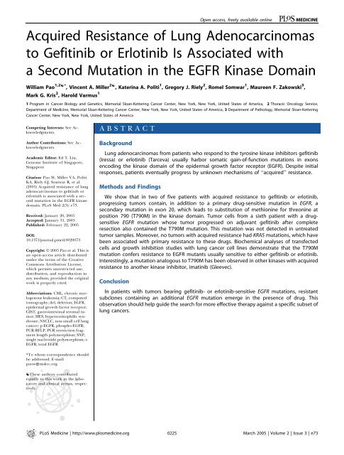
Open access, freely available onlinePLoS MEDICINE<strong>Acquired</strong> <strong>Resistance</strong> <strong>of</strong> <strong>Lung</strong> <strong>Adenocarcinomas</strong><strong>to</strong> Gefitinib or Erlotinib Is Associated witha Second Mutation in the EGFR Kinase DomainWilliam Pao 1,2[* , Vincent A. Miller 2[ , Katerina A. Politi 1 , Gregory J. Riely 2 , Romel Somwar 1 , Maureen F. Zakowski 3 ,Mark G. Kris 2 , Harold Varmus 11 Program in Cancer Biology and Genetics, Memorial Sloan-Kettering Cancer Center, New York, New York, United States <strong>of</strong> America, 2 Thoracic Oncology Service,Department <strong>of</strong> Medicine, Memorial Sloan-Kettering Cancer Center, New York, New York, United States <strong>of</strong> America, 3 Department <strong>of</strong> Pathology, Memorial Sloan-KetteringCancer Center, New York, New York, United States <strong>of</strong> AmericaCompeting Interests: See Acknowledgments.Author Contributions: See Acknowledgments.Academic Edi<strong>to</strong>r: Ed T. Liu,Genome Institute <strong>of</strong> Singapore,SingaporeCitation: Pao W, Miller VA, PolitiKA, Riely GJ, Somwar R, et al.(2005) <strong>Acquired</strong> resistance <strong>of</strong> lungadenocarcinomas <strong>to</strong> gefitinib orerlotinib is associated with a secondmutation in the EGFR kinasedomain. PLoS Med 2(3): e73.Received: January 20, 2005Accepted: January 31, 2005Published: February 22, 2005DOI:10.1371/journal.pmed.0020073Copyright: Ó 2005 Pao et al. This isan open-access article distributedunder the terms <strong>of</strong> the CreativeCommons Attribution License,which permits unrestricted use,distribution, and reproduction inany medium, provided the originalwork is properly cited.Abbreviations: CML, chronic myelogenousleukemia; CT, computed<strong>to</strong>mography; del, deletion; EGFR,epidermal growth fac<strong>to</strong>r recep<strong>to</strong>r;GIST, gastrointestinal stromal tumor;HES, hypereosinophilic syndrome;NSCLC, non-small cell lungcancer; p-EGFR, phospho-EGFR;PCR-RFLP, PCR restriction fragmentlength polymorphism; SNP,single nucleotide polymorphism; t-EGFR, <strong>to</strong>tal EGFRABSTRACTBackground<strong>Lung</strong> adenocarcinomas from patients who respond <strong>to</strong> the tyrosine kinase inhibi<strong>to</strong>rs gefitinib(Iressa) or erlotinib (Tarceva) usually harbor somatic gain-<strong>of</strong>-function mutations in exonsencoding the kinase domain <strong>of</strong> the epidermal growth fac<strong>to</strong>r recep<strong>to</strong>r (EGFR). Despite initialresponses, patients eventually progress by unknown mechanisms <strong>of</strong> ‘‘acquired’’ resistance.Methods and FindingsWe show that in two <strong>of</strong> five patients with acquired resistance <strong>to</strong> gefitinib or erlotinib,progressing tumors contain, in addition <strong>to</strong> a primary drug-sensitive mutation in EGFR, asecondary mutation in exon 20, which leads <strong>to</strong> substitution <strong>of</strong> methionine for threonine atposition 790 (T790M) in the kinase domain. Tumor cells from a sixth patient with a drugsensitiveEGFR mutation whose tumor progressed on adjuvant gefitinib after completeresection also contained the T790M mutation. This mutation was not detected in untreatedtumor samples. Moreover, no tumors with acquired resistance had KRAS mutations, which havebeen associated with primary resistance <strong>to</strong> these drugs. Biochemical analyses <strong>of</strong> transfectedcells and growth inhibition studies with lung cancer cell lines demonstrate that the T790Mmutation confers resistance <strong>to</strong> EGFR mutants usually sensitive <strong>to</strong> either gefitinib or erlotinib.Interestingly, a mutation analogous <strong>to</strong> T790M has been observed in other kinases with acquiredresistance <strong>to</strong> another kinase inhibi<strong>to</strong>r, imatinib (Gleevec).ConclusionIn patients with tumors bearing gefitinib- or erlotinib-sensitive EGFR mutations, resistantsubclones containing an additional EGFR mutation emerge in the presence <strong>of</strong> drug. Thisobservation should help guide the search for more effective therapy against a specific subset <strong>of</strong>lung cancers.*To whom correspondence shouldbe addressed. E-mail:paow@mskcc.org[These authors contributedequally <strong>to</strong> this work in the labora<strong>to</strong>ryand clinical arenas, respectively.PLoS Medicine | http://www.plosmedicine.org0225March 2005 | Volume 2 | Issue 3 | e73
<strong>Resistance</strong> <strong>to</strong> EGFR Kinase Inhibi<strong>to</strong>rsIntroductionSomatic gain-<strong>of</strong>-function mutations in exons encoding theepidermal growth fac<strong>to</strong>r recep<strong>to</strong>r (EGFR) tyrosine kinasedomain are found in about 10% <strong>of</strong> non-small cell lungcancers (NSCLCs) from the United States [1,2,3], with higherpercentages observed in east Asia [2,4,5,6]. Some 90% <strong>of</strong>NSCLC-associated mutations occur as either multi-nucleotidein-frame deletions in exon 19, involving elimination <strong>of</strong>four amino acids, Leu-Arg-Glu-Ala, or as a single nucleotidesubstitution at nucleotide 2573 (TfiG) in exon 21, resultingin substitution <strong>of</strong> arginine for leucine at position 858(L858R). Both <strong>of</strong> these mutations are associated withsensitivity <strong>to</strong> the small-molecule kinase inhibi<strong>to</strong>rs gefitinibor erlotinib [1,2,3]. Unfortunately, nearly all patients whoexperience marked improvement on these drugs eventuallydevelop progression <strong>of</strong> disease. While KRAS mutations havebeen associated with some cases <strong>of</strong> primary resistance <strong>to</strong>gefitinib or erlotinib [7], mechanisms underlying ‘‘acquired’’or ‘‘secondary’’ resistance are unknown.<strong>Acquired</strong> resistance <strong>to</strong> kinase-targeted anticancer therapyhas been most extensively studied with imatinib, an inhibi<strong>to</strong>r<strong>of</strong> the aberrant BCR-ABL kinase, in chronic myelogenousleukemia (CML). Mutations in the ABL kinase domain arefound in 50%–90% <strong>of</strong> patients with secondary resistance <strong>to</strong>the drug (reviewed in [8]). Such mutations, which cluster infour distinct regions <strong>of</strong> the ABL kinase domain (the ATPbinding loop, T315, M351, and the activation loop), interferewith binding <strong>of</strong> imatinib <strong>to</strong> ABL [9,10,11]. Crystallographicstudies <strong>of</strong> various ABL mutants predict that most shouldremain sensitive <strong>to</strong> inhibi<strong>to</strong>rs that bind ABL with lessstringent structural requirements. Using this insight, newsmall-molecule inhibi<strong>to</strong>rs have been identified that retainactivity against the majority <strong>of</strong> imatinib-resistant BCR-ABLmutants [12,13].Although imatinib inhibits different kinases in variousdiseases (BCR-ABL in CML, KIT or PDGFR-alpha in gastrointestinalstromal tumors [GISTs], and PDGFR-alpha inhypereosinophilic syndrome [HES]) (reviewed in [14]), sometumors that become refrac<strong>to</strong>ry <strong>to</strong> treatment with imatinibappear <strong>to</strong> have analogous secondary mutations in the kinasecodingdomain <strong>of</strong> the genes encoding these three enzymes.For example, in CML, a commonly found mutation is a CfiTsingle nucleotide change that replaces threonine withisoleucine at position 315 (T315I) in the ABL kinase domain[9,10,11]. In GIST and HES, respectively, the analogous T670Imutation in KIT and T674I mutation in PDGFR-alpha havebeen associated with acquired resistance <strong>to</strong> this drug [15,16].To determine whether lung cancers that acquire clinicalresistance <strong>to</strong> either gefitinib or erlotinib display additionalmutations in the EGFR kinase domain, we have examined thestatus <strong>of</strong> EGFR exons 18 <strong>to</strong> 24 in tumors from five patientswho initially responded but subsequently progressed while onthese drugs. These exons were also assessed in tumor cellsfrom a sixth patient whose disease rapidly recurred while ongefitinib therapy after complete gross tumor resection.Because <strong>of</strong> the association <strong>of</strong> KRAS mutations with primaryresistance <strong>to</strong> gefitinib and erlotinib [7], we also examined thestatus <strong>of</strong> KRAS in tumor cells from these six patients. In aneffort <strong>to</strong> explain the selective advantage <strong>of</strong> cells with a newlyidentified ‘‘resistance’’ mutation in EGFR—a T790M aminoacid substitution—we further characterized the drug sensitivity<strong>of</strong> putatively resistant EGFR mutants versus wild-type ordrug-sensitive EGFR mutants, using both a NSCLC cell linefortui<strong>to</strong>usly found <strong>to</strong> contain the T790M mutation and lysatesfrom cells transiently transfected with wild-type and mutantEGFR cDNAs.MethodsTissue ProcurementTumor specimens, including paraffin blocks, fine needlebiopsies, and pleural effusions, were obtained throughpro<strong>to</strong>cols approved by the Institutional Review Board <strong>of</strong>Memorial Sloan-Kettering Cancer Center (pro<strong>to</strong>col 92–055[7] and pro<strong>to</strong>col 04–103 [Pro<strong>to</strong>col S1]). All patients providedinformed consent.Mutational Analyses <strong>of</strong> EGFR and KRAS in <strong>Lung</strong> TumorsGenomic DNA was extracted from tumor specimens, andprimers for EGFR (exons 18–24) and KRAS2 (exon 2) analyseswere as published [3,7]. All sequencing reactions wereperformed in both forward and reverse directions, and allmutations were confirmed at least twice from independentPCR isolates.A specific exon 20 mutation (T790M) was also detected bylength analysis <strong>of</strong> fluorescently labeled (FAM) PCR productson a capillary electrophoresis device (ABI 3100 Avant,Applied Biosystems, Foster City, California, United States),based on a new NlaIII restriction site created by the T790Mmutation (2369 CfiT), using the following primers: EGFREx20F, 59-FAM-CTCCCTCCAGGAAGCCTACGTGAT-39 andEGFR Ex20R 59-TTTGCGATCTGCACACACCA-39. Usingserially mixed dilutions <strong>of</strong> DNA from NSCLC cell lines(H1975, L858R- and T790M-positive; H-2030, EGFR wildtype)for calibration, this assay detects the presence <strong>of</strong> theT790M mutation when H1975 DNA comprises 3% or more <strong>of</strong>the <strong>to</strong>tal DNA tested, compared <strong>to</strong> a sensitivity <strong>of</strong> 6% fordirect sequencing (data not shown).RT-PCRThe following primers were used <strong>to</strong> generate EGFR cDNAfragments spanning exon 20: EGFR 2095F 59-CCCAAC-CAAGCTCTCTTGAG-39 and EGFR 2943R 59-ATGACAAGG-TAGCGCTGGGGG-39. PCR products were ligated in<strong>to</strong>plasmids using the TOPO TA-cloning kit (Invitrogen,Carlsbad, California, United States), as per manufacturer’sinstructions. Minipreps <strong>of</strong> DNA from individual clones weresequenced using the T7 priming site <strong>of</strong> the cloning vec<strong>to</strong>r.Functional Analyses <strong>of</strong> Mutant EGFRsTwo numbering systems are used for EGFR. The firstdenotes the initiating methionine in the signal sequence asamino acid 24. The second, used here, denotes themethionine as amino acid þ1. Commercial suppliers <strong>of</strong>antibodies, such as the Y1068-specific anti-phospho-EGFR,use the first nomenclature. To be consistent, we considerY1068 as Y1092. Likewise, the T790M mutation reported herehas also been called T766M. Mutations were introduced int<strong>of</strong>ull-length wild-type and mutant EGFR cDNAs using aQuikChange Site-Directed Mutagenesis Kit (Stratagene, LaJolla, California, United States) and cloned in<strong>to</strong> expressionvec<strong>to</strong>rs as described [3]. The following primers were used <strong>to</strong>generate the deletion (del) L747–E749;A750P mutant: forward59-TAAAATTCCCGTCGCTATCAAGGAGCCAA-PLoS Medicine | http://www.plosmedicine.org0226March 2005 | Volume 2 | Issue 3 | e73
<strong>Resistance</strong> <strong>to</strong> EGFR Kinase Inhibi<strong>to</strong>rsTable 1. Specimens Analyzed in This Study for Mutations in the EGFR Tyrosine Kinase Domain (Exons 18 <strong>to</strong> 24) and KRAS (Exon 2)Patient Pathology Specimen Analyzed Date Obtained Percent Tumor Cells a EGFR KRAS1 Transbronchial biopsy Day 0 Scant Wild-type Wild-typeProgressing lung lesion 12 mo .85% L858R þ T790M Wild-typePleural effusion 14 mo .85% L858R þ T790M Wild-type2 Original lung lesion Day 0 .85% del L747–E749;A750P Wild-typeProgressing spine lesion 75 mo .85% del L747–E749;A750P þ T790M Wild-typeProgressing lung lesion 77 mo .85% del L747–E749;A750P þ T790M Wild-type3 Original pleural biopsy Day 0 n/a Unavailable UnavailableRe-resection lung lesion 68 mo .85% del E746–A750 Wild-typePleural effusion 76 mo .50% del E746–A750 þ T790M Wild-typeThe transbronchial biopsy in patient 1 had scant tumor cells; sequencing analysis revealed only wild-type sequence (see text).a Percent tumor cells is defined by assessment <strong>of</strong> corresponding his<strong>to</strong>pathological slides.n/a, not applicable.DOI: 10.1371/journal.pmed.0020073.t001CATCTCCGAAAGCCAACAAGG-39 and reverse 59-CCTTGTTGGCTTTCGGAGATGTTGGCTCCTTGATAGC-GACGGGAATTTTA-39. The following primers were used <strong>to</strong>introduce the T790M mutation: forward 59-AGCTCATCATG-CAGCTCAT-39 and reverse 59-ATGAGCTGCATGAT-GAGCT-39. The L858R mutant cDNA was generatedpreviously [3]. All mutant clones were fully re-sequencedbidirectionally <strong>to</strong> ensure that no additional mutations wereintroduced. Various EGFRs were transiently expressed in293T human embryonic kidney cells as published [3]. Cellswere treated with different concentrations <strong>of</strong> gefitinib orerlotinib.ImmunoblottingSee methods and supplementary methods in [3] for detailson cell lysis, immunoblotting, and antibody reagents. At leastthree independent experiments were performed for allanalyses.Cell CultureThe NSCLC cell lines H1650, H1975, H2030, H2347, H2444,H358, and H1734 were purchased from American TypeCulture Collection (Manassas, Virginia, United States). H3255was a gift <strong>of</strong> B. Johnson and P. Janne. Cells were grown incomplete growth medium (RPMI-1640; American TypeCulture Collection catalog no. 30–2001) supplemented with10% fetal calf serum, 10 units/ml penicillin, and 10 lg/mlstrep<strong>to</strong>mycin) at 37 8C and 5% CO 2 . For viability studies, cellswere seeded in complete growth medium in black 96-wellclear bot<strong>to</strong>m ViewPlates (PerkinElmer, Wellesley, Massachusetts,United States) at a density <strong>of</strong> 5,000 (H1975 and H2030)or 7,500 cells per well (H3255). Following overnight incubation,cells were grown for 24 h in the supplemented RPMI-1640 medium with 0.1% serum. Cells (in supplemented RPMI-1640 medium containing 0.1% serum) were then incubatedfor 48 h in the continued presence <strong>of</strong> gefitinib or erlotinib.Viability AssayCell viability was assayed using Calcein AM (ace<strong>to</strong>xymethylester <strong>of</strong> Calcein, Molecular Probes, Eugene, Oregon, UnitedStates). Following incubation with gefitinib or erlotinib,monolayers were washed twice with PBS (containing calciumand magnesium) and incubated with 7.5 lmol Calcein AM insupplemented RPMI-1640 (no serum) for 30 min. Labelingmedium was removed, and cells were washed three times withPBS. Calcein fluorescence (Ex, 485 nm; Em, 535 nM) wasdetected immediately using a Vic<strong>to</strong>r V multi-label platereader (PerkinElmer). Three independent experiments wereperformed for each cell line; each experiment included four<strong>to</strong> eight replicates per condition.ResultsCase ReportsWe identified secondary EGFR mutations in three <strong>of</strong> sixindividuals whose disease progressed on either gefitinib orerlotinib (Table 1). Brief case his<strong>to</strong>ries <strong>of</strong> these three patientsare presented below.Patient 1. This 63-y-old female ‘‘never smoker’’ (smokedless than 100 cigarettes in her lifetime) initially presentedwith bilateral diffuse chest opacities and a right-sided pleuraleffusion. Transbronchial biopsy revealed adenocarcinoma.Disease progressed on two cycles <strong>of</strong> systemic chemotherapy,after which gefitinib, 250 mg daily, was started. Comparison<strong>of</strong> chest radiographs obtained prior <strong>to</strong> starting gefitinib(Figure S1A, left panel) and 2 wk later (Figure S1A, middlepanel) showed dramatic improvement. Nine months later, achest radiograph revealed progression <strong>of</strong> disease (Figure S1A,right panel). Subsequently, the patient underwent a computed<strong>to</strong>mography (CT)–guided biopsy <strong>of</strong> an area in the rightlung base (Figure 1A, left panel). Despite continued treatmentwith gefitinib, either with chemotherapy or at 500 mg daily,the pleural effusion recurred, 12 mo after initiating gefitinib(Figure 1A, right panel). Pleural fluid was obtained formolecular studies. In <strong>to</strong>tal, this patient had three tumorspecimens available for analysis: the original lung tumorbiopsy, a biopsy <strong>of</strong> the progressing lung lesion, and pleuralfluid. However, re-review <strong>of</strong> the original transbronchialbiopsy showed that it had scant tumor cells (Table 1).Patient 2. This 55-y-old woman with a nine pack-yearhis<strong>to</strong>ry <strong>of</strong> smoking underwent two surgical resections within2 y (right lower and left upper lobec<strong>to</strong>mies) for bronchioloalveolarcarcinoma with focal invasion. Two years later, herdisease recurred with bilateral pulmonary nodules andfurther progressed on systemic chemotherapy. Thereafter,the patient began erlotinib, 150 mg daily. A baseline CT scan<strong>of</strong> the chest demonstrated innumerable bilateral nodules(Figure S1B, left panel), which were markedly reduced innumber and size 4 mo after treatment (Figure S1B, middlePLoS Medicine | http://www.plosmedicine.org0227March 2005 | Volume 2 | Issue 3 | e73
<strong>Resistance</strong> <strong>to</strong> EGFR Kinase Inhibi<strong>to</strong>rspleural effusion recurred 4 mo later (Figure S1C, right panel),at which time pleural fluid was collected for analysis. In <strong>to</strong>tal,this patient had two clinical specimens available for analysis:tumor from the surgical resection and pleural fluid (Table 1).Figure 1. Re-Biopsy Studies(A.) Patient 1. CT-guided biopsy <strong>of</strong> progressing lung lesions after 10mo on gefitinib (left panel). Two months later, fluid from a rightsidedpleural effusion (right panel) was collected for molecularanalysis.(B) Patient 2. CT-guided biopsy <strong>of</strong> a progressing thoracic spine lesion(left panel) and fluoroscopic-guided biopsy <strong>of</strong> a progressing lunglesion (right panel). The biopsy needles are indicated by white arrows.DOI: 10.1371/journal.pmed.0020073.g001panel). After 14 mo <strong>of</strong> therapy, the patient’s dose <strong>of</strong> erlotinibwas decreased <strong>to</strong> 100 mg daily owing <strong>to</strong> fatigue. At 23 mo <strong>of</strong>treatment with erlotinib, a CT scan demonstrated anenlarging sclerotic lesion in the thoracic spine. The patientunderwent CT-guided biopsy <strong>of</strong> this lesion (Figure 1B, leftpanel), and the erlotinib dose was increased <strong>to</strong> 150 mg daily.After 25 mo <strong>of</strong> treatment, she progressed within the lung(Figure S1B, right panel). Erlotinib was discontinued, and afluoroscopically guided core needle biopsy was performed ata site <strong>of</strong> progressive disease in the lung (Figure 1B, rightpanel). In <strong>to</strong>tal, this patient had three tumor specimensavailable for analysis: the original resected lung tumor, thebiopsy <strong>of</strong> the enlarging spinal lesion, and the biopsy <strong>of</strong> theprogressing lung lesion (Table 1).Patient 3. This 55-y-old female ‘‘never smoker’’ was treatedfor nearly 4.5 y with weekly paclitaxel and trastuzumab [17]for adenocarcinoma with bronchioloalveolar carcinomafeatures involving her left lower lobe, pleura, and mediastinallymph nodes. Treatment was discontinued owing <strong>to</strong> fatigue.Subsequently, the patient underwent surgical resection.Because <strong>of</strong> metastatic involvement <strong>of</strong> multiple mediastinallymph nodes and clinical features known at that time <strong>to</strong> bepredictive <strong>of</strong> response <strong>to</strong> gefitinib (female, never smoker,bronchioloalveolar variant his<strong>to</strong>logy), she was placed on‘‘adjuvant’’ gefitinib 1 mo later (Figure S1C, left panel). Thisdrug was discontinued after 3 mo when she developed a newleft-sided malignant pleural effusion (Figure S1C, middlepanel). Despite drainage and systemic chemotherapy, thePatients’ Tumors Contain EGFR Tyrosine Kinase DomainMutations Associated with Sensitivity <strong>to</strong> EGFR TyrosineKinase Inhibi<strong>to</strong>rsWe screened all available tumor samples from these threepatients for previously described drug-sensitive EGFR mutations,by direct DNA sequencing <strong>of</strong> exons 19 and 21 [3].Tumor samples from patient 1 showed a TfiG change atnucleotide 2573, resulting in the exon 21 L858R amino acidsubstitution commonly observed in drug-responsive tumors.This mutation was present in the biopsy material from theprogressing lung lesion (Figure S2A, upper panels) and fromcells from the pleural effusion (Figure S2A, lower panels),both <strong>of</strong> which on cy<strong>to</strong>pathologic examination consisted <strong>of</strong> amajority <strong>of</strong> tumor cells (Table 1). Interestingly, comparisons<strong>of</strong> the tracings suggest that an increase in copy number <strong>of</strong> themutant allele may have occurred. Specifically, while the ratio<strong>of</strong> wild-type (nucleotide T) <strong>to</strong> mutant (nucleotide G) peaks atposition 2573 was approximately 1:1 or 1:2 in the lung biopsyspecimen (Figure S2A, upper panels), sequencing <strong>of</strong> DNAfrom the pleural fluid cells demonstrated a dominant mutantG peak (Figure S2A, lower panels). Consistent with this, asingle nucleotide polymorphism (SNP) noted at nucleotide2361 (A or G) demonstrated a corresponding change in theratios <strong>of</strong> A:G, with a 1:1 ratio in the transbronchial biopsy,and a nearly 5:1 ratio in the pleural fluid (Figure 2A). Notably,we did not detect the 2573 TfiG mutation in the originaltransbronchial biopsy specimen (Table 1; data not shown). Asstated above, this latter specimen contained scant tumor cells,most likely fewer than needed for detection <strong>of</strong> an EGFRmutation by direct sequencing (see [7]).All three specimens from patient 2, including the originallung tumor and the two metastatic samples from bone andlung, showed an exon 19 deletion involving elimination <strong>of</strong> 11nucleotides (2238–2248) and insertion <strong>of</strong> two nucleotides, Gand C (Figure S2B, all panels; Table 1). These nucleotidechanges delete amino acids L747–E749 and change aminoacid 750 from alanine <strong>to</strong> proline (A750P). A del L747–E749;A750P mutation was previously reported with differentnucleotide changes [2]. In all samples from patient 2, the wildtypesequence predominated at a ratio <strong>of</strong> about 3:1 over themutant sequence.Both <strong>of</strong> the available tumor samples from patient 3contained a deletion <strong>of</strong> 15 nucleotides (2236–2250) in exon19 (Table 1; data not shown), resulting in elimination <strong>of</strong> fiveamino acids (del E746–A750). This specific deletion has beenpreviously reported [3]. The ratio <strong>of</strong> mutant <strong>to</strong> wild-typepeaks was approximately 1:1 in both specimens (data notshown).Collectively, these results demonstrate that tumors from allthree patients contain EGFR mutations associated withsensitivity <strong>to</strong> the tyrosine kinase inhibi<strong>to</strong>rs gefitinib anderlotinib. In addition, these data show that within individualpatients, metastatic or recurrent lesions <strong>to</strong> the spine, lung,and pleural fluid contain the same mutations. These latterobservations support the idea that relapsing and metastatictumor cells within individuals are derived from originalprogeni<strong>to</strong>r clones.PLoS Medicine | http://www.plosmedicine.org0228March 2005 | Volume 2 | Issue 3 | e73
<strong>Resistance</strong> <strong>to</strong> EGFR Kinase Inhibi<strong>to</strong>rsFigure 2. Sequencing Chroma<strong>to</strong>grams with the T790M EGFR Exon 20 Mutation in Various Clinical Specimens and the NSCLC Cell Line H1975(A–C) In all three patients—patient 1 (A), patient 2 (B), and patient 3 (C)—the secondary T790M mutation was observed only in lesions obtainedafter progression on either gefitinib or erlotinib.(D) Cell line H1975 contains both an exon 21 L858R mutation (upper panel) and the exon 20 T790M mutation (lower panel). The asterisksindicate a common SNP (A or G) at nucleotide 2361; the arrows indicate the mutation at nucleotide 2369 (CfiT), which leads <strong>to</strong> substitution <strong>of</strong>methonine (ATG) for threonine (ACG) at position 790. In the forward direction, the mutant T peak is blue. In the reverse direction, the mutantpeak is green, while the underlying blue peak represents an ‘‘echo’’ from the adjacent nucleotide.DOI: 10.1371/journal.pmed.0020073.g002A Secondary Missense Mutation in the EGFR KinaseDomain Detected in Lesions That Progressed while onTreatment with Either Gefitinib or ErlotinibTo determine whether additional mutations in the EGFRkinase domain were associated with progression <strong>of</strong> disease inthese patients, we performed direct sequencing <strong>of</strong> all <strong>of</strong> theexons (18 through 24) encoding the EGFR catalytic region inthe available tumor specimens.Analysis <strong>of</strong> patient 1’s pre-gefitinib specimen, whichcontained scant tumor cells (Table 1; see above), notsurprisingly showed only wild-type EGFR sequence (Table 1;data not shown). However, careful analysis <strong>of</strong> the exon 20sequence chroma<strong>to</strong>grams in both forward and reversedirections from this patient’s lung biopsy specimen obtainedafter disease progression on gefitinib demonstrated anadditional small peak at nucleotide 2369, suggesting a CfiTmutation (Figure 2A, upper panels; Table 1). This nucleotidechange leads <strong>to</strong> substitution <strong>of</strong> methionine for threonine atposition 790 (T790M). The 2369 CfiT mutant peak was evenmore prominent in cells from the patient’s pleural fluid,which were obtained after further disease progression ongefitinib (Figure 2A, lower panels; Table 1). The increase inthe ratio <strong>of</strong> mutant <strong>to</strong> wild-type peaks obtained from analyses<strong>of</strong> the lung specimen and pleural fluid paralleled the increasein the ratio <strong>of</strong> the mutant G peak (leading <strong>to</strong> the L858Rmutation) <strong>to</strong> the wild-type T peak at nucleotide 2573 (seeabove; Figure S2A), as well as the increase in the ratio <strong>of</strong> theA:G SNP at position 2361 (Figure 2A). Collectively, thesefindings imply that the exon 20 T790M mutation was presen<strong>to</strong>n the same allele as the exon 21 L858R mutation, and that asubclone <strong>of</strong> cells harboring these mutations emerged duringdrug treatment.In patient 2, the tumor-rich sample obtained prior <strong>to</strong>treatment with erlotinib did not contain any additionalmutations in the exons encoding the EGFR tyrosine kinasedomain (Figure 2B, upper panels; Table 1). By contrast, herprogressing bone and lung lesions contained an additionalsmall peak at nucleotide 2369, suggesting the existence <strong>of</strong> asubclone <strong>of</strong> tumor cells with the same CfiT mutationobserved in patient 1 (Figure 2B, middle and lower panels;PLoS Medicine | http://www.plosmedicine.org0229March 2005 | Volume 2 | Issue 3 | e73
<strong>Resistance</strong> <strong>to</strong> EGFR Kinase Inhibi<strong>to</strong>rsTable 2. Status <strong>of</strong> NSCLC Cell Lines Analyzed for EGFR TyrosineKinase Domain (Exons 18 <strong>to</strong> 24) and KRAS (Exon 2) MutationsCell Line EGFR KRASH1650 del E746–A750 Wild-typeH3255 L858R Wild-typeH1975 L858R þ T790M Wild-typeH2030 Wild-type G12CH358 Wild-type G12CH2444 Wild-type G12VH1734 Wild-type G13CH2347 Wild-type L19FSee Methods for further details.DOI: 10.1371/journal.pmed.0020073.t002Table 1). The relative sizes <strong>of</strong> the 2369 T mutant peaks seen inthese latter two samples appeared <strong>to</strong> correlate with therelative size <strong>of</strong> the corresponding peaks <strong>of</strong> the exon 19deletion (Figure S2B). Interestingly, the SNP at nucleotide2361 (A or G) was detected in specimens from patient 2before but not after treatment with erlotinib, suggesting tha<strong>to</strong>ne EGFR allele underwent amplification or deletion duringthe course <strong>of</strong> treatment (Figure S2B).Patient 3 showed results analogous <strong>to</strong> those <strong>of</strong> patient 2. Atumor-rich pre-treatment specimen did not demonstrateEGFR mutations other than the del E746–A750 exon 19deletion; specifically, in exon 20, no secondary changes weredetected (Figure 2C, upper panels; Table 1). However,analysis <strong>of</strong> DNA from cells in the pleural effusion thatdeveloped after treatment with gefitinib showed the CfiTmutation at nucleotide 2369 in exon 20 (Figure 2C, lowerpanels; Table 1), corresponding <strong>to</strong> the T790M mutationdescribed above. There was no dramatic change between thetwo samples in the ratio <strong>of</strong> the A:G SNP at position 2361.The mutant 2369 T peak was small, possibly because gefitinibhad been discontinued in this patient for 4 mo at the timepleural fluid tumor cells were collected; thus, there was noselective advantage conferred upon cells bearing the T790Mmutation.To determine whether the 2369 CfiT mutation was apreviously overlooked EGFR mutation found in NSCLCs, were-reviewed exon 20 sequence tracings derived from analysis<strong>of</strong> 96 fresh-frozen resected tumors [3] and 59 paraffinembeddedtumors [7], all <strong>of</strong> which were removed frompatients prior <strong>to</strong> treatment with an EGFR tyrosine kinaseinhibi<strong>to</strong>r. We did not detect any evidence <strong>of</strong> the T790Mmutation in these 155 tumors (data not shown; see Discussion).Collectively, our results suggest that the T790Mmutation is associated with lesions that progress while ongefitinib or erlotinib. Moreover, at least in patients 1 and 2,the subclones <strong>of</strong> tumor cells bearing this mutation probablyemerged between the time <strong>of</strong> initial treatment with a tyrosinekinase inhibi<strong>to</strong>r and the appearance <strong>of</strong> drug resistance.In three additional patients (case his<strong>to</strong>ries not describedhere) with lung adenocarcinomas who improved but subsequentlyprogressed on therapy with either gefitinib orerlotinib, we examined DNA from tumor specimens obtainedduring disease progression. In all three patients, we foundEGFR mutations associated with drug sensitivity (all exon 19deletions). However, we did not find any additional mutationsin exons 18 <strong>to</strong> 24 <strong>of</strong> EGFR, including the CfiT change atposition 2369 (data not shown). These results imply thatalternative mechanisms <strong>of</strong> acquired drug resistance exist.Patients’ Progressive Tumors Lack KRAS MutationsMutations in exon 2 <strong>of</strong> KRAS2 occur in about one-fourth <strong>of</strong>NSCLCs. Such mutations rarely, if ever, accompany EGFRmutations and are associated with primary resistance <strong>to</strong>gefitinib or erlotinib [7]. To evaluate the possibility thatsecondary KRAS mutations confer acquired resistance <strong>to</strong>these drugs, we performed mutational pr<strong>of</strong>iling <strong>of</strong> KRAS2exon 2 from tumor specimens from patients 1 <strong>to</strong> 3, as well asthe three additional patients lacking evidence <strong>of</strong> the T790Mmutation. None <strong>of</strong> the specimens contained any changes inKRAS (Table 1; data not shown), indicating that KRASmutations were not responsible for drug resistance andtumor progression in these six patients.An Established NSCLC Cell Line Also Contains Both T790Mand L858R MutationsWe pr<strong>of</strong>iled the EGFR tyrosine kinase domain (exons 18 <strong>to</strong>24) and KRAS exon 2 in eight established NSCLC lines (Table2). Surprisingly, one cell line—H1975—contained the sameCfiT mutation at position 2369 (T790M) as described above(Figure 2D, lower panel). This cell line had previously beenshown by others <strong>to</strong> contain a 2573 TfiG mutation in exon 21(L858R) [18], which we confirmed (Figure 2D, upper panel); inaddition, H1975 was reported <strong>to</strong> be more sensitive <strong>to</strong>gefitinib inhibition than other lung cancer cell lines bearingwild-type EGFR [18]. Only exons 19 and 21 were apparentlyexamined in this published study.In our own analysis <strong>of</strong> H1975 (exons 18 <strong>to</strong> 24), the mutant2369 T peak resulting in the T790M amino acid substitutionwas dominant, suggesting an increase in copy number <strong>of</strong> themutant allele in comparison <strong>to</strong> the wild-type allele. The ratio<strong>of</strong> mutant <strong>to</strong> wild-type peaks was similar <strong>to</strong> that <strong>of</strong> the mutant2573 G (corresponding <strong>to</strong> the L858R amino acid substitution)<strong>to</strong> wild-type T peaks (Figure 2D, all panels), implying that theT790M and L858R mutations were in the same amplifiedallele. To further investigate this possibility, we performedRT-PCR <strong>to</strong> generate cDNAs that spanned exon 20 <strong>of</strong> EGFRand included sequences from exon 19 and 21. PCR productswere then cloned, and individual colonies were analyzed forEGFR mutations. Sequencing chroma<strong>to</strong>grams <strong>of</strong> DNA fromfour <strong>of</strong> four clones showed both the 2369 CfiT and 2573TfiG mutations, confirming that both mutations were in thesame allele (data not shown).Other NSCLC cell lines carried either EGFR or KRASmutations, but none had both (Table 2). As reported, H3255contained an L858R mutation [19] and H1650 contained anexon 19 deletion [18]. No other cell lines analyzed containedadditional mutations in the exons encoding the EGFRtyrosine kinase domain.A Novel PCR Restriction Fragment Length PolymorphismAssay Independently Confirms the Absence or Presence <strong>of</strong>the T790M MutationAs stated above, the mutant peaks suggestive <strong>of</strong> a T790Mmutation in exon 20 were small in some sequence chroma<strong>to</strong>grams.To eliminate the possibility that these peaks were due<strong>to</strong> background ‘‘noise,’’ we sought <strong>to</strong> confirm the presence <strong>of</strong>the 2369 CfiT mutation in specific samples, by developing anPLoS Medicine | http://www.plosmedicine.org0230March 2005 | Volume 2 | Issue 3 | e73
<strong>Resistance</strong> <strong>to</strong> EGFR Kinase Inhibi<strong>to</strong>rsindependent test, based on a fluorescence detection assaythat takes advantage <strong>of</strong> a PCR restriction fragment lengthpolymorphism (PCR-RFLP) generated by the specific missensemutation. After PCR amplification with exon-20-specific primers spanning nucleotide 2369, wild-type sequencecontains specific NlaIII sites, which upon digestionyield a 106-bp product (see Methods; Figure 3A). Presence <strong>of</strong>the mutant 2369 T nucleotide creates a new NlaIII restrictiondigest site, yielding a slightly shorter product (97 bp), readilydetected by fluorescent capillary electrophoresis. This test isabout 2 -fold more sensitive than direct sequencing (seeMethods; data not shown).We first used DNA from the H1975 cell line (which containsboth T790M and L858R mutations) <strong>to</strong> confirm the specificity<strong>of</strong> the PCR-RFLP assay. As expected, analysis <strong>of</strong> these cellsproduced both the 97- and 106-bp fragments. By contrast,analysis <strong>of</strong> DNA from H2030 (which contains wild-type EGFR;Table 2) showed only the 106-bp fragment (Figure 3A). Thesedata show that this test can readily indicate the absence orpresence <strong>of</strong> the mutant allele in DNA samples. However, thistest was only semi-quantitative, as the ratio <strong>of</strong> the mutant 97-bp product versus the wild-type 106-bp product varied inindependent experiments from approximately 1:1 <strong>to</strong> 2:1.We next used this PCR-RFLP assay <strong>to</strong> assess various patientsamples for the presence <strong>of</strong> the specific 2369 CfiT mutationcorresponding <strong>to</strong> the T790M amino acid substitution. DNAfrom the progressing bone and lung lesions in patient 1produced both the 97- and 106-bp fragments, but DNA fromthe original lung tumor did not (Figure 3B). The ratio <strong>of</strong>mutant <strong>to</strong> wild-type products was higher in the cells from thepleural fluid, consistent with the higher peaks seen on thechroma<strong>to</strong>grams from direct sequencing <strong>of</strong> exon 20 (seeFigure 2A). Likewise, DNA from progressive lesions frompatients 2 and 3 yielded both 97- and 106-bp fragments in thePCR-RFLP assay (Figure 3B), whereas the pre-treatmentspecimens did not produce the 97-bp product. Collectively,these data from an independent assay confirm that theT790M mutation was present in progressing lesions from allthree patients. We were also unable <strong>to</strong> detect the T790Mmutation in any specimens from the three additional patientswith acquired resistance that failed <strong>to</strong> demonstrate secondarymutations in EGFR exons 18 <strong>to</strong> 24 by direct sequencing (datanot shown).Biochemical Properties <strong>of</strong> EGFR MutantsTo determine how the T790M mutation would affect EGFRproteins already containing mutations associated with sensitivity<strong>to</strong> EGFR tyrosine kinase inhibi<strong>to</strong>rs, we introduced thespecific mutation in<strong>to</strong> EGFR cDNAs that encoded the exon 21and 19 mutations found in patients 1 and 2, respectively.Corresponding proteins ([i] L858R and L858R plus T790M,[ii] del L747–E749;A750P and del L747–E749;A750P plusT790M, and [iii] wild-type EGFR and wild-type EGFR plusT790M) were then produced by transient transfection withexpression vec<strong>to</strong>rs in 293T cells, which have very low levels <strong>of</strong>endogenous EGFR [3]. Various lysates from cells that wereserum-starved and pre-treated with gefitinib or erlotinibwere analyzed by immunoblotting. Amounts <strong>of</strong> <strong>to</strong>tal EGFR (t-EGFR) were determined using an anti-EGFR monoclonalantibody, and actin served as an indica<strong>to</strong>r <strong>of</strong> relative levels <strong>of</strong>protein per sample. To assess the drug sensitivity <strong>of</strong> thevarious EGFR kinases in surrogate assays, we used a Y1092-Figure 3. A Novel PCR-RFLP Assay Independently Confirms Presence <strong>of</strong>the T790M Mutation in Exon 20 <strong>of</strong> the EGFR Kinase Domain(A) Design <strong>of</strong> the assay (see text for details). ‘‘F’’ designates thefluorescent label, FAM. At the bot<strong>to</strong>m <strong>of</strong> this panel, the assaydemonstrates with the 97-bp NlaIII cleavage product the presence <strong>of</strong>the T790M mutation in the H1975 cell line; this product is absent inH2030 DNA. The 106-bp NlaIII cleavage product is generated bydigestion <strong>of</strong> wild-type EGFR.(B) The PCR-RFLP assay demonstrates that pre-drug tumor samplesfrom the three patients lack detectable levels <strong>of</strong> the mutant 97-bpproduct, while specimens obtained after disease progression containthe T790M mutation. Pt, patient.DOI: 10.1371/journal.pmed.0020073.g003phosphate-specific antibody (i.e., phospho-EGFR [p-EGFR])<strong>to</strong> measure the levels <strong>of</strong> ‘‘au<strong>to</strong>phosphorylated’’ Tyr-1092 onEGFR in relation <strong>to</strong> levels <strong>of</strong> t-EGFR protein. We also assessedthe global pattern and levels <strong>of</strong> induced tyrosine phosphorylation<strong>of</strong> cell proteins by using a generalized anti-phosphotyrosinereagent (RC-20).Gefitinib inhibited the activity <strong>of</strong> wild-type and L858REGFRs progressively with increasing concentrations <strong>of</strong> drug,as demonstrated by a reduction <strong>of</strong> tyrosine-phosphorylatedproteins (Figure 4A) and a decrease in p-EGFR:t-EGFR ratios(Figure 4B). By contrast, wild-type and mutant EGFRscontaining the T790M mutation did not display a significantchange in either phosphotyrosine induction or p-EGFR:t-EGFR ratios (Figure 4A and 4B). Similar results were obtainedusing erlotinib against wild-type and del E747–L747;A750PEGFRs in comparison <strong>to</strong> the corresponding mutants containingthe T790M mutation (Figure 4C). These resultsPLoS Medicine | http://www.plosmedicine.org0231March 2005 | Volume 2 | Issue 3 | e73
<strong>Resistance</strong> <strong>to</strong> EGFR Kinase Inhibi<strong>to</strong>rsFigure 5. Sensitivity <strong>to</strong> Gefitinib Differs Among NSCLC Cell LinesContaining Various Mutations in EGFR or KRASThe three indicated NSCLC cell lines, H3255 (L858R mutation),H1975 (both T790M and L858R mutations), and H2030 (wild-typeEGFR, mutant KRAS) (see Table 2), were grown in increasingconcentrations <strong>of</strong> gefitinib, and the density <strong>of</strong> live cells after 48 h<strong>of</strong> treatment was measured using a Calcein AM fluorescence assay.Fluorescence in vehicle-treated cells is expressed as 100%. Results arethe mean 6 standard error <strong>of</strong> three independent experiments inwhich there were four <strong>to</strong> eight replicates <strong>of</strong> each condition. Similarresults were obtained with erlotinib (see Figure S3).DOI: 10.1371/journal.pmed.0020073.g005Figure 4. EGFR Mutants Containing the T790M Mutation Are Resistant <strong>to</strong>Inhibition by Gefitinib or Erlotinib293T cells were transiently transfected with plasmids encoding wildtype(WT) EGFR or EGFR mutants with the following changes:T790M, L858R, L858R þ T790M, del L747–E749;A750P, or del L747–E749;A750P þ T790M. After 36 h, cells were serum-starved for 24 h,treated with gefitinib or erlotinib for 1 h, and then harvested forimmunoblot analysis using anti-p-EGFR (Y1092), anti-t-EGFR, antiphosphotyrosine(p-Tyr), and anti-actin antibodies as described inMethods. The EGFR T790M mutation, in conjunction with eitherwild-type EGFR or the drug-sensitive L858R EGFR mutant, preventsinhibition <strong>of</strong> tyrosine phosphorylation (A) or p-EGFR (B) by gefitinib.Analogously, the T790M mutation, in conjunction with the drugresponsivedel L747–E749;A750P EGFR mutant, prevents inhibition<strong>of</strong> p-EGFR by erlotinib (C).DOI: 10.1371/journal.pmed.0020073.g004suggest that the T790M mutation may impair the ability <strong>of</strong>gefitinib or erlotinib <strong>to</strong> inhibit EGFR tyrosine kinase activity,even in EGFR mutants (i.e., L858R or an exon 19 deletion)that are clinically associated with drug sensitivity.<strong>Resistance</strong> <strong>of</strong> a NSCLC Cell Line Harboring Both T790Mand L858R Mutations <strong>to</strong> Gefitinib or ErlotinibTo further explore the functional consequences <strong>of</strong> theT790M mutation, we determined the sensitivity <strong>of</strong> variousNSCLC cells lines grown in the presence <strong>of</strong> either gefitinib orerlotinib, using an assay based upon Calcein AM. Uptake andretention <strong>of</strong> this fluorogenic esterase substrate by vehicleversusdrug-treated live cells allows for a comparison <strong>of</strong>relative cell viability among cell lines [20]. The H3255 cellline, which harbors the L858R mutation and no other EGFRTK domain mutations (Table 2), was sensitive <strong>to</strong> treatmentwith gefitinib, with an IC 50 <strong>of</strong> about 0.01 lmol (Figure 5). Bycontrast, the H1975 cell line, which contains both L858R andT790M mutations (Table 2), was approximately 100-fold lesssensitive <strong>to</strong> drug, with an IC 50 <strong>of</strong> about 1 lmol (Figure 5). Infact, the sensitivity <strong>of</strong> H1975 cells was more similar <strong>to</strong> that <strong>of</strong>H2030, which contains wild-type EGFR (exons 18 <strong>to</strong> 24) andmutant KRAS (Figure 5). Very similar results were obtainedwith erlotinib (Figure S3).DiscussionSpecific mutations in the tyrosine kinase domain <strong>of</strong> EGFRare associated with sensitivity <strong>to</strong> either gefitinib or erlotinib,but mechanisms <strong>of</strong> acquired resistance have not yet beenreported. Based upon analogous studies in other diseases withanother kinase inhibi<strong>to</strong>r, imatinib, a single amino acidsubstitution from threonine <strong>to</strong> methionine at position 790in the wild-type EGFR kinase domain was predicted <strong>to</strong> lead <strong>to</strong>drug resistance, even before the association <strong>of</strong> exon 19 and 21mutations <strong>of</strong> EGFR with drug responsiveness in NSCLC wasreported. The T790M mutation was shown in vitro in thecontext <strong>of</strong> wild-type EGFR <strong>to</strong> confer resistance <strong>to</strong> gefitinib[21] and a related quinazoline inhibi<strong>to</strong>r, PD153035 [22].We show here, through molecular analysis <strong>of</strong> tumormaterial from three patients and one NSCLC cell line, asPLoS Medicine | http://www.plosmedicine.org0232March 2005 | Volume 2 | Issue 3 | e73
<strong>Resistance</strong> <strong>to</strong> EGFR Kinase Inhibi<strong>to</strong>rsTable 3. Analogous Mutations in Four Kinases Associated with <strong>Resistance</strong> <strong>to</strong> Kinase Inhibi<strong>to</strong>rsProtein Mutation Disease Drug ReferenceBCR-ABL T315I CML Imatinib 11PDGFR-alpha T674I HES Imatinib 16KIT T670I GIST Imatinib 15EGFR T790M NSCLC Gefitinib and erlotinib This studyDOI: 10.1371/journal.pmed.0020073.t003well as additional biochemical studies, that acquired clinicaldrug resistance <strong>to</strong> gefitinib or erlotinib is indeed associatedwith the T790M mutation. Importantly, we find that theT790M mutation confers drug resistance not just <strong>to</strong> wild-typeEGFR but also <strong>to</strong> mutant EGFRs associated with clinicalresponsiveness <strong>to</strong> EGFR tyrosine kinase inhibi<strong>to</strong>rs [1,2,3]. Ourresults further demonstrate that an analogous mechanism <strong>of</strong>acquired resistance exists for imatinib and EGFR tyrosinekinase inhibi<strong>to</strong>rs (Table 3), despite the fact that the variousagents target different kinases in distinct diseases.In tumors from patients not treated with either gefitinib orerlotinib, the 2369 CfiT mutation (T790M) appears <strong>to</strong> beextremely rare. We have not identified this mutation in 155tumors (see above), and among nearly 1,300 lung cancers inwhich analysis <strong>of</strong> EGFR exons 18 <strong>to</strong> 21 has been performed[1,2,3,4,5,6], only one tumor (which also harbored an L858Rmutation) was reported <strong>to</strong> contain the T790M mutation.Whether the patient from which this tumor was resected hadreceived gefitinib or erlotinib is unclear, and the report didnot note an association with acquired resistance <strong>to</strong> eitherdrug [5].How tumor cells bearing the T790M mutation emergewithin gefitinib- or erlotinib-treated patients is a matter <strong>of</strong>investigation. Subclones bearing this mutation could arise denovo during treatment. However, based upon analogousstudies in CML, it is also possible that NSCLC subclonesbearing this secondary mutation pre-exist within the primarytumor clone in individual patients, albeit at low frequency[23]. In either scenario, treatment with gefitinib or erlotinibsubsequently allows these resistant subclones <strong>to</strong> becomeapparent, because most cells bearing sensitivity-conferringmutations die, while cells with the T790M mutation persist.From analysis <strong>of</strong> the crystal structure <strong>of</strong> the EGFR kinasedomain bound <strong>to</strong> erlotinib, it is has been shown that the wildtypethreonine residue at position 790 is located in thehydrophobic ATP-binding pocket <strong>of</strong> the catalytic region,where it forms a critical hydrogen bond with the drug [24].The related compound, gefitinib, is predicted <strong>to</strong> interact withthis threonine residue as well. Substitution <strong>of</strong> the threonineat position 790 by a larger residue like methionine wouldprobably result in steric clash with the aromatic moieties onthese two drugs [25]. By contrast, ATP would likely notdepend on the accessibility <strong>of</strong> the same hydrophobic cavityand is therefore probably not affected by the incorporation<strong>of</strong> a bulky methionine side chain [25]. Consistent with this,the T790M mutation has been shown not <strong>to</strong> abrogate thecatalytic activity <strong>of</strong> wild-type EGFR [22].The T790M mutation could also affect the kinase activity oralter the substrate specificity <strong>of</strong> mutant EGFRs, such that aproliferative advantage would be conferred upon cellsbearing the mutation. Consistent with this, the H1975 NSCLCcell line reported here <strong>to</strong> contain both T790M and L858R didnot <strong>to</strong> our knowledge undergo any prior treatment withgefitinib or erlotinib; the doubly mutated cells must havebecome dominant over time through multiple passages invitro. This scenario could explain the seemingly contradic<strong>to</strong>ryreport by others who found the H1975 cell line <strong>to</strong> behighly sensitive <strong>to</strong> gefitinib [18]; our H1975 cells couldrepresent a subclone that emerged over time. Analysis <strong>of</strong>earlier passages <strong>of</strong> H1975 cells for the T790M mutation wouldbe informative in this regard.Recently, new small-molecule inhibi<strong>to</strong>rs have been identifiedthat retain activity against the majority <strong>of</strong> imatinibresistantBCR-ABL mutants. The new drugs bind <strong>to</strong> ABL in an‘‘open’’ conformation, as opposed <strong>to</strong> imatinib, which bindsABL in a ‘‘closed’’ conformation [12,13]. Analogously, it maybe possible <strong>to</strong> find EGFR tyrosine kinase inhibi<strong>to</strong>rs that bind<strong>to</strong> the EGFR kinase domain in different ways than gefitiniband erlotinib. For example, the crystal structure <strong>of</strong> anotherEGFR inhibi<strong>to</strong>r, lapatinib (GW572016), was recently solvedbound <strong>to</strong> EGFR [26]. This study revealed that the quinazolinerings <strong>of</strong> erlotinib and lapatinib interact differently with theEGFR kinase domain, suggesting that while the T790Mmutation may affect inhibition by erlotinib and gefitinib, itmay not affect inhibition <strong>of</strong> EGFR by compounds similar <strong>to</strong>lapatinib. To our knowledge, no NSCLC patient who initiallyresponded <strong>to</strong> but then progressed on either gefitinib orerlotinib has yet been treated with lapatinib.In some <strong>of</strong> the patient specimens analyzed, the actualsequencing peaks demonstrating the T790M mutation weresmaller than originally anticipated. These results differ fromthose <strong>of</strong> acquired resistance mutation in CML [10], GIST[15,27], and HES [16]. However, in contrast <strong>to</strong> all <strong>of</strong> thesediseases, in which tumor cells are readily accessible, lungcancer-relatedtumors are more difficult <strong>to</strong> access, asillustrated by the limited manner in which we were able <strong>to</strong>obtain tumor cells from various sites <strong>of</strong> disease (see Figure 1).Moreover, re-biopsy <strong>of</strong> patients with lung cancer is notroutinely performed. The use <strong>of</strong> position emission <strong>to</strong>mographyscans <strong>to</strong> identify the most metabolically active lesionsfor biopsy could possibly circumvent this fac<strong>to</strong>r in the future,as long as such lesions are resectable. Additionally, as moremolecularly tailored treatment options become available forlung cancer, re-biopsy <strong>of</strong> progressive sites <strong>of</strong> disease shouldbecome a standard procedure, especially for patients onclinical trials <strong>of</strong> targeted agents.Since tumor specimens from three additional patients withacquired resistance <strong>to</strong> EGFR tyrosine kinase inhibi<strong>to</strong>rs didnot demonstrate the T790M mutation, this specific lesiondoes not account for all mechanisms <strong>of</strong> acquired resistance <strong>to</strong>PLoS Medicine | http://www.plosmedicine.org0233March 2005 | Volume 2 | Issue 3 | e73
<strong>Resistance</strong> <strong>to</strong> EGFR Kinase Inhibi<strong>to</strong>rsgefitinib or erlotinib. Given the paradigm established withimatinib, other drug-resistance mutations in EGFR, eitherwithin or outside the tyrosine kinase domain, are likely <strong>to</strong>exist. It is also possible that EGFR amplification itself plays arole in acquired resistance, since imatinib-resistant cloneshave been shown <strong>to</strong> lack resistance mutations but containamplified copies <strong>of</strong> BCR-ABL [11,28]. Nonetheless, studiespresented here provide a basis for the rational development<strong>of</strong> ‘‘second generation’’ kinase inhibi<strong>to</strong>rs for use in NSCLC.Supporting InformationFigure S1. Imaging Studies from Patients 1, 2, and 3(A) Patient 1. Serial chest radiographs from before (day 0) and duringgefitinib treatment (14 d and 9 mo), demonstrating initial responseand subsequent progression.(B) Patient 2. Serial CT studies <strong>of</strong> the chest before (day 0) and duringerlotinib treatment (4 mo and 25 mo), demonstrating initial responseand subsequent progression.(C) Patient 3. Serial chest radiographs before (day 0) and duringadjuvant gefitinib treatment (3 mo), following complete resection <strong>of</strong>grossly visible disease. The left-sided pleural effusion seen at 3 morecurred 4 mo later, at which time fluid was collected for molecularanalysis.Found at DOI: 10.1371/journal.pmed.0020073.sg001 (951 KB PPT).Figure S2. Sequencing Chroma<strong>to</strong>grams with the EGFR Exon 19 and21 Mutations Identified in Patients 1 and 2(A) Status <strong>of</strong> EGFR exon 21 in tumor specimens from patient 1. DNAfrom the growing lung lesion and the pleural effusion demonstrated aheterozygous TfiG mutation at position 2573, leading <strong>to</strong> thecommon L858R amino acid substitution.(B) All three specimens from patient 2 showed the same heterozygousexon 19 deletion, removing residues 747–749 and changing thealanine at position 750 <strong>to</strong> proline.Found at DOI: 10.1371/journal.pmed.0020073.sg002 (104 KB PPT).Figure S3. Sensitivity <strong>to</strong> Erlotinib Differs among NSCLC Cell LinesContaining Various Mutations in EGFR or KRASSee legend for Figure 5.Found at DOI: 10.1371/journal.pmed.0020073.sg003 (153 KB PPT).Pro<strong>to</strong>col S1. Memorial Sloan-Kettering Cancer Center IRB Pro<strong>to</strong>col04–103Found at DOI: 10.1371/journal.pmed.0020073.sd003 (566 KB PDF).Accession NumbersThe LocusLink (http://www.ncbi.nlm.nih.gov/LocusLink/) accessionnumber for the KRAS2 sequence discussed in this paper is 3845;the GenBank (http://www.ncbi.nlm.nih.gov/Genbank/) accession numberfor the KRAS2 sequence discussed in this paper isNT_009714.16. Reference EGFR sequence was obtained fromLocusLink accession number 1956 and GenBank accession numberNT_033968.AcknowledgmentsThe work was performed in the labora<strong>to</strong>ry <strong>of</strong> HV. We thank all thepatients who participated in this study; J. Doherty for PCR andsequencing expertise; T. Wang for help with sequencing; J. Somar forPCR-RFLP analyses; M. Ladanyi for advice and critical reading <strong>of</strong> themanuscript; C. Azzoli, A. Chiang, L. Tyson, members <strong>of</strong> theinterventional radiology service, and multiple others for assistancein obtaining patient samples; B. Johnson and P. Janne for providingH3255 cells; R. Heelan for radiologic evaluation; P. Yurttas and N.Pavletich for helpful discussions about EGFR crystal structures; M.McClellan and R. Wilson from the Genome Sequencing Center atWashing<strong>to</strong>n University in St. Louis for re-reviewing exon 20 sequencechroma<strong>to</strong>grams from 96 fresh-frozen lung tumor specimens; D.Wong, M. Blackman, and D. Tabarini from the Memorial Sloan-Kettering Cancer Center (MSKCC) DNA Sequencing Core Facility; A.Ciro and H. Djaballah from the MSKCC High-Throughput ScreeningCore Facility for assistance with cell viability assays; and AstraZenecaand Genentech for providing gefitinib and erlotinib, respectively.This work was supported by an anonymous donor. KAP receivedsupport from the Labrecque Foundation and the American CancerSociety (PF-05–078-01-MGO); GJR from the National Institutes <strong>of</strong>Health (T32 CA 09207), RS from the Canadian Institutes <strong>of</strong> HealthResearch, and WP from the CHEST Foundation <strong>of</strong> the AmericanCollege <strong>of</strong> Chest Physicians and the LUNGevity Foundation. Thefunders had no role in study design, data collection and analysis,decision <strong>to</strong> publish, or preparation <strong>of</strong> the manuscript.Author contributions. WP conceived and designed the study;acquired, analyzed, and interpreted the data; and drafted the articleand revised the manuscript. VAM conceived and designed the clinicalaspects <strong>of</strong> the study, analyzed and interpreted the data, and helpeddraft the article. KAP helped design aspects <strong>of</strong> the study; acquired,analyzed, and interpreted the data; and helped draft the article. GJRhelped acquire the clinical data and specimens and critically read themanuscript. RS designed and performed the cell viability studies andcritically read the manuscript. MFZ acquired the pathologic data andcritically read the manuscript. MGK helped acquire the clinical dataand specimens and critically read the manuscript. HV contributed <strong>to</strong>the conception and design <strong>of</strong> the study and <strong>to</strong> the interpretation <strong>of</strong>the data, and edited the article and the revised manuscript.Competing Interests. VAM has received research funding fromGenentech (co-developer <strong>of</strong> erlotinib) and has received honorariafrom AstraZeneca (maker <strong>of</strong> gefitinib) for consultancy. MGK hasreceived research funding from AstraZeneca and research fundingand consulting fees from Genentech and has represented Astra2Zenecabefore the United States Food and Drug Administration. WP,VAM, MFZ, and HV, represented by the Sloan-Kettering Institute forCancer Research, filed on June 1, 2004, a provisional patentapplication entitled ‘‘Use <strong>of</strong> mutations in EGFR kinase as an indica<strong>to</strong>r<strong>of</strong> therapeutic efficacy <strong>of</strong> erlotinib in the treatment <strong>of</strong> NSCLC,’’patent 60/576,275. HV is Co-founder and Chair <strong>of</strong> the Board <strong>of</strong>Direc<strong>to</strong>rs <strong>of</strong> the Public Library <strong>of</strong> <strong>Science</strong>.&References1. Lynch TJ, Bell DW, Sordella R, Gurubhagavatula S, Okimo<strong>to</strong> RA, et al.(2004) Activating mutations in the epidermal growth fac<strong>to</strong>r recep<strong>to</strong>runderlying responsiveness <strong>of</strong> non-small-cell lung cancer <strong>to</strong> gefitinib. N EnglJ Med 350: 2129–2139.2. Paez JG, Janne PA, Lee JC, Tracy S, Greulich H, et al. (2004) EGFRmutations in lung cancer: Correlation with clinical response <strong>to</strong> gefitinibtherapy. <strong>Science</strong> 304: 1497–1500.3. Pao W, Miller V, Zakowski M, Doherty J, Politi K, et al. (2004) EGF recep<strong>to</strong>rgene mutations are common in lung cancers from ‘‘never smokers’’ and areassociated with sensitivity <strong>of</strong> tumors <strong>to</strong> gefitinib and erlotinib. Proc NatlAcad Sci U S A 101: 13306–13311.4. Huang SF, Liu HP, Li LH, Ku YC, Fu YN, et al. (2004) High frequency <strong>of</strong>epidermal growth fac<strong>to</strong>r recep<strong>to</strong>r mutations with complex patterns in nonsmallcell lung cancers related <strong>to</strong> gefitinib responsiveness in Taiwan. ClinCancer Res 10: 8195–8203.5. Kosaka T, Yatabe Y, Endoh H, Kuwano H, Takahashi T, et al. (2004)Mutations <strong>of</strong> the epidermal growth fac<strong>to</strong>r recep<strong>to</strong>r gene in lung cancer:Biological and clinical implications. Cancer Res 64: 8919–8923.6. Shigematsu H, Lin L, Takahashi T, Nomura M, Suzuki M, et al. (2004)Clinical and biological features <strong>of</strong> epidermal growth fac<strong>to</strong>r recep<strong>to</strong>rmutations in lung cancers. J Natl Cancer Inst. In press.7. Pao W, Wang TY, Riely GJ, Miller VA, Pan Q, et al. (2005) KRAS mutationsand primary resistance <strong>of</strong> lung adenocarcinomas <strong>to</strong> gefitinib or erlotinib.PLoS Medicine 2: e17.8. Deininger M, Buchdunger E, Druker BJ (2004) The development <strong>of</strong>imatinib as a therapeutic agent for chronic myeloid leukemia. Blood:Epub ahead <strong>of</strong> print.9. Al-Ali HK, Heinrich MC, Lange T, Krahl R, Mueller M, et al. (2004) Highincidence <strong>of</strong> BCR-ABL kinase domain mutations and absence <strong>of</strong> mutations<strong>of</strong> the PDGFR and KIT activation loops in CML patients with secondaryresistance <strong>to</strong> imatinib. Hema<strong>to</strong>l J 5: 55–60.10. Gorre ME, Mohammed M, Ellwood K, Hsu N, Paquette R, et al. (2001)Clinical resistance <strong>to</strong> STI-571 cancer therapy caused by BCR-ABL genemutation or amplification. <strong>Science</strong> 293: 876–880.11. Shah NP, Nicoll JM, Nagar B, Gorre ME, Paquette RL, et al. (2002) MultipleBCR-ABL kinase domain mutations confer polyclonal resistance <strong>to</strong> thetyrosine kinase inhibi<strong>to</strong>r imatinib (STI571) in chronic phase and blast crisischronic myeloid leukemia. Cancer Cell 2: 117–125.12. O’Hare T, Pollock R, St<strong>of</strong>fregen EP, Keats JA, Abdullah OM, et al. (2004)Inhibition <strong>of</strong> wild-type and mutant Bcr-Abl by AP23464, a potent ATPbasedoncogenic protein kinase inhibi<strong>to</strong>r: Implications for CML. Blood104: 2532–2539.13. Shah NP, Tran C, Lee FY, Chen P, Norris D, et al. (2004) Overridingimatinib resistance with a novel ABL kinase inhibi<strong>to</strong>r. <strong>Science</strong> 305: 399–401.14. Sawyers C (2004) Targeted cancer therapy. Nature 432: 294–297.15. Tamborini E, Bonadiman L, Greco A, Albertini V, Negri T, et al. (2004) Anew mutation in the KIT ATP pocket causes acquired resistance <strong>to</strong>PLoS Medicine | http://www.plosmedicine.org0234March 2005 | Volume 2 | Issue 3 | e73
<strong>Resistance</strong> <strong>to</strong> EGFR Kinase Inhibi<strong>to</strong>rsimatinib in a gastrointestinal stromal tumor patient. Gastroenterology 127:294–299.16. Cools J, DeAngelo DJ, Gotlib J, S<strong>to</strong>ver EH, Legare RD, et al. (2003) Atyrosine kinase created by fusion <strong>of</strong> the PDGFRA and FIP1L1 genes as atherapeutic target <strong>of</strong> imatinib in idiopathic hypereosinophilic syndrome. NEngl J Med 348: 1201–1214.17. Krug LM, Miller VA, Crapanzano J, Ng KK, Pizzo B, et al. (2001)Randomized phase II trial <strong>of</strong> trastuzumab (tras) plus either weeklydocetaxel (doc) or paclitaxel (pac) in previously untreated advanced nonsmallcell lung cancer (NSCLC). Proc Am Soc Clin Oncol 20: 1328.18. Sordella R, Bell DW, Haber DA, Settleman J (2004) Gefitinib-sensitizingEGFR mutations in lung cancer activate anti-apop<strong>to</strong>tic pathways. <strong>Science</strong>305: 1163–1167.19. Tracy S, Mukohara T, Hansen M, Meyerson M, Johnson BE, et al. (2004)Gefitinib induces apop<strong>to</strong>sis in the EGFRL858R non-small cell lung cancercell line H3255. Cancer Res 64: 7241–7244.20. Bozyczko-Coyne D, McKenna BW, Connors TJ, Neff NT (1993) A rapidfluorometric assay <strong>to</strong> measure neuronal survival in vitro. J NeuroscienceMeth 50: 205–216.21. Blencke S, Zech B, Engkvist O, Greff Z, Orfi L, et al. (2004) Characterization<strong>of</strong> a conserved structural determinant controlling protein kinase sensitivity<strong>to</strong> selective inhibi<strong>to</strong>rs. Chem Biol 11: 691–701.22. Blencke S, Ullrich A, Daub H (2003) Mutation <strong>of</strong> threonine 766 in theepidermal growth fac<strong>to</strong>r recep<strong>to</strong>r reveals a hotspot for resistanceformation against selective tyrosine kinase inhibi<strong>to</strong>rs. J Biol Chem 278:15435–15440.23. Kreuzer KA, Le Coutre P, Landt O, Na IK, Schwarz M, et al. (2003)Preexistence and evolution <strong>of</strong> imatinib mesylate-resistant clones in chronicmyelogenous leukemia detected by a PNA-based PCR clamping technique.Ann Hema<strong>to</strong>l 82: 284–289.24. Stamos J, Sliwkowski MX, Eigenbrot C (2002) Structure <strong>of</strong> the epidermalgrowth fac<strong>to</strong>r recep<strong>to</strong>r kinase domain alone and in complex with a 4-anilinoquinazoline inhibi<strong>to</strong>r. J Biol Chem 277: 46265–46272.25. Daub H, Specht K, Ullrich A (2004) Strategies <strong>to</strong> overcome resistance <strong>to</strong>targeted protein kinase inhibi<strong>to</strong>rs. Nat Rev Cancer 3: 1001–1010.26. Wood ER, Truesdale AT, McDonald OB, Yuan D, Hassell A, et al. (2004) Aunique structure for epidermal growth fac<strong>to</strong>r recep<strong>to</strong>r bound <strong>to</strong>GW572016 (Lapatinib): Relationships among protein conformation, inhibi<strong>to</strong>r<strong>of</strong>f-rate, and recep<strong>to</strong>r activity in tumor cells. Cancer Res 64: 6652–6659.27. Chen LL, Trent JC, Wu EF, Fuller GN, Ramdas L, et al. (2004) A missensemutation in KIT kinase domain 1 correlates with imatinib resistance ingastrointestinal stromal tumors. Cancer Res 64: 5913–5919.28. Gorre ME, Sawyers CL (2002) Molecular mechanisms <strong>of</strong> resistance <strong>to</strong>STI571 in chronic myeloid leukemia. Curr Opin Hema<strong>to</strong>l 9: 303–307.Patient SummaryBackground. Normal cells in our body have safety mechanisms thatkeep them from growing out <strong>of</strong> control. Tumor cells have somehowfound ways around these safety mechanisms, in some cases throughactivating particular growth-promoting genes. One <strong>of</strong> these, the EGFRgene, is <strong>of</strong>ten activated in lung cancer. Two drugs, gefitinib (also knownas Iressa) and erlotinib (also called Tarceva), have been developed <strong>to</strong>inhibit activated EGFR, and studies have shown that they can shrinktumors in some patients. Most patients who respond <strong>to</strong> these drugshave tumors that carry an alteration (or mutation) in the EGFR gene,which somehow makes their tumors responsive <strong>to</strong> the drugs.Why Was This Study Done? In those patients in whom the drugs work,the tumors shrink initially, but after a while they s<strong>to</strong>p responding and thecancer comes back. The cancer has, as researchers describe it, becomeresistant <strong>to</strong> the drugs. Understanding how tumors become resistant isimportant <strong>to</strong> develop new and better drugs.What Did the Researchers Do? They asked patients who initiallyresponded <strong>to</strong> erlotinib or gefitinib but then became resistant <strong>to</strong> consent<strong>to</strong> studies allowing further analysis <strong>of</strong> tumor tissue during and after drugtreatment. They then re-examined the EGFR gene in these tumorsamples.What Did They Find? They found that tumors from all patients carriedmutations in the EGFR gene that are known <strong>to</strong> make them responsive <strong>to</strong>the drugs. In addition, three <strong>of</strong> the post-treatment tumors had anidentical second mutation in their EGFR gene. Biochemical studiesshowed that these secondary alterations made the original drugsensitiveEGFR less sensitive <strong>to</strong> drug treatment. The numbers are smallbut suggest that this secondary resistance mutation could be quitecommon. Tumor cells from the three other patients didn’t have thismutation, which suggests that there are other ways for lung cancers <strong>to</strong>become resistant <strong>to</strong> gefitinib and erlotinib.What Next? Larger studies are needed <strong>to</strong> confirm that this particularmutation is a major cause <strong>of</strong> resistance against the two drugs. It is alsoimportant <strong>to</strong> find out what causes resistance in the other cases. Andknowing about this resistance mutation will help researchers <strong>to</strong> developdrugs that will work even against tumors with the mutation.More Information Online The following pages contain some informationon the EGFR kinase inhibi<strong>to</strong>rs.U. S. Food and Drug Administration information page on Iressa(gefitinib): http://www.fda.gov/cder/drug/infopage/iressa/iressaQ&A.htmCancer Research UK information page about erlotinib (Tarceva): http://www.cancerhelp.org.uk/help/default.asp?page=10296PLoS Medicine | http://www.plosmedicine.org0235March 2005 | Volume 2 | Issue 3 | e73