Combined Medial and Lateral Condyle Elbow Fractures in a 3-Year ...
Combined Medial and Lateral Condyle Elbow Fractures in a 3-Year ... Combined Medial and Lateral Condyle Elbow Fractures in a 3-Year ...
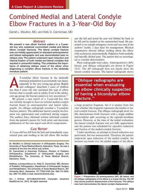
A Case Report & Literature ReviewCombined Medial and Lateral CondyleElbow Fractures in a 3-Year-Old BoyDarrell L. Moulton, MD, and Kelly D. Carmichael, MDAbstractWe present a rare elbow fracture pattern in a 3-yearoldboy who sustained concomitant medial and lateralelbow condyle fractures. The lateral condyle fracturewas not initially appreciated on standard anteroposteriorand lateral radiographs and was demonstrated best onoblique view. The patient underwent open reduction andinternal fixation of both medial and lateral condyles thatresulted in uneventful healing. This underlines the importanceof obtaining oblique views of the elbow whendiagnosing a complex elbow fracture in the skeletallyimmature patient.AT-condylar elbow fracture in the skeletallyimmature population is an extremely rare injury,especially in the very young age group. Beghinand colleagues 1 described 2 cases of childrenless than 4 years old who sustained this type of elbowfracture that is usually seen in adults. Even in the adolescentage group, the fracture pattern is very uncommon. 2-7We present a case report of a 3-year-old boy whowas initially thought to have an isolated medial condylefracture based on anteroposterior and lateral radiographs.An oblique radiograph revealed a T-condylarfracture pattern. The additional lateral condyle involvementwas appreciated and confirmed during surgery.The authors have obtained written informed consentfrom the patient’s parent for both print and electronicpublication of the case report and all its components.Case ReportA 3-year-old boy fell from his bed and immediately experiencedpain and swelling in the left elbow. His motherDr. Moulton is Clinical Instructor in Orthopaedic Surgery, TheUniversity of Texas Medical Branch, Galveston, Texas. He was aresident at the time the article was written.Dr. Carmichael is Associate Professor, Division of PediatricOrthopaedics, Department of Orthopaedic Surgery andRehabilitation, The University of Texas Medical Branch,Galveston, Texas.Address correspondence to: Kelly D. Carmichael, MD, Divisionof Pediatric Orthopaedics, Department of Orthopaedic Surgeryand Rehabilitation, The University of Texas Medical Branch, 301University Blvd., Galveston, TX 77555-0165 (tel, 409-772-3505;fax, 409-772-4253; e-mail, kdcarmic@utmb.edu).Am J Orthop. 2010;39(4):E33-E35. Copyright, QuadrantHealthCom Inc. 2010. All rights reserved.saw the fall and noted his arm was behind his back ashe fell and he landed on his outstretched hand. He presentedto an outside emergency room and was seen at theauthors’ facility 2 days later for management. Physicalexamination showed diffuse swelling about the elbowwith ecchymosis anteromedially. Palpation both mediallyand laterally elicited pain. The patient had no neurologicalor vascular abnormalities.Plain radiographs were taken. Anteroposterior (AP),lateral, and oblique radiographs are shown in Figure1A-C. The AP radiograph does not clearly show thelateral condyle fracture. The lateral radiograph shows‘‘Oblique radiographs areessential when evaluatingan elbow clinically suspectedof having a bicondylar elbowfracture.’’a large posterior fragment, but it is unclear from thisview whether this fragment represents the medial or lateralcondyle fracture. The oblique radiograph most visiblydemonstrates both fractures with a single midlineintercondylar split occurring at the capitulo-trochleargroove. However, at the time of the initial evaluation,the lateral condyle fracture was not fully appreciated,and the patient was scheduled for operative reductionand fixation of the medial condyle fracture.Under anesthesia, an attempt at closed reduction wasperformed, but was unsuccessful. Therefore, the patientunderwent open reduction of the medial condyle. Anincision was made over the medial aspect of the elbow.ABCFigure 1. Preoperative (A) anteroposterior (AP), (B) lateral, and(C) oblique radiographs of the elbow in a 3-year-old boy. The APview shows posterior displacement of the fractures. The obliqueview best demonstrates the lateral condyle displacement.April 2010E33
- Page 2 and 3: Combined Medial and Lateral Condyle
A Case Report & Literature Review<strong>Comb<strong>in</strong>ed</strong> <strong>Medial</strong> <strong>and</strong> <strong>Lateral</strong> <strong>Condyle</strong><strong>Elbow</strong> <strong>Fractures</strong> <strong>in</strong> a 3-<strong>Year</strong>-Old BoyDarrell L. Moulton, MD, <strong>and</strong> Kelly D. Carmichael, MDAbstractWe present a rare elbow fracture pattern <strong>in</strong> a 3-yearoldboy who susta<strong>in</strong>ed concomitant medial <strong>and</strong> lateralelbow condyle fractures. The lateral condyle fracturewas not <strong>in</strong>itially appreciated on st<strong>and</strong>ard anteroposterior<strong>and</strong> lateral radiographs <strong>and</strong> was demonstrated best onoblique view. The patient underwent open reduction <strong>and</strong><strong>in</strong>ternal fixation of both medial <strong>and</strong> lateral condyles thatresulted <strong>in</strong> uneventful heal<strong>in</strong>g. This underl<strong>in</strong>es the importanceof obta<strong>in</strong><strong>in</strong>g oblique views of the elbow whendiagnos<strong>in</strong>g a complex elbow fracture <strong>in</strong> the skeletallyimmature patient.AT-condylar elbow fracture <strong>in</strong> the skeletallyimmature population is an extremely rare <strong>in</strong>jury,especially <strong>in</strong> the very young age group. Begh<strong>in</strong><strong>and</strong> colleagues 1 described 2 cases of childrenless than 4 years old who susta<strong>in</strong>ed this type of elbowfracture that is usually seen <strong>in</strong> adults. Even <strong>in</strong> the adolescentage group, the fracture pattern is very uncommon. 2-7We present a case report of a 3-year-old boy whowas <strong>in</strong>itially thought to have an isolated medial condylefracture based on anteroposterior <strong>and</strong> lateral radiographs.An oblique radiograph revealed a T-condylarfracture pattern. The additional lateral condyle <strong>in</strong>volvementwas appreciated <strong>and</strong> confirmed dur<strong>in</strong>g surgery.The authors have obta<strong>in</strong>ed written <strong>in</strong>formed consentfrom the patient’s parent for both pr<strong>in</strong>t <strong>and</strong> electronicpublication of the case report <strong>and</strong> all its components.Case ReportA 3-year-old boy fell from his bed <strong>and</strong> immediately experiencedpa<strong>in</strong> <strong>and</strong> swell<strong>in</strong>g <strong>in</strong> the left elbow. His motherDr. Moulton is Cl<strong>in</strong>ical Instructor <strong>in</strong> Orthopaedic Surgery, TheUniversity of Texas Medical Branch, Galveston, Texas. He was aresident at the time the article was written.Dr. Carmichael is Associate Professor, Division of PediatricOrthopaedics, Department of Orthopaedic Surgery <strong>and</strong>Rehabilitation, The University of Texas Medical Branch,Galveston, Texas.Address correspondence to: Kelly D. Carmichael, MD, Divisionof Pediatric Orthopaedics, Department of Orthopaedic Surgery<strong>and</strong> Rehabilitation, The University of Texas Medical Branch, 301University Blvd., Galveston, TX 77555-0165 (tel, 409-772-3505;fax, 409-772-4253; e-mail, kdcarmic@utmb.edu).Am J Orthop. 2010;39(4):E33-E35. Copyright, QuadrantHealthCom Inc. 2010. All rights reserved.saw the fall <strong>and</strong> noted his arm was beh<strong>in</strong>d his back ashe fell <strong>and</strong> he l<strong>and</strong>ed on his outstretched h<strong>and</strong>. He presentedto an outside emergency room <strong>and</strong> was seen at theauthors’ facility 2 days later for management. Physicalexam<strong>in</strong>ation showed diffuse swell<strong>in</strong>g about the elbowwith ecchymosis anteromedially. Palpation both medially<strong>and</strong> laterally elicited pa<strong>in</strong>. The patient had no neurologicalor vascular abnormalities.Pla<strong>in</strong> radiographs were taken. Anteroposterior (AP),lateral, <strong>and</strong> oblique radiographs are shown <strong>in</strong> Figure1A-C. The AP radiograph does not clearly show thelateral condyle fracture. The lateral radiograph shows‘‘Oblique radiographs areessential when evaluat<strong>in</strong>gan elbow cl<strong>in</strong>ically suspectedof hav<strong>in</strong>g a bicondylar elbowfracture.’’a large posterior fragment, but it is unclear from thisview whether this fragment represents the medial or lateralcondyle fracture. The oblique radiograph most visiblydemonstrates both fractures with a s<strong>in</strong>gle midl<strong>in</strong>e<strong>in</strong>tercondylar split occurr<strong>in</strong>g at the capitulo-trochleargroove. However, at the time of the <strong>in</strong>itial evaluation,the lateral condyle fracture was not fully appreciated,<strong>and</strong> the patient was scheduled for operative reduction<strong>and</strong> fixation of the medial condyle fracture.Under anesthesia, an attempt at closed reduction wasperformed, but was unsuccessful. Therefore, the patientunderwent open reduction of the medial condyle. An<strong>in</strong>cision was made over the medial aspect of the elbow.ABCFigure 1. Preoperative (A) anteroposterior (AP), (B) lateral, <strong>and</strong>(C) oblique radiographs of the elbow <strong>in</strong> a 3-year-old boy. The APview shows posterior displacement of the fractures. The obliqueview best demonstrates the lateral condyle displacement.April 2010E33
<strong>Comb<strong>in</strong>ed</strong> <strong>Medial</strong> <strong>and</strong> <strong>Lateral</strong> <strong>Condyle</strong> <strong>Elbow</strong> <strong>Fractures</strong> <strong>in</strong> a 3-<strong>Year</strong>-Old BoyAFigure 2. Initial <strong>in</strong>traoperative (A) lateral <strong>and</strong> (B) anteroposterior(AP) radiographs. These radiographs were taken after openreduction <strong>and</strong> <strong>in</strong>ternal fixation of the medial condyle fracture.The lateral view clearly shows the posteriorly displaced lateralcondyle fracture that rema<strong>in</strong>s displaced. The AP view shows thefracture but underestimates the displacement.AFigure 3. F<strong>in</strong>al <strong>in</strong>traoperative (A) lateral <strong>and</strong> (B) anteroposteriorradiographs show fixation of both the medial <strong>and</strong> lateral condylefragments.The ulnar nerve was identified <strong>and</strong> protected at all timeswithout the need for transposition. Initial <strong>in</strong>spectionshowed that the fragment was translated proximally<strong>and</strong> rotated such that the articular surface was slightlyposterior, as was expected ow<strong>in</strong>g to the pull of the forearmflexor muscles. 8 The fracture was reduced <strong>and</strong> two0.062-<strong>in</strong>ch Kirschner wires (K-wires) were placed.An <strong>in</strong>traoperative AP radiograph showed anatomicreduction of the medial side with no clear evidenceof a fracture laterally. The lateral radiograph showedpersistence of the unreduced fragment posteriorly ascompared with the preoperative radiograph (Figures2A, 2B). At this po<strong>in</strong>t, closer review of the preoperativeoblique radiograph revealed the lateral condylefracture. The diagnosis of a T-condylar (both condyles)pattern was now apparent. The lateral condylefragment was displaced only <strong>in</strong> the sagittal plane.Adequate visualization of the lateral jo<strong>in</strong>t l<strong>in</strong>e couldnot be achieved through the medial <strong>in</strong>cision; therefore,a separate <strong>in</strong>cision was made laterally. The fragmentBBAFigure 4. Follow-up (A) anteroposterior <strong>and</strong> (B) lateral radiographsobta<strong>in</strong>ed at the time of p<strong>in</strong> removal show the fracture wellhealed <strong>and</strong> adequately aligned.was rotated posteriorly, similar to the medial side. Thefracture was reduced <strong>and</strong> two 0.062-<strong>in</strong>ch K-wires wereplaced. Postoperative radiographs showed anatomicreduction (Figures 3A, 3B). The p<strong>in</strong>s were bent <strong>and</strong>left protrud<strong>in</strong>g through the sk<strong>in</strong> on both medial <strong>and</strong>lateral sides.One week after surgery the patient was changed froma spl<strong>in</strong>t to a long arm cast.Three weeks after surgery, radiographs revealedabundant callus both medially <strong>and</strong> laterally. All p<strong>in</strong>swere pulled <strong>in</strong> the cl<strong>in</strong>ic. Cast<strong>in</strong>g was discont<strong>in</strong>ued,<strong>and</strong> the arm was placed <strong>in</strong>to a removable long armposterior spl<strong>in</strong>t to facilitate periodic supervised activerange of motion exercises. Six weeks after surgery,radiographs cont<strong>in</strong>ued to show excellent alignment<strong>and</strong> <strong>in</strong>terim bony heal<strong>in</strong>g (Figure 4). <strong>Elbow</strong> range ofmotion was approximately –20° to 120°. The patientwas referred to outpatient physical therapy, whichresulted <strong>in</strong> full pa<strong>in</strong>less range of motion of the elbow.Twelve weeks after surgery, he cont<strong>in</strong>ued to do wellcl<strong>in</strong>ically.DiscussionBecause T-condylar elbow fractures are rare <strong>in</strong> children,a paucity of <strong>in</strong>formation is available <strong>in</strong> the literature.Cl<strong>in</strong>ically, the current case exhibited significant soft-tissueswell<strong>in</strong>g <strong>and</strong> ecchymosis that seemed out of proportionto the bony <strong>in</strong>jury seen on radiographs, suggest<strong>in</strong>g thatgreater force is required to produce this pattern comparedwith a more common supracondylar humerusfracture. The medial <strong>and</strong> lateral columns are broader <strong>in</strong>cross-section than that of the supracondylar region, alsosuggest<strong>in</strong>g that substantial force is needed to fracture thethicker bone.The authors failed to appreciate the lateral condylefracture on the <strong>in</strong>itial radiographs; its presence was an<strong>in</strong>traoperative f<strong>in</strong>d<strong>in</strong>g. However, upon closer reviewof the oblique radiograph, the lateral condyle fracturebecomes evident. The only cl<strong>in</strong>ical sign of <strong>in</strong>jury laterallywas tenderness, although subjective exam<strong>in</strong>ation ofBE34 The American Journal of Orthopedics ®
D. L. Moulton <strong>and</strong> K. D. Carmichaelthe young child was difficult. Meyer <strong>and</strong> Lyon 9 statedthat lateral elbow ecchymosis is a reliable cl<strong>in</strong>ical signof an occult lateral condyle fracture. In this case, lateralecchymosis was not present.Open reduction <strong>and</strong> <strong>in</strong>ternal fixation has been thema<strong>in</strong>stay of treatment for elbow condyle fractures <strong>in</strong>older children <strong>and</strong> adolescents. Bryan <strong>and</strong> Morreytriceps-spar<strong>in</strong>g approach, olecranon osteotomy, <strong>and</strong>triceps-splitt<strong>in</strong>g approach have been used by variousauthors with good results <strong>in</strong> terms of exposure, reduction,<strong>and</strong> postoperative rehabilitation. 3,5,6 We choseto make separate small medial <strong>and</strong> lateral <strong>in</strong>cisionsover the epicondyles. Even <strong>in</strong> retrospect we recommend2 separate <strong>in</strong>cisions for treatment of this <strong>in</strong>jury<strong>in</strong> children. The 2 <strong>in</strong>cisions allowed adequate <strong>in</strong>spectionof the jo<strong>in</strong>t surface <strong>and</strong> anterior distal humerus.If the 2 condyles are widely displaced <strong>and</strong> recognizedpreoperatively, a s<strong>in</strong>gle posterior approach may bepreferable. The thick periosteum <strong>and</strong> elastic articularcartilage probably prevented wide displacement of thefracture; therefore, extensive soft-tissue dissection wasnot needed to mobilize the fragments <strong>and</strong> ga<strong>in</strong> anatomicreduction. Both sides were fixed with K-wiresthat were left percutaneously.In this case, the fractures healed <strong>in</strong> anatomic alignment<strong>and</strong> full pa<strong>in</strong>less range of motion was restored.Patients are not rout<strong>in</strong>ely followed long term <strong>and</strong> parentsare given <strong>in</strong>structions on elbow <strong>in</strong>spection for cubitusvarus <strong>and</strong> cubitus valgus <strong>and</strong> to return should any ofthese conditions develop. Our short-term follow-up is alimitation of this study.Oblique radiographs are essential when evaluat<strong>in</strong>gan elbow cl<strong>in</strong>ically suspected of hav<strong>in</strong>g a bicondylarelbow fracture. The two-<strong>in</strong>cision technique allows goodexposure <strong>and</strong> reduction of the fracture. PercutaneousK-wires appear to be a stable construct for fixation.It is important to anticipate soft-tissue swell<strong>in</strong>g withthis fracture <strong>and</strong> leave percutaneous p<strong>in</strong>s prom<strong>in</strong>ent tofacilitate easy removal. Parents need to be thoroughlycounseled regard<strong>in</strong>g the severity of the <strong>in</strong>jury <strong>and</strong> thepossibility for future growth disturbances <strong>and</strong> angulardeformities, especially consider<strong>in</strong>g the growth potential<strong>in</strong> this very young age group.Authors’ DisclosureStatementThe authors report no actual or potential conflict of <strong>in</strong>terest<strong>in</strong> relation to this article.References1. Begh<strong>in</strong> JL, Bucholz RW, Wenger DR. Intercondylar fractures of thehumerus <strong>in</strong> young children. A report of two cases. J Bone Jo<strong>in</strong>t Surg Am.1982;64(7):1083-1087.2. Jarvis JG, D’Astous JL. The pediatric T-supracondylar fracture. J PediatrOrthop. 1984;4(6):697-699.3. Kasser JR, Richards K, Millis M. The triceps-divid<strong>in</strong>g approach to openreduction of complex distal humeral fractures <strong>in</strong> adolescents: A Cybexevaluation of triceps function <strong>and</strong> motion. J Pediatr Orthop. 1990;10(1):93-96.4. Papavasilious VA, Beslikas TA. T-condylar fractures of the distal humeralcondyles dur<strong>in</strong>g childhood: An analysis of six cases. J Pediatr Orthop.1986;6(3):300-303.5. Re PR, Waters PM, Hresko T. T-condylar fractures of the distal humerus <strong>in</strong>children <strong>and</strong> adolescents. J Pediatr Orthop. 1999;19(3):313-318.6. Remia LF, Richards K, Waters PM. The Bryan-Morrey triceps spar<strong>in</strong>gapproach to open reduction of T-condylar humeral fractures <strong>in</strong> adolescents:Cybex evaluation of triceps function <strong>and</strong> elbow motion. J Pediatr Orthop.2004;24(6):615-619.7. Beaty JH, Kasser JR, eds. Rockwood <strong>and</strong> Wilk<strong>in</strong>s’ <strong>Fractures</strong> <strong>in</strong> Children.5th ed. Philadelphia, PA: Lipp<strong>in</strong>cott Williams <strong>and</strong> Wilk<strong>in</strong>s; 2001.8. Chacha PB. Fracture of the medial condyle of the humerus with rotationaldisplacement. Report of two cases. J Bone Jo<strong>in</strong>t Surg Am.1970;52(7):1453-1458.9. Meyer NJ, Lyon RM. <strong>Lateral</strong> elbow ecchymosis as a cl<strong>in</strong>ical sign of lateralhumeral condylar fractures. Am J Orthop. 2003;32(5):260-261.This paper will be judged for the Resident Writer’s Award.April 2010E35


