Introduction to the study of anatomy for students
Introduction to the study of anatomy for students
Introduction to the study of anatomy for students

You also want an ePaper? Increase the reach of your titles
YUMPU automatically turns print PDFs into web optimized ePapers that Google loves.
<strong>Introduction</strong> <strong>to</strong> <strong>the</strong> <strong>study</strong> <strong>of</strong> ana<strong>to</strong>my <strong>for</strong> <strong>students</strong> <strong>of</strong> general medicineand dentistryMiloš Grimwinter semester 2012/201/2013
Institute <strong>of</strong> Ana<strong>to</strong>my, Charles University in PragueFirst Faculty <strong>of</strong> Medicine
http://anat.lf1.cuni.cz/internet.htminput code: practice, dissecting rooms - 2012protective footwear <strong>for</strong> dissection room, medical coat, disposable medical gloves,ana<strong>to</strong>mical <strong>for</strong>ceps
Course <strong>of</strong> Ana<strong>to</strong>my (B82238, B82239, B80617, B82240) <strong>for</strong> Students <strong>of</strong>General Medicine in <strong>the</strong> first and second semester <strong>of</strong> Academic Year 2012– 2013. Subject Clinical Topographic Ana<strong>to</strong>my (B81312) is taught in <strong>the</strong>seventh semester.Course Head: Pr<strong>of</strong>. Karel Smetana, MD, DSc.Head <strong>of</strong> English-taught courses: Pavel Šnajdr, MD. Ph.D.Curricular timing: Ana<strong>to</strong>my is taught mainly in <strong>the</strong> first and second semesterscontaining following parts: lectures (120 hrs), practical classes (60 hrs), seminars(30 hrs) and two dissection blocks (<strong>to</strong>ge<strong>the</strong>r 58 hrs). Advanced course <strong>of</strong>clinically oriented ana<strong>to</strong>my is taught in <strong>the</strong> beginning <strong>of</strong> winter semester <strong>of</strong> <strong>the</strong>4th year (36 hrs). Total teaching hrs: 304Attendance <strong>to</strong> practical lessons and dissections is obliga<strong>to</strong>ry, attendance <strong>to</strong>lectures is recommended.Content: Macroscopic and microscopic ana<strong>to</strong>my <strong>of</strong> organs and organ systems,<strong>the</strong>ir development, regional ana<strong>to</strong>my with respect <strong>to</strong> functional and clinicalapplications. Neuroana<strong>to</strong>my includes both macroscopic and microscopicstructures and functional pathways <strong>of</strong> <strong>the</strong> central nervous system.The list <strong>of</strong> Recommended Textbooks follows at <strong>the</strong> end <strong>of</strong> this text.
Lectures: 4 hrs per week in each <strong>of</strong> semesters according <strong>to</strong> syllabus.First semester: Ana<strong>to</strong>mical terminology, locomo<strong>to</strong>r apparatus including limbs,basic ana<strong>to</strong>mical concept <strong>of</strong> vessels and nerves, central lymphatic organs,regional ana<strong>to</strong>my <strong>of</strong> limbs including <strong>the</strong>ir blood supply and innervation,gastrointestinal system and respira<strong>to</strong>ry system including <strong>the</strong>ir blood supply andinnervation.Second semester: Urogenital system, heart, endocrine system, central andperipheral nervous system, regional ana<strong>to</strong>my <strong>of</strong> <strong>the</strong> head and neck, sensoryorgans, skin.Attendance at lectures is recommended.Seventh semester (4th year) clinically oriented <strong>to</strong>pographical (regional)ana<strong>to</strong>my.Practical classes/seminars: 3 hrs per week in <strong>the</strong> first and secondsemesters according <strong>to</strong> syllabus. The main goal is demonstration <strong>of</strong> organs,evaluation <strong>of</strong> <strong>students</strong>´ knowledge by means <strong>of</strong> written tests and oralexaminations. Clinically relevant seminars are given by <strong>students</strong> <strong>the</strong>mselves.Attendance is obliga<strong>to</strong>ry; first semester is closed by <strong>the</strong> credit, secondsemester by <strong>the</strong> credit and final exam, seventh semester by <strong>the</strong> credit withmark.
Gross ana<strong>to</strong>my dissection courses 1, 2: Courses are organized in <strong>the</strong>afternoon during both semesters according <strong>to</strong> syllabus and take <strong>to</strong>ge<strong>the</strong>r 58hours. Attendance is obliga<strong>to</strong>ry; each dissection course is closed by <strong>the</strong> credit(oral examination, identification and description <strong>of</strong> dissected structures).The goal <strong>of</strong> dissection is <strong>to</strong> dissect and learn all structures <strong>of</strong> <strong>the</strong> body and <strong>the</strong>ir<strong>to</strong>pographical relations.During <strong>the</strong> courses <strong>students</strong> take turns in dissecting <strong>of</strong> different regions <strong>of</strong> <strong>the</strong>body.Ana<strong>to</strong>mic dissection 1 is focused on all ana<strong>to</strong>mical limb structures and trunkmuscles; ana<strong>to</strong>mic dissection 2 is focused on all ana<strong>to</strong>mical structures <strong>of</strong> head,neck, thorax, back, abdomen and pelvis.Knowledge and skills <strong>to</strong> be acquired: Theoretical and practical knowledge <strong>of</strong><strong>the</strong> macroscopic and microscopic ana<strong>to</strong>my <strong>of</strong> organs, <strong>the</strong>ir development,knowledge <strong>of</strong> <strong>to</strong>pographical relations with emphases on clinical applications,knowledge <strong>of</strong> nomenclature used <strong>to</strong> describe <strong>the</strong> human body.Eligible subjects recommended <strong>for</strong> <strong>students</strong> with deeper interest in Ana<strong>to</strong>my andmolecular medicine.New Trends in Experimental and Clinical Ana<strong>to</strong>my (B81303)
Requirements <strong>for</strong> successfully passing <strong>the</strong> Ana<strong>to</strong>my Course1) The <strong>study</strong> <strong>of</strong> Ana<strong>to</strong>my 1, Ana<strong>to</strong>mic Dissections 1, 2 are concluded by <strong>the</strong>credit, Ana<strong>to</strong>my 2 is concluded by <strong>the</strong> credit and final exam.2) Requirements <strong>for</strong> receiving <strong>the</strong> credita) obliga<strong>to</strong>ry attendance (absences must be substituted immediately aspossible)b) knowledge <strong>of</strong> <strong>the</strong> subject evaluated by successful passing <strong>of</strong> written and oraltests, activity at seminars.c) Credits: in case <strong>the</strong> credit has not been obtained at <strong>the</strong> end <strong>of</strong> a particularsemester during <strong>the</strong> last practical <strong>of</strong> <strong>the</strong> semester or at <strong>the</strong> end <strong>of</strong> dissectioncourse, <strong>the</strong> student is entitled <strong>to</strong> two re-examinations during examination period(written test - Ana<strong>to</strong>my 1, 2; oral test - Dissections)3) Prerequisites <strong>for</strong> <strong>the</strong> admission <strong>to</strong> <strong>the</strong> final exam: credit from Ana<strong>to</strong>my 1and Ana<strong>to</strong>my 2, credit from Ana<strong>to</strong>mic Dissections 1 and 2,4).The final exam is organized during summer examination period. It consists <strong>of</strong>three parts:a) written testb) practical part: dissection <strong>of</strong> selected region and demonstration <strong>of</strong> selectedorgans including <strong>the</strong>ir X-ray, MR and CT pictures
c) <strong>the</strong>oretical part based on <strong>the</strong> list <strong>of</strong> questions.Satisfac<strong>to</strong>ry result <strong>of</strong> written test is prerequisite <strong>for</strong> <strong>the</strong> admission <strong>to</strong> o<strong>the</strong>r parts <strong>of</strong><strong>the</strong> exam. The exam can be terminated at any part without even commencing <strong>the</strong>oral part and evaluates <strong>the</strong> student “failed”. This provision will not apply in case <strong>of</strong> asecond re-examination, when <strong>the</strong> exam continues even in case <strong>of</strong> unsatisfac<strong>to</strong>ryresult <strong>of</strong> <strong>the</strong> written test.Successfully written test and practical part <strong>of</strong> <strong>the</strong> final exam is not necessary <strong>to</strong>retake in case <strong>of</strong> re-examination, <strong>the</strong>y are valid during <strong>the</strong> whole exam period,however at longest <strong>for</strong> 4 months.5) Students with Individual Study Plan are recommended <strong>to</strong> discuss <strong>the</strong> extentand schedule <strong>of</strong> <strong>the</strong> subject with <strong>the</strong> Head <strong>of</strong> <strong>the</strong> Institute (Pr<strong>of</strong>. Smetana) at <strong>the</strong>beginning <strong>of</strong> particular semester.All actual in<strong>for</strong>mation is available on:<strong>of</strong>ficial board in <strong>the</strong> lobby <strong>of</strong> our Departmentwww.lf1.cuni.czhttp://anat.lf1.cuni.cz/internet.htmPrague, September 27, 2012Pr<strong>of</strong>. Karel Smetana MD., DrSc.
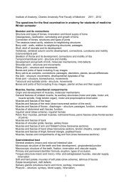
Institute <strong>of</strong> Ana<strong>to</strong>my, Charles University First Faculty <strong>of</strong> Medicine 2012 - 2013The list <strong>of</strong> questions <strong>for</strong> <strong>the</strong> final examination in microscopic and grossana<strong>to</strong>my including organogenesis <strong>for</strong> <strong>students</strong> <strong>of</strong> general medicine.Each question covers both microscopic and macroscopic aspects <strong>of</strong> organstructure, its syn<strong>to</strong>py, development and <strong>the</strong> most frequent birth defectsSkele<strong>to</strong>n and its connectionsStructure and types <strong>of</strong> bones, innervation and blood supply <strong>of</strong> boneOsteogenesis, ossification, remodeling and growth <strong>of</strong> boneConnection <strong>of</strong> bones, structure and types <strong>of</strong> jointsThe osseous nasal cavity, relations <strong>to</strong> neighboring structuresBony orbit - walls, relation <strong>to</strong> neighboring structures, passagesSkull, skull <strong>of</strong> neonate and its developmentVertebrae, vertebral column and its development, connections, curvatures andmotilityCraniovertebral jointSkele<strong>to</strong>n <strong>of</strong> thorax and its development, connections and motility <strong>of</strong> ribsTemporomandibular joint - structure and motilityDevelopment and growth <strong>of</strong> limb, molecular mechanisms, limb defectsShoulder joint – structure and movementsElbow joint – structure and movementsBones and joints <strong>of</strong> hand including reading <strong>of</strong> X-ray images
Recommended TextbooksAna<strong>to</strong>myPlatzer: Color Atlas Ana<strong>to</strong>my – Vol.1 Locomo<strong>to</strong>r System, Thieme 2008, 6 th ed.Fritsch, Kuehnel Color Atlas Ana<strong>to</strong>my – Vol.2 Internal Organ, Thieme 2008, 5th ed.Kahle, Frotscher: Color Atlas Ana<strong>to</strong>my – Vol.3 Nervous System and SensoryOrgans, Thieme 2010, 6th editionor Snell: Clinical Ana<strong>to</strong>my by systems, Lippincott Williams and Wilkins 2007His<strong>to</strong>logyMescher: Junqueira's Basic His<strong>to</strong>logy: Text and Atlas, McGraw-Hill Medical 2009,12 th ed.EmbryologySadler: Langman's Medical Embryology, Lippincott Williams +Wilkins 2009, 11 th ed.AtlasesSobotta: Atlas <strong>of</strong> Human Ana<strong>to</strong>my, Churchill Livings<strong>to</strong>ne 2011, 15th ed.or Netter: Atlas <strong>of</strong> Human Ana<strong>to</strong>my, Saunders 2010, 5th editionor Agur, Dalley: Grant´s Atlas <strong>of</strong> Ana<strong>to</strong>my, Lippincot Williams and Wilkins 2008,12 th ed.or Gilroy, MacPherson, Ross, Schuenke, Schultze, Schumacher: Atlas <strong>of</strong> Ana<strong>to</strong>my,Thieme 2008, 1st editionor Köpf-Maier: Wolf-Heidegger’s Atlas <strong>of</strong> Human Ana<strong>to</strong>my Vol.1+2, Karger 2005,6 th ed.
Dissection manualGrant's Dissec<strong>to</strong>r, Lippincot Williams and Wilkins 2008, 14th ed.Complementary textbooks and AtlasesDauber: Pocket Atlas <strong>of</strong> Human Ana<strong>to</strong>my, Founded by Heinz Feneis, Thieme 2007Crossman: Neuroana<strong>to</strong>my, An Illustrated Colour Text, Churchill Livings<strong>to</strong>ne 2010, 4 <strong>the</strong>d.Fitzgerald MJT, Gruener G: Clinical neuroana<strong>to</strong>my and neuroscience, Elsevier,2012, 6 th ed.Kierszenbaum, His<strong>to</strong>logy and Cell Biology: An <strong>Introduction</strong> <strong>to</strong> Pathology, Mosby2012, 3rd ed.Moore, Agur, Dalley: Essential Clinical Ana<strong>to</strong>my, Lippincot Williams and Wilkins2010, 4 th ed.Petrovický: Basic Neuroana<strong>to</strong>my I. and II. Praha, Karolinum 1997Seichert: Little Ana<strong>to</strong>mical Atlas. Praha 1995 Prague,February, 2012, Pr<strong>of</strong>. Karel Smetana MD., DrSc.
Ana<strong>to</strong>my 1 (B80597) <strong>for</strong> <strong>students</strong> <strong>of</strong> general medicineSyllabus <strong>of</strong> lectures in winter semester 2012/2013
Objectives: <strong>to</strong> show importance <strong>of</strong> <strong>the</strong> ana<strong>to</strong>my in practice. Seminars are focused on <strong>the</strong>presentation <strong>of</strong> <strong>the</strong> ana<strong>to</strong>mic background applied on selected clinical cases. (Diagnosisand treatment methods are not object <strong>of</strong> <strong>the</strong>se presentations). It is recommended <strong>to</strong> discussseminar lecture with course teacher.Form: spoken lecture per<strong>for</strong>mance (10 min maximum). Method: diagrams drawn on <strong>the</strong>board, power-point projection, back projection, videoprojection, practical demonstration <strong>of</strong> <strong>the</strong>specimens, X-ray pictures. Active participation in <strong>the</strong> seminars is one <strong>of</strong> <strong>the</strong> aspects <strong>to</strong>grant a semester credit.
The building <strong>of</strong> <strong>the</strong> Institute <strong>of</strong> Ana<strong>to</strong>my <strong>of</strong> <strong>the</strong> Charles – FerdinandUniversity from 1874 – 7
Extension <strong>of</strong> <strong>the</strong> building in 1924 - 5 (3rd floor)
1929
pitevny
27. 5. 2010
To <strong>the</strong> memory <strong>of</strong> those who donated <strong>the</strong>ir bodies<strong>for</strong> <strong>the</strong> education <strong>of</strong> medical <strong>students</strong> in ana<strong>to</strong>my. .
Vesalius, Andreas (1514-1564) : De humani corporis fabrica libriseptem (Jan Stephanus Calcar), Basel: Joannes Oporinus, 1543http://www.nlm.nih.gov/exhibition/his<strong>to</strong>ricalana<strong>to</strong>mies/browse.html
J. Jessenius 1566 - 1621
Christian Sebastian a Zeidlern (1620 ? – 1689): Soma<strong>to</strong><strong>to</strong>pia anthropologica. Praha 1686
Christian Sebastian a Zeidlern(1620 ? – 1689) Johann Georg Ilg (1771 – 1836)
W. Staněk created Czechana<strong>to</strong>mical terminology
Atlas <strong>of</strong> ana<strong>to</strong>mical dissectionWáclav Staněk (1804 – 1871), Prague 1840
Pr<strong>of</strong>. Dr. Vaclav Steffal,<strong>the</strong> first head <strong>of</strong> The Institute<strong>of</strong> Ana<strong>to</strong>my <strong>of</strong> <strong>the</strong> CzechMedical Faculty1883 - 1894
J. Janošík (1894 - 1926) K. Weigner (1826 - 1937) L. Borovanský (1838 – 1970)
Terminology used in descriptive ana<strong>to</strong>myVerticalHorizontalMedianCoronalSagittalRightLeftIntermediateMedialLateralAnteriorPosteriorVentralDorsalFrontalOccipitalSuperiorInferiorCranialCaudalRostralApicalBasalBasilarMiddleTransverseTransverseLongitudinalAxialExternalInternalLuminalSuperficialDeepProximalDistalCentralPeripheralRadialUlnarFibular;PeronealTibialPalmar;VolarPlantarFlexorExtensor
Planes and directions
General ana<strong>to</strong>myGeneral TermsPrincipal Planes,Principal Axis,Directions in Space,Direction <strong>of</strong> MovementsParts <strong>of</strong> <strong>the</strong> BodyFrontal planesCoronal planesMedian planeSagittal planesTransverse planes
What is Ana<strong>to</strong>my:Dissection or <strong>the</strong> separation <strong>of</strong> <strong>the</strong> body in<strong>to</strong> its parts. However, ït is not sufficient <strong>to</strong>name <strong>the</strong> parts. Contemporary ana<strong>to</strong>my integrates <strong>the</strong> normal structure with <strong>the</strong>normal function <strong>for</strong> better understanding <strong>of</strong> human body. It serves <strong>the</strong> needs <strong>of</strong>surgeon and physicians and contributes <strong>to</strong> development <strong>of</strong> new diagnostic and<strong>the</strong>rapeutic methods. Much <strong>of</strong> adult ana<strong>to</strong>my can only be unders<strong>to</strong>od by know itsprenatal his<strong>to</strong>ry.
Research activitiesResearch is focused on cell and developmental biology, tissue engineering,experimental morphology, experimental medicine, neurosciences and clinicalana<strong>to</strong>my.More specifically, research is focused on genes influencing limb and musculaturepatterning, on <strong>the</strong> epi<strong>the</strong>lial-mesenchymal transition in <strong>the</strong> neural crest, on isolationand characterization <strong>of</strong> neural crest stem cells, on <strong>the</strong> influence <strong>of</strong> hypoxia ondeveloping vessels and heart.Fur<strong>the</strong>r research studies <strong>the</strong> phenotype including glycophenotype. The epidermalstem cells as well as <strong>the</strong>ir epi<strong>the</strong>lial-mesenchymal interactions are investigatedunder normal conditions, during wound healing and in cancer.The development <strong>of</strong> <strong>the</strong> heart including <strong>the</strong> pathogenesis <strong>of</strong> embryonic heart failureis studied under both normal and altered hemodynamic conditions.On <strong>the</strong> clinical side we are focused on development <strong>of</strong> ligamen<strong>to</strong>us apparatus andits relation <strong>to</strong> growing skele<strong>to</strong>n.Neuroscience <strong>to</strong>pic deals with volumetry <strong>of</strong> basal ganglia in normal ana<strong>to</strong>micalpreparations and in MRI <strong>of</strong> patient with Parkinson disease, Alzheimer disease andcarotid artery occlusion. Especially amygdala is in <strong>the</strong> focus <strong>of</strong> attention
GROWTHAllometric growth in humansPostnatal growth <strong>of</strong> <strong>the</strong> skull
Adult male skele<strong>to</strong>n:anterior and posterior view
Ossification,classification <strong>of</strong> bones(long, short, flat irregular,pneumatized)Ossification <strong>of</strong> a long boneDiaphysis <strong>of</strong> <strong>the</strong> femurEndosteum, Periosteum
Fibrous joints (sutures, gomphosis)Synovial joint (hip joint)
Skele<strong>to</strong>n<strong>of</strong> neonatalinfantSection <strong>of</strong> a fetal hand:primary ossification<strong>of</strong> cartilaginous models
Radiograph <strong>of</strong> <strong>the</strong> right hand at 11 years(female). Epiphyses are separated fromdiaphyses by epiphyseal growth plate.The hand´s skele<strong>to</strong>n
Pelvis - its joints and ligaments
Anteroposterior radiograph <strong>of</strong> adult female pelvis
Dorsal aspect <strong>of</strong> bones <strong>of</strong> <strong>the</strong> right foot; transverse section <strong>to</strong> show joints
Lateral views <strong>of</strong> head and neck <strong>to</strong> show surface ana<strong>to</strong>my,bones <strong>of</strong> <strong>the</strong> skull, facial musculature, parotid gland, branches<strong>of</strong> <strong>the</strong> facial nerveand branches<strong>of</strong> <strong>the</strong> carotid artery
Sagittal section <strong>of</strong> <strong>the</strong> skull; ana<strong>to</strong>mical illustration <strong>of</strong> individual bones
Lateral radiograph <strong>of</strong> adult skull
Frontal views <strong>of</strong> head <strong>to</strong> show surface ana<strong>to</strong>my,facial musculature and bones <strong>of</strong> <strong>the</strong> skull
How <strong>to</strong> illustrate <strong>the</strong> ana<strong>to</strong>mical structures
Frontal views <strong>of</strong> trunk <strong>to</strong> show surface ana<strong>to</strong>my and skele<strong>to</strong>n
Frontal views <strong>of</strong> trunk <strong>to</strong> show musculatureand projection <strong>of</strong> some thoracic and abdominal organs
Projections <strong>of</strong> organs on <strong>the</strong> anterior body wall
Different types <strong>of</strong> ana<strong>to</strong>mic illustrationsDuodenum, pancreas, aorta, bile duct, inferior vena cava
plexus lumbalis, plexus sacralis
Stavba bronchů, septuminteralveolaretunica mucosa,tunica fibromusculocartilagineatunica adventitia (peribronchium)
Syn<strong>to</strong>pie pars cervicalis tracheae
Female genital systemInternal genital organsOvary, Uterine tubeSalpinx), Uterus (Metra,Hystera), VaginaExternal genital organsPudendum (vulva)Mons pubisLabium majusLabium minusPudendal cleftLabium minusVestibuleBulb <strong>of</strong> vestibuleCli<strong>to</strong>ris
Magneticresonanceimage <strong>of</strong> female pelvis in sagittal plane
Illustration <strong>of</strong> distribution <strong>of</strong> mo<strong>to</strong>neurons innervating individualmuscle groups
Catani a,M.et al. :Virtual inVivoInteractiveDissection<strong>of</strong> WhiteMatterFasciculiin <strong>the</strong>HumanBrainNeuroImage17, 77–94(2002)Capsula interna
Pr<strong>of</strong>esors EmeritiMUDr. Radomír Čihák, DrSc.MUDr. Václav Seichert, DrSc.Pr<strong>of</strong>esorsMUDr. Jan Bar<strong>to</strong>níček, CSc.MUDr. Rastislav Druga, DrSc.MUDr. Oldřich Eliška, DrSc.MUDr. Miloš Grim, DrSc.Dr. Med. Zdenek HalataMUDr. Pavel Petrovický, DrSc.MUDr. Karel Smetana ml., DrSc. headAssoc. Pr<strong>of</strong>essorsMUDr. Ondřej Naňka, Ph.D.MUDr. David Sedmera, D.Sc.Assist. Pr<strong>of</strong>essorsMUDr. Rastislav HromádkaMUDr. Martin Chovanec, Ph.D.MUDr. Ivo Klepáček, CSc.MUDr. Jiří Klempíř, Ph.D.RNDr. Hana Kolesová, Ph.D.Ing. Eliška Krejčí, Ph. D.MUDr. Lukáš Lacina, Ph.D.MUDr. Veronika Němcová, CSc.MUDr. Pavel Šnajdr, Ph.D.Assistants, Ph.D. StudentsMUDr. Jiří Beneš (PhDs)MUDr. Jana DudováMUDr. Zdeněk Fík (PhDS)Dr. Ayesha HaqueMUDr. Ondřej Kodet (PhDS)MUDr. Zdeňka NovákováMUDr. Živorad Peševski (PhDS)MUDr. David Stehlík (PhDS)Mgr. Pavol SzaboMgr. Barbora Šaňková (PhDS)ScientistsMUDr. Zdeněk Čada, Ph.D.RNDr. Barbora Dvořánková, Ph.D.Ivan Helekal, akademický malířMgr. Jan KacvinskýMgr. Alena KvasilováMgr. Markéta PleschnerováMUDr. Petr Valášek, Ph.D.Lecturers – <strong>students</strong> <strong>of</strong> 1. LF27 studentů