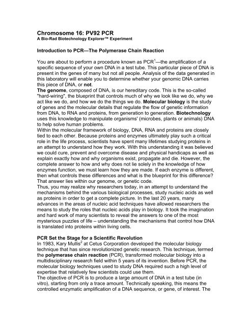
Chromosome 16: PV92 PCR
Chromosome 16: PV92 PCR
Chromosome 16: PV92 PCR

You also want an ePaper? Increase the reach of your titles
YUMPU automatically turns print PDFs into web optimized ePapers that Google loves.
<strong>Chromosome</strong> <strong>16</strong>: <strong>PV92</strong> <strong>PCR</strong>A Bio-Rad Biotechnology Explorer ExperimentIntroduction to <strong>PCR</strong>—The Polymerase Chain ReactionYou are about to perform a procedure known as <strong>PCR</strong> 1 —the amplification of aspecific sequence of your own DNA in a test tube. This particular piece of DNA ispresent in the genes of many but not all people. Analysis of the data generated inthis laboratory will enable you to determine whether your genomic DNA carriesthis piece of DNA, or not.The genome, composed of DNA, is our hereditary code. This is the so-called"hard-wiring", the blueprint that controls much of why we look like we do, why weact like we do, and how we do the things we do. Molecular biology is the studyof genes and the molecular details that regulate the flow of genetic informationfrom DNA, to RNA and proteins, from generation to generation. Biotechnologyuses this knowledge to manipulate organisms' (microbes, plants or animals) DNAto help solve human problems.Within the molecular framework of biology, DNA, RNA and proteins are closelytied to each other. Because proteins and enzymes ultimately play such a criticalrole in the life process, scientists have spent many lifetimes studying proteins inan attempt to understand how they work. With this understanding it was believedwe could cure, prevent and overcome disease and physical handicaps as well asexplain exactly how and why organisms exist, propagate and die. However, thecomplete answer to how and why does not lie solely in the knowledge of howenzymes function, we must learn how they are made. If each enzyme is different,then what controls these differences and what is the blueprint for this difference?That answer lies within our genome, or genetic code.Thus, you may realize why researchers today, in an attempt to understand themechanisms behind the various biological processes, study nucleic acids as wellas proteins in order to get a complete picture. In the last 20 years, manyadvances in the areas of nucleic acid techniques have allowed researchers themeans to study the roles that nucleic acids play in biology. It took the imaginationand hard work of many scientists to reveal the answers to one of the mostmysterious puzzles of life – understanding the mechanisms that control how DNAis translated into proteins within living cells.<strong>PCR</strong> Set the Stage for a Scientific RevolutionIn 1983, Kary Mullis 2 at Cetus Corporation developed the molecular biologytechnique that has since revolutionized genetic research. This technique, termedthe polymerase chain reaction (<strong>PCR</strong>), transformed molecular biology into amultidisciplinary research field within 5 years of its invention. Before <strong>PCR</strong>, themolecular biology techniques used to study DNA required such a high level ofexpertise that relatively few scientists could use them.The objective of <strong>PCR</strong> is to produce a large amount of DNA in a test tube (invitro), starting from only a trace amount. Technically speaking, this means thecontrolled enzymatic amplification of a DNA sequence, or gene, of interest. The
template strands can be any form of double-stranded DNA such as genomicDNA. A researcher can take trace amounts of genomic DNA from a drop ofblood, a single hair follicle or cheek cell (in theory, only a single template strandis needed to copy and generate millions of new identical DNA molecules) andmake enough to study. Prior to <strong>PCR</strong>, this would have been impossible. It is theability to amplify the precise sequence of DNA of interest that is the true power of<strong>PCR</strong>.<strong>PCR</strong> has made an impact on four main areas of genetic research: gene mapping,cloning, DNA sequencing and gene detection. <strong>PCR</strong> is now used as a medicaldiagnostic tool to detect specific mutations that may cause genetic disease 3 , isused in criminal investigations and courts of law to identify suspects on themolecular level 4 , and has been a powerful tool in the sequencing of the humangenome 5 . Prior to <strong>PCR</strong> the use of molecular biology techniques for therapeutic,forensic, pharmaceutical, agricultural or medical diagnostic purposes was notpractical nor cost effective. The development of <strong>PCR</strong> technology transformedmolecular biology from a difficult science to one of the most accessible andwidely used disciplines of biotechnology.Now, let’s extract some of your own DNA.Procedure for DNA Extraction and Template PreparationTo obtain DNA for use in the polymerase chain reaction you will extract the DNAfrom your own living cells. It is interesting to note that DNA can be also extractedfrom mummies and fossilized dinosaur bones. In this lab activity, you will beisolating DNA from epithelial cells that line the inside of your cheek . This isaccomplished by using a sterile pipet tip to gently scrape the inside of both yourcheeks about 10 times each to scoop up the cells lining the surface. You will thenboil the cells to rupture them and release the DNA they contain.To obtain pure DNA for <strong>PCR</strong> you will use the following procedure.The cheek cells in the pipet tip are transferred into a micro test tube containing200 µl of InstaGene matrix. This particulate matrix is made up of negativelycharged microscopic beads that "chelate", or grab metal ions out of solution. Itacts to trap metal ions, such as Mg2+, which are required as catalysts orcofactors in enzymatic reactions. Your cheek cells will then be lysed or rupturedby heating to release all of their cellular constituents, including enzymes thatwere once contained in the cheek cell lysosomes. Lysosomes are sacs within thecells cytoplasm that contain powerful enzymes, such as DNases, which are usedby cells to digest the DNA of invading viruses. When you rupture the cells, theseDNases can digest the released DNA of interest. However, when the cells arelysed in the presence of the chelating beads, the cofactors are adsorbed and arenot available to the enzymes. This virtually blocks all enzyme degradation of theextracted DNA and results in a population of intact genomic DNA molecules thatwill be used as the template in your <strong>PCR</strong> reaction.Your isolated cheek cells are first suspended in the InstaGene matrix andincubated at 56 °C for 10 minutes. This "preincubation" step helps to soften theplasma membranes and release clumps of cells from each other. The increased
temperature also acts to inactivate enzymes such as DNases, which will degradethe DNA template. After this 10 minute incubation period, the cells are thenplaced into a boiling (100°C) water bath for 6 minutes.The boiling ruptures the cells and releases the DNA from the cell nucleus. Yourextracted genomic DNA will then be used as the target template for <strong>PCR</strong>amplification.DNA Extraction and Template PreparationWorkstation ChecklistYour workstation. Materials and supplies that should be present at yourworkstation prior to beginning this lab are listed below.Student workstations Number (√)Screwcap tubes with InstaGene matrix 8 Styrofoam microtube rack 2 P-20 micropipet 1 P-200 micropipet 1 Pipet tips (filter type), 20–200 µl 1 box Lab marker 1 Waste container 1 Copy of Quick Guide or protocol 1 Instructor's (common) workstation. A list of materials, supplies, andequipment that should be present at a common location to be accessed by yourteam is also listed below.P-200 micropipet 1–8 Pipet tips (filter type), 20–200 µl 1 box Water baths (56 and 100°C) 2 Centrifuge 1 Lab Protocol1. Each member of your team should have two screwcap tubes, each containing200 µl of InstaGene matrix. Label the tube on the cap and on the side with yourinitials. In addition, label the tubes as "tube 1" and "tube 2". Each person shouldwash his or her hands before beginning step 2.2. Using a sterile 20–200 µl filter pipet tip, gently scrape the inside of both cheeks10 times each with the tip. This is most easily done by pinching and extendingthe corner of your mouth with one hand, and scraping the cheek with the tip inthe other hand. Use firm, but gentle pressure. The goal is to remove the surfacelayer of epithelial cells from your cheek lining. You should see a small volume ofwhite cells in the pipet tip.
3. Place the tip that contains your cheek cells into the screwcap tube labeled as"tube 1".4. Using a second sterile 20–200 µl filter pipet tip, gently scrape the inside of bothcheeks 10 times each with the tip. Place the tip that contains your cheek cellsinto your screwcap tube labeled as "tube 2".5. Place the tips on the end of a P-200 micropipet that is set on a 100 µl setting.Pipet up and down 5 times into the InstaGene matrix – the action of pipetting upand down mixes and transfers your cheek cells into the InstaGene matrix.6. Screw the lid tightly on your tubes and place in the foam microtube holder.When all members of your team have collected their samples, float the rack andtubes in a 56°C water bath for 10 min. At the halfway point (5 minutes), remix thecontents of the tubes by shaking or vortexing several times and place back in thewater bath for the remaining 5 minutes.7. Remove the tubes from the water bath and remix by shaking the tubes severaltimes.Now float the rack of tubes in a 100°C water bath for 6 minutes.8. After 6 minutes, remove the tubes from the 100°C water bath and shake orvortex gently several times to resuspend the sample. Place the eight tubes in abalanced arrangement in the centrifuge. Pellet the matrix by spinning for 5minutes at 6,000 x g (or 10 minutes at 2,000 x g) in the centrifuge.
9. Using a 200 µl pipet tip, remove 170 µl of the supernatant from one of yourscrewcap tubes and transfer the supernatant into the other. You will now be leftwith one screwcap tube that contains your isolated genomic DNA.<strong>PCR</strong> AmplificationIt is estimated that there are 30,000–50,000 individual genes in the humangenome.The true power of <strong>PCR</strong> is the ability to target and amplify a specific piece of DNA(or gene) out of a complete genome.The recipe for a <strong>PCR</strong> amplification of DNA requires a simple mixture ofingredients.To replicate a given piece of DNA, the reaction mixture requires the followingcomponents.1. Intact DNA template—containing the sequence of DNA to be amplified.2. Individual deoxynucleotide bases (A, T, G and C)—raw material of DNA.3. DNA polymerase—an enzyme that assembles the nucleotides into a new DNAchain.4. Magnesium ions—a cofactor (catalyst) required by DNA polymerase to createthe DNA chain.5. Oligonucleotide primers—pieces of DNA complementary to the template thattell DNA polymerase exactly where to start making copies.6. Salt buffer—provides the optimum ionic environment and pH for the <strong>PCR</strong>reaction.The template DNA in this exercise is genomic DNA that was extracted from yourcheek cells. When all the other components are combined under the rightconditions a copy of the original double stranded template DNA molecule ismade—doubling the number of template strands. Each time this cycle isrepeated, copies are made from copies and the number of template strandsdoubles—from 2 to 4 to 8 to <strong>16</strong> and so on—until after 20 cycles there are1,048,554 exact copies of the target sequence.<strong>PCR</strong> makes use of the same basic processes that cells use to duplicatetheir DNA.1. Complementary DNA strand hybridization
2. DNA strand synthesis via DNA polymeraseThe two DNA primers provided in this kit are designed to flank, or bracket, a DNAsequence within your genome and thus provide the exact start signal for the DNApolymerase to "zero-in on" and begin synthesizing (replicating) copies of thattarget DNA.Complementary strand hybridization takes place when the two different primersanneal, or hybridize, to each of their respective complementary base pairsequences on the template DNA molecule.The primers are two short single-stranded DNA molecules (23 bases long), onethat is complementary to a portion of the 5'–3' strand, and another that iscomplementary to the portion of the 3'–5' strand of your DNA. These primershybridize to the separated template strands and serve as starting points for DNAamplification.The enzyme, called Taq DNA polymerase, extends the annealed primers'sequence by "reading" the strand and synthesizing the complementarysequence. In this way, Taq replicates the template DNA strand(s). Thispolymerase has been isolated from a heat stable bacterium(Thermus aquaticus) which in nature lives within the steam vents in theYellowstone National Park. 6 For this reason the enzymes within these bacteriahave evolved to withstand high temperatures (94°C) also used in each cycle ofthe <strong>PCR</strong> reaction.<strong>PCR</strong>—Step by Step<strong>PCR</strong> amplification includes three main steps, a denaturation step, an annealingstep and an extension step (these steps are summarized in Figure 1). Duringdenaturation, the reaction mixture is heated to 94°C for 1 minute, which results inthe melting out or separation of the double-stranded DNA template into twosingle stranded molecules. In <strong>PCR</strong> amplification, DNA templates must beseparated before the polymerase can generate a new copy. The harshtemperature required to melt the DNA strands normally would destroy the activityof most enzymes, but because Taq polymerase has been isolated from bacteriathat thrive in the high temperatures of hot springs, it remains active.
Fig. 1. A complete cycle of <strong>PCR</strong>.During the annealing step, the oligonucleotide primers "anneal to" or find theircomplementary sequences on the two single-stranded template strands of DNA.In these annealed positions, they can act as "primers" for Taq DNA polymerase.They are called primers because they "prime" the single stranded DNAmolecules by providing a short sequence of double stranded DNA for Taqpolymerase to extend and build a new complementary strand. Binding of theprimers to their template sequences is also highly dependent on temperature. Inthis lab exercise, a 60°C annealing temperature is optimum for primer binding.During the extension step, the job of Taq DNA polymerase is to add nucleotides(A, T, G or C) one at a time to the primer to create a complementary copy of theDNA template. During polymerization, the reaction temperature is increased to72°C, the temperature that produces optimal Taq polymerase activity. The threesteps of denaturation, annealing, and extension, combine to form one "cycle" of<strong>PCR</strong>, and a complete <strong>PCR</strong> amplification undergoes 40 cycles.The entire 40 cycle reaction is carried out in a test tube which is placed into thethermal cycler (also called a Gene Cycler). The Gene Cycler contains analuminum block that holds the samples and can be rapidly heated and cooledacross extreme temperature differences.The rapid heating and cooling of this thermal block is defined as "temperaturecycling" or "thermal cycling".
<strong>PCR</strong> AmplificationWorkstation ChecklistYour workstation. Materials and supplies that should be present at yourworkstation prior to beginning this lab are listed below.Student workstations Number (√)<strong>PCR</strong> tubes 4 Microtubes, capless 4 Master mix 1 P-20 micropipet 1 Pipet tips (filter type), 2–20 µl 8 tips Foam microtube rack 2 Lab marker 1 Ice bucket with ice 1 Waste container 1 Copy of Quick Guide or protocol 1 Instructor's (common) workstationCentrifuge 1 Gene Cycler or MyCycler 1 Lab Protocol1. Centrifuge your tubes for 2 minutes at 6,000 x g or for 5 minutes at 2,000 x gin a centrifuge.2. Each member of the team should obtain a <strong>PCR</strong> tube and capless microtubeadaptor.Label each tube with your initials and place the <strong>PCR</strong> tube into the caplessmicrotube (which acts as a convenient holder for the small <strong>PCR</strong> tube). Place the<strong>PCR</strong> tube in its holder in the foam rack.3. Transfer 20 µl of your DNA template from your screwcap tube into the bottomof the <strong>PCR</strong> tube. Do not transfer any of your matrix beads into the <strong>PCR</strong>reaction—the reaction will be inhibited.4. Locate the tube of yellow <strong>PCR</strong> master mix (labeled "Master") in your icebucket.Transfer 20 µl of the master mix into your <strong>PCR</strong> tube. Mix by pipetting up anddown 2–3 times.
5. Remove your <strong>PCR</strong> tube from its holder and place the tube in the Gene Cycler.6. When all of the <strong>PCR</strong> samples are in the Gene Cycler, the teacher will beginthe <strong>PCR</strong> reaction. The reaction will undergo 40 cycles of amplification, which willtake approximately 3 hours.2 nd <strong>PV92</strong> <strong>PCR</strong> Lab Period: Analyzing Your DNA Using Gel ElectrophoresisWhat Are You Looking At?Before you analyze your <strong>PCR</strong> products, it is important to review the details of thetarget sequence being explored.What Can Genes and DNA Tell Us?It is estimated that the 23 pairs, or 46 chromosomes, of the human genome(23 chromosomes come from the mother and the other 23 come from the father)contain approximately 30,000–50,000 genes. Each chromosome contains aseries of specific genes. The larger chromosomes contain more DNA, andtherefore more genes, compared to the smaller chromosomes. Each one of thehomologous chromosomes (pairs) contains similar genes.Each gene holds the code for a particular protein. Interestingly, the 30,000–50,000 genes only comprise 5% of the total chromosomal DNA. The other 95% isnon-coding DNA. This non-coding DNA is interspersed in blocks betweenfunctional segments of genes and within genes, splitting them into segments.The exact function of the non-coding (intergenic) DNA is not yet known, althoughit is thought that non-coding DNA allows for the accumulation of mutations andvariations within organisms.When DNA is first transcribed into RNA, exons are separated from each other byintrons. While the RNA is still in the nucleus intergenic sequences, or introns (in= stay within the nucleus), are removed from the RNA while the exons (ex = exitthe nucleus) are spliced together to form the complete messenger RNA codingsequence for the protein (see Figure 2).This process is called RNA splicing and is carried out by specializedenzymes called spliceosomes.
Fig. 2. Splicing of introns from genes.Surprisingly, introns often vary in their size and sequence among individualswhile exons do not. This variation is thought to be the result of the differentialaccumulation of mutations in DNA throughout evolution. These mutations aresilently passed on to our descendants. We do not notice mutations in our noncodingDNA because they do not affect our phenotypes. However, thesedifferences in our DNA represent the molecular basis of DNA fingerprinting usedin human identification and studies in population genetics.The Target Sequence–Can You Say "Alu"?The genome contains small repetitive DNA elements that have become randomlyinserted into the human genome over millions of years. One such repetitiveelement is called the "Alu sequence" 7 (see Figure 3). This is a DNA sequenceabout 300 base pairs long that is repeated, one copy at a time, almost 500,000times throughout the human genome 8 . The origin and function of these randomlyrepeated sequences is not yet known.Fig. 3. Location of an Alu repetitive element within an intron.Some of these Alu sequences have characteristics that make them very useful togeneticists. When present within introns of certain genes, they can either beassociated with a disease or merely used to estimate relatedness amongindividuals. In this exercise, analysis of a single Alu repeat is used to estimate itsfrequency in the population and as a simple measure of molecular geneticvariation—with no reference to disease or relatedness among individuals.In this lab you will be hunting for an Alu element in the <strong>PV92</strong> region ofchromosome <strong>16</strong>. This particular Alu element is dimorphic, meaning that theelement is present in some individuals and not others. Some people have theinsert in one copy of their <strong>16</strong> th chromosomes (one allele), others may have theinsert in both copies of chromosome <strong>16</strong> (two alleles), while some may not have
the insert on either copy of the chromosome (see Figure 4). The presence orabsence of this insert can be detected using the polymerase chain reactionfollowed by agarose gel electrophoresis.Since you are amplifying a region of DNA contained within an intron, the regionof DNA is never really used in your body. So if you don’t have it, don’t worry.The primers in this kit are designed to bracket the region within the <strong>PV92</strong> regionthat is 641 base pairs in length if the intron does not contain the Alu insertion or941 base pairs in length if Alu is present. This increase in size is due to the 300base pair sequence contributed by the Alu insert. When your <strong>PCR</strong> products areelectrophoresed on an agarose gel, there are three distinct outcomes that can bevisualized in the gel.If both chromosomes contain Alu inserts, then each amplified <strong>PCR</strong> product willbe 941 base pairs long. On a gel they will migrate at the same speed so there willbe one band that corresponds to 941 base pairs. If neither chromosome containsthe insert then each amplified <strong>PCR</strong> product will be 641 base pairs and they willmigrate as one band that corresponds to 641 base pairs. If you have an Aluinsert on one chromosome but not the other, then there will be one <strong>PCR</strong> productof 641 base pairs and one of 941 base pairs. The resulting gel will reveal twobands.Fig. 4. The presence or absence of the Alu insert within the <strong>PV92</strong> region ofchromosome <strong>16</strong>.Electrophoresis separates DNA fragments according to their relative size(molecular weight). DNA fragments are loaded into an agarose gel slab, which isplaced into a chamber filled with a conductive liquid buffer solution. A directcurrent is passed between wire electrodes at each end of the chamber. DNAfragments are negatively charged, and when placed in an electric field will bedrawn toward the positive pole and repelled by the negative pole. The matrix ofthe agarose gel acts as a molecular sieve through which smaller DNA fragments
can move more easily than larger ones. Over a period of time smaller fragmentswill travel farther than larger ones. Fragments of the same size stay together andmigrate in what appear as single "bands" of DNA in the gel. In the sample gelbelow (Fig. 5), <strong>PCR</strong> amplified bands of 941 bp and 641 bp are separated basedupon their sizeFig. 5. Separation of DNA bands based on size. This gel depicts theelectrophoretic separation of the EZ Load DNA molecular mass ruler, whichcontains 1,000 bp, 700 bp, 500 bp, 200 bp and 100 bp fragments (lane 1), twohomozygous (+/+) 941 bp fragments (lanes 2 and 6), three homozygous (-/-)641 bp fragments (lanes 3, 5, and 8), and two heterozygous (+/-) 941/641 bpfragments (lanes 4 and 7).
Analyzing Your DNA Using Gel ElectrophoresisWorkstation ChecklistYour workstation. Materials and supplies that should be present at yourworkstation prior to beginning this lab are listed below.Student workstations Number/Station (√)Agarose gel 1 Student <strong>PCR</strong> samples 1/student MMR-DNA standard 1 LD loading dye 1 P-20 micropipet 1 Pipet tips (filter type), 2–20 µl 11 tips Lab marker 1 Styrofoam microtube rack 1 Gel box and power supply 1 Gel staining tray 1 Waste container 1 Copy of Quick Guide or protocol 1 Instructor's workstation1x TAE electrophoresis buffer 275 ml/gel box Bio-Safe DNA stain—1x solution 500 ml MMR-DNA standard 1 Positive control samples (two each) 6 <strong>PV92</strong> homozygous (+/+)<strong>PV92</strong> homozygous (-/-)<strong>PV92</strong> heterozygous (+/-)Centrifuge (optional equipment) 1 Lab Protocol1. Remove your <strong>PCR</strong> samples from the freezer and place in the microtube rack.If a centrifuge is available, place the <strong>PCR</strong> tubes in the adaptor and pulse spin thetubes (~3 seconds at 2,000 x g) to bring the condensation that formed on the lidsto the bottom of the tubes. Add 10 µl of 5x loading dye to each of your <strong>PCR</strong>tubes.2. Obtain a pre-poured agarose gel. Place the casting tray with the solidified gelin it, into the platform in the gel box. The wells should be at the (-) cathode end ofthe box, where the black lead is connected.3. Pour ~275 ml of electrophoresis buffer into the electrophoresis chamber. Pourbuffer into the gel box until it just covers the wells.4. Using a separate tip for each sample, load the samples into the first sevenwells of the gel in the following order:
Lane SampleVolume Loaded1 MMR-DNA Standard 10 µl2 Homozygous (+/+) control 20 µl3 Homozygous (-/-) control 20 µl4 Heterozygous (+/-) control 20 µl5 Student 1 20 µl6 Student 2 20 µl7 Student 3 20 µl8 Student 4 20 µl5. Secure the lid on the gel box. The lid will attach to the base in only oneorientation: red to red and black to black. Connect electrical leads to the powersupply.6. Turn on the power supply. Set it to 100 V and electrophorese the samples for30 minutes.Staining of Agarose GelsThe moment of truth has arrived. What is your genotype? Are you a homozygoteor a heterozygote? To find out, you will have to stain your agarose gel. SinceDNA is naturally colorless, it is not immediately visible in the gel. Unaided visualexamination of gel after electrophoresis indicates only the positions of the loadingdyes and not the positions of the DNA fragments. DNA fragments are visualizedby staining the gel with a blue dye called Fast Blast DNA stain. The blue dyemolecules are positively charged and have a high affinity for the DNA. Theseblue dye molecules strongly bind to the DNA fragments and allow DNA tobecome visible. These visible bands of DNA may then be traced, photographed,sketched, or retained as a permanently dried gel for analysis.Directions for Using Fast Blast DNA StainWARNINGAlthough Fast Blast DNA stain is nontoxic and noncarcinogenic, latex orvinyl gloves should be worn while handling the stain or stained gels tokeep hands from becoming stained blue. Lab coats or other protectiveclothing should be worn to avoid staining clothes.Quick Staining of Agarose Gels in 100x Fast Blast DNA StainThis protocol allows quick visualization of DNA bands in agarose gels within 15minutes. For quick staining, Fast Blast DNA stain (500x) should be diluted to a100x concentration. We recommend using 120 ml of 100x Fast Blast to stain two
7 x 7 cm or 7 x 10 cm agarose gels in individual staining trays provided in Bio-Rad’s education kits. If alternative staining trays are used, add a sufficientvolume of staining solution to completely submerge the gels.Following electrophoresis, agarose gels must be removed from their gel traysbefore being placed in the staining solution. When the electrophoresis iscomplete, turn off the power and remove the lid from the gel box. Carefullyremove the gel tray and the gel from the gel box. Be careful, the gel is veryslippery. Nudge the gel off the gel tray with your thumb and carefully slide it intoyour plastic staining tray. This is easily accomplished by holding the base of thegel tray in one hand and gently pushing out the gel with the thumb of the otherhand. Because the gel is fragile, special attention must be given when handlingit. We highly recommend using a large spatula or other supportive surface totransfer the gel from one container to another. Destaining requires the use of atleast one large-volume container, capable of holding at least 500 ml, at eachstudent workstation. Each student team may utilize separate washing containersfor each wash step, or simply use a single container that is emptied after eachwash and refilled for the next wash.1. Mark the staining tray with your initials and class period. You willstain 2 gels per tray.2. Stain gels (2-3 minutes)Remove each gel from the gel tray and carefully slide it into the stainingtray. Pour approximately 120 ml of 100x stain into the staining tray. Ifnecessary, add more 100x stain to completely submerge the gels. Stainthe gels for 2-3 minutes, but not for more than 3 minutes. Using a funnel,pour the 100x stain into a storage bottle and save it for future use. Thestain can be reused at least 7 times.3. Rinse gelsTransfer the gels into a large container containing 500-700 ml of clean,warm (40-55°C) tap water. Gently shake the gels in the water for ~10seconds to rinse.4. Wash gelsTransfer the gel into a large container with 500-700 ml of clean, warm tapwater. Gently shake the gels in the water for 1 minute, transfer to a freshcontainer of warm tap water and repeat this procedure for a total of 5minutes.5. Record and analyze resultsExamine the stained gels for expected DNA bands. The bands mayappear fuzzy immediately after the second wash, but will begin to developinto sharper bands with 5-15 minutes after washing. This is due to FastBlast dye molecules migrating into the gel and binding more tightly to theDNA molecules.To obtain maximum contrast, additional washes in warm water may benecessary.
Place your gel on a light background and record your results by making adiagram as follows. Place a clear sheet of plastic sheet over the gel. Witha permanent marker, trace the wells and band patterns onto the plasticsheet to make a replica picture of your gel. Remove the plastic sheet forlater analysis.With the help of your instructor, determine whether you are homozygousor heterozygous for the Alu insertion. First look at the control samples andnote the migration patterns of the homozygous +/+, the homozygous −/−,and the heterozygous +/− samples. You may notice that in theheterozygous sample the smaller 641 base pair band is more intense thanthe larger 941 bp band. This difference is due to the fact that the smallerfragment is amplified more efficiently than the larger fragment. Copies ofthe shorter fragment can be made at a faster rate than the biggerfragment, so more copies of the shorter fragment are created per cycle.
Possible questions to consider:DNA Extraction and Template Preparation Focus Questions1. Why is it necessary to trap the metal ions in the cheek cell solution before theboiling/lysis step at 100 °C? What would happen if you did not put in theInstaGenematrix?2. What is needed from the cheek cells in order to conduct the polymerase chainreaction?3. What structures must be broken in order to release the DNA from a cell?4. Why do you think the extracted cheek cell DNA is stored cold in the InstaGenematrix after boiling the samples?<strong>PCR</strong> Amplification Focus Questions1. Why is it necessary to have a primer on each side of the DNA segment to beamplified?2. How did Taq polymerase acquire its name?3. Why are there nucleotides (A, T, G, and C) in the master mix? What are theother components of the master mix and what are their functions?4. Describe the three main steps of each cycle of <strong>PCR</strong> amplification and whatreactions occur at each temperature.5. Explain why the precise length target DNA sequence doesn’t get amplifieduntil the third cycle. You may need to use additional paper and a drawing toexplain your answer.Analyzing Your DNA Using Gel ElectrophoresisFocus Questions1. Explain the difference between an intron and an exon.2. Why do the two possible <strong>PCR</strong> products differ in size by 300 base pairs?3. Explain how agarose electrophoresis separates DNA fragments of interest.Why does a smaller DNA fragment move faster than a larger one?4. What kind of controls are run in this experiment? Why are they important?Could there be others used?
References1. Practice of the polymerase chain reaction (<strong>PCR</strong>) process requires alicense. The Gene Clycler and MyCycler thermal cycler are AuthorizedThermal Cyclers may be used with <strong>PCR</strong> licenses available from AppliedBiosystems. Their use with Authorized Reagents also provides a limited<strong>PCR</strong> license in accordance with the label rights accompanying suchreagents. Some applications may also require licenses from other thirdparties.2. Mullis KB et al., Enzymatic amplification of DNA in vitro: the polymerasechain reaction, Cold Spring Harb Symp Quant Biol 51, 263-273 (1986)3. Hollstein MC et al., p53 mutations in human cancers, Science 253, 49-53(1991)4. Walsh PS et al., Report of the blind trial of the Cetus Amplitype HLA DQαforensic deoxyribonucleic acid (DNA) amplification and typing kit, JForensic Sci 36, 1551-1556 (1991)5. Olson M et al., A common language for physical mapping of the humangenome, Science 245, 1434-1435 (1989)6. Saiki RK et al., Primer-directed enzymatic amplification of DNA with athermostable DNA polymerase, Science 239, 487-491 (1988)7. Deininger PL, SINEs, short interspersed repeated DNA elements in highereukaryotes. In Berg DE and How MM (eds), Mobile DNA. Washington DC,ASM Press, 619-636 (1989)8. Batzer MA et al., Amplification dynamics of human-specific (HS) Alu familymembers, Nucleic Acids Res 19, 3619-3623 (1991)