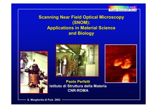
Scanning Near Field Optical Microscopy (SNOM): Applications in ...
Scanning Near Field Optical Microscopy (SNOM): Applications in ...
Scanning Near Field Optical Microscopy (SNOM): Applications in ...
Create successful ePaper yourself
Turn your PDF publications into a flip-book with our unique Google optimized e-Paper software.
S. Margherita di Pula 2003Summary:• <strong>Near</strong>-field microscopy (<strong>SNOM</strong>) <strong>in</strong> anutshell• Practical examples of results:– PtSi/Si– Spectroscopic <strong>SNOM</strong>: Diamond films– Biological growth medium– Pancreatic cells– LiF– Internal photoemission, Schottky Barrier– Boron nitride– Fluorescent markers <strong>in</strong> cells• The future: new sources -- 4GLS etc.
NashvillecountrymusicSwiss cheese& Frascatiw<strong>in</strong>eThanks to all the partners of this work:• Frascati - ISM-CNR: A. Cricenti, G. Longo, V. Marocchi, V. Mussi, M. Girasole, R. Generosi, M.Luce, P. Perfetti• Monterotondo - ISM-CNR: F. Cattaruzza, A. Flam<strong>in</strong>i, T. Prosperi• EPFL: D. Vobornik, G. Margaritondo, S. Catsicas, P. Ste<strong>in</strong>er, H. Hirl<strong>in</strong>g• Vanderbilt: D. W. Piston, M. A. Rizzo, J. K. Miller, B. Ivanov, N. H. Tolk• US - NRL: D. Talley, P. Thielen, J. S. Sanghera, I. D. Aggarwal• Frascati - ENEA: G. Baldacch<strong>in</strong>i, F. Bonfigli, F. Flora, T. Marolo, R. M. Montereali• MISDC -VNIIFTRI Mendeleevo, Moscow: A. Faenov, T. Pikuz• University of Roma: A. Congiu CastellanoS. Margherita di Pula 2003
The scann<strong>in</strong>g near-field opticalmicroscope (<strong>SNOM</strong>): like the stethoscopeHeart:Frequency 30-100 HzWavelength λ 102 mAccuracy <strong>in</strong> localization 10 cm λ /1000SmallapertureCoatedsmall-tipopticsfiberSmalldistanceMicroscopiclightemitt<strong>in</strong>gobject<strong>SNOM</strong> resolution: wellbelow the “diffractionlimit” of standardmicroscopy ( λ)S. Margherita di Pula 2003
The diffraction limit and how we can beat itFar-field microscope:∆yDiffraction by the lens aperture blurs theimages of the two objects: the m<strong>in</strong>imumseparation ∆y that can be resolved is λ/2λ<strong>Near</strong>-field scann<strong>in</strong>gmicroscope:diffraction does notplay a roleTapered optic fiberS. Margherita di Pula 2003
oscillat <strong>in</strong>gd λ (far field) only the term 1/r survives andthe Poynt<strong>in</strong>g vector S and its flux have a well def<strong>in</strong>ite valueand the travel<strong>in</strong>g wave can be detected far from the dipoleFor r≅λ (near field) The terms 1/r 2 and 1/r 3 do notcontribute to S, they represent the near field whose<strong>in</strong>tensity decay exponentially to zeroS. Margherita di Pula 2003
A simple approach to <strong>SNOM</strong>Task: resolv<strong>in</strong>g two small objects illum<strong>in</strong>ated by a light-wave<strong>in</strong> this case two slits separated by a distance x 0wave, k = 2π/λxzThe uncerta<strong>in</strong>ty pr<strong>in</strong>ciple applied tothe wave requires: ∆x ∆ k x> 2πthe m<strong>in</strong>imum ∆x corresponds tothe maximum ∆ k x ,butx 0k z =(k 2 -k x2 ) 1/2 =[(2π/λ) 2 -k x2 ] 1/2first casewe may have two casesk z is real, then -2π/λ< k x < 2π/λthe maximum value for ∆k x is 4π/λand the m<strong>in</strong>imum value for ∆x is,from the uncerta<strong>in</strong>ty pr<strong>in</strong>cipleλ/2, far field limitS. Margherita di Pula 2003
second casek z =(k 2 -k x2 ) 1/2 =[(2π/λ) 2 -k x2 ] 1/2k z is imag<strong>in</strong>ary (evanescent waves): then ∆k xcan be larger than 4π/λand one can have∆x< λ /2 (near field limit)How to get an imag<strong>in</strong>ary k zwave, k = 2π/λaAfter a p<strong>in</strong>hole of width a, the wave willhave a strong Fourier component alongx-direction, with k x =π/2a, correspond<strong>in</strong>g tok z =(k 2 -k x2 ) 1/2 =[(2π/λ) 2 -(π /2a) 2 ] 1/2if a< λ /4 then one has an evanescent wavewith imag<strong>in</strong>ary k zS. Margherita di Pula 2003z .
<strong>SNOM</strong> transmission modesampleplane waveDETECTORsmall aperturea < < λS. Margherita di Pula 2003E. H. Synge Philos. Mag. 6, 356 (1928)
<strong>SNOM</strong> transmission modesampleplane waveDETECTORsmall aperturea < < λS. Margherita di Pula 2003
Multi-purpose <strong>SNOM</strong> module built at the ISM-CNR (Istitutodi Struttura della Materia), Frascati [Antonio Cricenti et al.]Fiber tipModes of operation:SampleTransmission Reflection CollectionS. Margherita di Pula 2003
IR <strong>SNOM</strong> -- the strong po<strong>in</strong>ts:• It beats the diffraction limit thus reach<strong>in</strong>g highlateral resolution• It can work <strong>in</strong> any environment and (almost)without any particular sample preparation• It provides real optical signal• It can perform spectroscopic measurements• It does not touch the sample: totally nondestructivetechnique[Image from theJasco Co. UKHomepage]S. Margherita di Pula 2003
Fiber Preparation:Chemical etch<strong>in</strong>gfor IR fibers:Tampon solution(Tetra-methylpenta-decane)Chalcogenidefiber+ 30% H 2 S 2Piranha Solution70% H 2 SO 4MicroscopeS. Margherita di Pula 2003
F<strong>in</strong>al structure(tapered chalcogenide fiber with Au coat<strong>in</strong>g):190 µmAuAs 38 Se 62As 40 Se 60120 µmS. Margherita di Pula 2003
Manag<strong>in</strong>g the distance fiber tip-sample:S. Margherita di Pula 2003
…our f<strong>in</strong>al component -- the Vanderbilt FEL!S. Margherita di Pula 2003
WHAT IS A SASE-FEL RADIATION SOURCE?SPONTANEOUS RADIATION:λ ph ≅ λ u /2γ 2 (1 + K 2 )Beam divergence ≅ 1/γ = m e c 2 /E e ≅ 1 00 µradI ph ≅ Ν eΝ e number of electrons (≅ 10 9 )S. Margherita di Pula 2003
THE INTERACTION OF THE ELECTRONS WITH THE SPONTANEOUSRADIATION CAUSES MICROBUNCHING AND THE SELF-AMPLIFICATION OFSPONTANEOUS EMISSION (SASE)IN THE SASE MODE THE INTENSITY:I ph ≅ Ν 2eSINCE Ν e number of electrons (≅ 10 9 ) THE AMPLIFICATIONGIVES AN EXTRAORDINARY HIGH PHOTON FLUXBeam divergence (few µrad.)S. Margherita di Pula 2003
S. Margherita di Pula 2003
S. Margherita di Pula 2003
S. Margherita di Pula 2003
<strong>SNOM</strong> IN THE SPETTROSCOPIC MODETake the image at λ valueswhere the sample has a spectroscopicf<strong>in</strong>gerpr<strong>in</strong>tS. Margherita di Pula 2003•reflectivity•absorption•fluorescence• ……
This is an example of how we can usea <strong>SNOM</strong> to <strong>in</strong>vestigate bulk opticalproperties with lateral resolutionwell below the diffraction limitQuickTime and aGIF decompressorare needed to see this picture.S. Margherita di Pula 2003
Absorption Spectrum of Artificial Diamond Filmsλ=3.5 µmS. Margherita di Pula 2003
Artificial CVD Diamond Film<strong>Near</strong> <strong>Field</strong><strong>Optical</strong> imagesTopographicImagesS. Margherita di Pula 2003
<strong>SNOM</strong>/FEL Images of Biological Growth Medium•Surfaces immersed <strong>in</strong> water under physiological conditions arerapidly covered with biofilms, which consist of microrganismsembedded <strong>in</strong> a matrix of extracellular substance•Intensive research is <strong>in</strong> progress to elucidate the <strong>in</strong>flence of biofilmactivity on a variety of <strong>in</strong>dustrial, medical and environmentalprocesses•The first step of this research it to study the growth medium ofbiofilms•The growth medium is a m<strong>in</strong>imal salt medium with severalconstituents like sulphur compounds and nitrogen oxideS. Margherita di Pula 2003
First IR spectroscopic <strong>SNOM</strong> results on biosystems:postgate growth medium on siliconNoise:
First IR spectroscopic <strong>SNOM</strong> results on biosystems:resolution tests20 µmL<strong>in</strong>e scan:Resolution< 150 nm = λ/40S. Margherita di Pula 2003
Absorption of a Pancreatic Cellλ = 6.1 µmλ = 6.45 µmS. Margherita di Pula 2003
Shear-Force and <strong>SNOM</strong> Images (20x20 µm)of Pancreatic Cells with photons of 6.1 µmShear-ForceReflectivityS. Margherita di Pula 2003
IR spectroscopic <strong>SNOM</strong> results onbio-systems: fixed (with paraformaldehyde) cells <strong>in</strong>water from a rat pancreatic β cell l<strong>in</strong>e (INS-1)λ = 6.95 µm(sulfide stretch)20 µmλ = 6.45 µm(amide II)λ = 6.1 µm (amide I,C=O stretch)TopographyReflectionS. Margherita di Pula 2003
IR spectroscopic <strong>SNOM</strong> study of metal (CdCl 2 )contam<strong>in</strong>ation of pancreatic cellstopographyλ = 0.488 µm 0.630 µm 0.785 µm 1.25 µm 4.05 µmspectroscopyS. Margherita di Pula 2003
First IR spectroscopic <strong>SNOM</strong> results onbio-systems: COS-7 cells (not fixed) <strong>in</strong> PBScellTopographyPBScrystalThe nucleusis best seenhere8.05 µm (phosphates, DNA)7.6 µm C-H bend<strong>in</strong>g6.95 µm (sulfide)only thePBS crystalreflects6.45 µm (amide II)6.1 µm (amide I)S. Margherita di Pula 200320 µm
How important is IR <strong>SNOM</strong>,<strong>in</strong> particular for IR bio-research?Very important: due to the long wavelengths, IRtechniques are severely affected by the diffraction limit!20 µmSpectroscopic<strong>SNOM</strong> image of celltaken at 6.1 µmSame image withdiffraction-limitblurr<strong>in</strong>gS. Margherita di Pula 2003
First example ofIR Spectroscopic<strong>SNOM</strong>:diamond filmsS. Margherita di Pula 2003
Lithium fluoride (LiF) films on siliconLiF - an <strong>in</strong>terest<strong>in</strong>g material for waveguides, microcavities,laser sources and generally optoelectronics devices:• Non hygroscopic• Irradiation by x-rays, e-beams and ion beams producesstable color centers (F-center) that can be used <strong>in</strong> devicemanufactur<strong>in</strong>g. X-rays give high spatial resolution.+ -e -We need to clarify how the film growth parameters <strong>in</strong>fluencethe color<strong>in</strong>g process and to compare film color<strong>in</strong>g with bulkcolor<strong>in</strong>gS. Margherita di Pula 2003
LiF crystal film on Si “colored” with X-rays through a12.7 µm grid.Topography, fluorescence <strong>SNOM</strong> andreflectivity <strong>SNOM</strong>FluorescenceTopographyReflectivityat 6.1 µmThe <strong>SNOM</strong> contrast at 9.2, 6.1,5.8 and 5.5 µm reveals acoloration-<strong>in</strong>duced change <strong>in</strong>the refractive <strong>in</strong>dexS. Margherita di Pula 2003
LiF film pattern created with X-rays on a Si substrateShear-force (topography)S. Margherita di Pula 2003Reflectivity at 6.1 µmThe <strong>SNOM</strong> contrast at 9.2, 6.1, 5.8 and 5.5 µm reveals acoloration-<strong>in</strong>duced change <strong>in</strong> the refractive <strong>in</strong>dex
S. Margherita di Pula 2003
<strong>SNOM</strong> and Internal Photoemission (IPE)an improved local probeS. Margherita di Pula 2003
The first steps: “FELIPE” (FEL Internal photoemission)Conduction bandFELSchottky Barrier φ bE FhνValence bandSemiconductor MetalPhotocurrentpAThe Schottky Barrier is measured directlyhνS. Margherita di Pula 2003
S. Margherita di Pula 2003
<strong>SNOM</strong> as a Useful Probe <strong>in</strong> Microelectronicse.g. <strong>SNOM</strong> Images of Boron Doped Silicon (100)Ion implantation is the key technique <strong>in</strong><strong>in</strong>tegrated Circuits TechnologyDepend<strong>in</strong>g on the Boron doses the post annealedsamples may present clusters formation or other defects<strong>SNOM</strong>/IPE Techniques may be used as powerfuldiagnostic toolsS. Margherita di Pula 2003
IR Spectroscopic <strong>SNOM</strong>:Boron-doped (ion-implanted, annealed) silicon10 µmWavelength:1.33 µmS. Margherita di Pula 2003
IR Spectroscopic <strong>SNOM</strong>: BN th<strong>in</strong> films (+ oxides)<strong>SNOM</strong> topography imageWurtziteL<strong>in</strong>e scan: featuresnarrower than λ/9<strong>SNOM</strong> reflectionspectroscopic imagesHexagonalS. Margherita di Pula 2003Cubic
A negative but important result:carboxylic acid term<strong>in</strong>ated Si and silicon nitride surfaces(chemical process<strong>in</strong>g for biological sensors)Surface—(CH 2 ) n (COOH)AFMtopographyReflectivityλ = 6.1~ µm6 µm20 µm20 µmIR spectroscopic images taken at this and otherwavelengths show no lateral fluctuationsS. Margherita di Pula 2003
Spectroscopic <strong>SNOM</strong> results on bio-systems:prelim<strong>in</strong>ary tests on <strong>in</strong>ternal cell structureslabeled by a fluorescent markerFluorescentmarkerCells at PetriDish bottomDetector(avalanchephotodiode)S. Margherita di Pula 2003
Intest<strong>in</strong>al adeno carc<strong>in</strong>oma epithelial cells labeledwith Fluoresce<strong>in</strong> Isothiocynate25 x 25 µmS. Margherita di Pula 2003
Fibroblast cells (COS) with Oregon greenfluorophore (excitation at 496 nm, emission at522 nm) fixed to Transferr<strong>in</strong>Neuron cells withOregon greenfluorophore directlyattached to vesicles<strong>in</strong>side the cellS. Margherita di Pula 2003
S. Margherita di Pula 2003Fluorescence: <strong>SNOM</strong> vs far-field
<strong>SNOM</strong> Fluorescence study of magnetic field (24 hrs50 Hz, 1 mT) effects on human kerat<strong>in</strong>ocyte cellsunexposedcellsTopographyTopography<strong>SNOM</strong> fluorescence<strong>SNOM</strong> fluorescenceexposedcellsS. Margherita di Pula 2003
New types of synchrotron sources:• Ultrabright storage r<strong>in</strong>gs (SLS, newGrenoble project) approach<strong>in</strong>g thediffraction limit• Self-amplified spontaneous emission(SASE) X-ray free electron lasers• VUV FEL’s (such as CLIO)• Energy-recovery mach<strong>in</strong>es• Inverse-Compton-scatter<strong>in</strong>g table-topsourcesS. Margherita di Pula 2003
THE “4 GLS” CONCEPT AT DARESBURYS. Margherita di Pula 2003
Conclusions:1. <strong>SNOM</strong> experiments with an FEL are possible and yieldresolution levels well beyond the diffraction limit2. Vibrational spectroscopy <strong>SNOM</strong> provides chemical<strong>in</strong>formation on a microscopic scale3. Results on biological systems f<strong>in</strong>ally open up the possibiltyto perform <strong>in</strong>frared microscopy of cells and <strong>in</strong>ternal cellstructures4. IR Spectroscopic <strong>SNOM</strong> tests were successfully performed<strong>in</strong> different <strong>SNOM</strong> modes5. The experimental performances did not yet reach saturationand substantial improvements can be foreseen6. In the near future, even better IR sources will further bost thecapabilities of IR <strong>SNOM</strong>S. Margherita di Pula 2003
A powerful <strong>in</strong>ternational coalition:NashvillecountrymusicSwiss cheese& Frascatiw<strong>in</strong>eS. Margherita di Pula 2003
The first steps: “FELIPE” (FEL Internal photoemission)Conduction bandLightsourceConduction banddiscont<strong>in</strong>uityhνValence bandSemiconductor A Semiconductor BPhotocurrentpAProblem: the high value of the photon energy“mixes” the conduction band discont<strong>in</strong>uity and thelocal gapS. Margherita di Pula 2003hν
The first steps: “FELIPE” (FEL Internal photoemission)FELConduction bandhνValence bandSemiconductor A Semiconductor BS. Margherita di Pula 2003pAThe FEL removes the problem and the conductionband discont<strong>in</strong>uity is measured directly
Another problem, however, still rema<strong>in</strong>s: is thediscont<strong>in</strong>uity the same for the entire <strong>in</strong>terface?Solution: FELIPE(essentially, a near-field <strong>SNOM</strong> system)First <strong>SNOM</strong> results with an FEL!S. Margherita di Pula 2003Lateral fluctuations!
<strong>SNOM</strong>: why does it work?yUncerta<strong>in</strong>ty pr<strong>in</strong>ciple:Photon beamx∆y ∆k y > 2 → ∆y > 2/∆k y∆k y < k y = (k 2 - k x2 ) 1/2k x real → ∆k y < k = 2/λ∆y > λ(Abbé’s diffraction limit)This conclusion is no longer valid ifk x is imag<strong>in</strong>ary(absorbed, evanescent waves)S. Margherita di Pula 2003
IR <strong>SNOM</strong>: a multi-purpose module built at theISM (Istituto di Struttura della Materia, CNR),Frascati [Antonio Cricenti et al.]S. Margherita di Pula 2003Resolution< 0.15 µm
…our f<strong>in</strong>al component -- the Vanderbilt FEL!S. Margherita di Pula 2003
NashvillecountrymusicSwiss cheese& Frascatiw<strong>in</strong>eThanks to all the partners of this work:• Frascati - ISM-CNR: A. Cricenti, G. Longo, V. Marocchi, V. Mussi, M. Girasole, R.Generosi, M. Luce, P. Perfetti• Monterotondo - ISM-CNR: F. Cattaruzza, A. Flam<strong>in</strong>i, T. Prosperi• EPFL: D. Vobornik, G. Margaritondo, S. Catsicas, P. Ste<strong>in</strong>er, H. Hirl<strong>in</strong>g• Vanderbilt: D. W. Piston, M. A. Rizzo, J. K. Miller, B. Ivanov, N. H. Tolk• US - NRL: D. Talley, P. Thielen, J. S. Sanghera, I. D. Aggarwal• Frascati - ENEA: G. Baldacch<strong>in</strong>i, F. Bonfigli, F. Flora, T. Marolo, R. M. Montereali• MISDC -VNIIFTRI Mendeleevo, Moscow: A. Faenov, T. Pikuz• University of Roma: A. Congiu CastellanoS. Margherita di Pula 2003


