Crystalline cell inclusions - Phycology Research Group, Ghent ...
Crystalline cell inclusions - Phycology Research Group, Ghent ...
Crystalline cell inclusions - Phycology Research Group, Ghent ...
Create successful ePaper yourself
Turn your PDF publications into a flip-book with our unique Google optimized e-Paper software.
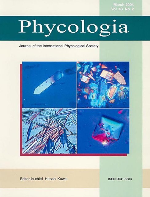
Phycologia (2004) Volume 43 (2), 189-203 Published 29 March 2004<strong>Crystalline</strong> <strong>cell</strong> <strong>inclusions</strong>: a new diagnostic character in theCladophorophyceae (Chlorophyta)FREDERIK LELIAERT* AND ERIC COPPEJANS<strong>Research</strong> <strong>Group</strong> <strong>Phycology</strong>, Biology Department, <strong>Ghent</strong> University, 9000 <strong>Ghent</strong>, BelgiumF. LELIAERT AND E. COPPEJANS. 2004. <strong>Crystalline</strong> <strong>cell</strong> <strong>inclusions</strong>: a new diagnostic character in the Cladophorophyceae(Chlorophyta). Phycologia 43: 000–000.<strong>Crystalline</strong> <strong>cell</strong> <strong>inclusions</strong> were observed in 45 species of Cladophorophyceae. The crystals can be classified into eightmorphological types, including needle shaped, prismatic, octahedral, tetrahedral, cubic and globular, and they were foundto occur in clusters or as single crystals. In addition to the different morphological types, the crystals are characterized bydifferent chemical compositions. Chemical tests distinguished the crystals as being composed of calcium oxalate, calciumcarbonate or proteins. The diversity of crystal types raises the possibility that these structures have systematic value. Theoccurrence of crystalline structures is compared with previously published phylogenies of the Cladophorophyceae. Sometypes of crystals were found to be genus or species specific, whereas other types occurred in distantly related groups. Thecrystalline <strong>cell</strong> <strong>inclusions</strong> can be useful diagnostic characters. For example, Cladophoropsis sundanensis and Cladophoracoelothrix are distantly related but have similar thallus architecture, and they can be distinguished from one another by thepresence or absence of crystals.INTRODUCTIONThe Cladophorophyceae (van den Hoek et al. 1995) encompassesa mainly marine class of coenocytic Chlorophyta. Thallusarchitecture includes branched and unbranched uniseriatefilaments, blade-like thalli, pseudoparenchymatic thalli andthalli consisting of inflated <strong>cell</strong>s. Diagnostic morphologicalcharacters are scarce and generally unsuitable for retrievingevolutionary relationships, due to repeated convergence andparallel evolution (Leliaert et al. 2003).A wide variety of crystalline structures have been observedin vascular plants and macroalgae; however, they have longbeen neglected in the latter. Vascular plants accumulate crystalsof calcium oxalate (CaOx) in a diversity of shapes, sizes,amounts and spatial locations. These crystals may be importantin structural reinforcement, calcium regulation (Volk etal. 2002) and defence against grazers. Both the morphologyand distribution of CaOx crystals within plants exhibit species-specificpatterns, indicating that their development is geneticallycontrolled. The morphology and distribution of crystalsvary widely between genera and often between closelyrelated species; however, they have been shown to be systematicallyuseful in some cases (Franceschi & Horner 1980;Webb 1999). In macroalgae, CaOx crystals have been recordedonly rarely, but the reports that do exist represent a broadsample of algae (Pueschel 2001). In the Chlorophyta, needleshapedCaOx crystals have been reported in vacuoles of thebryopsidophycean genera Penicillus Lamarck (Friedmann etal. 1972; Turner & Friedmann 1974; Böhm et al. 1978), ChlorodesmisHarvey & Bailey (Ducker 1967: pp. 158, 161, table27d; Menzel 1987; Coppejans & Prud’homme van Reine1989: p. 127, fig. 12) and Codium Stackhouse (E. Coppejans,unpublished observations). Cruciate CaOx crystals have beenobserved in the zygnematophycean genus Spirogyra Link* Corresponding author (frederik.leliaert@UGent.be).(Klein 1877; Pueschel 2001). Protein crystals have been reportedin a wide range of Rhodophyta (Feldmann-Mazoyer1941; Fritsch 1945; Pueschel 1992) and in the brown algaHaplogloia kuckuckii Kylin (Pueschel 1994). Few recordshave been found of crystalline, proteinaceous <strong>inclusions</strong> inChlorophyta.Reports of crystalline <strong>cell</strong> <strong>inclusions</strong> in members of the Cladophorophyceaeare sparse. Børgesen (1905, fig. 13; 1913, fig.32b) casually illustrated needle-shaped crystals in Cladophoropsismembranacea (Hofman Bang ex C. Agardh) Børgesen.Klein (1882), Chemin (1931), Jónsson (1962) and van denHoek & Chihara (2000, p. 96, fig. 44G) briefly described proteincrystalloids with various morphologies in several speciesof Cladophora Kützing, including C. pellucida (Hudson)Kützing, C. prolifera (Roth) Kützing, C. rupestris (Linnaeus)Kützing, C. sakaii Abbott and C. ohkuboana Holmes.This study describes the morphological variation of crystalline<strong>cell</strong> <strong>inclusions</strong> observed in a survey of numerous taxaof Cladophorophyceae. The chemical nature of the crystals isbriefly considered by the use of solubility tests and stainingmethods. The major aim of this study is to detect any possiblesystematic value of the various types of crystalline <strong>cell</strong> <strong>inclusions</strong>.MATERIAL AND METHODSThe <strong>cell</strong> contents of 66 species of Cladophorophyceae wereexamined to detect the presence of crystalline structures. Thespecimens studied are listed in Appendix 1 (herbarium abbreviationsfollow Holmgren et al. 1990). The specimens werecollected during various field trips or requested from differentherbaria; 24 type specimens are included in the study. Collectedsamples were prepared as herbarium specimens or preservedin 4% formalin–sea water. When possible, fresh specimenswere studied in the field with a Nikon hand microscope
Phycologia, Vol. 43 (2), 2004Table 1. Morphological types of crystalline <strong>cell</strong> <strong>inclusions</strong> in the Cladophorophyceae, taxa where these types have been observed and chemicalsolubility, staining and birefringence under polarized light.Morphological typeTaxaChemical solubility, 1staining 2 andbirefringenceType 1acrystals single, elongated prismatic(Figs. 1–9)Boodlea spp. (except B. vanbosseae)Phyllodictyon anastomosansStruveopsis siamensisChamaedoris orientalisCladophoropsis (pro parte)⎫Type 1bType 2crystals single, broad prismatic,hexagonal to diamond shapedor triangular (Figs. 10–16)crystals single, needle shaped, attenuatingto one or both ends,or elongated rod shaped (Figs.17–22)Chamaedoris peniculumCladophoropsis magnusPhyllodictyon spp. (p.p.)Struvea plumosaApjohnia laetevirens⎪⎬HCl: dissolveAcet. acid: intactSod. hyp.: intactY: positiveL: negativebirefringenceType 3crystalline structures elongatedelliptical to irregular rodshaped, single or clustered incruciate to star-shaped aggregates(Figs. 23–27)Dictyosphaeria cavernosaDictyosphaeria versluysii⎪⎭Type 4 octahedral crystals (Figs. 28–32) Valoniopsis pachynemaCladophora dotyanaSiphonocladus tropicus: rareType 5Type 6Type 7globular aggregates of rod- orcone-shaped crystals (Figs.33–35)tetrahedral crystals; in most species(marked with *) growing intofour armed (apparently threearmed), star-shaped structures(Figs. 36–44)cubical <strong>cell</strong> <strong>inclusions</strong>, single orfused (Figs. 45, 46)Valonia spp.Ventricaria ventricosa(Cladophora dotyana: rare)Cladophoropsis herpesticaChamaedoris auriculata*Chamaedoris delphinii*Phyllodictyon papuense (stipe)Valonia aegagropila*Valonia fastigiata*Valonia utricularis*Cladophora proliferaCladophora rugulosaCladophora rupestris⎫⎪⎬⎪⎭HCl: dissolveAcet. acid: dissolveSod. hyp.: intactY: negativeL: negativebirefringenceHCl: intactAcet. acid: intactSod. hyp.: dissolveY: negativeL: Dark brownstainingno birefringenceType 8star-shaped or irregular clustersof fine, needle-shaped crystals(Figs. 47–49)Boodlea vanbosseaeChaetomorpha spp.Chamaedoris spp.Cladophora coelothrixMicrodictyon tenuiusSiphonocladus tropicusValonia spp.Valoniopsis pachynemaVentricaria ventricosaHCl: intactAcet. acid: intactSod. hyp.: intactY: negativeL: negativebirefringence1HCl, hydrochloric acid; Acet. acid, acetic acid; Sod. hyp., sodium hypochlorite.2Y, Yasue method for CaOx staining; L, staining with Lugol iodine.
Leliaert & Coppejans: <strong>Crystalline</strong> <strong>cell</strong> inclusion in the CladophorophyceaeFigs 1–9. Elongate prismatic crystals (Type 1a) in Phyllodicyton anastomosans.Figs 1, 2. Crystals present in the apical <strong>cell</strong>s but absent from the tenacular <strong>cell</strong>. Scale bars 50 m.Figs 3, 4. Crystals abundant in the <strong>cell</strong>s of the main axes. Scale bars 50 m.Fig. 5. Aggregate of crystals. Birefringence under polarizing optics demonstrates crystallinity of <strong>inclusions</strong>. Scale bar 25 m.Figs 6–8. Morphological variation of crystals within a single <strong>cell</strong>. Scale bars 10 m.Fig. 9. Fusion of two crystals to form cruciate structures. Scale bar 10 m.
Phycologia, Vol. 43 (2), 2004Figs 10–16. Broad hexagonal or diamond-shaped prismatic crystals (Type 1b).Figs 10–12. Type-1b crystals in Phyllodictyon orientale.Fig. 10. Aggregate of crystals. Birefringence under polarizing optics demonstrates crystallinity of <strong>inclusions</strong>. Scale bar 50 m.Figs 11, 12. Morphological variation of the crystals in a single <strong>cell</strong>. Scale bars 25 m.Figs 13–16. Type-1b crystals in Cladophoropsis magnus. Scale bars 10 m.(Nikon, Tokyo, Japan). Preserved specimens were studied ona Leitz-Diaplan light microscope (LM) (Leitz, Wetzlar, Germany).To improve visualization of the <strong>cell</strong>-<strong>inclusions</strong>, <strong>cell</strong>swere dissected and the <strong>cell</strong> contents examined. Photographswere taken with a Olympus-DP50 camera (Olympus, Tokyo,Japan) mounted on the LM.To confirm the presence of crystalline structure, <strong>cell</strong>s wereexamined under differential interference (Nomarski) contrast(DIC) for characteristic crystalline birefringence. Chemicalsolubilities of the <strong>inclusions</strong> were tested by viewing specimensexposed to 1.0 N hydrochloric acid, 5% sodium hypochloriteor 67% aqueous acetic acid (Yasue 1969; Friedmannet al. 1972; Pueschel 2001). The Yasue (1969) method forCaOx staining was performed as follows: filaments wereplaced in 5% aqueous AgNO 3 for 10 min, rinsed in water andthen mounted on a microscope slide in a solution of 70%ethanol saturated with dithiooxamide (Sigma Chemical Co.,St Louis, MO, USA). After a few minutes, water was addedto the edge and the effect monitored (Pueschel 2001). Proteincrystalloids were stained with aniline blue and Lugol iodineas a general staining method for proteins (Gurr 1965).RESULTSOf the 66 species examined, 45 were found to possess somekind of crystalline <strong>cell</strong> inclusion (Appendix 1). A large morphologicalvariety of crystals was observed in the differentgenera: fine needle-shaped or elongated hexagonal rod-shapedcrystals, single or grouped in clusters; broad hexagonal, diamond-shapedor triangular structures; tetrahedrons; octahedronsand star-shaped structures. The morphological variationof the <strong>cell</strong> <strong>inclusions</strong> can be classified into eight morphologicaltypes (Table 1). Cells of some species contain only onecrystal type, whereas those of others (e.g. some species ofValonia C. Agardh) have up to three morphological crystal
Leliaert & Coppejans: <strong>Crystalline</strong> <strong>cell</strong> inclusion in the CladophorophyceaeFigs 17–22. Needle or spindle shaped crystals (Type 2) in Apjohnia laetevirens.Figs 17, 18. Dense accumulation of Type-2 crystals in the <strong>cell</strong>s. Scale bars 50 m.Figs 19–22. Morphological variation of crystals within a single <strong>cell</strong>. Scale bars 10 m.Fig. 19. Needle-shaped crystal with one attenuating end.Figs 20, 21. Spindle-shaped crystals with both ends attenuating.Fig. 22. Rod-shaped crystal without attenuating ends.types. In some taxa, crystals are present in all <strong>cell</strong>s of thethallus, whereas in others, crystals are restricted to the older<strong>cell</strong>s or the axial filaments. The number of crystals varies froma few to thousands per <strong>cell</strong>. The vacuolated nature of the <strong>cell</strong>sin the Cladophorophyceae makes it difficult to preserve thetonoplast and to determine the sub<strong>cell</strong>ular location of the <strong>inclusions</strong>.This problem was also encountered by Pueschel(1995) who studied CaOx crystals in the <strong>cell</strong>s of AntithamnionNägeli.Crystal Types 1–4 (Table 1) could be detected by brightfieldlight microscopy (BF) but are especially conspicuouswhen viewed with DIC, under which they appear bright dueto birefringence; birefringence indicates a crystalline substructure.The crystals stay intact when treated with acetic acid orsodium hypochlorite; this eliminates the possibility that thestructures are composed of calcium carbonate or protein, respectively.The crystals dissolve very quickly in hydrochloricacid, eliminating the possibility of silica composition. The Yasuemethod for CaOx staining resulted in dark staining of thecrystals. The chemical solubility tests and staining methodindicate that the chemical composition of these types of crystalsis CaOx.Elongate prismatic crystals (Type 1a: Figs 1–9), single orgrouped in loose aggregates, are present in the <strong>cell</strong>s of speciesof Boodlea G. Murray & De Toni (except B. vanbosseae),Phyllodictyon anastomosans, Struveopsis siamensis, Siphonocladusrigidus and the Cladophoropsis Børgesen species C.carolinensis, C. macromeres, C. membranacea, C. philippinensisand C. vaucheriaeformis. The crystals occur in all <strong>cell</strong>sof the thallus except the tenacular <strong>cell</strong>s (Figs 1–8); the numberper <strong>cell</strong> ranges from 1 to 30 in the apical <strong>cell</strong>s (Figs 1, 2) tomore than 200 in the <strong>cell</strong>s of the main axes (Figs 3, 4). Crystalsare 1.5–5(–8) m in diameter, 25–70 m long, with alength–width ratio of 3–14. Infrequently, two crystals fuse andform cruciate structures (Fig. 9). Crystals in C. sundanensisare rectangular to elongated rod shaped, present in most <strong>cell</strong>s(up to 7 crystals per <strong>cell</strong>), 2–15 m in diameter, 15–25 mlong, with a length–width ratio of 1–12. In S. rigidus, crystalsare present in a small number of <strong>cell</strong>s (up to five per <strong>cell</strong>), 3–12 m in diameter and up to 60 m long. No Type-1 crystalswere observed in the Cladophoropsis species C. herpesticaand C. javanica. In Chamaedoris orientalis, crystals are abundantin all <strong>cell</strong>s of the capitulum filaments (over 200 per <strong>cell</strong>)but are absent from the stipe <strong>cell</strong>; crystals are 0.5–1.5 m indiameter and up to 30 m long.Broad hexagonal or diamond-shaped prismatic crystals
Phycologia, Vol. 43 (2), 2004Figs 23–27. Clusters of elliptical or rod-shaped crystals (Type 3). Scale bars 10 m.Figs 23, 24. Type-3 crystals in Dictyosphaeria cavernosa. Clusters of two and three rod-shaped crystals.Figs 25–27. Type-3 crystals in Dictyosphaeria versluysii. Elliptical crystalline structures, single or clustered.(Type 1b: Figs 10–16) were observed in Chamaedoris peniculum,Cladophoropsis magnus, Phyllodictyon J.E. Gray (P.gardineri, P. orientale, P. papuense and P. pulcherrimum) andStruvea plumosa. In Chamaedoris peniculum the crystals arediamond shaped and occur exclusively in the <strong>cell</strong>s of the capitulumfilaments with up to five crystals per <strong>cell</strong>; crystals are12–24 m in diameter, 18–35 m long, with a length–widthratio of c. 1.5. The diamond-shaped crystals in Cladophoropsismagnus occur in all <strong>cell</strong>s of the thallus with numbers rangingfrom c. 20 to more than 100 per <strong>cell</strong>; crystals are 10–30m in diameter, 20–55 m long, with a length–width ratio of1.2–1.7. In the Phyllodictyon species, crystals are present inmost <strong>cell</strong>s (except the tenacular <strong>cell</strong>s); the number of crystalsper <strong>cell</strong> ranges from one to five. In P. orientale, P. pulcherrimumand P. papuense the crystals are generally hexagonal,15–25 µm in diameter,20–65 m long, with a l/w ratioof c. 2. In P. gardineri, the crystals are generally diamondshaped or triangular,20–50 m in diam., 30–75 µm long, witha length–width ratio of 1.5–2. In S. plumosa, crystals are diamondshaped, triangular or pentagonal and present in all<strong>cell</strong>s, except the young laterals, with numbers per <strong>cell</strong> rangingfrom one to five; crystals are 5–27 m in dia., 8–32 µm long,with a length–width ratio of 1.2–1.5.Needle- or spindle-shaped crystals with one or both endsattenuating to elongate rod-shaped crystals (Type 2: Figs 17–22) were found only in Apjohnia laetevirens. Crystals are presentin all <strong>cell</strong>s of the thallus and may exceed 1000 per <strong>cell</strong>,with diameters of 2–6 m and lengths of up to 125 m.Elongate elliptical to irregular rod-shaped crystals (Type 3:Figs 23–27) differ from Type-1 crystals in their shape and thefact that they often form cruciate to star-shaped clusters. Thistype of crystal is present in the <strong>cell</strong>s of Dictyosphaeria cavernosaand D. versluysii, but was not found in D. o<strong>cell</strong>ata.The number of crystals per <strong>cell</strong> is difficult to determine butis probably in the range of 1000 or more. Individual crystalsare 1–3 m in diameter and up to 20 m long; the clustersare up to 25 m in diameter.Octahedral crystals (Type 4: Figs 28–32) are present in Valoniopsispachynema, where they occur in all <strong>cell</strong>s of the thallus;the number of crystals per <strong>cell</strong> ranges from 100 to morethan 1000, with diameters of 5–50 m. Stellate morphologiesresulting from two fusing crystals are sometimes found (Fig.32). This type of crystal is less abundant in the <strong>cell</strong>s of Cladophoradotyana and Siphonocladus tropicus; crystals inthese species are 15–25 m in diameter.Globular aggregates of rod- or cone-shaped crystals (Type5: Figs 33–35) are present in Valonia fastigiata, V. macrophysa,V. utricularis and Ventricaria ventricosa. The crystalscould only be observed by dissecting the <strong>cell</strong> and viewing itscontents under LM. The crystals can be detected with BF butare especially noticeable when viewed with DIC due to birefringence.The crystals stay intact when treated with sodiumhypochlorite but dissolve in acetic acid and hydrochloric acid,suggesting their chemical composition to be calcium carbonate.The aggregates are 25–40 m in diameter.The colourless crystalline <strong>cell</strong> <strong>inclusions</strong> of Types 6 and 7are not birefringent under polarized light and are thereforedifficult to detect, especially when masked by the thick <strong>cell</strong>walls in some genera (e.g. Valonia). The <strong>inclusions</strong> can beobserved by dissecting the <strong>cell</strong>s and viewing the <strong>cell</strong> contentsin LM. The structures dissolve when treated with sodium hypochlorite;they stain dark blue with methylene blue and dark
Leliaert & Coppejans: <strong>Crystalline</strong> <strong>cell</strong> inclusion in the CladophorophyceaeFigs 28–32. Octahedral crystals (Type-4).Figs 28, 29. Type-4 crystals in Cladophora dotyana. Scale bars 10 m.Figs 30–32. Type-4 crystals in Valoniopsis pachynema.Fig. 30. Crystals scattered within a <strong>cell</strong>. Scale bar 100 m.Fig. 31. Surface view of an octahedral crystal. Scale bar 25 m.Fig. 32. Stellations resulting from two fusing octahedral crystals. Scale bar 25 m.Figs 33–35. Globular aggregates of rod- or cone-shaped crystals (Type-5).Figs 33, 34. Type-5 crystals in Valonia fastigiata. Scale bars 10 m.Fig. 33. Globular aggregate of cone-shaped structures.Fig. 34. Globular aggregate of rod-shaped crystals.Fig. 35. Type-5 crystals in Ventricaria ventricosa: globular aggregate of cone-shaped structures. Scale bar 25 m.
Phycologia, Vol. 43 (2), 2004
Leliaert & Coppejans: <strong>Crystalline</strong> <strong>cell</strong> inclusion in the Cladophorophyceaebrown with Lugol iodine, suggesting a proteinaceous nature(dark blue staining with Lugol iodine would indicate the presenceof starch). Protein crystals stain easily due to their porousstructure.Cell <strong>inclusions</strong> of Type 6 (Figs 36–44) are scattered amongthe chloroplasts. This type of crystal is abundant in <strong>cell</strong>s ofValonia aegagropila, V. fastigiata and V. utricularis and inthe stipe <strong>cell</strong>s of Chamaedoris auriculata, C. delphinii and P.papuense. The crystals in these species are tetrahedral whensmall (with diameters of up to 40 m) and grow into fourarmedstructures with serrated edges, up to 230 m across.Under LM, these <strong>inclusions</strong> appear as three-armed structuresbecause the axis of the fourth arm is placed perpendicular tothe plane of the slide. In Cladophoropsis herpestica the crystalsare less frequent and remain relatively small and tetrahedral,with diameters of up to 40 m. The structures appearto have concentric bands, observable when stained with methyleneblue. The bands replicate the crystals’ morphology ona smaller scale; up to five bands per crystal were observed(Fig. 40).Cubic <strong>cell</strong> <strong>inclusions</strong> (Type 7: Figs 45, 46) are present inthe <strong>cell</strong>s of Cladophora prolifera, C. rugulosa and C. rupestris.In C. rugulosa, the crystalloids are especially abundantin the large basal <strong>cell</strong>s. The diameters of the cubes are 5–15(–20) m inC. prolifera and C. rupestris, and up to 65 m inC. rugulosa. In C. rugulosa, two or more crystalloids are oftenfused in larger aggregates; frequently the structures are penetratedby crevices and are partially eroded (Fig. 45).Star-shaped clusters of fine needle-shaped crystals (Type 8:Figs 47–49) were observed in a number of taxa (Boodleavanbosseae, Chaetomorpha aerea, C. brachygona, Chamaedorisauriculata, C. delphinii, Cladophora coelothrix, Microdictyontenuius, Siphonocladus tropicus, V. aegagropila,V. fastigiata, V. macrophysa, Valoniopsis pachynema andVentricaria ventricosa). The structures appear bright underpolarized light due to birefringence and can therefore be easilydetected in taxa with relatively thin <strong>cell</strong> walls (e.g. some speciesof Chaetomorpha Kützing, Microdictyon Decaisne andSiphonocladus Schmitz); in Valonia and Ventricaria Olsen &J. West dissection of the <strong>cell</strong>s is needed to observe the <strong>inclusions</strong>.The crystalline structures stay intact when treated withhydrochloric acid, acetic acid and sodium hypochlorite. Theclusters vary from being densely packed (Fig. 48) to beingloosely aggregated into indefinite shapes (Fig. 49). The diametersof dense clusters is 10–40 m; loose aggregates areup to 150 m in diameter. The needle-shaped crystals composingthe clusters are straight or slightly curved, 0.2–1 min diameter and 10–35 m long. The number of clusters per<strong>cell</strong> ranges between one and seven.DISCUSSION<strong>Crystalline</strong> <strong>cell</strong> <strong>inclusions</strong> have previously been noticed insome members of the Cladophorophyceae but their taxonomicsignificance has never been examined. The needle-shapedcrystals described in Cladophoropsis membranacea byBørgesen (1905, fig. 13; 1913, fig. 32b) were also observedin this study and are categorized as CaOx crystals of Type 1a.The first record of protein crystalloids in the class was madeby Klein (1882), who found cubic crystals in Cladophora prolifera.Later Chemin (1931) and Jónsson (1962) described tetrahedralprotein crystals in C. pellucida and cubic proteincrystals in C. rupestris. Recently, van den Hoek & Chihara(2000, p. 96, fig. 44G) detected similar <strong>cell</strong> <strong>inclusions</strong> in JapaneseCladophora species, but did not examine their chemicalnature; C. sakaii Abbott had tetrahedral crystals and C. ohkuboanaHolmes cubic crystals. Protein crystals with similarmorphologies were also observed in the present study and arecategorized as Type-6 and -7 protein <strong>inclusions</strong>.The chemical nature of the various crystalline structures inthis study is tentatively determined on the basis of chemicalsolubility tests, staining methods and examination of birefringenceunder polarized light. Crystals of Types 1–4 are presumedto be composed of CaOx. CaOx crystals occur in awide variety of organisms, including angiosperms and red andgreen algae (Khan 1995; Pueschel 2001). Plant <strong>cell</strong>s makeCaOx crystals in an intriguing variety of shapes and theirdevelopment is found to be genetically controlled (Franceschi& Horner 1980; Webb 1999). Two forms of CaOx crystalsare found in biological systems: dihydrated CaOx (mineralogicalname: Weddellite) and monohydrated CaOx (mineralogicalname: Whewellite). CaOx monohydrate commonly appearsas flat, elongated, six-sided prismatic crystals, correspondingto Type 1a in the present study. CaOx dihydratecrystals are typically octahedral, corresponding to Type-4crystals in this study. Crystals of Types 1–4 have been observedacross a wide range of unrelated organisms. Needleshapedcrystals of Type 1a closely resemble the CaOx crystalsfound in the siphonous chlorophytes Penicillus (Friedmann etal. 1972) and Chlorodesmis (Ducker 1967) and the red algaAntithamnion kylinii N.L. Gardner (Pueschel 1995). NeedleshapedCaOx crystals, resembling Type-2 crystals in thisstudy, are common in a wide variety of higher plants, where←Figs 36–44. Tetrahedral <strong>cell</strong> <strong>inclusions</strong> (Type 6).Figs 36–38. Type-6 crystals in the stipe <strong>cell</strong>s of Chamaedoris auriculata.Fig. 36. Dense accumulation of Type-6 crystals in the stipe <strong>cell</strong>. Scale bar 50 m.Figs 37, 38. ‘Young’ and ‘mature’ Type-6 crystals. Scale bars 25 m.Figs 39, 40. Type-6 crystals in Cladophoropsis herpestica.Fig. 39. Tetrahedral crystal eroded at one end. Scale bar 25 m.Fig. 40. Concentric bands within the structures observable when stained with methylene blue. Scale bar 10 m.Fig. 41. Tetrahedral Type-6 crystal in Valonia aegagropila growing into a four-armed star-shaped structure. Scale bar 10 m.Figs 42, 43. Four-armed star-shaped structures (the fourth arm is not visible) in V. fastigiata. Scale bars 50 m.Fig. 42. Dark blue staining with methylene blue.Fig. 43. Typical serrated edges of the star-shaped structures. Chloroplasts surround the crystalline structure.Fig. 44. Serrated edges of the star-shaped structures in V. utricularis. Chloroplasts surround the crystalline structure. Scale bar 50 m.
Phycologia, Vol. 43 (2), 2004Figs 45, 46. Cubic <strong>cell</strong> <strong>inclusions</strong> (Type 7) in the <strong>cell</strong>s of Cladophora rugulosa. Scale bars 25 m.Fig. 45. Accumulation of cubic structures. Older structures are penetrated by crevices and are partially eroded.Fig. 46. Fusion of several cubic crystalloids forming larger aggregates.they are generally called rhaphides (Franceschi & Horner1980; Prychid & Rudall 1999, 2000). The Type-2 crystals inApjohnia Harvey, however, do not occur in bundles within acommon organic matrix as rhaphides do in higher plants. Thecrystalline <strong>inclusions</strong> of Type 3 occur singly or in clusters of2–10 elongated crystals; these resemble the crystalline <strong>inclusions</strong>found in Spongomorpha aeruginosa (Linnaeus) van denHoek (Acrosiphoniales) by Jónsson (1962), and the cruciateCaOx crystals found in Spirogyra hatillensis Transeau (Zygnematales)by Pueschel (2001). The octahedral Type-4 crystalsfound in Valoniopsis pachynema, Cladophora dotyanaand Siphonocladus tropicus are similar to the CaOx dihydratecrystals found on the surface of the lichen Pyxine subcinereaStirton (Modenesi et al. 2001) or to the renal stones composedof CaOx dihydrate in mammals (Driessens & Verbeeck 1990).The globular aggregates of rod-shaped crystals (Type 5) arepossibly composed of calcium carbonate, based on the factthat the <strong>inclusions</strong> dissolve in acetic acid. In various speciesof green, brown and red algae, the thallus is encrusted withcalcium carbonate that occurs in two different crystallinestates: calcite and aragonite (Borowitzka 1987; Simkiss &Wilbur 1989; Lobban & Harrison 1994). Aragonite is themore common form and is deposited outside the <strong>cell</strong> wall ofsome Chlorophyta (e.g. Halimeda Lamouroux and AcetabulariaLamouroux) and Phaeophyceae (e.g. Padina Adanson),within the <strong>cell</strong> walls in some Rhodophyta (e.g. Bangiales, Gigartinalesand Nemaliales) or in an organic matrix betweenthe <strong>cell</strong>s in some members of the Nemaliales. Calcite crystalsare produced in the <strong>cell</strong> walls of the Corallinales and in someCharophyceae. In some angiosperms and some members ofFigs 47–49. Star-shaped clusters of fine needle-shaped crystals (Type 8).Fig. 47. Cluster of straight needle-shaped crystals in Chaetomorpha aerea. Scale bar 10 m.Fig. 48. Star-shaped cluster of needle-shaped crystals in Siphonocladus tropicus. Scale bar 10 m.Fig. 49. Cluster of curved needle-shaped crystals in Valonia macrophysa. Scale bar 50 m.
Leliaert & Coppejans: <strong>Crystalline</strong> <strong>cell</strong> inclusion in the CladophorophyceaeFig. 50. Cladogram of the Cladophorophyceae based on partial large-subunit ribosomal RNA sequence analysis (Leliaert et al. 2003), with thedifferent types of crystalline <strong>cell</strong> <strong>inclusions</strong> highlighted on different copies of the same plot.
Phycologia, Vol. 43 (2), 2004the Peyssonneliaceae, amorphous calcium carbonate occurs ascystoliths within <strong>cell</strong>s (Boudouresque & Denizot 1975, pp. 10,38, figs 48, 49, 56, 57, 60; Watson & Dallwitz 1992; Womersley1994, p. 153, figs 45, 46).The colourless crystalline <strong>cell</strong> <strong>inclusions</strong> of Types 6 and 7are presumably proteinaceous crystals. Jónsson (1962) demonstratedthe proteinaceous nature of the <strong>cell</strong> <strong>inclusions</strong> in C.rupestris and C. pellucida by xanthoprotein, biuret and Millonreactions. The concentric bands observed in the tetrahedralcrystals of Cladophoropsis herpestica are comparable to theones found in the protein crystals of Haplogloia kuckuckii(Phaeophyceae) (Pueschel 1994, 1995). Protein crystals areespecially well known in the Rhodophyta where they are distributedamong at least 11 orders (Pueschel 1992). Tetrahedraland cubic crystals, similar to the ones found in the presentstudy, have been observed in the Ceramiaceae by Feldmann-Mazoyer (1941).The chemical nature of the crystalline <strong>inclusions</strong> of Type 8remains uncertain. Because the <strong>inclusions</strong> stay intact whentreated with hydrochloric acid, they might be composed ofsilica. Star-shaped clusters of fine crystalline structures, similarto the crystals of Type 8, have been observed in Spongomorphaaeruginosa and Acrosiphonia spinescens (Kützing)Kjellmann (Acrosiphoniales) by Jónsson (1962).The eight types of <strong>cell</strong> <strong>inclusions</strong> are plotted on a cladogrambased on partial large-subunit ribosomal RNA (LSUrRNA) sequence analysis (Leliaert et al. 2003) (Fig. 50). Althoughmany species treated in the present study are missingfrom the phylogenetic analysis, the plotting of characters providesa general idea of the distribution of crystalline <strong>inclusions</strong>in the Cladophorophyceae. We decline to draw conclusionsabout the evolution of these <strong>cell</strong>ular <strong>inclusions</strong> for thefollowing reasons. (1) The absence of a particular crystal typein a taxon does not necessarily mean that the genes are absentor even that the product is not formed in a soluble form. Anidentical protein, for example, may crystallize in one speciesbut not in the second. Also, various kinds of <strong>inclusions</strong> arenot necessarily alternate states of a single character. (2) Differentshapes of protein crystals could be the result of differencesof the same protein, or crystals of different morphologiescould represent entirely different proteins (C.M. Pueschel,unpublished observations). (3) Only CaOx crystals ofType 1a are characteristic for a single clade. The other typeseither occur in two or more separate lineages (Types 1b, 4, 5,6 and 8) or are only represented by a single taxon in thecladogram (Types 3 and 7).The presence of a particular type of CaOx crystal is constantwithin a species, indicating that its presence is not environmentallydependent. Moreover the morphology of thesecrystals appears to be species or genus specific, indicating thattheir development would be genetically controlled and thereforehave systematic value. The presence or absence of crystalscan be used to distinguish between unrelated species withsimilar thallus architectures, for example, C. sundanensis andCladophora coelothrix. Both species are characterized by acushion-like growth form, the presence of hapteroid rhizoids,delayed cross-wall formation and both have comparable <strong>cell</strong>dimensions. Cladophoropsis sundanenis can be distinguishedfrom Cladophora coelothrix by the presence of elongate rectangularcrystals, which are absent in the latter. Some speciesof Cladophoropsis possess CaOx crystals, whereas in otherspecies these crystals are absent. Molecular studies based onsmall-subunit and LSU rRNA gene sequences (Bakker et al.1994; Hanyuda et al. 2002; Leliaert et al. 2003) demonstratethat Cladophoropsis is polyphyletic. One group of Cladophoropsisspecies is closely related to Boodlea and Struvea Sonder,whereas the other species are more closely related to certainspecies of Cladophora. The first (i.e. Cladophoropsis philippinensis,C. vaucheriaeformis and C. membranacea) can becharacterized by the presence of CaOx crystals, whereas theother species [C. javanica (Kützing) P. Silva and C. herpestica]lack this type of crystal. Additional species should beincluded in molecular analyses to test the usefulness of thischaracter to distinguish between the two unrelated Cladophoropsisgroups.Jónsson (1962) demonstrated that the crystals found inadult thalli of Cladophora pellucida and C. rupestris wereabsent from germinating plants. Several authors have suggestedthat protein crystals might have a storage function(Jónsson 1962; Wetherbee et al. 1984; Pueschel 1992, 1994).This hypothesis was confirmed by Pueschel & Korb (2001),who demonstrated that protein bodies in Laminaria solidungulaJ. Agardh were present in N-replete conditions but absentfrom N-starved thalli. If the presence of proteinaceous <strong>inclusions</strong>is environmentally controlled, as the above studies suggest,their taxonomic application is undermined (Pueschel1992).As stated previously, morphological characters are scarcein the Cladophorophyceae. Many characters, such as growthform and branching pattern, have been found to be environmentallycontrolled and therefore have no value as taxonomiccharacters. The present study demonstrates that crystalline <strong>cell</strong><strong>inclusions</strong> provide useful diagnostic characters at the specificlevel.ACKNOWLEDGEMENTSWe are grateful to Dr Curt Pueschel, Dr Tom Beeckman andDr Hilda Raes for helpful suggestions. We also thank Dr CurtPueschel and an anonymous reviewer for their valuable commentson the manuscript. We thank the curators of the followingherbaria for loans: AK, B, BM, C, L, LD, M, MEL,MICH, NY, PC and S. Financial support was provided by theFWO <strong>Research</strong> Project (3G002496).REFERENCESBAKKER F.T., OLSEN J.L., STAM W.T. & VAN DEN HOEK C. 1994. TheCladophora complex (Chlorophyta): new views based on 18SrRNA gene sequences. Molecular Phylogenetics and Evolution 3:365–382.BÖHM L., FÜTTERER D. & KAMINSKI E. 1978. Algal calcification insome Codiaceae (Chlorophyta): ultrastructure and location of skeletaldeposits. Journal of <strong>Phycology</strong> 14: 486–493.BøRGESEN F. 1905. Contributions à la connaissance du genre SiphonocladusSchmitz. Oversigt over det Kongelige Danske VidenskabernesSelskabs Forhandlinger 1905: 259–291.BøRGESEN F. 1913. The marine algae of the Danish West Indies. Part1. Chlorophyceae. Dansk Botanisk Arkiv 1(4): 1–158.BOROWITZKA M.A. 1987. Calcification in algae: mechanisms and therole of metabolism. CRC Critical Reviews in Plant Sciences 6: 1–45.
Leliaert & Coppejans: <strong>Crystalline</strong> <strong>cell</strong> inclusion in the CladophorophyceaeBOUDOURESQUE C.F. & DENIZOT M. 1975. Révision du genre Peyssonnelia(Rhodophyta) en Méditerranée. Bulletin du Museumd’Histoire Naturelle de Marseille 35: 7–92.CHEMIN E. 1931. Les cristaux protéiques chez quelques espèces marinesdu genre Cladophora. Comptes Rendus Hebdomadaires desSéances de l’Académie des Sciences (Paris) 193: 742–745.COPPEJANS E. & PRUD’HOMME VAN REINE W. 1989. Seaweeds of theSnellius-II Expedition. Chlorophyta: Caulerpales (except Caulerpaand Halimeda). Blumea 34: 119–142.DRIESSENS F.C.M. & VERBEECK R.M.H. 1990. Biominerals. CRCPress, Boston, Massachusetts. 307 pp.DUCKER S.C. 1967. The genus Chlorodesmis (Chlorophyta) in theIndo-Pacific region. Nova Hedwigia 13: 145–182.FELDMANN-MAZOYER G. 1941. Recherches sur les Céramiacées de laMéditerranée occidentale. Imprimérie Minerva, Alger, Algeria. 510pp.FRANCESCHI V.R. & HORNER H.T. JR. 1980. Calcium oxalate crystalsin plants. Botanical Review 46: 361–427.FRIEDMANN E.I., ROTH W.C., TURNER J.B. & MCEWEN R.S. 1972. Calciumoxalate crystals in the aragonite producing green alga Penicillusand related genera. Science 177: 891–893.FRITSCH F.E. 1945. The structure and reproduction of the algae. Vol.2. Foreword, Phaeophyceae, Rhodophyceae, Myxophyceae. CambridgeUniversity Press, Cambridge. 939 pp.GURR E. 1965. The rational use of dyes in biology and general stainingmethods. Leonard Hill, London. 299 pp.HANYUDA T., WAKANA I., ARAI S., MIYAJI K., WATANO Y. & UEDA K.2002. Phylogenetic relationships within Cladophorales (Ulvophyceae,Chlorophyta) inferred from 18S rRNA gene sequences, withspecial reference to Aegagropila linnaei. Journal of <strong>Phycology</strong> 38:564–571.HOLMGREN P.K., HOLMGREN N.H. & BARNETT L.C. 1990. Index herbariorum.Part 1. The herbaria of the world, ed. 8. New YorkBotanical Garden, New York. 693 pp.JÓNSSON S. 1962. Recherches sur des Cladophoracées marines: structure,reproduction, cycles comparés, conséquences systématiques.Annales des Sciences Naturelles, Botanique 12(3): 25–263.KHAN S.R. 1995. Calcium oxalate in biological systems. CRC Press,Boca Raton, Florida. 375 pp.KLEIN J. 1877. Algologische Mitteilungen. Flora 60: 315–319.KLEIN J. 1882. Die Krystalloïde der Meeresalgen. Jahrbücher für WissenschaftlicheBotanik 13: 23–73.LELIAERT F., ROUSSEAU F., DE REVIERS B. & COPPEJANS E. 2003. Phylogenyof the Cladophorophyceae (Chlorophyta) inferred from partialLSU rRNA gene sequences: is the recognition of a separateorder Siphonocladales justified? European Journal of <strong>Phycology</strong> 38:233–246.LOBBAN C.S. & HARRISON P.J. 1994. Seaweed ecology and physiology.Cambridge University Press, New York. 366 pp.MENZEL D. 1987. Fine structure of vacuolar <strong>inclusions</strong> in the siphonousgreen alga Chlorodesmis fastigiata (Udoteaceae, Caulerpales)and their contribution to plug formation. Phycologia 26: 205–221.MODENESI P., BOMBARDI V., GIORDANI P., BRUNIALTI G. & CORALLO A.2001 Dissolution of weddellite, calcium oxalate dihydrate in Pyxinesubcinerea. Lichenologist 33: 261–266.PRYCHID C.J. & RUDALL P.J. 1999. Calcium oxalate crystals in monocotyledons:structure and systematics. Annals of Botany 84: 725–739.PRYCHID C.J. & RUDALL P.J. 2000. Distribution of calcium oxalatecrystals in monocotyledons. In: Monocots – systematics and evolution(Ed. by K.L. Wilson & D.A. Morrison), pp. 159–162. Proceedingsof the Second International Conference on the ComparativeBiology of the Monocots, Sydney, vol. 1. CSIRO, Melbourne.PUESCHEL C.M. 1992. An ultrastructural survey of the diversity ofcrystalline, proteinaceous <strong>inclusions</strong> in red algal <strong>cell</strong>s. Phycologia31: 489–499.PUESCHEL C.M. 1994. Protein crystals in Haplogloia kuckuckii (Chordariales,Phaeophyceae), another mechanism for nitrogen storage inbrown algae? Phycologia 33: 91–96.PUESCHEL C.M. 1995. Calcium oxalate crystals in the red alga Antithamnionkylinii (Ceramiales): cytoplasmic and limited to indeterminateaxes. Protoplasma 189: 73–80.PUESCHEL C.M. 2001. Calcium oxalate crystals in the green alga Spirogyrahatillensis (Zygnematales, Chlorophyta). International Journalof Plant Science 162: 1337–1345.PUESCHEL C.M. & KORB R.E. 2001. Storage of nitrogen in the formof protein bodies in the kelp Laminaria solidungula. Marine EcologyProgress Series 218: 107–114.SIMKISS K. & WILBUR K.M. 1989. Biomineralization: <strong>cell</strong> biology andmineral deposition. Academic Press, Orlando, Florida. 340 pp.TURNER J.B. & FRIEDMANN E.I. 1974. Fine structure of capitular filamentsin the coenocytic green alga Penicillus. Journal of <strong>Phycology</strong>10: 125–134.VAN DEN HOEK C. & CHIHARA M. 2000. A taxonomic revision of themarine species of Cladophora (Chlorophyta) along the coasts ofJapan and the Russian Far-East. Monographs, National Science Museum(Tokyo) 19: 1–242.VAN DEN HOEK C., MANN D.G. & JAHNS H.M. 1995. Algae. An introductionto phycology. Cambridge University Press, Cambridge. 623pp.VOLK G.M., LYNCH-HOLM V.J., KOSTMAN T.A., GOSS L.J. & FRANCES-CHI V.R. 2002. The role of druse and raphide calcium oxalate crystalsin tissue calcium regulation in Pistia stratiotes leaves. PlantBiology 4: 34–45.WATSON L. & DALLWITZ M.J. 1992. The families of flowering plants:descriptions, illustrations, identification, and information retrieval.Available at: http://biodiversity.uno.edu/delta/. Accessed 10/02/2004.WEBB M.A. 1999. Cell-mediated crystallization of calcium oxalate inplants. Plant Cell 11: 751–761.WETHERBEE R., JANDA D.M. & BRETHERTON G.A. 1984. The structure,composition and distribution of proteinaceous crystalloids in vegetative<strong>cell</strong>s of the red alga Wrangelia plumosa. Protoplasma 119:135–140.WOMERSLEY H.B.S. 1994. The marine benthic flora of southern Australia.Rhodophyta. Part 3A. Bangiophyceae and Florideophyceae(Acrochaetiales, Nemaliales, Gelidiales, Hildenbrandiales and Gigartinalessensu lato). Australian Biological Resources Study, Canberra.508 pp.YASUE T. 1969. Histochemical identification of calcium oxalate. ActaHistochemica et Cytochemica 2: 83–95.Received 11 March 2003; accepted 8 August 2003Communicating editor: E. Henry
Phycologia, Vol. 43 (2), 2004Appendix 1. Species and specimens examined, with collector, locality and herbarium. Species with an asterisk (*) were found to accumulatecrystalline <strong>cell</strong> <strong>inclusions</strong>. The following specimens are housed in GENT: Copp & PvR (E. Coppejans & W. Prud’homme van Reine), FL (F.Leliaert), HEC (E. Coppejans), KZN (E. Coppejans et al.), PH (F. Leliaert et al.), SEY (E. Coppejans et al.), Snellius-II (E. Coppejans et al.),SOC (F. Leliaert) and WA (T. Schils).Anadyomene brownii (J. Gray) J. Agardh; Snellius-II 10516, Sumba, IndonesiaA. plicata C. Agardh; Copp & PvR 13259, Madang, Papua New Guinea; HEC 6312, Port Moresby, Papua New GuineaA. stellata (Wulfen) C. Agardh; HEC 5450, Corsica*Apjohnia laetevirens Harvey; Hussey s.n., Port Elliot, South Australia (NY); WA 138, Perth, West AustraliaBoergesenia forbesii (Harvey) J. Feldmann; FL 628, Zanzibar, Tanzania; ODC 566, Dahab, Egypt; HEC 6334, Port Moresby, Papua NewGuinea*Boodlea composita (Harvey) Brand; Telfair s.n., Mauritius (holotype, BM); Tilden 539, Oahu, Hawaii (NY); FL 927, FL 986 and FL 1007,Zanzibar, Tanzania*B. montagnei (Harvey ex J. Gray) Egerod; Harvey, Algae Insul. Amicorum Exsicc. No. 89, Tonga (holotype, BM); Weber-van Bosse 1040,Flores, Indonesia (L 938 028 062); PH 646, Mactan Island, The Philippines; FL 978, Zanzibar, Tanzania*B. siamensis Reinbold; Reinbold s.n., Ko Chang Archipelago, Thailand (holotype, M); Børgesen 1068, St Thomas, Virgin Islands (NY); FL905, Mbudya Island, Tanzania*B. vanbosseae Reinbold; Reinbold s.n., Lucipara Island, Indonesia (holotype, M); Snellius-II 10117, Maisel Island, Indonesia; SEY 603,Poivre Island, Seychelles; HEC 11411, Pemba Island, Tanzania*Chaetomorpha aerea (Dillwyn) Kützing; HEC 4187, North France; FL 639, Zanzibar, Tanzania; KZN 908, KwaZulu-Natal, South Africa*C. brachygona Harvey; Binney s.n., Key West, Florida, USA (syntype, BM); Børgesen 1362, St Croix, Virgin Islands (BM); HEC 6096,Mombasa, Kenya; FL 981, Zanzibar, TanzaniaC. crassa (C. Agardh) Kützing; HEC 5632, Mombasa, Kenya; PH 666, Siquijor, The Philippines; FL 983, Zanzibar, TanzaniaC. gracilis Kützing; HEC 7317, Shimoni, Kenya; HEC 12944, Mnazi Bay, TanzaniaC. spiralis Okamura; HEC 11621, Sri Lanka; KZN 814, Palm Beach, KwaZulu-Natal, South Africa; PH 239, Olango Island, The Philippines*Chamaedoris auriculata Børgesen; Børgesen 5447, Dwarka, India (holotype, C); FL 906, Mbudya Island, Tanzania; SOC 344, S. Socotra;KZN 83, KwaZulu-Natal, South Africa*C. delphinii (Hariot) J. Feldmann & Børgesen; Ferlus s.n., Fort-Dauphin, Madagascar (holotype, PC); Weber-van Bosse s.n., Durban, SouthAfrica (L 936 73 446); KZN 215, KwaZulu-Natal, South Africa*C. peniculum (Ellis & Solander) Kuntze; Børgesen 1575, St Croix, Virgin Islands (NY); Howe 4430, Puerto Rico (NY); Vickers 34, Barbados(L 937 183 160); HEC 5032, Long Island, Bahamas*C. orientalis Okamura & Higashi; Yamada s.n., Ryukyu Islands, Japan (S); HEC 12289, Bulusan, The Philippines (21 April 98)Cladophora capensis (C. Agardh) De Toni; FL 79, Kommetjie, South Africa; FL 169, Platboom, South AfricaC. catenata (Linnaeus) Kützing; KZN 398, Mabibi, KwaZulu-Natal, South Africa; KZN 767, Kosi Bay, KwaZulu-Natal, South Africa*C. coelothrix Kützing; HEC 9394, Mombasa, Kenya; KZN 321, Sodwana Bay, KwaZulu-Natal, South Africa; FL 953, Zanzibar, Tanzania*C. dotyana Gilbert; Gilbert 9214, Hokipa Park, East Maui, Hawaii (holotype, MICH); HEC 11240, Mafia Island, Tanzania; PH 289, Bulusan,Sorsogon, The Philippines; KZN 1676, KwaZulu-Natal, South AfricaC. liebetruthii Grunow in Piccone; KZN 802, Palm Beach, KwaZulu-Natal, South AfricaC. montagneana Kützing; FL 900, Kunduchi, TanzaniaC. ordinata (Børgesen) van den Hoek; KZN 959, Trafalgar, KwaZulu-Natal, South Africa; FL 342, Port Edward, KwaZulu-Natal, SouthAfrica*C. prolifera (Roth) Kützing; HEC 1790, South Turkey*C. rugulosa G. Martens; HEC 11015, KwaZulu-Natal, South Africa; FL 225, KwaZulu-Natal, South Africa; KZN 819, KwaZulu-Natal,South Africa*C. rupestris (Linnaeus) Kützing; HEC 10781, North FranceC. sibogae Reinbold; HEC 5688, Mombasa, Kenya; FL 910, Mbudya Island, TanzaniaC. socialis Kützing; SEY 307, La Digue Island, Seychelles; FL 918, Mbudya Island, Tanzania*Cladophoropsis carolinensis Trono; Yoshinaga s.n., Nikumaroro Atoll, Kiribati (GENT)*C. gracillima Dawson; Dawson 3233, Punta Palmilla, Baja California Sur, Mexico (holotype, NY)*C. javanica (Kützing) P. Silva; Zollinger 2379, Java, Indonesia (PC); Taylor 46–287, Romurikku Island, Marshall Islands (NY)*C. herpestica (Montagne) Howe; Hombron 3, New Zealand (PC); Womersley 234/b2, Elliston, South Australia (MEL 3010); HEC 6001,Mombasa, Kenya*C. macromeres Taylor; Taylor 903, Dry Tortugas, Florida (holotype, MICH); HEC 5624, HEC 8669B and HEC 9398, Mombasa, Kenya*C. magnus Womersley; Womersley A 13,615, Smoky Bay, S. Australia (isotype, MEL 666096)*C. membranacea (Hofman Bang ex C. Agardh) Børgesen; s.n., St Croix, Virgin Islands (syntype, LD 7287); Papenfuss 10505, Oahu, Hawaii(UC 970829); Wynne 8209, Guadeloupe (MICH); van den Hoek 68/62, Curaçao (L 993 113 339); Womersley A56405, Cape Lannes,South Australia (L 991 058 095)*C. philippinensis Taylor; Bartlett A-195, Little Santa Cruz Island, opposite Zamboanga, The Philippines (holotype, MICH); PH 567, Cebu,The Philippines*C. sundanensis Reinbold; Weber-van Bosse s.n., Indonesia (syntype, L 937 279 372); FL 975, FL 995 and FL 1000, Zanzibar, Tanzania*C. vaucheriaeformis (Areschoug) Papenfuss; FL 954 and FL 989, Zanzibar, Tanzania*Dictyosphaeria cavernosa (Forsskål) Børgesen; s.n., Mokha, Yemen (holotype, C); FL 976, Zanzibar, TanzaniaD. o<strong>cell</strong>ata (Howe) Olsen-Stojkovich; FL 630 and FL 667, Zanzibar, Tanzania*D. versluysii Weber-van Bosse; PH 634, Cebu, The Philippines; FL 993, Zanzibar, Tanzania; KZN 662, KwaZulu-Natal, South Africa*Ernodesmis verticillata (Kützing) Børgesen; s.n., St Croix, Virgin Islands (holotype, L 937 183 51); Vickers 1533, Barbados (L 951 246105); Dawson 3278, Baja California, Mexico (L 952 78 801)Microdictyon kraussii J. Gray; HEC 10939, Durban, KwaZulu-Natal, South Africa; KZN 334, Sodwana Bay, KwaZulu-Natal, South AfricaM. okamurae Setchell; Snellius-II 11644, Tukang Besi Islands, Indonesia; Snellius-II 11644, Salayer, IndonesiaM. palmeri Setchell; Copp & PvR 13653, Madang Province, Papua New Guinea; HEC 4690, Hansa Bay, Papua New Guinea*M. tenuius J. Gray; HEC 9808, Mafia Island, Tanzania; HEC 10691, Zanzibar, TanzaniaM. vanbosseae Setchell; Snellius-II 10887, Komodo Island, Indonesia*Phyllodictyon anastomosans (Harvey) Kraft & Wynne; Harvey 582a, Fremantle, W. Australia (BM); HEC 6670, Laing Island, Papua NewGuinea; FL 994, Zanzibar, Tanzania; SOC 253, Socotra
Leliaert & Coppejans: <strong>Crystalline</strong> <strong>cell</strong> inclusion in the CladophorophyceaeAppendix 1. Continued.*P. gardineri (A. Gepp & E. Gepp) Kraft & Wynne; Gardiner 10, Cargados Carajos (holotype, BM)*P. orientale (‘orientalis’) (A. Gepp & E. Gepp) Kraft & Wynne; Gardiner, Sealark Expedition s.n., Amirante Islands (holotype, BM); HEC6154, Bi Ya Doo Island, The Maldives; SEY 301, Bird Island, Seychelles; SEY 775, Plate Island, Seychelles*P. papuense nom. prov.; HEC 4548, HEC 4696 and HEC 7760, Laing Island, Papua New Guinea*P. pulcherrimum J. Gray; Howe 7228, Puerto Rico (NY); cancap 7403, Cape Verde Islands (L 997 062 499); Curtiss s.n., Florida (Microdictyoncurtissiae Taylor, Herb. Taylor 22712, MICH)Rhizoclonium africanum Kützing; HEC 11291, Dar es Salaam, Tanzania; FL 725, Zanzibar, Tanzania*Siphonocladus rigidus Howe; Howe 1502, Key West, Florida, USA (holotype, NY)*S. tropicus (P. Crouan & H. Crouan) J. Agardh; Mazé 193, Guadeloupe (syntype, BM); Howe 4939, Jamaica (NY); HEC 9795, Mafia Island,Tanzania; SOC 186, N. Socotra*Struvea elegans Børgesen; Mortensen s.n., between St Thomas and St Jan (syntype, C 1762); Taylor 313, Dry Tortugas, Florida (NY); HEC10437, Port Moresby, Papua New Guinea*S. plumosa Sonder; Preiss s.n., W. Australia (holotype, MEL 502116); Harvey 567a, Fremantle, West Australia (MEL 666900); Womersley169, Elliston, South Australia (AK 144137)*Struveopsis siamensis (Egerod) P. Silva; ODC 665, Mbudya Island, Tanzania; FL 677, Zanzibar, Tanzania*Valonia aegagropila C. Agardh; Bosc s.n., Venezia, Italy (lectotype, LD 15978); HEC 9393, Mombasa, Kenya; FL 627, FL 688 and FL990, Zanzibar, Tanzania*V. fastigiata Harvey ex J. Agardh; Harvey 74, Sri Lanka (syntype, BM); Snellius-II 10810, Komodo Island, Indonesia; FL 688 and FL 729,Zanzibar, Tanzania*V. macrophysa Kützing; FL 355 and KZN 90, KwaZulu-Natal, South Africa*V. utricularis (Roth) C. Agardh; Copp & PvR 13511, Madang, Papua New Guinea; FL 922 and FL 957, Zanzibar, Tanzania*Valoniopsis pachynema (G. Martens) Børgesen; s.n., Sumatra, Indonesia (lectotype, L 936 181 388); HEC 11581, Sri Lanka; SEY 212, BirdIsland, Seychelles; FL 698 and FL 1006, Zanzibar, Tanzania*Ventricaria ventricosa (J. Agardh) Olsen & J. West; FL 952, Zanzibar, Tanzania


