New Pest Response Guidelines - Phytosanitary Resources
New Pest Response Guidelines - Phytosanitary Resources
New Pest Response Guidelines - Phytosanitary Resources
- No tags were found...

You also want an ePaper? Increase the reach of your titles
YUMPU automatically turns print PDFs into web optimized ePapers that Google loves.
United StatesDepartment ofAgricultureMarketing andRegulatoryProgramsAnimal andPlant HealthInspectionService<strong>New</strong> <strong>Pest</strong> <strong>Response</strong><strong>Guidelines</strong>Phytophthora species in the Environment and NurserySettingsPlant Protectionand QuarantineCooperatingStateDepartments ofAgricultureJuly 09, 2010i
Phytophthora spp.<strong>New</strong> <strong>Pest</strong> <strong>Response</strong> <strong>Guidelines</strong>Phytophthora species in the Environment and Nursery SettingsThe U.S. Department of Agriculture (USDA) prohibits discrimination in all its programsand activities on the basis of race, color, national origin, age, disability, and whereapplicable, sex, marital status, familial status, parental status, religion, sexual orientation,genetic information, political beliefs, reprisal, or because all or part of any individualsincome is derived from any public assistance program. (Not all prohibited bases apply toall programs). Persons with disabilities who require alternative means for communicationo program information (Braille, large print, audiotape, etc.) should contact USDATARGET Center at (202) 720-2600 (voice and TDD). To file a complaint ofdiscrimination, write to USDA, Director, Office of Civil Rights, 1400 IndependenceAvenue, SW., Washington, DC 20250-9410, or call (800) 795-3272 (voice) or (202) 720-6382 (TDD). USDA is an equal opportunity provider and employer.This document is not intended to be complete and exhaustive. It provides a foundationbased upon available literature to assist in the development of appropriate and relevantregulatory activities. Some key publications were not available at the time of writing, andnot all specialists and members of the research community were consulted in thepreparation of this document.References to commercial suppliers or products should not be construed as anendorsement of the company or product by the USDA.All uses of pesticides must be registered or approved by appropriate Federal, State,and/or Tribal agencies before they can be applied. It is a violation of Federal law for anyperson to use a pesticide in a manner inconsistent with its label. The informationprovided on pesticide labels may not reflect all of the actual information, includingprecautions and instructions for use, which you are required to follow in your specificState or locality. It is the responsibility of persons intending to use a pesticide to read andabide by the label, including labeling approved for the particular State or locality inwhich the chemical is to be used, and to comply with all Federal, State, Tribal, and locallaws and regulations relating to the use of the pesticide. APHIS program staffs areresponsible for their compliance with applicable environmental regulations.CAUTION: <strong>Pest</strong>icides can be injurious to humans, domestic animals, desirable plants,and fish or other wildlife—if they are not handled or applied properly. Use all pesticidesselectively and carefully. Follow recommended practices for the disposal of surpluspesticides and pesticide containers.Version 1.0 October 7, 201007/09/10 ii
Phytophthora spp.CREDITSCite this report as follows: U.S. Department of Agriculture, Animal and Plant HealthInspection Services (APHIS). 2010. Phytophthora species in the Environment andNursery Settings <strong>New</strong> <strong>Pest</strong> <strong>Response</strong> <strong>Guidelines</strong>, USDA–APHIS–PPQ–Emergency andDomestic Programs–Emergency Management, Riverdale, Maryland.Further information regarding this <strong>New</strong> <strong>Pest</strong> <strong>Response</strong> <strong>Guidelines</strong> can be obtained from:USDA-APHIS-PPDEmergency Management4700 River Road, Unit 160Riverdale, MD 20737-1237Phone: 301-734-8247FAX: 301-734-8584CONTRIBUTORSThese guidelines were prepared by:Writers:Melinda Sullivan, Ph.D. USDA, APHIS, PPQ, CPHSTRuss Bulluck, Ph.D. USDA, APHIS, PPQ, CPHSTEditor:Lara Boeck, USDA, APHIS, PPQ, Registration,Identification, Permits, and Plant SafeguardingCover Photo:Courtesy of Alina Greslebin and Carlos Baroll “Phytophtora austrocedrae”07/09/10 iii
Phytophthora spp.AcknowledgementsWe acknowledge the kind assistance provided by the following individuals during thepreparation of this document:Philip Berger, USDA APHIS PPQ, Center for Plant Health, Science and Technology(CPHST), Raleigh, NCJonathan Jones, USDA APHIS PPQ Emergency and Domestic Programs (EDP),Riverdale, MDPrakash Hebbar, USDA APHIS PPQ, EDP, Riverdale, MDMatthew Royer, USDA APHIS PPQ, EDP, Riverdale, MDPatrick Shiel, USDA APHIS PPQ, CPHST, Raleigh, NCRoger Magarey, North Carolina State UniversityThank you also to reviewers:Lindsey Seaston, USDA APHIS PPQ CPHSTMatteo Garbelotto, University of California, BerkeleyDavid Rizzo, University of California, DavisSusan Frankel, USDA Forest ServiceHeather M. Hartzog, USDA APHIS PPQ CPHSTLisa Ferguson, USDA APHIS PPQ CPHSTValerie DeFeo, USDA APHIS PPQ EDPGloria Abad, USDA APHIS PPQ PHPMichael Benson, North Carolina State UniversityH. David Shew, North Carolina State University07/09/10 iv
Phytophthora spp.CONTENTSSECTIONACKNOWLEDGEMENTSCONTENTSLIST OF TABLES1. INTRODUCTIONo Purposeo Disclaimerso <strong>Pest</strong> Statuso Exotic Phytophthora Species Preventiono Program Safetyo Contactso Initiating an Emergency <strong>Pest</strong> <strong>Response</strong> Programo Support for Program Decision Makingo Exotic Phytophthora Species Included in <strong>New</strong> <strong>Pest</strong> <strong>Response</strong>Guidelineo Exotic Phytophthora Species Not Included in <strong>New</strong> <strong>Pest</strong><strong>Response</strong> Guideline2. PEST INFORMATIONo Nomenclatureo Phytophthora alnio Phytophthora alticola, P. frigidao Phytophthora austrocedraeo Phytophthora boehmeriaeo Phytophthora captiosa, P. fallaxo Phytophthora colocasiaeo Phytophthora gallicao Phytophthora idaeio Phytophthora iranicao Phytophthora italicao Phytophthora kernoviaeo Phytophthora meloniso Phytophthora multivesiculatao Phytophthora multivorao Phytophthora pinifoliao Phytophthora polonicao Phytophthora porrio Phytophthora primulaeo Phytophthora psychrophilao Phytophthora quercinao Phytophthora tentaculatao Phytophthora uliginosaPAGEiiivix1.11.21.21.31.31.31.41.51.71.92.42.42.72.92.112.142.152.182.192.202.212.222.262.282.292.312.332.342.372.392.402.422.4307/09/10 v
Phytophthora spp.3. SURVEY PROCEDURESo Introductiono Precautions for Inspectorso National Surveyo Sampling Procedures/Survey Strategieso Survey TypesGeneral Detection SurveysDelimiting SurveyMonitoring Surveyo Survey SitesNursery SiteNatural Area Siteo Host Range, Symptoms, and Survey Information for ExoticPhytophthora spp.Phytophthora alniPhytophthora alticolaPhytophthora austrocedraePhytophthora boehmeriaePhytophthora captiosaPhytophthora colocasiaePhytophthora fallaxPhytophthora frigidaPhytophthora gallicaPhytophthora idaeiPhytophthora iranicaPhytophthora italicaPhytophthora kernoviaePhytophthora melonisPhytophthora multivesiculataPhytophthora multivoraPhytophthora pinifoliaPhytophthora polonicaPhytophthora porriPhytophthora primulaePhytophthora psychrophilaPhytophthora quercinaPhytophthora tentaculataPhytophthora uliginosa4. Diagnostics and Identificationo Importanceo Authoritieso IdentificationPhytophthora alniPhytophthora alticolaPhytophthora austrocedraePhytophthora boehmeriae3.13.13.23.43.63.63.73.73.83.83.163.243.243.253.263.273.283.293.303.313.313.313.323.323.333.343.353.363.373.373.383.393.403.413.423.434.24.24.34.34.44.44.507/09/10 vi
Phytophthora spp.Phytophthora captiosaPhytophthora colocasiaePhytophthora fallaxPhytophthora frigidaPhytophthora gallicaPhytophthora idaeiPhytophthora iranicaPhytophthora italicaPhytophthora kernoviaePhytophthora melonisPhytophthora multivesiculataPhytophthora multivoraPhytophthora pinifoliaPhytophthora polonicaPhytophthora porriPhytophthora primulaePhytophthora psychrophilaPhytophthora quercinaPhytophthora tentaculataPhytophthora uliginosao Sample Packaging and Documentationo Sample Numbering, Labeling, and Record Keepingo State or Other Diagnostic Screening Laboratories Resultso Approved Laboratory or Confirmatory Testingo Potentially Actionable Suspect Sample (PASS) Policy forExotic Phytophthora specieso Diagnostic and Identification Protocolso Completing the PPQ Form 391 Determination Sectiono Saturday Deliveryo Notification of State Officials of Sample Submissions andResultso Sample Processing Timeo Morphological characteristics of exotic Phytophthora spp.5. REGULATORY PROCEDURESo Instructions to Officerso Quarantine Actions and Authoritieso Tribal Governmentso Overview of Regulatory Program for Exotic Phytophthoraspecies After a U.S. Detectiono Record Keepingo Issuing an Emergency Action Notificationo Regulated Articleso Quarantine Actiono Regulated Establishmento Primary Plant Host Listo Regulatory Treatments for Exotic Phytophthora spp.4.54.64.74.74.74.84.94.94.104.114.124.134.144.144.154.164.164.174.184.184.194.194.204.204.204.214.214.214.224.224.235.15.15.25.35.35.45.55.55.55.65.607/09/10 vii
Phytophthora spp.o Quarantine Areao Grower Requirements Under Quarantineo Establishing a Federal Quarantineo Removing Areas from Quarantineo Regulatory Records6. CONTROLo Overviewo Control Decisions and Oversighto Control of Exotic Phytophthora spp.o Control Procedures for Positive Commercial NurseryDetectionso Destruction of Wild and Cultivated Host Plantso Cultural Control of Exotic Phytophthora spp.o Chemical Control of Exotic Phytophthora spp.o Biological Control of Exotic Phytophthora spp.o Host Resistance for Control of Exotic Phytophthora spp.o Control Recordso Environmental Monitoring7. PATHWAYS FOR INTRODUCTIONa. A Quarantine <strong>Pest</strong> and Its Pathwayb. Questions Regarding Detectionc. Examination of Pathway Risk8. ENVIRONMENTAL COMPLIANCEo Overviewo Disclaimero National Environmental Policy Acto Categorical Exclusionso Environmental Assessmentso Environmental Impact Statementso Environmental Monitoringo Biological Assessment5.75.75.75.75.86.16.16.16.66.76.76.106.126.126.146.147.17.27.38.18.18.18.28.28.28.38.39. RESEARCH NEEDS 9.110. DEFINITIONS 10.111. APPENDIX A. Disinfecting Equipment A.112. APPENDIX B. Symptoms of Exotic Phytophthora spp. B.113. APPENDIX C FormsPPQ Form 391, Specimens for DeterminationPPQ Form 523, Emergency Action NotificationC.1C.207/09/10 viii
Phytophthora spp.14. APPENDIX D. References D.115. APPENDIX E. Diagnostic Protocols E.116. APPENDIX F. List of Experts on P. ramorum F.1LIST OF TABLES1-1. Exotic Phytophthora species included in this <strong>New</strong> <strong>Pest</strong> <strong>Response</strong> Guideline(NPRG).1-2. Exotic Phytophthora species not included in this <strong>New</strong> <strong>Pest</strong> <strong>Response</strong>Guideline (NPRG).3-1. Host range, symptoms, and survey information for exotic Phytophthoraspp.3-2. Determining the number of hosts for visual inspection of disease symptomsassociated with Phytophthora spp. within a nursery.3-3. Number of composite soil or growing samples collected based on nurseryblock size for Phytophthora spp.3-4. Number of water samples collected based on pond size.3-5. Sampling table for examination of treated sites for Phytophthora ramorum.4-1. Morphological characteristics of exotic Phytophthora spp.1.71.93.243.83.133.153.224.2307/09/10 ix
01. Introduction Phytophthora spp.2B2BCONTENTSSECTION1. INTRODUCTIONo Purposeo Disclaimerso <strong>Pest</strong> Statuso Exotic Phytophthora Species Preventiono Program Safetyo Contactso Initiating an Emergency <strong>Pest</strong> <strong>Response</strong> Programo Support for Program Decision Makingo Exotic Phytophthora Species Included in <strong>New</strong><strong>Pest</strong> <strong>Response</strong> Guidelineo Exotic Phytophthora Species Not Included in<strong>New</strong> <strong>Pest</strong> <strong>Response</strong> GuidelinePAGE1.11.21.21.31.31.31.41.51.71.9INTRODUCTIONPurposeThis <strong>New</strong> <strong>Pest</strong> <strong>Response</strong> Guideline (NPRG) presents availableinformation for designing a site specific action plan implementingdetection, diagnosis, containment and control, or eradication ofPhytophthora species from a forest or nursery setting. Specificemergency program activity should be based on information available atthat time. Any new detection may require the establishment of anIncident Command System to facilitate management of personnelassociated with the emergency. This document is meant to provide thenecessary information to launch a response to a detection of an exoticPhytophthora in a nursery or forest setting.Please note: Detections of Phytophthora ramorumare to follow protocols set forth by the USDA P.ramorum National Program. This NPRG is not tosupersede any current protocols of this program, asthe national program has the most recent and up-todateinformation on survey and detection methodsfor this specific pest. For more informationregarding the P. ramorum National Program, pleasevisithttp://www.aphis.usda.gov/plant_health/plant_pest_info/pram/index.shtml.The document provides background information on the diseases, theirhosts, and the causal pathogens. This management approach is anamalgam of methods employed by USDA programs in this country and07/09/101.1
01. Introduction Phytophthora spp.programs in other countries. It is intended to provide a starting point fora control/eradication or management program, with modifications to bemade as the program develops.The United States Department of Agriculture (USDA), Animal andPlant Health Inspection Service (APHIS), Plant Protection andQuarantine (PPQ) agency developed these guidelines throughdiscussion, consultation, or agreement with other APHIS staff, theAgricultural Research Service (ARS), Forest Service (FS), universityadvisors, states, tribal groups, and industry. It is to be used inconjunction with other agency regulations, guidelines, and manualswhen conducting program activities. The information contained in theseguidelines is based on the best scientific information available at thetime of writing. Specific emergency program actions should be basedon the best information available at the time of the incident.DisclaimersDocument Comprehensiveness: This document is not intended to becomplete and exhaustive, but provides a basic foundation, based uponthe literature available, to assist further work.Commercial Suppliers or Products: Any references to commercialsuppliers or products should not be construed as an endorsement of thecompany or product by the USDA.<strong>Pest</strong> Status The genus Phytophthora encompasses approximately 100phytopathogenic species. Phytophthora species negatively impactornamental nurseries and forests throughout the world. In addition tocausing large losses of nursery stock, exotic Phytophthora species alsothreaten natural ecosystems, where they can cause catastrophic damage.Crop and forage species can also be impacted by Phytophthora species.Leaf spots and lesions, dieback, cankers, wilt, small leaves, chlorosis,necrosis, and root rot are examples of symptoms that develop as a resultof Phytophthora infection.Some important, well-known Phytophthora species include P. infestans,P. sojae, and P. ramorum. Phytophthora infestans caused the Irishpotato famine in the 19 th century and is considered the most seriousdisease of potatoes. Phytophthora sojae costs the soybean industrymillions of dollars annually. Phytophthora ramorum is the causal agentof sudden oak death (SOD), a disease that has been damaging importantnursery and forest plants in North America and Europe (Rizzo et al.,2005). Full genome sequences are available for P. infestans, P.ramorum, P. sojae (Tyler et al., 2006; Haas et al., 2009); given the largeinvestment of time and money necessary to sequence a completegenome, the availability of this wealth of data highlights the importanceof this group of pathogens.07/09/101.2
01. Introduction Phytophthora spp.Phytophthora species are Oomycetes, also called water molds (Erwinand Ribeiro, 1996). These fungal-like organisms can produce motilezoospores, sporangia and chlamydospores (asexual structures), andoogonia and antheridia (sexual structures) (Erwin and Ribeiro, 1996).Oospores (from oogonia and antheridia) and chlamydospores aretolerant of a wide range of climatic conditions and can provide longtermsurvival for some Phytophthora species. In addition, mycelia,sporangia, and encysted zoopores have been noted to survive in woodand other host tissues (Erwin and Ribeiro, 1996). If conducive climateand susceptible host material are available, sporangia and zoospores canbe produced multiple times during a season and promote the disease toepidemic levels (Erwin and Ribeiro, 1996).Several ‘new’ species of Phytophthora have been described, indicatingthat taxonomic knowledge of this genus is potentially incomplete.Additionally the discovery of species hybrids, such as P. alni subsp.alni, indicates that new Phytophthora species are evolving, possibly innursery locations, where related but geographically isolated speciescome in contact. This <strong>New</strong> <strong>Pest</strong> <strong>Response</strong> Guideline will focus onspecies of Phytophthora that can impact either forests or nurseries andare exotic to the conterminous United States (Table 1-1). Species thatimpact crops are not covered in this guideline unless they also impactforest or nursery species. Species with considerable taxonomicconfusion are also not covered in this guideline (Table 1-2).ExoticPhytophthoraSpecies PreventionProgramSafetyContacts07/09/10Federal and state regulatory officers must conduct inspections and applyprescribed measures to ensure that the disease or pathogen does notspread within or between properties. Federal and state regulatoryofficers conducting inspections should follow the sanitation guidelinesin the beginning of the Survey section to prevent spreadingcontaminated plant material or tools to other facilities before enteringand upon leaving each property.The safety of the public as well as the program personnel is a priorityconsideration in preprogram planning and training, and throughoutprogram operations. Safety officers and supervisors must enforce onthe-jobsafety procedures.When an emergency program for the pest has been implemented, itssuccess depends on the cooperation, assistance, and understanding ofother groups. The appropriate liaison and information officers shoulddistribute news of program progress and developments to interestedgroups, including:Other Federal, county, and municipal agricultural and forestryofficials1.3
01. Introduction Phytophthora spp.Grower groups (such as specific commodity or industry groups)Commercial interestsAcademic entities with agricultural/forestry interestsLand-grant universities with Cooperative Extension ServicesState and local law enforcement officialsPublic health agenciesNative American Tribal GovernmentsForeign agricultural interestsNational and local news media, andThe publicInitiating anEmergency <strong>Pest</strong><strong>Response</strong> ProgramAn emergency pest response program consists of detection anddelimitation, and may be followed by programs in regulation,containment, eradication, and control. The <strong>New</strong> <strong>Pest</strong> Advisory Group(NPAG) will evaluate the pest. After assessing the risk to U.S. planthealth, and consulting with experts and regulatory personnel, NPAGwill recommend a course of action to PPQ management.Follow this sequence when initiating an emergency pest responseprogram:1. A new or reintroduced pest is discovered and reported.2. The pest is examined and pre-identified by regional or area identifier.3. The pests identity is confirmed by a national taxonomic authorityrecognized by USDA–APHIS–PPQ–National Identification System.4. Existing <strong>New</strong> <strong>Pest</strong> <strong>Response</strong> <strong>Guidelines</strong> are consulted or a newNPAG is assembled in order to evaluate the pest.5. Depending on the urgency, official notifications are made to theNational Plant Board, cooperators, and trading partners.6. A delimiting survey is conducted at the site of detection.7. An Incident Assessment Team may be sent to evaluate the site.07/09/101.4
01. Introduction Phytophthora spp.8. A recommendation is made, based on the assessment of surveys,other data, and recommendation of the Incident Assessment Team or anNPAG, as follows: Take no action Regulate the pest Contain the pest Suppress the pest Eradicate the pest9. State Departments of Agriculture are consulted.10. If appropriate, a control strategy is selected.11. A PPQ Deputy Administrator authorizes a response.12. A command post is selected and the Incident Command System isimplemented.13. State Departments of Agriculture cooperate with parallel actionsusing a unified command.14. Traceback and trace-forward investigations are conducted.15. Field identification procedures are standardized.16. Data reporting is standardized.17. Regulatory actions are taken.18. Environmental Assessments are completed as necessary.19. Treatment is applied for required pest generational time.20. Environmental monitoring is conducted, if appropriate.21. <strong>Pest</strong> monitoring surveys are conducted to evaluate program success.22. Programs are designed for eradication, containment, or long-termuse.Support forProgramDecision-Making07/09/10The USDA/APHIS/PPQ Center for Plant Health, Science andTechnology (CPHST) provides technical support, in consultation withother scientists and technical working groups when appropriate, toemergency pest response program directors concerning riskassessments, survey methods, control strategies, and other aspects of1.5
01. Introduction Phytophthora spp.pest response programs. PPQ managers consult with state departmentsof agriculture in developing guidelines and policy for pest responseprograms.07/09/101.6
01. Introduction Phytophthora spp.Table 1-1. Exotic Phytophthora species included in this <strong>New</strong> <strong>Pest</strong> <strong>Response</strong> Guideline (NPRG).Phytophthora speciesPhytophthora alni (subsp. alni, multiformis,and uniformis)Phytophthora alticolaPhytophthora austrocedraePhytophthora boehmeriaePhytophthora captiosaPhytophthora colocasiaePhytophthora fallaxPhytophthora frigidaPhytophthora gallicaPhytophthora idaeiPhytophthora iranicaPhytophthora italicaPhytophthora kernoviaePhytophthora melonisPhytophthora multivesiculataWhy is pest included in the NPRG?Forest pathogen – An aggressive root and collar rot of riparian,plantation, and shelterbelt aldersPotential nursery and wildland pathogen – Eucalyptus, theprimary host, is a major landscape taxon (mostly in California).Forest pathogen in South America – Aggressive species causingwithering, defoliation, and death.Pathogen of Pinus spp.Potential nursery and wildland pathogen – Eucalyptus, theprimary host, is a major landscape taxon (mostly in California).The number of hosts of this pathogen and countries where thepathogen occurs makes this species important as a potential riskfor nurseries. Reported to occur in the United States (CA, NC,HI), but reports from conterminous United States have not beenverified.Potential nursery and wildland pathogen – Eucalyptus, theprimary host, is a major landscape taxon (mostly in California).Potential nursery and wildland pathogen – Eucalyptus, theprimary host, is a major landscape taxon (mostly in California).Tree pathogenPotentially present in raspberry cultivarsOccurs on solanaceous plants, including tomato, which can besold in nurseries, or planted in gardens.Myrtus communis (true myrtle) – A tree species – is the reportedhost.Forest and nursery pathogen. Rhododendron is a host.Crop and garden species are hosts; threat to nurseries.Ornamental hosts (Cymbidium, boat orchids)07/09/10 1.7
01. Introduction Phytophthora spp.Phytophthora multivoraPhytophthora pinifoliaPhytophthora polonicaPhytophthora porriPhytophthora primulaePhytophthora psychrophilaPhytophthora quercinaPhytophthora tentaculataPhytophthora uliginosaPotential nursery and wildland pathogen – Eucalyptus, theprimary host, is a major landscape taxon (mostly in California).Forest pathogenIsolated from soil associated with Alnus glutinosa. Potentialforest pathogen.Nursery and crop species are potential hosts.Nursery/ornamental hosts (Primula)Isolated from soil below Quercus robur, Q. ilex, Q. pubescens,and Q. petraea. Potential forest pathogen.Forest pathogenNursery/ornamental hostsIsolated from soil below Quercus robur and Q. petraea.Potential forest pathogen.07/09/10 1.8
01. Introduction Phytophthora spp.Table 1-2. Exotic Phytophthora species not included in this <strong>New</strong> <strong>Pest</strong> <strong>Response</strong> Guideline.Phytophthora speciesWhy pest is not included in the NPRG?Phytophthora andinaTaxonomic confusion. Initial assessment is that this species isnot different from P. infestans; most experts feel this is not avalid species.Phytophthora botyrosaHosts are rare under cultivation as landscape plantings or asornamentals.Phytophthora x cactorum-nicotianae A hybrid of P. cactorum and P. nicotianae. Both parents arepresent in the United States.Phytophthora cajaniCrop host (minor crop – pigeon pea)Phytophthora canavaliae There is a single report of this species – Waterhouse (1970)included a translation of the description by Hara. This speciesdesignation requires validation.Phytophthora cinnamomi var. parvispora Taxonomic confusion. Variety designation requires validation bymolecular techniques. Only reported host is a minor landscapeplant (Beaucarnea spp.) in tropical areas.Phytophthora cinnamomi var. robiniae Appears to be a single report from China; Molecular research isnecessary to clarify the status of ‘variety’.Phytophthora clandestinaPhytophthora cyperi-bulbosiCrop (alfalfa) and forage (clover) hosts only.Single observation in 1953 on Cyperus bulbosus in India. Notreported on any other host or from any other country and has notbeen cultured (Erwin and Ribeiro, 1996).Phytophthora humicolaNatural host is unknown.Phytophthora ipomoeae Taxonomic confusion. Reported as indistinguishable from P.infestans and P. mirabilis on ITS-1 rDNA; only morphology andhost specificity is used to differentiate. More research is neededusing sequences of additional isolates and recent collections.Phytophthora japonicaCrop host (rice) only1.9
01. Introduction Phytophthora spp.Phytophthora leersiaePhytophthora lepironiaePhytophthora macrochlamydosporaPhytophthora megakaryaKnown only from a herbarium specimen. No cultures areavailable for study. Sequencing analysis is needed to validate thespecies.Known only from a herbarium specimen. No cultures areavailable for study. Sequencing analysis is needed to validate thespecies.Crop host (soybean) onlyHosts (Theobroma cacao and Irvingia spp.) are rare undercultivation as landscape plantings or as ornamentals.Phytophthora mexicana Taxonomic confusion. P. mexicana is very similar to P.palmivora or P. capsici but appears to be phylogeneticallydistinct from P. capsici.Phytophthora mirabilisPhytophthora morindaePhytophthora oryzo-bladisPhytophthora palmivora var. heterocysticaPhytophthora parsianaPhytophthora plurivoraPhytophthora polygoniTaxonomic confusion. There is a strong indication that it is asynonym of P. infestans.Outside of Hawaii, where species originally described (Nelsonand Abad, 2010), the host (noni- Morinda citrifolia) is rareunder cultivation as landscape plantings or as ornamentals.Crop host (rice) onlyTaxonomic confusion. Unclear whether this variety ismorphologically distinct. No cultures are available and it isreported only from the initial publication.Taxonomic confusion. <strong>New</strong> species reported for hightemperature isolates that had been determined to be P. cryptogeaor P. drechsleri. Some sequences cited in paper of P. parsianacorrespond to other species. One isolate from study was from theUnited States.Sequence data from United States indicates that it is alreadypresent in the United States but was misclassified as P. citricola.Weedy host plants (Polygonum spp. and Rumex dentatus)1.10
01. Introduction Phytophthora spp.Phytophthora quinineaPhytophthora verrucosaPhytophthora vignaeHosts (Cinchona officinalis and C. pubescens) are only grown inHawaii – introduced there.Species requires validation.Minor crop host – Vigna spp. (cowpea)1.11
02. <strong>Pest</strong> Information Phytophthora spp.CONTENTSSECTION1. PEST INFORMATIONo Nomenclatureo Phytophthora alni• Background Information• History and Distribution• Damage and Hosts• Economic Impact and Ecological Range• Life Cycle and Biology• Plant Hostso Phytophthora alticola and P. frigida• Background Information• History and Distribution• Damage and Hosts• Economic Impact and Ecological Range• Life Cycle and Biology• Plant Hostso Phytophthora austrocedrae• Background Information• History and Distribution• Damage and Hosts• Economic Impact and Ecological Range• Life Cycle and Biology• Plant Hostso Phytophthora boehmeriae• Background Information• History and Distribution• Damage and Hosts• Economic Impact and Ecological Range• Life Cycle and Biology• Plant Hostso Phytophthora captiosa and P. fallax• Background Information• History and Distribution• Damage and Hosts• Economic Impact and Ecological Range• Life Cycle and Biology• Plant Hostso Phytophthora colocasiae• Background Information• History and Distribution• Damage and Hosts• Economic Impact and Ecological RangePAGE2.42.42.42.52.52.62.72.72.72.72.82.82.82.92.92.92.92.102.102.102.102.112.112.112.112.112.122.132.142.142.142.142.152.152.152.152.152.152.152.162.162.1
02. <strong>Pest</strong> Information Phytophthora spp.• Life Cycle and Biology• Plant Hostso Phytophthora gallica• Background Information• History and Distribution• Damage and Hosts• Economic Impact and Ecological Range• Life Cycle and Biology• Plant Hostso Phytophthora idaei• Background Information• History and Distribution• Damage and Hosts• Economic Impact and Ecological Range• Life Cycle and Biology• Plant Hostso Phytophthora iranica• Background Information• History and Distribution• Damage and Hosts• Economic Impact and Ecological Range• Life Cycle and Biology• Plant Hostso Phytophthora italica• Background Information• History and Distribution• Damage and Hosts• Economic Impact and Ecological Range• Life Cycle and Biology• Plant Hostso Phytophthora kernoviae• Background Information• History and Distribution• Damage and Hosts• Economic Impact and Ecological Range• Life Cycle and Biology• Plant Hostso Phytophthora melonis• Background Information• History and Distribution• Damage and Hosts• Economic Impact and Ecological Range• Life Cycle and Biology• Plant Hostso Phytophthora multivesiculata• Background Information2.172.182.182.182.182.182.192.192.192.192.192.192.202.202.202.202.202.202.202.202.202.212.212.212.212.212.212.212.222.222.222.222.222.232.242.252.252.262.262.272.272.272.272.272.282.282.2
02. <strong>Pest</strong> Information Phytophthora spp.• History and Distribution• Damage and Hosts• Economic Impact and Ecological Range• Life Cycle and Biology• Plant Hostso Phytophthora multivora• Background Information• History and Distribution• Damage and Hosts• Economic Impact and Ecological Range• Life Cycle and Biology• Plant Hostso Phytophthora pinifolia• Background Information• History and Distribution• Damage and Hosts• Economic Impact and Ecological Range• Life Cycle and Biology• Plant Hostso Phytophthora polonica• Background Information• History and Distribution• Damage and Hosts• Economic Impact and Ecological Range• Life Cycle and Biology• Plant Hostso Phytophthora porri• Background Information• History and Distribution• Damage and Hosts• Economic Impact and Ecological Range• Life Cycle and Biology• Plant Hostso Phytophthora primulae• Background Information• History and Distribution• Damage and Hosts• Economic Impact and Ecological Range• Life Cycle and Biology• Plant Hostso Phytophthora psychrophila• Background Information• History and Distribution• Damage and Hosts• Economic Impact and Ecological Range• Life Cycle and Biology2.282.282.292.292.292.292.292.302.302.302.302.302.312.312.312.312.322.332.332.332.332.342.342.342.342.342.342.342.342.352.352.362.362.372.372.372.372.382.382.382.392.392.392.392.392.402.3
02. <strong>Pest</strong> Information Phytophthora spp.• Plant Hostso Phytophthora quercina• Background Information• History and Distribution• Damage and Hosts• Economic Impact and Ecological Range• Life Cycle and Biology• Plant Hostso Phytophthora tentaculata• Background Information• History and Distribution• Damage and Hosts• Economic Impact and Ecological Range• Life Cycle and Biology• Plant Hostso Phytophthora uliginosa• Background Information• History and Distribution• Damage and Hosts• Economic Impact and Ecological Range• Life Cycle and Biology• Plant Hosts2.402.402.402.402.412.412.412.422.422.422.422.432.432.432.432.432.432.442.442.442.442.44PEST INFORMATIONNomenclaturePhylum: OomycotaClass: OomycetesOrder: PythialesFamily: PythiaceaeGenus: PhytophthoraSynonyms: Vary with the specific pathogen.Preferred Common Name: Varies with the specific pathogen.Phytophthora alniBackgroundInformationforPhytophthoraalniPhytophthora alni is a hybrid pathogen of alder (Alnus spp.). Because thisPhytophthora hybrid does not consist of a single entity but comprises a rangeof phenotypically diverse allopolyploid genotypes, P. alni was split into threesubspecies: P. alni subsp. alni (Paa), P. alni subsp. uniformis (Pau), and P.alni subsp. multiformis (Pam) (Brasier et al., 2004). The variants appear torange in their virulence and pathogenicity on European alders. Phytophthoraalni subspecies alni appears to be the most aggressive and pathogenic toEuropean alder species (Brasier and Kirk, 2001). The two other variants, Pau2.4
02. <strong>Pest</strong> Information Phytophthora spp.and Pam, appear to be significantly less aggressive than Paa, though stillconsidered pathogenic. The Paa variant is considered the primary agentkilling alders in Europe, and is the most frequent subspecies observed inEurope at this time (Brasier, 2003).History andDistributionforPhytophthoraalniIn 1993, a previously unknown and lethal disease of alder was described insouthern Britain (Gibbs, 1995). Initially it was thought to be caused byPhytophthora cambivora, a fungus well-known as a pathogen of broadleavedtrees but not previously reported from alder. Even though the alderpathogen exhibited female gametangia (oogonia) with distinctive surfaceornamentation and two-celled amphigynous (collar-like) male gametangia(antheridia), it quickly became clear that the pathogen was an entirely newspecies. The pathogen had several unusual properties and was suggested tobe a new species hybrid, which had probably originated relatively recently(Brasier et al., 1995). The pathogen was self-fertile rather than outcrossing,had submerged instead of an aerial colony type, and had markedly lowercardinal temperatures for growth. It also exhibited an unusually high level ofzygotic abortion (Brasier et al., 1995).Brasier et al. (1999) used molecular analysis to show that the alder funguswas a hybrid most likely between P. cambivora and a fungus close to P.fragariae (a pathogen of strawberry). Researchers originally thought thatPam and Pau were genetic breakdowns of Paa (Brasier et al., 1999). Morerecently, Ioos et al. (2006) demonstrated that P. cambivora and P. fragariaeare not the progenitors of P. alni. They showed that Paa is actually a hybridof Pam and Pau (Ioos et al., 2006; Ioos et al., 2007a, b).The disease is widespread in southern England and in Europe in general. Thedisease has been reported from 17 countries: Austria, Belgium, CzechRepublic, England, Estonia, France, Germany, Hungary, Ireland, Italy,Lithuania, The Netherlands, Poland, Scotland, Slovakia, Slovenia, Spain, andSweden (Webber et al., 2004; Solla et al., 2009). Reports from Denmark donot now appear to have involved the alder Phytophthora (Gibbs et al., 2003).Until 2006, the alder Phytophthora had not been found outside of Europe. Areport suggests that a similar pathogen may have been found in a nursery inMinnesota, infecting woody ornamental species (Schwingle et al., 2007a;Schwingle and Blanchette, 2008). Additionally, Phytophthora alni subsp.uniformis was found in Alaska in 2007 (Adams et al., 2008). P. alni is notknown to occur in the conterminous United States.Damage toHosts byPhytophthoraalniP. alni causes a serious disease of alder (Alnus spp.), including lower stembark lesions, root and collar necrosis, and crown dieback typical of otherPhytophthora diseases. Diseased trees do not show crown symptoms untilmost of the bark at the base of the tree has been killed.2.5
02. <strong>Pest</strong> Information Phytophthora spp.From a distance, diseased alders attract attention in mid-to-late summer,because the leaves are abnormally small, yellow, and sparse. They often fallprematurely, leaving the branches bare. In a tree with severe crownsymptoms, the lower part of the stem is often marked with a black or rustycolored exudate known as ‘tarry spots’ that can occur up to 2-3 meters fromthe ground. The spots indicate that the underlying bark is necrotic or dead(Thorain et al., 2007). Over the next few years, the fine twig structure, thebark, and eventually the trunk will break up. It is quite common for narrowstrips of bark to remain alive and to support a limited growth of new shootsfrom the trunk and major branches (Webber et al., 2004). Adventitous rootsmay be seen on the stems of trees as a result of prolonged flooding of theroot system, in response to death of the bark, or as a result of Phytophthoradisease (Gibbs et al., 2003). Although not a specific symptom, thedevelopment of adventitious roots can be a useful indication of the presenceof a bark lesion further down the stem. Early and often excessivefructification with unusually small cones is also observed (Jung andBlaschke, 2004).EconomicImpact andEcologicalRange ofPhytophthoraalniThere are four alder species native to Europe: the common alder (Alnusglutinosa), the gray alder (A. incana), the Italian alder (A. cordata), and thegreen alder (A. viridis). In general, members of the genus Alnus are pioneerspecies, able to colonize bare, open ground rapidly and with a great ability totolerate wet sites (Webber et al., 2004). The roots have specialized nodulesthat fix atmospheric nitrogen as a result of a symbiotic association with theactinomycete Frankia. Common alder in particular has considerablelandscape value along waterways; it plays a vital role in riparian ecosystemsand the root system helps to stabilize riverbanks (Webber et al., 2004).Very high losses have occurred in some localities (parts of France andGermany); while in others the disease impact has been relatively small(Webber et al., 2004). The susceptibility of North American Alnus species iscurrently unknown, so it is difficult to assess the economic and ecologicalrange of P. alni. Cech (1998), however, reports that strains of the alderPhytophthora were pathogenic to Alnus rubra (red alder), which is present inthe United States and is one of the few commercial hardwood species in thewestern United States (Cree, 2006). Jung et al. (2007) developed a model topredict the potential distribution of P. alni in Bavaria, Germany in order tohave a tool for assessing the potential hazard posted by P. alni to forests inother regions of the globe. Preliminary results of an application of the multicriteriamodel (drainage, streams, climate, distribution of alder species,distribution of wholesale and retail nurseries, and urban settlements) to theUnited States, indicates that there are regions with susceptible alder forests.Heavy loss of alders due to Phytophthora infection could result in significantecological effects including changes in forest composition, soil composition,wildlife food and habitat, and increased soil erosion (Cree, 2006).2.6
02. <strong>Pest</strong> Information Phytophthora spp.Life Cycle andBiology ofPhytophthoraalniLittle is known about the biology of the pathogen under field conditions.Most species of Phytophthora infect their hosts mainly by motile spores(zoospores) that are dispersed through water and in the soil. This couldexplain the high incidence of the disease on alder in the riparian zone,although the disease may also occur in sites remote from waterways (orchardshelter belts and woodland plantations). According to Brasier (2003), it israre to isolate the pathogen from river water or from soil around infectedalder trees. Ioos et al. (2005), however, detected P. alni in a river using aPCR test, which confirms that P. alni can spread naturally in surface water.Zoospores of the pathogen have been shown to be attracted to fine roots ofalder, but it is not known whether such roots are infected in the field. On thecontrary, it appears that infection may take place through the bark near theroot collar (Webber et al., 2004). Mechanical injury may not be necessary forinfection. Foliar and crown symptoms do not occur until the root collar hasbeen largely girdled. Thus, many years may elapse between infection and theappearance of visible disease in the crown of affected trees (Cree, 2006).Baiting tests in Germany showed that the alder Phytophthora was present inrootstocks of alders from three out of four nurseries that regularly brought inalder plants for re-sale, but not in rootstocks from four nurseries that grewtheir own alder from seed (Jung and Blaschke, 2004). This suggests that oneavenue of spread for P. alni is via the planting of nursery stock.Little is known about the role of oospores in the biology of the pathogen. Thealder Phytophthora is homothallic, and produces oospores in culture, butviability as determined by the tetrazolium bromide method, is very low (26 to31 percent) and no germination was observed in more than 4,000 oosporesanalyzed (Delcan and Brasier, 2001).Optimum temperature of the pathogen in culture is 22.5-25.0 o C with theupper temperature limit about 30 o C (Brasier et al., 1995; Santini, 2001).Plant HostsforPhytophthoraalniPhytophthora alni causes disease of alder (Alnus cordata, A. glutinosa, A.incana, and A. viridis).In greenhouse inoculation trials, Castanea sativa (chestnut), Juglans regia(walnut), and Prunus avium (sweet cherry) were shown to be experimentalhosts (Santini et al., 2003; Santini et al, 2006).2.7
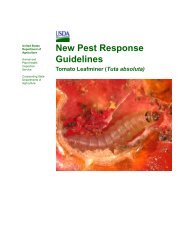
Phytophthora disease of alders: Phytophthora alni (Peronosporales: Peronosporaceae)http://www.invasive.org/species/subject.cfm?sub=10987Page 1 of 211/2/2010Home | About | Join / Sign In | ContactsSearch:GOInvasives 101 Species Images Publications Maps Videos Control EDRR CWMAs/CISMAs How to ... GlobalPhytophthora disease of aldersPhytophthora alni Brasier & S.A. KirkTaxonomic Rank: Oomycetes: Peronosporales: PeronosporaceaeNAPIS: FICBPUTBayer code:Selected Images from Invasive.orgView All Images at Invasive.orgFeature(s); Smooth-walledoogonium of P. alni (Swedishvariant) with oospore andamphigynous antheridium.Thomas Jung, , Bugwood.orgAdditional Resolutions & Image UsageSymptoms; Common alder (A.glutinosa) in a non-flooded forestplantation with root and collar rotcaused by P. alni.Thomas Jung, , Bugwood.orgAdditional Resolutions & Image UsageSymptoms; Grey alder (A. incana)with collar rot caused by P. alni;note the typical tarry spots at theouter bark and the tongue-shapedorange-brown necrosis of the innerbark.Thomas Jung, , Bugwood.orgAdditional Resolutions & Image UsageSymptoms; Root system of a 2-year-old nursery grown commonalder (A. glutinosa) with necroticlesions caused by P. alni.Thomas Jung, , Bugwood.orgAdditional Resolutions & Image UsageSymptoms; 2-yr-old sprouts of of an8-yr-old coppiced Alnus glutinosashowing wilting due to root andcollar rot caused by P. alni.Thomas Jung, , Bugwood.orgAdditional Resolutions & Image UsageSymptoms; Alder plantation onformer agricultural land; noteinfected grey alder (A. incana) withsparse, chlorotic and small-sizedfoliage.Thomas Jung, , Bugwood.orgAdditional Resolutions & Image UsageTaxonomic References:Index Fungorum. Paul Kirk. CABI, CBS and Landcare Research. http://www.indexfungorum.org/Invasive Listing Sources:National Cooperative Agricultural <strong>Pest</strong> Survey Target Species 2006, 2007, 2009North American Forest Commission Exotic Forest <strong>Pest</strong> Information SystemPartners
Phytophthora disease of alders: Phytophthora alni (Peronosporales: Peronosporaceae)http://www.invasive.org/species/subject.cfm?sub=10987Page 2 of 211/2/2010Invasive.org: Center for Invasive Species and Ecosystem HealthThe University of Georgia - Warnell School of Forestry and Natural <strong>Resources</strong> andCollege of Agricultural and Environmental Sciences - Dept. of EntomologyLast updated on Tuesday, February 02, 2010 at 10:08 AM
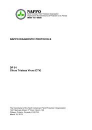
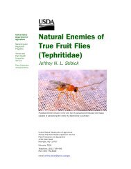
02. <strong>Pest</strong> Information Phytophthora spp.Phytophthora alticola and P. frigidaBackgroundInformationforPhytophthoraalticola, P.frigidaCold-tolerant Eucalyptus spp. are grown extensively for pulp-woodproduction in summer rainfall areas of South Africa with an altitude above1150 m. During the mid-1980’s, an increased demand for pulpwood led tothe expansion of cold-tolerant Eucalyptus plantations, the establishment of abreeding program for cold-tolerant species, and the introduction of severalalternative Eucalyptus spp. from seeds collected in natural stands inAustralia.Phytophthora collar and root rot is a widespread disease affecting a numberof cold-tolerant Eucalyptus spp. in South Africa. This disease hampersprogress towards introducing alternative Eucalyptus spp. yielding high pulpvolumes. Phytophthora spp. known to be associated with collar and root rotof Eucalyptus spp. in South Africa include P. boehmeriae, P. cinnamomi,and P. nicotianae. In 2001, P. nicotianae caused disease outbreaks onseveral cold-resistant Eucalyptus spp. in South Africa. This was particularlyinteresting as P. cinnamomi, rather than P. nicotianae, has typically beenassociated with mortality of cold-tolerant Eucalyptus spp.History andDistributionforPhytophthoraalticola, P.frigidaDamage toHosts byPhytophthoraalticola, P.frigidaMaseko et al. (2007) conducted a survey to assess the presence of P.nicotianae and other invasive Phytophthora spp. in Eucalyptus spp. in SouthAfrica. The study resulted in the isolation of two putative new Phytophthoraspecies, P. alticola and P. frigida associated with symptoms of Phytophthoracollar and root rot.Both species, P. alticola and P. frigida, are reported from South Africa. P.alticola has also been reported from Swaziland.The most common disease symptom of Phytophthora collar and root rot isprogressive wilting of the leaves due to girdling of the root collars. When thebark is removed, brown lesions extending from the roots are typicallyobserved. Other disease symptoms include root disease, bleeding lesionsfrom diseased stem tissues, and the formation of epicormic shoots on thestems of dying trees. Dying trees are usually present in small patchesthroughout the plantations, especially in areas prone to water-logging duringthe rainy seasons.2.8
02. <strong>Pest</strong> Information Phytophthora spp.EconomicImpact andEcologicalRange ofPhytophthoraalticola, P.frigidaPhytophthora collar and root rot disease hampers progress towardsintroducing alternative Eucalyptus spp. in South Africa, potentially yieldinghigh pulp volumes.Although P. frigida and P. alticola were proven to be pathogenic onEucalyptus dunnii, they were substantially less pathogenic than P.cinnamomi. Their relative importance as tree pathogens and in thePhytophthora complex associated with collar and root rot will need to bedetermined.Life Cycle andBiology ofPhytophthoraalticola, P.frigidaDue to the recent descriptions of P. alticola and P. frigida, little is knownabout their lifecycles and biology.P. alticola is homothallic in culture and thus, likely to be an inbreedingspecies. The cardinal temperatures for P. alticola were 15 o C and 30 o C. Noneof the isolates examined grew below 10 o C or above 30 o C. The optimaltemperature was 25-30 o C for growth. The pathogen is sensitive tohymexazol.The distinctive morphological features of P. alticola, which include papillateand caducous sporangia, indicate that it is adapted for wind or splashdispersal.P. frigida, in contrast, is a heterothallic species and has predominantly beenfound on E. smithii, planted in areas with an altitude above 1150m in SouthAfrica.The cardinal temperatures for P. frigida isolates examined were 10 o C and30 o C. None of the P. frigida isolates grew at 5 o C or 3 o C. The ability to growat temperatures lower than 15 o C indicates adaptation to a cool temperateclimate. It has not been associated, however, with shoot dieback of forest treespecies examined. Distinctive morphological characteristics include a stellateto petalloid growth pattern, and the ability to utilize L-asparagine better thannitrate as sole nitrogen source. The pathogen is tolerant to hymexazol.Plant HostsforPhytophthoraalticola, P.frigidaEucalyptus badjensis, E. dunnii, E. macarthurii are reported hosts for P.alticola.Acacia decurrens, A. mernsii, Eucalyptus dunnii, and E. smithii werereported as a host for P. frigida.The pathogens were isolated from rhizosphere soil around diseased plants.2.9
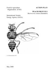
A B CDEAboveground and belowground diseasesymptoms of Phytopthhora root and collar rot inEucalyptus spp. (A) Young E. badjensis killed by P.alticola in Midillovo progeny trial. (B–D) Collar rotand formation of epicormic shoots in E. badjensis.(E) Root rot of E. macarthurii caused by P. alticola.(F) Root rot of E. saligna caused by P. frigida. (G)Discolouration of the inner collar and kinoexudation of E. smithii infected by P. frigida.>F GPictures courtesy of Michael J. Wingfield
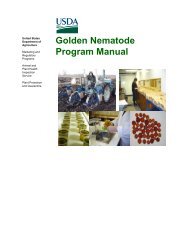
02. <strong>Pest</strong> Information Phytophthora spp.Phytophthora austrocedraeBackgroundInformationforPhytophthoraaustrocedraeAustrocedrus chilensis (Cordilleran cypress) is an endemic tree in theCupressaceae found in southern Argentina and Chile. It forms pure andmixed stands with Nothofagus spp. and, among the few conifers inhabitingsouthern Argentina, it has the largest distribution, covering approximately160,000 hectares.Mortality of Austrocedrus chilensis, termed ‘Mal del Cipres’ or ‘cypresswither’, in Argentina was first detected in 1948. The mortality had beenstudied for many years but its cause had remained unclear. Several studies ofbiotic and abiotic factors have tried to elucidate the origin and causes of thisdisease, but in spite of the amount of information gathered, a satisfactoryetiology had not emerged.Pythiaceous fungi have been suggested by several authors as a possiblecausal agent. For this reason, a survey of Phytophthora spp. of Austrocedrusforests was conducted. Five Phytophthora species were detected inhabitingsoil of declining A. chilensis forests but none of them showed a clearrelationship with the decline (Greslebin et al., 2005).History andDistributionforPhytophthoraaustrocedraeDamage toHosts byPhytophthoraaustrocedraeEconomicImpact andEcologicalRange ofPhytophthoraaustrocedraeGreslebin et al. (2007) isolated a new Phytophthora species, P. austrocedrae,from necrotic lesions of stem and roots of Austrocedrus chilensis.The disease is confirmed to be present in Argentina. According to Filip andRosso (1999), the disease possibly occurs in south-eastern Chile.The main symptom of the disease is progressive withering and subsequentdefoliation of the tree, which finally dies while standing. The first visiblesymptom in individual trees is chlorotic foliage. Trees may die rapidly, inwhich case foliage changes from chlorotic to red (Filip and Rosso, 1999).On the rootlets of affected trees, dead tissue can be observed. This becomessevere during dry summers. Basal resinous exudates and red-brown necroticlesions are seen in the inner bark extending up the bole from killed roots.Brown cubic rots and sapwood caused by wood-decomposer fungi arefrequently, but not always, associated with dead or dying trees (Greslebin etal., 2007). Trees of all sizes are affected.A. chilensis is economically valuable because of the high quality of its woodused for construction and woodworking, and it has great tourist and scenicappeal (Greslebin et al., 2005).The disease affects tourism, recreation, and commercial forestry in Argentina(Greslebin et al., 2005; Manna et al., 2008b). Austrocedrus forests surroundmost of the tourist cities and villages. It is nearly impossible to carry outappropriate silvicultural management of affected stands because the2.10
02. <strong>Pest</strong> Information Phytophthora spp.appearance and evolution of the disease cannot be predicted. Another seriousconsequence of the disease is the replacement of native forests with exoticintroduced species. This is due mainly to the lack of effective control leadingthe public institutions in charge of forest management to authorize landowners with affected forests to fell the dead trees and replace them with otherspecies.Life Cycle andBiology ofPhytophthoraaustrocedrae‘Mal del cipres’ begins in the root system. The distribution and pattern ofspread of mortality in a stand (patchy) is consistent with a soil-bornepathogen. The disease appears to be associated with areas of poor drainage,fine-textured soils, and areas of higher precipitation (Manna et al., 2008b).Allophanic volcanic soils do not appear to favor disease development(Manna and Rajchenberg, 2004). The disease has been associated with lowcanopy cover, organic horizon thickness, elevated pH, and high understorycover (Manna et al., 2008a).P. austrocedrae is a homothallic species characterized by semipapillatesporangia, oogonia with amphigynous antheridia, and very slow growth (1-2mm/day on V-8 agar at 17.5 o C optimum temperature). Growth was favoredby cool temperatures. Isolation from diseased trees was successful whenplates were incubated at 17 o C but unsuccessful at temperatures of 20 o C orabove.Plant HostsforPhytophthoraaustrocedraeAustrocedrus chilensis2.11
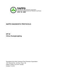
P. austrocedrae, aboveground symptomsPictures courtesy of Alina Greslebin and Carlos Baroll
P. austrocedrae, aboveground symptoms (2)Pictures courtesy of AlinaGreslebin and Carlos Baroll
P. austrocedrae, inner bark redbrownnecrotic lesionsPictures courtesy of Alina Greslebinand Carlos Baroll
P. austrocedrae, inner bark redbrownnecrotic lesions
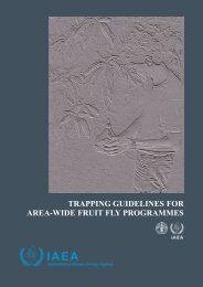
02. <strong>Pest</strong> Information Phytophthora spp.Phytophthora boehmeriaeBackgroundInformationforPhytophthoraboehmeriaeHistory andDistributionforPhytophthoraboehmeriaeDamage toHosts byPhytophthoraboehmeriaePhytophthora boehmeriae was first isolated from Boehmeria nivea (ramie, orChinese silk tree), by Sawada (1927) in Taiwan. It has not been reported inTaiwan since then (Ho, 1990), but has been reported from multiple othercountries.The disease is present in Argentina, Australia, Brazil, China, Greece, India,Japan, Korea, Mexico, Papua <strong>New</strong> Guinea, South Africa, Taiwan, and <strong>New</strong>Zealand.There is a herbarium record of P. boehmeriae from the United States in 1946,however, no locality or host information is provided.(http://194.203.77.76/herbIMI/DisplayResults.asp?strName=Phytophthora+boehmeriae). P. boehmeriae is not believed to be present in the UnitedStates.In nature, P. boehmeriae can infect, damage, and blight the seedlings andbolls of cotton, the leaves and stems of ramie, the leaves of paper mulberry(Broussonetia papyrifera), the fruits of citrus, and the roots of pine.Cotton: On seedlings, P. boehmeriae produces round or irregular, watersoaked,dark-green spots or lesions on cotyledons or true leaves, resulting inpremature leaf fall or wilting of some or all leaves under cold, humidconditions. On the roots and root-stem transition zones, the disease firstappears as brown streaks and then as a brown rot, resulting in wilting anddeath of whole seedlings.On bolls, dark-green, water-soaked lesions generally form at basal cracks ortips, causing rotting of the tissues within, with a layer of fungal growth onthe surface under humid conditions. Sporangia may be present on the surfaceof rotten bolls and oospores may be present on the cotton lint and internalcarpel surface of infected bolls. As the spots develop, whole diseased bollsmay rot away. Infected tissues may turn almost black. Infection is restrictedto the lower two-thirds of the cotton plant (Elena and Paplomatas, 1998).Ramie: The pathogen attacks leaves and stems. The leaf spots are round orirregular, water-soaked and light green at first, then dark brown or darkgreen. At later stages, the spots turn yellowish brown or gray at the center,with a brown margin. Diseased leaves tend to fall early. Elliptical, darkbrownstem lesions are present, mainly at the bases, resulting in rotting ofwhole bases.Paper mulberry and Chinese wingnut: Leaf spots similar to those found oncotyledons or leaves of cotton are observed. Root rot is also observed on2.12
02. <strong>Pest</strong> Information Phytophthora spp.Chinese wingnut.Citrus: The pathogen causes brown fruit rot and root stock gummosis.Pine: The pathogen causes root rot on pine (Oxenham and Winks, 1963).Geraldton wax plant: Plants show progressive yellowing of the branchesfrom the base to the top of the stems. Leaves of diseased plants becomegrayish green, then yellow, and finally straw-colored. Leaves remainattached to the branches after the plants die. Root and stem discoloration wasobserved and the root cortex sloughed off (Wolcan and Lori, 2001).Black wattle: Disease caused by P. boehmeriae is evident by dark lesions atthe trunk base without gum exudation up to 10 m in height (Dos Santos et al.,2006).EconomicImpact andEcologicalRange ofPhytophthoraboehmeriaeP. boehmeriae has been reported on multiple hosts in multiple studies.Economic and ecological information is lacking on most hosts with theexception of cotton in China.In China, P. boehmeriae is the principal agent causing cotton blight andramie blight. Cotton blight, including cotton seedling blight and cotton bollblight, is one of the main diseases in cotton in mainland China. A severeattack can kill 30-50 percent of seedlings in the field in cool, wet weather.Seedling blight mortality up to 60 percent has been recorded in fields (Tang,1990). Cotton boll blight injures bolls, resulting in rotting. The rotting rateof bolls is 10 to 30 percent in ordinary years, with a maximum over 50percent in rainy years. In addition to yield loss, the disease affects cottonquality, resulting in reduced fiber length and decreased ginning outrun.In Greece, P. boehmeriae boll rot has been reported as a new threat to cottoncultivation. In Australia, it has damaged citrus and pine, causing rot of fruitsand roots, respectively.P. boehmeriae was reported as one of the causal agents of the gummosiscomplex in black wattle in Brazil and is now of quarantine importance for thecitrus industry. This was the first report of the involvement of P. boehmeriaein the etiology of the gummosis complex of black wattle in Brazil (DosSantos et al., 2006).Life Cycle andBiology ofPhytophthoraboehmeriaeP. boehmeriae is homothallic and oospores form abundantly in host tissues.When the tissues (leaves, bolls, etc.) infected by P. boehmeriae decompose,oospores formed in the diseased tissue are released into the soil. Undersuitable conditions, they germinate by germ tubes to form sporangia ormycelia, which may produce sporangia. The germination mode of sporangiais affected mostly by temperature. It has been observed that at 18 to 20 o C, all2.13
02. <strong>Pest</strong> Information Phytophthora spp.sporangia germinated indirectly to produce zoospores, whereas at 22 to 24 o Cmost sporangia germinated directly and terminated with secondary sporangiacapable of releasing zoospores (CABI, 2006).All spore forms may be carried long distances by water or soil and allgerminate in water. When the spores lodge (encyst) against a stem or root ofa certain host, such as ramie or pine, or splash onto a leaf or fruit of a suitablehost plant, such as cotton, they germinate to produce appressoria or myceliathat infect the plant through the cuticle or via stomata or wounds. The role ofthe chlamydospore in the lifecycle is not clear. Some researchers suggest thatthe fungus may overwinter as chlamydospores; others suggest thatchlamydospores are seldom present in P. boehmeriae, and oospores canoverwinter and survive in the soil (CABI, 2006).The pathogen is favored by high humidity and warm temperatures; therefore,it frequently occurs in low, wet areas in rainy years. The optimumtemperature for mycelial growth in culture is 25 to 30 o C, minimum 9 o C, andmaximum 34.5 o C. The minimum temperatures for sporangium and oosporeformation are 25 o C and 25 to 28 o C, respectively. Darkness stimulatesmycelial growth and oospore formation, whereas illumination inhibits both(CABI, 2006).There is evidence that P. boehmeriae is seedborne in cotton and can bedisseminated by seeds (CABI, 2006).Antheridia are amphigynous and almost spherical in shape. Often with aresidual oil globule. Antheridia have been shown to change to paragynous onmedia with low nutrient levels (Gao et al., 1998).P. boehmeriae is one of the most sensitive to metalaxyl of all Phytophthoraspecies and has been used as a bioassay for metalaxyl in soil.Plant HostsforPhytophthoraboehmeriaeAcacia mearnsii (black wattle), Ailanthus altissima (tree-of-heaven),Araucaria hetrophila (Norfolk Island Pine), Avicennia spp. (mangrove),Boehmeria frutescens var. concolor (nakai), Boehmeria nivea (ramie),Broussonetia papyrifera (paper mulberry), Cedrus deodara (Deodar cedar),Chamelaucium uncinatum (Geraldton waxplant), Citrus sinensis (orange),Citrus spp., Eucalyptus dunnii, Eucalyptus grandis, Eucalyptus macarthurii,Eucalyptus pilularis, Eucalyptus smithii, Ficus spp., Gossypium hirsutum,Gossypium spp., Malus domestica (apple), Malus sylvestris (apple), Perseaamericana (avocado), Persoonia longifolia (long-leaf Personnia), Pinuspatula (Mexican yellow pine), Pinus spp., Pterocarya stenoptera (Chinesewingnut), and Solanum melogena (eggplant) are hosts for P. boehmeriae.Experimental hosts for P. boehmeriae: Allium fistulosum (Japanese bunchingonion), Benincasa hispida (Chinese waxgourd), Capsicum annuum (bell2.14
02. <strong>Pest</strong> Information Phytophthora spp.pepper), Cephalonoplos segetum (common cephalanoplos), Chenopodiumalbum (fathen), Convolvulus arvensis (field bindweed), Corchorus capsularis(white jute), Cucumis sativus (cucumber), Cucurbita moschata (butternutsquash), Ipomoea batatas (sweet potato), Ixeris denticulata (stebbins), Ixerislaevignata (stebbins), Lycopersicon esculentum (tomato), Nicotiana rustica(tobacco), Nicotiana tabacum (tobacco), Phaseolus vulgaris (bean),Portulaca oleracea (little hogweed), Pyrus spp. (pear), Rehmannia glutinosa(Chinese-foxglove), Ricinis communis (castorbean), Solanum tuberosum(potato), Taraxacum mongolicum (Mongolian dandelion), Vicia bungei(vetch), and Xanthium sibericum (Siberian cocklebur).2.15
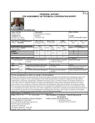
P. boehmeriae symptoms on Tree of Heaven (Ailanthus altissiman)Pictures courtesy of Byung-Soo Kim
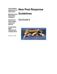
02. <strong>Pest</strong> Information Phytophthora spp.Phytophthora captiosa and P. fallaxBackgroundInformationforPhytophthoracaptiosa, P.fallaxHistory andDistributionforPhytophthoracaptiosa, P.fallaxA locally severe crown disease of Eucalyptus trees has been recorded since1986 in <strong>New</strong> Zealand. The main species affected are Eucalyptus saligna, E.botryoides, E. regnans, E. delegatensis, and E. fastigata. Dieback has beenrecorded in the central North and in Southland. The organisms responsibleare two species of Phytophthora, which have recently been described asPhytophthora captiosa and P.fallax (Dick et al., 2006). This disorder, incontrast to most Phytophthora diseases, can occur high in the canopy ofsusceptible trees.The two fungi (Phytophthora captiosa and P. fallax) are the firstPhytophthora species to be described only from <strong>New</strong> Zealand and the first tobe closely associated with the foliage of Eucalyptus spp. The majority ofPhytophthora spp. are present in the soil and cause root disease; thosespecies affecting aerial plant parts usually occurring on small plants.Canopies of Eucalyptus can be as high as 20 meters. Their modes ofinfection and dispersal are unknown.Phytophthora captiosa and P. fallax are only known to occur in <strong>New</strong>Zealand.Damage toHosts byPhytophthoracaptiosa, P.fallaxEconomicImpact andEcologicalRange ofPhytophthoracaptiosa, P.fallaxLife Cycle andBiology ofPhytophthoracaptiosa, P.fallaxLeaves, petioles, seed capsules, peduncles, and twigs may become infected.The effects of the disease range from minor leaf spots to major foliage lossand sometimes dieback.The disease is associated with crown disease and causes premature leaf dropand twig dieback upon inoculation.At this time, no information is available on the economic impact andecological range of these species.P. captiosa and P. fallax have non-papillate, non-caducous sporangia andboth are self-fertile (homothallic). Non-papillate, non-caducous sporangia areusually associated with soil-and root-inhabiting Phytophthora species ratherthan aerially dispersed species. Additionally, sporulation of both species hasnot been observed for either species in the field. The mode of infection andspread of these non-caducous Phytophthora species in the Eucalyptus treecanopy remains unknown. Studies have been proposed to assess whetherinvertebrates might be acting as vectors from foliage.2.16
02. <strong>Pest</strong> Information Phytophthora spp.Plant HostsforPhytophthoracaptiosa, P.fallaxEucalyptus botryoides and E. saligna are reported hosts for Phytophthoracaptiosa.E. delegatensis, E. fastigata, E. nitens, and E. regnans are reported hosts forPhytophthora fallax.2.17
P. Fallax symptoms on leaves of E. fastigataPicture courtesy of Margaret Dick
02. <strong>Pest</strong> Information Phytophthora spp.Phytophthora colocasiaeBackgroundInformationforPhytophthoracolocasiaeHistory andDistributionforPhytophthoracolocasiaeThe causal organism of leaf spot on taro (Colocasia esculenta) was firstdescribed as Phytophthora colocasiae in Java (Indonesia) (Raciborsky,1900). There is little information on the origin of P. colocasiae, but there areindications of an Asiatic origin (Zentmyer, 1988). Only the A1 mating typeof P. colocasiae occurrs in Hawaii and only the A2 mating type is reportedfrom Taiwan, indicating that they are not likely from the center of origin ofthe pathogen (Ko, 1979; Ann et al., 1986). Zhang et al. (1994) showed thatHainan Island, an offshore island in the tropical region of southern China, isinside the center of origin of P. colocasiae. P. colocasiae has beendistributed in vegetatively propagated material and by most likely via soil.The disease is currently present in the following countries/territories:American Samoa, Argentina, Bangladesh, Belau, Borneo, Brazil, Brunei,Burma, Ceylon, China, Dominican Republic, Equatorial Guinea (Bioko),Ethiopia, Federated States of Micronesia, Guam, India, Indonesia, Japan,Korea, Madagascar, Malaysia, Mauritius, Myanmar, Nepal, NorthernMariana Islands, Pakistan, Palau, Papua <strong>New</strong> Guinea, Puerto Rico,Philippines, Reunion, Samoa, Seychelles Islands, Solomon Islands, SriLanka, Taiwan, Thailand, and Trinidad and Tobago.United States: There are numerous reports from Hawaii, where the pathogenhas a widespread distribution. Reports from continental United States (NorthCarolina and California), however, need to be confirmed. Abad et al. (1994)report P. colocasiae on American ginseng in North Carolina, but accordingto Abad (personal communication) the isolate needs to be confirmed viamolecular methods.Damage toHosts byPhytophthoracolocasiaeAffected leaves initially show small dark brown to olive-green spots (oftenwater-soaked), which enlarge rapidly and turn purplish brown with yellowishmargins. The lesions frequently form concentric zones (zonate) and exudedrops of yellowish liquid. The exudate and leaf spot symptoms are bestviewed at night or in the early morning hours. Some of the diseased tissuesmay be covered with a whitish fuzz of sporangia. When the petiole or leafstalk becomes infected, rots are usually long, brown, and occur anywhere onthe stalk. When the rot expands, the stalk becomes soft, is often unable tosupport the weight of the leaf, and breaks (Brooks, 2005; CABI, 2006).As the disease progresses, the lesions (mostly along the leaf margin) continueto expand and frequently coalesce. Diseased tissues disintegrate, formingholes of irregular size, and affected leaves collapse within 20 days ofunfurling, compared to 40 days for healthy leaves (Jackson and Gollifer,1975). Exudates associated with diseased tissue become yellow to brown andform small crusts. The normal six to seven leaves per plant can be reduced tothree or four leaves per plant by severe disease incidence, which reduces net2.18
02. <strong>Pest</strong> Information Phytophthora spp.photosynthesis and corm yield. If weather conditions are favorable, the entirefield can be blighted in 7 to10 days (Trujillo, 1965). Highly susceptiblecultivars appear to be ‘melting’ in the field, producing smaller and smallerleaves on shorter petioles (Brooks, 2005; CABI, 2006).In both tolerant and resistant cultivars, diseased tissue falls away from spots,forming holes (often referred to as ‘shot-holes’) in the leaf (Brooks, 2005;CABI, 2006).After harvest, gray-brown to dark-blue lesions occur on undamaged corms.These lesions enlarge rapidly and coalesce. The boundary between healthyand diseased tissues is usually indistinct and soft. Affected corms are almostcompletely decayed 8 days after harvest in wet conditions.EconomicImpact andEcologicalRange ofPhytophthoracolocasiaeLife Cycle andBiology ofPhytophthoracolocasiaeP. colocasiae is a limiting factor in the production of taro in Southeast Asiaand the Pacific Islands, where it is consumed as a staple food crop on thefarms in developing countries (Brooks, 2008; Gallegy and Hong, 2008). Areduction in yield of 30 to 50 percent may occur if conditions are favorablefor disease development (Erwin and Ribeiro, 1996).Taro leaf blight was reported in the Samoan Archipelago in 1993 (Brooks,2008). ‘Niue’ was the favored cultivar at the time, comprising over 75percent of an estimated US $10 million in annual exports for Western Samoaand most of the 357,000 kg grown for local use in American Samoa. By1995, the epidemic had decimated the susceptible cultivar ‘Niue’, reducingSamoa’s export market to $60,750 (US) and American Samoan production toa reported 5,000 kg (Brooks, 2008).The mycelium of P. colocasiae is hyaline, coenocytic, inter- or intra-cellular.The haustoria are slender, long, and unbranched (Thankappan, 1985). Thesporangiophores are very slender, unbranched, extremely narrow at the tip,and measure up to 50 µm in length. The sporangia are elongated, lemon- orpear-shaped, and measure 38-60 x 18-26 µm. They germinate directly orindirectly depending on weather conditions. When indirect germinationoccurs, as many 12 reniform, biflagellate zoospores are released. Thickwalled,round, hyaline chlamydospores are also formed sometimes.The oogonium is spherical and yellow. Amphigynous antheridium persists atthe base of the oogonium for a considerable period after the oospores areformed. The oospores are spherical, have a 20-28 µm diameter, and lie freein the oogonium.Leaf blight of taro is mainly a foliar disease and occurs under warmconditions. P. colocasiae grows at 15 to 35 o C; with an optimum of 27 to30 o C. Epidemics are likely when zoospore release is maximized by repeatednight time temperatures close to 20 o C and relative humidity of 90 to 1002.19
02. <strong>Pest</strong> Information Phytophthora spp.percent (Trujillo, 1965; Narula and Mehrotra, 1984). Visible lesions appeartwo to four days after inoculation and, under humid conditions, can destroylarge taro leaves with a 30- to 40-day lifespan in 5 to 10 days. Yield lossesmay reach 50 to 60 percent under severe blight conditions and susceptibletaro cultivars can be destroyed completely (Brooks, 2008).Inoculum is always available in some areas due to continuous cropping, butnot in all areas where the crop is seasonal. Survival of P. colocasiae in thewild is poorly understood. Neither chlamydospores nor oospores have beenreported under field conditions, although they form readily in agar culture.Additionally, the pathogen is heterothallic, requiring two mating types toform oospores. Mycelium within stored corms used in propagating is apossibility for pathogen survival. Quintugua and Trujillo (1998) showed thatspores can remain viable for three months (as resting zoosporangia and/orchlamydospores); however, they also predicted that survival was less thanone year in the absence of a host, due to the poor saprophytic ability of thepathogen.Long distance dispersal occurs by means of vegetatively propagated materialand probably through soil. Local dissemination is via rain splash or winddrivenrain. In wetland (flooded) taro production, sporangia and zoosporesare spread between plants and fields by paddy water (Brooks, 2005).Giant African snails (Achatina fulica) have been shown to transmit P.colocasiae in taro (USDA APHIS, 2007).Plant HostsforPhytophthoracolocasiaeAlocasia macrorhiza (giant taro), Alocasia spp. (taro), Amorphophalluscampanulatus (elephant-foot yam), Bougainvillea spectabilis (bouganvilla),Cantharanthus roseus (periwinkle), Colocasia antiquorum (elephant’s ear),Colocasia esculenta (taro), Colocasia spp., Dracontium polyphyllum(guapa),, Hevea brasilensis (rubber), Panax quinquefolius (Americanginseng), Piper betle (betel), Piper nigrum (black pepper), Ricinis communis(castorbean), Vinca rosea (periwinkle), Xanthosoma sagittifolium (yautia),and Xanthosoma violacea (blue taro) are reported hosts of P. colocasiae.2.20
02. <strong>Pest</strong> Information Phytophthora spp.Phytophthora gallicaBackgroundInformationforPhytophthoragallicaHistory andDistributionforPhytophthoragallicaDamage toHosts byPhytophthoragallicaPhytophthora gallica was isolated from soil beneath a declining matureQuercus robur (English oak) and from the littoral zone of a lake withPhragmites australis (common reed) and Salix alba (white willow) asdominants. The pathogen was informally designated as Phytophthora taxon‘G’.The pathogen has been found in France and Germany (Jung and Nechwatal,2008). Recently the pathogen has been identified in Alaska in surveys ofalder stands (Trummer, 2009).In pathogenicity tests, P. gallica was only weakly aggressive to Q. robur andS. alba, and non-pathogenic to Phragmites australis. Therefore, its ecologicalniche, whether as a pathogen or saprotroph, remains uncertain. However, asaprophytic lifestyle seems unlikely given the much higher growth rates ofthe Pythium species ubiquitous in these wet ecosystems (Jung andNechwatal, 2008).P. gallica was also moderately aggressive to Alnus glutinosa and Fagussylvatica and non-pathogenic to Fraxinus excelsior (Jung and Nechwatal,2008).EconomicImpact andEcologicalRange ofPhytophthoragallicaThe economic and ecological impact of P. gallica is unclear at this time.Life Cycle andBiology ofPhytophthoragallicaBecause the species has only recently been described, there is littleinformation available on the life cycle and biology of P. gallica. P. gallica isa non-papillate, slow growing Phytophthora species; P. boehmeriae and P.kernoviae are its closest relatives. P. gallica produces colonies with limitedaerial mycelium and variable growth patterns. Gametangia are not formed insingle or mixed cultures with tester strains of known mating types, indicatingthat P. gallica may be sexually sterile. P. gallica produces globose andelongated irregular chlamydospores, of which a high proportion are abortive.In water culture, irregular hyphal swellings and non-papillate persistentsporangia are formed abundantly (Jung and Nechwatal, 2008).2.21
02. <strong>Pest</strong> Information Phytophthora spp.Plant HostsforPhytophthoragallicaQuercus robur (English oak), Salix alba (white willow), Alnus glutinosa(alder), and Fagus sylvatica (European beech) are reported hosts for P.gallica.2.22
Taro leaf blight lesions caused by P. colocasiae on cultivars of C. esculenta.1 21. Taro leaf blight (TLB) lesion with silveryring of sporangia2. Target-like lesion3. TLB lesions at 48 hours3Pictures courtesy of Fred Brooks
Taro leaf blight lesions caused by P. colocasiae on cultivars of C. esculenta.4564. Lesions with chlorotic halos5. <strong>New</strong> lesions after a rain6. Early lesion developmentPictures courtesy of Fred Brooks
Taro leaf blight lesions caused by P. colocasiae on cultivars of C. esculenta.77. Plant exudate from youngTLB lesion8. Leaf damage8Pictures courtesy of Fred Brooks
02. <strong>Pest</strong> Information Phytophthora spp.Phytophthora idaeiBackgroundInformationforPhytophthoraidaeiHistory andDistributionforPhytophthoraidaeiDamage toHosts byPhytophthoraidaeiEconomicImpact andEcologicalRange ofPhytophthoraidaeiLife Cycle andBiology ofPhytophthoraidaeiPlant HostsforPhytophthoraidaeiIn 1987, raspberry plants from several cultivars raised from root cuttings atthe Scottish Crop Research Institute (SCRI) were found to be infected with aPhytophthora spp. The plants had been used as uninoculated checks in a testto screen raspberry plants for resistance to P. fragariae var. rubi and hadbeen placed under conditions suitable for the development of the disease.Although the check plants showed no aerial symptoms, the root systems ofsome of the plants showed up to 20 percent root rot, and numerous oosporeswere seen in the cortex of the root (Kennedy and Duncan, 1995). Papillatesporangia were observed after floating pieces of root for 2 to 3 days at 15 o C.Kennedy and Duncan (1995) formally described the species Phytophthoraidaei.The pathogen is currently known only from England and Scotland.Root rot is observed with P. idaei, but no aerial symptoms are observed.Research at SCRI showed that a trend for reduced cane height was commonto most cultivars for plants grown in the ground, not under greenhouse/glasshouse conditions (Young et al., no date).Information is not available on the economic and ecological impact, butlevels of root rot in stocks at the SCRI rarely exceeded 5 percent of the rootsystem.The pathogen is potentially spread by cultivation of apparently healthyplants. The pathogen is homothallic with paragynous antheridia.Chlamydospores and hyphal swellings are not formed. Growth, sporangialformation, and oogonial and oospore formation are optimal at 20-22 o C.Rubus idaeus (red raspberry) is the only known host for P. idaei.2.23
Phytophthora idaei D.M. Kenn. 1995G. Abad, USDA-APHIS-PPQ-PHP-RIPPS-MDLG. Abad, USDA-APHIS-PPQ-PHP-RIPPS-MDLG.G.Abad,Abad,USDA-APHIS-PPQ-PHP-RIPPS-MDLUSDA-APHIS-PPQ-PHP-RIPPS-MDLG. Abad, USDA-APHIS-PPQ-PHP-RIPPS-MDL
Raspberry plants inoculated with P.idaei and P. rubia b ca. P. idaeib. Controlc. P. rubiPicture courtesy of David E.L.Cooke, Scottish Crop Research Institute
02. <strong>Pest</strong> Information Phytophthora spp.Phytophthora iranicaBackgroundInformationforPhytophthorairanicaHistory andDistributionforPhytophthorairanicaDamage toHosts byPhytophthorairanicaEconomicImpact andEcologicalRange ofPhytophthorairanicaLife Cycle andBiology ofPhytophthorairanicaPlant HostsforPhytophthorairanicaPhytophthora iranica was first isolated and described in Varamin, Iran byErshad (1971) from roots of eggplant. When artificially inoculated, tomatofruits, sugar beet roots, and potato tubers were susceptible, but eggplant,cucumber, sweet orange and apple fruits were not susceptible.IranP. iranica was isolated from eggplant but was not pathogenic to eggplant byexperimental inoculation (Ershad, 1971). P. iranica caused a soft rot ofpotato tubers, which turned pink when exposed to air. This symptom is alsocharacteristic of pink rot of potatoes caused by P. erthroseptica.Information is not available on the economic and ecological impact of P.iranica.The minimum temperature for growth is 10 o C, optimum 27.5 o C, andmaximum 35 o C (Erwin and Ribeiro, 1996).Sporangia are papillate (sometimes with two papillae per sporangium);ovoid, obpyriform, ellipsoid to subspherical, 30 to 72 µm long x 22 to 51 µmwide (average 47.9 x 36.8 µm) (length-breadth ratio 1.3:1); and persistent onthe stalk. Sporangiophores are short and sympodially branched (Ershad,1971). Chlamydospores are rare; 17 to 41 µm in diameter, average 28.7 µm;mostly intercalary; and rarely terminal. Hyphal swellings are not formed.Oogonia are formed in a single culture (homothallic) and are subsphericaland 21 to 45 µm in diameter, average 34 µm. Antheridia are mostlyparagynous, but occasionally an amphigynous antheridium is found;oospores are aplerotic, 15 to 37 µm in diameter, average 29.3 µm; theoospore wall is 1 to 6 µm thick, average 3 µm (Ershad, 1971).Solanum melongena (eggplant) (isolated from), Solanum tuberosum (potato),Lycopersicon esculentum (tomato), and Beta vulgaris (sugar beet) arereported hosts for P. iranica.2.24
Phytophthora iranica Ershad 1971G. Abad, USDA-APHIS-PPQ-PHP-RIPPS-MDL G. Abad, USDA-APHIS-PPQ-PHP-RIPPS-MDLG. Abad, USDA-APHIS-PPQ-PHP-RIPPS-MDLG. Abad, USDA-APHIS-PPQ-PHP-RIPPS-MDL
02. <strong>Pest</strong> Information Phytophthora spp.Phytophthora italicaBackgroundInformationforPhytophthoraitalicaHistory andDistributionforPhytophthoraitalicaDamage toHosts byPhytophthoraitalicaEconomicImpact andEcologicalRange ofPhytophthoraitalicaLife Cycle andBiology ofPhytophthoraitalicaPlant HostsforPhytophthoraitalicaPhytophthora italica was originally described as P. iranica. It was isolatedand proved to be pathogenic to roots of myrtle in Italy (Belisario et al.,1993). Cacciola et al. (1996) compared the myrtle isolate with an authenticP. iranica isolate. The myrtle isolate could be distinguished from P. iranica,as well as P. citricola and P. pseudotsugae by morphological characteristicsand by polyacrylamide gel electrophoresis of mycelial proteins and esterase.ItalyP. italica causes root rot of myrtle.Information is not currently available on the economic and ecological impactof P. iranica.Growth occurs between 10 and 35 o C, with optimum at 26 o C.Sporangia are papillate (frequently bipapillate, sometimes with three papillaeper sporangium); ovoid, obpyriform, subspherical or broadly ellipsoid, 14 to56 µm long x 11 to 38 µm wide (average 39 x 29 µm) (length-breadth ratio1.1:1.5); and persistent on the stalk. Sporangiophores are short andsympodially branched. Chlamydospores are rare. Oogonia are formed in asingle culture (homothallic) and are subspherical, often slightly flattened orpyriform with short stalk and smooth walls. The diameter of oogonia is 15 to29 µm, average 24 µm. Antheridia are paragynous and subspherical.Oospores are aplerotic, 11 to 23 µm in diameter, average 19 µm (Cacciola etal., 1996).Myrtle communis is the primary host of P. italica. The pathogen is reportedto be weakly pathogenic to tomato seedlings and apple fruit (browndiscoloration results).2.25
02. <strong>Pest</strong> Information Phytophthora spp.Phytophthora kernoviaeBackgroundInformationforPhytophthorakernoviaePhytophthora kernoviae was first discovered in Cornwall in October 2003during detailed surveys for P. ramorum. An unknown Phytophthora wasisolated from a large (>1m 2 ) aerial bleeding lesion on a mature Europeanbeech, Fagus sylvatica, in a woodland in Cornwall, southwest England(Braiser et al., 2005). The same Phytophthora was isolated concurrently fromRhododendron ponticum at another woodland site in the same area. The newPhytophthora was informally designated as “Phytophthora taxon C” and wasofficially named P. kernoviae by Brasier et al. (2005). The name is derivedfrom Kernow, the Cornish name for Cornwall where the organism was firstobserved. A nursery in the northwest of England (Cheshire) was found to becontaminated with P. kernoviae, but all infected plants were destroyed, andthe outbreak is considered eradicated.The pathogen was first detected in 2006 in <strong>New</strong> Zealand (North Island), intwo samples (soil and Annona spp. (cherimoya, custard apple). Whencollections and earlier studies of Phytophthora species were revisited, it wasfound that P. kernoviae was already occurring in stands in the 1950s to1960s. At present, P. kernoviae has been detected at several sites in soilsamples in the North Island has not caused extensive damage.History andDistributionforPhytophthorakernoviaeDamage toHosts byPhytophthorakernoviaeEngland, Scotland, Ireland, and <strong>New</strong> ZealandNote: The full host range of P. kernoviae is not known and needs to befurther investigated.Symptoms of P. kernoviae include bleeding trunk cankers on beech thateventually girdle and kill the tree. Leaf spots, blight, and shoot diebackdevelop on foliage hosts where the pathogen sporulates. The pathogen isdispersed by rain to bole hosts, such as beech and oak, where cankersdevelop.Tree hosts:Fagus sylvatica (beech): Initial symptoms are bleeding lesions on the trunkfound anywhere from ground level up to 12 meters. The bleeding is usuallydark brown to blue-black, and similar to symptoms caused by P. ramorum.Underneath, orange-pink to pinkish-brown active lesions in the inner bark arevisible. Sometimes girdling of the entire tree can occur. Older lesions appearsunken.Quercus robur (English oak): Bleeding lesions on the trunk are similar to2.26
02. <strong>Pest</strong> Information Phytophthora spp.those on F. sylvatica but may be more difficult to see both internally andexternally due to the thick outer bark ridges and outer bark plates of oak.Bleeding can occur from cracks between the bark ridges. Older cankers maynot become sunken as with beech.Quercus ilex (Holm oak): Symptoms are entirely limited to severe necroticleaf lesions and dieback of epicormic shoots (new shoots arising from nearthe base of the plant). There has been no evidence of sunken bleedingcankers on the stems of infected trees.Liridodendron tulipifera (tulip tree): Disease symptoms occur on foliage,shoots and trunk. Multiple bleeding lesions are formed on the trunk fromground level up to 9 meters, and the bark becomes highly corrugated as aresult. Internal lesions range in color from pale chocolate to dark chocolate toblue-black. Lesions tend to be limited in size (approximately 15 x 20 cm).Lesions can also develop on leaves; these are fairly restricted to leaf-tips(approximately 10-15 mm in length) and on the leaf margins. The necrotictissue dries out to a dark black color. Shoot dieback also occurs, and infectedshoots are defoliated.Magnolia spp.: Distinctive symptoms are found on infected foliage withinfection occurring anywhere on the leaf surface. Multiple infections areevident as numerous dark brown necrotic patches, giving leaves a spottyappearance. There is a tendency for these necrotic spots to merge anddevelop towards the center of the leaf (midrib), and as lesions become welldeveloped, leaves are noticeably mottled. The mottling may have angularedges, and uninfected tissue between necrotic areas becomes chlorotic(yellowing). Infections that take place at the leaf margin and cause it tocollapse and form a hard, dry rim. The petioles (leaf stalk) can be infectedand disease often progresses along the base following petiole infection. Budscan also become diseased, turning light khaki-gray.Shrub hosts:Rhododendron spp: The majority of the findings of P. kernoviae have beenon R. ponticum and rhododendron hybrids. Early leaf symptoms include ablackening of the leaf petiole that often extends into the base of the leaf. Thisnecrotic (dead and dying) lesion may progress farther into the leaf tissue andin extreme cases affect the whole leaf. Occasionally, however, onlyblackening of the leaf tip is observed. Both old and young leaves appear to beaffected equally. Leaves infected with P. kernoviae fall within a few weeksof infection, which is unusual for a Phytophthora infection of rhododendron.Shoot dieback and cankers can occur, which may girdle the stem tissue andcause leaves above the lesion to wilt. In severe infections, the whole bushmay be killed. Leaf and stem infections can be found at any height orposition on a rhododendron bush (Beales et al., 2006).2.27
02. <strong>Pest</strong> Information Phytophthora spp.Pieris spp. and Michelia doltsopa: Leaf blight symptoms similar torhododendron are seen, with no obvious dieback or cankers. Infection onMichelia doltsopa is characterized by necrotic leaf lesions on the leaves thatprogress along the leaf margin and into the tissue of the leaf blade. Necroticleaf tissue is characteristically a dark black-brown color. Lesions on leaves ofPieris spp. are typically a light tan to rusty brown color. Necrosis progressesdirectly towards the central (midrib) vein and along the vein, causing avisually striking leaf blight (Beales et al., 2006).Differences in pathogenicity have been observed between <strong>New</strong> Zealand andUnited Kingdom isolates of P. kernoviae (Widmer, 2008). On Magnoliastellata and Rhododendron ‘Cunningham’s White’, <strong>New</strong> Zealand isolatesshowed higher necrosis than isolates from the United Kingdom.EconomicImpact andEcologicalRange ofPhytophthorakernoviaeLife Cycle andBiology ofPhytophthorakernoviaeLike Phytophthora ramorum, P. kernoviae presents a risk to both the forestryand nursery industries. In Europe, Fagus sylvatica is an important forest treeand is planted for amenity purposes. Rhododendrons are commonly grownornamentals, but R. ponticum is considered an invasive plant in woodlands.Other ornamental hosts are valuable trees and shrubs. Comparativeinoculations of P. kernoviae and P. ramorum on beech stems suggest thatlesion development and tissue colonization are significantly more rapid for P.kernoviae than for P. ramorum (Brasier et al., 2005). Thus, the rate of spreadand mortality in woodlands is potentially greater for P. kernoviae, given thatthe more slowly colonizing P. ramorum has already proven to be a seriousthreat to invaded forests (Benson et al., 2008). Potential hosts for P.kernoviae occur in both east and west coast forests of the United States.Further studies are needed on the biology and epidemiology of P. kernoviaesince this is a newly described species of Phytophthora. Like P. ramorum, P.kernoviae causes widespread foliar necrosis and shoot dieback of the oftendense understory rhododendrons. Also like P. ramorum, P. kernoviae hascaducous sporangia (containing zoospores) and is most likely aerially orsplash dispersed.Under suitable conditions, asexual reproduction takes place, producing newsporangia. Numerous sporangia develop on the surface of leaf lesions duringperiods of leaf wetness. During periods of rain, sporangia are splashed orblown by the wind to other hosts, where motile zoospores are liberated.Zoospores presumably infect through the bark of beech and other tree hostsin a manner similar to those of P. ramorum. Long-distance spread can beaccomplished by the movement of infected rhododendron, beech, and otherhosts. P. kernoviae is apparently not a root pathogen, but it can be isolatedfrom soil. Movement of soil, litter, or plant debris can also spread thedisease. Spread of P. kernoviae within a woodland or drainage basin also canoccur when propagules of the pathogen are introduced into the runoff watereither directly or when associated host debris enters streams.2.28
02. <strong>Pest</strong> Information Phytophthora spp.Sporangia are not formed on bleeding stem cankers, but are found oninfected leaves, shoots, and fruit (Denman et al., 2006). Canker hosts are,therefore, not considered important for subsequent pathogen dispersal, butmay be important for pathogen survival. Additionally, P. kernoviae has beenfound to produce sporangia on asymptomatic foliage (Denman et al., 2008).Since the pathogen is homothallic, the possibility exists for the production ofoospores on mycelium colonizing the phloem and xylem tissue in trunkcanker lesions. Oospore production might provide a potential pathway forlong-term survival when trees die and bark is shed; however, oosporeproduction has yet to be documented in nature (Benson et al., 2008).Chlamydospores have not been observed in the laboratory or under fieldconditions.P. kernoviae has papillate, caducous to mouse-shaped sporangia (averagesize 39.6 x 25.6 µm, range 30 to 50 x 20 to 32 µm) and is homothallic withamphigynous antheridia approximately 11 x 10 µm and oogonia with anaverage diameter of 25 to 27 µm.Plant HostsforPhytophthorakernoviaeAnnona cherimola (cherimoya/custard apple), Castanea sativa (Europeanchestnut), Drimys winteri (winter’s bark), Fagus sylvatica (beech), Gevuinaavellana (Chilean hazelnut), Hedera helix (ivy), Ilex aquifolium ‘Variegata’(variegated holly), Liriodendron tulipfera (tulip tree), Lomatia myricoides(river lomatia), Magnolia spp. (M. amoena, brooklynensis, cylindrica,delavayi, kobus, liliflora, mollicomata, salicifolia, sargentiana, sprengeri,stellata, and wilsonii), Magnolia Gresham hybrid ‘Joe McDaniel’, MagnoliaGresham hybrid ‘Sayonara’, Magnolia Leonard Messel, Magnolia xsoulangeana, Michelia doltsopa (sweet michelia), Pieris Formosa(Wakehurst pieris), Pieris japonica (Japanese pieris), Podocarpus salignus(willow podocarp), Prunus laurocerasus (cherry laurel), Quercus ilex (Holmoak), Quercus robur (English oak), Rhododendron spp., Rhododendronponticum (pontic rhododendron), Sesquiadendron giganteum (giant sequoia),and Vaccinium myrtillus (bilberry) are reported hosts of P. kernoviae (Defra,2009).A finding in <strong>New</strong> Zealand in the 1950s from below a stand of Pinus radiatahas been shown by molecular analysis to be Phytophthora kernoviae (Defra,2009).Denman et al. (2006) list susceptibility of detached leaves to infection by P.kernoviae. Liriodendron tulipfera (tulip tree), Magnolia solangeana, andRhododendron catawabiense showed high susceptibility.2.29
02. <strong>Pest</strong> Information Phytophthora spp.Phytophthora melonisBackgroundInformationforPhytophthoramelonisPhytophthora melonis was first isolated from Katsura (1968, 1976) fromdiseased cucumber (Cucumis sativus) plants in Japan. The new species wascharacterized by homothallism, amphigynous antheridia, semipapillatesporangia, and the production of chlamydospores. A similar disease wasreported on cucumber and other cucurbits in mainland China, Taiwan, Iran,Egypt, Turkey, Korea, and India. Isolates from all of these countries weresimilar to P. melonis except the sporangia were non-papillate and proliferatedinternally; also, chlamydospores were not found. Isolates were consideredheterothallic. The latter isolates have been identified as P. melonis or as P.drechsleri. One isolate causing cucumber blight in China was treated as anew species, P. sinensis (Yu and Zhang, 1982).The original description by Katsura is erroneous (Erwin and Ribeiro, 1996).Ho et al. (2007) published a redescription of Phytophthora melonis to correcterrors in the original description.Ho and Jong (1986) considered Phytophthora melonis a synonym of P.drechsleri but this was not supported by molecular analysis (Cook et al.,2000). Ho (1986) examined isolates of P. melonis, P. sinensis, andP.drechsleri from Cucumis spp. and suggested they were synonymous.Despite confusion, it is now clear that whilst P. melonis and P. sinensis maybe synonymous, they are unrelated to P. drechsleri (Mills et al., 1991; Cookeet al., 2000).History andDistributionforPhytophthoramelonisDamage toHosts byPhytophthoramelonisThe current distribution for P. melonis includes: China, India, Iran, Japan,Korea, TaiwanCucumber: P. melonis causes a late blight of leaves, stems, and fruits. Soonafter heavy rains, infection is initiated in the collar region, which turnsgreenish brown and becomes soft. The stem eventually shrinks, and the plantfalls over. Lesions on leaves become darker green than the rest of the leafand slowly progresses toward the pedicel if moist conditions prevail.Infection on fruit is usually initiated at the tip, resulting in deeply depressed,dark green, water-soaked lesions. A soft rot of the fruit follows (Erwin andRibeiro, 1996). Rapid wilting and death of the plants is also common.Pointed Gourd: A fruit and vine rot is caused by P. melonis.Pistachio: Gummosis, characterized by the exudation of gum from the crown,is observed. The tissues under the bark of the crowns, lower trunk of affectedtrees, and roots turn brown or dark brown and rot, often resulting in death of2.30
02. <strong>Pest</strong> Information Phytophthora spp.the tree.EconomicImpact andEcologicalRange ofPhytophthoramelonisLife Cycle andBiology ofPhytophthoramelonisLittle information is available on the ecological and economic impact of P.melonis. Since 1983, mortality was estimated as averaging 10-12 percent inpistachio in Iran (Mirabolfathy et al., 2001). Losses have been on theincrease. Due to rotting of cucurbit fruit, considerable yield loss would beexpected.Phytophthora melonis was easily dispersed by zoospores and sporangiacarried in irrigation water and via wind-blown rain splash in Taiwan (Lin andWu, 1985). P. melonis is most active after heavy rains. Rain wateraccumulating around the stem of the plant results in invasion of the collarregion. In Japan, the disease is most severe from the middle of May to theend of June (Erwin and Ribeiro, 1996).Lin and Wu (1985) showed that soil moisture strongly influenced thesurvival of P. melonis. The fungus persisted only for four weeks in floodedsoil. In soil with 6 percent or 20 percent moisture content, a condition thatpermitted soil aeration, however, the fungus could be detected from theinfested soil for a period of 20 and 5 weeks, respectively.Plant HostsforPhytophthoramelonisCucumis sativus (cucumber), Cucumis melo (cantaloupe), Citrullus lanatus(watermelon), Pistacia vera (pistachio), Trichosanthes dioica (pointedgourd) are hosts for P. melonis (Guharoy et al., 2006). Lens culinaris (lentil)was also identified as a host (Esmaili-Shirazifard and Banihashemi, 2008).Banihashemi and Mirtalebi (2008) used safflower seedlings as a selectivehost to discriminate between the morphologically similar P. melonis and P.drechsleri. P. melonis could not infect safflower seedlings; while P.drechsleri from various hosts attacked safflower within a short period. Atleast six other Phytophthora species (P. asparagi, cactorum, cryptogea,erythroseptica, palmivora, and quercina) are capable of infecting safflower.This method should only be used once a morphological identity has beenestablished. Additionally P. melonis does not induce pink rot symptoms inpotato tubers unlike P. drechsleri (Mostowfizadeh-Ghalamfarsa et al., 2005;Esmaili-Shirazifard and Banihashemi, 2008).2.31
Symptoms of the disease caused by P. melonis in cantaloupesCucumis melo)Pictures courtesy of Somnath Bhattacharyya
Symptoms of the disease caused by P. melonis in Pointed gourd(Trichutsanthes diocia Roxb.)Pictures courtesy of Somnath Bhattacharyya
02. <strong>Pest</strong> Information Phytophthora spp.Phytophthora multivesiculataBackgroundInformationforPhytophthoramultivesiculataHistory andDistributionforPhytophthoramultivesiculataDamage toHosts byPhytophthoramultivesiculataIlieva et al. (1998) examined Phytophthora isolations from diseasedCymbidium orchid plants samples received from growers by the Dutch PlantProtection Service since 1991 that could not be assigned to a known speciesof the genus. The authors named a new species, Phytophthoramultivesiculata, as the causal agent based on pathogen morphology andisozyme analysis.P. multivesiculata is reported from the Netherlands, <strong>New</strong> Zealand (NorthIsland), and Australia.Note: The specimen from Australia is from a single herbarium specimenoriginally identified as Phytophthora syringae, which has now been reidentifiedas P. multivesiculata (Cunnington et al., 2009).The leafy parts of the plants and the pseuduobulbs (modified stems) areattacked. Infection appears on the leaves after prolonged periods of rain or onindoor plants after they have been irrigated. According to Hill (2004), thedisease first appears as dark green lesions that expand rapidly under humidconditions to form large irregular patches of water-soaked tissue on matureleaves.The appearance of leaf lesions often signals the presence of a more damagingform of the disease. Infectious propagules that reach the foliage may bewashed down to the base of the plant and, if immature leafy shoots arepresent, down into the center of these growths. There, the immature leaftissues are attacked and a severe internal rot rapidly develops. Initially the rotis not visible and, often, the first indication that infection is present is achange in the color of the leaves. Infected shoots become gray-green andrapidly lose turgor. Often by this stage, the infection has already spreadthrough the attachment point of the shoot into the adjacent pseudobulb. If theplants are young and have only one or two pseuduobulbs, the infection mayprove fatal. Infected young pseudobulbs have a distinctive internal, blueblackor purplish-brown discoloration and a sour odor. The roots do notbecome infected and remain grayish-white apparently unaffected even afterthe rest of the plant has turned brown (Hill, 2004).According to Ilieva et al. (1998), dry rot of leaves (with a somewhat waxylooking surface) with a change of color to brown with typical horizontalzebra-like stripes, about 0.5 cm wide with lighter discoloration in the middleand a dark brown to black margin are observed on Cymbidium plants infectedwith P. multivesiculata. The base of the Cymbidium bulbs showed wetbrown-black discolored tissue.2.32
02. <strong>Pest</strong> Information Phytophthora spp.EconomicImpact andEcologicalRange ofPhytophthoramultivesiculataLife Cycle andBiology ofPhytophthoramultivesiculataPlant HostsforPhytophthoramultivesiculataInformation on the economic and ecological impact is not currently available.Hill (1982) observed that the Phytophthora spp. attacking Cymbidium plantsin <strong>New</strong> Zealand tended to be more active and damaging in the late summerand autumn, when temperatures were at their seasonal highs. Ilieva et al.(1998) found that P. multivesiculata has a relatively fast growth rate at 20 o Cand a high temperature for growth of 35 o C, the highest of any of thepresently known Group IV Phytophthora species (Hill, 2004).Cymbidium spp. are the only reported hosts for P. multivesiculata.2.33
Phytophthora multivesiculata Ilieva, Man in ‘t Veld,Veenbaas‐Rijkset Pieters. (1998)G. Abad, USDA-APHIS-PPQ-PHP-RIPPS-MDLG. Abad, USDA-APHIS-PPQ-PHP-RIPPS-MDLG. Abad, USDA-APHIS-PPQ-PHP-RIPPS-MDLAPHIS PPQ PHP G. Abad, USDA-APHIS-PPQ-PHP-RIPPS-MDL G. Abad, USDA-APHIS-PPQ-PHP-RIPPS-MDL
02. <strong>Pest</strong> Information Phytophthora spp.Phytophthora multivoraBackgroundInformationforPhytophthoramultivoraAs a result of wide scale forest quarantine and management of Phytophthoracinnamomi (an introduced species causing jarrah (Eucalyptus marginata)dieback) in Western Australia, extensive and regular testing of soil and planttissue samples for P. cinnamomi at the Vegetation Health Service (VHS)laboratory of the Department of Environment and Conservation has led to theisolation of a large range of Phytophthora spp. and undescribedPhytophthora taxa.In May and June 2007, Phytophthora isolates were recovered from therhizosphere of declining Eucalyptus gomphocephala, E. marginata, andAgonis flexuosa in Yalgorup National Park. These isolates morphologicallyresembled P. citricola and had similar growth rates at 25 o C. Recentevaluation of the VHS culture collection using molecular techniques hasidentified most of these isolates as a new taxon (Phytophthora spp. 4) in theP. citricola complex (Burgess et al., 2009). Phylogenetic analyses of the ITSand cox1 gene show that Phytophthora spp. 4 is unique and comprises adiscrete cluster within the major ITS clade 2 of Cooke et al. (2000) with itspresent closest relative being P. citricola. Scott et al., (2009a) formallydescribed Phytophthora spp. 4 as the new species Phytophthora multivora.History andDistributionforPhytophthoramultivoraDamage toHosts byPhytophthoramultivoraP. multivora is widespread in southwest Western Australia.In GenBank, 11 ITS sequences, designated as P. citricola, are identical to P.multivora. Seven are from unpublished studies in Hungary, Canada,Switzerland, Korea, Japan, and three isolates are from Spain (Zea-Bonilla etal., 2007; Moralejo et al., 2009). In addition, an isolate designated as P. sojaein a study from Japan almost has an identical sequence (Villa et al., 2006).Due to the widespread distribution of P. multivora across natural ecosystemsin Western Australia, it is likely that Western Australia may be the source ofdispersal, possibly via the nursery trade (Scott et al., 2009a).Phytophthora multivora causes dieback symptoms and an overall decline.Eucaplyptus gomphocephala and E. marginata: Severe dieback and mortalityare associated with P. multivora. Crown symptoms include thinning,clustering of leaves, and dieback of branches and parts of the crown.Banksia attenuata: Sudden wilting and death due to the girdling of the collar.Tongue-shaped, orange-brown necrosis of the inner bark can be observed.EconomicImpact andEcologicalRange ofPhytophthora multivora, as its name indicates, can affect a wide range ofspecies. Experts believe this pathogen could kill up to half of the plantspecies in Western Australia, equating to more than 3000 plant varieties(Mandurah Mail, 2009). Information on the economic impact is not currently2.34
Images of P. multivora, sporangiaPictures courtesy of Dr. Peter Scott
Images of P. multivora, sporangiaPictures courtesy of Dr. Peter Scott
02. <strong>Pest</strong> Information Phytophthora spp.PhytophthoramultivoraLife Cycle andBiology ofPhytophthoramultivoraavailable.Phytophthora multivora is the first record of a Phytophthora speciesassociated with Eucalyptus gomphocephala decline and has been shown toproliferate on calcareous soils, believed to be suppressive to otherPhytophthora species, including P. cinnamomi.Initial results indicate that P. multivora attacks the fine roots of E.gomphocephala and may not colonize the stem collar under normalconditions. The high oospore wall index (ratio between the volume of theoospore wall and the volume of the whole oospore) indicates that P.multivora may be adapted to tolerate periods of prolonged drought (Scott etal., 2009b).Plant HostsforPhytophthoramultivoraPhytophthora multivora is pathogenic to Eucalyptus gomphocephala and E.marginata (jarrah) and is believed to be involved with the decline syndromeof both Eucalyptus species within the tuart woodland in south-west WesternAustralia.Other (mostly native) Australian hosts include: Agonis flexuosa (pepperminttree), Banksia attenuata (candlestick banksia), B. grandis (giant banksia), B.littoralis (swamp banksia), B. menziesii (firewood banksia), B. prionotes(acorn banksia), Bossiaea spp., Conospermum spp., Gastrolobium spinosum(prickly poisonbush), Leucopogon verticillatus (tassle flower), Patersoniaspp., Pinus radiata (Monterey pine - plantation), Podocarpus drouyniana(Drouyn's podocarpus), and Xanthorrhoea gracilis (graceful grass tree).Based on sequence data, isolates from Spain on mango (Mangifera indica)are P. multivora (Zea-Bonilla et al., 2007).Phytophthora pinifoliaBackgroundInformationforPhytophthorapinifoliaHistory andDistributionforPhytophthorapinifoliaSince 2004, a new disease of Pinus radiata, referred to ‘Daño Foliar delPino’ (DFP) has appeared in the Arauco province of Chile and subsequentlyspread to other areas. Duran et al. (2008) officially name the pathogenPhytophthora pinifolia.In February 2004, unusual tree mortality appeared in a 6-year old P. radiatastand of about 70 ha on the Arauco coast of Chile. In October 2004 and in thesame area, a serious needle blight disease was observed and associated withthis mortality. The damage increased dramatically expanding toapproximately 60,000 ha by the end of 2006 (Duran et al., 2008).Attempts to isolate the pathogen that caused the needle blight led to several2.35
02. <strong>Pest</strong> Information Phytophthora spp.fungi, but upon inoculation these fungi did not reproduce the symptoms ofthe disease and were thought to represent opportunistic endophytes orsecondary inhabitants.Isolations from diseased needles in July 2007 on Phytophthora selectivemedia consistently yielded a Phytophthora species. Pathogenicity of thisPhytophthora species was confirmed and symptoms were reproduced onPinus radiata seedlings (Duran et al., 2008).Phytophthora pinifolia is presently only known from Chile.Damage toHosts byPhytophthorapinifoliaThe pathogen causes serious needle blight.Mature Trees (up to 18-year-old): Symptoms begin with a reddening of thepast year’s needles in early winter. The first needles to display symptoms arethose on the lower sides of the branches. Needles die and assume a distinctlygray color and begin to fall. Initially, dead and dying needles are retained onthe branches giving the trees the appearance of having been severelyscorched. Needles then fall from the trees, which can be almost entirelydefoliated. <strong>New</strong> needle growth is not affected and the trees appear to recoveruntil infection re-occurs in the following season. After two or three years ofdefoliation, trees occasionally die and this appears to be hastened due toinfection by Diplodia pinea (Diplodia blight of pine), which is a well-knownopportunistic pathogen (Duran et al., 2008).One of the earliest symptoms on affected needles is the emergence of darkresinous bands on the green needles, which appear transparent when viewedwith backlighting. These bands are found at various positions, either close tothe bases or higher up on the needles. Drops of resin are often found at thebase of the needles and the tissue within the papery brachyblasts iscommonly collapsed and has a light gray color. Needles often collapse fromtheir bases just above the branches and hang at right angles from thebranches. When the bark is removed, a distinct brown or reddish browndiscoloration can be seen in the phloem and cambium, particularly where it isassociated with dying needles.Young Trees (up to one and 4-years-old): Symptoms appear different tothose on mature trees. Damage to young trees is most common and mostsevere where stands occur alongside larger trees affected by DFP. One of thefirst and most obvious symptoms to appear on young trees is where younggrowing terminal shoots wilt and die rapidly. Lesions are typically found onthese shoots or on the needles associated with them, depending on the time ofthe year. The damage resulting in the wilted growing shoots is typicallyfound on parts of branches or shoots a short distance below the obviouslyaffected tissue. Close inspection reveals needles with symptoms similar tothose on older trees with infected bases leading to girdling of the stems. Dark2.36
02. <strong>Pest</strong> Information Phytophthora spp.resinous bands can be found on the needles in the early stages of symptomdevelopment and infection appears to be concentrated within the paperybrachyblasts at the base of the needle fascicles. On younger trees, necrosis ofthe cambium is more pronounced than that on mature trees and the impact ofinfection is commonly more severe. Resin can be found exuding profuselyfrom the brachyblasts or the bases of the needles. Branches can have largenumbers of needles hanging at right angles, apparently from their bases, andinfections begin on the lower sides of branches. Removal of the outer barkreveals distinct lesions below the needle bases and in many cases, due tomultiple infections, they coalesce to form cankers in the phloem and outercambium. These cankers result in girdling of the stems or branches andwilting of the needles and shoots proximal to them. Naturally regeneratedplants and newly planted seedlings are equally affected by DFP, and appearto wilt and die rapidly due to their small size (Duran et al., 2008).EconomicImpact andEcologicalRange ofPhytophthorapinifoliaForestry companies plant species of Pinus, Eucalyptus, and Acacia, nonnativetrees, in forestry plantations. Using these species, forestry companiesproduce solid-wood products and pulp for paper and rayon production(Duran et al., 2008). Growth of these non-native trees is in many casesexceptional and typically far beyond that associated with the trees in thenatural environment, which leads to greater economic value for these species.Daño Foliar del Pino is generally accepted in Chile to be the most seriousproblem yet to have affected pine forestry in the country (Duran et al., 2008).The United States produces nearly $67 billion in pine annually (NPAG,2008). The introduction of P. pinifolia into the United States could have adevastating effect on the pine industry. With the United States importing$327 million in lumber and wood in the rough from Chile and exporting over$340 million in pine products in 2006, the establishment of P. pinifolia in theUnited States would have a great impact on pine products and trade in theUnited States (NPAG, 2008).The discovery of an aerial Phytophthora spp. causing a serious disease of P.radiata in Chile, adds to a number of new and serious tree-infecting aerialPhytophthora spp. that have recently been discovered. <strong>New</strong>ly plantedseedlings and naturally regenerated plants die if infected in the first year ofgrowth, making P. pinifolia a serious threat to reforestation.Life Cycle andBiology ofPhytophthorapinifoliaThe pine needle blight in Chile has a distinctly seasonal pattern ofoccurrence. Trees begin to show symptoms in early winter from about Julyonwards when the temperatures begin to drop to between 6 and 12°C. This isalso the start of the rainy season and there appears to be a very closeassociation of the disease with rainfall. A spatial analysis has shown that thesouthern slopes (higher humidity, most free water, and low solar radiation)are most severely affected (Duran et al., 2008).2.37
02. <strong>Pest</strong> Information Phytophthora spp.While the symptoms of DFP in Chile are consistent with the biology of anaerial Phytophthora sp., there are many questions regarding the biology ofthe pathogen that remain to be answered. While it is assumed that thesporangia are the infective propagules, this has yet to be shownexperimentally. These structures were not abundant in culture and morenatural conditions under which to produce them will need to be developed.Likewise, infection studies with zoospores and the infection biology and lifecycle of the pathogen remain to be understood.Plant HostsforPhytophthorapinifoliaBackgroundInformationforPhytophthorapolonicaHistory andDistributionforPhytophthorapolonicaDamage toHosts byPhytophthorapolonicaEconomicImpact andEcologicalRange ofPhytophthorapolonicaLife Cycle andBiology ofPhytophthorapolonicaPinus radiata (Monterey pine) is the only known host for P. pinifolia.Phytophthora polonicaIn a survey of Phytophthora associated with alder decline in Poland, severalisolates of a homothallic Phytophthora spp., which could not be assigned toother taxa including Phytophthora alni subspecies, were consistentlyrecovered from rhizosphere soil samples. Based on morphology andsequence data, the Phytophthora was found to be a new species. Belbahri etal. (2006) described P. polonica as a new species of Phytophthora.PolandPhytophthora polonica was slightly pathogenic to alder twigs with tissuediscoloration progressing a few millimeters beyond the inoculation woundafter 10 days. The pathogenic status of P. polonica remains unclear andfurther research is necessary.Information on the economic and ecological impact is not currently available.Little information is currently available on the life cycle and biology of P.polonica. P. polonica is characterized by moderate to slow growth rate of itscolony on carrot agar at 20°C, high optimal (30°C), and maximum (38°C)growth temperatures, formation of catenulate, often lateral, hyphal swellings,large chlamydospores in agar media and in soil extract, persistent, ovoid, toellipsoid nonpapillate sporangia, and large oogonia with paragynous and2.38
02. <strong>Pest</strong> Information Phytophthora spp.sometimes amphigynous arrangement.Plant HostsforPhytophthorapolonicaAlnus glutinosa (alder) is the only known host for P. polonica.Phytophthora porriBackgroundInformationforPhytophthoraporriHistory andDistributionforPhytophthoraporriPhytophthora porri was originally described as a pathogen of leek (Alliumporrum), causing waterlogged areas followed by a whitening of the tips ofleaves and other affected parts of this plant (Foister, 1931). Later, thepathogen was isolated principally from leek and other members of theLiliaceae and a few dicotyledonous hosts.The pathogen is reported from Australia, Belgium, Canada, China, England,Greece, Ireland, Japan, Netherlands, Norway, Scotland, and South Africa(Taylor, 1965; Von Maltitz and von Broembsen, 1984; Erwin and Ribeiro,1996). The reports from Canada on carrot, however, may be a distinct species(Stelfox and Henry, 1978; Ho, 1983).France, Germany, Italy, <strong>New</strong> Zealand, Switzerland, and United States arealso reported in the distribution for P. porri. This distribution is reported forP. porri from Brassica spp. The Phytophthora species from Brassica spp.,however, is now included in P. brassicae (Man In’t Velt et al., 2002).Damage toHosts byPhytophthoraporriAllium spp. – Initially, yellowing and dying of the tips of leaves is observed,followed by this area turning white. The white area may measure half an inchto six inches long, and while most bend backwards, becoming disfigured bysaprophytic fungi, some turn crisp, curl, and do not bend. Sometimes theapical attack is replaced by a marginal infection at any place from near thetip to about half-way down the leaf. As this area dies, the tissues contract andthe leaf becomes twisted. Usually associated with these symptoms is a waterloggedarea developed half-way down or at the base of the leaf (Foister,1931).Campanula spp.: The crown region is gray-brown to chocolate brown; thediscoloration spreads upwards into the leaf bases and flowering shoots anddownwards into the roots. The upper parts of the roots are pale pink to brownand later turn dark brown and disintegrate, freeing the leafy rosette from theroots (Legge, 1951).Dianthus caryophyllas (carnation): Collar rot is the primary symptomassociation with P. porri. Dark, wet lesions that later became gray and dryappear on the stalks (Kouyeas, 1977).2.39
02. <strong>Pest</strong> Information Phytophthora spp.Gladiolus spp: Wet rot of the bottom leaves is seen, which leads to plantdeath (Kouyeas, 1977).Lactuca sativa (lettuce): Diseased plants wilt and collapse completely within2 to 4 weeks. Examination of wilted plants shows a dark, firm rot of stemsthat extends to the soil level upwards while the roots appear healthy (Sitepuand Bumbieris, 1981).EconomicImpact andEcologicalRange ofPhytophthoraporriLife Cycle andBiology ofPhytophthoraporriPhytophthora porri is a serious disease of winter leek in Europe in intensivevegetable growing areas. The disease results in lower yields and loss ofquality (marketable yield) (Dobson and Clarkson, 1989). Epidemics destroymore than 50 percent of the crop before January to April, when winter leek isharvested (CABI, 2006). Griffen and Jones (1977) reported yield losses of30-50 percent in autumn-sown salad onions in the United Kingdom due to P.porri.Phytophthora porri is homothallic. Oospores, and possibly chlamydosporeswhich are very similar in shape, form in infected leaves, may enter the soilwith leaf debris and survive the crop-free period from February till July, andprobably longer. In autumn, some oospores may germinate immediately toform sporangia, which may release zoospores. Infections via oospore occuronly on above ground plant parts (Smilde et al., 1996a). Sporangia are notdischarged easily by wind. The zoospores may be transported to the leaves ofhosts by rain splash, thus triggering a new epidemic (Smilde et al., 1996a).Prolonged dew periods and rain events (free water) have been shown to oftenprecede the development of disease (Taylor, 1965; Smilde et al., 1996a).Smilde et al. (1996b) reported oospore germination in vitro. Naturalsecondary spread of P. porri to neighboring rows was observed after 3 weeksin a leek field (Smilde and van Ness, 1992). There is no infection of theroots.Phytophthora porri is characterized by non-papillate to semi-papillatesporangia, both amphigynous and paragynous antheridia, slow growth, lowmaximum growth temperatures, and coiling growth of hyphae (Man In’tVeld et al., 2002). Cardinal temperatures for mycelial growth of P. porriwere 11°C (Smilde et al.,1996b). Optimum and maximum temperatures for the P. porri isolate fromlettuce were about 8 to 10°C lower than those from other hosts (Sitepu andBumbieris, 1981).Disease is patchy and is usually most severe in depressions where water2.40
02. <strong>Pest</strong> Information Phytophthora spp.accumulates after rain or irrigation.Plant HostsforPhytophthoraporriAllium ascalonicum (shallot), A. bakeri (Chinese onion), Allium cepa (onion,shallot), A. chinense (rakkyo), A. fistulosum (Japanese bunching onion), A.geyeri (Geyer’s onion), Allium nipponicum (Japanese garlic), Allium porrum(leek), Allium sativum (garlic), A. tuberosum (Chinese chive), A. wakegii,Campanula persicifolia (peach-bells), Chrysanthemum spp., Daucus carotasubsp. sativus (carrot), Dianthus caryophyllus (carnation), Gladiolus spp.,Hyacinthus orientalis (hyacinth), Lactuca sativa (lettuce), Partheniumargentatum (guayule), Pastinaca sativa (parsnip), Rosa spp. (rose), andTulipa spp. (tulips) are reported hosts for P. porri.The reports from Canada on carrot, however, may be a distinct species(Stelfox and Henry, 1978; Ho, 1983).Isolates from the Brassica spp. are now referred to as a new speciesPhytophthora brassicae (Man In’t Veld et al., 2002).Isolates of P. porri appear to be host specific. Isolates from Allium were onlyweakly pathogenic to cabbage and vice versa (De Cock et al., 1992). Isolatesfrom Campanula persicifolia and leek failed to cross-infect (Legge, 1951).Also isolates from carnation did not infect leek and vice versa. The isolatefrom lettuce, described by Sitepu and Bumbieris (1981) was not pathogenicto either leek or cabbage. Tichelaar and von Kesteren (1967) found that anisolate from onion could not infect leek.2.41
Symptoms of disease caused by P. porri in leek (Allium porrum)Pictures courtesy of Dr. Diederik Smilde
02. <strong>Pest</strong> Information Phytophthora spp.Phytophthora primulaeBackgroundInformationforPhytophthoraprimulaeHistory andDistributionforPhytophthoraprimulaeDamage toHosts byPhytophthoraprimulaeIn May 1949, diseased specimens of polyanthus (Primula polyantha) andPrimula of an unknown species were received at the Ministry of Agricultureand Fisheries’ Plant Pathology Laboratory, Harpenden, from Lewes, Sussex(England). Later that same month, polyanthus plants showing the samesymptoms were received from Cornwall. All plants were infected by aspecies of Phytophthora (Tomlinson, 1952). Tomlinson (1952) officiallydescribed Phytophthora primulae as a new species.England, Denmark, Germany, Greece, Netherlands, <strong>New</strong> Zealand, andScotland (Elena and Grigoriou, 2008).Primula spp: The symptoms of the disease closely resemble those of the RedCore disease of strawberry. Diseased plants are characteristically dwarfed,and the leaves are wilted and collapsed in succession from the outside to thecenter of the rosette. The wilt and collapse symptoms are observed mainly inthe late spring (late April to May) in England. There is a poorly developedroot system with a conspicuous absence of fine lateral roots. Some of themain roots were decayed backwards from the tip, giving a ‘rat-tail’ effect andaffected roots, when cut longitudinally, showed a complete discoloration ofthe vascular tissue as in strawberry Red Core, although the discoloration wasbrown. In earlier stages of root infection, some roots may be healthy exceptfor partial or general watersoaking and decay of laterals; while other rootsmay show partial loss of laterals and partial discoloration of the stele. Thesteles of old infected roots contain large numbers of oospores (Tomlinson,1952).Parsley: Symptoms include root and crown rot, chlorosis, and wilting ofleaves (Elena and Grigoriou, 2008). Patches of stunted plants with yellowingfoliage may be the first indication of a crown rot problem (Bennison andGreen, n.d.). When the roots and crown are cut open, a brown discolorationis present, which becomes darker as the infection progresses. The outercortex of the root may easily slough away. Severe infection results in foliagecollapse and eventual plant death.Experimental Hosts: Pathogenicity to apple was demonstrated using woundinoculation and resulted in a brown, circular rot spreading from the wound(2.5 cm in diameter after 10 days at laboratory temperature). All plants oftomato developed a dark lesion on the stem around the site of inoculation andthe part of the plant above it collapsed and died (Tomlinson, 1952).2.42
02. <strong>Pest</strong> Information Phytophthora spp.EconomicImpact andEcologicalRange ofPhytophthoraprimulaeLife Cycle andBiology ofPhytophthoraprimulaePolyanthus is cultivated commercially out-of-doors in England, mainly in thesouthern counties, and for the cut-flower trade. In one nursery, Tomlinson(1952) noted that P. primulae was causing disease in several thousand plantsand was ‘becoming a menace’. Bennison and Green (n.d.) report that parsleyroot and crown rot caused by P. primulae is the most common Phytophthoradisease of herbs in the United Kingdom. Specific information on theeconomic and ecological impact of P. primulae, however, is not currentlyavailable.The optimum, minimum, and maximum growth temperatures of thepolyanthus fungus were respectively 15-20°C, slightly above freezing point,and slightly below 27°C. At 17°C, P. primulae produces smooth hyphae andabundant sexual organs, whereas at 24°C, hyphal swellings occur and sexualorgans are absent. By comparison with other species of Phytophthora andPythium, the fungus appears to be a ‘slow-growing’ organism with apreference for low temperatures (Tomlinson, 1952).Tomlinson (1952) observed that the fine lateral roots became affected firstafter being dipped in a zoospore suspension of the P. primulae for 24 hours.Brown discolored steles were detected in the roots after 2-3 weeks, first nearthe tip of root and spreading towards the main root. It seemed unlikely thatthe fungus gains entry into the steles of the main roots via the cortical tissues,but it appeared to reach them only after infection and growth through thelateral roots.In England, Tomlinson (1952) found that the source of inoculum of P.primulae in one nursery originated in the seedling boxes. The fungus wascarried over during planting operations in soil adhering to the roots or in theroots of slightly affected young plants. The disease is usually associated withpoor drainage, especially in overwintered crops (EPPO, 2000).Plant HostsforPhytophthoraprimulaePrimula spp. (polyanthus, primrose) and Petroselinum crispum (parsley) arereported hosts for P. primulae.Elena and Grigoriou (2008) showed that the parsley isolate of P. primulaewas fairly host specific to parsley. Inoculations of tomato, lettuce,cauliflower, broccoli, red cabbage, white cabbage, leek, Brussels sprout,carrot, potato, onion, and Primula acaulis with the parsley isolate of P.primulae did not result in disease development. Apple fruits developed abrown rot from the wound to the center.Experimental hosts included: Lycopersicon spp. (tomato) and Malussylvestris (crab apple). The pathogen was not pathogenic to potato tubers,onion bulbs, and cultivated or wild strawberries (Tomlinson, 1952).2.43
02. <strong>Pest</strong> Information Phytophthora spp.Phytophthora psychrophilaBackgroundInformationforPhytophthorapsychrophilaHistory andDistributionforPhytophthorapsychrophilaDamage toHosts byPhytophthorapsychrophilaWhen a baiting procedure was preformed on soil collected beneath decliningmature Quercus robur, Q. petraea, and Q. ilex from Germany and SouthernFrance, an undescribed Phytophthora spp. was discovered. Jung et al. (2002)describe this species as Phytophthora psychrophila and two additionalPhytophthora species from European oak forests.Phytophthora psychrophila is present in France and GermanyThe pathogen is associated with oak decline in Europe. Abovegroundsymptoms of oak decline include dieback of branches and parts of the crown,formation of epicormic shoots, high transparency of the crown, yellowingand wilting of leaves, and tarry exudates from the bark. All symptoms areindicative of water stress.In soil infestation tests with Quercus robur seedlings, P. psychrophila wasnearly non-pathogenic with damage not significantly different from thecontrol inoculations (Jung et al., 2002). The authors attributed this toincubation temperatures higher than optimum (18-22°C). In another soilinfestation test with an incubation temperature of 12-20°C during thegrowing season and a 3-month winter period at 5°C, three isolates of P.pyschrophila were able to produce necrotic bark lesions on suberized taproots of some Q. robur seedlings. P. psychrophila was unable to cause decayin apple fruits (Jung et al., 2002).EconomicImpact andEcologicalRange ofPhytophthorapsychrophilaLife Cycle andBiology ofPhytophthorapsychrophilaThe ecological role of P. psychrophila in the forest community remainsunclear, but it is likely to be involved in the complex interactions betweenPhytophthora fine root damage, site conditions, climatic perturbations,anthropogenic nitrogen input into forest soils, insect defoliations, andsecondary attacks by parasites and pathogens, which are considered to beresponsible for the widespread decline of oaks throughout Europe. Specificinformation on the economic and ecological impact of P. psychrophila is notcurrently available (Jung et al., 2002).Phytophthora psychrophila was isolated from non-hydromorphic soils. Theoptimum temperature for growth was 15-17°C. The maximum temperaturefor growth was slightly below 25°C. With its low cardinal temperatures andthe limitation of successful isolations to the cool season, P. psychrophila is atypical low-temperature species, well-adapted to activity in the winter andspringtime, and not active during the summertime (Jung et al., 2002).2.44
02. <strong>Pest</strong> Information Phytophthora spp.Plant HostsforPhytophthorapsychrophilaBackgroundInformationforPhytophthoraquercinaQuercus spp. are the only known hosts of P. psychrophila.Phytophthora quercinaEuropean oaks (Quercus robur and Q. patraea) have experienced waves ofmortality due to unknown causes since the 1980s. In the mid-1990s, a rangeof root infecting Phytophthora species were shown to be associated withclassic oak dieback, including the newly described Phytophthora quercina.Several studies demonstrated a link between this newly detected pathogenand the dieback, including Jung et al. (1999) in Germany and Vetttraino etal. (2002) in Italy. Molecular evidence supports the designation of P.quercina as a distinct species (Cooke et al., 1999).Jung et al. (2000) showed that at least two different complex diseases arereferred to under the name of ‘oak decline’. On sites with a mean soil pH 3.5or greater and sandy-loamy to clayey soil texture, Phytophthora species werecommonly isolated from rhizosphere soil, and highly significant correlationsexisted between crown transparency and various root parameters. In contrast,in stands with sandy to sandy-loamy soils and a mean soil pH of 3.9 or less,Phytophthora species were not found. Usually a combination of abioticstresses such as drought, frost, and soil compaction along with biotic factorssuch as P. quercina and several other Phytophthora spp. that have commonlybeen isolated from small necrotic roots of declining trees are involved in ‘oakdecline’.History andDistributionforPhytophthoraquercinaThe current distribution of P. quercina includes: Austria, Belgium, England,France, Germany, Hungary, Italy, Luxemburg, Serbia and Montenegro,Scotland, Sweden, Turkey, and the United Kingdom (Hansen and Delatour,1999; Jung et al., 1999; Balcý and Halmschlager, 2002a,b; Jönsson et al.,2003a).Although there is a report of this species from Missouri (Schwingle et al.,2007b), morphological and ecological differences separate these isolatesfrom P. quercina in Europe. This species is not considered to be establishedin the conterminous United States.Damage toHosts byPhytophthoraquercinaThe pathogen is involved in oak decline in Europe. Aboveground symptomsof oak decline include dieback of branches and parts of the crown, formationof epicormic shoots, high transparency of the crown, yellowing and wiltingof leaves, and tarry exudates from the bark. All symptoms are indicative ofwater stress.The pathogen causes disease of the tree’s fine feeder roots. Apart fromdieback of nonsuberized and suberized roots, the pathogen has been shown to2.45
02. <strong>Pest</strong> Information Phytophthora spp.cause abnormal root branching (Jung et al., 1996b) and to produce elicitins(proteins that may cause necrosis of the leaves and induce yellowing andwilting of the infected host plant) (Heiser et al., 1999; Brummer et al, 2002).Loss of roots leads to yellowing, crown thinning, wilting of leaves andeventual death of the trees. In pathogenicity tests under artificial conditions,Q. robur seedlings showed severe dieback, root necrosis, and leaf necrosis(Jung et al., 1999). P. quercina has also been shown to cause substantialreductions in the fine-root length of mature Q. robur trees under naturalconditions (Jönsson, 2004; Jönsson et al., 2003b; Jönsson-Belyazio andRosenbren, 2006).EconomicImpact andEcologicalRange ofPhytophthoraquercinaLife Cycle andBiology ofPhytophthoraquercinaThe extent of economic damage caused by P. quercina and oak decline inEurope is not known. The organism is one of many possible biotic andabiotic contributors to an environmentally and economically damaging forestcondition known to occur in Europe since the 1980s (Cree, 2005). P.quercina has been shown, however, to be pathogenic to at least someEuropean Quercus species.Phytophthora quercina has been isolated from roots and from rhizospheresoil from oaks exhibiting symptoms of oak decline. The exact role of thefungus in oak decline has yet to be determined. P. quercina was describedonly recently by Jung et al. (1999) and very little is known about its biology.Inoculation of roots of oak seedlings with zoospores of the fungus resulted indieback of unsuberized and suberized long roots, and distinct, sometimesgirdling necroses on suberized roots, which mostly developed from infectionof lateral roots (Jung et al., 1999). Microscopic examination of infected rootsrevealed the presence of hyaline, non-septate, irregular to corraloid hyphae,and thick-walled oospores. The fungus is homothallic. Optimum growth inculture has reported to occur at 20°C (Jung et al., 1999) and 25°C (Barzantiet al., 2001), although it is able to grow at temperatures as high as 27.5°C(Barzanti et al., 2001). The thick-walled oospores of P. quercina are capableof surviving unfavorable conditions for several years and not very sensitiveto the fungicide metalaxyl (Jung, 2003a,b).Phytophthora quercina is quite adaptable with regard to site conditions andcan be found at dry sites that normally do not favor the survival ofPhytophthora species (Balcý and Halmschlager, 2002b). Jönsson et al.(2003a) found that P. quercina was the most frequently found species inacidic soils (pH 3.5-5.5) in Sweden but did not occur in soils less than pH3.5.Phytophthora quercina has been found in nurseries in Germany and France,P. quercina is unlikely to be detected during regular nursery inspectionssince the period that oak plants for forestry use are growing in well-drainednursery beds (2-3 years) is too short for developing extensive fine rootdamage that would lead to above ground symptoms. Moreover, the fine root2.46
02. <strong>Pest</strong> Information Phytophthora spp.symptoms caused by P. quercina are not readily distinguishable for thosecaused by other Phytophthora species. P. quercina is a soilborne pathogenand it is reasonable to conclude that soil, plant products contaminated withsoil, or infected nursery stock could introduce P. quercina into new areas.Soil particles adhering to logging machines, machines for construction offorest roads, and trecking boots can also spread the pathogen. P. quercinaspreads within infested stands via zoospores and surface water.Cooke et al. (2005), after conducting AFLP analyses of Europeanpopulations of P. quercina, suggested that the pathogen was recentlyintroduced into Europe and has been most likely spread via forestry practicesand plant trade due to the limited genetic diversity (within and between sites)and lack of genetic substructuring according to geographic origin or hostspecies. The two subgroups that were distinguished may reflect an initialintroduction of isolates with two different genetic backgrounds.Plant HostsforPhytophthoraquercinaBackgroundInformationforPhytophthoratentaculataHistory andDistributionforPhytophthoratentaculataDamage toHosts byPhytophthoratentaculataOak (Quercus cerris, Q. hartwissiana, Q. frainetto, Q. ilex, Q. robur, Q.petraea, Q. pubescens, Q. suber, and Q. vulcanica) are reported hosts for P.quercina.Phytophthora tentaculataKröber and Marwitz (1993) first isolated Phytophthora tentaculata from theroots and stems of greenhouse-grown Chrysanthemum frutescens, C.leucanthemum, Delphinium ajacis, and Verbena. The pathogen was named asa new species based on morphological differences from established species.The current distribution of P. tentaculata includes: China, Germany, Italy,Netherlands, and Spain.African Daisy: The pathogen causes blighted leaves and crown and stem rot(Cristinzio et al., 2006).Aucklandia lappa: Symptoms of P. tentaculata include stalk rot, wilting,plant death (Meng and Wang, 2008).Chrysanthemum spp., Delphinium ajacis, Verbena spp.: P. tentaculata causesroot, collar, and stalk rot (Kröber and Marwitz, 1993; Moralejo et al., 2004).Oregano: Symptoms include leaf russeting and chlorosis, wilt, defoliation,and dieback of twigs, browning and rot of the basal stem, root rot, andsubsequent collapse of the entire plant (Martini et al., 2009).2.47
02. <strong>Pest</strong> Information Phytophthora spp.Santolina chamaecyparissus: Root rot and brown, wilted, and dead foliagehave been observed after infection by P. tentaculata (Alvarez et al., 2006).EconomicImpact andEcologicalRange ofPhytophthoratentaculataLife Cycle andBiology ofPhytophthoratentaculataLittle information is available on the economic impact and ecological rangeof P. tentaculata. The pathogen is a problem in nurseries and the host rangecontinues to expand. Presence of P. tentaculata in the United States wouldlikely cause severe economic impacts to the nursery trade, as well asenvironmental impacts on native species. In Italy, root and basal stem rotcaused by P. tentaculata is the most serious soilborne disease of oreganoreported (Martini et al., 2009). Approximately 80 percent of infected plantsdie within 30 days after the appearance of symptoms in the canopy. Stockplants were also affected.Little information about the lifecycle and biology of P. tentaculata iscurrently available outside of the original species description. The minimumtemperature reported for growth is 7°C, optimum 15 to 25°C, and maximum32°C. Mycelium of P. tentaculata is arachnoid (like a spider web), papillatesporangia are formed, chlamydospores and hyphal swellings are observed,antheridia are paragynous, and the pathogen is homothallic.P. tentaculata may be moved via infected nursery stock. Meng and Wang(2008) speculate introduction of the pathogen into China via seed.Plant HostsforPhytophthoratentaculataAucklandia lappa, Chrysanthemum spp. (frutescens, leucanthemum),larkspur (Delphinium ajacis), African daisy (Gerbera jamesonii), oregano(Origanum spp.), Verbena, and lavender cotton (Santolinachamaecyparissus).2.48
Phytophthora tentaculata Kröber & Marwitz (1993)G. Abad, USDA-APHIS-PPQ-PHP-RIPPS-MDL G. Abad, USDA-APHIS-PPQ-PHP-RIPPS-MDLG. Abad, USDA-APHIS-PPQ-PHP-RIPPS-MDL G. Abad, USDA-APHIS-PPQ-PHP-RIPPS-MDL
02. <strong>Pest</strong> Information Phytophthora spp.Phytophthora uliginosaBackgroundInformationforPhytophthorauliginosaHistory andDistributionforPhytophthorauliginosaDamage toHosts byPhytophthorauliginosaWhen a baiting procedure was performed on soil collected beneath decliningmature Quercus robur and Q. petraea from Poland and Germany, anundescribed Phytophthora spp. was discovered. Jung et al. (2002) describethis species as Phytophthora uliginosa and two additional Phytophthoraspecies from European oak forests. P. uliginosa is reported as “Phytophthoraspp. 5” in other works.Phytophthora ulginosa has been reported from Poland and Germany.Two unknown non-papillate homothallic Phytophthora isolates from oakforests in Austria showed all the characteristic features of P. uliginosa andthe size of the oogonia and sporangia as well as their growth rates on V8 agarfell within the range of species as described; they can therefore be assignedto P. uliginosa (Jung et al., 2002).The pathogen is associated with oak decline in Europe. Abovegroundsymptoms of oak decline include dieback of branches and parts of the crown,formation of epicormic shoots, high transparency of the crown, yellowingand wilting of leaves, and tarry exudates from the bark. All symptoms areindicative of water stress.With a mean root rot of 43 percent, a mean reduction in fine root length andnumber of fine rot tips of 44 and 48 percent respectively, and dieback of thetap roots of most seedlings in a soil infestation test, P. uliginosa waspathogenic to Quercus robur (Jung et al., 2002).EconomicImpact andEcologicalRange ofPhytophthorauliginosaLife Cycle andBiology ofPhytophthorauliginosaThe extent of economic damage caused by P. uliginosa and oak decline inEurope is not known. The organism is one of many possible biotic andabiotic contributors to an environmentally and economically damaging forestcondition known to occur in Europe since the 1980s. P. uliginosa has beenshown, however, to be pathogenic to at least some European Quercusspecies.Phytophthora uliginosa is homothallic with paragynous antheridia and hasnon-papillate sporangia with a shallow apical thickening. The type isolateproduced oogonia rarely after isolation from forest soil, but abundantly afterreisolation from fine roots of artificially inoculated Q. robur.The optimum temperature for growth was 17-18°C. The maximumtemperature for growth was slightly below 29°C (Jung et al., 2002).This species may be restricted to soils with permanently or seasonally highwater tables, and was in two out or three stands isolated together with aSaprolegnia species, a freshwater mold. P. uliginosa was only recovered inthe springtime.2.49
02. <strong>Pest</strong> Information Phytophthora spp.Plant HostsforPhytophthorauliginosaOaks (Quercus robur, Q. patraea) are reported hosts for P. uliginosa.2.50
03. Survey Phytophthora spp.CONTENTSSECTION3. SURVEY PROCEDURESo Introductiono Precautions for Inspectorso National Surveyo Sampling Procedures/Survey Strategieso Survey Typeso Survey Siteso Table 3-1. Host range, symptoms, and surveyinformation for exotic Phytophthora spp.PAGE3.13.13.23.43.63.83.24SURVEY PROCEDURESIntroductionPlant regulatory officials conduct detection, delimiting, andmonitoring surveys. Detection surveys are performed toascertain the presence or absence of a pest in an area where itis not known to occur. Delimiting surveys are performed todefine the extent of an infestation. Monitoring surveys areperformed to determine the success of control or mitigationactivities conducted against a pest.Use this chapter as a guide to conducting a survey forPhytophthora spp. in the natural environment and nurseries.Precautions forInspectorsTake the following precautions before starting a survey:<strong>Pest</strong>icide ApplicationsBefore starting inspections, always determine whether therehave been recent pesticide applications that would make itunsafe to inspect the nurseries. Check with property owners ornursery managers for this information. Look for posted signsindicating recent pesticide applications, particularly incommercial operations.QuarantinesDetermine whether any quarantines for other forest or nurserypathogens, such as Phytophthora ramorum, are in effect forthe area being surveyed. Comply with any and all quarantinerequirements.07/09/103.1
03. Survey Phytophthora spp.Private PropertyObtain permission from the landowner before entering a newproperty. See Regulatory Procedures on page 5.1 forpertinent information.SanitationWhen visiting commercial operations to conduct surveys orto take samples, everyone, including regulatory, officialsmust take strict measures to prevent contamination by plantpathogens and other pests between properties.All activities that involve entry into a property or nurserywhere disease is suspected present a danger of inadvertentspread of the disease. Before entering a new property, makecertain that footwear is clean and free of soil, or use disposableboots to avoid moving soilborne pests from one property toanother. Disposable gloves must be worn or hands washedthoroughly with soap and water before exiting each field orgarden. Hands must be washed on site in order to avoidcontaminating other areas. Footwear must be disinfested withquaternary ammonium between nurseries and properties. Inaddition, all tools and equipment that come in contact withplants or soil must be disinfected between fields by washingwith a quaternary ammonium compound. See Appendix A fordetails about disinfecting tools, supplies, and equipment.National SurveyPPQ may conduct a national detection survey, similar to theNational Survey conducted for Phytophthora ramorum, forexotic Phytophthora spp. in nurseries or forests, to find anyinfestations in the United States. A National Survey wouldmost likely occur following a known positive finding anddelimiting survey for a particular Phytophthora sp.APHIS has established a protocol, Phytophthora ramorumNursery Survey Manual, for systematic survey of P. ramorumin nursery settings. This manual is available online at:http://www.aphis.usda.gov/plant_health/plant_pest_info/pram/downloads/surveyplan/surveymanual.pdf. States areencouraged to utilize the guidance in this document to bestdetermine where to conduct survey activities for exoticPhytophthora spp. and as a source of general methodologiesthat may be employed to survey for Phytophthora spp. Selectportions of this procedure are discussed below.The USDA Forest Service Forest Health Monitoring Program07/09/103.2
03. Survey Phytophthora spp.established a protocol for a systematic survey of P. ramorumin forest environments, which was employed to conduct anational P. ramorum survey using vegetation and watersampling. The survey strategy, based on risk-based polygons,reflected understanding of the biology and ecology of P.ramorum, known and potential hosts based on laboratorytesting or taxonomic similarity, and the likely pathways forintroduction. The following factors were used to assign riskand to develop the sampling polygons: 1) presence of knownP. ramorum host species, host genera, and closely relatedgenera; 2) locations of nurseries receiving P. ramorum hostrhododendron stock; 3) length of yearly mesic/moist weatherperiod; and 4) area outside limiting temperature extremescurrently associated with P. ramorum. A similar risk-basedsampling strategy may be employed in a national survey forthe exotic Phytophthora spp. in a natural environment or forestsetting after an initial U.S. detection. However, a singlestrategy may not be applicable to every exotic Phytophthoraspp.; factors used to develop polygons and assign risk mayneed to change for each species, based on pathogen biology,epidemiology, and host range.Note: At this time, some pertinent host range and biologicalinformation may be unknown for a particular exoticPhytophthora spp. covered within this NPRG. Therefore,extensive research may need to be conducted prior toemploying a risk-based sampling strategy.A risk-based stream sampling protocol is available thatclassifies streams from extreme to low risk, withcorresponding methodology. This stream baiting protocolemployed for P. ramorum can be found athttp://www.fs.fed.us/foresthealth/fhm/sp/sod/methods/stream_baiting_protocol.doc07/09/103.3
03. Survey Phytophthora spp.SamplingProcedures/Survey StrategiesVisual inspection:A visual inspection of host plants for characteristic symptomsor signs of a pathogen can be an effective surveymethodology, providing that the symptoms or a combinationof symptoms are characteristic of a particular pathogen.Characteristic symptoms (if any) associated with exoticPhytophthora species covered in this <strong>New</strong> <strong>Pest</strong> <strong>Response</strong><strong>Guidelines</strong> are given in Table 3-1. Since symptoms ofPhytophthora are not diagnostic, however, a laboratoryconfirmation will be necessary to confirm the presence of aPhytophthora spp. In general, during visual inspections,symptomatic plant tissue is collected and submitted forlaboratory analysis (see Diagnostic Section 4).There are many types of symptoms associated withPhytophthora spp. that may occur singly, or in combination.Common symptoms associated with Phytophthora infectioninclude: root rot, stem canker, leaf blight, leaf spots, twigblight or dieback, lesions, internal and external discoloration,wilt, defoliation, production of abnormal exudates, chlorosis,abnormal leaf coloration, and plant death.Signs associated with Phytophthora spp. can include: thepresence of mycelium and/or microscopic spores on planttissue or in soil.Inspectors should receive training in identifying symptomsassociated with Phytophthora spp. on host plants. At aminimum, inspectors should review photographs of the widerange of symptoms associated with Phytophthora infectionbefore starting the survey.Soil and Growing Media Sampling:In natural areas, soil cores are collected using a standard soilprobe to sample rhizosphere soil surrounding a symptomaticplant. In a nursery situation, growing media samples may alsoneed to be collected. Soil and growing media samples shouldbe collected as composite samples, but these two sample typesshould not be mixed. A composite sample consists of amixture of sub-samples. Sub-samples are small amounts ofsoil (or media) removed from the ground (or pot) and addedtogether to form a composite sample. The number ofcomposite samples collected will depend upon the size of thenatural area or nursery block being sampled, and is covered inthe sections below.07/09/103.4
03. Survey Phytophthora spp.Infested soil or growing media will look exactly the same asun-infested soil or growing media. Therefore, all soil andmedia must be handled carefully. All tools used to collect soilor media samples must be disinfected with 10 percent bleachsolution or quaternary ammonium solution, or flame-sterilizedwith a propane torch between areas or blocks. All soil andorganic material should be removed from the tools prior todisinfection. Care should also be taken not to transfer soil orgrowing media from one area or block to the next via shoes orclothing. All sampling equipment should be cleaned anddisinfected prior to entering a new area or nursery block. Caremust be taken to ensure that un-infested soil or growing mediais not contaminated by infested material.Soil samples are usually processed as shown below using abaiting technique. Soil baits are washed, rinsed, and drainedwell. The soil sample is covered and saturated with with about2.5 cm (1") of sterile deionized water. The soil and water arenot to be mixed. A minimum of two leaves of the preferredbait for the exotic Phytophthora spp. (Table 3-1) per soilsample should be used. Baits are immersed halfway in thesoil/water solution for at least 48 hours at room temperature,then removed from the solution and washed. The baits are thenincubated in a moist chamber at room temperature for 7 daysto allow any potential disease symptoms develop. Baits areexamined daily for developing symptoms; pieces are cut fromthe edge of a developing lesion or leaf spot and placed on amicrobiological medium. Plates are sealed, incubated untilgrowth is seen, and sent for identification if a Phytophthoraspp. is suspected. Table 3-1 gives the baits that have been usedin the literature for each Phytophthora spp. and themicrobiological medium that has been used to culture eachspecies.Bait SelectionHealthy source plants are an absolute requirement. Sourceplants should be free of dieback and leaf symptoms. Use olderleaves free of leaf symptoms (spots, blight, and chlorosis),insect damage, and mechanical damage. Do not use newlyformed, succulent leaves. Leaves formed in the present yearmay be used after full-leaf expansion and a period ofhardening in summer. For P. ramorum, baits arerecommended to be at least one year old. Bait leaves wrappedin paper towels moistened with chlorinated tap or sterile water07/09/103.5
03. Survey Phytophthora spp.and sealed in a plastic bag may be stored refrigerated for up to1 week before use. Do not use well water or stream water forwashing or storing leaves, as these may harbor otherPhytophthora spp. or other pathogens.Water Sampling: The genus Phytophthora is in the classOomycetes. Collectively these organisms are called “watermolds” and are taxonomically closer to algae than to fungi.For this reason, it is necessary to test water collected frompotentially infested nursery blocks or streams from potentiallyinfested natural areas for the presence of Phytophthora.There are two potential methods provided here to detectPhytophthora species in water. The first uses leaf baits inmesh bags followed by moist chamber incubation of the leafbaits. Known host material is floated in potentiallyPhytophthora-infested water. If Phytophthora motilezoospores are present, they are attracted to the host material.Infected leaves are plated onto microbiological media and thepathogen is cultured and then identified using morphologicalor molecular methods (see Diagnostic Section 4).The second method uses water filtration. In general, the wateris collected from a pond or stream and filtered with sterilefilters. The filters are then placed on microbiological media toculture species present on the filter.Survey TypesGeneral Detection Surveys: Perform a detection survey toascertain the presence or absence of a pest in an area where itis not known to occur.The purpose of a general detection survey is to determinewhether a pest is present in a defined area. This can be broadin scope, assessing the presence of the disease over largedistances or it may be restricted to determining if a specificpest is present in a more narrowly focused area.Statistically, a detection survey is not a valid tool to claim thata pest does not exist in an area, even if results are negative.Negative results can be used to provide clues as to mode ofdispersal and temporal occurrence. Industry practices are animportant consideration, particularly when combined withresults from similar areas that are topographically, spatially, orgeographically similar.07/09/103.6
03. Survey Phytophthora spp.Delimiting Surveys: Delimiting surveys are performed todefine the extent of an infestation for a detection of aconfirmed pathogen. When the presence of disease caused byan exotic species of Phytophthora or viable propagules isconfirmed in an area, a delimiting survey will be implementedto determine the size and level of the infestation.Traceback/Traceforward Investigations: Tracebacktraceforwardinvestigations help determine priorities fordelimiting survey activities after an initial U.S. detection.Traceback investigations attempt to determine the source ofinfection (e.g., seed source). Traceforward investigationsattempt to define further potential dissemination throughmeans of natural and artificial spread (e.g., distribution ofinfected plant material, areas that share common equipment,etc.). Once a positive detection is confirmed, investigationsare conducted to determine the extent of the infestation orsuspect areas in which to conduct further investigations.For positive detections on homeowner properties, inquiries areconducted with the owner of the infected material to determinewhere it originated (seed source, nursery where purchased,etc.) and where it might have been further distributed (toneighbors, relatives, etc.).Delimiting Survey after Initial U.S. Detection: Once apositive detection of an exotic Phytophthora species hasoccurred, additional surveys of natural areas or nurseries in thearea will be necessary. Surveys after the first U.S. detectionshould be most intensive around the known positivedetection(s) and at locations discovered through traceback /traceforward investigations.Monitoring surveys: Monitoring surveys are performed todetermine the success of control or mitigation activitiesconducted against a pest.Data Collection: Data collection can be simplified by the useof pre-programmed hand-held units that allow ease of datarecording with GPS capability. The data collected duringsurveys should include:1) date of collection2) sample number from predetermined numberingsystem3) collector’s name and agency07/09/103.7
03. Survey Phytophthora spp.4) full address including county5) type of property, i.e., residential, natural area, nursery6) property ID numbers if appropriate7) GPS coordinates of the host plant and property8) host species, and cultivar9) observations of the number of infected plants10) general conditions or any other relevant information11) positive or negative results from testing (recordedlater).Recording negative results in surveys is just as important aspositive detections, because it helps define an area ofinfestation. Data collection should include an efficienttracking system for suspect samples to document their status atvarious stages and laboratories in the confirmation process.Survey SitesNursery SiteVisual:The greatest chance of detecting Phytophthora infections isthrough the collection of ANY unhealthy-looking plant tissuefor laboratory analysis. Prior to beginning the inspections, avisual assessment of the nursery as a whole needs to beconducted. During this survey, any low-lying areas or areaswith standing water that may favor Phytophthora infectionmust be identified, and the nursery layout, general condition ofthe plants, and nursery environment assessed. Thisinformation is used to help guide inspection.The following protocol for surveying nurseries draws frommultiple sources for surveying nurseries for Phytophthoraramorum, and will serve as the baseline information forsurveying for exotic Phytophthora spp. Slight deviations inmethodology (e.g., type of bait used) may be necessary tosurvey for a particular exotic Phytophthora species.Determining the Number of Plants to Inspect:A minimum number of host plants in each nursery should bevisually inspected at random based on Table 3-2. For differentexotic Phytophthora species, the host plants will varyconsiderably. At the discretion of the inspector, more plantsmay be visually inspected and sampled if conditions suggestthis is necessary. Sampling and baiting downslope adjacent tothe cull and/or compost pile can be an effective way ofdetecting the presence Phytophthora spp. in the nursery. Lowlyingareas or areas with standing water may favor07/09/103.8
03. Survey Phytophthora spp.Phytophthora infection, and should be examined.Table 3-2. *Determining the number of hosts for visualinspection of disease symptoms associated withPhytophthora spp. within a nursery.Hosts Plants Per Nursery 95 percent ConfidenceLimit of Detecting 0.5percent Diseasen
03. Survey Phytophthora spp.drop. Some Phytophthora spp. can cause premature leaf drop,yielding infected plants that appear to be asymptomatic. As aresult, leaves found in the pot or on the ground below the plantshould also be checked for symptoms and collected forlaboratory analysis.Any and all plant tissue that appears unhealthy needs to becollected and sampled. If there is a large amount of unhealthytissue, collection of as many samples as needed to fullyrepresent the symptoms seen on a genus/species/variety/blockbasis should be carried out. This does not mean samplingevery symptomatic plant, but sampling enough of them in anygiven block so that the lab is sure to have the material it needsto make a correct diagnosis. Lack of certainty as to whatPhytophthora symptoms might look like should not dictatewhether to sample, as other pathogens, includingPhytophthora spp. known to be present in the United States, aswell as environmental stressors, can cause similar symptomsthat cannot be identified based on visual inspection.Samples should not be taken from healthy, asymptomaticplants. If no unhealthy plants are observed, note how manyhealthy host plants were inspected.Each sample should consist of a minimum of five leaves; forsmall leaf hosts the terminal 3 inches of branch tips arecollected. The testing laboratory may be consulted fordetailed guidance on sample collection and documentation.A Nursery Survey Data Collection Form or equivalent shouldbe completed for each location. Surveyors may wish to draw amap of the nursery and indicate areas inspected and sampled.This can be very useful if re-sampling is necessary.These minimum decontamination procedures must befollowed between nurseries and between hosts within anursery:• Decontaminate all tools or equipment used to takesamples between blocks of nursery stock and beforeleaving a nursery. A dilute (10 percent) bleach solutionor a quaternary ammonium solution must be sprayedover all tools between nursery blocks.• Loose dirt must be brushed from boots and shoes, which07/09/103.10
03. Survey Phytophthora spp.must be sprayed with disinfection solution betweennursery blocks.Collecting Sample Tissue by Tissue Type:Leaves:• Collect symptomatic or discolored leaves.o Symptomatic fallen leaves within the pot of asymptomatic plant can be included in the sampleprovided they are not exhibiting extensive decay.o For plants with very small leaves or needles, samplescan be submitted as twig sections with the leavesattached. In these cases try to ensure that the samplehas a total of approx. a 3" x 3" leaf surface area.Twigs:• Cut the twigs below the cankered regions (lesions,dieback) (well into healthy tissue).• Sterilize pruning equipment between samples using adilute (10 percent) bleach solution or a quaternaryammonium solution.Branches of Trees:• Follow procedures in your state for surveying andsampling trees.• In some states, nursery inspectors may sample trees;in other states forestry or other officials may beasked to sample trees.Roots:• Collect fine roots while collecting soil from therhizosphere.Handling and Preparing Samples:• Samples should be bagged in a moisture-retainingcontainer, such as a polyethylene bag to prevent drying.• Do not add extra moisture to the sample to keep it fresh.The extra moisture will actually speed deterioration ofthe sample.07/09/103.11
03. Survey Phytophthora spp.• Keep the samples cool (around 3-6 o C) – place them infoam cooler.• Mail or deliver the sample as soon as possible to preservefreshness (if mailing use overnight mail).• Remove gloves and place sample bag in a secondprotective bag.• After double-bagging the sample, complete a labsample form and attach it to the bag (you may also placethe lab sample form inside the second bag; this reducesthe risk that the form could deteriorate during shipping).o Use the sample submission form required by thereceiving lab.o For samples going to an APHIS lab, use the PPQForm 391.• Always write identifying label remarks on the outside ofthe bag with a permanent marker.o Attach labels on the outside of bags; labelsinside the bag may deteriorate due to moisture andbecome illegible.o Include on all labels with a permanent marker: time,date, collector’s identification number, location ofsample site, sample number.• Keep the sample cool and out of the sun (have a foamcooler with cold packs available). Do not allow it todry out or overheat.Notifying the Lab:Contact receiving lab and let staff know the samples are beingsent.Soil:Soil and growing media samples should be collected ascomposite samples. Composite samples of growing mediashould be kept separate from soil samples. A composite07/09/103.12
03. Survey Phytophthora spp.sample consists of a mixture of sub-samples. Sub-samplesare small amounts of soil (or media) removed from the ground(or pot) and added together to form a composite sample. Theuse of sub-sampling increases the chances of findingPhytophthora spp. if they are present. Samples should containa maximum of 500-ml (volume) of soil and/or growing media(1/2 of a quart-size Ziploc bag) for P. ramorum. The numberof composite samples collected will depend upon the size ofthe nursery block being sampled (see Table 3-3). There shouldbe at least two samples, one for growing media and one forsoil, unless all plants and associated growing media weredestroyed or the plants are not on soil (e.g., on concrete orasphalt). If the surface of soil is covered with gravel, take subsamplesfrom the soil beneath the gravel. If water permeableweed block is present, either covered with gravel or undergravel, the weed block should be removed prior to soilsamplingTable 3-3: Number of composite soil or growing samplescollected based on nursery block size for Phytophthora spp.Size of site (acres) Square feet No. of soil andgrowing mediasamples collected(total)*0.00< n < 0.25 n
03. Survey Phytophthora spp.samples should be collected according the pattern in thediagram below (Figure 3-1). Alternatively, if fallen leaves orother debris from the infected plants are present; sub-samplingmay be targeted towards those areas. The location of eachcomposite sample should be maintained (preferably by GPSbut at least by flagging) in case follow-up treatment of the soilor growing media for P. ramorum is required. Compositesamples may also be collected from neighboring blocks ofuninfested plants using the same steps. If you are collectingfrom blocks of un-infested plants, collect the compositesoil/growing media samples from these blocks first tominimize the risk of contaminating un-infested soil/growingmedia. If all potentially infested growing media has beendestroyed with the infected plants, collect compositesamples from the remaining host plants within 2- to 10-m ofthe originally infected plants that have been placed on hold.Preferentially target the growing media of those plants that aredownslope of the originally infected plants, based on wateringpatterns.Figure 3-1: Recommended pattern for collection of subsamplesfor composite soil and/or growing media samples.Soil Baiting:It is possible to follow the above procedure and notsuccessfully bait and culture Phytophthora spp. This may bedue to Phytophthora spp. not being present, but may also bedue to dormancy of Phytophthora spp. To address thisdormancy potential and to better enable the diagnostician todetect Phytophthora spp. when present, mix the soil well andsplit the soil samples when they arrive in the laboratory. Oncethe samples are well mixed and split, place one of the samplehalves into cold storage at approximately 4°C for one month.Bring samples out from cold room after one month has passed,07/09/103.14
03. Survey Phytophthora spp.leave samples at room temperature for two days and repeatsoil baiting process. The samples should be processed asshown below. To prepare soil bait, briefly soak pears (selectunripe green pears), Rhododendron leaves or leaves of the baitof choice for a particular Phytophthora spp. in a milddetergent solution to remove any pesticide residues. Rinse thebaits well and drain. Leaving the soil in the Ziploc bag, addenough sterile deionized water to saturate and cover soil withabout 2.5 cm (1") of water. Do not mix the soil and water. Usetwo pears or leaves per soil sample. With a black permanentmarker, label one side of the pears or leaves with the soilsample number and date processed.Carefully push each pear or leaf into the wet soil and wateruntil the bait is immersed halfway. Leave the labeled side ofthe bait out of the water. Seal the Ziploc bag and leavebait in the soil/water mixture for at least 48 hours at roomtemperature. After 48 hours, remove the baits and wash offany clinging soil into Ziploc bag. Set the bait on a moistenedpaper towel in a sealed container at room temperature for 7days to let any potential disease symptoms develop. Thesoil/water mixture must be autoclaved before disposal.Examine the bait daily for developing symptoms. Under alaminar flow hood, cut eight to 10 pieces of pear or leaf fromthe edge of a developing lesion or leaf spot and insert into theoomycete selective medium (PARP). Write the sample numberand date processed on the underside of the Petri dish. Seal thedish with parafilm, incubate, and send for identification.Water:Water samples should be collected in a sterile wide-mouthbottle and kept at 5-10°C. Water samples should be taken fromthe surface to increase the likelihood of obtaining zoospores ofPhytophthora. Sample size should be approximately 1000 ml.Samples should be processed within 48 hours of collection.Number of samples is determined by the size of the nurserypond or stream to be sampled. Information for the number ofsamples collected by pond size is given for P. ramorum inTable 3-4. A similar sample size is suggested for otherPhytophthora species.07/09/103.15
03. Survey Phytophthora spp.Table 3-4: Number of water samples collected based onpond size.Size of pond/stream (acres)0.00 - 0.25 50.26 - 0.5 100.50 - 1.0 20>1.00 30No. of water samplescollected (liters)Baiting: For P. ramorum, research supports usingrhododendron leaves at least one year old. Rhododendronleaves are prepared as bait by trimming off the petiole end ofeach leaf. Three-to-four leaves are placed into each labeledmesh bag. The bags are labeled with a plastic tag listing thedate, water source (location), and site name (i.e., nurserylicense number). The mesh bags are placed into the watersource for a minimum of 48 hours, preferably up to one week(baits will begin to decompose if left in the water longer than 1week, so exceeding this period is not recommended). The bagsare placed such that the leaves will remain submerged theentire time, even if water levels fluctuate within the watersource. If possible, the baits are placed near the influent fromthe area closest to or containing the infested plants.Suspect lesions that develop on the rhododendron leaves areplated on PARP at 18-20°C (64-68°F). Any Phytophthoraspecies growing on the PARP need to be transferred to CornMeal Agar (CMA) or V8 agar for identification to species. Acontrol sample using a leaf bait in distilled water should be runsimultaneously with the leaf bait sample. Specific host speciesused as baits for the exotic Phytophthora species covered inthis <strong>New</strong> <strong>Pest</strong> <strong>Response</strong> Guideline and microbiological mediaused for culturing each species are given in Table 3-1.Filtering: For P. ramorum, water is removed from the pond orstream and filtered with sterile filters, which are then placedon PARP. Once the filter is removed from PARP, anyresultant Phytophthora colonies are transferred to Corn MealAgar or V8 agar and identified to species.Natural Area SiteGeneral detection and monitoring protocols from the State ofCalifornia and the State of Oregon are presented in this section07/09/103.16
03. Survey Phytophthora spp.as examples of the various methods used to address differentlocal conditions and objectives. Select the relevant parts andcustomize to fit your areas and the Phytophthora species forwhich you are surveying. Monitoring surveys are only neededwhen the pest is known to occur and information ondistribution, treatment efficacy, and the like is needed.Protocol will vary for each type of survey.State of CaliforniaPrepared by Donald R. Owen, California Department ofForestry and Fire Protection. March 2003.Don.Owen@fire.ca.govDetection Survey:Visual: The best method for conducting a detection survey isto traverse the area to be surveyed following a series ofparallel, evenly spaced transect lines, continually looking fordisease symptoms as you walk. In effect, the area beingsurveyed is a strip that extends outward a certain distance oneither side of transect lines. All known hosts are visuallyscanned for symptoms, both to the right and left of the transectline, within the boundaries of the strip. All transects should bewalked in the course of the survey, but the surveyor should bewilling to make deviations from survey strips in order tofurther investigate areas of specific interest, e.g., areas with aconcentration of symptoms, or areas suspected to have anabundance of hosts but are not within the boundaries of thesurvey strip.The intensity of the survey will determine the likelihood offinding Phytophthora spp. if present. Intensity can bemeasured as a percentage of the area visually inspected, andwill vary based on the width and spacing of survey strips.From a practical standpoint, the width of the strip is roughlyestimated based on the distances to the right and left that asurveyor can effectively scan for symptoms. This is obviouslynot a precise measurement, but it does provide a means fordetermining the approximate area of land that has beenvisually surveyed. For small areas (under 10 acres), it may bepossible to survey close to 100 percent of the property; thiswould be impractical for larger properties. As a minimumstandard for a detection survey, a 20 percent strip survey isrecommended.Figure 3-2 illustrates how this could be achieved. Parallel07/09/103.17
03. Survey Phytophthora spp.transect lines are plotted on the map at 100 m intervals. Thesurveyor uses a strip width of 20 m (10 m on either side of thetransect line) and visually scans all hosts within the boundariesof the strip as the transects are walked. This example could beused with any size or configuration of land to achieve a 20percent survey. Strips of the same width spaced at 50 mintervals would achieve a 40 percent survey. Strip width canbe varied to meet the preferences of the surveyor(s) or tobetter conform to site conditions (e.g., heavy vegetative covermay warrant narrower strips). If strip width is decreased,spacing between strips will also need to be decreased tomaintain survey intensity. From a practical standpoint, about20 m is the maximum strip width that a single surveyor shouldconsider using.Figure 3-2: Transects spaced at 100 mTopography can have significant influence on host and diseasedistribution. For this reason, it is generally best to plot transectlines roughly perpendicular to contour lines (up and down theslope). If lines are plotted parallel to contour lines, certaintopographic features (e.g., ridgelines, stream bottoms) may bemissed or poorly represented in the survey. It may not be07/09/103.18
03. Survey Phytophthora spp.necessary or practical to use a single method for surveying anentire property. For larger properties in particular, the bestapproach may be to partition the property into more uniformsubunits. Topographic maps, aerial photos, and other sourcesof information can aid in this process. Changes in topographymay warrant changes in the orientation of transect lines.Changes in vegetative cover may warrant different surveyintensities. Some areas may be devoid of hosts and beexcluded from the survey, while others may be of lower orhigher risk for disease depending on the kinds and numbers ofhosts present.During the survey, closely inspect all symptomatic hosts todecide whether a diagnostic sample is warranted. Determiningthe presence of Phytophthora spp. requires lab confirmation.This is essential when conducting a detection survey in an areawhere the pathogen’s presence is uncertain. Keep a record ofPhytophthora hosts and symptoms encountered. Record thelocations of symptomatic hosts and where diagnostic samplesare taken, so that these areas can easily be returned to. Eachsampling location should have a unique identifier.Even with the most thorough survey, there is always thepossibility the disease will not be detected even though it ispresent. It is also possible that symptoms will be found, butthe pathogen cannot be detected in samples submitted for labanalysis. The best time for symptom recognition and samplecollection will vary with climate type and Phytophthora spp.For example, for P. ramorum, the best time in California isduring winter and spring. In Washington State, fall sampling isrecommended. Also, the pathogen may be more readilyisolated from foliar samples than from bark and wood or fromcertain host plants. All samples should be kept cool andprocessed as quickly as possible. Survey and sample collectioncan occur at any time of year, but the aforementioned factorsmay influence the outcome of the survey. Follow-up surveysmay be warranted.Keep different host plant species collected on the sametransect double bagged and separate from each other. Do nottransport plant parts or bark samples unless double bagged,labeled inside and out and sealed, along with an accompanying“Chain of Custody” form. A clean dry paper towel should beinserted in bag with leaf samples to absorb excess moisture.Bark samples should be covered in plastic wrap, double07/09/103.19
03. Survey Phytophthora spp.bagged, and labeled inside and out. All samples should beprotected from direct sunlight and kept in a cooler, sealedcoolant bag, or refrigerator until shipped. Mail samples to theappropriate diagnostic lab via overnight mail.Delimitation or Delineation SurveyA delimitation survey has a different purpose than a detectionsurvey, but the basic survey methodology is the same, i.e.,traverse the area following a series of parallel, evenly spacedtransect lines while visually scanning all known hosts within acertain distance, both to the right and left, of transect lines(refer to the discussion under detection survey). The purposemay simply be to map the area-wide distribution (presence orabsence) of disease, but more than likely additionalinformation will be desired: On which hosts is the diseasepresent? What are the relative levels of infestation (high,medium, low, or absent)? Diagnostic samples may not benecessary if there is good evidence that disease already existsin the general area, in which case disease presence or level ofinfestation is inferred from the symptoms observed.Diagnostic verification is needed for regulatory action tooccur.The intensity of the survey will depend upon the desiredaccuracy and the relative abundance of disease. If a high levelof resolution is desired and/or the disease is believed to berelatively rare, greater survey intensity is warranted, i.e.,consider a strip survey of >20 percent. Also consider doing thesurvey in stages. For example, a 5-10 percent strip surveymight be sufficient to establish the general disease distributionacross the property. This could be followed by more intensesurveys that are limited to particular areas of special interest.Meandering searches, i.e., those that do not follow transectlines, can be used to better define distribution patterns andtheir boundaries. As was described for the detection survey,the property can be partitioned into more uniform subunits thatare surveyed according to their particular attributes. Adelineation survey need not follow a strict protocol; be flexiblein designing a survey that best meets your needs.Distribution data is best expressed as a continuum. A majoradvantage of a systematic strip survey is that it allows you toeffectively sample a large area of land and to view conditionsof interest as a continuum as you walk the property. If you areinterested in specific information, e.g., the distribution of07/09/103.20
03. Survey Phytophthora spp.symptomatic oak trees, it may be relatively easy to map theoccurrence of each symptomatic and non-symptomatic oakwithin the boundaries of the strip survey. Collecting greateramounts of information, however, can be overwhelming andtime consuming, e.g., attempting to map the occurrence ofevery symptomatic species of host plant. Keep in mind thepurpose of the survey. Because you are doing a delineationsurvey, it is important that you continually observe conditionsas you walk – this will enable you to better discern distributionpatterns. Especially look for changes or unusual conditionsand record them. Other data, which provides details to youroverall observations, need not be recorded on a continualbasis. You may decide to collect detailed data only at givenpoints along the transect. To avoid bias, it is best topredetermine how this will be done. For example, every 40 malong the transect, stop and record data on conditions within agiven distance of your position.If symptoms of a particular Phytophthora spp. are known toexist in the survey area, diagnostic samples should only betaken to confirm potentially new or unusual occurrences ofdisease, e.g., symptoms on an unusual portion of a host plant,or suspicious symptoms on a non-host. If diagnostic samplesare taken, record each sampling location and provide it with aunique identifier. You may also consider taking samples todetermine if other pathogens are in the area.State of Oregon:Oregon Post-treatment P. ramorum monitoring protocolNancy Osterbauer, nosterba@oda.state.or.usMonitoring Survey: Pathogen is known to be established.Phytophthora ramorum has been detected in a small part ofone county in Oregon. The Oregon Department of Agriculture(ODA) issued a quarantine against Phytophthora ramorum toprevent further spread and to protect Oregon’s agricultural andtimber industries and natural resources. The following surveyfor P. ramorum has been implemented to assist in thedetection and eradication of the pathogen. This post-treatmentP. ramorum monitoring standard operating procedure isfollowed when surveying and processing samples from knownpositive sites in Curry County, Oregon for the pathogenPhytophthora ramorum.Sample Collection and Sample HandlingThe P. ramorum survey will be conducted every winter,07/09/103.21
03. Survey Phytophthora spp.spring, and summer for 24 months after the initial eradicationtreatment. All oak and tanoak samples collected must beprocessed in the field. All other samples should be processedin the field whenever possible. Samples will be delivered tothe laboratory in sealed plastic bags (e.g., Ziploc bags) in acooler and then processed within 48 hours upon arrival. Platesshould arrive sealed and in a cooler. Samples without a samplesubmission form will not be accepted.Aseptic technique will be used with all sample plating. Platedsamples will be stored in the absence of light at roomtemperature. Footwear must be sprayed thoroughly with a 10percent bleach solution after each site inspection to avoidspread of the pathogen. Vehicle tires must be washed clean ofsoil before leaving the area. All field and laboratory tools mustbe sanitized/sterilized after each use. All samples and platedsamples must be sterilized in the autoclave at 121°C and 15psi for 30 minutes at the conclusion of the survey.ProcedureVisually survey each treated site for host plants symptomaticfor P. ramorum. Use Table 3-5 to determine the acreage to bevisually inspected and then run a transect survey through eachtreated site. The number of plant and soil samples to becollected from each treated site is also listed in Table 3-5.Determine the transect to be followed. Survey as much of thetreated site as possible within the guidelines given in Table 3-5. Examine all host plants within the transect for P. ramorumlikesymptoms. Collect host and soil samples while walkingthe transect(s). To determine how often to collect a sample,divide the length of the transect by the number of samples(host and soil) to be collected. Using Example 1 (8.6 acretreated site) and the information given in Table 3-5, a hostsample would be collected every 24 yards along the transectand a soil sample every 48 yards. Soil samples need only becollected during the winter and spring survey periods.Collect plant samples from hosts with suspicious symptoms. Ifno symptoms are present, collect samples from asymptomatichosts. Mark the location of the host with GPS. Record thisinformation and a description of the sample on the samplesubmission form. Assign each plant and soil sample a uniquenumber. Label the host with yellow flagging and an aluminummarking tag. Write the sample number and date on theflagging and tag.07/09/103.22
03. Survey Phytophthora spp.During the winter and spring survey periods only: Collect soilsamples at the base of host plants. Preferentially collect soilsamples at the base of symptomatic hosts. If no symptoms arepresent, collect samples at the base of asymptomatic hosts.Mark the location of the nearest host with GPS. Record thisinformation and a description of the sample on the samplesubmission form. Label the nearest host with an aluminummarking tag. Write the sample number and date on the tag.Assign each soil sample a unique number. Wash the soles ofyour shoes and your tools with a 10 percent bleach solutionusing a hand-held sprayer before leaving the area.Table 3-5. Sampling table for examination of treated sitesfor Phytophthora ramorum.Size ofTreated Site(acres)Areavisuallyinspected( percent)No. ofPlantSamplesCollected0.00 – 1.00 80.0 8 41.01 – 1.25 72.0 10 51.26 – 1.50 67.0 12 61.51 – 2.00 55.0 16 82.01 – 2.50 48.0 20 102.51 – 3.00 43.3 24 123.01 – 4.00 35.0 32 164.01 – 5.00 30.0 40 205.01 – 6.00 26.2 48 246.01 – 7.00 23.6 56 287.01 – 8.00 21.9 64 328.01 – 9.00 20.6 72 369.01 – 10.00 19.5 80 4010.01 – 50.00 4.0 80 4050.01 – 100.00 2.5 80 40100.01 ormore2.0 80 40No. of SoilSamplesCollected07/09/103.23
03. Survey Phytophthora spp.Table 3-1. Host range, symptoms, and survey information for exotic Phytophthora spp.PhytophthoraspeciesPhytophthora alni(subsp. alni,multiformis, anduniformis)Hosts Symptoms SurveySpecificsAlnus Leaves: Small, sparse,glutinosa, A. yellow, prematureincana, A. drop.cordata, and A.viridis (alder).Crown/collar: Overalldecline, rot.Stem: Lower portionmarked by black/rustycolored exudate (‘tarryspots’), adventitiousroots.Fruit: Early andexcessive fructificationwith unusually smallcones.Visual surveybased onsymptoms.Isolatedprimarilyfrom barklesions orfromrhizospheresoil aroundsymptomatictrees. Alsoisolated fromwater butefficiency waslow.Baits usedRhododendron,alder twigs,apple,eucalyptus, oakleafletsMedia usedfor culturingPVPH (Tsaoand Guy,1977),PARPNH(Jung et al.,1996a), CornMeal Agar,and PARBHy(Streito et al.,2002).KeyReference(s)BaitingMethodology-AlaskaBrasier et al.(1995)Jung andBlashke (2004)Roots: Rot.07/09/103.24
03. Survey Phytophthora spp.PhytophthoraspeciesPhytophthoraalticolaHosts Symptoms SurveySpecificsEucalyptus spp. Leaves: Progressive(E. badjensis, E. wilting.dunnii, and E.macarthurii).Crown/collar: Rot,girdling.Stem: Bleedinglesions and formationof epicormic shoots.Roots: Rot.Four soilsamples weretaken from thetop 10 cm atthe base ofdying treesand pooled.Plant tissuewas collectedfrom infectedroot collars.Baits usedCitrus leaf disks(5mm, diam.) orEucalyptussiebericotyledons.Media usedfor culturingNARPH(Huberli etal., 2000)KeyReference(s)Maseko et al.(2007)07/09/103.25
03. Survey Phytophthora spp.PhytophthoraspeciesPhytophthoraaustrocedraeHosts Symptoms SurveySpecificsAustrocedrus Overall: Progressivechilensis withering and(Cordilleran defoliation of the tree,cypress). which dies whilestanding.Leaves: Chloroticfoliage, may changefrom yellow to red incolor.Stem: Basal resinousexudates and redbrownnecrotic lesionin the inner bark arevisible extending up thestem from killed roots.Direct platingof necrotictissues ontoselectivemedium;immediatelyaftercollection andafter washingnecrotic tissuewith runningtap water for24-48 hoursBaits usedNone ListedMedia usedfor culturingPARNBP,PAR, NAR,and BARP(Greselebin etal., 2007).KeyReference(s)Greselebin et al.(2007)Roots: Rot, deadtissue.07/09/103.26
03. Survey Phytophthora spp.PhytophthoraspeciesPhytophthoraboehmeriae07/09/10Hosts Symptoms SurveySpecificsSymptoms vary withhost (see Section 2).Acacia mearnsii(black wattle),Ailanthus altissima(tree-of-heaven),Araucariahetrophila (NorfolkIsland Pine),Avicennia spp.(mangrove),Boehmeria spp.(nakai, ramie),Broussonetiapapyrifera (papermulberry), Cedrusdeodara (Deodarcedar),Chamelauciumuncinatum(Geraldtonwaxplant), Citrusspp. (citrus),Eucalyptus spp.(eucalyptus), Ficusspp. (ficus),Gossypium spp.(cotton), Malus spp.(apple), Perseaamericana(avocado),Persoonia longifolia(long-leafPersonnia), Pinusspp. (pine),Pterocaryastenoptera (Chinesewingnut), andSolanum melogena(eggplant).Overall: Wilting, andplant death (cotton).Leaves: Leafspots(paper mulberry,Chinese wingnut),round water-soakedlesions, and leaf fall(cotton).Trunk:Gummosis, lesions atthe trunk base withgum exudation (blackwattle).Roots: Root rot (citrus,pine, cotton), brownstreaks (cotton), androot stock gummosis(citrus).Direct plating,soil sampling.Soil and rootsamples weretaken fromfour trees persite. One soilsample wassampled in therhizosphere ofeach tree.The top 5 cmof soil wasremoved and~1 kg of soilsampled at adepth of 5-25cm.Root samplesconsisted ofdiseasedsections ofadventitiousroots withfeeder roots.Baits usedEucalyptussiebericotyledons,lupin, citrus leafdisksMedia usedfor culturingPDA, CMA,CMA +A, 3P,PVPH, PARP,PARPH,NARPHKeyReference(s)Gerettson-Cornell (1976)Linde et al.(1994)3.27
03. Survey Phytophthora spp.PhytophthoraspeciesPhytophthoracaptiosaHosts Symptoms SurveySpecificsOverall: Dieback.Eucalyptusspp.(eucalyptus) (E.botryoides, andsaligna).Leaves: Leaf spots,lesions.Crown/collar:Dieback, defoliation.Stem/petioles/twig/branches: Lesions.Direct platingof diseasedleaf lamina,petiole, ortwig material;soil near thebase ofdiseased trees.Baits usedEucalyptuspetiole materialwith a smallportion of leaflamina stillattached.Media usedfor culturingModifiedPARPH –malt extractbase, CAKeyReference(s)Dick et al.(2006)Seeds: Lesions.Symptoms can beobserved as high as 20meters up in thecanopy.07/09/103.28
03. Survey Phytophthora spp.PhytophthoraspeciesPhytophthora colocasiaeHosts Symptoms SurveySpecificsAlocasia spp. Leaves: Small dark(taro),brown to olive greenAmorphophallus spots, lesions, enlargecampanulatus to turn purplish brown(elephant-footwith yellow margins.yam),Lesions are zonate andBougainvilleaspectabilisexude drops of(bouganvilla), yellowish liquid.Cantharanthus Petiole infection leadsroseusto long, brown rot.(periwinkle),Colocasia spp. Stem/stalk: Tissue(elephant’s ear, softens, breakstaro),Dracontium Corms: Gray-brown topolyphyllumdark blue lesions,(guapa), Heveadecay or rot.brasilensis(rubber), Panaxquinquefolius(Americanginseng), Piperspp. (betel, blackpepper), Riciniscommunis(castorbean),Vinca rosea(periwinkle), andXanthosoma spp.(yautia, bluetaro).Direct platingof diseasedleaves andpetioles.Isolated fromsoils usingleaf baits orby sieving asoil-watersolution.Baits usedTaro whole leafbaits, taro leafdisksMedia usedfor culturingNAP, V8,PARP, 3P-LBA (Golliferet al., 1980).KeyReference(s)Gollifer et al.(1980)Narula andMehrotra(1984)Ann et al.(1986)Quintugua andTrujillo(1998)07/09/103.29
03. Survey Phytophthora spp.PhytophthoraspeciesPhytophthora fallaxHosts Symptoms SurveySpecificsEucalyptus spp. Overall: Dieback.(eucalyptus) (E.delegatensis, E. Leaves/Petioles: Leaffastigata, E. spots, lesions.nitens, and E.regnans).Crown/collar:Dieback, defoliation.Stems/Twigs/Branches:Lesions.Direct platingof diseasedleaf lamina,petiole, ortwig material;soil near thebase ofdiseased trees.Baits usedEucalyptuspetiole materialwith a smallportion of leaflamina stillattached.Media usedfor culturingModifiedPARPH –malt extractbase, CAKeyReference(s)Dick et al.(2006)Seeds: Lesions.Symptoms can beobserved as high as 20meters up in thecanopy.07/09/103.30
03. Survey Phytophthora spp.PhytophthoraspeciesHosts Symptoms SurveySpecificsPhytophthora frigida Acacia spp. Leaves: Progressive(black wattle) wilting.(A. decurrensand A. mernsii), Crown/collar: Rot,Eucalyptus girdling.spp.(eucalyptus)(E. dunnii and Stem: BleedingE. smithii) lesions and formationof epicormic shoots.Phytophthora gallica Querus robur(English oak),Salix alba(white willow),Alnus glutinosa(alder), andFagus sylvatica(Europeanbeech).Roots: RotOverall: Decline,lesions on cuttings(experimentalinoculation)Four soilsamples weretaken from thetop 10 cm atthe base ofdying treesand pooled.Plant tissuewas collectedfrom infectedroot collars.Isolated fromrhizospheresoil ofdeclining oakand commonreed(Phragmitesaustralis)Baits usedCitrus leaf disks(5mm, diam.) orEucalyptussiebericotyledonsQuercus roburleafletsMedia usedfor culturingNARPH(Huberli etal., 2000)PARPNHKeyReference(s)Maseko et al.(2007)Jung andNechwatal(2008)Phytophthora idaei Rubus ideaus(red raspberry).Roots: Rot.Reduced cane heightfor cultivars grown inthe ground not undergreenhouse conditions.Direct platingof necroticroot and canebasesNone listedModifiedFrench BeanAgar (FBA)(Kennedy andDuncan,1995)Kennedy andDuncan (1995)07/09/103.31
03. Survey Phytophthora spp.PhytophthoraspeciesPhytophthorairanicaHosts Symptoms SurveySpecificsSolanum Isolated from eggplant, Direct platingmelongena but was not pathogenic of eggplant(eggplant) to eggplant by root tissue.(isolated from), experimentalSolanum inoculation.tuberosum(potato), Caused a pink rot inLycopersicon potato tubers whenesculentum exposed to air.(tomato), andBeta vulgaris(sugar beet).Baits used Media used Keyfor culturing Reference(s)None listed Not known Ershad (1971)Erwin andRibeiro (1996)Phytophthora italicaMyrtlecommunis(myrtle).Weaklypathogenic toLycopersiconesculentum(tomato) Malusspp. (apple).Roots: Rot.Wilted seedlings.Tomato and apple –brown discoloration.Direct platingof rotted rootsof wiltedseedlings.None listedBNPRAH(Masago etal., 1977).Belisario et al.(1993)07/09/103.32
03. Survey Phytophthora spp.PhytophthoraspeciesPhytophthorakernoviaeHosts Symptoms SurveySpecificsAnnona cherimola Leaves: Leaf blights(cherimoya), (blackening of leafCastanea sativapetiole, leaf tip, leaf(Europeanchestnut), Drimys base), necrotic lesions,winteri (winter’s dieback, wilting.bark), Fagus Rhododendron leavessylvatica (beech), often fall within a fewGevuina avellanaweeks of infection.(Chilean hazelnut),Hedera helix (ivy),Ilex aquifolium Stem: Bleeding bark(variegated holly), cankers, can girdle/killLiriodendron trees, dieback.tulipfera (tulipBleeding cankers maytree), Lomatiamyricoides (river be sunken orlomatia), Magnolia demarcated by blackspp. (magnolia) lines.Michelia doltsopa(sweet michelia),Pieris spp. (pieris),Podocarpussalignus (willowpodocarp), Prunuslaurocerasus(cherry laurel),Quercus spp.( oak),Rhododendronspp.,(rhododendron), Sesquiadendrongiganteum (giantsequoia), andVacciniummyrtillus (bilberry).Direct platingofsymptomatictissue(necroticinner bark orleaf lesions).Brown andBrasier (2007)recommendattempting toisolate fromthe xylem ifdiscolorationis present.Baits usedRhododendron‘Cunningham’sWhite leaf disks(Benson et al.,2008)Media usedfor culturingSMA + MRP(Elliot et al.,1966),PARPHKeyReference(s)Brasier et al.(2005)Beales et al.(2006)07/09/103.33
03. Survey Phytophthora spp.PhytophthoraspeciesPhytophthoramelonisHosts Symptoms SurveySpecificsCucumis sativus Overall: Rapid wilting, Direct plating(cucumber), plant death.ofCucumis melosymptomatic(cantaloupe), Leaves: Lesions, late tissue.Citrullus blight (cucumber).lanatus(watermelon), Crown/collar:Lens culinaris Gummosis (pistachio),(lentil), Pistacia rot (cucurbits).vera (pistachio),andStem/Vine: Soft,Trichosanthes shrinks, falls over, rotdioica (pointed (cucumber, pointedgourd). gourd).Baits usedDiseasedcantaloupeswere dividedand mixed withsterile soil;cantaloupeseeds planted ininfested soil;germinatedseedlings hadsymptoms.Media usedfor culturingPCNB-Gratedcarrot agar(Maden andKarahan,1980).CMA, PARP(withcarbendazim)KeyReference(s)Alavi andStrange (1979)Guharoy et al.(2006)Fruit: Water-soakedlesions, soft rot(cucumber, pointedgourd).Roots: Rot, turnbrown. (pistachio).07/09/103.34
03. Survey Phytophthora spp.PhytophthoraspeciesPhytophthoramultivesiculataHosts Symptoms SurveySpecificsCymbidium spp.(orchids)Leaves: Under wetconditions, lesions thatexpand rapidly to formlarge irregular patchesof water-soaked tissue;Under dry conditions, achange of color tobrown with typicalzebra-like stripes, about0.5 cm wide withlighter discoloration inthe middle and darkbrown to black margin.Direct platingofsymptomatictissue.Baits usedNone listedMedia usedfor culturingCherrydecoctionagar, WAKeyReference(s)Ilieva et al.(1998)Shoots turn gray-greencolor and lose turgorPseudobulbs(modified stems):Internal rot; distinctiveinternal, blue-black orpurplish-browndiscoloration and a sourodor.07/09/103.35
03. Survey Phytophthora spp.PhytophthoraspeciesPhytophthoramultivoraHosts Symptoms SurveySpecificsEucalyptus spp. Overall: Dieback, wilt,(jarrah) (E. decline, mortality.gomphocephalaand marginata), Leaves: Clustering ofAgonis flexuosa leaves.(pepperminttree), Banksia Crown/collar:spp. (banksias), Dieback, thinning,Bossiaea spp.,girdling of collar.Conospermumspp.,Stem/Branches:GastrolobiumDieback. Tongueshaped,orange-brownspinosum(pricklypoisonbush),necrosis of the innerLeucopogonbark (Banksia).verticillatus(tassle flower),Patersonia spp.,Pinus radiata(Monterey pine -plantation),Podocarpusdrouyniana(Drouyn'spodocarpus),andXanthorrhoeagracilis(graceful grasstree).Rhizospheresoil sampling,baiting, andisolation.Soil sampledbelow theupper 5 cmorganic layerto a depth of30 cm(samplingalong mainlateral roots).Foursubsamplesfrom each treewere bulkedand baited.Baits usedJuvenile leavesof Quercus ilex,Q. suber, andPittosporumundulatum.Media usedfor culturingPARPNHKeyReference(s)Scott et al.(2009)07/09/103.36
03. Survey Phytophthora spp.PhytophthoraspeciesPhytophthorapinifoliaPhytophthorapolonicaHosts Symptoms SurveySpecificsPinus radiate Overall: Decline,(Monterey Pine) wilting, mortality.Alnus glutinosa(alder)Leaves: Needle blight(reddening to graycolor), exudation ofresin at the bases ofneedle brachyblasts.Dead needles areinitially retained on thebranches. Whenneedles fall, can lead tonear completedefoliation.Stem/Branches:Necrotic lesions in thephloem and cambium,which eventually girdlethe branchesTwigs: discoloration –slightly pathogenicafter inoculation.Direct platingofsymptomatictissue (basesof newlyinfectedneedles,resinousbands onneedles,phloem tissuebelowinfectedneedles on thebranches andstems)Rhizospheresoil samples.Baits usedNone listedRhododendronleavesMedia usedfor culturingNARP – CornMeal AgarbasedPARP,PARPNHKeyReference(s)Duran et al.(2008)Belbahri et al.(2006)07/09/103.37
03. Survey Phytophthora spp.PhytophthoraspeciesPhytophthora porriHosts Symptoms SurveySpecificsAllium spp. Overall: Wilt,(onion, leek, complete collapse ofgarlic), plants, rot.Campanulapersicifolia Leaves: Yellowing,(peach-bells), dying of leaf tips,Chrysanthemum followed by the areaspp., Daucus turning white, leafcarota subsp. twisting, water-loggedsativus (carrot), areas (Allium spp.); wetDianthus rot (Gladiolus spp.)caryophyllus(carnation), Crown: Gray-brown toGladiolus spp., chocolate discoloration,Hyacinthus spreading upwards andorientalis downwards(hyacinth), (Campanula spp.); RotLactuca sativa (carnation)(lettuce),Parthenium Stem/Stalk: Dark, wetargentatum lesions that later(guayule), become gray and dryPastinaca (carnation); firm rot ofsativa (parsnip), stems (lettuce).Rosa spp.(rose), andTulipa spp.(tulips).Roots: Pale pink tobrown and later turndark brown.(Campanula spp.).Direct platingofsymptomatictissue.Baits usedNone listedMedia usedfor culturingPVP, CMAamended withvancomycin,oat extractagarKeyReference(s)Griffen andJones (1977)Sitepu andBumbieris(1981)Smilde et al.(1996b)07/09/103.38
03. Survey Phytophthora spp.PhytophthoraspeciesPhytophthoraprimulaeHosts Symptoms SurveySpecificsPrimula spp. Overall: Wilt, collapse(polyanthus, dwarfed/stunted plants;primrose) and plant death.Petroselinumcrispum(parsley)Leaves: Wilt, collapse,in succession fromoutside to the center;chlorosis, wilting ofleaves (parsley).Direct platingofsymptomatictissue (roots,plants)Baits usedNone listedMedia usedfor culturingMaize extractagar, CornMeal AgarKeyReference(s)Tomlinson(1952)Crown: Rot (parsley).Roots: Poorlydeveloped root system;with a conspicuousabsence of fine lateralroots; some main rootsare decayed backwardsfrom the tip, completebrown discoloration ofvascular tissue(Primula spp. andparsley); the steles ofold infected rootscontain large numbersof oospores (Primulaspp.); rot (parsley).07/09/103.39
03. Survey Phytophthora spp.PhytophthoraspeciesPhytophthorapsychrophilaHosts Symptoms SurveySpecificsQuercus spp. Associated with oak(oak) (Q. ilex, decline in Europe butQ. petraea, Q. role unknown.robur),Roots: Necrotic barklesions on suberized taproots (Q. roburseedlings)Rhizospheresoil samplesaround baseof Quercusrobur and Q.petraea.One liter ofsoil sampleswerecollected,mixed, and200 mlsubsamplesflooded to adepth of 3 cmand baited.Baits usedOak leaflets (2-5 day old)(Quercusrobur), Cypress(Chamaecyparis lawsoniana)twigsMedia usedfor culturingPARPNH,CARPHBHyKeyReference(s)Jung et al.(2002)07/09/103.40
03. Survey Phytophthora spp.PhytophthoraspeciesPhytophthoraquercinaHosts Symptoms SurveySpecificsOak (Quercus Associated with oakcerris, Q. decline throughouthartwissiana, Europe.Q. frainetto, Q.ilex, Q. robur, Overall: YellowingQ. petraea, Q. and wilting; dieback,pubescens, Q. eventual tree death.suber, and Q.vulcanica).Leaves: Yellowing,wilting, necrosis.Crown: Thinning.Rhizospheresoil samples,includingnecrotic fineroots aroundthe stem andbase ofdeclining oaktrees; directplating ofnecrotic rootsBaits usedYoung oakleaves;Apple and pearbaits wereunsuccessful.Media usedfor culturingPARPNHKeyReference(s)Jung et al.(1999)Balcy andHalmschlager(2002b)Jönsson-Belyazio andRosengren(2006)Roots: Dieback(necrosis) ofnonsuberized andsuberized roots,abnormal rootbranching.07/09/103.41
03. Survey Phytophthora spp.PhytophthoraspeciesPhytophthoratentaculataHosts Symptoms SurveySpecificsAucklandia Overall: Wilting; plantlappa (mu deathxiang),Chrysanthemum Leaves: Blightedspp. (frutescens, leaves (African Daisy),leucanthemum), Leaf russeting, wilt,Delphinium and chlorosisajacis(oregano).(larkspur),Gerbera Collar/Crown: Rotjamesonii (African Daisy,(African daisy), Chrysanthemum,Origanum spp. Verbena, Delphinium)(oregano),Verbena spp.,and Santolinachamaecyparissus (lavendercotton).Stem/Stalk: Rot(African Daisy,Chrysanthemum,Verbena, Delphinium),defoliation, anddieback of twigs,browning and rot of thebasal stem (oregano).Soil, directplating ofsymptomatictissue.Baits usedAppleMedia usedfor culturingPARBH,PARPBNPRAHKeyReference(s)Moralejo et al.(2004)Alvarez et al.(2006)Martini et al.(2007)Roots: Rot(Chrysanthemum,Verbena, Delphinium,oregano, Santolinachamaecyparissus).07/09/103.42
03. Survey Phytophthora spp.PhytophthoraspeciesPhytophthorauliginosaHosts Symptoms SurveySpecificsQuercus spp. Associated with oak(oak) (Q. decline in Europe butpetraea, Q. role unknown.robur),Roots: Reduction offine root length andnumber of fine roottips, dieback of taproots (Q. roburseedlings).Rhizospheresoil samplesaround baseof Quercusrobur and Q.petraea.May berestricted tosoils withpermanentlyor seasonallyhigh watertables.Baits usedOak leaflets (2-5 day old)(Quercusrobur), Cypress(Chamaecyparis lawsoniana)twigsMedia usedfor culturingPARPNH,CARPHBHyKeyReference(s)Jung et al.(2002)One liter ofsoil sampleswere taken,mixed, and200 mlsubsamplesflooded to adepth of 3 cmand baited.07/09/103.43
04. <strong>Pest</strong> Diagnostics & Identification Phytophthora spp.CONTENTSSECTION4. Diagnostics and Identificationo Importanceo Authoritieso Identification• Phytophthora alni• Phytophthora alticola• Phytophthora austrocedrae• Phytophthora boehmeriae• Phytophthora captiosa• Phytophthora colocasiae• Phytophthora fallax• Phytophthora frigida• Phytophthora gallica• Phytophthora idaei• Phytophthora iranica• Phytophthora italiae• Phytophthora kernoviae• Phytophthora melonis• Phytophthora multivesiculata• Phytophthora multivora• Phytophhtora pinifolia• Phytophthora polonica• Phytophthora porri• Phytophthora primulae• Phytophthora psychrophila• Phytophthora quercina• Phytophthora tentaculata• Phytophthora ulginosao Sample Packaging and Documentationo Sample Numbering, Labeling, and RecordKeepingo State or Other Diagnostic ScreeningLaboratories Resultso Approved Laboratory or Confirmatory Testingo Potentially Actionable Suspect Sample (PASS)Policy for Exotic Phytophthora specieso Diagnostic and Identification Protocolso Completing the PPQ Form 391 DeterminationSectiono Saturday Deliveryo Notification of State Officials of SampleSubmissions and Resultso Sample Processing TimePAGE4.24.24.34.34.44.44.54.54.64.74.74.74.84.94.94.104.114.124.134.144.144.154.164.164.174.184.184.194.194.204.204.204.214.214.214.224.2207/09/10 4.1
04. <strong>Pest</strong> Diagnostics & Identification Phytophthora spp.o Table 4-1. Morphological characteristics ofexotic Phytophthora spp.4.23DIAGNOSTICS AND IDENTIFICATIONImportanceAuthoritiesAccurate identification Phytophthora spp. is pivotal to assessingits potential risk, developing a survey strategy, and deciding thelevel and manner of control.Plant Protection ActThe Plant Protection Act permit requirements apply to all plantpests and infected plant material, including diagnostic samples,regardless of their quarantine status. A permit is required for anylaboratory receiving such material shipped interstate. For furtherguidance on permitting of plant pest material, consult the PPQpermit website at: http://www.aphis.usda.gov/ppq/permits/ orcontact PPQ Permit Services at (301) 734-8758.IdentificationEnzyme-linked immunosorbent assay (ELISA) is a biochemicaltechnique used to detect the presence of an antibody or anantigen in a sample. ELISA can be used as a screening test forthe presence of Phytophthora species in a sample. ELISA is notspecies-specific and cannot be used to diagnosis whichPhytophthora spp is causing the infection.Characteristic symptoms or combination of symptoms can beuseful for identification for each exotic Phytophthora spp.Characteristic symptoms (if available) are provided below foreach species covered in this NPRG. To confirm a particularPhytophthora species, however, additional laboratory testing willbe required.Identification of Phytophthora spp. can be made throughisolation of the organism by culturing on various microbiologicalmedia, and subsequent identification via morphologicalsimilarities or differences with other species, and throughmolecular methodologies, including PCR. Morphologicalidentification will be necessary for a number of Phytophthoraspp. in this <strong>New</strong> <strong>Pest</strong> <strong>Response</strong> <strong>Guidelines</strong>, based on similarityand differences between fungal structures, including gametangia,sporangia, colony type and more. Specific information isprovided for each Phytophthora spp. below.Culture methods and PCR can detect the organism in plant tissue,07/09/10 4.2
04. <strong>Pest</strong> Diagnostics & Identification Phytophthora spp.plant debris, soil, potting media, and water. Recovery ratesduring culturing may vary with season and host, and arefacilitated by the use of the selective medium, PARP. PCRmethods are very sensitive and seem less influenced by thesefactors.Phytophthora alni:Characteristic symptoms: Crown decline and the tarry spotsymptoms occurring together reliably indicate the presence of abasal stem necrosis produced by Phytophthora. After removingthe outer bark layers around the tarry spots a red-brown to blackdiscolored necrotic area is exposed. It is mostly tongue-shaped,growing upwards as well as in a periclinal direction (Cech,1998).Morphological: Phloem tissue from the outer edges of necroticlesions may be washed in running water and plated directly ontoa selective medium and incubated at 20°C (Tsao and Guy, 1977).The pathogen has been cultured using the apple method(Campbell, 1949; Brasier and Strouts, 1976).Morphologically, the gametangia of P. alni are similar to P.cambivora, but mycologists can distinguish these pathogens in alaboratory through colony type, their ability to self-fertilize(homothallic), oogonia shape, lower optimal growth temperature,and other characteristics (Brasier et al., 1999; Brasier et al.,2004; Jung and Blaschke, 2004). Main morphologicalcharacteristics are given in Table 4-1. Brasier et al. (2004) givesthe complete species descriptions for each subspecies of P. alni.Molecular: According to Olsson (1999), an improved DAS-ELISA technique developed for Phytophthora spp. fromstrawberry and raspberry are suitable for mass testing of plantmaterial and for diagnosing the alder Phytophthora in Sweden.The sensitivity of this method was found to be comparable to thatof DNA-based methods using PCR, although specific data arenot given. The pathogen can be isolated by direct plating ofnecrotic bark tissue and from water using alder twigs as bait, butthe efficiency of this method is low (Streito et al., 2002).De Merlier et al. (2005) developed a PCR test to identify P. alnisubsp. alni and P. alni subsp. uniformis. However, this test didnot detect P. alni subsp. multiformis (Ioos et al., 2005). Ioos etal. (2005) developed a PCR-based test that detects anddifferentiates the various subspecies of P. alni. Where the DeMerlier et al. (2005) method used DNA extracted from07/09/10 4.3
04. <strong>Pest</strong> Diagnostics & Identification Phytophthora spp.laboratory-grown cultures, the Ioos et al. (2005) methodextracted DNA directly from soil, water, or wood. Additionally,Bakonyl et al. (2006) used SAP and SWAP PCR primers toidentify all three subspecies of P. alni from pure culture andfrom plant tissue. This test can detect a minimum of 20 pg ofDNA from pure cultures or DNA extracted from as few as 10zoospores.Note: Single-strand conformation polymorphism (SSCP) analysisof PCR-amplified ribosomal DNA internal transcribed region Ihas now been used to identify and provide molecular fingerprintsfor 59 Phytophthora species, including P. alni subsp. alni (Konget al., 2003; Kong et al., 2004; Gallegly and Hong, 2008).Phytophthora alticola:Morphological: Symptoms of P. alticola are not characteristic asthey are common for all Phytophthora species causing root andcrown/collar rot. Identification requires the use of morphologicalcharacteristics (Table 4-1). According to Maseko et al. (2007), P.alticola is similar morphologically to P. arecae. Note: Erwin andRibeiro (1996) note that P. arecae should be merged with P.palmivora. P. arecae differs from P. alticola in culturemorphology, sporangial shape and size, and heterothallism.Maseko et al. (2007) give the complete species description for P.alticola.Phytophthora austrocedrae:Morphological: Characteristic symptoms on Austrocedruschilensis coupled with morphology are key to identifying thispathogen. Symptoms include: progressive withering andsubsequent defoliation of the tree, which dies while standing,foliage chlorotic to red, basal resinous exudates and a red-brownnecrotic lesion in the inner bark that is visible extending up thestem from killed roots, and root rot. This new species ischaracterized by a combination of a very slow growth rate,semipapillate, non-caducous and non-proliferating sporangia,oogonia with amphigynous antheridia formed in a single culture,and low (17°C) optimal temperature for growth (Table 4-1). Inaddition, the morphology of the mycelium is often characteristicand may be useful for identification of the species incombination with other characters. Greslebin et al. (2007) givesthe complete species description for P. austrocedrae.P. austrocedrae can be distinguished from its closest relative, P.syringae, by colony pattern and antheridia. P. syringae has apetaloid colony pattern on V8, TA, and PDA, and oogonia with07/09/10 4.4
04. <strong>Pest</strong> Diagnostics & Identification Phytophthora spp.paragynous antheridia that, in isolates from Argentina, areusually formed only in media amended with oil and attemperatures below 12°C.Phytophthora boehmeriae:Morphological: Symptomatology can be combined withmorphology to identify P. boehmeriae. Sawada (1927) publishedthe original species description, but the description is not inEnglish. Erwin and Ribeiro (1996) summarize the Sawada paperand others. This information is provided in Table 4-1. P.boehmeriae can be recognized by the production of abundantoogonia and oospores in single culture, amphigynous antheridia,and broadly ovoid to subspherical conspicuously papillatesporangia. P. boehmeriae differs from P. parasitica and P.citrophthora by production of oospores in single culture andfrom P. megasperma by production of papillate sporangia.Molecular: Shen et al. (1995) developed a PCR assay for themolecular detection of P. boehmeriae in infected cotton. PCRprimers PB1 and PB2 resulted in the amplification of a productof approximately 750 base pairs only from isolates of P.boehmeriae. Using primers PB1 and PB2, detection sensitivitywas approximately 10 fg DNA/µl. In inoculated plant material,P. boehmeriae could be detected in tissue one day afterinoculation prior to the appearance of symptoms.Note: SSCP analysis of PCR-amplified ribosomal DNA internaltranscribed region I, as described above for P. alni, has now beenused to identify and provide molecular fingerprints for P.boehmeriae (Gallegly and Hong, 2008).Phytophthora captiosa:Morphological: Foliar symptomatology on Eucalyptusbotryoides and E. saligna can be combined with morphology toidentify P. captiosa. Dick et al. (2006) gives the completespecies description for P. captiosa. P. captiosa can bedistinguished culturally from similar group VI species(Waterhouse, 1963) by its combination of homothallism,sometimes rather conical amphigynous antheridia, sporangialdimensions, the absence of hyphal swellings andchlamydospores, and its growth-temperature relationships. It hassome similarities with P. cajani and P. melonis. The first formedsporangia of P. cajani, however, are larger than those of P.captiosa at 60 x 32 µm, and later formed sporangia of P. cajaniare more irregular in shape. P. cajani is also reportedly hostspecificto pigeon pea (Cajanus cajan). P. melonis is most07/09/10 4.5
04. <strong>Pest</strong> Diagnostics & Identification Phytophthora spp.probably heterothallic. A factor that can clearly distinguish P.melonis from P. captiosa is its minimum, optimum, andmaximum temperatures for growth of 9°C, 28-32°C, and 37°Crespectively, compared with 0-5°C, 25°C, and 32°C, for P.captiosa (Dick et al., 2006).Among the Phytophthora species in ITS clades 9 and 10, P.captiosa is most similar to P. richardiae. P. captiosa, however,does not produce paragynous antheridia and has a lowerminimum growth temperature than P. richardiae (2°C), whichdoes not grow below 10°C. Morphologically, P. captiosa can bedistinguished from P. insolita by its formation of antheridia, asP. insolita has oogonia devoid of antheridia. It can bedistinguished from P. macrochlamydospora by the absence ofchlamydospores (Dick et al., 2006).Phytophthora colocasiae:Characteristic symptoms: On taro, symptoms are quitecharacteristic for this disease. Symptoms include: small darkbrown to olive-green spots (often water-soaked) on leaves, whichenlarge rapidly and turn purplish brown with yellowish margins.The lesions frequently form concentric zones and exude drops ofyellowish liquid. As the disease progresses, the lesions (mostlyalong the leaf margin) continue to expand and frequentlycoalesce. Exudates associated with disease tissue become yellowto brown and form small crusts. Affected corms are almostcompletely decayed at 8 days after harvest in wet conditions. Inthe early stages of the disease, it may be confused withPhyllosticta colocasiae. This pathogen causes brown spots withyellow borders similar to those caused by Phytophthoracolcasiae, but the spots do not enlarge or coalesce, and thecenters usually fall out, causing a ‘shot hole’ type of symptom(‘shot holes’ are only seen in Phytophthora colocasiae in tolerantor resistant cultivars). A postharvest rot of the corms can alsooccur. This diseased tissue is firm and leathery in contrast to thesoft rot caused by Pythium or Botryodiploidia species (Erwin andRiberio, 1996).Morphological: On other hosts, symptoms coupled withpathogen morphology are key to identifying this pathogen. Thecausal organism of leaf spot on taro (Colocasia esculenta) wasfirst described as Phytophthora colocasiae in Java (Indonesia)(Raciborsky, 1900). Morphological characteristics aresummarized in Erwin and Ribeiro (1996) and given in Table 4-1.Ho (1992) and Ho et al. (1995) give dichotomous keys forPhytophthora species occurring in Taiwan, including P.07/09/10 4.6
04. <strong>Pest</strong> Diagnostics & Identification Phytophthora spp.colocasiae.Molecular: Mishra et al. (2010) developed a species-specificPCR assay for P. colocasiae. SSCP analysis of PCR-amplifiedribosomal DNA internal transcribed region I, as described above,has now been used to identify and provide molecular fingerprintsfor P. colocasiae (Gallegly and Hong, 2008).Phytophthora fallax:Morphological: Foliar symptomatology on Eucalyptusdelegatensis, E. fastigata, E. nitens, and E. regnans can becombined with morphology to identify P. fallax. Dick et al.(2006) gives the complete species description for P. fallax. P.fallax can be distinguished from other Phytophthora species ingroups V and VI of Waterhouse’s classification system by itscombination of both amphigynous and paragynous antheridia,and by its often distorted sporangia. Among the species groupedtogether on the phylogenetic tree, P. fallax most closelyresembles P. richardiae and P. captiosa. It is distinguished fromthese species by the occurrence of paragynous antheridia and thepresence of distorted sporangia. Also, it can be distinguishedfrom P. richardiae by a lower minimum growth temperature, andfrom P. captiosa by its rather different growth-temperatureresponses (Dick et al., 2006).Phytophthora frigida:Morphological: Symptoms of P. frigida are not characteristic, asthey are common for all Phytophthora species causing root andcrown/collar rot. Identification requires the use of morphologicalcharacteristics (Table 4-1). P. frigida is similar morphologicallyto P. multivesiculata (also exotic to the United States andcovered in this NPRG). In terms of mating behavior, P. frigida isheterothallic, whereas P. multivesiculata is homothallic. P.frigida has papillate sporangia rather than semi-papillatesporangia. P. frigida also has corraloid hyphae rather than coiledhyphae, and large spherical hyphal swellings rather than thecatenulate hyphal swellings found in P. multivesiculata. Masekoet al. (2007) give the complete species description for P. frigida.Phytophthora gallica:Morphological: Symptoms of P. gallica are not characteristic, asthey are common for all Phytophthora species causing overalldecline. Identification requires the use of morphologicalcharacteristics (Table 4-1). Jung and Nechwatal (2008) give thecomplete species description for P. gallica. Phytophthora gallicais an apparently sterile species with nonpapillate sporangia, and07/09/10 4.7
04. <strong>Pest</strong> Diagnostics & Identification Phytophthora spp.belongs to group VI of the Waterhouse (1963) morphologicalclassification system. DNA sequence analyses places P. gallicain ITS clade 10 of the Cook et al. (2000) and clade 8 of Kroon etal. (2004), and therefore, it is related to P. kervoviae and P.boehmeriae (both exotic species to the United States covered inthis NPRG). P. kernoviae can easily be distinguished from P.gallica by its production of oogonia in single culture, absence ofchlamydospores and hyphal swellings, production of papillatecaducous sporangia, different colony growth patterns on CA, anddifferent cardinal temperatures for growth. Moreover, P. gallicahas, as yet, only been recovered from soil at wet sites, whereas P.kernoviae spreads aerially in ornamental gardens and beechwoodlands with dense rhododendron understory. P. boehmeriaeis distinguished from P. gallica by its homothallism, theproduction of papillate caducous sporangia, the absence ofelongated pyriform, club-shaped, and irregular chlamydospores,different colony growth patterns and higher cardinaltemperatures for growth, different host ranges, and different ITSand mtDNA sequences (Jung and Nechwatal, 2008).Phytophthora idaei:Morphological: Symptoms of P. idaei are not characteristic, asthey are common for all Phytophthora species causing root rot.Identification requires the use of morphological characteristics(Table 4-1). Kennedy and Duncan (1995) give the completespecies description for P. idaei. P. idaei isolates are clearlyhomothallic, and with their predominately paragynous antheridiaand distinctly papillate sporangia, they fit into Group I of the keyto species of Phytophthora de Bary (Waterhouse, 1963). P. idaeidiffer from other species in Group I: from P. iranica by notproducing brown, intercalary chlamydospores; from P.pseudotsugae in increased sporangial numbers in liquid culture,more branched sporangiophores, and the pigmentation producedon CHT medium; and from P. clandestina by the absence ofhyphal swellings, persistent sporangia, subterminal or digitateantheridia, and much smaller levels of amphigyny. They alsogrow faster and have no prominent basal plug in the sporangium.P. idaei isolates are distinguished easily from the two speciesbelonging to Group III that have been isolated from raspberry,namely P. citricola and P. syringae, by the depth of the apicalthickening of the sporangium, as well as other characters. Otherpapillate species on raspberry belonging to Group II can bedistinguished from P. idaei in that they are primarilyheterothallic, have amphigynous antheridia, and have highercardinal temperatures for growth (Kennedy and Duncan, 1995).07/09/10 4.8
04. <strong>Pest</strong> Diagnostics & Identification Phytophthora spp.Kennedy and Duncan (1995) note that P. idaei and P. cactorumcan be distinguished by a range of cultural and morphologicalcharacteristics including but not limited to: a uniform colonygrowth on MEA compared to the rosette appearance of P.cactorum, wider hyphae than P. cactorum, persistent sporangiaand slightly larger oogonia and oospores, reduced numbers ofsporangia, slower growth rate on solid medium, and sympodialbranching of sporangiophores in liquid culture.Molecular: The Scottish Crop Research Institute mentions that,“A sensitive, species-specific polymerase chain reaction (PCR)-based detection assay capable of detecting P. idaei wasdeveloped at SCRI(http://www.fruitdisease.co.uk/RootRotResearchPage1.asp), buta scientific paper with the details of the PCR reaction could notbe located. A poster presented by Cooke et al. (n.d.) illustrates aprimer that is specific to P. cactorum/P. idaei in strawberry andraspberry, but a species-specific primer is not mentioned.Note: SSCP analysis of PCR-amplified ribosomal DNA internaltranscribed region I, as described above, has now been used toidentify and provide molecular fingerprints for P. idaei (Galleglyand Hong, 2008).Phytophthora iranica:Morphological: Symptoms of P. iranica are not characteristic, asthey are common for all Phytophthora species causing root rotand are similar to P. erthyroseptica causing pink rot of potatoes.Identification requires the use of morphological characteristics(Table 4-1). Ershad (1971) gives the complete speciesdescription for P. iranica, but the description is not in English.Erwin and Ribeiro (1996) summarize the Ershad speciesdescription. P. iranica differs from P. cactorum by its productionof noncaducous sporangia, sporangia with more than one papilla,and thicker oospore walls.Molecular: SSCP analysis of PCR-amplified ribosomal DNAinternal transcribed region I, as described above for P. alni, hasnow been used to identify and provide molecular fingerprints forP. iranica (Gallegly and Hong, 2008).Phytophthora italica:Morphological: Symptoms of P. italica are not characteristic, asthey are common for all Phytophthora species causing root rot.Identification requires the use of morphological characteristics(Table 4-1). P. italica, isolated from myrtle in Italy, was initially07/09/10 4.9
04. <strong>Pest</strong> Diagnostics & Identification Phytophthora spp.misidentified as P. iranica (Belisario et al., 1993). Cacciola et al.(1996) compared the myrtle isolate with an authentic P. iranicaisolate. The myrtle isolate could be distinguished from P. iranicaand P. citricola and P. pseudotsugae by morphologicalcharacteristics as well as by polyacrylamide gel electrophoresisof mycelial proteins and esterase. The complete speciesdescription is given by Cacciola et al. (1996).The oogonia of P. italica are similar in shape and size to those ofP. iranica. The antheridia of P. italica are always paragynous,whereas antheridia of P. iranica are either amphigynous orparagynous (most frequent) and on cylindrical amphigynousantheridia, fingerlike appendages often form. The averagediameter of antheridia is consistently smaller for P. italica thanfor P. cactorum, P. citricola, P. iranica, and P. pseudotsugae.Hyphae produced by P. italica are straight to irregularlybranched, but hyphae of P. iranica are sinuate with endstypically coiled. P. italica produce distinctly different proteinand esterase patterns when compared to P. cactorum, P.citricola, P. iranica, and P. pseudotsugae.Phytophthora kernoviae:Serological: In addition to the ELISA procedure described above,a lateral flow device developed by Pocket Diagnostics (CentralScience Lab, York, U.K.) will also detect Phytophthora at thegenus level (Lane et al., 2007). This device was tested for P.ramorum and P. kernoviae but did not differentiate the twospecies.Morphological: Symptoms of P. kernoviae are not characteristic,as they are common for other forest and nursery Phytophthoraspecies, including P. ramorum. Identification requires the use ofmorphological characteristics (Table 4-1) or moleculartechniques. Brasier et al. (1995) give the complete speciesdescription for P. kernoviae. P. kernoviae is homothallic andproduces amphigynous antheridia and caducous andconspicuously papillate sporangia. P. boehmeriae is the onlyknown species that has the same characters. P. kernoviae,however, can be easily separated from P. boehmeriae by theshape of oogonial stalks (tapered vs. not tapered) andsporangium (often asymmetric vs. spherical/ovoid), and pedicellength (medium vs. short). Other species with similarmorphology include P. botryosa, P. heveae, P. hibernalis, P.ilicis, P. katsurae, P. meadii, P. megakarya, and P. nemorosa,but they all have distinct morphological characteristics from P.kernoviae (Gallegly and Hong, 2008). Specifically, P. heveae07/09/10 4.10
04. <strong>Pest</strong> Diagnostics & Identification Phytophthora spp.and P. katsurae are homothallic with tapered oogonial stalks andamphigynous antheridia; their sporangia are papillate but theyare non-caducous. Similarly, P. ilicis, P. hibernalis and P.nemorosa are homothallic, produce amphigynous antheridia andcaducous sporangia with medium-length pedicels, but they aresemi-papillate. In addition, P. botryosa, P. meadii, and P.megakarya produce amphigynous antheridia and caducouspapillate sporangia, but they are heterothallic (Brasier et al.,1995; Benson et al., 2008).Molecular: Presently, there are two real-time TaqMan diagnosticprocedures for P. kernoviae that have been reported in theliterature, one relying on the ITS region developed at the CentralScience Laboratory, York, U.K. (Defra, 2005) and the otherusing the spacer region in the ras-related protein Ypt1 gene(Schena et al., 2006; Schena et al., 2008). In addition to P.kernoviae, the multiplex real-time PCR developed by Schena etal. (2006) can also detect P. ramorum, P. citricola, and P.quercina with a detection limit of 100 fg of target DNA. Schenaet al. (2008) developed a PCR-based molecular tool box toidentify 15 Phytophthora species that damage forests and trees:P. cactorum, P. cambivora, P. cinnamomi, P. citricola, P.europaea, P. inundata, P. lateralis, P. megasperma, P.nemorosa, P. kernoviae, P. pseudosyringae, P. psychrophila, P.quercina, P. ramorum, and P. ilicis. Detection limits rangedbetween 100 and 10 pg target DNA/µ1. Amplification withPhytophthora-genus-specific primers prior to amplification withthe various species-specific primers (nested PCR) increased thesensitivity of detection over amplification with species-specificprimers only (100 fg/µ1). Nested PCR was required for soil andwater samples. Note: Primers designed for P. cactorum and P.ilicis cross-reacted with P. idaei and P. nemorosa.Phytophthora melonis:Morphological: Symptoms of P. melonis are not characteristic, asthey are common for other Phytophthora species. Identificationrequires the use of morphological characteristics (Table 4-1) ormolecular techniques. Phytophthora melonis was first describedby Katsura (1968, 1976) from diseased cucumber in Japan. Ho etal. (2007) published a redescription of Phytophthora melonis tocorrect errors in the original description.Phytophthora melonis is morphologically quite similar (oftenindistinguishable) to P. drechsleri, but quite different at themolecular level. Banihashemi and Mirtalebi (2008) usedsafflower seedlings as a selective host to discriminate between07/09/10 4.11
04. <strong>Pest</strong> Diagnostics & Identification Phytophthora spp.the morphologically similar P. melonis and P. drechsleri. P.melonis could not infect safflower seedlings; while P. drechslerifrom various hosts attacked safflower within a short period. Atleast six other Phytophthora species (asparagi, cactorum,cryptogea, erythroseptica, palmivora, and quercina) are capableof infecting safflower. This method should only be used once amorphological identity has been established. Additionally P.melonis does not induce pink rot symptoms in potato tubersunlike P. drechsleri (Mostowfizadeh-Ghalamfarsa et al., 2005;Esmaili-Shirazifard and Banihashemi, 2008).Molecular: Wang et al. (2007) developed a PCR assay for thedetection of Phytophthora melonis in plant tissue, water, andsoil. Traditional, nested, and real-time assays were developedusing PCR primers specific to P. melonis (Pm1 and Pm2), whichwere designed using sequences from the ITS region of nuclearribosomal DNA.SSCP analysis of PCR-amplified ribosomal DNA internaltranscribed region I, as described above for P. alni, has now beenused to identify and provide molecular fingerprints for P. melonis(Gallegly and Hong, 2008).Phytophthora multivesiculata:Characteristic symptoms: The combination of symptoms onCymbidium spp. is quite characteristic of P. multivesiculata. Theleafy parts of the plants and the pseuduobulbs (modified stems)are attacked. According to Hill (2004), the disease first appearsas dark green lesions that expand rapidly under humid conditionsto form large irregular patches of water-soaked tissue on matureleaves. The appearance of leaf lesions often signals the presenceof a more damaging form of the disease (severe internal rot).Initially the rot is not visible and, often, the first indication thatinfection is present is a change in the color of the leaves. Infectedshoots become gray-green and rapidly lose turgor. Often by thisstage, the infection has already spread through the attachmentpoint of the shoot into the adjacent pseudobulb. If the plants areyoung and have only one or two pseuduobulbs, the infection mayprove fatal. Infected young pseudobulbs have a distinctiveinternal, blue-black or purplish-brown discoloration and a sourodor. The roots do not become infected and remain grayishwhite,apparently unaffected even after the rest of the plant hasturned brown (Hill, 2004).According to Ilieva et al. (1998), dry rot of leaves (with asomewhat waxy-looking surface) that change color to brown07/09/10 4.12
04. <strong>Pest</strong> Diagnostics & Identification Phytophthora spp.with typical horizontal zebra-like stripes (about 0.5 cm wide withlighter discoloration in the middle and a dark brown to blackmargin) are observed on Cymbidium plants infected with P.multivesiculata.Morphological: Final species identification requires the use ofmorphological characteristics (Table 4-1). Ilieva et al. (1998)give the complete species description for P. multivesiculata. P.multivesiculata resembles P. porri and P. megasperma. Severalmorphological characteristics can be used to separate these threespecies. P. porri isolates: 1) sparsely form sporangia on solidmedium, 2) have sporangia more often with a tapered base thanP. multivesiculata, 3) never have internal proliferation ofsporangia, 4) have differences in hyphal swellings from P.multivesiculata, 5) have paragynous antheridia, 6) exhibit slowergrowth on V8 agar, 7) have differences in culture characteristicsfrom P. multivesiculata, and 8) have differences in isozymeanalysis from P. multivesiculata. P. megasperma isolates: 1)produce sporangia only in water culture, 2) more commonlyshow internal proliferation of sporangia than P. multivesiculata,3) rarely produce of hyphal swellings, 4) have paragynousantheridia 5) exhibit faster growth on V8 agar than P.multivesiculata, 6) show differences in culture characteristicsfrom P. multivesiculata, and 7) have differences in isozymeanalysis from P. multivesiculata.Phytophthora multivora:Morphological: Symptoms of P. multivora are not characteristic,as they are common for other Phytophthora species.Identification requires the use of morphological characteristics(Table 4-1) or molecular phylogenetic techniques. Scott et al.(2009a) give the complete species description for P.multivesiculata. P. multivesiculata is very similar to P. citricolaand has been misidentified as P. citricola in the past.Phylogenetic analyses of the ITS and cox1 gene regions showthat P. multivora is unique and comprises a discrete clusterwithin the major ITS clade 2 of Cook et al. (2000); its closestrelative is P. citricola. P. multivora and P. citricola (ex-typeisolate) produce different colony growth patterns on V8, MEA,and CMA with the most distinct variation on V8 agar. P.multivora has a clear optimum growth temperature of 25 °Cwhile the optimum growth rate of P. citricola is at 22.5 °C.Overall for the whole temperature range (except the optimaltemperature of 25 °C), P. multivora isolates are slower growingthan P. citricola. Sporangial shapes of P. multivora are generallymore uniform while in P. citricola sporangia are more variable07/09/10 4.13
04. <strong>Pest</strong> Diagnostics & Identification Phytophthora spp.and the frequency of distorted shapes is significantly higher. P.multivora produces on average significantly smaller oogonia andoospores, and significantly thicker oospore walls than P.citricola (Scott et al., 2009a).Phytophthora pinifolia:Characteristic symptoms: P. pinifolia is the only species ofPhytophthora known to infect green shoots and needles of aPinus spp. Other Phytophthora species infecting pine aresoilborne and do not infect pine shoots and needles. Thepathogen causes serious needle blight. Overall decline, wilting,and mortality are common. Exudation of resin at the bases ofneedle brachyblasts is also observed. Dead needles are initiallyretained on the branches. When needles fall, it can lead to nearcompletedefoliation. Necrotic lesions in the phloem andcambium will eventually girdle the branches.Morphological: Final identification requires the use ofmorphological characteristics (Table 4-1). Duran et al. (2008)gives the complete species description for P. pinifolia. Theclosest relatives of P. pinifolia based on DNA sequence data areP. megasperma, P. gonapodyides, P. humicola, and P. inundata.These species all reside in Clade 6 in the Cook et al. (2000)phylogenetic classification of Phytophthora spp. Members of thisclade mostly occur in forests or riparian ecosystems, and culturesare mostly sexually sterile or inbreeding. These species areunlikely to be confused with P. pinifolia, because they are allsoilborne pathogens and have nested or extended sporangiumproliferation unlike P. pinifolia. It is, therefore, easy todiscriminate between P. pinifolia and these species ofPhytophthora even in the absence of oogoonia, simply on thebasis of habit (aerial vs. soilborne) and morphology of sporangia(Duran et al., 2008).Phytophthora polonica:Morphological: Symptoms of P. polonica are not characteristic,as they are common for other forest Phytophthora species,including P. alni. Identification requires the use ofmorphological characteristics (Table 4-1) or moleculartechniques. Belbahri et al. (2006) give the complete speciesdescription for P. polonica. P. polonica belongs to Group V ofthe morphological classification scheme, as it is homothallic withparagynous antheridia, and bears nonpapillate sporangia withinternal proliferation (Waterhouse, 1963). Other similarPhytophthora species with Group V include: P. fragariae var.typ., P. fragariae var. rubi, P. humicola, P. insolita, P.07/09/10 4.14
04. <strong>Pest</strong> Diagnostics & Identification Phytophthora spp.megasperma, P. quininea. Unlike P. polonica, both varieties ofP. fragariae, as well as species within the ‘P. megaspermacomplex’, can be readily distinguished by having lower cardinalgrowth temperatures. P. humicola has unusually high optimaltemperatures like P. polonica but does not form chlamydospores.P. insolita superficially resembles P. polonica in its cardinaltemperatures and colony pattern, and in the formation of hyphalswellings and small chlamydospores, but it is easilydistinguished by its parthenogenetic oospores (i.e., withoutattached antheridia). P. quininea differs by producing largeroogonia (Belbahri et al., 2006).Phytophthora porri:Characteristic symptoms: On Allium spp., symptoms are quitecharacteristic for this disease. Symptoms include: yellowing anddying of the tips of leaves, followed by this area turning white(hence the name for the disease ‘white tip’); leaves becomingtwisted; and water-logged area developing half-way down or atthe base of the leaf (Foister, 1931). Roots are not affected. Asimilar disease of onions and shallots known as ‘shanking’ hasbeen reported in England. In this case, infection is spreadthrough roots and bulbs. The roots are completely destroyed, thescales of the bulbs become soft and water soaked, and the leavesturn yellow and shrivel (Erwin and Ribeiro, 1996).Morphological: On other hosts, symptoms (rot and wet rot) arenot specific. Symptoms coupled with pathogen morphology arekey to identifying this pathogen. Morphological characteristicsare given in Table 4-1. Foister (1931) gives the complete speciesdescription for P. porri. A key morphological characteristic thatseparates P. porri from other Phytophthora species is thepresence of coiling hyphae on solid media (Smilde et al., 1996b).Isolates of P. brassicae (a new species that infects Brassica spp.and was once included in P. porri) are morphologically similar toP. porri but they show rather different mitochondrial DNArestriction-fragment patterns, abundantly produce sporangia onagar, lack oogonial production after subculturing, and havemainly amphigynous antheridia (De Cock et al., 1992).Molecular: Restriction fragment patterns of mitochondrial DNAwas used to separate P. porri from P. brassicae (De Cock et al.,1992). SSCP analysis of PCR-amplified ribosomal DNA internaltranscribed region I, as described above for P. alni, has now beenused to identify and provide molecular fingerprints for P. porri(Gallegly and Hong, 2008).07/09/10 4.15
04. <strong>Pest</strong> Diagnostics & Identification Phytophthora spp.Phytophthora primulae:Morphological: Symptoms of P. primulae are not characteristic,as they are common for other Phytophthora species, including P.fragariae (Red Core disease). Identification requires the use ofmorphological characteristics (Table 4-1) or moleculartechniques. Tomlinson (1952) gives the complete speciesdescription for P. primulae. P. primulae forms coiled hyphae onsolid media similar to those of P. porri. Sporangia of P. primulaedo not form new sporangia by external or internal proliferation.P. fragariae gives rise to new sporangia by proliferation; P. porrihas both paragynous and amphigynous antheridia, and producesnew sporangia by sympodial branching (Tomlinson, 1952; Erwinand Ribiero, 1996).Molecular: SSCP analysis of PCR-amplified ribosomal DNAinternal transcribed region I, as described above for P. alni, hasnow been used to identify and provide molecular fingerprints forP. primulae (Gallegly and Hong, 2008).Phytophthora psychrophila:Morphological: Symptoms of P. psychrophila are notcharacteristic, as they are common for other forest Phytophthoraspecies. Identification requires the use of morphologicalcharacteristics (Table 4-1) or molecular techniques. Jung et al.,(2002) gives the complete species description for P.psychrophila. P. psychrophila belongs to morphological GroupIV (Waterhouse, 1963) characterized by homothallic specieswith amphigynous antheridia and semipapillate sporangia. ITS-DNA sequences indicate a distinct species belonging to Clade 3of Cook et al. (2000), allied with P. ilicis. P. psychrophila can bedistinguished from P. ilicis by the former’s significantly largersporangia with more variable shapes and with shorter pedicels(
04. <strong>Pest</strong> Diagnostics & Identification Phytophthora spp.SSCP analysis of PCR-amplified ribosomal DNA internaltranscribed region I, as described above for P. alni, has now beenused to identify and provide molecular fingerprints for P.psychrophila (Gallegly and Hong, 2008).Phytophthora quercina:Morphological: Symptoms of P. quercina are not characteristic,as they are common for other forest Phytophthora species.Identification requires the use of morphological characteristics(Table 4-1) or molecular techniques. Jung et al., (1999) gives thecomplete species description for P. quercina. Jung et al. (1999)summarizes the major morphological differences among P.quercina, P. cactorum, P. clandestina, P. idaei, P. iranica, P.psuedotsugae, P. tentaculata, and P. citricola (Group III).Molecular: Schubert et al. (1999) developed primers for PCRbaseddetection of Phytophthora citricola, P. cambivora, and P.quercina. Nechwatal et al. (2001) developed a PCR techniqueutilizing these species-specific primers and could detect P.quercina and P. citricola from baited soil samples taken fromoak stands. The pathogen could be detected from the oak baits,and from the water used in the baiting tests after removal offloating organic matter. Nested PCR with the primers allowedthe detection of as few as five zoospores of P. citricola and 300zoospores of P. quercina in a volume of 100 µl. This methodallowed detection and identification of species of Phytophthorain soil without the need for direct extraction of the soil samplesand without the specific knowledge required to make amorphological identification of each species.Presently, there is a real-time TaqMan diagnostic procedure forP. quercina that has been reported in the literature using thespacer region in the ras-related protein Ypt1 gene (Schena et al,2006; Schena et al., 2008). In addition to P. quercina, themultiplex real-time PCR developed by Schena et al. (2006) canalso detect P. ramorum, P. citricola, and P. kernoviae with adetection limit of 100 fg of target DNA. As previously describedfor other species that damage forests and trees, the PCR-basedmolecular tool box developed by Schena et al. (2008), can beused to identify P. quercina.SSCP analysis of PCR-amplified ribosomal DNA internaltranscribed region I, as described above for P. alni, has now beenused to identify and provide molecular fingerprints for P.quercina (Gallegly and Hong, 2008).07/09/10 4.17
04. <strong>Pest</strong> Diagnostics & Identification Phytophthora spp.Phytophthora tentaculata:Morphological: Symptoms of P. tentaculata are notcharacteristic, as they are common for other Phytophthoraspecies. Identification requires the use of morphologicalcharacteristics (Table 4-1) or molecular techniques. Kroeber andMarwitz, (1993) give the complete species description for P.tentaculata. Erwin and Ribeiro (1996) summarize the mainmorphological characteristics of P. tentaculata. In Group I, P.tentaculata differs from P. cactorum by production of hyphalswellings, the different shape of sporangia, and by its lownumber of caducous sporangia. P. cactorum sporangia are allnearly caducuous. P. tentaculata has larger oogonia and oosporesthan P. cactorum does, and also differs from both P. clandestinaand P. pseudotsugae, in that it produces chlamydospores,whereas P. clandestina and P. tentaculata do not (Erwin andRibeiro, 1996).Molecular: Camele et al. (2005) conducted RFLP and sequenceanalysis of PCR amplified nuclear ribosomal DNA to confirmthe presence of P. tentaculata in Italy. SSCP analysis of PCRamplifiedribosomal DNA internal transcribed region I, asdescribed above for P. alni, has now been used to identify andprovide molecular fingerprints for P. tentaculata (Gallegly andHong, 2008).Phytophthora uliginosa:Morphological: Symptoms of P. uliginosa are not characteristic,as they are common for other forest Phytophthora species.Identification requires the use of morphological characteristics(Table 4-1) or molecular techniques. Jung et al., (2002) gives thecomplete species description for P. uliginosa. P. uliginosabelongs to morphological Group V (Waterhouse, 1963) withhomothallic species with paragynous antheridia and nonpapillatesporangia with a shallow apical thickening. ITS-DNA sequencesindicate a distinct species belonging to Clade 7a of Cook et al.(2000), allied with P. cambivora, P. fragariae, P. alni, and P.europaea. P. uliginosa shares many morphological and growthfeatures with members of the P. cambivora clade. P. uliginosadiffers from other species in this clade in its combination ofsmooth-walled, homothallic oogonia with one-celled paragynousantheridia. P. uliginosa is distinguished from P. europaea byhaving larger oogonia without tapering base, larger oospores,wider exit pores, slower growth rates, lower optimum andmaximum temperature for growth, different colony morphologyon CMA, V8, MEA, and PDA, and different ITS-DNA07/09/10 4.18
04. <strong>Pest</strong> Diagnostics & Identification Phytophthora spp.sequences. Furthermore, it seems to be more aggressive to Q.robur than P. europaea.SamplePackaging andDocumentationSample Labeling,Numbering, andRecord KeepingAll samples must be packaged in a large zip-lock bag made leakproof, then placed in a sturdy cardboard outer box with insulationto prevent movement within the box during shipping. Include thecompleted PPQ form 391, and any relevant tags or barcodes thatcame with the sample.An electronic data collection system for survey and samplecollection is currently being developed. The IncidentCommander or Program officials should provide the appropriateequipment for recording the sample collection information. Untilthe protocols for that system are finalized, complete a PPQ form391 (Specimens for Determination) for each sample.Place a hard copy of the PPQ form 391 inside the outside bag ofdouble-bagged samples. Assign and record for each sample aunique ID sample number with a predetermined format. Assurethat the sample is linked to any survey data collected for thatsample by including the Survey ID number on the form. Thiswill enable the linkage of the sample to all the field collectioninformation.In Block 1 of the PPQ Form 391, enter and label the assignedsample ID number first: the first two letters designated the statecode. Also enter the survey ID in parenthesis. A state-specificlab sample accession number can also be added for recordkeeping. Use the following format:Sample ID # XX-00000 (Survey ID # _________)In the remarks section (Block 22), provide the name of the officeor diagnostic laboratory forwarding the sample, plus a contactname, e-mail address, and phone number. Include the dateforwarded to state diagnostic lab, USDA-APHIS NIS, or theUSDA Beltsville laboratory.In Block 23, enter the preliminary diagnosis (e.g., “High suspectPhytophthora spp.”).Inspectors must provide all relevant collection information withsamples. This information should be communicated within aState and with the regional office program contact. If a sampletracking database is available at the time of the detection, entercollection information in the system as soon as possible.07/09/10 4.19
04. <strong>Pest</strong> Diagnostics & Identification Phytophthora spp.State or OtherDiagnosticScreeningLaboratoriesResultsWhen plant samples are collected they should be completed andsubmitted to the state or university diagnostic facility within thestate of collection. Samples are not to be directly submitted to theUSDA Beltsville laboratory. Each state land grant universitydiagnostic laboratory, as part of the National Plant DiagnosticNetwork (NPDN), has a protocol for preliminary identification ofPhytophthora spp. from plant material, and instructions forsending suspicious samples to the USDA lab for confirmationand species identification. Diagnostic screening laboratoriesreceiving samples are to communicate the date of receipt to theirState Plant Regulatory Official and/or State Plant HealthDirector. All relevant sample information, along with thediagnostic lab’s determinations, must be communicated as soonas possible within a State and with the PPQ regional officeprogram contact.ApprovedLaboratory forConfirmatoryTestingPotentiallyActionableSuspect Sample(PASS) Policy forExoticPhytophthoraspeciesMorphological identification of Phytophthora spp. will beperformed by the National Mycologist at the USDA-APHIS-PPQPlant Safeguarding and <strong>Pest</strong> Identification National IdentificationServices in Beltsville, MD. Confirmatory molecular testing andvalidation of primers for PCR or serological tests for thepathogens is performed at the USDA, APHIS, PPQ CPHST Labin Beltsville, MarylandPotentially Actionable Suspect Sample and FederalConfirmation in PPQA Potentially Actionable Suspect Sample (PASS), in its simplestform, is a sample of a pest or pathogen of regulatory concern(including bacteria, viruses, nematodes, weeds, insects, mites,mollusks, etc.) that has been presumptively identified by alaboratory without federal confirmatory authority. Under thesecircumstances, confirmatory testing by an appropriatelyapproved Federal laboratory is required. These pests are definedby the Plant Protection Act (2000) as “new to or not widelydistributed” and are regulated by APHIS in order to protectforeign and interstate commerce. Confirmation by an APHISapprovedlab means that Federal action will result from theidentification, and/or that Federal funds will be utilized inresponse to the identification of the pest or pathogen.Presumptive identification of a PASS by a non-approvedlaboratory results in the sample(s)/specimen(s) or its biologicalextract (DNA, RNA, proteins, etc.) being forwarded to a07/09/10 4.20
04. <strong>Pest</strong> Diagnostics & Identification Phytophthora spp.designated Federal laboratory for confirmatory testing.Subsequent ‘suspect’ positive samples from within an APHISdefinedregulated area of the first PASS do not require federalconfirmatory testing, but new finds outside of the defined areaare considered a new PASS. Typically, a sample considered aPASS would also encompass any sample that involves unusual orunexpected circumstances, (e.g., a new host, new location, anatypical biology, or potential bio-terrorism act). The PASSpolicy for a specific pest or pathogen is based on what is knownabout the biology of the organism and epidemiology of thedisease.PASS Policy for Exotic Phytophthora spp.A PASS policy similar to that of P. ramorum is used for otherexotic Phytophthora spp. The PASS policy for P. ramorum isavailable at:http://www.aphis.usda.gov/plant_health/plant_pest_info/pram/downloads/pdf_files/passpolicy_mar08.pdfDiagnostic andIdentificationProtocolsCompleting thePPQ form 391DeterminationSectionSoil sample extraction and morphological identificationprotocols are being developed. Molecular protocols are currentlybeing validated at the CPHST Beltsville Laboratory andprotocols are expected in the future. When available, allprotocols will be posted on the PPQ website:http://www.aphis.usda.gov/plant_health/plant_pest_info/Diagnostic screening laboratories must report theirdeterminations for each sample using the PPQ form 391 thatcame with the sample. Include the name and phone number ofthe responsible diagnostician, keep a copy, and follow the samplepackaging instructions described above.SaturdayDeliveryNormally, it is not recommended that samples be sent onThursdays or Fridays to NIS or the CPHST Beltsville Labbecause of the possibility of deterioration over the weekend.Depending on need and when approved by APHIS officials,samples may be sent on Thursdays or Fridays by FedEx ®because it is possible to have Saturday delivery by overnightcarriers to the Beltsville facility. However this must bedetermined by consultation and arrangement with APHISand state officials prior to assuming that the laboratory willbe operating on Saturday. If you verify with APHIS officialsthat samples will be accepted on Saturday, the FedEx ® trackingnumber to the CPHST Beltsville Lab must be provided, viaemail, no later than 2 PM EST Friday so the Fedex ® local office07/09/10 4.21
04. <strong>Pest</strong> Diagnostics & Identification Phytophthora spp.can be notified in time to authorize Saturday delivery.Notification ofState Officials ofSampleSubmissions andResultsNotify the State Plant Health Director and State Plant RegulatoryOfficials in the sample state of origin and fax the PPQ regionaloffice of any sample forwarding information and completeddocumentation, including overnight freight tracking information.Once results are known, States will be notified through theofficial communication chain.All communication of sample results will be carried out inaccordance with the official communication policies of theUSDA and APHIS PPQ. Do not contact the CPHST BeltsvilleLab or MDL for sample results. This information will bereported through the appropriate and approved reportinglines to the regions and States from PPQ headquarters assoon as they are available. The NPGBL and MDL will directany inquiries to PPQ headquarters.SampleProcessing TimeGrowers, cooperators, and laboratories need to be aware that,once received by the CPHST Beltsville Lab or MDL, sampleprocessing and testing time of at least 48 hours is required. Thisis in addition to the time it takes to process and forward samplesfrom the intermediate state or cooperating university diagnosticlaboratories.07/09/10 4.22
04. <strong>Pest</strong> Diagnostics & Identification Phytophthora spp.Table 4-1: Morphological characteristics of exotic Phytophthora spp.Phytophthoraspp.Phytophthoraalni subsp. alniMainhyphaeHyphalswellingsare notreported.Sporangia Chlamydospores Heterothallicvs.HomothallicNot seen on CA,sparse on peabroth.Born singly onlongsporangiophores,ellipsoid, nonpapillate,noncaducous,with abroad exit pore.Length range 35-70 µm. Breadthrange 27.5-50µm. Lengthbreadthratio:1.32-1.62.None observed. Homothallic Abundant.Oogonia Oospores Antheridia GrowthCharacteristics*Withtaperedstalks,variablywarty withbullateproturbances.Diameterrange 37-55 µm.Some withsmalldiametersof 25-35µm.Plerotic.Diameterrange –27.5-50µm.Predominatelytwo-celled andamphigynous.Length overallrange from 20-30 µm. Widthoverall rangeof 15-20 µm.CA- uniform,appresed-feltywith no or verysparse aerialmycelium;coloniesirregular inoutline.Maximumtemperature forgrowth is 29 °C.After zoosporerelease showingnested andinternalproliferation.Highproportioneitherfullyaborted orwith thinwalledoospores.07/09/10 4.23
04. <strong>Pest</strong> Diagnostics & Identification Phytophthora spp.Phytophthoraspp.Phytophthoraalni subsp.multiformisMainhyphaeHyphalswellingsare notreported.Sporangia Chlamydospores Heterothallicvs.HomothallicSimilar to P. alnisubsp. alni.None observed. Homothallic Range ofdifferentformsfrom nearsmooth toextremelyornamented.Oogonia Oospores Antheridia GrowthCharacteristics*Diametersrange 45-65 µm.Information notprovided.Wide varietyof types fromsingle celled totwo celledamphigynousandoccasionallypredominatelyparagynous.CA- irregular,often withmoderate todense aerialmycelium.Comprises arange of formswith differentphenotypes.07/09/10 4.24
04. <strong>Pest</strong> Diagnostics & Identification Phytophthora spp.Phytophthoraspp.Phytophthoraalni subsp.uniformisMainhyphaeHyphalswellingsare notreported.Sporangia Chlamydospores Heterothallicvs.HomothallicSimilar to P. alnisubsp. alni.None observed. Homothallic Gametangiagenerallyfrequent.Oogonia Oospores Antheridia GrowthCharacteristics*Oogoniamostlysmoothwalled,but someslightlywavyedgedverrucose.Occasionally,oogoniahavelarge,distortedbeak-likeproturbances.Diameterrange37.5-55µm.Diameterrange –30-47.5µm.Consistentlytwo-celled andamphigynous.Length overallrange from17.5-27.5 µm.CA- irregular,appressedcolony, oftenwith a littlewooly aerialmycelium in thecolony centerbut submergedgrowth at theedge.Some culturesareas highlyunstable andchimeric, withgametangiaproduced onlybeneath patchesof aerialmycelium.07/09/10 4.25
04. <strong>Pest</strong> Diagnostics & Identification Phytophthora spp.Phytophthoraspp.PhytophthoraalticolaMainhyphaeSmooth upto 5 µm.Hyphalswellingsnot present.Sporangia Chlamydospores Heterothallicvs.HomothallicSporangiapapillate,occasionallybipapillate,variable in sizeand shape(ovoid, globose,obturbinate,limoniform, andvarious distortedshapes).Terminalsporangiacaducous withshort pedicel andconspicuousbasal plugs. Exitpore diameter 4-8 µm (mean 6µm). Lengthbreadth30-45µm x 20-35 µm(mean 36 x 28µm). Lengthbreadthratiomean 1.4.Rarely produced,terminal andspherical in shapebetween 20 and35 µm (mean 28µm).HomothallicOogonia Oospores Antheridia GrowthCharacteristics*Smoothwalled,terminalwithtaperedstalks withdiametersrangingfrom 24-31 µm(mean 26µm).Thickinnerwalls.Markedlyaplerotic24-36 µm.Mainlyamphigynousbutparagynousantheridia alsopresent.Antheridia hada tendency todetach fromthe oogonia.V8, CA, MEA,CMA, andPDA-Nodistinctivegrowth pattern.Myceliumdome-shapedand fluffy withscant tomoderate aerialmycelium onV8, CMA, andMEA.Colonies tend tobe appressedwith thinlyspread aerialmycelium onCMA.Sensitive tohymexazol.07/09/10 4.26
04. <strong>Pest</strong> Diagnostics & Identification Phytophthora spp.Phytophthoraspp.PhytophthoraaustrocedraeMainhyphaeQuitevariablebased onmediumused, 4-8µm indiameter.Hyphalswellingsusuallyformed insolid andliquidmedia, butmoreabundant insolidmedium.Globose tosubgloboseandcatenulated,sometimeswithdistortedshapes.Growth veryslow andfavored bycooltemperatures(17.5 °C)Sporangia Chlamydospores Heterothallicvs.HomothallicSporangiophoressimple, 3-11 µmdiam., frequentlywith hyphalswellings.Sporangia borneterminally.Ovoid,obpyriform,limoniform orellipsoidal,infrequently withdistorted shapes;semi-papillate,papilla 1-3 µmthick, nonpapillatesporangia wereinfrequentlyobserved; 22-83x 15-58 µm.Length-breadthratio on average1.4 +0.2.Sporangia withhyphalprojections,lateralattachment of thesporangiophorewere frequentlyobserved.Non-caducous,non-proliferatingsporangia.None reported. Homothallic Oogoniaformedmorequickly onmediawithantibioticsthan thatwithoutantibiotics(20 days).Oogonia Oospores Antheridia GrowthCharacteristics*Globoseor nearlyso, range22-56 µmwithhyaline tolightbrown,smoothwalls.Plerotic,globose,17-48 µm,hyaline,withsmoothwalls 1-3µm thick.Amphigynous,hyaline, onecelled10-30 x8-20 µm(average18+3.5 x 14+2µm)V8, TA, andTAβ -colonyuniform,without pattern,cottony domeshapedin thecenter,appressed ormostlysubmerged atthe margins.CMA - colonywas appressed,with little or noaerialmycelium; thesubmergedmyceliumshowed anarachnoidpattern. PDAcolonywasuniform,without pattern,densely felty towooly, withabundant anddense aerialmycelium anddefinedmargins.07/09/10 4.27
04. <strong>Pest</strong> Diagnostics & Identification Phytophthora spp.Phytophthoraspp.PhytophthoraboehmeriaeMainhyphaeGnarledhyphae,grows in atortuousmanner.No hyphalswellingsobserved.Sporangia Chlamydospores Heterothallicvs.HomothallicPapillate,spherical, ovoid,ellipsoidal, orobturbinate. Thespores measure20-72 x 20-51µm. Lengthbreadthratiosreported are 1.25to 1.4. Sporangiaare caducouswith a pedicellength of 5.0 µmor less.Sporangiophoresare sympodial.Producedinfrequently.Diameters rangefrom 17 to 51 µm.Walls are 2 µmthick.Homothallic.Zhou et al.(1997) showedthat growingP. boehmeriaeon mediumcontaining thefungicideethazol causedself-sterilesectors(change inmating type).Oogonia Oospores Antheridia GrowthCharacteristics*Smoothwalledandhyaline toyellow.Diametersare 21.7-41 µm.Abundantin hosttissues.Oosporesnearly filltheoogonium(plerotic).The wallis 2 µm orless inthickness.Diameterfrom 16-34 µm.Amphigynousand almostspherical inshape, oftenwith a residualoil globule.Dimensionsreported varyfrom 8-21 x12.5-21 µm.Gao et al.(1998) showedthat highconcentrationof nutrients inculture mediafavoredamphigynousantheridiaproduction;lowconcentrationof nutrientsfavoredparagynousantheridia.Colonies areuniform andcompact withwell-definedmargins anddense aerialmycelia.Oospores, andsporangia formabundantly inan agar mediumin 7 to 10 days.07/09/10 4.28
04. <strong>Pest</strong> Diagnostics & Identification Phytophthora spp.Phytophthoraspp.PhytophthoracaptiosaMainhyphaeNo hyphalswellingsobserved.Sporangia Chlamydospores Heterothallicvs.HomothallicNon-papillate,non-caducous,ovoid sporangiaare sometimesproduced on hostpetiole materialfloated in soilwater.Sporangiaaverage 24+4.5 x25.5 +4.5 µmwith isolatemeans of 30.5-40.5 x 20.5-29.5µm. Lengthbreadthratio is1.2-1.8.Sporangialproliferation isboth internal andexternal from thebase of theexistingsporangium.Nochlamydosporesobserved.HomothallicOogonia Oospores Antheridia GrowthCharacteristics*Spherical,30-41.5µm, oftenwith ataperedbase,generallyturningpalebrownwith age.Oogonialstalk oftentwocelled.Oogoniaproducedin hosttissue arebrown andslightlysmallerthan thoseproducedon CA27.5 + 0.5µm indiameter.Initiallyplerotic,occasionallybecomingaplertoicwith age.Averaging29.5 + 4µm indiameter.Oosporewallaveraging1.5 + 0.5µm thick;occasionally up to 3µm.Amphigynous,ranging fromcylindrical toconical, oftenwith coiledhyphae at thebase,occasionallytwo-celled, thebottom cellabout one thirdthe size of thetop cell,averaging 19.5+ 3 x 14 + 2.5µm.Optimaltemperature: 25°C. Moderategrowth on CAat 20 °C in thedark. Forms astellate torosaceous,appressed tofluffy coloniesafter 10 days onCA in darkness.07/09/10 4.29
04. <strong>Pest</strong> Diagnostics & Identification Phytophthora spp.Phytophthoraspp.PhytophthoracolocasiaeMainhyphaeNo hyphalswellingsreported.Sporangia Chlamydospores Heterothallicvs.HomothallicSporangiophoresin culture areirregularlybranched, but onleaf surfaces areshort andunbranched.Semipapillate,caducoussporangia(pedicel length3.5-10 µm);ovoid, ellipsoid,or sometimesfusiform; and 40-70 µm long x 17-28 µm wide.Length-breadthratio of 1.6:1 anda tapered basewith anoccasional lateralattachment,sometimesintercalary. Aconspicuousbasal plug ispresent at thepoint ofattachment of thesporangium tothesporangiophore.Abundant in someisolates and rarein others. Oftenhyaline in color.They are 17-38µm in diameter(average 27 µm),and the wall is 2-3µm thick.Formation iseither terminal orintercalary in themycelium).Heterothallic.Oogonia Oospores Antheridia GrowthCharacteristics*Oogoniaare 20-35µm indiameter,average29.0 µm,sphericalandyellowish.Oosporesare 18-30µm indiameter,average23.0 µmandaplerotic.Antheridia areamphigynousandsubterminal.The minimumgrowthtemperature is>10 °C,optimum 27-30°C, andmaximum >35°C.07/09/10 4.30
04. <strong>Pest</strong> Diagnostics & Identification Phytophthora spp.Phytophthoraspp.PhytophthorafallaxMainhyphaeNo hyphalswellings.Sporangia Chlamydospores Heterothallicvs.HomothallicNon-papillate,non-caducous,obpyriform todistortedsporangia, oftenwith a distinctiveelongated neckand conspicuousbasal plug.Sporangiaaverage 55.5+5 x32 +3 µm withisolate means of50.5-61.5 x 28-34 µm. Lengthbreadthratio isabout 1.7.Hyphalprojections areoften seen at theapex of maturesporangia.Sporangiaproliferate bothinternally andexternally fromthe base of anexistingsporangia.Spherical,terminalchlamydosporesmeasuring 12-26µm in diameterwere produced insoil water in twoisolates.HomothallicOogonia Oospores Antheridia GrowthCharacteristics*Spherical,30-39 µm,oftenbecomingpalebrownwith age.Initiallyplerotic,becomingapleroticwith age.Averaging31.5 + 2.5µm indiameter.Oosporewallaveraging2 + 0.5µm thick.Antheridiabothamphigynousandparagynous.Structuredifficult todetermine dueto coiled anddistortedhyphae aroundthe stalk of theoogonia.Antheridia areoccasionallyabsent (olderculture).Amphigynousantheridia arecylindrical,single celledand average18.5+4 x 14+1µm.Paragynousantheridia areglobose, andaverage 9+2µm, most oftenattached nearthe stalk.Optimaltemperature: 20°C. Slowgrowth on CAat 20 °C in thedark. Forms astellate torosaceous, oftenwith denselyfluffy myceliumafter 10 days onCA in darkness.07/09/10 4.31
04. <strong>Pest</strong> Diagnostics & Identification Phytophthora spp.Phytophthoraspp.PhytophthorafrigidaMainhyphaeCorraloid,irregular,andsympodiallybranched,fairlyuniform, upto 5 µm.Hyphalswellingsglobose andintercalary.Sporangia Chlamydospores Heterothallicvs.HomothallicSporangiophoresthin branches,arising near ordirectly fromhyphal swellings.Sporangiaterminal orsometimesintercalary,readily producedin solid or liquidmedia,conspicuouslypapillate.Caducous withshort pedicels.Exit pore 3-7 µm(mean 5 µm),ovoid,obpyriform, orirregular shaped.Length x breadth24-40 µm x 20-33 µm (mean 33x 27 µm).Terminal,globose, meandiameter 20-35µm (mean 25µm), thin or thickwalled,andbrown.HeterothallicOogonia Oospores Antheridia GrowthCharacteristics*Terminal,sphericalwithsmoothwalls,oftenthickerandgoldenbrownwith ageandmostly 25-42 (mean33 µm) indiameter.Aplerotic19-38 µm(mean 28µm)diameter,wall 1.5-3µm thick,often lightyellow orcolorless.Amphigynous(95 percent),elongated,cylindrical orspherical toellipsoidal.V8, CA, MEA,CMA, andPDA-stellate torosaceouscolony type.Cottonycolonies withirregular growthpatterns areproduced onV8, CA, MEA,and PDA.Submergedcolonies withonly sparseaerial myceliumon CMA.Tolerant tohymexazol.07/09/10 4.32
04. <strong>Pest</strong> Diagnostics & Identification Phytophthora spp.Phytophthoraspp.PhytophthoragallicaMainhyphaePrimaryhyphae onCA 2.5-7.5µm (average4.5+1.5 µm)wide.Sphericaland irregularhyphalswellingsproduced inwaterculture.Sporangia Chlamydospores Heterothallicvs.HomothallicSporangiaproducedabundantly inwater culture.Borneterminally, onunbranchedsporangiophoresor more rarely inlax sympodia.Non-papillate,proliferatinginternally in anextended or morerarely in a nestedway. Shapesvary fromobpyriform,ovoid, peanutshaped,orlimoniform.Average 52.5+11x 27+5 µm.Length-breadthratio 2+0.5.Zoosporesdischargedthrough an exitpore 7.5-19 µmwide.Globose andelongated,pyriform, clubshaped,andirregular.Attachedterminally orlaterally. Globosechlamydosporesaveraging 47.5+7µm. Elongatedpyriform, clubshaped,andirregularchlamydosporesaveraging 72+19x 34+7 µm.Mostchlamydosporesstarting to abort afew days afterformation.SterileOogonia Oospores Antheridia GrowthCharacteristics*Notproducedin singleculture orin pairedcultures.- - Coloniesgenerallyappressed withsubmergedmargins anddendroidsectors of aerialmycelium onCA, largelysubmerged witha rosaceouspattern onMEA, andfaintly stellatewith narrowlobedtochrysanthemummargins onCMA.07/09/10 4.33
04. <strong>Pest</strong> Diagnostics & Identification Phytophthora spp.Phytophthoraspp.PhytophthoraidaeiMainhyphaeHyphaehyaline,non-septate,4.8-6 µmwide.Hyphalswellingsnot reported.Sporangia Chlamydospores Heterothallicvs.HomothallicSporangiophoressimple orsparinglybranched withirregular laxsympodium;width 2.4-3.6µm, lengthvariableaveraging 3.1-3.3 µm.Sporangiapersistent,terminal oroccasionallyintercalary,proliferatingexternally;spherical toovoid, 41.4-55.2x 29.9-43.7 µm,papillaconspicuousprotrudingbeyond contourof sporangium;apical thickeninghyaline, depth2.4-6 µm.Chlamydosporesnot reported.HomothallicOogonia Oospores Antheridia GrowthCharacteristics*Oogoniaterminalon stalks5.5-10.5µm wideand 6.1-20.9 µmlong,spherical,smoothwalled,27-38.4µm indiameter,sometimespigmentedwhenmature.Aplerotic22.8-31.2µmdiameterwith awall 1.9-2.7 µm.Hyaline,simple, clubshaped,occasionallyspherical, 9.1-15.7 µm wideand 12.6-26.2µm long, withstalks 4.1-6.6µm wide and6.9-23.1µmlong.Predominately(97 percent)paragynousand attachmentcommonlynear oogonialstalk.Colonies wereuniform with noobvious patternon CMA, MEA,V8, and SAMA.They formed noaerial myceliumon CMA butproducedlimited aerialmycelium onV8, MEA, andSAMA.Minimumgrowth at orbelow 5 °C,optimum 20 -22.5 °C, andmaximum 27°C.Zoospores 12-16.8 µm x 7.2-9.1 µmdischargedthrough a pore 6-7.4 µm wide.07/09/10 4.34
04. <strong>Pest</strong> Diagnostics & Identification Phytophthora spp.Phytophthoraspp.PhytophthorairanicaMainhyphaeHyphaeweresinuate, withtypicallycoiling ends.Hyphalswellingsare notformed.Sporangia Chlamydospores Heterothallicvs.HomothallicSporangiophoresare short andsympodiallybranched.Sporangiapapillate(sometimesbipapillate),ovoid,obpyriform,ellipsoid tosubspherical, 30-72 µm long x 22-51 µm wide.Length-breadthratio: 1.3:1; andpersistent on thestalk.Rare, 17-41 µm indiameter, average28.7 µm; mostlyintercalary; andrarely terminal.HomothallicOogonia Oospores Antheridia GrowthCharacteristics*Oogoniasubspherical and 21-45 µm indiameter(average34 µm).Aplerotic,15-37 µmindiameter(average29.3 µm).The wallis 1-5 µmthick(average 3µm).Mostlyparagynous,butoccasionallyanamphigynousantheridium isfound.Amphigynousantheridiawerecylindrical andoften showedfinger-likeappendices.More than oneantheridiumper oogoniumwas observedin this species.The minimumgrowthtemperature is10 °C, optimum27.5 °C, andmaximum 35°C.07/09/10 4.35
04. <strong>Pest</strong> Diagnostics & Identification Phytophthora spp.Phytophthoraspp.PhytophthoraitalicaMainhyphaeHyphaehyaline, 3-6µm indiameter,poorlybranched,becomingseptate withage.Hyphalswellingsare notformed.Sporangia Chlamydospores Heterothallicvs.HomothallicSporangiophoressimple,sympodial.Sporangiaterminal,occasionallyintercalary,distinctlypapillate,frequentlybipapillate,sometimes withthree papillae,persistent,varying in shapeand dimensions.On V8 agarsporangiaovoid,obpyriform,subspherical orbroadly ellipsoid,14-56 µm long x11-38 µm wide;length-breadthratio: 1.1-1.5.Rarely produced. Homothallic Oogoniasubspherical, oftenslightlyflattenedorpyriform,with ashort stalkandsmoothwalls,varying insize;diameteron V8agar 15-29µm at thepoles(meandiameter22+3.4µm).Oogonia Oospores Antheridia GrowthCharacteristics*Single,markedlyaplerotic,11-23 µmindiameter(mean16.8+2.9µm).Single,unicellular,paragynous,mostlysubspherical(meandiameter7.1+1.6 µm),sometimesovoid, 6-12µm long x 4-10 µm wide.On PDA-slightstellate patterns;On V8-uniform, whiteto yellowishbrownwithaerial myceliumwooly anddense like felt.On CMA-white,appressed andslightly radiate,with sparseaerial myceliumwhen mature.Growth occursbetween 10 and35 °C, withoptimum at 26°C.07/09/10 4.36
04. <strong>Pest</strong> Diagnostics & Identification Phytophthora spp.Phytophthoraspp.PhytophthorakernoviaeMainhyphaeHyphaesometimesdenticulateortuberculate.Sporangia Chlamydospores Heterothallicvs.HomothallicSporangiaoccasional onCA, common onplugs immersedin soil water orsoil leachate;sympodialsporangiophores.Sporangiapapillate,caduceus, fromregular ovoid orlimoniform todistinctlyasymmetrical or‘mouse-shaped’with one roundedand one flatterside. Most have aconspicuousvacuole.Sporangia lengthx width range ofmeans 38.5-45.5x 22.5-27 µm.Length-breathration: average1.5 µm.Sporangialpedicels range ofmeans 8.6-14.1µm.Nochlamydosporesobserved.HomothallicOogonia Oospores Antheridia GrowthCharacteristics*Oogonia,diameterrange ofmeans23.5-25.5µm; oftenwithtaperedstalks.Plerotic,diameterrange ofmeans21.1-22.5µm; wallthicknessaveragesabout 3.5µm.Amphigynous.Length/widthrange of means11.5-12.5 x10-10.8 µm.On CA-largelysubmerged indarkness. Onexposure tolight, smallcentral patchyaerialmycelium.Diurnal light,alternating ringsof aerialmycelium.Maximumgrowthtemperature 26°C, optimum at18 °C.07/09/10 4.37
04. <strong>Pest</strong> Diagnostics & Identification Phytophthora spp.Phytophthoraspp.PhytophthoramelonisMainhyphaeMainhyphaetubular,nonseptate,smooth, 4-6µm wide,branchingfreely butbecomingirregularwith age.Hyphalswellingsoccasionallyproduced inwater,spherical tooval (under25 µm),terminal,intercalary,or in smallnetworks.Sporangia Chlamydospores Heterothallicvs.HomothallicSporangia sparseon agar;abundant inwater. Sporangiasingle, terminal,nonpapillate,noncaducous, onlong mostlyunbranched orlax, sympodiallybranchedundifferentiatedsporangiophores,ovoid toellipsoid,average 35-84 x14-44 µm;length-breadthration 1.4-3.1;internalproliferationdominant. Emptysporangiapartially collapseafter zoosporerelease with exitpores 10-15 µmwide. Encystedzoospores 8.3-9.4 µm diameter.Not observed. Heterothallic Yellow togoldenbrown,globose,smooth,26-49 µmdiameter.Oogonia Oospores Antheridia GrowthCharacteristics*Single,globose,hyaline,plerotic toslightlyaplerotic,19-42 µmindiameter.Wall 2-4µm thick.Amphigynous,cylindrical tooval,unicellular butsometimebicellular, 16-18 x 16-17µm.On V8-coloniesslightly radiateto rosette withscant toabundant aerialmycelium.Cardinaltemperatures10-13 °C(minimum), 27-28 °C(optimum), and36-37 °C(maximum).07/09/10 4.38
04. <strong>Pest</strong> Diagnostics & Identification Phytophthora spp.Phytophthoraspp.PhytophthoramultivesiculataMainhyphaeMyceliumbranched,main hyphae6.0 µm inCMA.Hyphalswellingsnumerous;rounded,ellipsoid,catenulate,andclustered.Sporangia Chlamydospores Heterothallicvs.HomothallicSporangiophoreslong, slender (2-3 µm), oftentwisted,proliferatingpercurrentlythrough theemptysporangium,sometimessympodiallyfrom near thebase.Sporangia 30-60x 20-41 µm,ovoid,obpyriform, nonpapillaeor semipapillate;mostwith a roundedbase,occasionally on atapered base; exitpore 8-14 µmwide.None reported. Homothallic Smoothwalled,spherical,readilyproducedin hosttissue andculture.On V8 –28-50 µm.Oogonia Oospores Antheridia GrowthCharacteristics*Mostlyaplerotic24-42 µm.Antheridiamostlyamphigynous(95 percent),somediclinous.Irregular andslightly fluffy.07/09/10 4.39
04. <strong>Pest</strong> Diagnostics & Identification Phytophthora spp.Phytophthoraspp.PhytophthoramultivoraMainhyphaePrimaryhyphae 3.8-4.6 µm indiameter.Sporangia Chlamydospores Heterothallicvs.HomothallicSporangiasemipapillateand either ovoid;limoniform,ellipsoid, orobpyriform,sometimes withshallow apicalthickening, noncaducous,occasionallyforming aconspicuousbasal plug.Sporangia with 2or 3 papillae ordistorted shapeswere formed attimes. Typicallyborne terminally.Externalproliferation wasregularlyobserved, eitherirregular or inlax or densesympodia. Meansporangialdimensions wereoverall 25-97 x13-63 µm withlength-breadthratio of 1.3-3.3µm overall.None reported. Homothallic Oogoniaborneterminallywithsmoothwalls,globose toslightlysubglobose. Meandiameterof26.5+1.9µm.Oogonia Oospores Antheridia GrowthCharacteristics*Globose,nearlyplerotic,23.9+2µm. Thickoosporewalls2.6+0.5µm(overallrange 1.4-4.6 µm).Oosporewall index(0.52+0.07).Ovoid, clubshaped,orirregular,almostexclusivelyparagynous,diclinous andtypicallyattached closeto oogonialstalk.Intercalary andamphigynousantheridiawere rarelyobserved.V8- producestellate growthpatterns withclearlydelimited,submergedmargin.MEA- faintlystellateCMA-Appressed tosubmergedcolonies with afaintly stellateto petaloidpattern.PDA- petaloidfelty to fluffycolonies.07/09/10 4.40
04. <strong>Pest</strong> Diagnostics & Identification Phytophthora spp.Phytophthoraspp.PhytophthorapinifoliaMainhyphaeHyphaecoralloidwith unusualsinglesphericalswellings,sometimeswithradiatinghyphae,swellings 4-8 µmdiameter.Sporangia Chlamydospores Heterothallicvs.HomothallicSporangia bornepredominatelyon unbranched,simplesporangiophores(22-44 µm long).Sporangia nonpapillate,39-61x 27-45 µm,semiglobose toovoid, caducous,no internalproliferation, exitpore 15 µmwide, occasionalfree withpedicels 14-32µm long.None reported Heterothallic? NotKnown –P.pinifoliafromChile mayconsist ofa singlematingtypeunable toundergosexualrecombination.Oogonia Oospores Antheridia GrowthCharacteristics*NotKnownNot KnownCultures wereNARP-Submergedwith coralloidmorphology.CA-V8- whitewith fluffyaerialmycelium,regular marginor a rosaceousor petallatepattern.Optimaltemperature forgrowth 25 °C.07/09/10 4.41
04. <strong>Pest</strong> Diagnostics & Identification Phytophthora spp.Phytophthoraspp.PhytophthorapolonicaMainhyphaeMainhyphae 8µm wide.Hyphalswellingsabundantly,usually large(up to 50 µmlong), singleor morefrequentlycatenulate,intercalaryor lateral, oraggregated,inflated,toruloid,irregularlyshaped toglobose.Sporangia Chlamydospores Heterothallicvs.HomothallicNone on culturemedia or inwater soilextract, few ingelatine solution.Borne on longnonbranchingsporangiophores,mostly ovoid toellipsoid, 52-67x 32-44 µm,noncaducous,nonpapillate,proliferatinginternally, oftennested orcatenulate.Zoosporesdischargedthrough an exitpore 10-18 µmwide.Abundant,spherical tosubglobose orpyriform, averagediameter 48.4 µm(ranging from 16to 69 µm),moderately thinwalled(1-2 µm),intercalary, lateralor terminal onshort branches. Inwater, formingextensivenetworks ofgeniculate hyphaewith lateralchlamydosporesat the joints.HomothallicOogonia Oospores Antheridia GrowthCharacteristics*Abundant.Mostlyborne onstalkedbranches,sphericaltoglobose,smoothandthinwalled,41.8 +2.8SD µmdiameter.Fromapleroticto nearlyfilling theoogonia,38.1+2.5µmdiameter.A highproportionof abortedoogoniawere seen.Mostly clavateto irregularlyshaped, lessfrequentlyspherical tobarrel shaped,16.2+2.8 µm x13+2.1 µmwide; borne onlong stalks,mostlyattached nearthe oogonialbase,occasionallywith hyphalextensions.Predominatelyparagynousbut sometimesamphigynous.CA- aerialmyceliumappressed tolimited, slightlystellate torosaceous;concentricgrowth ringssomewhatnoticeable onunderside ofPetri dishCMAsubmergedtosomewhatradiateMEA-aerialmyceliumappressed,fairly felty,markedlyrosaceousPDA-felty,broadly lobed,rosaceous.Optimumtemperature 30°C, minimumtemperature 5°C, andmaximumtemperature 38°C.07/09/10 4.42
04. <strong>Pest</strong> Diagnostics & Identification Phytophthora spp.Phytophthoraspp.PhytophthoraporriMainhyphaeMyceliumbranched,non-septatewhen young,septate andempty whenold, coiledin spirals inregion ofsexualactivity.Hyphalswellingsare round,ellipsoid, orangular andoccur singlyor in chains.Formabundantlyin culture.Sporangia Chlamydospores Heterothallicvs.HomothallicSporangiaobpyriform,oval, or ellipsoid,51 x 35 µm (31-82 x 23-52 µm),produced at endofsporangiophoresor intercalary,semipapillate,persistent on thestalk,nonproliferating,and bornesuccessively.Length-breadthratio is 1.29-1.46.On Allium spp.occasionallywith 5-12 x 10-12 µm; apicalthickening 2.5-5.0 µm andhyaline.Form in culturemedium afterprolongedincubation inwater andmeasure 20.8-35.3µm in diameter,average 30 µm.HomothallicOogonia Oospores Antheridia GrowthCharacteristics*Spherical,with anunevenlythickenedwall, 38-39 µm(29-46µm),fertilizedmostly byparagynousantheridia,sometimesbyamphigynousantheridia,andsometimesby bothtypes atonce.Spherical,honeyyellowwhen old,32-33 µm(22-39µm), witha thickwall 3-4.5µm,aplerotic.Oval orflattenedspheres,terminal orintercalary.Not on thesame hyphaeas oogonium;amphigynous7.3-19.4 µm(avg. 12.5 µm)andparagynous7.3-10 µm.AlthoughFoister (1931)originallyreported amaximumtemperature forgrowth that was
04. <strong>Pest</strong> Diagnostics & Identification Phytophthora spp.Phytophthoraspp.PhytophthoraprimulaeMainhyphaeMyceliumhyaline,non-septatewhen young,septate anddevoid ofprotoplasmwhen old;occasionallycoiled inspirals.Hyphalswellingsnot reportedbyTomlinson(1952), butare commonin otherisolates(Galleglyand Hong,2008).Sporangia Chlamydospores Heterothallicvs.HomothallicSporangiaterminal,developed onundifferentiatedsporangiophores,variable in shapeand size, some,single structuresovoid, bluntlyellipsoid,limoniform, orobpyriform. 33-165 x 23-52 µm;others compoundforms, elongatedconsisting of 2 to11 spherical ormore irregularlyshapedsporangialsegmentsdelimited at oneor both ends bysepta or hyalineplugs andconnected inchain fashion,100-351 x 6-36µm;semipapillate,noncaducous.Zoospores 10-15µm.None reported Homothallic Oogoniaterminalor lateral,globose,23-43 µm(avg. 37µm) indiameter.Oogonia Oospores Antheridia GrowthCharacteristics*Spherical,hyalinewhenyoung,sometimeslightyellowwhen old,17-33 µm(avg. 30µm) indiameter,withsmoothwall 3 µminthickness,aplerotic.Terminal,rarelyintercalary,diclinous andparagynous,mostly 14-18µm indiameter.Temperaturesfor growth:minimum(1°C), optimum(15-20 °C), andmaximum (
04. <strong>Pest</strong> Diagnostics & Identification Phytophthora spp.Phytophthoraspp.PhytophthorapsychrophilaMainhyphaePrimaryhyphaesometimesshowsympodialbranchingwith amotherhyphaeending in ashortprojection.Hyphalswellingshave notbeenreported.Sporangia Chlamydospores Heterothallicvs.HomothallicSporangiasemipapillate,sometimesbipapillate, somecaducous withpedicel lengthsless than 5 µm.Sporangia arevariable in shapeand size, usuallyovoid toobpyriform witha rounded base,but sporangiawith unusualfeathers such ascurved apices,lateraldisplacement ofpapilla, lateralattachment ofsporangiophore,short hyphalprojections, or aconspicuousbasal plug arecommon.Sporangiaaverage 58+10.3x 39+5.9 µm.Length-breadthration: 1.5.Proliferate onlyexternally.None reported. Homothallic 34-39 µm.Oogonialwalls areoftenmarkedlythickerthanoosporewalls.Oogonia Oospores Antheridia GrowthCharacteristics*Pleroticusually,but can beapleroticin oogoniawithdistortedshapes.Oogoniawith twooosporesareoccasionally found.Diameteraverage29+3.0µm.Oosporewallsaverage2.4+0.5µm indiameter,sometimesturninggoldenyellowwhenaging.Oosporeabortioncan befound inallisolates.Amphigynous,one-celled, andaverage15+2.2 x13+1.5 µm.CMA- faintpetaloid patternsparse aerialhyphae, VA andMEA- domeshapedandfluffy, petaloidto broad-lobedgrowth patter,PDA-irregularto stoloniferouspattern withappressed tosubmergedgrowth, feltyand domeshaped.Optimaltemperature(15-17°C),maximum(25°C).07/09/10 4.45
04. <strong>Pest</strong> Diagnostics & Identification Phytophthora spp.Phytophthoraspp.PhytophthoraquercinaMainhyphaePrimaryhyphae were3.8-9.2 µmwide.Terminal,sympodialbranchingwith motherhyphaeending in ashortproturbance.Hyphaeweresometimesundulate.Sporangia Chlamydospores Heterothallicvs.HomothallicSporangiophoressimple orforming irregularlax sympodium,1.5-5.8 µm indiameter,sometimes widernear the point ofattachment to thesporangium.Terminal,occasionallyintercalary.Noncaducous,papillate orrarely bipapillatewith a papilliumdepth of 2.3-6µm. Sometimeswith aconspicuousbasal plug.Variable in sizeand shape, 18.8-112.5 x 13.8-47.5 µm. Avg.length-breathratio: 1.45.Range fromsubglobose,ovoid andobpyriform toampulliform,banana- orpeanut-likeOccasionallyproduced by someisolates, spherical,terminal, orintercalary 17-35µm diameter.HomothallicOogonia Oospores Antheridia GrowthCharacteristics*Borneterminally, range inshapefromsphericalto ovoidandellipsoid,45 percentbeingmarkedlyelongated.On MEA,19-45 µmindiameter.Oogonialwalls weresmoothandranged inthicknessfrom 0.5-2.1 µm.Sphericalto ovoid,17.9-38µm indiameter,markedlyapleroticand thickwalled(0.8-5µm).Olderoosporewallsoftenturnedgoldenyellow.Hyaline,single,terminal,spherical orclub-shaped toirregular,paragynous,8.3-26 x 6.3-15 µm.Usuallyinserted nearthe oogonialstalk.V8, MEAdomeshapedand fluffy;becomingappressed withagePDA- appresseddense-felty anddome-shapedCMA-sparseaerialmycelium.07/09/10 4.46
04. <strong>Pest</strong> Diagnostics & Identification Phytophthora spp.Phytophthoraspp.MainhyphaeSporangia Chlamydospores Heterothallicvs.Homothallicdistorted shapes.Oogonia Oospores Antheridia GrowthCharacteristics*PhytophthoratentaculataOn CA, themyceliumisarachnoid.Hyphalswellingsarerelativelysmall andoccurwheremyceliumbranches.Papillate, somearebipapillate.Spherical orovoidto obpyriform;some aredistorted.A few arecaducous with ashort pedicel.They are 10-81 x13-52 µm(average 35.7 x27.4 µm).Length-breadthration is 1.3:1.Intercalary orterminal, sphericalthin-walled, and10-45 µm indiameter (average26.6 µm)HomothallicSpherical,somelobed atthe base,mostlythickwalled,hyaline,20-49 µmindiameter(average34 µm).Spherical,hyaline,aplerotic,and 14-38µm indiameter(average28.1 µm)Spherical toelliptical 13-23x 9-16 µm.One to two,rarely threeparagynousantheridia areattached tooogonium; afew areamphigynous.Antheridia arediclinous.Antheridiastalks are longand sometimesbranched. Afew stalkscompletelyencircle theoogonium.Some stalkshave toothshapedprojections.Temperaturesfor growth:minimum(7°C), optimum(15-25 °C), andmaximum (
04. <strong>Pest</strong> Diagnostics & Identification Phytophthora spp.Phytophthoraspp.PhytophthorauliginosaMainhyphaeMain andlateralhyphae areirregular indiameter andshort thicklateralhyphaewithoutrestrictedbases, aswell asirregularhyphalswellingsarecommon. Inwater,clusters ofhyphae withchains ofdeltoid toirregularswellingsare formed.Hyphaeoften tapertowards apoint wheretheysuddenlybecomewider again;hyphal tipsappeararrow-like.Sporangia Chlamydospores Heterothallicvs.HomothallicNonpapillatesporangia withaflattened broadapex, a verythinapicalthickening,and anextremelywide exit pore.Empty sporangiaare sometimesfunnel-shapedwith slightlycurled wallstoward the exitpore. Broadlyellipsoid orobpyriform,sometimes withan undulatingwall orconspicuousbasal plug,averaging 49+6 x30+4.2 µm.Length-breadthratio is about 1.6.Sporangialproliferation isexternal orinternalsometimesnested.None reported Homothallic Oogoniasingle,globose,terminal,average43+4.4µm.Oogonia Oospores Antheridia GrowthCharacteristics*Pleroticandapleroticoosporeswith thickwalls(average4+0.7µm),globose,andaverage39+4.4 indiameter.Paragynous,single,terminal,unicellular,hyaline, about17+3.3 x 13+2.6 µm, andattached nearthe stalk.CMA, V8, andMEA – colonyis uniformwithout anygrowth patter,with someaerial myceliumbut not domeshaped.PDA- almost nogrowth.Optimaltemperature(17-18°C),maximum(29°C).07/09/10 4.48
04. <strong>Pest</strong> Diagnostics & Identification Phytophthora spp.* CA- Carrot Agar, CMA- Corn Meal Agar, MEA- Malt Extract Agar, PDA- Potato Dextrose Agar, SAMA – Defined sucrose,asparagine, and mineral salts media containing β-sitosterol, TA- Tomato Juice Agar, V8- V8 Juice Agar, β after any medium indicatedaddition of β-sitosterol07/09/10 4.49
05. Regulatory Procedures Phytophthora spp.CONTENTSSECTION5. Regulatory Procedureso Instructions to Officerso Quarantine Actions and Authoritieso Tribal Governmentso Overview of Regulatory Program for ExoticPhytophthora Species After a U.S. Detectiono Record Keepingo Issuing an Emergency Action Notificationo Regulated Articleso Quarantine Actionso Regulated Area Establishmento Primary Plant Host Listo Regulatory Treatments for Exotic Phytophthoraspp.o Quarantine Areao Grower Requirements Under Quarantineo Establishing a Federal Quarantineo Removing Areas from Quarantineo Regulatory RecordsPAGE5.15.15.25.35.35.45.55.55.55.65.65.75.75.75.75.8REGULATORY PROCEDURES07/09/10InstructionsTo OfficersQuarantineActions andAuthoritiesAgricultural officers must follow instructions for regulatorycontrol measures, treatments, or other procedures whenauthorizing the movement of regulated articles. A fullunderstanding of the instructions and procedures is essential whenexplaining procedures to persons interested in moving articlesaffected by the quarantine and regulations. Only authorizedtreatments may be used in accordance with labeling restrictions.During all field visits, please ensure that proper sanitationprocedures are followed as outlined in the Survey section.After an initial suspect positive detection, an Emergency ActionNotification (PPQ form 523, Appendix D) may be issued to holdarticles or facilities, pending positive identification by a USDA,APHIS, PPQ recognized authority and/or further instruction fromthe PPQ Deputy Administrator. If necessary, the DeputyAdministrator will issue a letter directing PPQ field offices toinitiate specific emergency action under the Plant Protection Actuntil emergency regulations can be published in the FederalRegister.5.1
05. Regulatory Procedures Phytophthora spp.The Plant Protection Act of 2000 (Statute 7 USC 7701-7758)provides for authority for emergency quarantine action. Thisprovision is for interstate regulatory action only; intrastateregulatory action is provided under state authority. Statedepartments of agriculture normally work in conjunction withfederal actions by issuing their own parallel hold orders andquarantines for intrastate movement. However, if the U.S.Secretary of Agriculture determines that an extraordinaryemergency exists and that the measures taken by the state areinadequate, USDA can take intrastate regulatory action, providedthat the governor of the state has been consulted and a notice hasbeen published in the Federal Register. If intrastate action cannotor will not be taken by a state, PPQ may find it necessary toquarantine an entire state.PPQ works in conjunction with state departments of agricultureand forestry to conduct surveys, enforce regulations, and takecontrol actions. PPQ employees must have permission of theproperty owner before entering private property. Under certainsituations during a declared extraordinary emergency or if awarrant is obtained, PPQ can enter private property in the absenceof owner permission. PPQ prefers to work with the state tofacilitate access when permission is denied. Each stategovernment, however, has varying authorities regarding enteringprivate property. A General Memorandum of Understanding(MOU) exists between PPQ and each state that specifies variousareas where PPQ and the state department of agriculturecooperate. For clarification, check with your State Plant HealthDirector (SPHD) or State Plant Regulatory Official (SPRO) in theaffected state.TribalGovernmentsPPQ also works with Federally Recognized Indian tribes toconduct surveys, enforce regulations and take control actions.Each tribe stands as a separate governmental entity (sovereignnation) with powers and authorities similar to state governments.Permission is required to enter and access tribal lands.Executive Order 13175, Consultation and Coordination withIndian and Tribal Governments, states that agencies must consultwith Indian tribal governments about actions that may havesubstantial direct effects on tribes. Whether an action issubstantial and direct is determined by the tribes. Effects are notlimited to current tribal land boundaries (reservations) and mayinclude effects on off-reservation land or resources which tribescustomarily use, or even effects on historic or sacred sites in07/09/105.2
05. Regulatory Procedures Phytophthora spp.states where tribes no longer exist.Consultation is a specialized form of communication andcoordination between the federal government and tribalgovernment. Consultation must be conducted early in thedevelopment of a regulatory action to ensure that tribes haveopportunity to identify resources which may be affected by theaction and to recommend the best ways to take actions on triballands or affecting tribal resources. Communication with triballeadership follows special communication protocols.For additional information, contact PPQ’s Tribal Liaison. Todetermine whether there are Federally Recognized Tribes in astate, contact the state plant health director. To determinewhether there are sacred or historic sites in an area, contact theState Historic Preservation Officer (SHPO).For clarification, check with your state plant health director(SPHD) or State Plant Regulatory Official (SPRO) in the affectedstate.Overview ofRegulatoryProgram forExoticPhytophthoraSpecies Aftera U.S.DetectionOnce an initial U.S. detection is confirmed, holds may be placedon the property by the issuance of an Emergency ActionNotification (EAN). Immediately place a hold on the property toprevent the removal of any potential Phytophthora host plantsand/or soil. All potential host plants for Phytophthora spp.should be included until delimiting surveys on the property can beperformed. However, particular emphasis should be placed on theknown positive plant host(s).Trace-back/trace-forward investigations from the property willdetermine the need for subsequent holds for testing and/or takingfurther regulatory actions. Further delimiting surveys and testingwill identify positive properties requiring holds and regulatorymeasures prescribed.RecordKeepingRecord keeping and documentation is important for any holds andsubsequent actions taken. Rely on receipts, shipping records, andinformation provided by homeowners and nurseries to determinewhere plant material was shipped, how plant material may havemoved within the facility, and any cultural or sanitation practicesemployed.Keep a detailed accounting of the numbers and types of plantmaterial held, destroyed, or requiring treatments or controlactions. Consult a master list of properties distributed with the07/09/105.3
05. Regulatory Procedures Phytophthora spp.lists of suspect properties based on trace-back/trace-forwardinvestigations. Draw maps of the facility layout to locate suspectplants and other potentially infected areas. When appropriate, takephotographs of the plant symptoms and property layout, anddocument plant propagation methods, labeling, and any othersituation that may be useful for further investigations andanalysis.Keep all written records filed with EAN (PPQ form 523) copies,including copies of sample submission forms, documentation ofcontrol activities, and related State-issued documents if available.Issuing anEmergencyActionNotificationAn EAN is issued to hold all host plant material at facilities thathave the suspect plant material directly or indirectly connected topositive confirmations. Once an investigation determines theplant material is not suspect or testing determines there is no risk,the material may be released and the release documented on theEAN.The EAN may also be issued to hold plant material in naturalareas or nurseries pending positive identification of suspectsamples. When a decision to destroy plants is made, or in thecase of submitted samples, once positive confirmation is received,the same EAN for which the plants are on hold is also used todocument any actions taken such as destruction and disinfection.Additional quarantine action may be warranted in the case ofadditional areas or nurseries with positive finds of exoticPhytophthora spp.If plant lots or shipments are to be held as separate units, it isadvisable to issue separate EAN’s for each held unit of suspectplant material associated with that unit. EAN’s are issued underthe authority of the Plant Protection Act of 2000 (statute 7 USC7701-7758). It is advised that States issue their own hold ordersparallel to the EAN to ensure that plant material cannot moveintrastate.When using EAN’s to hold articles, it is most important that theEAN language clearly specify the actions to be taken. An EANissued for positive testing and positive associated plant materialmust clearly state that the material must be disposed of, ordestroyed, and that areas must be disinfected. Include languagethat these actions will take place at the owner’s expense and willbe supervised by a regulatory official. If the EAN is used to issuea hold order for further investigations and testing of potentiallyinfested material, be sure to document on the same EAN any07/09/105.4
05. Regulatory Procedures Phytophthora spp.disposal, destruction, and disinfection orders resulting frominvestigations or testing.For Block 1, enter the name of location of the nearest PPQ office.Under “Name of Article” in Block 3, enter the host scientificname and cultivar. In Block 4, enter the property address, or fieldnumber, or other information indicating the location of the plantmaterial held. Under “Shipper” Block 6, enter the plant materialsource if known. Blocks 7 and 8 can be left blank unless thatinformation is known.To place plant material on a property on “Hold,” in Block 12 ofthe EAN, enter for the <strong>Pest</strong>: “Exotic Phytophthora species”. Theauthority under which actions are taken is The Plant ProtectionAct of 2000, Statute 7 USC 7701-7758. In Block 15, under“Action Required” suggested text is as follows:“All host plants of the Exotic Phytophthora species areprohibited from movement from the property pendingfurther notification by USDA APHIS PPQ and/or the Statedepartment of agriculture. No other host material may leavethe property until further evaluations can be made. Afterfurther investigations are conducted on the listed plants andother host material, if a positive detection is confirmed on theproperty, material will be treated/destroyed undersupervision, with approved methods in accordance withUSDA and state policies.”07/09/10RegulatedArticlesQuarantineActionsRegulatedAreaEstablishmentThe following articles may be regulated if an ExoticPhytophthora species is detected in a nursery or forest setting: Nursery hosts and forest host plants and host plant parts Soil Equipment (harvesting and cultivation as well asconveyance) Nurseries Forests or regionsRegulatory action may be required if: An Exotic Phytophthora species is found during a nurseryor forest survey.Field personnel will attempt to detect the pest within the regulatedarea at all establishments where regulated articles are sold, grown,handled, moved or processed. Establishments that might beincluded are airports, landfill sites, processing plants, produce andflea markets, nurseries, horticultural shops, flower shops, farms,5.5
05. Regulatory Procedures Phytophthora spp.home gardens, and any other establishment that handles regulatedarticles. Surveys may be set up at establishments deemed to be atrisk by project personnel.Primary PlantHost ListRegulatoryTreatmentsfor ExoticPhytophthoraspp.The primary host plants for Exotic Phytophthora species differwith each species. Consult the biology and life cycle section formore definitive information.Approved regulatory treatments appropriate for this pest aredetermined by program management and/or a Technical AdvisoryCommittee in conjunction with the Center for Plant Health,Science, and Technology. Check the HUPPQ Treatment ManualUH forcurrent recommendations.Treatment options include:Sanitation, and/orDestruction of wild and cultivated hosts, and/orApplication of fumigants and/orApplication of other cultural controls, and/orApplication of biological controlsFour treatments are currently available after an exoticPhytophthora spp. has been found:1. Destruction of wild and cultivated hosts: This can beaccomplished by double bagging and proper disposal in anapproved landfill, incineration to ash (T415-a), or steamsterilization (T415b).2. Fumigation: After removal of plant material, nurseriesare fumigated with methyl bromide, basamid or similarbiocidal fumigant. If post-treatment sampling revealsactive Exotic Phytophthora species, fumigations should berepeated.3. Disinfection of all equipment and implements that havecome in contact with host plants or soil.4. Prohibition of host crops in nurseries in the regulatedarea should remain in effect until the pathogen isconsidered eradicated.Other treatments may be available in the near future but willrequire validation (see research recommendations). Thisscreening should be initiated immediately to identifyalternatives rapidly, but longer-term studies may be necessary07/09/105.6
05. Regulatory Procedures Phytophthora spp.to address all regulatory issues.QuarantineAreaGrowerRequirementsUnderQuarantineQuarantine Areas will be based on an assessment of the numberof areas or nurseries found positive for Exotic Phytophthoraspecies.Depending upon decisions made by Federal and State regulatoryofficials in consultation with the Exotic Phytophthora TechnicalWorking Group, quarantine areas may have certain otherrequirements for commercial nurseries in that area.Any chemicals used to control Exotic Phytophthora species willrequire labeling for their specific use, or emergency exemptionsmust be in place to allow the use of other materials.Establishing aFederalQuarantineRegulatory actions undertaken using EAN’s continue to be ineffect until the prescribed action is carried out and documented byregulatory officials. These may be short-term destruction ordisinfection orders or longer-term requirements for growers thatinclude prohibiting the planting of host crops for a period of time.Over the long term, producers, shippers, and processors may beplaced under compliance agreements and permits issued to moveregulated articles out of a quarantine area or property under anEAN.Results analyzed from investigations, testing, and risk assessmentwill determine the area to be designated for a Federal and parallelState quarantine. Risk factors will take into account positivetesting, and positive associated or potentially infested exposedfields. Boundaries drawn may include a buffer area based on riskfactors and epidemiology.PPQ may issue a Federal Order, followed by an interim ruleestablishing a quarantine to be published in the Federal Register,which is normally drafted by PPQ headquarters staff inconsultation with the region, SPHD, and SPRO. The conditionsthat growers must abide by within a quarantine area are includedin the rule. Regulated articles and conditions allowing movementof articles out of the regulated area are determined and includedin the regulation, along with other administrative requirements.RemovingAreas fromQuarantineProject managers identify and remove areas from quarantinerequirements after the exotic Phytophthora spp. is declarederadicated. Eradication is assumed when sufficient time haspassed since the last specimen recovery. APHIS will publish aNotice of Quarantine Revocation in the Federal Register when07/09/105.7
05. Regulatory Procedures Phytophthora spp.areas are removed from quarantine requirements.RegulatoryRecordsMaintain standardized regulatory records and database(s) insufficient detail to carry out an effective, efficient, andresponsible regulatory program.07/09/105.8
06. Control Procedures Phytophthora spp.CONTENTSSECTION6. Control Procedureso Overviewo Control Decisions and Oversighto Control of Exotic Phytophthora Specieso Control Procedures for Positive CommercialNursery Detectionso Destruction of Wild and Cultivated Host Plantso Cultural Control of Exotic Phytophthora spp.o Chemical Control of Exotic Phytophthora spp.o Biological Control of Exotic Phytophthora spp.o Host Resistance for control of ExoticPhytophthora spp.o Control Recordso Environmental MonitoringPAGE6.16.16.16.66.76.76.106.126.126.146.14CONTROL PROCEDURESOverviewControl Decisionsand OversightPlant Protection and Quarantine develops control measures and makesthem available to involved states. Environmental Protection Agency(EPA)-approved and labeled treatments will be recommended whenavailable. If additional treatments selected are not labeled for useagainst the organism or in a particular environment, an emergencyexemption can be requested and obtained under Section 18, or 24(c),special local need (SLN), of FIFRA (Federal Insecticide, Fungicide, andRodenticide Act), as amended.All quarantine actions related to destruction are to be witnessed,supervised, and documented by a federal and/or state plant regulatoryofficial whenever possible.Once it becomes clear that an Exotic Phytophthora sp. is present on aproperty, an assessment and appropriate delimiting survey should beaccomplished. After assessment and an effective delimiting survey,control or containment of the Exotic Phytophthora sp. may include anumber of control options. Recommendations from the ExoticPhytophthora Technical Working Group may include host plant removaland destruction, as well as fumigation within an area.Control of ExoticPhytophthoraSpecies07/09/10Exclusion. The best control strategy for any Phytophthora sp. isexclusion. Avoiding the introduction of a pest into the natural or nurseryenvironment is the only method that guarantees a given disease will notdevelop. Exclusion can occur by regulating (not allowing) plants or seed6.1
06. Control Procedures Phytophthora spp.from countries or areas where a disease is known to occur.Exclusion can also occur by:Authorized and trained personnel visually inspecting allincoming nursery stock (buy-ins, transfers, and returns),regardless of origin, for symptoms of Phytophthora spp. prior tointroduction into the nursery facility.Offloading incoming high-risk plant shipments to an area thatcan be cleaned of leafy debris; sweeping incoming plant debrisfrom the receiving area and the delivery truck; and collectingdebris and disposing of it appropriately are ways to removepotential sources of disease inoculum.Monitor sanitation practices of delivery trucks that ship high-riskplants. Assure that trucks are properly cleaned of plant debris,and that mud or soil is washed off tires and truck body betweenshipments, because basic sanitization can remove sources ofdisease, and trucks are a potential source of inoculum if notcleaned properly.Eradication. Once an exotic Phytophthora spp. is detected, the overallAPHIS strategy for nurseries infested with exotic Phytophthora speciesis eradication after delimiting surveys to determine the extent of theinfestation. Fumigation, destruction of host plants, and other methodswill likely be employed in the regulated area. Quarantines will be issuedto limit the spread of the exotic Phytophthora species. Quarantines canrestrict the movement of infested commodities out of the regulatedarea(s) and reduce the potential for infested nursery stock to moveoutside of an infested nursery. Furthermore, proper identification of theorganism is required to identify the location and regulate the movementof the pathogen.Integrated <strong>Pest</strong> Management Methods. If eradication is not possible,adequate control/management of exotic Phytophthora species, as withcommon Phytophthora diseases, will require the integration of chemical,cultural, biological control, and host resistance methods to reducedisease losses. In most cases, an integrated approach for pathogenmanagement gives the best results and is less toxic to the environment.For best results, cultural practices and chemical controls should beapplied preventatively.Chemical control. Fungicides are chemical compounds that eitherdestroy a fungus or inhibit or suppress its growth. Most conventionalfungicides are not effective against Phytophthora species, because they07/09/106.2
06. Control Procedures Phytophthora spp.are not true fungi. There are several classes of compounds that haveactivity against Phytophthora species by protecting the plants frominfection. These compounds, however, rarely kill Phytophthora if theplant is already infected, and may mask symptom development and leadto inadvertent transport of infected nursery stock and spread the disease.Infested soil or infected roots may also provide hidden pathways forpathogen dispersal, evading detection, and thwarting quarantine efforts.Fungicides that do not completely eliminate the pathogen need to beavoided.Certain systemic compounds, called the phenylamide fungicides, whichincludes metalaxyl (now mefenoxam), are capable of moving upward(acropetally) from soil or foliage. Because phenylamide fungicides havea specific mode of action, metalaxyl-resistant strains of Phytophthoraspp. can displace the sensitive wild-type isolates in field populations.Management of fungicide resistance has become a problem for controlof Phytophthora spp.; resistance development must be monitored ifusing fungicides (Erwin and Ribeiro, 1996).Fosetyl-Aluminum (Fosetyl-Al) (an ethyl phosphonate) is another typeof systemic fungicide used for Phytophthora species. It is taken up intothe plants and translocated both upward and downward (basipetally).Fosetyl-Al has been effective for control of many Phytophthora-causeddiseases but not all Phytophthora-caused diseases, including P.infestans.Phosphonates are currently being evaluated for control of P. ramorumand other Phytophthora spp. and may prove useful for managingdiseases caused by exotic species in agronomic, nursery, and forestsettings (Garbeloto et al., 2002, 2003, 2007, 2009). Comparison ofmetalaxyl-M, phosphonic acid (a phosphonate), and copper hydroxidewas conducted in vitro and in planta for activity against P. ramorum. P.ramorum was only moderately sensitive to phosphonic acid in vitro, butwas highly sensitive to copper hydroxide. In planta experimentsindicated the broad efficacy of phosphonic acid injections and of copperhydroxide sprays in preventing growth of P. ramorum in oaks and baylaurels, respectively. Although metalaxyl-M was effective in vitro,drenches of potted oak trees using this active ingredient were largelyineffective in reducing the growth rate of the pathogen in planta(Garbelotto et al., 2009). In contrast, Linderman and Davis (2008)showed that metalaxyl (as Subdue MAXX) was the most effectivechemical for suppressing infections on a range of Phytophthora spp. (P.ramorum, P. citricola, P. citrophthora, and P. nicotianae) that occurcommonly in nursery crops. Agri-Fos (potassium phosphite), Alliete(fosetyl-Al), Banaol (propamocarb hydrochloride), Biophos (potassiumphosphate), Fenamidone, Fosphite (potassium phoshite), Heritage07/09/106.3
06. Control Procedures Phytophthora spp.(azoxystrobin), Insignia (pyraclostrobin), Ranman (cyazofamid),StatureDM (dimethomorph), Subdue MAXX (mefenoxam), Tanos50WG (famoxadone, cymoxanil), Truban 30 WP (terrazol), and anuntreated control were also tested against the five Phytophthora species.Cultural control. Disease ensues when 1) a susceptible host is exposedto a 2) virulent pathogen under 3) optimal environmental conditions(sometimes referred to as the disease triangle). Manipulation of any ofthe three factors via cultural controls can help control disease.1. Moisture: The environmental factor to which plants are mostvulnerable and to which Phytophthora spp. have a critical dependency ismoisture.Avoid overhead irrigation of high risk plants and irrigate in a manner toavoid prolonged leaf wetness. Properly timed irrigation events reduceconditions favorable for disease development. Extended leaf wetness(such as overnight) is conducive to infection by the pathogen.Monitor, and annually test, untreated irrigation water from any sourceother than a well or a municipal water supply to confirm that it is freefrom the pathogen. For growing operations that utilize open irrigationwater sources (ponds, lakes, streams), or blend both well and surfacewater sources for irrigation purposes, proper water treatment (i.e.,ozonation, chlorination or other water disinfection program) isrecommended.Avoid or minimize accumulation of standing surface water in high riskplant beds, as Phytophthora spp. are transmitted via water. Repeat findsoccur more often in high-risk plant beds where standing wateraccumulates. The pathogen may potentially enter through the roots or bysplashing onto leaf surfaces.Good soil drainage and air movement will discourage the developmentof disease. Raising depressions to eliminate poor drainage has beenemployed to manage Phytophthora diseases. Avoid planting in poorlydrained sites, on heavy clay soils, and in low lying areas.2. Sanitation: Sanitation is defined as the prevention of deposition ofinoculum and/or removal of diseased plant material from the field.Proper sanitation and removal of plants and foliage with suspicioussymptoms is important (see exclusion above). Avoiding contact ofcontainers with soil, and cleaning and disinfection of equipment, shoes,and hands before moving into another planting area are important.3. Plant spacing: Use the widest in-row spacing economically feasible,07/09/106.4
06. Control Procedures Phytophthora spp.because the most severe and rapidly spreading outbreaks ofPhytophthora have been observed in very dense plantings. Additionally,having a physical barrier or placing/planting non-host plants near highrisk plants is useful to reduce spread from highly susceptible plants toother possible host plants.4. Crop rotation or leaving fields fallow has been used for agriculturalcrops. Crop rotation and fallowing fields have been used to reduce theinitial pathogen inoculum. Phytophthora spp. in general, form resistantspores that can persist for years in the absence of a host. The inoculumof Phytophthora, however, is subject to microbial antagonism, andPhytophthora spp. are not competitive saprophytically (i.e., they wouldnot be expected to persist in high populations in the absence of asusceptible host).5. Reducing injury to hosts: Injury provides an entry point forpathogenic organisms including Phytophthora species.6. Proper weed control: Weedy hosts may harbor the pathogen.7. Optimal fertilization practices: Over-fertilization can lead toincreased host susceptibility.8. pH: Increase in soil pH has been associated with Phytophthoraoutbreaks.9. Eliminate stress: Drought stress has been shown to be an importantfactor in disease caused by Phytophthora spp.Biological control.Biological control agents are useful for suppressing pest populations, butdo not eradicate them. Biological control can be useful if rigorousscreening on non-target organisms is tested. Proper permitting must beobtained prior to testing in the United States. They can be effectivewhen used in combination with other techniques. They arecharacterized as predators, parasites, parasitoids, or pathogens.Little information is currently available on possible biological optionsfor exotic Phytophthora spp. None have been widely adopted in fielduse, and efficacy information is limited.Antagonistic effects have been shown for a range of fungi againstPhytophthora species in general (Erwin and Ribeiro, 1996).Antagonistic fungi exert lytic effects on mycelium, chlamydospores,sporangia, and oospores. Although most of these effects aredemonstrable, very few have been utilized successfully in field07/09/106.5
06. Control Procedures Phytophthora spp.situations (Erwin and Ribeiro, 1996).Soils that favor the expression of disease are termed conducive; soilsinhospitable to some plant pathogens (i.e., pathogens cannot beestablished or, if established, cannot initiate disease are termed)suppressive soils. Conversely, in some cases with continued culture ofthe crop, the intensity of disease lessens. There are numerous examplesof suppression of P. cinnamomi in Australian soils (Broadbent et al.,1971; Broadbent and Baker, 1974 a, b; Baker, 1978; Weste andVithange, 1977, 1978 a, b; Malajczuk and McComb, 1977; Malajckuk1979 a, b; Halsall, 1982 a, b). Certain soils were shown to besuppressive to P. cactorum, the cause of root rot of ginseng in Korea(Chung et al., 1984). The seedling disease caused by P. palmivora wascontrolled simply by filling the transplanting holes with noninfested soil(Ko 1971, 1982).Myrothecium verucarria and Streptomyces spp. inhibited the growth ofP. cinnamomi on potato-dextrose agar. Munnecke (1984) used thestrategy of infesting field soils with the microorganisms after methylbromide fumigation of land naturally infested with P. cinnamomi in anavocado orchard. It was found that although methyl bromide hadsuppressed P. cinnamomi in microbe amended and non-amended plots,the fungus became reestablished after 9 months.Trichoderma significantly reduced mortality caused by P. cactorumafter natural and artificial inoculations of young apple trees(Valdebenito-Sanhueza, 1987). Root rot of soybean, caused by P. sojae,was partially controlled in a series of field experiments by rowapplication of spore formulations of Bacillus cereus strain UW85,previously reported as an antagonist of P. medicaginis by Handelsmanet al. (1990) (Osburn et al., 1995).Host resistance. Resistance is defined as the ability of the host to hinderdevelopment of the pathogen. Host resistance (partial, general,horizontal, field, rate-limiting) and/or single-gene (vertical, racespecific))is possible against many Phytophthora species depending onthe plant host. Deployment of single-gene (also known as verticalresistance) can lead to the development of pathogenic races, which canquickly overcome the resistance.Control Proceduresfor PositiveCommercialNursery DetectionsThe following guidelines may be used in commercial fields in the eventof an initial detection where there is no evidence of a general infectionin surrounding areas.Several actions need to occur immediately upon confirmation that a fieldis positive for an exotic Phytophthora species: 1) If not done at the time07/09/106.6
06. Control Procedures Phytophthora spp.of the suspect forwarding, an immediate quarantine hold must be placedon the property to prevent the movement of plant host material and soilfrom the premises. 2) Check all records to obtain information aboutseed source, sale of host plants from nurseries, etc. 3) Commencedelimitation survey operations as soon as possible.In consultation with the Exotic Phytophthora Technical Working Group,consideration should be given by state and federal regulatory officials toremoving all host material.Following completion of host removal operations, fumigation should becompleted. Trace-backs should begin as soon as possible, with initialsurvey directed at adjacent and contact fields based on risk andproximity to the index or infested field.Destruction of Wildand CultivatedHost PlantsUse the following methods to destroy all host material completely:Spray herbicide on cultivated host plants and weeds. Beforeany other activities, be sure to spray any underbrush and weeds.All are suspect until proven otherwise. Always follow labeldirections and wear protective equipment. Check areas to avoiddrift and damage/chemical exposure to adjacent areas.Burning, incinerating, and removal. The dead plant material issubsequently destroyed by burning or incineration, or removedand sent to an approved landfill. Collect, pile, and burn hostmaterial, including debris, if local ordinances permit.Cultural Controlof ExoticPhytophthora spp.Cultural controls are available that can be effective for all Phytophthoraspp. in general (see section above). For the exotic Phytophthora spp.covered in this NPRG specifically:P. alni: From the outset, it was realized that a ‘sanitation approach’based on destroying diseased alder trees was not feasible in the naturalor semi-natural environment because diseased trees do not show crownsymptoms until most of the bark at the base of the tree has been killed(Gibbs et al., 2003). By the time crown symptoms are observed, theaffected trees have produced inoculum for months, if not years. Theprocess of tree removal would also be extremely difficult and highlydestructive in the riparian habitat.Coppicing is a traditional method of managing riparian alder byencouraging new growth. Generally, trees are cut for coppicing 20-30cm above ground level, leaving a tall stump to develop new shootsunder favorable space and light conditions. Coppicing is especiallyimportant if the tree has a diseased root system that can no longer07/09/106.7
06. Control Procedures Phytophthora spp.support the entire crown; it prevents diseased trees from becomingunstable and causing damage to the anchoring riverbank. A number ofstudies have been established, but most are still in an early stage ofdevelopment. Preliminary results have shown that regrowth is abundantand vigorous where trees were healthy at the time of cutting, but someseverely diseased stumps can also produce healthy shoots (Gibbs et al.,2003). However, fewer shoots regenerate from diseased stumps thanfrom healthy ones (Webber et al., 2004). Jung and Blaschke (2004)found a low sprouting rate from diseased stumps, wilting of shootsduring hot periods, and re-infections of sprouts during the first two yearsof their study, which further reduced the number of surviving stumps.In a nursery/forest nursery situation, a 3-year fallow period betweenbare-rooted alder crops was recommended for control in infested areas,as the pathogen appears to have a relatively poor long-term survival ratein soil (Jung and Blaschke, 2004). Because infections occurpredominately via free water, it is recommended that alders not beplanted in areas prone to flooding (Cech, 1998). Alders and surfacewater in contact with alders should not be exposed to contaminated soilor diseased plants. Water from streams or rivers is not recommended forirrigation of plants, as a diseased alder stand may exist upstream fromthe point of extraction.P. austrocedrae: Although specific cultural controls have not beeninvestigated at this time, disease occurrence and the spatial pattern ofdisease is associated with soil properties related to poor internaldrainage, such as proximity to water streams, non-allophanized soils offine textures, and redoximorphic features (Mann and Rajchenberg,2004).P. boehmeriae: Elena and Paplomatas (1998) noted that the cotton fieldin Greece where bollrot was observed was irrigated prior to a heavyrainfall. Another field was sprinkler irrigated. The authors recommendusing drip irrigation instead of sprinkler irrigation to prevent soil withspores from splashing onto cotton fruits.After harvest, residues of diseased plants should be cleared promptly,and crop rotations should be practiced (if possible). Before sowing,seeds should be sunned and then treated in hot water after selection inorder to increase seed vigor. These practices will reduce carry-overand/or spread of disease. Reasonable plant densities and avoidingexcessive levels of nitrogen fertilizer will help decrease moisture stressand disease severity (Shen, 1992; Zhou, 1997).P. colocasiae: Cultural practices towards disease control includeminimizing the source of inoculum, use of disease-free plant material,07/09/106.8
06. Control Procedures Phytophthora spp.roguing (removing) infected leaves, and avoiding excessive levels ofmoisture. Where rainfall is high, taro leaf blight is difficult to control;cultural controls can be tried, but are often ineffective. <strong>New</strong> gardensshould be started as far away as possible from old ones. Young taroshould be inspected twice a week and infected leaves removed as soonas they are noticed. Removal of leaves, however, will only delay thedisease where rainfall is high. Suckers without leaves attached should beplanted. Taro stalks for planting should be cut only when the ‘tops’ aredry. Apart from the above, the best cultural practice is to make gardensin areas with cooler temperatures (Tsatsia and Jackson, n. d.).P. kernoviae: So far, control measures are based on the destruction ofinfected plants, and in particular of rhododendrons in infectedwoodlands. Leaf litter should also be removed and burned. Theremaining stumps should be treated with herbicide to prevent regrowth.Strict hygiene precautions for tools and machinery should also befollowed. More studies are needed on possible control measures. Allsteps in the cultural control section above for Phytophthora spp. areapplicable to P. kernoviae.P. melonis: Due to the confusion of P. melonis with P. drechsleri, littlespecific control information is available for P. melonis. P. melonis ismost active after heavy rains. Rainwater accumulating around the stemof the plant results in invasion of the collar region. Cucumbers should beplanted in well-drained areas (Erwin and Ribeiro, 1996).P. multivora: Nursery hygiene and the quarantine of infected areas isrequired to control pathogen spread (Scott et al., 2009b).P. pinifolia: It seems that only P. radiata is affected by P. pinifolia.Thus, other Pinus species may be chosen for plantation development.P. porri: Prevention of infection by P. porri can be obtained bymulching to avoid rain splash (Poll, 1996; Alofs and Pijnenburg, 1998;CABI, 2006). P. porri survives in soil on plant debris and may persistfor 3 years; thus, long rotations can be used to avoid crop reinfection.Soil solarization has not been shown to reduce initial inoculum level inthe Netherlands (Smilde et al., 1996b).P. primulae: Poorly drained sites subject to waterlogging should beavoided. In such situations, the crop may be grown on small ridges orraised beds (EPPO, 2000). Because P. primulae can remain viable in thesoil as oospores for several years, Polyanthus and other hosts should notbe planted in soil where the disease has occurred previously (Erwin andRibeiro, 1996).07/09/106.9
06. Control Procedures Phytophthora spp.P. quercina: Rotation of tree species between nursery beds may reduceP. quercina populations in the soil. Due to the long survival time of P.quercina oospores, this measure alone is not appropriate to eradicate thepathogen (Jung, 2003b). Heating and steaming of soil is effectiveagainst other soilborne Phytophthora species, and this may be an optionfor P. quercina in small nursery beds (Jung, 2003b). Lowering soil pHmay also reduce P. quercina populations in nursery beds (Jung, 2000;Jung, 2003b).Cultural controls have not been investigated for P. alticola, P. captiosa,P. fallax, P. frigida, P. gallica, P. idaei, P. iranica, P. italica, P.multivesiculata, P. polonica, P. psychrophila, P. tentaculata, and P.uliginosa.Chemical Controlof ExoticPhytophthora spp.P. alni: Chemical treatments remain untried but offer little scope foruse. The most promising candidate material is phosphate (phosphonate).P. boehmeriae: This is a seed-borne disease, so treating seeds withsystemic fungicides produces good protection for cotton and ramieseedlings. At the growing stage, fungicides effective against oomycetes,including systemic treatments such as metalaxyl, fosetyl-aluminum,cymoxanil, and protective treatments such as mancozeb andchlorothalonil, can be applied as foliar sprays or as root pours. For thecontrol of cotton blight, the chemicals can be sprayed at the seedlingstage for the control of seedling blight, and in the boll-bearing period forthe control of boll blight (Shen, 1992; Wang et al., 1997; CABI, 2006).Systemic and protective fungicides should be used alternately to preventor delay development of pathogen resistance to the fungicides (Gao etal., 1997, 1999).P. colocasiae: Fungicidal control is largely practiced against P.colocasiae in taro cultivation. Heavy rains often make repeatapplications necessary for chemical control. In many countries andisland nations, taro is a subsistence crop and routine chemical use isneither economically practical nor environmentally suitable (Brooks,2005). Currently, widely used products are systemic (metalaxyl) andnon-systemic fungicides (copper oxychloride, mancozeb, captafol,zineb), and are applied as foliar sprays. Copper-based fungicides(especially copper oxychloride) are available, but they have to be usedoften and are only recommended where taro is grown for sale. The useof metalaxyl (a systemic product) in combination with copper improvescontrol, but costs are high. Phosphoric acid alternating with mancozebhas used successfully in Samoa (Tsatsia and Jackson, n. d.; Semis et al.,1998).In India, spraying metalaxyl at intervals of 15 days was effective in07/09/106.10
06. Control Procedures Phytophthora spp.controlling the disease under field conditions and maximized netfinancial return (Ghosh and Pan, 1991). Aggarwal and Mehrotra (1987)showed the best control of the disease with chloroneb and captafol, goodcontrol with metalaxyl, fair control with copper fungicides, and poorcontrol with thiophanate and zineb. In another study, excellent controlwas obtained with captafol, good control with metalaxyl, and faircontrol with copper oxychloride (Aggarwal et al., 1987). Sahu et al.(1989) report that four sprays of zineb at 15-day intervals reduced theincidence of P. colocasiae and increased the yield. In Papua <strong>New</strong>Guinea five applications of metalaxyl at 3-week intervals resulted in analmost 50 percent increase of corm yields (Cox and Kasimani, 1990).Applications of mancozeb at 7-day intervals gave substantial diseasecontrol and increased yields in Hawaii (Bergquist, 1974). With mistblower application in the Solomon Islands, copper oxychloride at 2.25kg/ha was effective in controlling Phytophthora colocasiae andincreasing corn yield, but mancozeb at 3.6 kg/ha was not. Phytotoxicityfrom captafol at 1.8 and 3.6 kg/ha nullified any potential gain in yieldfrom control of blight (Jackson et al., 1980).P. melonis: Due to the confusion of P. melonis with P. drechsleri, littlespecific control information is available for P. melonis. Lin and Wu(1985) showed that Ridomil MZ (metalaxyl) at 1450 ppm inhibiteddisease development, but disease levels of greater than 50 percent werestill observed.P. multivora: Initial results indicated that infected E. gomphocephalarespond well to phosphate application (Scott et al., 2009b).P. porri: Chemical control, especially with protectant fungicides, isdifficult due to the unpredictable onset of epidemics. Dobson andClarkson (1989) tested a range of fungicides for control of white top onleeks, caused by P. porri, including metalaxyl plus mancozeb (Fubol),metalaxyl plus chlorothalonil (Folio), chlorothalonil, and captafol.Metalaxyl-containing fungicides significantly reduced the severity ofwhite tip when compared to the other fungicide treatments. At harvest,only the metalaxyl-containing fungicides significantly increasedmarketable and total yield. Metalaxyl resistance, however, has beenidentified in P. porri populations in the United Kingdom (Locke et al.,1997). Protective (preventative) programs of captafol anddithiocarbamates were shown to give adequate control of P. porri inleeks and salad onions, respectively (Griffin and Jones, 1977).P. primulae: High volume fungicide sprays of metalaxyl and thiramshould be applied when symptoms are first observed (EPPO, 2000).Clarkson and Phillips (1987) compared fungicides, etridiazole, fosetylaluminum,metalaxyl plus mancozeb, and propamocarb hydrochloride07/09/106.11
06. Control Procedures Phytophthora spp.for control of P. primulae in parsley. Metalaxyl plus mancozebincreased the percentage plant cover significantly on both beds andridges, and reduced percentage plants affect significantly on the ridges.P. quercina: It has been shown that soil drenching with metalaxyl doesnot eliminate the pathogen from infested soil (Jung, 2003b). Potassiumphosphonate may be effective against P. quercina, although test resultsare not available (Jung, 2003b).Chemical controls have not been investigated fully, and fungicidesensitivity data is not available, for P. alticola, P. austrocedrae, P.captiosa, P. fallax, P. frigida, P. gallica, P. idaei, P. iranica, P. italica,P. kernoviae, P. multivesiculata, P. pinifolia, P. polonica, P.psychrophila, P. tentaculata, and P. uliginosa.Biological Controlof ExoticPhytophthora spp.P. boehmeriae: Inhibitory effects of antagonists such as Trichodermaspp., Pythium oligandrum, Bacillus sp. (Ma and Shen, 1993; Ma et al.,1998) and Xenorhabdus spp. (Yang et al., 1998) have been reported,suggesting that these antagonists have potential for biocontrol of thediseases caused by P. boehmeriae.Biological control has not been investigated for P. alni, P. alticola, P.austrocedrae, P. captiosa, P. colocasiae, P. fallax, P. frigida, P. gallica,P. idaei, P. iranica, P. italica, P. kernoviae, P. melonis, P.multivesiculata, P. multivora, P. pinifolia, P. polonica, P. porri, P.primulae, P. psychrophila, P. quercina, P. tentaculata, and P. uliginosa.Host Resistance forcontrol of ExoticPhytophthora spp.P. alni: As yet, studies on host resistance have focused on Alnusglutinosa and have given disappointing results, suggesting thatresistance genes may be rare, and that a long-term resistance screeningprogram will be required (Gibbs et al., 2003).P. boehmeriae: Host cultivars resistant to cotton and ramie blightcaused by P. boehmeriae have been reported (Zhou, 1984; Huang andYao, 1995), and are widely used for control of these diseases (Chen,1986; Ji and Fan, 1988). Growing resistant cultivars can significantlydecrease losses caused by these blights.P. colocasiae: Host cultivars resistant to leaf blight have been the mostimportant method of disease control. However, desirable culturalcharacteristics and eating qualities are often lost during breeding.Current breeding efforts are focused on improving yield, suckering(desirable for vegetative propagation), time to maturity, taste, and07/09/106.12
06. Control Procedures Phytophthora spp.texture (Brooks, 2005).Breeding programs in Papua <strong>New</strong> Guinea and Samoa have producedplants resistant to taro leaf blight. In Solomon Islands, a hybrid, LA16,has been found to be resistant. In Bangladesh, among 50 lines tested byartificial inoculation in the field, two were highly resistant to P.colocasiae, five resistant, 12 moderately resistant and the restmoderately to highly susceptible (Goswami, 1993). Of 270 Colocasiaesculenta lines screened for natural resistance to leaf blight in the fieldat Trivandrum, India, 119 lines were resistant (Santha-Pillai et al.,1993). In tests carried out in Arunachal Pradesh, India, 23 varieties oftaro were screened for resistance to P. colocasiae: five varieties wereimmune and one was moderately resistant (Chaudhary and Rai, 1988).Of 11 cultivars screened under natural epiphytotics, Burdwar local wasthe best for commercial cultivation in west Bengal, India (Ghosh andPan, 1991). In the British Solomon Islands, none of the 181 localcultivars tested were highly resistant to the fungus (Gollifer and Brown,1974). More than 200 local varieties have been screened for resistanceto the fungus; only Abrueme has shown promise (Jackson and Gollifer,1975).Resistance to P. colocasiae was found in a wild taro (Colocasiaesculenta) accession introduced from Thailand and designated Bangkok.Data from crosses between Bangkok and local cultivars indicated thatresistance is controlled by a single dominant gene (Patel et al., 1984).P. kernoviae: No host resistance has been detected at this time, but littleresearch has been accomplished to date.P. melonis: Due to the confusion of P. melonis with P. drechsleri, littlespecific control information is available for P. melonis. Grafting ofcucumber plants onto Kurodane or Shintosa squash cultivars (Erwin andRibeiro, 1996) controlled the disease. The method may not be practical,however, outside the greenhouse environment.P. pinifolia: There is evidence that genotypes of P. radiata differ insusceptibility to P. pinifolia, indicating that host resistance may beavailable (Duran et al., 2008).P. porri: Different levels of partial resistance to P. porri were identifiedin winter leek and in wild and cultivated relatives (Smilde et al., 1995,1997).Adequate levels of host resistance against many exotic Phytophthoraspp., including P. alticola, P. austrocedrae, P. captiosa, P. fallax, P.frigida, P. gallica, P. idaei, P. iranica, P. italica, P. multivesiculata, P.07/09/106.13
06. Control Procedures Phytophthora spp.multivora, P. polonica, P. primulae, P. psychrophila, P. quercina, P.tentaculata, and P. uliginosa, have not been found at this time.ControlRecordsEnvironmentalMonitoringAttach any forms, receipts, etc. that document these actions. Programpersonnel must maintain records and maps noting the locations of alldetections, the number and type of plants subjected to control actions,and the materials and formulations used in each treated area. Attach alldocumentation to the office EAN copy.Contact PPQ headquarters for guidance on environmentaldocumentation and monitoring.07/09/106.14
07. Pathways Phytophthora spp.CONTENTSSECTION7. Pathways for Introduction of Exotic Phytophthoraspp.o A Quarantine <strong>Pest</strong> and its Pathwayo Questions Regarding Detectiono Examination of Pathway RiskPAGE7.17.27.3PATHWAYS FOR INTRODUCTION OF EXOTIC PHYTOPTHORA SPP.A Quarantine <strong>Pest</strong>and its PathwayIntroduction PotentialEarly detection will require that potential pathways for pest entry beregularly monitored, and that quality assurance be built in to assuretracking (monitoring) in each link in the pathway. This set of guidelineaddresses what needs to occur between the ports and those points at riskwithin the continental United States.Pathway Components – for Exotic Phytophthora spp.Exotic Phytophthora species covered in this NPRG are quarantine pestsof economic and environmental importance, and do not occur in theUnited States. The most likely method of entry into the United States isintentional (smuggling) or unintentional introduction of host materialinfected with the pathogen through the international trade of nurserystock or wood products. Different risks are associated with each of thesepotential pathways relating to their accessibility, transmissibility of thedisease organism, and likelihood of interdiction.Local spread may occur through the movement of fungal spores in soil,water, or in contaminated soil adhering to plants, equipment, implements,footwear or other carriers.Phytophthora infestations in nurseries may be introduced via threecritical pathways.• The movement of infected plant material from one nursery to another;• The natural environmental movement of spores from a nursery orinfected wild plants to infect plants in a nursery;• The transmission of the pathogen from non-plant pathways to plantmaterial (e.g., the introduction of infested soil, water, growing media,equipment, etc.)08/31/09 7.1
07. Pathways Phytophthora spp.Other pathways are possible, but are not yet known.QuestionsRegardingDetectioni. Is the inspection protocol in place for regulated commodityand plant material at the designated ports of entry?ii.iii.iv.Are signs and/or symptoms visible on commodity if thispest/pathogen is present: (Yes/No), Characteristic symptomsare given in Chapter 2 and Table 3-1 of this document.Is the inspection staff trained to detect the pathogen inquestion?Can the pest be detected by visual inspection?v. Can samples be processed in NPPO identification lab fornon-visible agents?vi.Is the sample size adequate to detect the pest/pathogen?(Yes/No) Yes only if inspectors are trained to inspect plantmaterial correctly. Confiscation of any contraband is to beexpected of inspectors.08/31/09 7.2
07. Pathways Phytophthora spp.Examination ofPathway Riski. Does environment at destination have exotic Phytophthorahosts? (Yes/No) A number of alternate hosts have beenidentified and may potentially serve as reservoirs for exoticphytophthoras (although whether these alternative hosts areepidemiologically important is debatable). It is important to note‘susceptible hosts’ sites, potential time of exposure, and relativeclimate, if known.ii. Is there a delivery system for infected/infested plant material,to host areas associated with Consumers, Distributors?(Yes/No) Consider trucking, rail and foot traffic. Consider enduse and disposal of dead or dying infected plant materials andassociated infested vector.iii. Are climatic and biotic conditions suitable if exoticPhytophthoras follows the pathway to destination? (Yes/No)Consider host material around ports if entry is along pathways tothe final disposition locations. Refer to exotic Phytophthora hostlist to determine if pathways are susceptible. If so, set updetection plans to evaluate and inspect pathways. This isespecially important in areas where the primary host affected bythis exotic Phytophthora grows.iv. Is disposal of discarded plant material at landfills, in gardens,as compost or fertilizer likely? (Yes/No) Determine thelongevity and disposal of imported products. Locate disposal sitesand waste areas that represent potential reservoirs for establishingnew infestations.08/31/09 7.3
08. Environmental Compliance Phytophthora spp.CONTENTSSECTION8. Environmental Complianceo Overviewo Disclaimero National Environmental Policy Acto Categorical Exclusionso Environmental Assessmentso Environmental Impact Statementso Environmental Monitoringo Biological AssessmentPAGE8.18.18.18.28.28.28.38.3OverviewDisclaimerNationalEnvironmentalPolicy Act07/09/10A key element in designing a program or an emergency response isconsultation with Environmental Services (ES), a unit of APHIS’Policy and Program Development Staff (PPD). ES preparesenvironmental documentation such as environmental impactstatements (EIS) and environmental assessments (EA) to aid inprogram operational decisions, as well as Endangered Speciesconsultation. ES also coordinates pesticide registration and approvalsfor APHIS pest control and eradication programs, ensuring thatregistrations and approvals meet program use needs and conform topesticide use requirements. Refer to the <strong>Resources</strong> Section of thisdocument for additional information.All uses of fumigants must be registered or approved by appropriateFederal, State, and/or Tribal agencies before they can be applied. Theinformation provided on pesticide labels may not reflect all of theactual information, including precautions and instructions for use,which you are required to follow in your specific State or locality. It isthe responsibility of persons intending to use a pesticide to read andabide by the label, including labeling that has been approved for theparticular State or locality in which the chemical is to be used, and tocomply with all Federal, State, Tribal, and local laws and regulationsrelating to the use of the pesticide. APHIS program staffs areresponsible for their compliance with applicable environmentalregulations.Agencies should prepare an Environmental Assessment (EA) orEnvironmental Impact Statement (EIS) concurrently and integratedwith environmental impact analyses, surveys, and studies required bythe Fish and Wildlife Coordination Act, National Historic PreservationAct of 1966, Endangered Species Act, and other laws and executiveorders. Environmental documents prepared to comply with other acts8.1
08. Environmental Compliance Phytophthora spp.also may be incorporated into National Environmental Policy Act(NEPA) documents as part of the NEPA process.CategoricalExclusionsCategorical exclusions (CE) are categories of actions that do not havea significant effect on the quality of the human environment and forwhich neither an environmental assessment (EA) nor an environmentalimpact statement (EIS) is generally required.APHIS managers are encouraged to use categorical exclusions whereappropriate to reduce paperwork and speed the decision-makingprocess. Proposed actions are subject to sufficient environmentalreview to determine whether they fall within the broadly definedcategories. Each time a specific categorical exclusion is used, therequired review must be done. An EA may be prepared for proposedactions otherwise excluded when the manager determines that theaction may have potential to significantly affect the environment or anEA would be helpful in planning or decision making.EnvironmentalAssessmentsAn environmental assessment (EA) is a concise public document thatbriefly provides sufficient evidence and analysis for determiningwhether to prepare an environmental impact statement (EIS) or findingof no significant impact (FONSI). An EA aids an agency's compliancewith the National Environmental Policy Act (NEPA) when no EIS isnecessary and facilitates the preparation of an EIS when necessary.Generally, an EA leads to a FONSI or an EIS, but it could also lead toabandonment of a proposed action.The content of an EA must include brief discussions of the need,alternatives, and potential environmental impacts of the proposal and alist of agencies and persons consulted.EnvironmentalImpactStatementsAn environmental impact statement (EIS) is a detailed statement thatmust be included in every recommendation or report on proposals forlegislation and other major Federal actions significantly affecting thequality of the human environment. The primary purpose of an EIS isto serve as an action-forcing device to insure that the policies andgoals defined in the National Environmental Policy Act (NEPA) areinfused into the ongoing programs and actions of the Federalgovernment. Generally, EIS’s are prepared when Federal agenciesrecognize that their actions have the potential for significantenvironmental effects (adverse or beneficial), or when anenvironmental assessment leads to a finding of potential significantimpact.APHIS prepares EIS's for administrative proceedings that establishbroad scale significant impact-generating strategies, methods, ortechniques such as large-scale aerial pesticide applications. This can07/09/108.2
08. Environmental Compliance Phytophthora spp.include contingency or emergency strategies that are comprehensive inscope or long-range plans with potential for significant environmentalimpact. APHIS also prepares programmatic EIS's to examinestrategies and options for dealing with issues with importantimplications for the maintenance and enhancement of environmentalquality.EnvironmentalMonitoringPPQ requests assistance from ES before PPQ personnel or funding areused for control operations. Additionally, program staff shouldconsult with the PPQ EDP Environmental Monitoring staff todetermine if an environmental monitoring plan is required for theoperation. State, regional, and national program managers determinecounties where treatments may be needed.Program personnel should evaluate the success of biological controlagents and herbicide treatments used in eradication or suppression ofthe target FNW or host weeds and avoid damage to non-target plants.BiologicalAssessmentA biological assessment (BA) is an analysis of the effects that aFederal agency action may have on listed or proposed endangered orthreatened species and designated critical habitat. The EndangeredSpecies Act (ESA) requires this analysis if the proposed action mayaffect a listed species. In such a case consultation with the U.S. Fishand Wildlife Service (FWS) or the National Marine Fisheries Service(NMFS) is required. Federal agencies are required to insure that anyaction authorized, funded, or carried out is not likely to jeopardizelisted species or result in adverse modification of designated criticalhabitat.07/09/108.3
09. Research Needs Phytophthora spp.CONTENTSSECTION9. Research Needso Overviewo Research NeedsPAGE9.19.1OverviewResearch Needs07/09/10Another very important part of the <strong>New</strong> <strong>Pest</strong> <strong>Response</strong> <strong>Guidelines</strong> isdetermining what is not known about the pathogen. Once theseresearch areas are identified, additional funding may be secured toevaluate the potential effectiveness of these different areas on controlof exotic Phytophthora species.For most of the Phytophthora species covered in this NPRG, littleinformation is available on the lifecycle and biology, especially underfield conditions. Information is also lacking on the economic andecological importance of each species. This information would bevery useful for designing a targeted survey for each exoticPhytophthora sp.Determination of the susceptibility of North American alderspecies to Phytophthora alni (all subspecies).Determination of the ecology and ecological importance ofPhytophthora alni in North America. Elucidation of the biology and life cycle of Phytophthora alniunder field conditions, including the role of the oospore in thebiology and survival of P. alni. Determination of the importance of Phytophthora alticola and P.frigida as tree pathogens in the Phytophthora complex associatedwith collar rot of cold-tolerant Eucalyptus spp.Elucidation of the biology and life cycle of Phytophthora alticolaand P. frigida.Elucidation of the biology and life cycle of Phytophthoraaustrocedrae.Clarification of the economic importance and ecologicalimportance of Phytophthora boehmeriae on hosts other thancotton.Elucidation of the biology and life cycle of Phytophthoraboehmeriae under field conditions, including the role of the9.1
09. Research Needs Phytophthora spp.chlamydospore and oospore in the survival of P. boehmeriae.Determination of the mode of infection and dispersal ofPhytophthora captiosa and P. fallax (e.g., how does inoculumreach Eucalyptus foliage as high as 20-60 meters?).Clarification of the economic and ecological importance ofPhytophthora captiosa and P. fallax.Determination of the center of origin for Phytophthora colocasiae.Clarification of the role of the chlamydospore and oospore in thesurvival of P. colocasiae where taro is grown seasonally (there isno evidence either structure is produced under field conditions).Determination of whether Phytophthora gallica is a pathogen or asaprotroph.Clarification of the economic and ecological importance ofPhytophthora gallica.Elucidation of the biology and life cycle of Phytophthora gallicaunder field conditions, including whether P. gallica is sexuallysterile.Clarification of the economic and ecological importance ofPhytophthora idaei.Elucidation of the biology and life cycle of Phytophthora idaei.Clarification of lifecycle, biology, and economic and ecologicalimportance of Phytophthora iranica.Clarification of lifecycle, biology, and economic and ecologicalimportance of Phytophthora italica.Determination of the full host range of Phytophthora kernoviae.Elucidation of the biology and life cycle of Phytophthorakernoviae, particularly survival strategies as the oospore has notbeen observed in nature.Clarification of the economic and ecological importance ofPhytophthora melonis.Clarification of lifecycle, biology, and economic and ecologicalimportance of Phytophthora multivesiculata.Clarification of lifecycle, biology, and economic and ecologicalimportance of Phytophthora multivora.Elucidation of the biology and life cycle of Phytophthorapinifolia.Clarification of pathogenic status, lifecycle, biology, and07/09/109.2
09. Research Needs Phytophthora spp.economic and ecological importance of Phytophthora polonica.Elucidation of the biology and life cycle of Phytophthoraprimulae.Clarification of lifecycle, biology, and economic and ecologicalimportance of Phytophthora psychrophila.Determination of the extent of economic damage and how muchPhytophthora quercina is involved in oak decline in Europe.Clarification of lifecycle, biology, and economic and ecologicalimportance of Phytophthora quercina.Clarification of lifecycle, biology, and economic and ecologicalimportance of Phytophthora tentaculata.Full knowledge of the potential host range of Phytophthoratentaculata, as the host range is expanding.Clarification of lifecycle, biology, and economic and ecologicalimportance of Phytophthora uliginosa.07/09/109.3
10. DefinitionsPhytophthora spp.DEFINITIONSAbioticActinomyceteAdventitious RootsAFLPAllopolyploidAmphigynousAntheridium (-ia (plural))ApleroticAppressedAppressoriaArachnoidBasalPertaining to the absence of life; diseasesnot caused by living organisms.A group of microorganisms similar tobacteria that produce long filaments.Arising from other than the usual place.Roots from a stem rather than as branchesof a root.Amplified Fragment LengthPolymorphism. A technique that uses PCRto amplify genomic DNA, cleaved byrestriction enzymes, in order to generateDNA fingerprints; it is a combination ofRFLP and arbitrary primer PCR. It doesnot require prior sequence knowledge.A polyploid has more than two sets ofchromosomes (e.g., triploid, tetraploid). Inan allopolyploid, the chromosomes arederived from two or more different species.In an autopolyploid, all the chromosomesare derived from the same species.Having an antheridium through which theoogonium grows, as in many Phytophthoraspecies (see paragynous).Male sexual organ (male gametangium)found in some fungi.An oospore, not filling the oogonium (seeplerotic).Pressed close to or lying flat.Swollen, flattened portion of a fungalfilament that adheres to the surface of ahigher plant, providing anchorage forinvasion by a fungus.Spider-web like.Located at the base.07/09/1010.1
10. DefinitionsPhytophthora spp.BiflagellateBioticBoleBrachyblastsBullateCaducousCalcareousCambiumCankersCatenulateChimericChlamydosporeChlorosisHaving two flagellaRelating to life; diseases caused by livingorganisms.The trunk of a tree.A short lateral branch.Appearing puckered as if blistered.Sporangia that detach easily from thesporangiophore by wind and water.Composed of, containing, or characteristicof calcium carbonate, calcium, orlimestone; chalky.Formative one-cell layer of tissue betweenxylem and phloem in most vascular plantsthat is responsible for secondary growth.A plant disease characterized (in woodyplants) by the death of cambium tissue andloss and/or malformation of bark, or (innon-woody plants) by the formation ofsharply delineated, dry, necrotic, localizedlesions on the stem; "canker" may also beused to refer to the lesion itself, particularlyin woody plants.Arranged in a series of rings or chains.Composed of parts of different origin.Thick-walled or double-walled asexualresting spore formed from hyphal cells(terminal or intercalary) or bytransformation of conidial cells that canfunction as an overwintering stage.Failure of chlorophyll development, causedby disease or a nutritional disturbance;fading of green plant color to light green,yellow, or white.07/09/1010.2
10. DefinitionsPhytophthora spp.ClavateCoenocyticCormCotyledonsCrownDecontaminationDelimiting SurveyDendroidDenticulateDicotyledonousDiebackEradicationElicitinsElipsoidResembling a club, becoming increasinglywide from the base to the distal end.Having multiple nuclei embedded incytoplasm without cross walls; nonseptate.Solid swollen underground bulb-shapedstem or stem base and serving as areproductive structure.Embryonic leaf in seed-bearing plants.The part of a plant, usually at ground level,where the stem and roots merge.The application of an approved chemical orother treatment to contaminatedimplements, material, or buildings forkilling or deactivating a pathogen. Alsocalled disinfection or disinfestation.After the initial first detection in an area,this type of survey is conducted to definethe geographic range of theinfection/infestation.Resembling a tree in form and branchingstructure.Having a very finely toothed margin.Flowering plant with two cotyledons; thestem grows by deposit on its outside.Progressive death of shoots, leaves, orroots, beginning at the tips.Control of plant disease by eliminating thepathogen after it is established or byeliminating the plants that carry thepathogen.Small cysteine-rich lipid-binding proteins.Surface whose plane sections are allellipses or circles.07/09/1010.3
10. DefinitionsPhytophthora spp.EncystedEndophytesEpicormic shootsExudateFasciclesFlagellumFructificationFungusFusiformGametangiaGeneral Detection SurveyGeniculateGirdleGloboseTo form a cyst or protective covering, tolose motility.An endosymbiont, often a bacterium orfungus, which lives within a plant for atleast part of its life without causingapparent disease.<strong>New</strong> shoots arising near the base of theplant.Liquid excreted or discharged fromdiseased tissues, from roots and leaves, orby fungi.Dense cluster or bundle.Hair-like, whip-like, or tinsel-likeappendage of a motile cell, bacterium orzoospore that provides locomotion.The bearing of fruit.A eukaryotic organism that is usuallyfilamentous (forming a mycelium) andheterotrophic, has cell walls composed ofchitin, and reproduces by sexual and/orasexual spores.Spindle-shaped; tapering at each end.Cell containing gametes or nuclei that actas gametes.A survey conducted over a large area todiscover new potentialinfestations/infections in areas where thepest/disease is not known to occur.Bent at a sharp angle.To circle and cut through a stem or the barkand outer few rings of wood, disrupting thephloem and xylem.Ball-shaped.07/09/1010.4
10. DefinitionsPhytophthora spp.GummosisHaustoriaHeterothallismHomothallismHostHyalineHybridHyphaeIdentification AuthorityIncident Command SystemInfectionIntercalaryIsozymeA plant disease in which the lesions exudea sticky liquid.Specialized branch of a parasite formedinside host cells to absorb nutrients.Condition in which sexual reproduction canoccur only in the presence of geneticallydifferent mycelia (see homothallism).Condition in which sexual reproductionoccurs with a single thallus; self-fertile (seeheterothallism).A plant which is invaded by a parasite orpathogen and from which it obtains itsnutrients.Transparent or nearly so; translucent; oftenused in the sense of colorless.Offspring of two individuals of differentgenotypes.Single, tubular filament of a fungal thallusor mycelium; the basic structural unit of afungus.Authority to confirm the presence of aparticular pest organism issued by theAPHIS National Identification Services todiagnosticians that have demonstratedproficiency in identifying.An expandable and contractible system tomanage emergencies, based on the ForestService’s Forest Fire Management System.The establishment of a parasite on orwithin a host plant.Inserted between.Enzymes that differ in amino acid sequencebut catalyze the same chemical reaction.07/09/1010.5
10. DefinitionsPhytophthora spp.Leaf SpotLesionLimoniformMonitoring or Evaluation SurveyMottleMyceliumNecrosisObligate ParasiteObpyriformOogoniaOosporeOvoidPapillaPapillateParagynousA plant disease lesion typically restricted indevelopment in the leaf after reaching acharacteristic size.Localized diseased area or wound.Shaped like a lemon.A survey conducted at a site where adisease was found and where an eradicationprogram is being performed.Disease symptom comprising light anddark areas in an irregular pattern, usuallycaused by a virus; often usedinterchangeably with mosaic.Mass of hyphae constituting the body(thallus) of a fungus.Death of cells or tissue, usuallyaccompanied by black or brown darkening.Organism that can grow only as a parasitein association with its host plant and cannotbe grown in artificial culture media.Resembling a pear which is attached at thenarrower end.Female gametangium of oomycetes,containing one or more gametes.Thick-walled, sexually-derived restingspore of oomycetes.Egg-shaped.Nipple-like projection; used to describe thetip of some sporangia and the localizedwall thickenings on the inner surface ofplant cell walls at sites penetrated by fungi.Having papilla.Having the antheridium contact the07/09/1010.6
10. DefinitionsPhytophthora spp.oogonium on the side, as in many Pythiumsp. (see amphigynous).PathogenPCRPediclePedunclePhenotypePhloemPleroticPotentially Actionable Suspect SamplePresumptive PositiveAny organism that can incite a disease.An acronym for Polymerase ChainReaction, a laboratory technique thatamplifies DNA sequences using pieces ofDNA called primers. These primers attachto different ends of specific segments of thetarget DNA and assemble the DNA fromopposite directions. This technique is usedin order to determine if a host is infectedwith a known pathogen.Small stalk.The stalk of an inflorescence or a stalkbearing a solitary flower in a one-floweredinflorescence.A phenotype is any observablecharacteristic or trait of an organism: suchas its morphology, development,biochemical or physiological properties, orbehavior.Tissue that conducts synthesized foodsubstances (e.g., from leaves) to partswhere needed; consists primarily of sievetubes.An oospore filling the oogonium.Also known as PASS, a presumptivepositive sample diagnosed or identified byprovisionally approved laboratory ordiagnostician with identification authoritythat would require confirmatory testing byan official APHIS Laboratory due to thenature of the plant sampled and thenecessity for Federal confirmation.Such a result may require confirmatorytesting if the sample is a PASS sample.07/09/1010.7
10. DefinitionsPhytophthora spp.ProtuberancePyriformReniformRhizosphereRiparianRosaceousSanitationSaprophyteSaprotrophSeptateSinuateSoilborneSporangiaSporesStellateSubgloboseSubmergedSomething, such as a bulge, knob, orswelling, that protrudes.Pear-shaped.Kidney-shaped.Microenvironment in the soil, immediatelyaround plant root.Relating to or located on the banks of ariver or stream.Like a rose.Destruction or removal of infected andinfested plants or plant parts;decontamination of tools, equipment,containers, work space, hands, etc.An organism that obtains nourishment fromnon-living organic matter.An organism that lives off of dead ordecaying organic material.With cross walls; having septa.Curved or curving in and out.Carried on or beneath the soil surface.Saclike fungal structure in which the entirecontents are converted into an indefinitenumber of asexual spores.Reproductive structure of fungi and someother organisms, containing one or morecells; a bacterial cell modified to survive anadverse environment.Arranged like rays or radii.Nearly globose.Beneath the surface.07/09/1010.8
10. DefinitionsPhytophthora spp.SubsphericalSymbioticSympodialSymptomTerminalTrace-backTrace-forwardTuberculateUndulateUninucleateVacuoleVerrucoseViableNearly spherical.Mutually beneficial association of twodifferent kinds of organisms.Pertaining to proliferation of axes, in whicheach successive spore or branch developsbehind and to one side of the previous apexwhere growth has ceased.The external and internal reactions oralterations of a plant as the result of adisease.Situated at the ends.To investigate the origin of infested plantsfrom initial detection location back throughintermediate steps in commercialdistribution channels to the origin.To investigate where infected plants mayhave been distributed from a knowninfestation through steps in commercialdistribution channels or wholesale or retailprocurement.Covered with tubercles (warty-likeprojections).To cause to move in a smooth wavelikemotion.A cell having one nucleus.Generally spherical organelle within a plantcell bound by a membrane and containingdissolved materials such as metabolicprecursors, storage materials, or wasteproducts.Covered with warts or projections thatresemble warts.The state of being alive; able to germinate,as seeds, fungus spores, sclerotia, etc.;07/09/1010.9
10. DefinitionsPhytophthora spp.capable of growth.Water-soakedWiltXylemZonateZoosporesZygoticDescribing disease symptom of plants orlesions that appear wet, dark, and usuallysunken and translucent.Drooping of leaves and stems from lack ofwater (inadequate water supply orexcessive transpiration); vascular diseasethat interrupts normal water uptake.The woody part of plants: the supportingand water-conducting tissue, consistingprimarily of tracheids and vessels.Marked with zones or bands; belted;striped.Fungal spore with flagella, capable oflocomotion in water.Relating to a zygote.07/09/1010.10
Appendix A. Disinfection ProtocolPhytophthora spp.APPENDIX A: Disinfection ProtocolIntroductionInstructionsPlant pathogens can persist on pruning shears, knives, harvesters,tractors and other implements used for cutting, digging, or taking soilsamples. Any piece of equipment that comes in contact with plantmaterial or soil could potentially harbor Phytophthora spp. Disinfectionof all equipment and footwear is required prior to leaving a site.Quaternary Ammonium CompoundsFor safety and comfort, applicators are required to wear rainwear suchas hats, coats, rubber boots and face shields. Soil should not beremoved from equipment before treatment if there is a possibility thesoil will contaminate the site. Once soil is saturated with quaternaryammonium or 10 percent sodium hypochlorite solution (bleach), it isno longer considered contaminated. Please follow all label directions.Use of any product inconsistent with label directions is illegal and maybe dangerous, and is not recommended.a. To disinfest storage areas, drench area thoroughly with a solutionof quaternary ammonium solution at labeled rates or 10 percentsodium hypochlorite solution. Do not rinse.b. To disinfest vehicles, portions of vehicles where soil is likely toadhere, such as tires, wheel wells, and under the chassis, shouldbe washed thoroughly with a 0.15 percent a.i. solution ofquaternary ammonium. Do not rinse for at least 1 hour (hr). After1 hr, equipment should be rinsed only if specifically required byowner or operators. Because quaternary ammonium can killvegetation on contact, it should be used to wash equipment innon-planted area. Equipment should be dry at the time oftreatment to facilitate efficacy of the solution. For large pieces ofequipment, a high pressure delivery system, such as a Hotsysteam pressure wash system, is recommended to penetrate the soiland debris which may still adhere. Equipment must be wet tosaturation with the quaternary ammonium solution.c. To disinfest tools and boots, remove adhering soil and thoroughlywet them with a 0.15 percent a.i. quaternary ammonium solution.Do not rinse.An emergency exemption may be required for the above rates anduses of quaternary ammonium compounds.07/09/10A.1
Appendix B. Symptoms of Exotic Phytophthora spp.Phytophthora spp.APPENDIX B: Symptoms of Exotic Phytophthora spp.07/09/10B.1
Appendix C. FormsPhytophthora spp.PPQ Form 39107/09/10C.1
Appendix C Forms Phytophthora spp.PPQ Form 52307/09/10C.2
Appendix D. ReferencesPhytophthora spp.3B3BREFERENCESAbad, Z.G., Creswell, T., Jones, R.K., and Shew, H.D. 1994. Occurrence ofPhytophthora species on various hosts in North Carolina. Plant Disease 78(8):830.Adams, G.C., Catal, M., Trummer, L., Hansen, E.M., Reeserm P., and Worrall, J.J. 2008.Phytophthora alni subsp. uniformis found in Alaska beneath thinleaf alders.Online. Plant Health Progress DOI: 10.1094/PHP-2008-1212-02-BR.Aggarwal, A. and Mehrotra, R.S. 1987. Control of Phytophthora leaf blight of taro(Colocasia esculenta) by fungicides and roguing. Phytoparasitica 15(4): 299-305.Aggarwal, A., Kaur, G., and Mehrotra, R.S. 1987. Activity of some antibiotics againstPhytophthora colocasiae incitant of leaf blight of Colocasia esculenta. Journal ofthe Indian Botanical Society 66(3-4): 301-304.Alavi, A. and Strange, R.N. A baiting technique for isolating Phytophthora dreschsleri,causal agent of crown rot of Cucumis species in Iran. Plant Disease Reporter 63:1084-1086.Alofs, W. and Pijnenburg, H. 1988. Bodembedekking voorkomt papiervlekken.Weekblad Groenten en Fruit 16 (December):58.Alvarez, L.A., Perez-Sierra, A., Leon, M., Armengol, J., and Garcia-Jimenez, J. 2006.Lavender cotton root rot: a new host of Phytophthora tentaculata found in Spain.Plant Disease 90(4): 523.Ann, P.J., Kao, C.W., and Ko, W.H. 1986. Mating-type distribution of Phytophthoracolocasiae in Taiwan. Mycopathologia 93: 193-194.Baker, K.F. 1978. Biological control of Phytophthora cinnamoni. In: International PlantPropagators Soc. Comb. Proc. 28: 72-79.Bakonyl, J., Nagy, Z.A., and Ersek, T. 2006. PCR-based DNA markers for identifyinghybrids within Phytophthora alni. Journal of Phytopathology 154: 168-177.Balcý, Y. and Halmschlager, E. 2002a. First confirmation of Phytophthora quercina onoaks in Asia. Plant Disease 84: 442.Balcý, Y. and Halmschlager, E. 2002b. First report of Phytophthora quercina from oakforests in Austria. Online. http://bspp.org.uk/publications/new-diseasereports/ndr.php?id=006002.07/09/10 D.1
Appendix D. ReferencesPhytophthora spp.Banihashemi, Z. and Mirtalebi, M. 2008. Safflower seedling a selective host todiscriminate Phytophthora melonis from Phytophthora drechsleri. Journal ofPhytopathology 156: 499-501.Barzanti, G.P., Capretti, P., and Ragazzi, A. 2001. Characteristics of some Phytophthoraspecies isolated from oak forest soils in central and northern Italy.Phytopathologia Mediterranea 40(2): 149-156.Beales, P.A., Lane, C.R., Barton, V.C., and Giltrap, P.M. 2006. Phytophthora kernoviaeon ornamentals in the UK. EPPO Bulletin 36: 377-379.Belbahri, L., Moralejo. E., Calmin, G., Oxzako, T., Garcia, J.A., Descals, E., and Lefort,F. 2006. Phytophthora polonica, a new species isolated from declining Alnusglutinosa stands in Poland. FEMS Microbiology Letters 261: 165-174.Belisario, A., Mangano Di San Lio, G., Pane, A., and Cacciola, S.O. 1993. Phytophthorairanica, a new root pathogen of Myrtle from Italy. Plant Disease 77: 1050-1055.Bennison, J. and Green, K. (n. d.). Horticultural Development Company. HDC Herb BestPractice Guide. Department of Environment, Food, and Rural Affairs. C-1Damping-off diseases and root rots. http://www.hdc.org.uk/herbs/page.asp?id=15Benson, M., Ivors, K., Koch, F., Fichtner, E., Garbelotto, M., Rizzo, D., Tjosvold, S.,Hansen, E., Parke, J., Hong, C., Chastagner, G., Jeffers, S., Woodward-Williams,J., Kim, S.H., Britton, K, DeNitto, G., Frankel, S., Micales, J., Grunwald, N.,Martin, F., Shishkoff, N., Smith, K., Tooley, P., Widmer, T., Bulluck, R., Burnett,G., Ferguson, L., Fowler, G., Garrett, L., Jones, J., Magarey, R., Randall-Schadel,B., Cardwell, K., Draper, M., and Chand-Goyal, T. 2008. Recovery Plan forPhytophthora kernoviae: Cause of bleeding trunk, cankers, leaf blight, and stemdieback in trees and shrubs.Bergquist, R.R. 1974. Effect of fungicide rate, spray interval, timing of spray application,and precipitation in relation to control of Phytophthora leaf blight of taro. Annalsof Botany 38: 213-221.Brasier, C.M. 2003. The hybrid alder Phytophthoras: their genetic status, pathogenicity,distribution, and competitive survival. Forestry Commission Bulletin 126: 39-54.Brasier, C.M. and Strouts, R.G. 1976. <strong>New</strong> records of Phytophthora on trees in Britain. 1.Phytophthora root rot and bleeding canker of horse chestnut (Aesculushippocastanum L.). European Journal of Forest Pathology 6: 129-136.Brasier, C.M. and Kirk, S.A. 2001. Comparative aggressiveness of standard and varianthybrid alder Phytophthoras, Phytophthora cambivora, and other Phytophthoraspecies on the bark of Alnus, Quercus, and other woody hosts. Plant Pathology50: 218-229.07/09/10 D.2
Appendix D. ReferencesPhytophthora spp.Braiser, C.M., Rose, J., and Gibbs, J.N. 1995. An unusual Phytophthora associated withwidespread alder mortality in Britain. Plant Pathology 44: 999-1007.Brasier, C.M., Cooke, D.E.L., and Duncan, J.M. 1999. Origin of a new Phytophthorapathogen through interspecific hybridization. Proceedings of the NationalAcademy of Sciences 96: 5878-5883.Brasier, C.M., Kirk, S.A., Delcan, J., Cooke, D.E.L., Jung, T., and Man In’t Veld, W.A.2004. Phytophthora alni sp. nov. and its variants: designation of emergingheteroploid hybrid pathogens spreading on Alnus trees. Mycological Research 108(10): 1172-1184.Brasier, C.M., Beales, P.A., Kirk, S.A., Denman, S., and Rose, J. 2005. Phytophthorakernoviae sp. nov., an invasive pathogen causing bleeding stem lesions on foresttrees and foliar necrosis of ornamentals in the UK. Mycological Research 109(8):853-859.Broadbent, P. and Baker, K.F. 1974a. Behavior of Phytophthora cinnamomi in soilssuppressive and conducive to root rot. Aust. J. Agric. 25: 121-137.Broadbent, P. and Baker, K.F. 1974b. Association of bacteria with sporangium formationand breakdown of sporangia of Phytophthora spp. Aust. J. Agric. Res. 25: 139-145.Broadbent, P., Baker, K.F., and Waterworth, Y. 1971. Bacteria and actinomycetesantagonistic to fungal root pathogens in Australian soils. Aust. J. Biol. Sci. 24:925-944.Brooks, F. 2005. Taro Leaf Blight. The Plant Health Instructor. DOI: 10.1094/PHI-I2005-0531-01.http://www.apsnet.org/education/LessonsPlantPath/taroleafblight/default.htm.Brooks, F.E. 2008. Detached-leaf bioassay for evaluating taro resistance to Phytophthoracolocasiae. Plant Disease 92: 126-131.Brown, A.V. and Brasier, C.M. 2007. Colonization of tree xylem of Phytophthoraramorum, P. kernoviae, and other Phytophthora species. Plant Pathology 56: 227-241.Brummer, M., Arend, M., Fromm, J., Schlenzig, A., and Oßwald, W.F. 2002.Ultrastructural changes and immunocytochemical localization of the elicitinquercinin in Quercus robur L. roots infected with Phytophthora quercina.Physiological and Molecular Plant Pathology 61: 109-120.07/09/10 D.3
Appendix D. ReferencesPhytophthora spp.Burgess, T.I., Webber, J.L., Ciampini, J.A., White, D., Hardy, G.E.StJ., and Stukely,M.J.C. 2009. Re-evaluation of Phytophthora species isolated during 30 years ofvegetation health surveys in Western Australia using molecular techniques. PlantDisease 215-223.CABI. 2006. Crop Protection Compendium. http://www.cabi.org/compendia/cpc/.Cacciola, S., O., Magnano Di San Lio, G., and Belisario, A. 1996. Phytophthora italicasp. nov. on myrtle. Phytopath. Medit 35: 177-190.Camele, I., Marcone, C., and Cristinzio, G. 2005. Detection and identification ofPhytophthora species in southern Italy by RFLP and sequence analysis of PCRamplifiednuclear ribosomal DNA. European Journal of Plant Pathology 113: 1-14.Campbell, W.A. 1949. A method for isolating Phytophthora cinnamomi directly fromsoil. Plant Disease Reporter 33: 134-135.Cech, T.L. 1998. Phytophthora decline of alder (Alnus spp.) in Europe. Journal ofArboriculture 24(6): 339-343.Chaudhary, R.G. and Rai, M. 1988. A note on the varietal screening of taro toPhytophthora blight. Haryana Journal of Horticultural Sciences 17(3-4): 278-279.Chen, H.F. 1986. Common diseases on ramie and their control. China’s Fibre Crops 3:25-27.Christinzio, G., Camele, I., and Marcone, C. 2006. First report of Phytophthoratentaculata on gerbera in Italy (Campania). Informatore Fitopatologico (Italy)56(2): 23-25.Chung, Y.R., Kim, H.J., Ohh, S.H., and Park, K.J. 1984. Comparison of rhizosphereenvironments in soils suppressive and conducive to ginseng root rot. Korean J.Plant Prot. 23: 142-146.Clarkson, J.D.S. and Phillips, A.N. 1987. Comparison and timing of fungicide drenchesto control Phytophthora crown rot of parsley. Tests of Agrochemicals andCultivars 8: 64-65.Cooke, D.E.L., Young, V., Guy, D., and Duncan, J.M. n. d. Validation and exploitationof a PCR-based diagnostic for detecting Phytophthora in strawberry andraspberry. Scottish Crop Research Institute.http://www.scri.ac.uk/webfm_send/548.07/09/10 D.4
Appendix D. ReferencesPhytophthora spp.Cooke, D.E.L., Drenth, A., Duncan, J.M., Wagles, G., and Brasier, C.M. 2000. Amolecular phylogeny of Phytophthora and related Oomycetes. Fungal Geneticsand Biology 30: 17-32.Cooke, D.E.L., Jung, T., Williams, N.A., Schubert, R., Bahnweg, G., Oßwald, W., andDuncan, J.M. 1999. Molecular evidence supports Phytophthora quercina as adistinct species. Mycological Research 103: 799-804.Cooke, D.E.L., Jung, T., Williams, N.A., Schubert, R., Oßwald, W., and Duncan, J.M.2005. Genetic diversity of European populations of the oak fine-root pathogen,Phytophthora quercina. Forest Pathology 35: 57-70.Cox, P.G., and Kasimani, C. 1990. Control of taro leaf blight using metalaxyl: effect ofdose rate and application frequency. Papua <strong>New</strong> Guinea Journal of Agriculture,Forestry and Fisheries 35(1-4): 49-55.Cree, L. 2005. Phytophthora quercina. EXFOR Database.http://spfnic.fs.fed.us/exfor/data/pestreports.cfm?pestidval=127&langdisplay=english.Cree, L. 2006. Phytophthora alni. EXFOR Database.http://spfnic.fs.fed.us/exfor/data/pestreports.cfm?pestidval=28&langdisplay=englishCunnington, J.H., Kanthi de Alwis, S., and Priest, M. 2009. Some notes on Phytophthorasyringae and P. multivesiculata in Australia. Australasian Plant Disease Notes 4:42-43.Defra. 2005. SID 5 Research Project Final Report. PH0310: Development of a PCRdiagnostic assay, and isolate profiling, for a new Phytophthora species (P.kernoviae) threatening UK trees and ornamentals. Central Science Laboratory,York, U.K.http://randd.defra.gov.uk/Document.aspx?Document=PH0310_3711_FRP.doc.Defra. 2009. Plants recorded as natural hosts of Phytophthora kernoviae. Department ofEnvironment, Food and Rural Affairs.http://www.fera.defra.gov.uk/plants/plantHealth/documents/kernhost.pdf.Delcan, J. and Brasier, C.M. 2001. Oospore viability and variation in zoospore andhyphal tip derivatives of the hybrid alder Phytophthoras. Forest Pathology 31: 65-83.De Cock, A.W.A.M., Neuvel, A., Bahnweg, G., De Cock, J.C.J.M., and Prell, H.H. 1992.A comparison of morphology, pathogenicity, and restriction fragment patterns ofmitochondrial DNA among isolates of Phytophthora porri Foister. NetherlandsJournal of Plant Pathology 98: 277-289.07/09/10 D.5
Appendix D. ReferencesPhytophthora spp.De Merlier, D., Chandelier, A., Debruxelles, N., Noldus, M., Laurent, F., Dufays, E.,Classens, H., and Cavelier, M. 2005. Characterization of alder Phytophthoraisolates from Wallonia and development of SCAR primers for their specificdetection. Journal of Phytopathology 153: 99-107.Denman, S., Kirk, S., Whybrow, A., Orton, E., and Webber, J.F. 2006. Phytophthorakernoviae and P. ramorum: host susceptibility and sporulation potential on foliageof susceptible hosts. EPPO Bulletin 36: 373-376.Denman, S., Moralejo, E., Kirk, S.A., Orton, E., and Whybrow, A. 2008. Sporulation ofPhytophthora ramorum and Phytophthora kernoviae on asymptomatic foliage andfruit. pp. 201-207. In Frankel, Susan J.; Kliejunas, John T.; Palmieri, KatherineM., technical coords. Proceedings of the sudden oak death third sciencesymposium. Gen Tech. Rep. PSW-GTR-214. Albany, CA: U.S. Department ofAgriculture, Forest Service, Pacific Southwest Research Station. 491 p.Dick, M.A., Dobbie, K., Cooke, D.E.L., and Brasier, C.M. 2006. Phytophthora captiosasp. nov. and P. fallax sp. nov. causing dieback of Eucalyptus in <strong>New</strong> Zealand.Mycological Research 110: 393-404.Dobson, D.C. and Clarkson, J.D.S. 1989. Comparison of fungicides for the control ofwhite tip (Phytophthora porri) on leeks. Ann. Appl. Biol. 114 (Supplement).Tests of Agrochemicals and Cultivars 10: 42-43.Dos Santos, A.F., Luz, E.D.M.N, and De Souza, J.T. 2006. First report of Phytophthoraboehmeriae on black wattle in Brazil. The British Society for Plant Pathology.http://www.bspp.org.uk/publications/new-disease-reports/ndr.php?id=013023.Duran, A., Gryzenhout, M., Slippers, B., Ahumada, R., Rotella, A., Flores, F., Wingfield,B.D., and Wingfield, M.J. 2008. Phytophthora pinifolia sp. nov. associated with aserious needle disease of Pinus radiata in Chile. Plant Pathology 57: 715-727.Elena, K., and Paplomatas, E.J. 1998. Phytophthora boehmeriae boll rot: a new threat tocotton cultivation in the Mediterranean region. Phytoparasitica 26(1): 20-26.Elena, K. and Grigoriou, A. 2008. First report of Phytophthora primulae in Greece:Identification based on morphology and DNA analysis and determination of itshost range. Hellenic Plant Protection Journal 1: 46-54.Elliot, C.G., Hendrie, M.R., and Knights, B.A. 1966. The sterol requirement ofPhytophthora cactorum. Journal of General Microbiology 42: 425-435.EPPO. 2000. EPPO Standards. <strong>Guidelines</strong> on good plant protection practice:Umbelliferous crops. archives.eppo.org/EPPOStandards/PP2_GPP/pp2-22-e.doc.07/09/10 D.6
Appendix D. ReferencesPhytophthora spp.Ershad, D. 1971. Beitrag zurKenntnis der Phytophthora – Aren in Iran and ihrerphytopathologischen Bedeutung. Mitt. Biol. Bundesanst. Land Forstwirtsch.Berlin-Dahlem 140: 3-84.Erwin, D.C. and Ribeiro, O.K. 1996. Phytophthora diseases worldwide. The AmericanPhytopathological Society. APS Press.Esmaili-Shirazifard, E. and Baniheshemi, Z. 2008. The role of Phytophthora melonis andP. drechsleri in cucurbit root rot in Iran. Iran Journal of Plant Pathology 44: 13-15. English summary.Filip, G.M. and Rosso, P.H. 1999. Cypress mortality (mal de cipres) in the PatagonianAndes: comparisons with similar forest diseases and declines in North America.European Journal of Forest Pathology 29: 89-96.Foister, B.A. 1931. The white tip disease of leeks and its causal fungus, Phytophthoraporri n. sp. Transactions of the Botanical Society of Edinburg 30: 257-281.Gallegly, M.E. and Hong, C. 2008. Phytophthora: Identifying species by morphology andDNA fingerprints. The American Phytopathological Society. APS Press.Gao, Z.M., Zheng, X.B., and Lu, J.Y. 1997. On inheritance of resistance of Phytophthoraboehmeriae to metalaxyl. Journal of Nanjing Agricultural University 29(3): 54-59.Gao, Z.M., Zheng, X.B., Lu, J.Y., and Ko, W.H. 1998. Effect of culture media onantheridial configuration in Phytophthora boehmeriae. Canadian Journal ofBotany 76: 2177-2179.Gao, Z.M., Zheng, X.B., and Lu, J.Y. 1999. Advances in study on inheritance ofresistance to fungicides in Phytophthora. Journal of Anhui AgriculturalUniversity 26(2): 155-161.Garbelotto, M., Rizzo, D.M., and Marais, L. 2002. Phytophthora ramorum and suddenoak death in California: IV. Preliminary studies on chemical control. USDAForest Service General Technical Report No. PSQ-GTR-184, 811-818.Garbelotto, M., Schmidt, D.J., Tjosvold, S., and Harnick, T.Y. 2003. Chemical treatmentstrategies for the control of sudden oak death in oaks and tanoaks. Phytopathology93: S28 (Abstr.).Garbelotto, M., Schmidt, D.J., and Harnik, T.Y. 2007. Phosphite infections and barkapplications of Phosphite + Pentrabark TM control sudden oak death in coast liveoak. Arboriculture and Urban Forestry 33: 309-317.07/09/10 D.7
Appendix D. ReferencesPhytophthora spp.Garbelotto, M., Harnik, T.Y., and Schmidt, D.J. 2009. Efficacy of phosphonic acid,metalaxyl-M and copper hydroxide against Phytophthora ramorum in vitro and inplanta. Plant Pathology 58: 111-119.Gerrettson-Cornell. 1976. Phytophthora heveae and P. boehmeriae in a Eucalyptuspilularis in <strong>New</strong> South Wales. APPS <strong>New</strong>sl. Aust. Plant. Pathol. Soc. 5(1): 8-9.Ghosh, S.K. and Pan, S. 1991. Control of leaf blight of taro (Colocasia esculenta (L.)Schott) caused by Phytophthora colocasiae Racib. through fungicides andselection of variety. Journal of Mycopathological Research 29(2):133-140.Gibbs, J.N. 1995. Phytophthora disease of alder in Britain. EPPO Bulletin 25: 661-664.Gibbs, J., van Dijk, C., and Webber, J. (eds.). 2003. Phytophthora Disease of Alder inEurope. Forestry Commission Bulletin 126. 82 pp.Gollifer, D.E. and Brown, J.F. 1974. Phytophthora leaf blight of Colocasia esculenta inthe British Solomon Islands. Papua <strong>New</strong> Guinea Agricultural Journal 25(1/2):6-11Gollifer, D.E., Jackson, G.V.H., and <strong>New</strong>hook, F.J. 1980. Survival of inoculum of theleaf blight fungus Phytophthora colocasiae infecting taro, Colocasia esculenta inthe Solomon Islands. Ann. Appl. Biol. 94: 379-390.Goswami, B.K., Zahid, M.I., and Haq, M.O. 1993. Screening of Colocasia esculentagermplasm to Phytophthora leaf blight. Bangladesh Journal of Plant Pathology9(1-2): 21-24.Greslebin, A.G., Hansen, E.M., Winton, L.M., and Rajchenberg, M. 2005. Phytophthoraspecies from declining Austrocedrus chilensis forests in Patagonia, Argentina.Mycologia 97(1): 218-228.Greslebin A.G., Hansen, E.M., and Sutton, W. 2007. Phytophthora austrocedrae sp. nov.,a new species associated with Austrocedrus chilensis mortality in Patagonia(Argentina). Mycological Research 111: 308-316.Griffen, M.J. and Jones, O.W. 1977. Phytophthora porri on autumn sown salad onions.Plant Pathol. 26: 149-150.Guharoy, S., Bhattachayya, S., Mukherjee, S.K., Mandal, K., and Khatua, D.C. 2006.Phytophthora melonis associated with fruit and vine rot disease of pointed gourdin India as revealed by RFLP and sequencing of ITS region. Journal ofPhytopathology 154: 612-615.Haas, B.J., Kamoun, S., Zody, M.C., Jiang, R.H.Y., Handsaker, R.E., Cano, L..M.,Grabherr, M., Kodira, C.D., Raffaele, S., Torto-Alalibo, T., Bozkurt, T.O.,07/09/10 D.8
Appendix D. ReferencesPhytophthora spp.Ah-Fong, A.M.V., Alvarado, L., Anderson, V.L., Armstrong, M.R., Avrova,A., Baxter, L., Beynon, J., Boevink, P.C., Bollman, S.R., Bos, J.I.B., Bulone,V., Cai, G., Cakir, C., Carrington, J.C., Chawner, M., Conti, L., Constanzo,S., Ewan, R., Fahlgren, N., Fischbach, M.A., Fuglestad, J., Gilroy, E.M.,Gnerre, S., Green, P.J., Grenville-Briggs, L.J., Griffith, F., Grunwald, N.J.,Horn, K., Horner, N.R., Hu, C.-H., Huitema, E., Jeong, D.-H., Jones, A.M.E.,Jones, J.D.G., Jones, R.W., Karlsson, E.K., Junjeti, S.G., Lamour, K., Liu, Z., Ma,L., MacLean, D., Chibucos, M.C., McDonald, H., McWalters, J., Meijer, H.J.G.,Morgan, W., Morris, P.F., Munro, C.A.,O’Neill, K., Ospina-Giraldo, M., Pinzon,A., Pritchard, L., Ramsahoye, B., Ren, Q., Restrepo, S., Roy, S., Sadanandom, A.,Savidor, A., Schornack, S., Schwartz, D.C., Schumann, U.D., Schwessinger, B.,Seyer, L., Sharpe, T., Silvar, C., Song, J., Studholme, D.J., Sykes, S., Thines, M.,van de Vondervoort, P.J.I., Phuntumart, V., Wawra, S., Weide, R., Win, J.,Young, C., Zhou, S., Fry, W., Meyers, B.C., van West, O., Ristaino, J., Govers,F., Birch, P.R.J., Whisson, S.C., Judelson, H.S., and Nusbaum, C. 2009. Genomesequence and analysis of the Irish potato famine Phytophthora infestans. Nature:461: 393-398.Halsall, D.M. 1982a. A forest soil suppressive to Phytophthora cinnamomi andconducive to Phytophthora cryptogea. I. Survival, germination and infectivity ofmycelium and chlamydospores. Aust. J. Bot. 30: 11-25.Halsall, D.M. 1982b. A forest soil suppressive to Phytophthora cinnamomi andconducive to Phytophthora cryptogea. II. Suppression of sporulation. Aust. J. Bot.30: 27-37.Handelsman, J., Raffel, S., Mester, E.H., Wunderlich, L., and Grau, C.R. 1990.Biological control of damping off of alfalfa seedlings with Bacillus cereus UW85.Appl. Environ. Microbiol. 56: 713-718.Hansen, E. and Delatour, C. 1999. Phytophthora species in oak forests in north-eastFrance. Ann. For. Sci. 56: 539-547.Heiser, I., Fromm, J., Giefing, M., Koehl, J., Jung, T., and Osswald, W. 1999.Investigations on the action of Phytophthora quercina, P. citricola, and P.gonapodyides toxins on tobacco plants. Plant Physiology and Biochemistry 37:73-81.Hill, C.F. 1982. <strong>New</strong> plant disease records in <strong>New</strong> Zealand. <strong>New</strong> Zealand Journal ofBotany 20: 355-359.Hill, C.F. 2004. First report of Phytophthora multivesiculata on cymbidium orchids in<strong>New</strong> Zealand. Australasian Plant Pathology 33: 603-604.Ho, H.H. 1983. Phytophthora porri from stored carrots in Alberta. Mycologia 75: 747-751.07/09/10 D.9
Appendix D. ReferencesPhytophthora spp.Ho, H.H. 1986. Phytophthora melonis and P. sinensis synonymous with P. drechsleri.Mycologia 78(6): 907-912.Ho, H.H. 1990. Taiwan Phytophthora. Bot. Bull. Acad. Sin. 31:89-106.Ho, H.H. 1992. Keys to species of Phytophthora in Taiwan. Plant Pathol. Bull. (Taiwan)1: 104-109.Ho, H.H. and Jong, S.C. 1986. A comparison between Phytophthora cryptogea and P.dreschsleri. Mycotaxon 27: 289-319.Ho, H.H., Ann, F.J., and Chang, H.S. 1995. The Genus Phytophthora in Taiwan. Instituteof Botany, Academia Sinica, Monograph Series 15. 86 pp.Ho, H.H., Gallegly, M.E., and Hong, C.X. 2007. Redescription of Phytophthora melonis.Mycotaxon 102: 339-345.Huang, B.L. and Yao, D.S. 1995. Studies on techniques for testing the resistance ofcotton cultivars against boll blight. Chinese Cotton 22(9): 18-19.Hüberli, D., Tommerup, I.C., Hardy, and G.E. St. J., 2000. False-negative isolations orAbsence of lesions may cause misdiagnosis of diseased plants infected withPhytophthora cinnamomi. Australasian Plant Pathology 29: 164-169.Ilieva, E., Man in’t Veld, W.A., Veenbaas-Rijks, W., and Pieters, R. 1998. Phytophthoramultivesiculata, a new species causing rot in Cymbidium. European Journal ofPlant Pathology 104: 677-678.Ioos, R., Husson, C., Audrieux, A., and Frey, P. 2005. SCAR-based PCR primers todetect the hybrid pathogen Phytophthora alni and its subspecies causing alderdisease in Europe. European Journal of Plant Pathology 112: 323-335.Ioos, R., Andrieux, A., Marcais, B., and Frey, P. 2006. Genetic characterization of thenatural hybrid species Phytophthora alni as referred from nuclear andmitochondrial DNA analyses. Fungal Genetics and Biology 43: 511-529.Ioos, R., Barres, B., Andrieux, A., and Frey, P. 2007a. Characterization of microsatellitemarkers in the interspecific hybrid P. alni ssp. alni, and cross-amplification withrelated taxa. Molecular Ecology Notes 7: 133-137.Ioos, R., Panabieres, F., Industri, B., Andrieux, A., and Frey, P. 2007b. Distribution andexpression of elicitin genes in the interspecific hybrid Oomycete Phytophthoraalni. Applied and Environmental Microbiology 73 (17): 5587-5597.07/09/10 D.10
Appendix D. ReferencesPhytophthora spp.Jackson, G.V.H. and Gollifer, D.E. 1975. Storage rots of taro (Colocasia esculenta) inthe British Solomon Islands. Annals of Applied Biology, 80(2):217-230.Jackson, G.V.H. and Gollifer, D.E. 1975. Disease and <strong>Pest</strong> Problems of Taro (Colocasiaesculenta. (L.) Schott) in British Solomon Islands. PANS 21: 45-53.Jackson, G.V.H., Gollifer, D.E., and <strong>New</strong>hook, F.J. 1980. Studies on the taro leaf blightfungus Phytophthora colocasiae in Solomon Islands: control by fungicides andspacing. Annals of Applied Biology 96(1): 1-10.Ji, S.Q., and Fan, B.H. 1988. A study of cotton boll rot in Shanxi Province and its control.Plant Protection 14(5): 5-6.Jönsson, U. 2004. Phytophthora species and oak decline – Can a weak competitor causesignificant root damage in a nonsterilized acidic forest soil? <strong>New</strong> Phytologist 162:211-222.Jönsson-Belyazio, U. and Rosengren, U. 2006. Can Phytophthora quercina have anegative impact on mature pedunculate oaks under field conditions? Ann. For.Sci. 63: 661-672.Jönsson, U., Lundberg, L., Sonesson, K., and Jung, T. 2003a. First records of soilbornePhytophthora species in Swedish oak forests. Forest Pathology 33: 175-179.Jönsson, U., Jung, T.., Rosengren, U., Nihlgard, B., and Sonesson, K. 2003b.Pathogenicity of Swedish isolates of Phytophthora quercina to Quercus robur intwo different soils. <strong>New</strong> Phytologist 158: 355-364.Jung, T. 2003a. <strong>Pest</strong> Risk Assessment Scheme: Phytophthora quercina. European andMediterranean Plant Protection Organization. Online.http://www.eppo.org/QUARANTINE/<strong>Pest</strong>_Risk_Analysis/PRAdocs_fungi/04-10804 percent20PRA percent20Phytophthora percent20quercina.doc.Jung, T. 2003b. EPPO Datasheet. Phytophthora quercina.Jung, T. and Blaschke, M. 2004. Phytophthora root and collar rot of alders in Bavaria:distribution, modes of spread, and possible management strategies. PlantPathology 53: 197-208.Jung, T. and Nechwatal, J. 2008. Phytophthora gallica sp. nov., a new species fromrhizosphere soil of declining oak and reed stands in France and Germany.Mycological Research 112: 1195-1205.Jung, T., Blaschke, H., and Neumann, P. 1996a. Isolation, identification, andpathogenicity of Phytophthora species from declining oak stands. EuropeanJournal of Forest Pathology 26: 253-272.07/09/10 D.11
Appendix D. ReferencesPhytophthora spp.Jung, T., Blaschke, H., and Neumann, P. 1996b. Phytophthora root rot in decliningforest trees. Phyton (Austria) 36: 95-101.Jung, T., Cooke, D.E.L., Blaschke, H., Duncan, J.M., and Oßwald, W. 1999.Phytophthora quercina sp. nov., causing root rot of European oaks. MycologicalResearch 103(7): 785-798.Jung, T., Blaschke, H., and Oßwald, W. 2000. Involvement of soilborne Phytophthoraspecies in Central European oak decline and the effect of site factors on thedisease. Plant Pathology 49: 706-718.Jung, T., Hansen, E.M., Winton, L., Oßwald, W., and Delatour, C., 2002. Three newspecies of Phytophthora from European oak forests. Mycological Research 103:397-411.Jung, T., Downing, M., Thomas, V., Blaschke, M. Tuffly, M., and Reich, R. 2007.Modeling of the potential distribution of Phytophthora alni root and collar rot ofalders in Bavaria and preliminary application of the model to estimate the USsusceptibility to P. alni. 4 th IUFRO Phytophthoras in Forests and NaturalEcosystems. August 2007. Abstr.http://nature.berkeley.edu/IUFRO2007/phytophthora/abstracts/20_jung.pdf.Katsura, K. 1968. Phytophthora melonis nov. sp. of cucumber. Ann. Phytopathol. Soc.Jpn. 34: 167 (In Japanese).Katsura, K. 1976. Two new species of Phytophthora causing damping off of cucumberand trunk rot of chestnut. Trans. Mycol. Soc. Jap. 17 (3/4): 238-242.Kennedy, D.M. and Duncan, J.M. 1995. A papillate Phytophthora species withspecificity to Rubus. Mycological Research 99(1): 57-68.Ko, W.H. 1971. Biological control of seedling root rot of papaya caused by Phytophthorapalmivora. Phytopathology 61: 780-782.Ko, W.H. 1979. Mating-type distribution of Phytophthora colocasiae on the island ofHawaii. Mycologia 71: 434-437.Ko, W.H. 1982. Biological control of Phytophthora root rot of papaya with virgin soil.Plant Disease 66: 446-448.Kong, P., Hong, C.X., Richardson, P.A., and Gallegly, M.E. 2003. Single-strandconformation polymorphism or ribosomal DNA for rapid species differentiationin genus Phytophthora. Fungal Genetics and Biology 39: 238-249.Kong, P., Hong, C.X., Tooley, P.W., Ivors, K., Garbelotto, M., and Richardson, P.A.07/09/10 D.12
Appendix D. ReferencesPhytophthora spp.2004. Rapid identification of Phytophthora ramorum using PCR-SSCP analysisof ribosomal DNA ITS-1. Lett. Appl. Microbiol. 38: 433-439.Kouyeas, H. 1977. Two new hosts of Phytophthora porri Foister. Annls. Inst. Phytopath.Benaki, (N.S.) 11: 357-360.Kröber, H. and Marwitz, R. 1993. Phytophthora tentaculata sp. nov. und Phytophthoracinnamomi var. parvispora var. nov, zwei neude von Zierpflanzen in Deutschland.Zeitschrift fur Pflanzenkrankheiten und Pflanzenschutz 100(3): 250-258. Germantext pertaining to P. tentaculata translated by Christina Southwick (CPHST – FortCollins).Kroon, L.P.N.M., Bakker, F.T., van den Bosch, G.B.M., Bonants, P.J.M., and Flier, W.G.2004. Phylogenetic analysis of Phytophthora species based on mitochondrial andnuclear DNA sequences. Fungal Genetics and Biology 41: 766-782.Lane, C.R., Hobden, E., Walker, L., Barton, V.C., Inman, A.J., Hughes, K.J.D., Swan,H., Colyer, A., and Barker, I. 2007. Evaluation of a rapid diagnostic field test kitfor identification of Phytophthora species, including P. ramorum and P.kernoviae at the point of inspection. Plant Pathology 56: 828-835.Legge, B.J. 1951. A Phytophthora crown rot of Campanula. Transactions of the BritishMycological Society 34: 293-303.Lin, Y.S. and Wu, R.S. 1985. Ecology and control of Phytophthora melonis in drainedpaddy field. Plant. Prot. Bull. (Taiwan, R.O.C.) 27: 257-266.Linde, C., Kemp, G.H.J., and Wingfield, M.J. 1994. Pythium and Phytophthora speciesassociated with eucalypts and pines in South Africa. European Journal of ForestPathology 24: 345-356.Linderman, R.G. and Davis, E.A. 2008. Evaluation of chemical agents for control ofPhytophthora ramorum and other species of Phytophthora on nursery crops.Online. Plant Health Progress. DOI: 10.1094/PHP-2008-01-RS.Locke, T., Scrace, J., and Peace, J.M. 1997. Resistance of Phytophthora porri tometalaxyl. <strong>Pest</strong>. Sci. 51: 371-374.Ma, P. and Shen, C.Y. 1993. Potential for biological control of Phytophthora boll rot ofcotton boll rot of cotton by Trichoderma and Pythium. Chinese Journal ofBiological Control 9(3): 122-125.Ma, P., Li, S.Z., Chen, X.H., Liu, Z.Z., and Du, J.Z. 1998. Biological control of cottonboll Phytophthora blight by using antagonists. Chinese Journal of BiologicalControl 14(2): 65-67.07/09/10 D.13
Appendix D. ReferencesPhytophthora spp.Madden, S. and Karahan, O. 1980. A new root and foot disease of melons (Phytophthoradrechsleri Tucker) in central Anatolia and its pathogenicity on common meloncultivars in this region. J. Turkish Phytopat. 9(1): 49-55.Malajczuk, N. 1979a. Biological suppression of Phytophthora cinnamomi in eucalyptusand avocados in Australia. In: Soilborne Plant Pathogens. B. Schippers and W.Gams (eds.). Pp. 635-652.Malajczuk, N. 1979b. The microflora of unsuberized roots of Eucalyptus calophylla R.Br. and Eucalyptus marginata Donn. Ex Sm. Seedlings grown in soils suppressiveand conducive to Phytophthora cinnamomi Rands. II. Mycorrhizal rootsassociated microflora. Aust. J. Bot. 27: 255-272.Malajczuk, N., and McComb, A.J. 1977. Root exudates from Eucalyptus calophylla R.Br.and Eucalyptus marginata Donn. Ex Sm. Seedlings and their effect onPhytophthora cinnamomi Rands. Aust. J. Bot. 25: 501-514.Mandurah Mail. Fairfax Media. (n. d.). Retrieved May 18, 2009, fromhttp://mandurah.yourguide.com.au/news/local/news/general/new percent20-diebackstrain-a-concern/1394214.aspx.Man In’t Veld, W.A. 2002. Gene flow analysis of Phytophthora porri reveals a newspecies: Phytophthora brassicae sp. nov. European Journal of Plant Pathology108: 51-62.Manna, L.L. and Rajchenberg, M. 2004. The decline of Austrocedrus chilensis forests inPatagonia, Argentina: soil features as predisposing factors. Forest Ecology andManagement 190: 345-357.Manna, L.L., Collantes, M., Bava, J., and Rajchenberg, M. 2008a. Seedling recruitmentof Austrocedrus chilensis in relation to cattle use, microsite environment, andforest disease. Ecologia Austral. 18: 27-41.Manna, L.L., Matteucci, S.D., and Kitzbeger, T. 2008b. Abiotic factors related to theincidence of the Austrocedrus disease syndrome at a landscape scale. ForestEcology and Management 256: 1087-1095.Martini, P., Scibetta, S., Allatta, C., Pane, A., and Cacciola, S.O. 2007. <strong>New</strong> reports ofPhytophthora hedraiandra, P. niederhauserii, and P. tentaculata in Italy. Journalof Plant Pathology 89 (3, Supplement): S47.Martini, P., Pane, A., Raudino, F., Chimento, A., Scibetta, S., and Cacciola, S.O. 2009.First Report of Phytophthora tentaculata causing root and stem rot of oregano inItaly. Plant Disease 93(8): 843.Masago, H. Yoshikawa, M., Fukada, M., and Nakanishi, N. 1977. Selective inhibition of07/09/10 D.14
Appendix D. ReferencesPhytophthora spp.Pythium spp. on a medium for direct isolation of Phytophthora spp. from soilsand plants. Phytopathology 67: 425-428.Maseko, B., Burgess, T.I., Coutinho, T.A., and Wingfield, M.J. 2007. Two newPhytophthora species from South African Eucalyptus plantations. MycologicalResearch 111: 1321-1338.Meng, J. and Wang, Y.C. 2008. First report of stalk rot caused by Phytophthoratentaculata on Aucklandia lappa in China. Plant Disease 92(9): 1365.Mills, S.D., Forsters, H., and Coffery, M.D. 1991. Taxonomic structure of Phytophthoracryptogea and P. drechsleri based on isozyme and mitochondrial DNA.Mycological Research 95: 31-48.Mirabolfathy, M., Cooke, D.E.L., Duncan, J.M., Williams, N.A., Ershad, D., andAlizadeh, A. 2001. Phytophthora pistaciae sp. nov. and P. melonis: the principalcauses of pistachio gummosis in Iran, Mycological Research 105: 1166-1175.Mishra, A.K., Jeeva, M.L., Vidyadharan, P., Misra, R.S., and Hegde, V. 2010. Rapid andsensitive detection of Phytophthora colocasiae associated with leaf blight of taroby species-specific polymerase chain assay. Ann. Microbiol. 60: 209-215.Moralejo, E., Puig, M., and Man in’t Veld, W.A. 2004. First report of Phytophthoratentaculata on Verbena sp. in Spain. Plant Pathology 53: 806.Moralejo, E., Perez-Sierra, A.M., Avarez, L.A., Belbahri, L., Lefort, F., and Descals, E.2009. Multiple alien Phytophthora taxa discovered on diseased ornamental plantsin Spain. Plant Pathology 58: 100-110.Mostowfizadeh-Ghalamfarsa, R., Banihashemi, Z., and Cooke, D.E.L. 2005. Potato pinkrot: a criterion for differentiation of Phytophthora melonis from P. drechsleri.Iran Journal of Plant Pathology 41: 75-77 (English Summary); 191-201.Munnecke, D.E. 1984. Establishment of micro-organisms in fumigated avocado soil toattempt to prevent reinvasion of the soils by Phytophthora cinnamomi. Trans. Br.Mycol. Soc. 83: 287-294.Narula, K.L. and Mehrotra, R.S. 1984. The saprophytic survival of Phytophthoracolocasiae in soils. Indian Phytopathology 37(2): 256-261.Nechwatal, J., Schlenzig, A., Jung, T., Cooke, D.E.L., Duncan, J.M., and Oßwald, W.F.2001. A combination of baiting and PCR techniques for the detection ofPhytophthora quercina and P. citricola in soil samples. Forest Pathology 31: 85-97.Nelson, S.C. and Abad, Z.G. 2010. Phytophthora morindae, a new species causing black07/09/10 D.15
Appendix D. ReferencesPhytophthora spp.flag disease on noni (Morinda citrifolia L.) in Hawaii. Mycologia 102(1): 122-134.<strong>New</strong> <strong>Pest</strong> Advisory Group (NPAG). 2008. Phytophthora pinifolia nom. Prov. Wingfield:Dano foliar del pino (foliar damage of plants). USDA-APHIS-PPQ-CPHST.Olsson, C.H.B. 1999. Diagnosis of root-infecting Phytophthora spp. Doctoral Thesis.Swedish University of Agricultural Sciences.Osburn, R.M., Milner, J.L., Oplinger, J.L., Smith, R.S., and Handelsman, J. 1995. Effectof Bacillus cereus UW85 on the yield of soybean at two field sites in Wisconsin.Plant Dis. 79: 551-556.Oxenham, B.L. and Winks, B.L. 1963. Phytophthora root rot of Pinus in Queensland.Queensland Journal of Agricultural Science 20:355-366.Patel, M.Z., Saelea, J., and Jackson, G.V.H. 1984. Breeding strategies for controllingdiseases of taro in Solomon Islands. Proceedings: sixth symposium of theInternational Society for Tropical Root Crops, Lima, Peru. 21-26 February, 1983.Pp. 143-149.Poll, J.T.K. 1996. Effect of soilcover on the yield and quality of vegetables. PublicatieProefstation voor de Akkerbouw en de Groenteteelt in de Vollegrond, Lelystad,No. 81B:103-109.Quitugua, R.J. and Trujillo, E.E. 1998. Survival of Phytophthora colocasiae in field soilat various temperatures and water matric potentials. Plant Disease 82: 203-207.Raciborski, M. 1990. Parsitische Algen und Pilze, Java’s (Java’s algae and fungi). IBatavia. Page 9.Rizzo, D.M., Garbelotto, M., and Hansen, E.M. 2005. Phytophthora ramorum:integrative research and management of an emerging pathogen in California andOregon forests. Annual Review of Phytopathology 43(1):3 09-335.Sahu, M.P., Singh, K.P., and Singh, J.R.P. 1989. Control of blight disease of taro. IndianFarming 39(2): 22-23.Santha-Pillai, V., Thankappan, M., and Misra, R.S. 1993. Leaf blight resistant hybrids oftaro. Journal of Root Crops 19: 66-68.Santini, A. 2001. A new Phytophthora root rot disease of alder in Italy. Plant Disease85(5): 560.Santini, A., Barzanti, G.P., and Capretti, P. 2003. Susceptibility of some mesophilichardwoods to alder Phytophthora. Journal of Phytopathology 151: 406-410.07/09/10 D.16
Appendix D. ReferencesPhytophthora spp.Santini, A., Biancalani, F., Barzanti, G.P., and Capretti, P. 2006. Pathogenicity of fourPhytophthora species on wild cherry and Italian alder seedlings. Journal of PlantPathology 154: 163-167.Sawada, K. 1927. Descriptive catalogue of the Formosan fungi. Rep. Dep. Agric. Gov.Res. Inst. Formosa Bull. 27: 1-62 (In Japanese).Schena, L., Hughes, K.J.D., and Cooke, D.E.L. 2006. Detection and quantification ofPhytophthora ramorum, P. kernoviae, P. citricola, and P. quercina insymptomatic leaves by multiplex real-time PCR. Molecular Plant Pathology 7(5):365-379.Schena, L., Duncan, J.M., and Cooke, D.E.L. 2008. Development and application of aPCR-based ‘molecular tool box’ for the identification of Phytophthora speciesdamaging forests and natural ecosystems. Plant Pathology 57: 64-75.Schubert, R., Bahnweg, G., Nechwatal, J., Jung, T., Cooke, D.E.L., Duncan, J.M.,Muller-Starck, G., Langebartels, C., Sandermann, H. Jr., and Oßwald, W. 1999.Detection and quantification of Phytophthora species which are associated withroot-rot diseases in European deciduous forests by species-specific polymerasechain reaction. European Journal of Forest Pathology 29: 169-188.Schwingle, B.W., Smith, J.A., and Blanchette, R.A. 2007a. Phytophthora speciesassociated with diseased woody ornamentals in Minnesota nurseries. PlantDisease 91: 97-102.Schwingle, B.W., Juzwik, J., Eggers, J., and Moltzan, B. 2007b. Phytophthora speciesassociated with declining and nondeclining oaks in Missouri forests. PlantDisease 91: 633.Schwingle, B.W. and Blanchette, R.A. 2008. Host range investigations of new,undescribed, and common Phytophthora spp. Isolated from ornamental nurseriesin Minnesota. Plant Disease 92: 642-647.Scott, P.M., Burgess, T.I., Barber, P.A., Shearer, B.L., Stukely, M.J.V., Hardy, G.E. St.J.,and Jung, T. 2009. Phytophthora multivora sp. nov., a new species recoveredfrom declining Eucalyptus, Banksia, Agonis, and other plant species in WesternAustralia. Persoonia 22: 1-13.Scott, P.M., Burgess, T.I., Barber, P.A., Shearer, B.L., Stukley, M.J.V., Jung,T., andHardy, G.E St.J. 2009b. Australasian Plant Pathology Society. Pathogen of theMonth. March 2009. Phytophthora multivora.http://www.australasianplantpathologysociety.org.au/regions/potm/Mar09percent20POTM.pdf.07/09/10 D.17
Appendix D. ReferencesPhytophthora spp.Semisi, S.T., Mauga, T., and Chan, E. 1998. Control of leaf blight disease, Phytophthoracolocasiae Racib. in taro Colocasiae esculenta (L.) Schott with phosphorous acid.J. South Pac. Agric. 5: 77-83.Shen, C.Y. 1992. Basic Research and Control of Cotton Diseases. Beijing, China:Scientific Publishers.Shen, G., Wang, Y.C., Zhang, W.L., and Zheng, X.B. 2005. Development of a PCR assayfor molecular detection of Phytophthora boehmeriae in infected cotton. Journal ofPhytopathology 153: 291-296.Sitepu, D. and Bumbieris, M. 1981. Stem rot of lettuce cased by a low temperaturePhytophthora porri in South Australia. Aust. Plant. Path. 10(3): 59-60.Smilde, W.D. and van Nes, M. 1992. Field inoculation of leek with Phytophthora porri.Neth. J. Pl. Path. 98: 157-159.Smilde, W.d., van Nes, M., and Reinink, K. 1995. Resistance to Phytophthora porri inleek and some of its wild relatives. Euphytica 83: 131-138.Smilde, W.D., van Nes, M., and Frinking, H.D. 1996a. Rain-driven epidemics ofPhytophthora porri in leak, European Journal of Plant Pathology 102: 365-375.Smilde, W.D., van Nes, M., and Frinking, H.D. 1996b. Effects of temperature onPhytophthora porri in vitro, in planta, and in soil. European Journal of PlantPathology 102: 687-695.Smilde, W.D., van Nes, M., Reinink, K., and Kik, C. 1997. Genetical studies ofresistance of Phytophthora porri in Allium porrum, using a new early screeningmethod. Euphytica 93; 345-352.Solla, A., Pérez-Sierra, A., Corcobado, T., Haque, M.M., Diez, J.J., and Jung, T. 2009Phytophthora alni on Alnus glutinosa reported for the first time in Spain. <strong>New</strong>Disease Reports 20: 23. http://www.ndrs.org.uk/article.php?id=020023.Stelfox, D. and Henry, A.W. 1978. Occurrence of the rubbery brown rot of stored carrotsin Alberta. Canadian Plant Disease Survey 58: 87-91.Streito, J.C., Jarnouen De Villartay, G., and Tabary, F. 2002. Methods for isolating thealder Phytophthora. Forest Pathology 32: 193-196.Tang, J.G., 1990. A serious outbreak of cotton seedling Phytophthora disease in Panzregion in 1989. Plant Protection, 16(4):46.Taylor, J.C. 1965. White tip of leeks. Plant Pathology 14: 189-190.07/09/10 D.18
Appendix D. ReferencesPhytophthora spp.Thankappan, M. 1985. Leaf blight of taro: a review. Journal of Root Crops 11: 1-8.Thoirain, B., Husson, C., and Marcais, B. 2007. Risk Factors for the Phytophthorainduceddecline of alder in northeastern France. Phytopathology 97:99-105.Tichelaar, G.M. and Van Kesteren, H.A. 1967. Aantasting van ui door Phytophthoraporri. Netherlands Journal of Plant Pathology 73: 103-104.Tomlinson, J.A. 1952. Brown core root rot of Primula caused by Phytophthora primulaen. sp. Transactions of the British Mycological Society 35: 221-235.Trujillo, E.E. 1965. The effects of humidity and temperature on Phytophthora blight oftaro. Phytopathology 55: 183-188.Trummer, L. 2009. Alder Phytophthora (Phytophthora alni subsp. uniformis) in Alaska.http://www.fs.fed.us/r10/spf/fhp/phytophthora/uniformis.html.Tsao, P.H. and Guy, S.O. 1977. Inhibition of Mortierella and Pythium in aPhytophthora-isolation medium containing hymexazol. Phytopathology 67: 796-801.Tsatsia, H. and Jackson, G. n. d. Extension Fact Sheet 14: Taro leaf blight.http://www.pacificdisaster.net/pdnadmin/data/original/MAL_SLB_Phytophthoracolocasiae_ExtDfsheet14.pdf.Tyler, B. M., Tripathy, S., Zhang, X., Dehal, P., Jiang, R.H.Y., Aerts, A., Arredondo,F.D., Baxter, L., Bensasson, D., Beynon, J.L., Chapman, J., Damasceno, C.M.B.,Dorrance, A.E., Dou, D., Dickerman, A.W., Dubchak, I.L., Garbelotto, M.,Gijzen, M., Gordon, S.G., Govers, F., Grunwald, N.J., Huang, W., Ivors, K.L.,Jones, R.W., Kamoun, S., Krampis, K., Lamour, K.H.,. Lee, M.-K.,McDonald, W.H., Medina, M., Meijer, H.J.G., Nordberg, E.K., Maclean, D.J.,Ospina-Giraldo, M.D., Morris, P.F., Phuntumart, V., Putnam, N.H., Rash, S.,Rose, J.K.C., Sakihama, Y., Salamov, A.A., Savidor, A., Scheuring, C.F., Smith,B.M., Sobral, B.W.S., Terry, A., Torto-Alalibo, T.A., Win, J., Xu, Z., Zhang, H.,Grigoriev, I.V., Rokhsar, D.S., and Boore, J.L. 2006. Phytophthora genomesequences uncover evolutionary origins and mechanisms of pathogenesis. Science313(5791):1261-1266.USDA-APHIS. 2007. <strong>New</strong> <strong>Pest</strong> <strong>Response</strong> <strong>Guidelines</strong>. Giant African Snails: Snail pests inthe Family Achatinidae.http://www.aphis.usda.gov/import_export/plants/manuals/emergency/downloads/nprg_gas.pdf.Valdebenito-Sanhuenza, R.M. 1987. Uso de foma’deido e Trichoderma para prevenir arecolonizcao de solo por Phytophthora em pomares do macieras (Use offormaldehyde and Trichoderma to prevent recolonization of soil by Phytophthora07/09/10 D.19
Appendix D. ReferencesPhytophthora spp.in apple orchards). Anais da II. Reduniao Sobre Controle Biologico de Doencasde Plantas. Campinas Funacao Cargill. Pp. 55.Vettraino, M., Barzanti, G.P., Bianco, M.C., Raggazi, A., Carpetti, P., Paoletti, E., Luisi,N., Anselmi, N., and Vannini, A. 2002. Occurrence of Phytophthora species inoak stands in Italy and their association with declining oak trees. Forest Pathology32(1): 19.Villa, N.O., Kageyama, K., Asano, T., and Suga, H. 2006. Phylogenetic relationships ofPythium and Phytophthora species based on ITS rDNA, cytochrome oxidase IIand β-tubulin gene sequences. Mycologia 98(3): 410-422.Von Maltitz, P.M. and von Broembsen, S.L. 1984. Phytophthora porri on onions inSouth Africa. Plant Disease 68(8): 732.Wang, Y., Ren, Z., Zheng, X., and Wang, Y. 2007. Detection of Phytophthora melonis insamples of soil, water, and plant tissue with polymerase chain reaction. Can. J.Plant Pathol. 29: 172-181.Waterhouse, G.M. 1963. Key to species of Phytophthora de Bary. Mycol. Pap. 92. 22pp.Commonw. Mycol. Inst. Kew, U.K.Waterhouse, G.M. 1970. The genus Phytophthora De Bary. Mycological Papers 132: 1-59. plus 21 plates.Webber, J., Gibbs, J., and Hendry, S., M.C. 2004. Phytophthora disease of alder. ForestryCommission, Edinburgh.http://www.forestry.gov.uk/PDF/fcin6.pdf/$FILE/fcin6.pdf.Weste, G. and Vithanage, K. 1977. Microbial populations of three forest soils: seasonalvariation and changes associated with Phytophthora cinnamomi. Aust. J. Bot. 25:377-383.Weste, G. and Vithanage, K. 1978a. Effect of Phytophthora cinnamomi on microbialpopulations associated with the roots of forest flora. Aust. J. Bot. 26: 153-167.Weste, G. and Vithanage, K. 1978b. Seasonal variation in numbers of thechlamydospores in Victorian forest soils infected with Phytophthora cinnamomi.Aust. J. Bot. 26: 657-662.Widmer, T.L. 2008. Comparing <strong>New</strong> Zealand and United Kingdom isolates ofPhytophthora kernoviae. Phytopathology 98: S171 (Abstr.).Wolcan, S. and Lori, G. 2001. Basal rot of Geraldton Waxplant caused by Phytophthoraboehmeriae. Plant Disease: 85(1): 98.07/09/10 D.20
Appendix D. ReferencesPhytophthora spp.Yang, X.F., Yang, H.W., Jian, H., and Luan X.Q. 1998. The antibiosis of Xenorhabdusspp. against Phytophthora boehmeriae. Chinese Journal of Biological Control14(1): 21-24.Young, V., Cooke, D, Baker, H., and Dolan, A. (n.d). Does Phytophthora idaei pose athreat to the raspberry crop? http://www.scri.ac.uk/webfm_send/658.Yu, Y., and Zhang, W. 1982. Phytophthora sinesis, a new species causing blight onCucumis sativa. Mycotaxon 14: 181-188.Zea-Bonilla, T., Martin-Sanchez, P.M., Hermoso, J.M., Carmona, M.P., Segundo, E., andPerez-Jimenez, R.M. 2007. First report of Phytophthora citricola on Mangiferaindica in Spain. Plant Pathology 56: 356.Zentmeyer, G.A. 1988. Origin and distribution of four species of Phytophthora.Transactions of the British Mycological Society 91(3): 367-378.Zhang, K.M., Zheng, F.C., Li, Y.D., Ann, P.J., and Ko, W.H. 1994. Isolates ofPhytophthora colocasiae from Hainan Island in China: evidence suggesting anAsian origin of this species. Mycologia 86(1): 108-112.Zhou, Z.H. 1984. A summary report on the cotyledon diseases, Phytophthora diseasesand anthracnose of ramie. China’s Fibre Crops 1: 34-38.Zhou, J.Y. 1997. Cotton seedling diseases. Shanghai Scientific and Technology Press.Shanghai, China.Zhou, Y.C., Zheng, X.B., Lu, J.Y., and Ko, W.H. 1997. Sexuality type change from selfinduction to cross-induction in Phytophthora boehmeriae. Mycological Research101(9): 1047-1050.07/09/10 D.21
Appendix E. Diagnostic ProtocolsPhytophthora spp.APPENDIX E: Diagnostic ProtocolsPurposeMaterials andEquipmentSafetyINSTRUCTIONSSub-samplingSievingChloroformExtractionCleaning andDrying SievesMicroscopicExamination ofSamples07/09/10 E.1
Appendix F. List of Experts on P. ramorumPhytophthora spp.Technical Working Group Members:None at this timeP. ramorum Science Panel, 2004Eric Allen, Canadian Food Inspection Agency; Al Barak, CPHST; MichaelBenson, North Carolina State University; Phil Berger, CPHST; TheodoreBoratynski, CPHST/PPQ WR; Larry Brown, CPHST; Russ Bulluck, USDA-APHIS PPQ EDP; Gary Chastagner, Washington State University; Gene Cross,NC Dept. Agriculture & Consumer Services; Bill Dickerson, CPHST; Wayne Dixon,FL Dept. of Agriculture & Consumer Services; Jennifer Dominiak, Maryland Dept. ofAgriculture; Laura Duffie, CPHST; Lisa Ferguson, CPHST; Susan Frankel,USDA-Forest Service, Pacific Southwest Research Station; Matteo Garbelotto,University of California, Berkeley; Lynn Garrett, CPHST; Don Givens USDAAPHIS PPQ WR; Jim Graham University of Florida; Nik Grunwald USDA-ARS-HCRL; Danny Hamon, USDA APHIS PPQ; Gray Haun, TN Dept. of Agriculture;Chuan Hong, Virginia Tech; Stephen Hunter, Plant Health Division, Departmentfor Environment, Food & Rural Affairs; Alan Inman, Food and EnvironmentResearch DEFRA; Kelly Ivors, North Carolina State University; Steve Jeffers,Clemson University; Jonathan Jones, USDA APHIS PPQ EDP; Richard Kaitany,Michigan Dept of Agriculture; David Kaplan, USDA APHIS PPQ EDP; Seong-Hwan Kim, Pennsylvania Dept of Agriculture; Dean Komm, CPHST; TomKubisiak, USDA-FS, Southern Institute of Forest Genetics; Kurt Lamour,University of Tennessee, Knoxville; Nolan Lemon, USDA APHIS LPA; RobertLinderman, USDA-ARS Asif Maan, California Dept. of Food and Agriculture;Roger Magarey, USDA/NCSU CIPM; Frank Martin, USDA-ARS; Phil Mason,USDA APHIS PPQ; Jackie Mullen, Auburn; Billy <strong>New</strong>ton, USDA APHIS PPQER; Steven Oak, USDA FS, Southern Region-Forest Health Protection; RobOrmrod, Canadian Food Inspection Agency; Nancy Osterbauer, OregonDepartment of Agriculture; Jennifer Parke, Oregon State University; Scott Pfister,Vermont Agency of Agriculture; Betsy Randall-Schadel, CPHST; Jean Ristaino,North Carolina State University; Charles G. “Terry” Shaw, USDA-FS, WesternWildland Environmental Threat Center; Pat Shiel, CPHST; Nina Shishkoff,USDA-ARS-FDWSRU; Robert Spaide, USDA APHIS PPQ; Suzanne Spencer,NC Dept. Agriculture & Consumer Services; Karen Suslow, Hines HorticultureInc.; Eileen Sutker, CPHST; Ted Swiecki, Phytosphere Research; Tim Tidwell,California Dept. of Food Agriculture; Steve Tjosvold, UC Cooperative Extension;Kayimbi M. Tubajika, CPHST; Art Wagner, APHIS; Daniel Williams PPQ; JeanWilliams-Woodward, University of Georgia.07/09/10 F.1