stan adriana, dragomir cristina, severin emilia, badila ... - Rombio.eu
stan adriana, dragomir cristina, severin emilia, badila ... - Rombio.eu
stan adriana, dragomir cristina, severin emilia, badila ... - Rombio.eu

You also want an ePaper? Increase the reach of your titles
YUMPU automatically turns print PDFs into web optimized ePapers that Google loves.
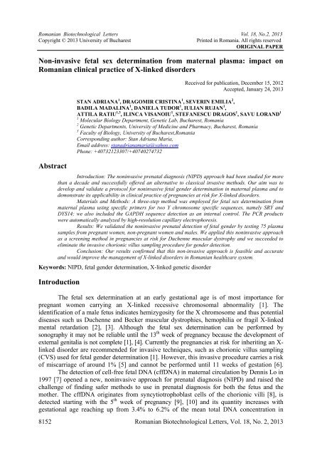
Romanian Biotechnological Letters Vol. 18, No.2, 2013Copyright © 2013 University of BucharestPrinted in Romania. All rights reservedORIGINAL PAPERNon-invasive fetal sex determination from maternal plasma: impact onRomanian clinical practice of X-linked disordersAbstractReceived for publication, December 15, 2012Accepted, January 24, 2013STAN ADRIANA 1 , DRAGOMIR CRISTINA 1 , SEVERIN EMILIA 2 ,BADILA MADALINA 1 , DANIELA TUDOR 1 , IULIAN RUJAN 1 ,ATTILA RATIU 1,3 , ILINCA VISANOIU 1 , STEFANESCU DRAGOS 1 , SAVU LORAND 11 Molecular Biology Department, Genetic Lab, Bucharest, Romania2 Genetic Departments, University of Medicine and Pharmacy, Bucharest, Romania3 Faculty of Biology, University of Bucharest,RomaniaCorresponding author: Stan Adriana Maria,Email address: <strong>stan</strong><strong>adriana</strong>maria@yahoo.comPhone: +40732123307/+40740274732Introduction: The noninvasive prenatal diagnosis (NIPD) approach had been studied for morethan a decade and successfully offered an alternative to classical invasive methods. Our aim was todevelop and validate a protocol for noninvasive fetal gender determination in maternal plasma and todemonstrate its applicability in clinical practice of pregnancies at risk for X-linked disorders.Materials and Methods: A three-step method was employed for fetal sex determination frommaternal plasma using specific primers for two Y chromosome specific sequences, namely SRY andDYS14; we also included the GAPDH sequence detection as an internal control. The PCR productswere automatically analyzed by high-resolution capillary electrophoresis.Results: We validated the noninvasive prenatal detection of fetal gender by testing 75 plasmasamples from pregnant women, non-pregnant women and males. We applied this noninvasive approachas a screening method in pregnancies at risk for Duchenne muscular dystrophy and we succeeded toeliminate the invasive chorionic villus sampling procedure for gender detection.Conclusion: Our results confirmed that this non-invasive approach is feasible and accurateand would improve the management of X-linked disorders in Romanian healthcare system.Keywords: NIPD, fetal gender determination, X-linked genetic disorderIntroductionThe fetal sex determination at an early gestational age is of most importance forpregnant women carrying an X-linked recessive chromosomal abnormality [1]. Theidentification of a male fetus indicates hemizygosity for the X chromosome and thus potentialdiseases such us Duchenne and Becker muscular dystrophies, hemophilia or fragil X-linkedmental retardation [2], [3]. Although the fetal sex determination can be performed bysonography it may not be reliable until the 13 th week of pregnancy because the development ofexternal genitalia is not complete [1], [4]. Currently the pregnancies at risk for inheriting an X-linked disorder are recommended for invasive techniques, such as chorionic villus sampling(CVS) used for fetal gender determination [1]. However, this invasive procedure carries a riskof miscarriage of around 1% [5] and cannot be performed until 11 weeks of gestation [6].The detection of cell-free fetal DNA (cffDNA) in maternal circulation by Dennis Lo in1997 [7] opened a new, noninvasive approach for prenatal diagnosis (NIPD) and raised thechallenge of finding safer methods to use in prenatal diagnosis for both the fetus and themother. The cffDNA originates from syncytiotrophoblast cells of the chorionic villi [8], isdetected starting with the 5 th week of pregnancy [9], [10] and its quantity increases withgestational age reaching up from 3.4% to 6.2% of the mean total DNA concentration in8152 Romanian Biotechnological Letters, Vol. 18, No. 2, 2013
STAN ADRIANA, DRAGOMIR CRISTINA, SEVERIN EMILIA, BADILA MADALINA, DANIELA TUDOR,IULIAN RUJAN, ATTILA RATIU, ILINCA VISANOIU, STEFANESCU DRAGOS, SAVU LORANDFigure 1. High-resolution capillary electrophoresis – electrophoregram view: A01 and A02 lanes <strong>stan</strong>d for SRYand GAPDH positive results; A03, A04 and A05 lanes <strong>stan</strong>d for SRY negative and GAPDH positive result; A06lane represents the non-template control for SRY assay; A07 and A08 lanes are the DYS14 and GAPDH positiveresult; A09 and A10 lanes correspond to DYS14 negative and GAPDH positive result; A11 lane represents thenon-template control for DYS14 assay; in A12 lane there is the pUC 18/Hae III marker.This interpretation considered the fact that DYS14 is a multi-copy marker and itsdetection was most robust comparing with the SRY marker. In this specific case werecommended the collection of a fresh new sample and the repetition of the test. Afterestablishing the result for each marker, we proceed to the final result interpretation bycombining them (Table I).Table I. The scoring model for (the results interpretation) interpreting the results obtained by combining theresults of the SRY and the DYS14 assays.SRY DYS14 Final resultpositive positive positivePCR resultpositive negative inconclusivenegative positive possible positive but repeat the testnegative negative negativeThe results obtained by analyzing plasma sample attained from the non-pregnantwomen and males were conclusive. The SRY was detected in 24 samples and the DYS14 in26 cases. The two cases were reported as male fetuses after repeating the test from anothermaternal plasma aliquot. In one case we had an inconclusive result for the DYS14 andrequested the collection of a new sample.The results obtained from all 45 pregnant women were confirmed by amniocentesisfollowed by the rapid prenatal diagnosis of trisomy 13, 18, 21 and sex chromosomean<strong>eu</strong>ploidies.We also tested 11 blood samples of pregnant women with gestational age between 9and 12 weeks of pregnancy who didn’t performed the invasive tests and have late confirmedNIPD results.This NIPD protocol for fetal gender determination was applied in two cases at risk forDuchenne muscular dystrophy (DMD) with long histories regarding the DMD geneheritability. In the first case, the pregnant woman was a heterozygous carrier for the exons 51-55 deletion and her five years old sun inherited the abnormal X chromosome (figure 2).Romanian Biotechnological Letters, Vol. 18, No. 2, 2013 8155
Non-invasive fetal sex determination from maternal plasma: impacton Romanian clinical practice of X-linked disordersFigure 1. The MLPA patters of the DMD gene in the pregnant woman with the 51-55 exons heterozygousdeletion and the male child with DMDThe maternal blood collection was performed at 9 weeks of pregnancy. The fetalgender was determined as female and the mother didn’t perform the CVS. Later in pregnancy,the amniocentesis for rapid an<strong>eu</strong>ploidy detection (RAD) of the 13, 18, 21, X and Ychromosomes confirmed the noninvasive result.The second pregnant woman tested had the heterozygous deletion of exons 30-43 deletion andthe noninvasive fetal gender detection was positive for the presence of a Y chromosome. Thevillous biopsy was performed at 11 week to analyze the fetal DMD gene. The peaks heightsbetween sex chromosomes markers were analyzed to exclude the maternal DNA8156 Romanian Biotechnological Letters, Vol. 18, No. 2, 2013
Non-invasive fetal sex determination from maternal plasma: impacton Romanian clinical practice of X-linked disordersThe DYS14 assay targeted a multi-copy sequence and therefore has a higher sensitivity thanSRY [1, 2], but as mentioned in other papers, the test specificity increases by combining bothmarkers [1]. The lack of a specific marker for female fetal DNA circulating in the maternalblood made questionable the report of a female fetus when both SRY and DYS14 arenegative. The large quantity of maternal DNA in the sample renders more difficult theinclusion of satisfactory internal control to test for successful amplification of fetal DNA [25].Although the inclusion of the GAPDH gene in the test wasn’t sufficient to provide thespecific control for the presence of the fetal DNA as well as other types of housekeepinggenes (β-globin, actins, HPRT), it demonstrated the success of our PCR approach.Another important trait of our method was the analysis of PCR products using theQIAxcel high-resolution gel cartridge thus providing a high resolution separation and adetection sensitivity of 0, 1 ng/µl DNA in undiluted amplification reactions. The fetal genderdetermination using the two assays for the SRY and DYS14 combined with the GAPDHcontrol was reliable and accurate. Very important, the interpretation of the results based onthe scoring model presented previously excluded false-negatives and false-positives results.The introduction of this NIPD test into the clinical practice of our laboratory wasperformed for pregnant women carriers for heterozygous DMD mutations. The obstetricianrecommended this NIPD test for fetal gender determination at 9 th weeks of pregnancy and inboth cases the results were conclusive. The results were confirmed by ultrasonography, fetalRAD by QF-PCR and karyotype.ConclusionThe results obtained in this study confirmed that this NIPD method for fetal genderdetermination is feasible and accurate and can be successfully implemented as a screening testfor all pregnancies at risk for X-linked disorders therefore eliminating the need for invasivesampling procedure in nearly as 50% of cases. A safe NIPD test for fetal sexing in the 9 thweek of pregnancy would improve the management of X-linked disorders in Romanianhealthcare system.References1. SCHEFFER PG, VAN DER SCHOOT CE, PAGE-CHRISTIAENS GCML, BOSSERS B, VAN ERP F,DE HAAS M - Reliability of Fetal Sex Determination Using Maternal Plasma. Obstet Gynecol: 2010, 115,1:117-1262. AVENT ND, CHITTY LS - Non-invasive diagnosis of fetal sex; utilisation of free fetal DNA in maternalplasma and ultrasound. Prenat Diagn 2006; 26: 598–6033. DAVALIEVA K, DIMCEV P, EFREMOV GD, PLASESKA-KARANFILSKA D. -Non-invasive fetal sexdetermination using real-time PCR. The Journal of Maternal-Fetal and Neonatal Medicine, 2006; 19(6):337–3424. DEVANEY SA, PALOMAKI GE, SCOTT JA, BIANCHI DW - Noninvasive Fetal Sex DeterminationUsing Cell-Free Fetal DNA: A Systematic Review and Meta-analysis. JAMA, 2011; 306 (6): 627-6365. MUJEZINOVIC F, ALFIREVIC Z. - Procedure-related complications of amniocentesis and chorionicvillous sampling: a systematic review. Obstet Gynecol 2007;110:687–694.6. WRIGHT CF, BURTON H - The use of cell-free fetal nucleic acids in maternal blood for non-invasiveprenatal diagnosis. Hum Reprod Update. 2009 Jan-Feb;15(1):139-51.7. LO YM, CORBETTA N, CHAMBERLAIN PF - Presence of fetal DNA in maternal plasma and serum.Lancet. 1997;350(9076):485-487.8158 Romanian Biotechnological Letters, Vol. 18, No. 2, 2013
STAN ADRIANA, DRAGOMIR CRISTINA, SEVERIN EMILIA, BADILA MADALINA, DANIELA TUDOR,IULIAN RUJAN, ATTILA RATIU, ILINCA VISANOIU, STEFANESCU DRAGOS, SAVU LORAND8. MADDOCKS DG, ALBERRY MS, ATTILAKOS G, MADGETT TE, CHOI K, SOOTHILL PW, AVENTND. - The SAFE project: towards non-invasive prenatal diagnosis. Biochem Soc Trans. 2009 Apr;37(Pt2):460-5.9. RIJNDERS RJ, VAN DER LUIJT RB, PETERS ED - Earliest gestational age for fetal sexing in cell-freematernal plasma. Prenat Diagn, 2003, 23: 1042–1104.10. MARTINHAGO CD, DE OLIVEIRA RM, TOMITÃO CANAS MDO C, VAGNINI LD, ALCANTARAOLIVEIRA JB, PETERSEN CG, FRANCO JUNIOR JG - Accuracy of fetal gender determination inmaternal plasma at 5 and 6 weeks of pregnancy. Prenat Diagn. 2006; 26(13):1219-23.11. LO YMD, ZHANG J, LEUNG TN, LAU TK, CHANG AMZ, HJELM NM. - Rapid clearance of fetalDNA from maternal plasma. Am J Hum Genet 1999c; 64:218–224.12. LO YM, TEIN MS, LAU TK, et al. 1998. - Quantitative analysis of fetal DNA in maternal plasma andserum: implications for noninvasive prenatal diagnosis. Am J Hum Genet 62: 768–775.13. LO YMD, HJELM NM, FIDLER C, SARGENT IL, MURPHY MF, CHAMBERLAIN PF, POON PMK,REDMAN CWG, WAINSCOAT JS. - Prenatal diagnosis of fetal RhD status by molecular analysis ofmaternal plasma. N Engl J Med,1998a;339:1734–1738.14. VAN DER SCHOOT CE, SOUSSAN AA, KOELEWIJN J, BONSEL G, PAGET-CHRISTIAENS LGC,DE HAAS M. Non-invasive antenatal RHD typing. Transfus Clin Biol 2006;13:53–57.15. ZHONG XY, HOLZGREVE W, HAHN S. - Risk free simultaneous prenatal identification of fetal RhesusD status and sex by multiplex real-time PCR using cell free fetal DNA in maternal plasma. Swiss MedWkly, 2001, 13: 70–74.16. GONZÁLEZ-GONZÁLEZ MC, GARCÍA-HOYOS M, TRUJILLO MJ, RODRÍGUEZ DE ALBA M,LORDA-SÁNCHEZ I, DÍAZ-RECASENS J, GALLARDO E, AYUSO C, RAMOS C. Prenatal detectionof a cystic fibrosis mutation in fetal DNA from maternal plasma. Prenat Diagn. 2002. 22(10):946-8.17. NASIS O, THOMPSON S, HONG T, SHERWOOD M, RADCLIFFE S, JACKSON L, OTEVREL T -Improvement in Sensitivity of Allele-specific PCR Facilitates Reliable Noninvasive Prenatal Detection ofCystic Fibrosis. Clinical Chemistry, 2004, 50:4,694–70118. LI Y, DI NARO E, VITUCCI A, GRILL S, ZHONG XY, HOLZGREVE W, HAHN S - Size fractionationof cell-free DNA in maternal plasma improves the detection of a paternally inherited β-thalassemia pointmutation by MALDI-TOF mass spectrometry. Fetal Diagn Ther, 2009, 25:246-249.19. PAPASAVVA T, KALIKAS I, KYRRI A, KLEANTHOUS M. - Arrayed primer extension for thenoninvasive prenatal diagnosis of beta-thalassemia based on detection of single nucleotidepolymorphisms.An n N Y Acad Sci. 2008;1137:302-8.20. LI Y, PAGE-CHRISTIAENS GC, GILLE JJ, HOLZGREVE W, HAHN S. Non-invasive prenatal detectionof achondroplasia in size-fractionated cell-free DNA by MALDI-TOF MS assay. Prenat Diagn. 2007.27(1):11-7.21. CHEN EZ, CHIU RWK, SUN H, AKOLEKAR R, CHAN KCA, LEUNG TY, JIANG P, ZHENG YWL,LUN FMF, CHAN LYS, JIN Y, GO ATJI, LAU ET, TO WWK, LEUNG WC, TANG RYK, AU-YEUNGSKC, LAM H, KUNG YY, ZHANG X, VAN VUGT KMG, MINEKAWA R, TANG MTH, WANG J,OUDEJANS CBM, LAU TK, NICOLAIDES KH, Y. M. DENNIS LO. - Noninvasive Prenatal Diagnosisof Fetal Trisomy 18 and Trisomy 13 by Maternal Plasma DNA Sequencing. PLoS ONE, www.plosone.org,July 2011, Volume 6, Issue 7, e2179122. CHIU RWK, CHAN KCA, GAO Y, LAU VYM, ZHENG W, et al. - Noninvasive prenatal diagnosis offetal chromosomal an<strong>eu</strong>ploidy by massively parallel genomic sequencing of DNA in maternal plasma. ProcNatl Acad Sci U S A, 2008, 105: 20458–20463.23. ZIMMERMANN B, HILL M, GEMELOS G, DEMKO Z, BANJEVIC M, BANER J, RYAN A,SIGURJONSSON S, CHOPRA N, DODD M, LEVY B, RABINOWITZ M. - Noninvasive prenatalan<strong>eu</strong>ploidy testing of chromosomes 13, 18, 21, X, and Y, using targeted sequencing of polymorphic loci.Prenat Diagn. 2012, oct 30:1-9 [Epub ahead of print]24. JOHNSON KL, DUKES KA, VIDAVER J, LESHANE ES, RAMIREZ I, WEBER WD, BISCHOFF FZ,HAHN S, SHARMA A, DANG DX, HIRE LM, BIANCHI DW, SIMPSON JL, HOLZGREVE W, ELIASS, KLINGER KW - Interlaboratory comparison of fetal male DNA detection from common maternalplasma samples by real-time PCR. Clin Chem. 2004; 50(3):516-21.25. DANIELS G, FINNING K, MARTIN P, MASSEY E. - Noninvasive prenatal diagnosis of fetal bloodgroup phenotypes: current practice and future prospects. Prenat Diagn. 2009, 29(2):101-7.Romanian Biotechnological Letters, Vol. 18, No. 2, 2013 8159