CIRRUS photo Certainty meets versatility - Carl Zeiss Meditec
CIRRUS photo Certainty meets versatility - Carl Zeiss Meditec
CIRRUS photo Certainty meets versatility - Carl Zeiss Meditec

You also want an ePaper? Increase the reach of your titles
YUMPU automatically turns print PDFs into web optimized ePapers that Google loves.
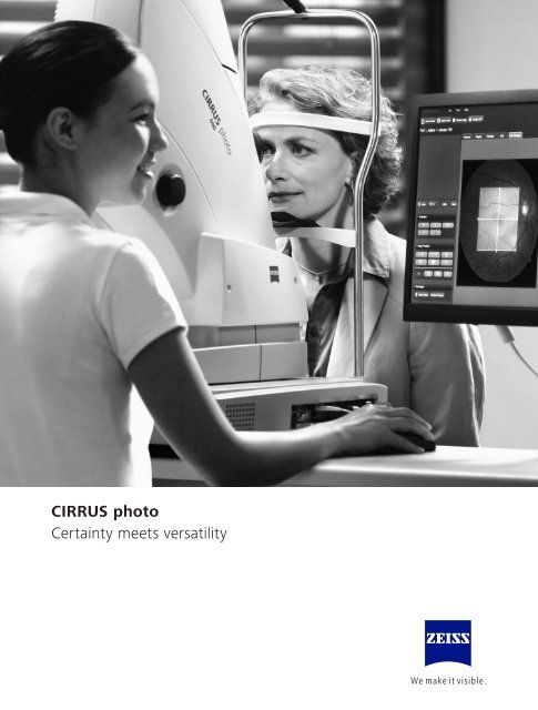
<strong>CIRRUS</strong> <strong>photo</strong><br />
<strong>Certainty</strong> <strong>meets</strong> <strong>versatility</strong>
<strong>CIRRUS</strong> <strong>photo</strong><br />
One system for fundus imaging and OCT<br />
Broader clinical insights, greater diagnostic certainty and added practice<br />
value – the new <strong>CIRRUS</strong> TM <strong>photo</strong> from ZeISS delivers all that in a single,<br />
integrated system for both fundus imaging and OCT.<br />
<strong>CIRRUS</strong> <strong>photo</strong> combines a full mydriatic/non-mydriatic fundus camera<br />
with proven <strong>CIRRUS</strong> HD-OCT technology in one compact and highly<br />
versatile system. Available in two models, <strong>CIRRUS</strong> <strong>photo</strong> 600 and<br />
<strong>CIRRUS</strong> <strong>photo</strong> 800, it provides multiple insights for comprehensive<br />
retina and posterior segment care.<br />
Visualize findings from various modalities. Correlate data from high-density<br />
OCT cubes, thickness and layer maps with results from superb color fundus<br />
images as well as fundus autofluorescence and fluorescein angiography*<br />
images. All in one convenient sitting.<br />
Achieve a more comprehensive clinical evaluation. Save time and space.<br />
enhance the examination experience for your patients and staff.<br />
Have it all in a single system for fundus imaging and OCT.<br />
* Only with <strong>CIRRUS</strong> <strong>photo</strong> 800<br />
3
4<br />
Broader clinical insights<br />
By simultaneously providing high-quality fundus images and<br />
OCT scans, <strong>CIRRUS</strong> <strong>photo</strong> facilitates broader, more comprehensive<br />
diagnostic insights. each modality by itself is a premier quality<br />
diagnostic instrument. Together, they enable you to characterize<br />
and examine the patient’s condition more completely and easily.<br />
A fundus camera …<br />
<strong>CIRRUS</strong> <strong>photo</strong> is a full-featured mydriatic/<br />
non-mydriatic fundus camera.<br />
Exceptional visualizations<br />
Legendary ZEISS optics let you visualize findings<br />
with high-resolution clarity and sharpness.<br />
Single-shot fundus autofluorescence<br />
Fundus autofluorescence imaging designed for<br />
fast and easy assessment of dry AMD.<br />
High-resolution angiographies<br />
Also available with fluorescein angiography* and<br />
indocyanine green angiography, <strong>CIRRUS</strong> <strong>photo</strong><br />
equips you with a more detailed diagnostic view.<br />
* Only with <strong>CIRRUS</strong> <strong>photo</strong> 800<br />
… and a <strong>CIRRUS</strong> HD-OCT<br />
<strong>CIRRUS</strong> <strong>photo</strong> incorporates unsurpassed<br />
OCT technology with its proven<br />
<strong>CIRRUS</strong> HD-OCT capabilities.<br />
Great detail density<br />
Highly dense OCT data cubes make even the<br />
smallest details clearly visible.<br />
Analysis you can trust<br />
Detailed OCT scans and change analyses provide<br />
highly reliable diagnostic data in seconds.
Interactive review<br />
The system’s one-of-a-kind MultiMode Navigator enables<br />
interactive analysis of registered fundus images and OCT cube scans –<br />
horizontal and vertical direction.<br />
Precise registration<br />
OCT scans are automatically registered with different types of<br />
fundus images including color fundus, angiography* and fundus<br />
autofluorescence* images, bringing depth to your analysis.<br />
Multimodal assessments<br />
<strong>CIRRUS</strong> <strong>photo</strong> allows you to conduct examinations with various<br />
modalities and to correlate the findings at one single workstation.<br />
every fundus image can also be registered independently of the<br />
acquisition sequence, along with other flexible combinations.<br />
(inset magnified to show detail)<br />
Orientation at a glance<br />
Whether for a quick overview or point-by-point comparisons,<br />
thumbnails provide at-a-glance insights.<br />
5
Versatile visualizations<br />
Harada’s disease<br />
Retinal<br />
pigment ephithelial<br />
ephithelial<br />
detachment<br />
Proliferative diabetic<br />
retinopathy<br />
Glaucoma<br />
6
1 7
8<br />
Greater diagnostic certainty<br />
Comprehensive, high-quality diagnostics form the basis for informed<br />
decisions. With its superb multimodality visualizations, <strong>CIRRUS</strong> <strong>photo</strong><br />
delivers exceptional insights, supporting greater diagnostic accuracy<br />
and certainty.<br />
Extraordinary image quality<br />
<strong>CIRRUS</strong> <strong>photo</strong> features standard-setting <strong>CIRRUS</strong> HD-OCT technology and a full-featured<br />
mydriatic/non-mydriatic fundus camera. The result – visualizations of a quality that is<br />
truly extraordinary.<br />
Overview at a glance See the details
Algorithm excellence<br />
ZeISS and its research collaborators have developed advanced algorithms to measure<br />
and display layers.<br />
Accurate centering<br />
FoveaFinder and AutoCenter automatically ensure that measurements are made in<br />
the correct locations, taking the pressure off the operator to perfectly center the scans.<br />
Comparison capabilities<br />
<strong>CIRRUS</strong> data cubes are automatically registered with data from prior visits, allowing for<br />
more detailed comparisons.<br />
Normative databases<br />
Diversified normative databases for ONH, RNFL and macular thickness facilitate even<br />
more at-a-glance assessments.<br />
Specific fundus details<br />
enabling efficient cross-modality analysis, <strong>CIRRUS</strong> <strong>photo</strong> allows easy switching<br />
between fundus images registered with OCT scans and maps.<br />
9
10<br />
Fundus and OCT details in one view<br />
<strong>CIRRUS</strong> <strong>photo</strong> delivers combined fundus and OCT reports that enable<br />
quick, at-a-glance assessments for a wide variety of retina and posterior<br />
segment disorders.
10a<br />
Get the complete picture in a single view<br />
ONH & RNFL OU Analysis<br />
Single Eye Analysis GPA Analysis
Enhanced HD Raster Single Line<br />
Enhanced HD 5 Line Raster<br />
Anterior Segment<br />
10b
Macular Thickness Analysis<br />
Macular Change Analysis<br />
Advanced Visualization<br />
<strong>CIRRUS</strong> <strong>photo</strong> offers a versatile<br />
suite of OCT analyses, which<br />
means more insight into your<br />
treatment decisions.<br />
11
12<br />
Added practice value<br />
As a highly efficient and versatile instrument, <strong>CIRRUS</strong> <strong>photo</strong> offers<br />
substantial value. In addition to streamlining your workflow and<br />
supporting more comprehensive assessments, it saves time and space.<br />
By eliminating the need to move patients to another instrument,<br />
it also enhances the examination experience – for patients and<br />
practice staff alike.<br />
<strong>CIRRUS</strong> <strong>photo</strong> – a single workstation<br />
complete with joystick, keyboard<br />
and TFT monitor for comprehensive<br />
clinical evaluations.
Konturdicke 8pt<br />
Konturfarbe 60% k<br />
Füllung 10% k<br />
Back office<br />
Fundus<br />
<strong>photo</strong>graphy<br />
Practice reception<br />
area<br />
Biometry OCT Visual Field<br />
More clinical efficiency<br />
With <strong>CIRRUS</strong> <strong>photo</strong>, you can add a color fundus<br />
image to an OCT examination for additional<br />
assessment – in seconds and without additional<br />
dilation.<br />
More time for patients<br />
easy, convenient and operator-independent<br />
storage of <strong>CIRRUS</strong> <strong>photo</strong> data is provided by<br />
FORUM ® . Via the FORUM Archive & Viewer,<br />
you can effortlessly exchange examination results<br />
with eMR systems and other diagnostic<br />
instruments – even with other practice sites.<br />
net port<br />
Instrument<br />
(non-DICOM)<br />
Exam lane 1 Exam lane 2<br />
Image courtesy of:<br />
Annette Brusis MD, Eye Center Heppenheim Dr. Wolff, Dr. Brusis, Dr. Köster, Germany (p. 2, 11)<br />
Antonio Ferreras MD, Miguel Servet University Hospital, Spain (p. 6, 7, 11)<br />
Matthias Jütte MD, Ophthalmic Practice Jütte, Jurkutat, Ilgner, Germany (p. 4, 8)<br />
Instrument<br />
(no network)<br />
FORUM Archive<br />
FORUM Viewer<br />
Electronic Medical Record<br />
More practice efficiency<br />
The ability to capture all necessary fundus images<br />
and HD-OCT scans in a single patient setup saves<br />
you time and space. As such, <strong>CIRRUS</strong> <strong>photo</strong> is<br />
ideally suited for comprehensive practices working<br />
with or without angiography.<br />
More flexibility<br />
Featuring a modular design, <strong>CIRRUS</strong> <strong>photo</strong> lets you<br />
individually choose the diagnostic modalities and<br />
clinical insights best suited for your practice needs –<br />
whether OCT, color and red-free fundus imaging,<br />
fundus autofluorescence, fluorescein angiography,<br />
ICG angiography, and anterior segment.<br />
13
Technical data<br />
Main system <strong>CIRRUS</strong> <strong>photo</strong> 600/800<br />
Field angle 45° and 30°<br />
Pupil diameter ≥ 4.0 mm; ≥ 3.3 mm (30° small pupil mode)<br />
≥ 2.0 mm for OCT scans only<br />
Refractive error compensation +35 D … -35 D, continuous<br />
Working distance 40 mm (patient’s eye – front lens)<br />
Fixation targets<br />
external and internal<br />
Internal<br />
Attention mode and free position or programmed sequences<br />
Database Patient information and images with field angle, FA time, R/L recognition and date of visit are stored<br />
Monitor 23” TFT (1920 x 1200)<br />
Instrument table Asymmetric, suitable for wheelchairs<br />
Accessories Network printer, sliding keyboard shelf, network isolator,<br />
FORUM eye care data management system<br />
Fundus camera<br />
Capture modes Color, red-free, blue, red and fundus autofluorescence pictures,<br />
as well as pictures of the anterior segment,<br />
<strong>CIRRUS</strong> <strong>photo</strong> 800 only: + fluorescein angiography and ICG angiography<br />
Filters Filters for green, blue and fundus autofluorescence images, UV/IR barrier filters<br />
<strong>CIRRUS</strong> <strong>photo</strong> 800 only: + FA + ICGA: exciter and barrier filters<br />
Capture sequence From 1.5 seconds (depends on flash energy)<br />
Capture sensor CCD 5.0 megapixels<br />
Xenon flash lamp 16 flash levels (30 Ws)<br />
<strong>CIRRUS</strong> <strong>photo</strong> 800 only: 24 flash levels (80 Ws)<br />
OCT<br />
Technology Spectral domain OCT<br />
Optical source Superluminescent diode (SLD), 840 nm<br />
Scan speed 27,000 A-scan per second<br />
A-scan depth 2.0 mm (in tissue), 1024 points<br />
Resolution Axial 5 µm (in tissue), transverse 15 µm (in tissue)<br />
Computer<br />
Operating system Windows embedded<br />
Hard drive Storage of over 30,000 fundus images with OCT cube scans (present size: 320 GB)<br />
Interfaces USB ports and network connectors, DVI port<br />
Export/import Image formats: BMP, TIFF, JPeG, PNG<br />
Patient list, DICOM MWL, DICOM storage<br />
Dimensions<br />
Main unit 410 mm x 480 mm x 680 mm (W 16.1 x D 18.9 x H 26.8 inches)<br />
Weight (main unit) 33 kg (72.7 lbs)<br />
Rated voltage 100 … 240 V ±10%<br />
Frequency 50 / 60 Hz<br />
Power consumption 400 VA (w/o instrument table)<br />
14
The moment a subtle change in pathology<br />
becomes a turning point in care.<br />
This is the moment we work for.<br />
// CIRRuS<br />
MADE By CARl ZEISS
� 0297<br />
Your local contact:<br />
Argentina<br />
<strong>Carl</strong> <strong>Zeiss</strong> Argentina S.A.<br />
Calle Nahuel Huapi 4015 / 25<br />
C1430 BCO Buenos Aires<br />
Argentina<br />
Phone: +54 11 45 45 66 61<br />
bruzzi@zeiss.com.ar<br />
Australia<br />
<strong>Carl</strong> <strong>Zeiss</strong> Pty Ltd<br />
Tenancy Office 4, Level 1<br />
40-52 Talavera Road<br />
North Ryde NSW 2113<br />
Australia<br />
Phone: +61 2 9020 1333<br />
med@zeiss.com<br />
Austria<br />
<strong>Carl</strong> <strong>Zeiss</strong> GmbH<br />
Laxenburger Str. 2<br />
1100 Vienna<br />
Austria<br />
Phone: +43 1 79 51 80<br />
austria@zeiss.org<br />
Belgium<br />
<strong>Carl</strong> <strong>Zeiss</strong> NV-SA<br />
Ikaroslaan 49<br />
1930 Zaventem<br />
Belgium<br />
Phone: +32 2 719 39 11<br />
info@zeiss.be<br />
Brazil<br />
<strong>Carl</strong> <strong>Zeiss</strong> do Brasil Ltda.<br />
Av. Naçoes Unidas, 21711<br />
CEP04795-100 São Paulo<br />
Brazil<br />
Phone: +55 11 5693 5521<br />
medbrasil@zeiss.org<br />
Canada<br />
<strong>Carl</strong> <strong>Zeiss</strong> Canada Ltd.<br />
45 Valleybrook Drive<br />
Toronto, ON M3B 2S6<br />
Canada<br />
Phone: +1 800 387 8037<br />
czcmed@zeiss.com<br />
China<br />
<strong>Carl</strong> <strong>Zeiss</strong> Shanghai Co. Ltd.<br />
1/f., Ke Yuan Building<br />
11 Ri Yin Nan Road<br />
Waigaoqiao Free Trade Zone<br />
2005 Yang Gao Bei Road<br />
Shanghai 200131<br />
China<br />
Phone: +86 21 5048 17 17<br />
sro@zeiss.com.cn<br />
<strong>Carl</strong> <strong>Zeiss</strong> <strong>Meditec</strong> AG<br />
Goeschwitzer Strasse 51–52<br />
07745 Jena<br />
Germany<br />
www.meditec.zeiss.com/cirrus<strong>photo</strong><br />
Czech Republic<br />
<strong>Carl</strong> <strong>Zeiss</strong> spol. s.r.o.<br />
Radlická 14/3201<br />
150 00 Prague 5<br />
Czech Republic<br />
Phone: +420 233 101 221<br />
zeiss@zeiss.cz<br />
France<br />
<strong>Carl</strong> <strong>Zeiss</strong> <strong>Meditec</strong> France SAS<br />
60, route de Sartrouville<br />
78230 Le Pecq<br />
France<br />
Phone: +33 1 34 80 21 00<br />
med@zeiss.fr<br />
Germany<br />
<strong>Carl</strong> <strong>Zeiss</strong> <strong>Meditec</strong> VG mbH<br />
<strong>Carl</strong>-<strong>Zeiss</strong>-Strasse 22<br />
73447 Oberkochen<br />
Germany<br />
Phone: +49 7364 20 6000<br />
vertrieb@meditec.zeiss.com<br />
Surgical Ophthalmology:<br />
Phone: +49 800 470 50 30<br />
iol.order@meditec.zeiss.com<br />
Hong Kong<br />
<strong>Carl</strong> <strong>Zeiss</strong> Far East Co. Ltd.<br />
Units 11-12. 25/F<br />
Tower 2, Ever Gain Plaza<br />
No. 88 Container Port Road<br />
Kwai Chung<br />
Hong Kong<br />
Phone: +852 2332 0402<br />
czfe@zeiss.com.hk<br />
India<br />
<strong>Carl</strong> <strong>Zeiss</strong> India Pvt. Ltd.<br />
Plot No.3, Jigani Link Road<br />
Bommasandra Industrial Area<br />
Bangalore - 560 099<br />
India<br />
Phone: +91 80 4343 8000<br />
info@zeiss.co.in<br />
Italy<br />
<strong>Carl</strong> <strong>Zeiss</strong> S.p.A.<br />
Viale delle Industrie 20<br />
20020 Arese (Milan)<br />
Italy<br />
Phone: +39 02 93773 1<br />
infomed@zeiss.it<br />
Japan<br />
<strong>Carl</strong> <strong>Zeiss</strong> <strong>Meditec</strong> Japan Co. Ltd.<br />
Shinjuku Ku<br />
Tokyo 160-0003<br />
22 Honchio-Cho<br />
Japan<br />
Ophthalmic instruments:<br />
Phone: +81 3 33 55 0331<br />
medsales@zeiss.co.jp<br />
Surgical instruments:<br />
Phone: +81 3 33 55 0341<br />
cmskoho@zeiss.co.jp<br />
Malaysia<br />
<strong>Carl</strong> <strong>Zeiss</strong> Sdn Bhd.<br />
Lot2, Jalan 243/51 A<br />
46100 Petaling Jaya<br />
Selangor Darul Ehsan<br />
Malaysia<br />
Phone: +60 3 7877 50 58<br />
malaysia@zeiss.com.sg<br />
Mexico<br />
<strong>Carl</strong> <strong>Zeiss</strong> de México S.A. de C.V.<br />
Avenida Miguel Angel de Quevedo<br />
496<br />
04010 Mexico City<br />
Mexico<br />
Phone: +52 55 59 99 0200<br />
cz-mexico@zeiss.org<br />
Netherlands<br />
<strong>Carl</strong> <strong>Zeiss</strong> B.V.<br />
Trapezium 300<br />
Postbus 310<br />
3364 DL Sliedrecht<br />
Netherlands<br />
Phone: +31 184 43 34 00<br />
info@zeiss.nl<br />
New Zealand<br />
<strong>Carl</strong> <strong>Zeiss</strong> (N.Z.) Ltd.<br />
15B Paramount Drive<br />
P.O. Box 121 - 1001<br />
Henderson, Auckland 0650<br />
New Zealand<br />
Phone: +64 9 838 5626<br />
med@zeiss.com<br />
Poland<br />
<strong>Carl</strong> <strong>Zeiss</strong> sp. Z o.o.<br />
ul. Lopuszanska 32<br />
02-220 Warsaw<br />
Poland<br />
Phone: +48 22 858 2343<br />
medycyna@zeiss.pl<br />
Singapore<br />
<strong>Carl</strong> <strong>Zeiss</strong> Ptd. Ltd.<br />
50 Kaki Bukit Place<br />
Singapore 415926<br />
Singapore<br />
Phone: +65 6741 9600<br />
info@zeiss.com.sg<br />
South Africa<br />
<strong>Carl</strong> <strong>Zeiss</strong> (Pty.) Ltd.<br />
363 Oak Avenue<br />
Ferndale<br />
Randburg 2194<br />
South Africa<br />
Phone: +27 11 886 9510<br />
info@zeiss.co.za<br />
South Korea<br />
<strong>Carl</strong> <strong>Zeiss</strong> Co. Ltd.<br />
Seoul 121-828<br />
Mapo-gu<br />
141-1, Sangsu-dong<br />
2F, BR Elitel Bldg.<br />
South Korea<br />
Phone: +82 2 3140 2600<br />
korea@zeiss.co.kr<br />
Spain<br />
<strong>Carl</strong> <strong>Zeiss</strong> <strong>Meditec</strong> Iberia S.A.U.<br />
Ronda de Poniente, 15<br />
Tres Cantos<br />
28760 Madrid<br />
Spain<br />
Phone: +34 91 203 37 00<br />
info@zeiss.es<br />
Sweden<br />
<strong>Carl</strong> <strong>Zeiss</strong> AB<br />
Tegeluddsvaegen 76<br />
10254 Stockholm<br />
Sweden<br />
Phone: +46 84 59 25 00<br />
info@zeiss.se<br />
Switzerland<br />
<strong>Carl</strong> <strong>Zeiss</strong> AG<br />
Feldbachstrasse 81<br />
8714 Feldbach<br />
Switzerland<br />
Phone: +41 55 254 7200<br />
med@zeiss.ch<br />
Thailand<br />
<strong>Carl</strong> <strong>Zeiss</strong> Thailand<br />
90 CyberWorld Tower A,<br />
36 th Floor, Unit A 3601<br />
230 Ratchadapisek Road<br />
Huaykhwang, Bangkok 10310<br />
Thailand<br />
Phone: +66 2 248 8787<br />
thailand@zeiss.com.sg<br />
United Kingdom<br />
<strong>Carl</strong> <strong>Zeiss</strong> Ltd.<br />
509 Coldhams lane<br />
Cambridge<br />
CAMBS<br />
CB1 3JS,<br />
United Kingdom<br />
Phone: +44 1707 871200<br />
info@zeiss.co.uk<br />
United States of America<br />
<strong>Carl</strong> <strong>Zeiss</strong> <strong>Meditec</strong>, Inc.<br />
5160 Hacienda Drive<br />
Dublin, CA 94568<br />
USA<br />
Phone: +1 925 557 4100<br />
info@meditec.zeiss.com<br />
Publication No: 000000-2031-489 CIR.4718 CZ-XI/2012<br />
The contents of the brochure may differ from the current status of approval of the product in your country. Please contact our regional representative for more information.<br />
Subject to change in design and scope of delivery and as a result of ongoing technical development. <strong>CIRRUS</strong>, FORUM, Advanced Visualization, Guided Progression Analysis, GPA, Fovea<br />
Finder, AutoCenter are either trademarks or registered trademarks of <strong>Carl</strong> <strong>Zeiss</strong> <strong>Meditec</strong> in the United States and/or other countries.<br />
© 2012 by <strong>Carl</strong> <strong>Zeiss</strong> <strong>Meditec</strong>, Inc. All copyrights reserved. 1112 2.5M