Chemical Examination of Urine - UNMC
Chemical Examination of Urine - UNMC
Chemical Examination of Urine - UNMC

Create successful ePaper yourself
Turn your PDF publications into a flip-book with our unique Google optimized e-Paper software.
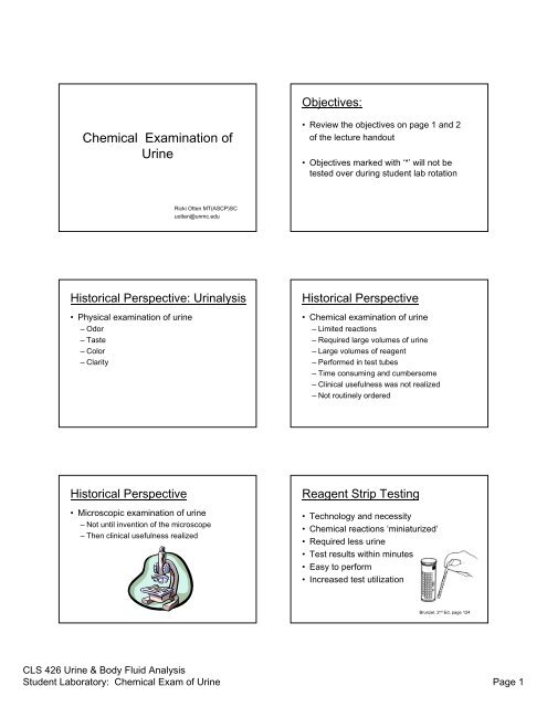
Reagent Strip Testing• Ideal qualitative screening tool– Sensitive: Low concentration <strong>of</strong> substancesNegative result = normal– Specific: Reacts with only one substanceFalse negative and false positive– Cost effective: Relatively inexpensive tool thatprovides information about the health status<strong>of</strong> the patientReagent Strip Testing• <strong>Chemical</strong>ly impregnated absorbentpads attached to an inert plastic strip• Each pad is a specific chemical reaction thattakes place upon contact with urine• <strong>Chemical</strong> reaction causes the color <strong>of</strong> the pad tochange• Color compared to a color chart for interpretationReagent Strip Testing• Qualitative or semi-quantitative results– Concentration units (mg/dl)– Negative, small, moderate large– Negative, 1+, 2+, 3+, 4+• Timing <strong>of</strong> chemical reactions is CRITICAL– Shortest time requirement on one end <strong>of</strong> strip: 30 sec– Longest time requirement on the other: 2 minReagent Strip Testing• Principle <strong>of</strong> chemical reactions– False negative reactions– False positive reactions– Color interferences• Alternative testing: used to confirm results thatyou may think are invalid due to– Interfering substance– Color interference (called color masking)Care and Storage (pg 4)Reading assignment:Textbook, chapter 7Page 124-130Confirmatory Testing (pg 6)Confirmatory Testing• Alternative testing establishes thecorrectness or accuracy <strong>of</strong> anotherprocedure• Often used when urine is highly pigmented– Bilirubin reagent strip ictotestCLS 426 <strong>Urine</strong> & Body Fluid AnalysisStudent Laboratory: <strong>Chemical</strong> Exam <strong>of</strong> <strong>Urine</strong> Page 2
Confirmatory Testing• Characteristics:– Differ in sensitivity• Ictotest vs Bilirubin reagent strip– Differ in specificity• SSA vs Protein reagent strip• Clinitest vs Glucose reagent strip– Differ in methodology/reactionIdeal:all 3Differ in Specificity• Clinitest reacts with all reducingsubstances• Glucose reagent strip reacts with onlyone reducing substance: glucose10 reagent strip tests– Specific gravity–pH–Protein–Glucose– Ketones–Blood– Bilirubin– Urobilinogen–Nitrite– Leukocyte Esterase• Purpose <strong>of</strong> the test• What is normal• What is abnormal• Reaction• Causes <strong>of</strong> invalidresultsSpecific Gravity: Purpose• Evaluates the concentrating and dilutingability <strong>of</strong> the kidney– Density is related to the amount <strong>of</strong>substances (solutes) in solution– Increased density ~ increased solute insolution ~ hypertonic urine ~ concentratedurine– Decreased density ~ decreased solute insolution ~ hypotonic urine ~ dilute urineSpecific Gravity: Normal• Normal: 1.002 – 1.035• Majority <strong>of</strong> urines: 1.010 – 1.025• Physiologically impossible: 1.000>1.040• Dependent upon hydration statusSpecific Gravity: Terms• Isosthenuria– Fixed at 1.010– Renal tubules lost absorption and secreting capability• Hypersthenuria– Increased specific gravity– Concentrated urine• Hyposthenuria– Decreased specific gravity– Dilute urineSensitivity issues:Pregnancy testingUrinary tract infectionCLS 426 <strong>Urine</strong> & Body Fluid AnalysisStudent Laboratory: <strong>Chemical</strong> Exam <strong>of</strong> <strong>Urine</strong> Page 3
Specific Gravity: Methods• Methods <strong>of</strong> measurement– Reagent strip test: indicates ionic solutes– Refractometer: indicates amount <strong>of</strong> total solutes• Two functions <strong>of</strong> the kidney– Maintain water balance– Maintain electrolyte homeostasisPerformed byrenal tubulesthroughconcentratingand diluting;reabsorbingand secretingwater andelectrolytes(ionic)Specific Gravity: Reaction• Based on a change in the pK a <strong>of</strong> apolyelectrolyte on the reagent pad• Increased ions in solution causes thepolyelectrolyte on the pad to produce free H +• Free H+ cause a change in pH on thereagent pad• Change in pH: bromthymol blue indicatorSpecific Gravity: ReactionSpecific Gravity• Sensitivity: 1.000• Specificity: detects only ionic substances– Radiographic dye– MannitolDoes not interfere– GlucosepH: Purpose• Kidneys regulate body’s acid-basebalance by selective handling <strong>of</strong> H + and HCO 3-– <strong>Urine</strong> pH reflects acid-base status <strong>of</strong> body• Treatment protocol may require urine pH bemaintained at a specific pH(Aids in identification <strong>of</strong> crystals (microscope))pH: Normal• Normal: ranges from 4.5 – 8.0• First morning void: acidic• Physiologically impossible: 8.01. <strong>Urine</strong> not handled properly2. Old urine3. Treatment inducedCLS 426 <strong>Urine</strong> & Body Fluid AnalysisStudent Laboratory: <strong>Chemical</strong> Exam <strong>of</strong> <strong>Urine</strong> Page 4
pH: Interpretation• Made in conjunction with– Acid-base status– Renal function– Presence <strong>of</strong> infection in urinary tract– Diet: high protein, low protein– Medications– Age <strong>of</strong> urine samplepH: Abnormal• Acid– Respiratory acidosis– High protein diet–Starvation–UTI• Alkaline– Respiratory alkalosis– Vegetarian diet– Renal tubular acidosis–UTIpH: ReactionpH:• Double indicator system–Methyl red– Bromthymol blueNeeded to measurethe wide pH range:acid to alkaline• Amount <strong>of</strong> free H + influences acidity <strong>of</strong>urine and cause pH indicator to changecolor• Invalid test results due to:– Improper handling <strong>of</strong> urine sample– Contamination <strong>of</strong> urine vessel prior tocollection– ‘Run-over’ phenomenonProtein: Purpose• Normal kidneys secrete LITTLE protein
Classification <strong>of</strong> Proteinuria• Functional• Orthostatic (postural)• Transient• Pathologic– Pre-renal (overflow)– Renal: glomerular– Renal: tubular– Post-renalProtein: Methods• Reagent strip test• SSA test• Foam test• Micro-albumin testProtein: Reagent Strip• The reagent pad is held at aconstant pH <strong>of</strong> 3 by a buffer• Proteins (anions) in solution cause anindicator dye to release H + causing a colorchange• ‘Protein error <strong>of</strong> indicators’Protein: Reagent Strip• Sensitivity: ~ 10-25 mg/dl• Specificity: reacts with albumin– False positive: highly alkaline urine (pH > 8.0)– False negative:Dilute urinePresence <strong>of</strong> other proteins(Tamm-Horsfall, globulins, myoglobin,free light chains, hemoglobin)Protein: SSA (Exton’s Test)• Sulfosalicylic Acid (SSA) Precipitation Test• Acid will precipitate proteins out <strong>of</strong> solutioncausing the solution to become cloudy• Amount <strong>of</strong> cloudiness is related to theamount <strong>of</strong> protein presentProtein: SSA (Exton’s Test)• Amount <strong>of</strong> cloudiness is evaluated, thusmust use centrifuged urine• Sensitivity: 5-10 mg/dl• Specificity: detects all proteinCLS 426 <strong>Urine</strong> & Body Fluid AnalysisStudent Laboratory: <strong>Chemical</strong> Exam <strong>of</strong> <strong>Urine</strong> Page 6
Protein: SSA (Exton’s Test)• False positive results:– Radiographic dyes– Turbid urine– Uncentrifuged urineProtein: Foam test• Shake aliquot <strong>of</strong> urine and observe color<strong>of</strong> resulting foam• White foam: protein present• False negative results:– Highly alkaline urine– Dilute urineProtein: Micro-albumin test• Measures very low concentration <strong>of</strong>albumin (better sensitivity than reagentstrip test for albumin)• Management <strong>of</strong> diabetic patient• Methods vary: reagent strip test,immunochemical reactionGlucose: Purpose• Healthy normal urine does not containglucose• Normally, glucose is filtered by theglomerulus and is reabsorbed back into thebloodstream through active transportmechanism• Glucose in urine is pathologicGlucose: Purpose• GlucosuriaGlycosuriaTerms usedinterchangeably• Caused by renal and non-renal disease– Pre-renal glycosuria: plasma glucose levelexceeds renal threshold (diabetes mellitus)Reducing Substances: Purpose• Reducing Substances:– Glucose– Other sugars: galactosemia (inheritedmetabolic disorder)– Renal glycosuria: plasma glucose levelbelow renal threshold, but tubules cannotreabsorb glucose back into bloodstreamCLS 426 <strong>Urine</strong> & Body Fluid AnalysisStudent Laboratory: <strong>Chemical</strong> Exam <strong>of</strong> <strong>Urine</strong> Page 7
Glucose, Reducing Substances• Normal: negative• Abnormal:– Diabetes mellitus: glucose– Impaired renal tubular reabsorption: glucoseMethods• Reagent strip: detects only glucose• Copper Reduction: detects reducingsubstances– Inborn error <strong>of</strong> metabolism: galactosemiaGlucose: Reagent Strip• Detects only glucose• Double sequential enzyme reactionGlucose: Reagent Strip• Sensitivity: ~ 30 mg/dl• Specificity:– Reacts only with glucose– False positive:• Strong oxidizing agents (bleach)• Peroxides– False negative:• Ascorbic acid (reducing agent)• Improperly stored urine: glycolysisClinitest Reaction• Copper Reduction Test:– Reducing substances are able to reducecopper sulfate to cuprous oxide– Pass-through phenomenon– All children
Ketones: Purpose• Ketones are intermediary products <strong>of</strong> fatmetabolism• Refer to diagram: text pg 152Ketones• Three ketone bodies– Acetone 2%– Acetoacetic acid 20%– Beta-hydroxybutyric acid 78%• Characteristic ‘fruity breath’ ~ acetoneKetones: Normal• Normal: negative• Abnormal:– Inability to utilize carbohydrates– Excessive loss <strong>of</strong> carbohydrates– Inadequate intake <strong>of</strong> carbohydratesKetones: Methods• Reagent strip• Acetest: tablet testKetones: Method• Glycine: also measures acetone– Reagent strip: check package insert– Acetest tablets: contain glycineKetones• Reagent strip– Sensitivity: 5-10 mg/dl– Specificity: acetoacetic acid and/or acetone• False positive: highly pigmented urine• False negative: improper specimen handling• Acetest– Specificity: acetoacetic acid and acetone• False positive: highly pigmented urien• False negative: improper specimen handlingCLS 426 <strong>Urine</strong> & Body Fluid AnalysisStudent Laboratory: <strong>Chemical</strong> Exam <strong>of</strong> <strong>Urine</strong> Page 9
Blood: Purpose• Blood in urine indicates pathology• Two forms found in urine– Intact RBC– Hemolyzed RBCBlood: Terms• Hematuria• Hemoglobinuria• MyoglobinuriaAll will give apositive bloodreactionBlood: Reagent stripBlood• Test can detect hemolyzed RBC• Heme moiety imparts peroxidase activityand catalyzes the reaction• Sensitivity• Specificity– Intact RBC– Hemolyzed RBC (hemoglobin)– Myoglobin– False positives: myoglobin, oxidizing agents– False negatives: ascorbic acidBlood:• Correlate reagent strip results– Microscopic findings– Color and clarityBilirubin and Urobilinogen• Textbook page 154• Bilirubin in urine is pathologic: liver disease• Urobilinogen in urine: normal to have asmall amount: 0.2 – 1 mg/dlCLS 426 <strong>Urine</strong> & Body Fluid AnalysisStudent Laboratory: <strong>Chemical</strong> Exam <strong>of</strong> <strong>Urine</strong> Page 10
Three mechanisms• Pre-hepatic: liver is healthy• Hepatic: liver disease• Post-hepatic: liver is healthy, obstructionindicatedBilirubin: Methods• Reagent strip• Ictotest: tablet test• Foam testBilirubin: Methods• Reagent strip• Ictotest: tablet test• Same reaction• Same specificity: conjugated bilirubin– False positive: urine color– False negative: low concentration, ascorbicacid, improper specimen handlingBilirubin: Methods• Reagent strip• Ictotest: tablet test• Sensitivity differsReagent strip: ~0.5 mg/dlIctotest: 0.05 – 0.1 mg/dlBilirubin: Methods• Possible to have a negative reagentstrip test and positive ictotest– Difference in sensitivity levelsBilirubin: Foam Test• Shake urine and observe resulting foam• Yellow foam = bilirubin• Always perform Ictotest when– <strong>Urine</strong> bilirubin test specifically ordered– <strong>Urine</strong> appearance– Positive reagent strip testCLS 426 <strong>Urine</strong> & Body Fluid AnalysisStudent Laboratory: <strong>Chemical</strong> Exam <strong>of</strong> <strong>Urine</strong> Page 11
Urobilinogen: Methods• Reagent strip test– Two reactions dependent upon manufacturer• Para-dimethylaminobenzaldehyde• Diazonium salt– Cannot determine absence <strong>of</strong> UBG• Watson-Schwartz assayUrobilinogen: Methods• Para-dimethylaminobenzaldehyde– Sensitivity: 0.2 mg/dl– Specificity:• False positive: any ‘Ehrlich reactive compound’; colormasking; urine at body temp• False negative: improper specimen handling• Diazonium salt– Sensitivity: 0.4 mg/dl– Specificity: reacts only with UBG• False positive: color masking• False negative: improper specimen handlingUrobilinogen: Watson Schwartz• Classic method used to differentiateurobilinogen from porphobilinogen using adifferential extraction method• Para-dimethylaminobenzaldehydeNitrite: Purpose• Bacteria that contain a specific enzymecan reduce dietary nitrates to nitrites• Rapid screening test for UTINitrite: Normal• Normal: negative• Abnormal:– Cystitis: bladder– Pyelonephritis: kidneyNitrite: Method• Reagent strip testNitrite + aromatic amine diazonium saltDiazonium salt + aromatic compound pink color• Sensitivity:0.06-0.1 mg/dl nitrite~ 10,000 organismsCLS 426 <strong>Urine</strong> & Body Fluid AnalysisStudent Laboratory: <strong>Chemical</strong> Exam <strong>of</strong> <strong>Urine</strong> Page 12
Nitrite: Method• Reagent strip testSpecificity:– False positive: improper specimen handling; colormasking– False negative: bacteria cannot reduce nitratesBladder time not sufficient: need 4 hoursLow nitrite levelsAscorbic acidAntibiotic inhibition <strong>of</strong> bacteriaFurther reduction <strong>of</strong> nitrites to nitrogenLeukocyte Esterase: Purpose• Increased WBC in urine is pathologic– Indicates inflammation, infection• Neutrophils most common type <strong>of</strong> WBCfound in urine• Can detect intact WBC and lysed WBCLeukocyte Esterase: Normal• Normal: negative• Abnormal:– Bacterial infection:cystitis, pyelonephritis, urethritis– Non-bacterial infection: yeast, trichomonasLeukocyte Esterase: Method• Reagent strip:Granules in cytoplasm <strong>of</strong> WBC contain anenzyme (esterase)Ester –esterase aromatic compoundAromatic compound + diazonium salt Purple colored complexLeukocyte Esterase• Sensitivity: 5-15 WBC/hpf• Specificity:– False positive: vaginal contamination; colormasking– False negative: strong oxidizing agents(bleach); lymphocytes (no granules)CLS 426 <strong>Urine</strong> & Body Fluid AnalysisStudent Laboratory: <strong>Chemical</strong> Exam <strong>of</strong> <strong>Urine</strong> Page 13