

Review - University of Oxford
Review - University of Oxford
Review - University of Oxford

Create successful ePaper yourself
Turn your PDF publications into a flip-book with our unique Google optimized e-Paper software.
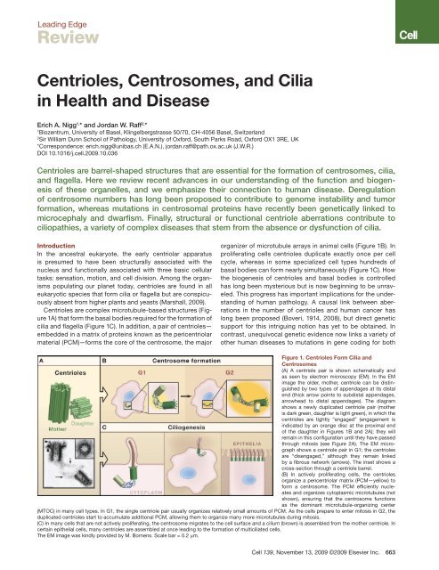
Leading Edge<strong>Review</strong>Centrioles, Centrosomes, and Ciliain Health and DiseaseErich A. Nigg 1, * and Jordan W. Raff 2, *1Biozentrum, <strong>University</strong> <strong>of</strong> Basel, Klingelbergstrasse 50/70, CH-4056 Basel, Switzerland2Sir William Dunn School <strong>of</strong> Pathology, <strong>University</strong> <strong>of</strong> <strong>Oxford</strong>, South Parks Road, <strong>Oxford</strong> OX1 3RE, UK*Correspondence: erich.nigg@unibas.ch (E.A.N.), jordan.raff@path.ox.ac.uk (J.W.R.)DOI 10.1016/j.cell.2009.10.036Centrioles are barrel-shaped structures that are essential for the formation <strong>of</strong> centrosomes, cilia,and flagella. Here we review recent advances in our understanding <strong>of</strong> the function and biogenesis<strong>of</strong> these organelles, and we emphasize their connection to human disease. Deregulation<strong>of</strong> centrosome numbers has long been proposed to contribute to genome instability and tumorformation, whereas mutations in centrosomal proteins have recently been genetically linked tomicrocephaly and dwarfism. Finally, structural or functional centriole aberrations contribute tociliopathies, a variety <strong>of</strong> complex diseases that stem from the absence or dysfunction <strong>of</strong> cilia.IntroductionIn the ancestral eukaryote, the early centriolar apparatusis presumed to have been structurally associated with thenucleus and functionally associated with three basic cellulartasks: sensation, motion, and cell division. Among the organismspopulating our planet today, centrioles are found in alleukaryotic species that form cilia or flagella but are conspicuouslyabsent from higher plants and yeasts (Marshall, 2009).Centrioles are complex microtubule-based structures (Figure1A) that form the basal bodies required for the formation <strong>of</strong>cilia and flagella (Figure 1C). In addition, a pair <strong>of</strong> centrioles—embedded in a matrix <strong>of</strong> proteins known as the pericentriolarmaterial (PCM)—forms the core <strong>of</strong> the centrosome, the majororganizer <strong>of</strong> microtubule arrays in animal cells (Figure 1B). Inproliferating cells centrioles duplicate exactly once per cellcycle, whereas in some specialized cell types hundreds <strong>of</strong>basal bodies can form nearly simultaneously (Figure 1C). Howthe biogenesis <strong>of</strong> centrioles and basal bodies is controlledhas long been mysterious but is now beginning to be unraveled.This progress has important implications for the understanding<strong>of</strong> human pathology. A causal link between aberrationsin the number <strong>of</strong> centrioles and human cancer haslong been proposed (Boveri, 1914, 2008), but direct geneticsupport for this intriguing notion has yet to be obtained. Incontrast, unequivocal genetic evidence now links a variety <strong>of</strong>other human diseases to mutations in gene coding for bothFigure 1. Centrioles Form Cilia andCentrosomes(A) A centriole pair is shown schematically andas seen by electron microscopy (EM). In the EMimage the older, mother, centriole can be distinguishedby two types <strong>of</strong> appendages at its distalend (thick arrow points to subdistal appendages,arrowhead to distal appendages). The diagramshows a newly duplicated centriole pair (motheris dark green, daughter is light green), in which thecentrioles are tightly “engaged” (engagement isindicated by an orange disc at the proximal end<strong>of</strong> the daughter in Figures 1B and 2A); they willremain in this configuration until they have passedthrough mitosis (see Figure 2A). The EM micrographshows a centriole pair in G1; the centriolesare “disengaged,” although they remain linkedby a fibrous network (arrows). The inset shows across-section through a centriole barrel.(B) In actively proliferating cells, the centriolesorganize a pericentriolar matrix (PCM—yellow) t<strong>of</strong>orm a centrosome. The PCM efficiently nucleatesand organizes cytoplasmic microtubules (notshown), ensuring that the centrosome functionsas the dominant microtubule-organizing center(MTOC) in many cell types. In G1, the single centriole pair usually organizes relatively small amounts <strong>of</strong> PCM. As the cells prepare to enter mitosis in G2, theduplicated centrioles start to accumulate additional PCM, allowing them to organize many more microtubules during mitosis.(C) In many cells that are not actively proliferating, the centrosome migrates to the cell surface and a cilium (brown) is assembled from the mother centriole. Incertain epithelial cells, many centrioles are assembled at once leading to the formation <strong>of</strong> multiciliated cells.The EM image was kindly provided by M. Bornens. Scale bar = 0.2 µm.Cell 139, November 13, 2009 ©2009 Elsevier Inc. 663
centrosomal and centriolar proteins (Bettencourt-Dias andGlover, 2007; Bond and Woods, 2006; Gerdes et al., 2009;Quinlan et al., 2008)Recent studies <strong>of</strong>fer exciting new insight into the physiologicalroles <strong>of</strong> centrioles and the pathologies that result from theirderegulation. When considered from a mechanistic perspective,human diseases resulting from centriole aberrations areexpected to reflect defects in (1) centriole biogenesis, (2) centrosomestructure, function, and positioning, or (3) the formationor maintenance <strong>of</strong> cilia and flagella. In this <strong>Review</strong> we willsummarize our current understanding <strong>of</strong> the function and biogenesis<strong>of</strong> centrioles and then focus on their role in disease.Our emphasis will be on the expected impact <strong>of</strong> centrosomedysfunction on tumorigenesis, the genetic evidence linkingcentrosomes to the regulation <strong>of</strong> brain and body size, and thecontribution <strong>of</strong> centriole aberrations to ciliopathies.The Functions <strong>of</strong> Centrosomes and CiliaCell DivisionIn most animal cells, centrosomes are a major source <strong>of</strong> spindlemicrotubules, and they are absolutely essential for cell division inseveral early embryonic systems. Perhaps the most striking example<strong>of</strong> this is provided by studies on the newly fertilized embryo <strong>of</strong>the frog Xenopus leavis (Klotz et al., 1990). As in many species,centrioles are eliminated from the developing oocyte in Xenopus,so the centriole is normally supplied to the egg by the fertilizingsperm. Pricking a Xenopus egg with a needle mimics fertilization,and the egg proceeds through several rounds <strong>of</strong> the cell cycle butfails to cleave at the end <strong>of</strong> each cycle. If, however, a centriole orcentrosome is coinjected into the egg as it is pricked, the egg cannow divide and, in some cases, develop into a morphologicallynormal tadpole using only the maternal complement <strong>of</strong> chromosomes.Thus, in Xenopus, the centriole is essential to allow theembryo to divide. Similarly, the fertilized embryos <strong>of</strong> both the fruitfly Drosophila and the nematode Caenorhabditis elegans absolutelyrequire functional centrosomes for the earliest stages <strong>of</strong>embryonic development (Kirkham et al., 2003; Leidel and Gonczy,2003; O’Connell et al., 2000; Stevens et al., 2007). Conversely, thepresence <strong>of</strong> extra centrosomes in many early embryonic systemsleads to the formation <strong>of</strong> multipolar spindles, which in turn leadsto abnormal chromosome segregation and division as the duplicatedchromosomes are segregated between the multiple spindlepoles. The strong conclusion from these studies is that centrosomesare the major driving force for bipolar spindle assemblyand cell division in most animal cells, a view that has dominatedthe centrosome field for most <strong>of</strong> the last 100 years.It is clear, however, that centrosomes are not absolutelyessential for division in many cell types. When centrosomes areabsent, either naturally (as in higher plants or in the female germcells <strong>of</strong> many animal species) or due to experimental manipulation,bipolar spindles can form in the vicinity <strong>of</strong> chromosomesthrough a centrosome-independent pathway that involves thesmall GTPase Ran and the action <strong>of</strong> microtubule motors andmicrotubule-bundling proteins (Kalab and Heald, 2008). Thispathway also presumably explains the surprising finding thatDrosophila mutants lacking the centriole duplication proteinDSas-4 appear to proceed normally through most <strong>of</strong> development,provided that a maternally supplied pool <strong>of</strong> DSas-4 is initiallypresent to allow centrosome formation during the earlieststages <strong>of</strong> embryogenesis (Basto et al., 2006). In these mutants,centrioles and centrosomes are undetectable in adult cells, yetadults appear morphologically normal and eclose with nearnormaltiming at near-normal Mendelian ratios. This is in starkcontrast to the rare larvae that develop to adulthood in manymutants that show dramatic defects in cell division (for criticaldiscussion see Gonzalez, 2008). Thus, although spindle assemblyis slowed in fly cells that lack centrosomes, flies appear toproceed through most <strong>of</strong> development relatively normally usingonly the centrosome-independent pathway <strong>of</strong> spindle assembly.Perhaps this reflects the fact that Drosophila cells have only fourchromosomes to segregate, and organisms with larger numbers<strong>of</strong> chromosomes may depend on the greater efficiency <strong>of</strong> spindleassembly afforded by centrosomes—an intriguing notionthat remains to be experimentally tested.Interestingly, Drosophila mutants that lack centrioles neverthelessdie soon after they eclose. Rather than reflecting theabsence <strong>of</strong> centrosomes, however, this death appears to resultfrom the lack <strong>of</strong> cilia that are essential for the function <strong>of</strong> certainmechano- and chemosensory neurons. These observationssupport the view that centrioles may have originally acquiredthe ability to form centrosomes not to increase the efficiency<strong>of</strong> cell division but rather to ensure that the centrioles associatewith the spindle poles and are thereby equally partitionedbetween the two daughter cells (Marshall, 2009).Although many somatic cells can clearly divide without centrosomes,there is compelling evidence that centrosomes arerequired for the efficient division <strong>of</strong> cells that split asymmetricallyto produce two daughter cells <strong>of</strong> different fates. Wellstudiedexamples are the early C. elegans embryo, Drosophilamale germline stem cells (GSCs), and Drosophila neuroblasts(the stem cell-like progenitors <strong>of</strong> the Drosophila nervous system)(Gonczy, 2008). In these cells, the astral microtubulesgenerated by centrosomes play an important part in aligningthe spindle relative to cortical fate determinants, thus ensuringthe accurate segregation <strong>of</strong> the determinants between thetwo daughter cells. In flies that lack functional centrosomes,~15% <strong>of</strong> neuroblast divisions appear to be symmetric (Bastoet al., 2006), whereas the asymmetric divisions <strong>of</strong> male GSCsare also compromised when centrosome function is perturbed(Yamashita et al., 2003). Thus, centrosomes have important,although not essential, roles in these asymmetric divisions.Remarkably, centrosomes themselves <strong>of</strong>ten behave asymmetricallyin many instances. In Drosophila neuroblasts andmale GSCs, for example, only one <strong>of</strong> the newly separatedcentrosomes initially nucleates a robust aster <strong>of</strong> microtubules,which allows this centrosome to maintain a stable positionat the apical side <strong>of</strong> the cell (Rebollo et al., 2007; Rusan andPeifer, 2007; Yamashita et al., 2007). As the cell enters mitosis,the other centrosome starts to nucleate microtubules,but the pre-positioning <strong>of</strong> the first centrosome ensures thatthe spindle forms in correct alignment with the cortical determinants.Interestingly, in male GSCs, the oldest centrosome(that contains the older, “mother,” centriole) is always retainedwithin the stem cell (Yamashita et al., 2007). This observationraises the possibility that an “immortal centrosome,” alwaysretained in the stem cell, could help determine stem cell fate664 Cell 139, November 13, 2009 ©2009 Elsevier Inc.
(Morrison and Spradling, 2008). Moreover, a gradual declinein the ability <strong>of</strong> the male GSCs to maintain proper centrosomeorientation as they age is correlated with a gradual loss <strong>of</strong> theirproliferative capacity, as GSCs appear unable to enter mitosiswith improperly aligned centrosomes (Cheng et al., 2008). Onthe other hand, centrosomes do not exhibit an obvious asymmetricbehavior during the asymmetric division <strong>of</strong> Drosophilafemale GSCs, and, unlike neuroblasts and male GSCs, theasymmetric division <strong>of</strong> female GSCs is not detectably perturbedin the absence <strong>of</strong> centrosomes (Stevens et al., 2007),arguing that an immortal centrosome is unlikely to be a ubiquitousmechanism for ensuring the maintenance <strong>of</strong> stem cellfate. A potentially interesting twist to the role <strong>of</strong> centrioles indetermining cell behavior comes from the observation that therelative age <strong>of</strong> the mother centrioles can determine the timing<strong>of</strong> primary cilium formation in the two daughter cells formedafter cell division (Anderson and Stearns, 2009). This intriguingdiscovery implies that differences in centriole age can conferan asymmetry to every animal cell division, which in turn mayhave far-reaching consequences for cell differentiation in vivo.Centrosomes and the Cell CycleAlthough centrosomes are not essential for cell division in allcell types, there is evidence that they contribute to efficientcell-cycle progression at both the G1/S and the G2/M transitions.In response to experimentally induced perturbation<strong>of</strong> centrosomes, some vertebrate cells undergo a G1 arrest,prompting speculation that a specific checkpoint might monitorthe functional integrity <strong>of</strong> the centrosome (Mikule et al.,2007). However, considering that this G1 arrest depends ona pathway involving p38 kinase and p53, it most likely reflectsa stress response rather than a specific novel checkpoint(Uetake et al., 2007; Srsen et al., 2006). During S phase, a subpopulation<strong>of</strong> cyclin E associates with centrosomes and possiblycontributes to the regulation <strong>of</strong> S phase entry (Matsumotoand Maller, 2004), and several cell-cycle regulatory proteinsare concentrated at centrosomes and spindle poles duringmitosis (Fry and Hames, 2004). These observations have led tothe proposal that centrosomes might function as “scaffolds” topromote interactions between various regulatory componentsduring the cell cycle (Doxsey et al., 2005). At the G2/M transition,the key mitotic kinases Cdk1/Cyclin B, Aurora-A, and Pol<strong>of</strong>amily members all accumulate at centrosomes, and the mitoticactivation <strong>of</strong> Cdk1 is first detected at centrosomes (Jackman etal., 2003). Furthermore, centrosomal Aurora-A has been implicatedin the timing <strong>of</strong> mitotic entry in C. elegans (Hachet etal., 2007; Portier et al., 2007), and, in Drosophila embryos, thedestruction <strong>of</strong> Cyclin B at the end <strong>of</strong> mitosis appears to be initiatedat centrosomes (Wakefield et al., 2000).There are also several reports indicating that components<strong>of</strong> the DNA-damage checkpoint are concentrated at centrosomes(Doxsey et al., 2005). Although the significance <strong>of</strong>these observations remains to be fully understood, a strongcase can be made for a functionally important link betweenthe DNA-damage response protein Chk2 and centrosomes inDrosophila syncytial embryos. In this case, the Chk2-dependentinactivation <strong>of</strong> centrosomes in response to DNA damagecauses the damaged nuclei to fall into the interior yolk region<strong>of</strong> the embryo, thereby preventing their incorporation into thedeveloping embryonic tissues (Takada et al., 2003). Althoughthis exact mechanism is not expected to operate in vertebratesomatic cells, connections between impaired DNA integrityand centrosomes have been observed in mammalian cells(Dodson et al., 2004; Hut et al., 2003). Furthermore, the proteinkinase Chk1, a major mediator <strong>of</strong> the DNA-damage responsecheckpoint, associates with centrosomes and contributes tothe regulation <strong>of</strong> the G2/M transition even in unperturbed cells(Kramer et al., 2004).Centriole and Centrosome Function in Differentiated CellsThe traditional emphasis on the role <strong>of</strong> centrosomes in celldivision should not detract from the fact that these organellescontribute to the organization <strong>of</strong> microtubule arrays throughoutinterphase <strong>of</strong> the cell cycle as well as in postmitotic, differentiatedcells. In turn, microtubule arrays are important for determiningthe shape, polarity, and motility <strong>of</strong> cells and organisms.For example, studies in Chlamydomonas have suggested thatthe proper positioning <strong>of</strong> the centriole is required for establishingthe overall geometry <strong>of</strong> the interphase cell (Feldman et al.,2007). Moreover, centrosomes adopt specific positions withinmany different types <strong>of</strong> polarized cells, such as migrating fibroblasts,epithelial cells, and neuronal cells (Gundersen, 2002;Higginbotham and Gleeson, 2007; Ueda et al., 1997). Centrosomesare generally thought to be positioned within the cellvia centrosome-associated microtubules that are preferentiallystabilized at specific cortical sites. In the case <strong>of</strong> cytotoxic Tlymphocytes (CTLs), for example, the centrosome migrates tothe site <strong>of</strong> contact between the CTL and its target cell, where ithelps deliver the lytic granules that will ultimately kill the targetcell (Stinchcombe et al., 2006).Taken together, the above studies suggest that centrosomescontribute to the proper spatial organization <strong>of</strong> many nondividingcells. Importantly, though, in many cases the specificspatial organization <strong>of</strong> microtubules may depend primarily onthe localization <strong>of</strong> specific PCM components, rather than onstructurally intact centrosomes. In particular, although microtubulenucleation and anchoring are <strong>of</strong>ten associated withcentrosomes, this is not necessarily the case in all cell types.Indeed, many differentiated cells do not contain the “textbook”arrangement <strong>of</strong> radial microtubule arrays emanating from acentrally located centrosome. Instead, microtubules may berunning parallel along the cell axis, as in many epithelial cells,or nucleate from the nuclear envelope, as in differentiated musclecells (Mogensen, 2004). Moreover, at least some interphasecells that have had their centrosomes removed experimentallycan re-establish relatively normal polarized microtubule arraysthat appear to lack centrosomes (Wadsworth and Khodjakov,2004). How these noncentrosomal microtubule arrays areorganized remains to be fully understood, but it seems thatcertain PCM components can dissociate from centrioles andbecome concentrated at different organizing centers such asthe plasma membrane or the nuclear envelope.Remarkably, there is also evidence that centrosomes havefunctions that are independent <strong>of</strong> their ability to organize microtubules.For example, the efficient breakdown <strong>of</strong> the nuclear envelopein C. elegans embryos requires the activity <strong>of</strong> centrosomeassociatedAurora-A in a way that is apparently independent <strong>of</strong>microtubules (Hachet et al., 2007; Portier et al., 2007).Cell 139, November 13, 2009 ©2009 Elsevier Inc. 665
Figure 2. The Centriole Duplication Cycle(A) A schematic representation <strong>of</strong> centriole behaviorduring the cell cycle. At the end <strong>of</strong> mitosis each newdaughter cell inherits a single pair <strong>of</strong> “disengaged”centrioles. Cells then progress into G1 or enter aquiescent state (G0), during which many cell typeswill form a cilium. In cycling cells the centrioles duplicatein S phase, with newly born procentrioles(light green) remaining tightly engaged with theirmother centrioles (dark green) and gradually elongatingthroughout S and G2. At the G2/M transition,the centrioles accumulate more pericentriolar material(PCM, yellow) and the two centrosomes startto separate from one another, eventually formingthe poles <strong>of</strong> the spindle in mitosis.(B) The top illustration depicts centriole duplicationin C. elegans embryos. SPD-2 recruits the proteinkinase ZYG-1 to mother centrioles, which thenrecruits a complex <strong>of</strong> SAS-6 and SAS-5. This promotesthe formation <strong>of</strong> a central tube (red) at rightangles to the mother centriole. SAS-6 and SAS-5then recruit SAS-4, which allows the centriolarmicrotubules (green) to associate with the centraltube, thus forming the procentriole. The proteinγ-tubulin is also required at about this time. The proteinshighlighted in red all have functional orthologsimplicated in centriole duplication in other species.The bottom illustration depicts the early events <strong>of</strong>centriole duplication in human cells. Cep192 is thehuman homolog <strong>of</strong> SPD-2, but this protein does notappear to be essential for centriole duplication inflies (Dix and Raff, 2007; Giansanti et al., 2008), andthere is conflicting data as to whether it is essentialin humans (Gomez-Ferreria and Sharp, 2008; Zhuet al., 2008). The protein kinase Plk4 (Sak in flies)is only distantly related to ZYG-1, but it appears toplay an analogous role to ZYG-1 in recruiting humanSAS-6 (HsSAS-6) to centrioles, and both Plk4and HsSAS-6 are essential for centriole duplication.HsSAS-6 seems to be required for the formation <strong>of</strong>a central “cartwheel” structure (rather than a centraltube) (red) and human SAS-4 (HsSAS-4, alsocalled CPAP) and γ-tubulin appear to be requiredto convert this structure to a procentriole. Severaladditional proteins, such as CP110 and Cep135/Bld10, have also been implicated in centriole duplication(Dobbelaere et al., 2008; Kleylein-Sohn etal., 2007). This notwithstanding, it is surprising thatthe duplication <strong>of</strong> a structure as elaborate as thecentriole appears to rely on such a small number<strong>of</strong> key proteins.The Functions <strong>of</strong> CiliaIn many nondividing cells the centrioles migrate to the cell surfacewhere the mother centriole forms a basal body that organizes theformation <strong>of</strong> a cilium or flagellum. The functional identity betweencentrioles and basal bodies is exemplified beautifully in the greenalgae Chlamydomonas, where the exact same microtubule barrelsfunction as centrioles within centrosomes at the spindlepoles during cell division and as basal bodies for the formation<strong>of</strong> flagella during interphase (Dutcher, 2003). Broadly speaking,there are two types <strong>of</strong> cilia: motile cilia that have a central pair <strong>of</strong>microtubules (known as the 9+2 organization) and nonmotile ciliathat lack the central pair <strong>of</strong> microtubules (the 9+0 organization).The importance <strong>of</strong> motile cilia in several epithelial tissues such asthe trachea and bronchial tubes (where the beating cilia functionto clear debris) has long been appreciated (Afzelius, 1976). In contrast,although most vertebrate cells can form a single nonmotileprimary cilium, it is only recently that the functions <strong>of</strong> primary ciliahave become more widely recognized. An important clue to thefunction <strong>of</strong> primary cilia came from the realization that targeteddisruption <strong>of</strong> the KIF3B kinesin in mice led to defects in left-rightasymmetry (situs inversus) that were correlated with the loss <strong>of</strong>primary cilia in the embryonic node (Nonaka et al., 1998). Surprisingly,these 9+0 primary cilia were actually motile, although theyexhibit an unusual twirling motion that is quite distinct from thebeating <strong>of</strong> 9+2 motile cilia. The movement <strong>of</strong> these primary ciliaappears to set up a “nodal flow” within the extracellular milieu surroundingthe node, and this leads to the asymmetric activation <strong>of</strong>signaling pathways within the embryo. Intriguingly, KIF3B knockoutmice also had a variety <strong>of</strong> other defects in neural, heart, andkidney development that could not readily be explained by theloss <strong>of</strong> cilia in the node cells, suggesting that primary cilia couldhave other important functions during development.The groundwork for understanding the origins <strong>of</strong> these additionaldefects came from pioneering studies in Chlamydomonas<strong>of</strong> a process termed intraflagellar transport (IFT) (Pedersenand Rosenbaum, 2008). IFT is the process by which various666 Cell 139, November 13, 2009 ©2009 Elsevier Inc.
cargos move along the ciliary microtubules from the cytoplasmto the tip <strong>of</strong> the cilia/flagella and then back down to the cellbody—a process that is essential for establishing and maintainingcilia/flagella organization. Indeed, defects in IFT arenow known to be the cause <strong>of</strong> the ciliary defects in the KIF3Bmutant mice. The cloning <strong>of</strong> one <strong>of</strong> the genes required forIFT in Chlamydomonas revealed that it was related to a genethat caused polycystic kidney disease when mutated in mice(Pazour et al., 2000). In contrast to the node cells, the cilia inkidney cells do not seem to generate liquid flow, but rather tosense it, and this is essential for normal kidney development(Jonassen et al., 2008).Subsequently, several mouse mutations initially identified onthe basis <strong>of</strong> a failure in sonic hedgehog (shh) signaling werealso found to reside in genes encoding proteins required forIFT (Corbit et al., 2005; Huangfu et al., 2003). The shh signalingpathway is highly conserved and is used in many tissuesthroughout development. It is also important in adult tissues,as it can promote the proliferation <strong>of</strong> adult stem cells (Ahn andJoyner, 2005) and is one <strong>of</strong> the most frequently upregulatedsignaling pathways in human cancers (Jiang and Hui, 2008).Remarkably, there is now strong evidence that cilia are essentialfor most, if not all, shh signaling in mice, and that severalproteins in the shh pathway are concentrated in cilia (Singlaand Reiter, 2006). Moreover, there is increasing evidence thatcilia can modulate several other signaling pathways such asthose activated by the Wnt and PDGFαα ligands. Unlike theirrole in the shh pathway, however, it seems that cilia are notessential for signaling via these other pathways; instead, theyexert a more indirect effect that modulates the strength <strong>of</strong> signaling(Singla and Reiter, 2006). Studies in frogs and fish haveconfirmed the importance <strong>of</strong> cilia in influencing several <strong>of</strong> thesedevelopmental pathways, although the molecular details varybetween species (Wessely and Obara, 2008). It remains to beunderstood why cilia are essential for shh signaling in mice butnot in Drosophila, where hedgehog signaling functions in manytissues that lack cilia.Biogenesis <strong>of</strong> Centrioles and CiliaCentriole FormationMost vertebrate cells contain either two centrioles (one centrosome)or two pairs <strong>of</strong> centrioles (two centrosomes), dependingon whether they are in G1 or G2 <strong>of</strong> the cell cycle (Figure 1). DuringS phase, the two centrioles duplicate through the formation,at a near-orthogonal angle, <strong>of</strong> exactly one new centriole close totheir proximal ends (Figure 2). Although centriole numbers areunder tight cell-cycle control in most proliferating cells, there arenotable exceptions. For example, cells lining the epithelia <strong>of</strong> therespiratory and reproductive tracts form hundreds <strong>of</strong> centriolesin order to provide the basal bodies for the formation <strong>of</strong> beatingcilia. These centrioles are generated nearly simultaneously fromfibrous aggregates termed deuterosomes. Until recently, centrioleformation was therefore thought to occur via one <strong>of</strong> twodistinct pathways, a “centriolar” and an “acentriolar” pathway.Implicit in this distinction was the assumption that the centriolarpathway is dependent on a “templating” function exertedby the pre-existing centrioles. It has now been recognized,however, that even cells that normally form centrioles throughthe centriolar pathway are competent to form new centriolesde novo, provided that the resident centrioles are removedbeforehand (Khodjakov et al., 2002; La Terra et al., 2005; Marshallet al., 2001). Furthermore, Polo-like kinase 4 (Plk4; alsoknown as Sak), a distant member <strong>of</strong> the Polo kinase family,was found to be a key regulator <strong>of</strong> centriole biogenesis in boththe centriolar (Bettencourt-Dias et al., 2005; Habedanck et al.,2005) and acentriolar pathways (Peel et al., 2007; Rodrigues-Martins et al., 2007). Thus, the emerging view is that the twopathways share key regulatory elements and that pre-existingcentrioles may function primarily as platforms for centriole biogenesisrather than as genuine “templates.” Compared to thede novo assembly <strong>of</strong> centrioles in the cytoplasm, a pre-existingcentriole may <strong>of</strong>fer a kinetic advantage to centriole formationby providing a surface that favors the assembly reaction (Loncarekand Khodjakov, 2009). One attractive possibility is thatthe PCM “cloud” surrounding each centriole may favor centrioleassembly by ensuring a higher local concentration <strong>of</strong> criticalassembly factors (Dammermann et al., 2004, 2008; Kirkham etal., 2003; Loncarek et al., 2008). Just as the PCM is importantfor centriole formation, the centrioles are in turn important forPCM assembly (Bobinnec et al., 1998), suggesting the development<strong>of</strong> a symbiotic relationship between the centrioles andthe PCM during evolution.Crucial insight into the molecular mechanisms underlyingcentriole duplication first emerged through a combination <strong>of</strong>genetic and RNA interference-based approaches with highresolutionlight and electron microscopy/tomography appliedto centriole biogenesis in C. elegans early embryos (Dammermannet al., 2004; Delattre et al., 2004; Kemp et al., 2004;Kirkham et al., 2003; Leidel et al., 2005; Leidel and Gonczy,2003; O’Connell et al., 2001). These pioneering studies revealedan ordered assembly pathway that involves the products <strong>of</strong>just five essential genes, termed zyg-1, spd-2, sas-4, sas-5,and sas-6 (Delattre et al., 2006; Pelletier et al., 2006) (Figure2B). Whereas SAS-4, SAS-5, SAS-6, and SPD-2 are coiled-coilproteins, ZYG-1 is a protein kinase. Shortly after fertilization <strong>of</strong>the egg, SPD-2 is recruited to the paternal centrioles, whichthen allows the centriolar recruitment <strong>of</strong> ZYG-1. Next, a complexcomprising SAS-5 and SAS-6 is recruited, which leads tothe formation <strong>of</strong> a “central tube” that is closely associated withthe original centriole. Finally, SAS-4 facilitates the assembly <strong>of</strong>microtubules onto the periphery <strong>of</strong> this tube, resulting in theformation <strong>of</strong> a procentriole (Pelletier et al., 2006). Importantly,the significance <strong>of</strong> these findings is not limited to nematodes.Although an ortholog <strong>of</strong> SAS-5 awaits definitive identification,SPD-2, SAS-4, and SAS-6 clearly have orthologs in human cellstermed Cep192 (Andersen et al., 2003), CPAP/HsSAS-4 (Hunget al., 2000), and HsSAS-6 (Leidel et al., 2005), respectively.Curiously, ZYG-1 does not have obvious structural orthologsoutside <strong>of</strong> nematodes, but the available evidence suggeststhat Plk4/Sak plays a functionally analogous role in Drosophilaand human cells (Bettencourt-Dias et al., 2005; Habedanck etal., 2005).The picture that emerges is that basic mechanisms underlyingcentriole biogenesis have been conserved during evolution(Figure 2B). Although proteomic studies have revealed asurprisingly large number <strong>of</strong> centrosome-associated proteinsCell 139, November 13, 2009 ©2009 Elsevier Inc. 667
(Andersen et al., 2003; Keller et al., 2005; Kilburn et al., 2007), itis striking that relatively few core proteins, notably the homologs<strong>of</strong> ZYG-1, SAS-4, and SAS-6, have been found to be essentialfor centriole biogenesis in all species examined. SPD-2, on theother hand, is essential in C. elegans (Pelletier et al., 2004), butnot in Drosophila (Dix and Raff, 2007; Giansanti et al., 2008).Thus, different organisms may show distinct requirements foradditional proteins in centriole duplication (Dobbelaere et al.,2008; Kleylein-Sohn et al., 2007) (Figure 2B). During centriolebiogenesis in human cells, the first proteins to assemble at thesite <strong>of</strong> procentriole formation include HsSAS-6, CPAP/HsSAS-4,Cep135, and γ-tubulin (Kleylein-Sohn et al., 2007). Clearly, theseproteins constitute attractive candidate substrates for the centrioleduplication kinase Plk4/Sak. Once a procentriolar seed hasformed, the protein CP110 then associates with the distal tip <strong>of</strong>the nascent procentriole, before α-/β-tubulin dimers are apparentlyinserted underneath a CP110 cap during centriole elongation(Kleylein-Sohn et al., 2007).Not surprisingly, some differences in centriole biogenesishave been noticed between cell types and organisms. Forinstance, the early stage <strong>of</strong> procentriole formation in Chlamydomonas,Tetrahymena, and vertebrate cells is characterizedby the appearance <strong>of</strong> a complex symmetrical structure termedthe cartwheel (Culver et al., 2009; Loncarek and Khodjakov,2009; Matsuura et al., 2004; Nakazawa et al., 2007) that ispositioned at the base <strong>of</strong> the newly born centriole, whereas inC. elegans a hollow tube structure initially forms, onto whichmicrotubules are then deposited (Pelletier et al., 2006). Consideringthat centriole duplication is an ancient process withkey regulators conserved during evolution, the observed differences(cartwheel versus tube, for example) most likelyrepresent variations on a common theme, perhaps reflectingdifferences in the half-lives <strong>of</strong> intermediate structures, ratherthan fundamentally distinct mechanisms. In strong support <strong>of</strong>this view, the protein SAS-6 is required for tube formation in C.elegans (Pelletier et al., 2006), whereas its homolog in Chlamydomonasand Tetrahymena clearly localizes to the cartwheel(Culver et al., 2009; Nakazawa et al., 2007).Once formed during early S phase, each new centriole elongatesthroughout the remainder <strong>of</strong> S and G2 phases. How thefinal length <strong>of</strong> the centriole is determined remains to be understood,but recent studies point to antagonistic roles for CP110and CPAP/HsSAS-4. Whereas CP110 appears to limit centriolarmicrotubule extension, overexpression <strong>of</strong> CPAP/HsSAS-4 resultsin the formation <strong>of</strong> long microtubule-based structures that extendbeyond the normal length <strong>of</strong> the centrioles (Kohlmaier et al., 2009;Schmidt et al., 2009; Tang et al., 2009). An additional proteinrecently implicated in centriole duplication and length control isthe WD40 domain protein POC1 (Keller et al., 2009).After cell division, the new centriole is able to function as aparental centriole during S phase, before it reaches full maturityduring late G2 or early M phase. This final maturation step isreflected by the acquisition <strong>of</strong> distal and subdistal appendages(Figure 2A). Thus, it takes more than one complete cell cyclefor a newly produced centriole to achieve complete structuralmaturity. As a result, the two centrioles present within a givenhuman centrosome can readily be distinguished by the fact thatonly the older (mature) one carries appendages. This structuraldifference is functionally important for at least two reasons:first, at least in some cell types, it is the mature centriole thatanchors most <strong>of</strong> the microtubules to the centrosome (Piel etal., 2000) and, second, it is also the mature centriole that isuniquely competent to function as a basal body during ciliogenesis(Ishikawa et al., 2005) (Figure 1). Thus, it would be <strong>of</strong>considerable interest to identify and functionally characterizecentriolar proteins that mark the mother centriole for the addition<strong>of</strong> appendages. One such candidate is the human POC5protein, which appears to be specifically required to properlyassemble the distal portion <strong>of</strong> the centriole (Azimzadeh et al.,2009). Intriguingly centrioles can behave asymmetrically in flystem cells—even though the mother centrioles in most Drosophilacells lack recognizable appendages (Callaini and Riparbelli,1990). This suggests that mother and daughter centriolescan be distinguished even when they both lack appendages.The Control <strong>of</strong> Centriole NumberBy analogy to the situation with DNA replication, the maintenance<strong>of</strong> constant centriole numbers in proliferating cells is likelyto require two types <strong>of</strong> control. A “cell-cycle control” is expectedto enforce the rule that centriole duplication occurs exactly oncein every cell cycle, whereas a “copy-number control” mustensure that only one new centriole is assembled next to eachpre-existing centriole (Nigg, 2007). With regard to cell-cyclecontrol, it has long been established that centriole duplicationrequires the activity <strong>of</strong> cyclin-dependent kinases and passage<strong>of</strong> cells beyond a stage in G1 where the retinoblastoma proteinis phosphorylated and E2F transcription factors become active(Nigg, 2002). Additional kinases, notably Plk2 (Warnke et al.,2004), Mps1 (Fisk et al., 2003; see, however, Stucke et al., 2002),and Ndr1 (Hergovich et al., 2007), as well as the DNA replicationproteins Orc1 (Hemerly et al., 2009) and geminin (Tachibana etal., 2005) have also been reported to play a role.Most importantly, elegant cell fusion studies have revealedthat there is a centrosome-intrinsic block to reduplication duringS and G2 phases, ensuring that duplicated centrioles do notnormally duplicate again until they have passed through M phase(Wong and Stearns, 2003). Newly assembled centrioles displaya tight, near-orthogonal association with the parental centriole,and this engagement persists from the time <strong>of</strong> centriole formationduring S phase until late mitosis (Figure 2A). The separation<strong>of</strong> the newly built centrosome from its parent, a process nowtermed disengagement (formerly disorientation), appears to constitutean essential prerequisite for a new round <strong>of</strong> duplication(Tsou and Stearns, 2006; Wong and Stearns, 2003), potentiallyexplaining why newly duplicated centrioles must pass throughmitosis before they can duplicate again. Although the detailedmechanism underlying centriole disengagement is unclear, thecysteine protease Separase as well as Plk1 have recently beenimplicated in the process (Tsou and Stearns, 2006; Tsou et al.,2009). The dependency <strong>of</strong> centriole disengagement on Separaseand Plk1 activity provides a plausible and attractive mechanismfor ensuring that a license for centriole duplication is issuedonly as cells pass through mitosis.How copy-number control operates to ensure the production <strong>of</strong>only a single centriole next to each pre-existing centriole remainsto be understood, but important first clues have emerged. Theobservation that centriole disengagement is required for centriole668 Cell 139, November 13, 2009 ©2009 Elsevier Inc.
duplication might suggest that there is only a single potential site<strong>of</strong> assembly on the pre-existing centriole and that this site mustbe liberated before the next round <strong>of</strong> duplication. However, thereclearly is no fundamental structural limitation to the simultaneousgrowth <strong>of</strong> multiple centrioles around the wall <strong>of</strong> a single “parent”centriole (Kleylein-Sohn et al., 2007). Remarkably, excess Plk4kinase activity can lead to the simultaneous formation <strong>of</strong> multiplecentrioles around a single parent, suggesting that Plk4 overexpressioncreates additional sites on “duplication-competent”parental centrioles (Habedanck et al., 2005; Kleylein-Sohn et al.,2007). Intriguingly, the overexpression <strong>of</strong> the centriole duplicationprotein SAS-6 also induces the formation <strong>of</strong> extra centrioles(Leidel et al., 2005; Peel et al., 2007; Rodrigues-Martins et al.,2007; Strnad et al., 2007). Thus, the number <strong>of</strong> centrioles producedduring each S phase is likely dictated by limiting amounts<strong>of</strong> active Plk4 that in turn recruit limiting amounts <strong>of</strong> SAS-6 to theparental centriole: excessive production <strong>of</strong> either protein can leadto the formation <strong>of</strong> extra daughter centrioles around a single parentwithin a single cell cycle.The near-simultaneous formation <strong>of</strong> hundreds <strong>of</strong> basal bodiesin ciliated epithelial cells has been extensively studied byboth light and electron microscopy, but little is known about themolecular aspects <strong>of</strong> this process, let alone its regulation. Withthe realization that proteins like Plk4/Sak and SAS-6 underlienot only the canonical (centriolar) pathway <strong>of</strong> centriole biogenesisbut almost certainly also the de novo pathway <strong>of</strong> basalbody biogenesis (Kuriyama, 2009; Peel et al., 2007; Rodrigues-Martins et al., 2007), this situation is already changing.The Biogenesis <strong>of</strong> CiliaIn cells that form primary cilia, the centrosome inherited duringthe last round <strong>of</strong> division migrates to the cortex <strong>of</strong> the cell where aGolgi-derived vesicle will encapsulate the distal end <strong>of</strong> the maturecentriole, thus initiating cilium extension (Satir and Christensen,2007). The centriolar appendages are thought to be required toanchor the mature centriole to the plasma membrane, but theirprecise role remains unknown. Likewise, it remains to be understoodhow the transition from centriole to basal body is regulated.Of interest in this context, recent experiments suggest that thecentriolar proteins Cep97 and CP110 need to be removed fromthe distal end <strong>of</strong> the mature centriole to allow the formation <strong>of</strong> acilium (Schmidt et al., 2009; Spektor et al., 2007).The building and maintenance <strong>of</strong> a cilium crucially dependson the trafficking <strong>of</strong> membrane vesicles, and this requires thesmall GTPase Rab8 (Yoshimura et al., 2007), as well as severalproteins encoded by genes mutated in Bardet-Biedl syndrome(BBS) (Blacque and Leroux, 2006). Interestingly, seven BBSproteins (BBS1, 2, 4, 5, 7, 8, and 9) have been shown to form acomplex <strong>of</strong> ~450 kDa (termed the BBSome) and to associatewith Rabin8, a GTP exchange factor specific for Rab8 (Nachuryet al., 2007). These findings confirm and extend the notion thatseveral small GTPases are important for cilia formation and/orfunction. The assembly and maintenance <strong>of</strong> cilia also dependon IFT particles as well as microtubule-dependent motor proteins(kinesin II and dynein for anterograde and retrogradetransport, respectively) for the delivery <strong>of</strong> cargo to the growingtip <strong>of</strong> the structure (Pedersen and Rosenbaum, 2008). How ciliaare resorbed upon cell-cycle re-entry is an important questionthat is only beginning to be addressed at a molecular level,but recent reports attribute a key role in this process to theAurora-A family <strong>of</strong> mitotic kinases (Pan et al., 2004; Pugachevaet al., 2007) as well as the ubiquitin conjugation system (Huanget al., 2009).Centrosomes and Cilia in Human DiseaseCentrosome Anomalies and CancerThe existence <strong>of</strong> an important connection between centrosomeabnormalities and cancer has long been proposed (Boveri,1914). In particular, Boveri advanced the hypothesis that cancerfrequently arises from chromosome segregation errors duringcell division and that numerical centrosome aberrations constitutea plausible source <strong>of</strong> such errors. Over the past decade,interest in Boveri’s hypothesis has been revitalized and severalfundamental questions concerning centrosome abnormalitiesare beginning to be addressed. First and foremost stands thequestion <strong>of</strong> whether centrosome abnormalities do indeed contributeto tumor development, and, if so, through what mechanisms.Second, it is <strong>of</strong> obvious interest to understand howcentrosome abnormalities arise in the first place, how cellsrespond to them, and what selective advantage they mightconfer to tumor cells. Finally, the question arises as to whethercentrosome abnormalities could be exploited for new diagnostic,prognostic, or therapeutic approaches.Centrosome Anomalies in TumorsCentrosome aberrations are commonly observed in many differentcancers and are sometimes already present in early, premalignantlesions (Lingle et al., 2002; Pihan et al., 2003). Furthermore,they are <strong>of</strong>ten accompanied by extensive chromosomeaberrations (D’Assoro et al., 2002; Pihan et al., 2003), a phenotypewhich in turn correlates with poor clinical outcome (Gisselsson,2003). Yet, a causal relationship between centrosomeabnormalities and cancer has been difficult to establish. Thederegulation <strong>of</strong> several oncogenes and tumor suppressor genesis well known to affect the number <strong>of</strong> centrosomes (Fukasawa,2007; Nigg, 2002), but there is a conspicuous absence <strong>of</strong> directgenetic evidence linking centrosomal proteins to human carcinogenesis.Considering the large number <strong>of</strong> proteins involvedin the assembly <strong>of</strong> a centrosome (Andersen et al., 2003), onecould argue that many genes coding for centrosomal proteinsare occasionally mutated in cancers, but that the frequency <strong>of</strong>mutations in any individual gene is low. Alternatively, it is plausiblethat alterations in the expression levels <strong>of</strong> centrosomal proteinsmay foster centrosome abnormalities.Although conclusive genetic evidence implicating specificcentrosomal proteins in human cancer is currently lacking,recent work has provided a compelling link between centrosomalabnormalities and tumorigenesis in flies. Viable and fertile fliesharboring extra centrosomes in ~60% <strong>of</strong> their somatic cellscould be generated by constitutive overexpression <strong>of</strong> the centrioleduplication kinase Plk4/Sak (Basto et al., 2008). Tumorigenesisis not observed in adults, but this is perhaps not surprising,as adult flies do not normally grow tumors, presumably becausethey have a short life span and there is relatively little cell divisionin most adult fly tissues. Strikingly, however, when larval braincells with extra centrosomes were transplanted into the abdomen<strong>of</strong> normal hosts, these cells could overproliferate and formdisseminating tumors that killed the host within weeks, a phe-Cell 139, November 13, 2009 ©2009 Elsevier Inc. 669
Figure 3. Centrosome Aberrations and Chromosomal Instability(A) A normal cell entering mitosis with two centrosomes (red) establishes abipolar spindle with the chromosomes (blue) aligned at the metaphase plate.(B) A cell entering mitosis with four centrosomes can form a tetrapolar spindle;when the cell divides, the chromosomes may segregate unevenly (left outcome).This occurs in many early embryos with extra centrosomes, providingthe basis for Boveri’s hypothesis that centrosome amplification can drivechromosomal instability. Many somatic cells with extra centrosomes, however,ultimately divide in a bipolar fashion due to the clustering <strong>of</strong> centrosomesand the partial inactivation <strong>of</strong> centrosomes that fail to cluster (middle outcome).Although this type <strong>of</strong> division does not appear to generate large-scalechromosomal instability, it can generate low-level chromosomal instability. Inparticular, it has recently been shown that the presence <strong>of</strong> extra centrosomesleads to an increase in merotelic chromosomal attachments (where both sisterkinetochores attach to microtubules from the same spindle pole—as shownhere with the purple chromosome). This can lead not only to chromosomemissegregation but also to a failure in cytokinesis, as chromosomes left behindat the spindle equator can block the final stages <strong>of</strong> cytokinesis (rightoutcome). Micrographs were kindly provided by F. Gergely.notype that was never seen when normal brain cells were transplanted.In an independent study using the same transplantationassay, mutations causing various centrosomal defects (includingthe complete loss <strong>of</strong> centrosomes) were also shown to causetumors (Castellanos et al., 2008) (Figure 4A).In addition to providing direct evidence in support <strong>of</strong> a causallink between centrosome aberrations and cancer, the aboveDrosophila studies also begin to elucidate the underlying mechanism.According to the prevailing model, well rooted in Boveri’soriginal hypothesis, centrosome aberrations are proposed topromote carcinogenesis by fostering aberrant spindle formationduring cell division, which in turn leads to chromosomalinstability and aneuploidy. Surprisingly, however, although mostDrosophila cells with extra centrosomes initially form multipolarspindles, they ultimately divide in a bipolar fashion, due tothe clustering <strong>of</strong> the extra centrosomes at just two dominantspindle poles, or to the progressive “inactivation” <strong>of</strong> any <strong>of</strong> theextra centrosomes that failed to cluster at one <strong>of</strong> the two poles(Figure 3) (Basto et al., 2008). As a consequence, centrosomeamplification led to only a very modest increase in chromosomalinstability, in line with the viability <strong>of</strong> these flies.Interestingly, the mitotic clustering <strong>of</strong> extra centrosomes hasalso been observed in vertebrate cells in culture (Kwon et al.,2008; Quintyne et al., 2005). Recent studies, however, revealthat centrosome amplification can lead to an increase in chromosomemissegregation even when the centrosomes cluster,due to increased rates <strong>of</strong> merotelic chromosomal attachmentsto the spindle (where a single sister kinetochore becomes simultaneouslyattached to two spindle poles—Figure 3B) (Ganemet al., 2009; Silkworth et al., 2009). These studies provide adirect mechanistic link between centrosome amplification andchromosomal instability and suggest that extra centrosomesmay initially facilitate the evolution <strong>of</strong> malignant phenotypesby promoting relatively low levels <strong>of</strong> chromosome missegregation.Considering the prevalence <strong>of</strong> centrosome amplification inhuman cancers, these aberrations may constitute a major cause<strong>of</strong> the chromosomal instability that is typical <strong>of</strong> tumor cells.Intriguingly, the studies in flies have also revealed a new potentiallyimportant role for aberrant centrosomes in tumorigenesis(Figure 4B). In previous abdominal transplantation studies, a strikingcorrelation had been observed between the ability <strong>of</strong> injectedmutant brain cells to form tumors and defects in the asymmetricdivisions <strong>of</strong> the corresponding mutant larval neuroblasts (Caussinusand Gonzalez, 2005). Defects in the asymmetric divisions <strong>of</strong>these cells can result in the expansion <strong>of</strong> the neuroblast population,which ultimately can lead to overproliferation (Betschingeret al., 2006; Lee et al., 2006). As described above, centrosomesare essential for the efficient asymmetric division <strong>of</strong> neuroblasts,and this process is <strong>of</strong>ten perturbed in neuroblasts with amplifiedor defective centrosomes (Basto et al., 2006, 2008). Thus,centrosome abnormalities could cause tumors through interferingwith the asymmetric divisions <strong>of</strong> neuroblasts, rather thanmerely through the promotion <strong>of</strong> chromosomal instability (Figure4B). Indeed, there is currently much interest in the possibility thatalterations in stem cell biology could be central to the generation<strong>of</strong> cancer, and the abdominal transplantation assay may provide apowerful system with which to address this hypothesis (Gonzalez,2007).It is also important to bear in mind that centrosome abnormalitiescould impact on tumor biology through processesthat are unrelated to cell division altogether. In particular, itmight be rewarding to explore the possibility that centrosomeabnormalities could contribute to tumor progression throughmicrotubule-mediated effects on the shape, polarity, or motility<strong>of</strong> interphase cells. Centrosome-dependent alterations inthese cellular properties might determine the architecture <strong>of</strong>the tumor tissue as well as the tumor’s propensity to metastasize(Nigg, 2002).Generating Centrosomal AnomaliesThe demonstration that extra centrosomes can lead to tumorformation, at least in flies, begs the question <strong>of</strong> how theseanomalies might arise in the first place. In principle, centrosomeamplification can arise through several distinct pathways. First,670 Cell 139, November 13, 2009 ©2009 Elsevier Inc.
Figure 4. Centrosome Aberrations andDisease(A) Studies in flies reveal that centrosomal aberrationscan lead to tumor formation. Green fluorescentprotein (GFP)-labeled larval brain tissueis transplanted into the abdomen <strong>of</strong> normal flies(white arrows highlight the scar tissue from theoperation, black arrows the transplanted tissue). Ifnormal brain tissue is transplanted (left), the tissuemaintains homeostasis and does not grow. If thebrain tissue harbors centrosome defects (middle,right), the transplanted tissue can overproliferate(middle, the example shown here is from a Sak/Plk4 mutant that has reduced numbers <strong>of</strong> centrosomes)or form aggressive tumors that ultimatelykill the host (right, the example shown hereis from a DSas-4 mutant that completely lackscentrosomes).(B) Centrosome aberrations may contribute totumorigenesis through interference with theasymmetric division <strong>of</strong> stem cells. This panelshows Drosophila neuroblasts dividing asymmetrically(top cell)—the centrosomal microtubulesensure that the spindle is correctly orientedrelative to cortical cell-fate determinants(yellow and purple). In neuroblasts with extracentrosomes (bottom cell) or that have no centrosomes(not shown), some neuroblasts appearto divide symmetrically.(C) Magnetic resonance imaging (MRI) scans froma microcephaly patient harboring a mutation inASPM/MCPH5 (right panels) and an unaffectedindividual (left panels).Micrographs were kindly provided by E. Castellanosand C. Gonzalez (A) (reproduced fromCastellanos et al., 2008) and G. Woods (C). Reprintedby permission fom MacMillan PublishersLtd: Nature Genetics, Bond et al., 2002, copyright(2002).it may reflect genuine deregulation <strong>of</strong> the centrosome cycle,due either to centriole overduplication (resulting from successiverounds <strong>of</strong> duplication in the same S phase) or excessivecentriole multiplication (resulting from the near-simultaneousformation <strong>of</strong> multiple centrioles around the pre-existing centriolepair). Second, it may reflect a failure <strong>of</strong> cell division, therebygenerating a tetraploid cell with four centrosomes. Third,centrosome amplification can arise through cell fusion, forinstance under the influence <strong>of</strong> fusogenic viruses. Finally, theformation <strong>of</strong> extra spindle poles may occasionally be triggeredby the fragmentation <strong>of</strong> the pericentriolar material, althoughthis mechanism does not represent a genuine amplification<strong>of</strong> centrosomes. The evidence available so far indicates thatderegulation <strong>of</strong> centrosome duplication and cell division failurerepresent common causes <strong>of</strong> centrosome amplification.One major and potentially important difference between thesetwo mechanisms is that only division failure will induce tetraploidization,which under in vivo conditions may favor the longtermsurvival <strong>of</strong> progeny from multipolar divisions (Nigg, 2002;Storchova and Pellman, 2004).Centrosome Anomalies as Potential Therapeutic TargetsConsidering that many tumors harbor centrosome abnormalities,the question arises as to whether these abnormalities could beexploited for the clinical management <strong>of</strong> tumors. One attractiveapproach stems from the realization that excess centrosomesput an extra burden on the cell division machinery, and cells withextra centrosomes clearly have requirements for the successfulcompletion <strong>of</strong> mitosis that exceed those <strong>of</strong> normal cells. Forinstance, the minus-end-directed kinesin-related motor HSET(Ncd in Drosophila) is not essential for mitosis in normal cells butis essential for efficient centrosome clustering in cells with extracentrosomes (Basto et al., 2008; Kwon et al., 2008). Furthermore,although many cells with extra centrosomes ultimately divide in abipolar fashion, the spindle assembly checkpoint (SAC, the systemthat monitors whether chromosomes are properly attachedto the mitotic spindle before allowing cells to exit mitosis) isneeded to provide the time necessary for centrosome clustering,so these cells are more dependent on a functional SAC than normalcells (Basto et al., 2008; Kwon et al., 2008; Yang et al., 2008).A striking visualization <strong>of</strong> this dependency is provided by studiesin Drosophila. In contrast to vertebrates, the SAC is not essentialduring unperturbed cell divisions in flies, so it is dispensable for flysurvival (Buffin et al., 2007). In flies with extra centrosomes, however,the SAC is essential for survival as cells require extra time t<strong>of</strong>orm a bipolar spindle; in the absence <strong>of</strong> the SAC, flies with extracentrosomes die during development due to an accumulation <strong>of</strong>mitotic defects.The fact that cells with extra centrosomes are dependentfor their survival on certain proteins or pathways that are lesscritical in normal cells implies that inhibiting these pathwayscould selectively kill cancer cells with extra centrosomeswhile leaving cells with a normal complement <strong>of</strong> centrosomesunscathed. In support <strong>of</strong> this view, perturbing HSET functionin human tumor cell lines effectively killed those lines with highCell 139, November 13, 2009 ©2009 Elsevier Inc. 671
levels <strong>of</strong> centrosome amplification, but not lines with a largelynormal complement <strong>of</strong> centrosomes (Kwon et al., 2008). A particularlyexciting aspect <strong>of</strong> this approach is that centrosomalanomalies usually increase as tumors develop an increasinglyaggressive behavior; thus, the more aggressive the cancer, themore susceptible it might be to this therapeutic strategy.It is also worth considering the possibility that centrosomeabnormalities might be exploited for diagnostic or prognosticapplications. In particular, many cancers display not onlynumerical but also structural centrosome aberrations, mostcommonly enlarged centrosomes and extracentriolar assemblies<strong>of</strong> PCM components (Lingle et al., 2002; Pihan et al., 1998).These structures probably result from the marked propensity<strong>of</strong> many centrosomal coiled-coil proteins to self-assembleand presumably reflect the deregulated expression or modification<strong>of</strong> these proteins (Nigg, 2002). Although the enlargedcentrosomes and PCM assemblies may appear superficiallysimilar, their protein composition and functional properties areexpected to differ as a function <strong>of</strong> the specific alterations ingene expression that characterize each tumor. So, dependingon the identity <strong>of</strong> the centrosomal proteins that are deregulatedin a particular tumor, microtubule nucleation may be enhancedor suppressed, and these alterations in turn are expected toinfluence the shape, polarity, adhesion, or motility <strong>of</strong> the correspondingcells. Thus, it is conceivable that future pr<strong>of</strong>ilingstudies will reveal correlations between specific patterns <strong>of</strong>structural centrosome aberrations and certain clinical properties<strong>of</strong> tumor cells, such as a predisposition to metastasize.Centrosome Anomalies and DevelopmentAlthough a direct causative link between centrosomal anomaliesand human cancer is lacking, there are now clear genetic linksbetween several centrosomal proteins and other human diseaseconditions. Primary autosomal recessive microcephaly is a rarecondition in which individuals are born with brains that are muchsmaller than normal, although the overall organization <strong>of</strong> the brainis usually unaffected and individuals <strong>of</strong>ten suffer only a relativelymild mental retardation (Woods et al., 2005) (Figure 4C). Positionalcloning has identified five genes that are mutated in this condition,and, most surprisingly, four <strong>of</strong> these genes have been shownto encode centrosome-associated proteins (CPAP/HsSAS-4,Cep215/Cdk5Rap2, ASPM, and STIL/SIL) (Bond et al., 2002,2005; Kumar et al., 2009), whereas the fifth (MCPH1) encodesa transcription factor linked to DNA-damage repair—and eventhis protein appears to associate with centrosomes (Brunk et al.,2007; Jeffers et al., 2008). CPAP/HsSAS-4 is essential for centrioleduplication (Figure 2) whereas Cep215/Cdk5Rap2, ASPM, andSIL/STIL are all members <strong>of</strong> protein families implicated in centrosomeand microtubule organization.It is difficult to rationalize why mutations in genes encodingcentrosomal proteins should produce such a specific defect inhuman brain development. One possibility is that human neuronalprogenitors may be particularly sensitive to centrosome defectsbecause they have to divide asymmetrically during normal braindevelopment (Bond and Woods, 2006), and very recent evidencesuggests that this is indeed the case (Wang et al., 2009). As discussedabove, centrosome defects do not dramatically perturbmitosis in most somatic fly cells, but the asymmetric division<strong>of</strong> larval neuroblasts is more noticeably disrupted. Mutations inDSas-4 (the fly homolog <strong>of</strong> CPAP/HsSAS-4) or Centrosomin (thefly homolog <strong>of</strong> Cep215/Cdk5Rap2) both lead to partially defectiveneuroblast divisions but not to a decrease in brain size. Thisindicates that flies can compensate for defective neuroblast divisionsduring development and thereby generate a brain that islargely <strong>of</strong> normal size (Basto et al., 2006; Lucas and Raff, 2007).Perhaps human brains cannot compensate for defective neuronaldivisions as flies do, leading to the development <strong>of</strong> a small brain inaffected individuals. Moreover, not only are DSas-4 mutant brainsnot smaller then normal, but they tend to overproliferate and formtumors in abdominal transplantation assays (Castellanos et al.,2008). No predisposition to tumors has been reported in humanswith primary autosomal microcephaly, but the significance <strong>of</strong> thisis hard to assess as the numbers <strong>of</strong> patients analyzed is small.Although the notion that defects in the asymmetric division<strong>of</strong> neuronal precursors may lead to microcephaly is attractive,brain development is much more complicated in humans thanin flies. Although there is evidence that neural progenitors candivide asymmetrically in mammalian systems, this is controversial,and the process is much less well understood than inflies (Huttner and Kosodo, 2005; see also Wang et al., 2009).Moreover, during brain development in mammals, centrosomesexhibit a series <strong>of</strong> complex behaviors not only during cell divisionbut also during neuronal migration (Higginbotham andGleeson, 2007). In this context it is interesting that defects in adifferent set <strong>of</strong> centrosome- and microtubule-associated proteins,exemplified by Lissencephaly-1 (Lis-1), appear to specificallyperturb centrosome and nuclear behavior during neuronalmigration (Tsai et al., 2007). This then leads to lissencephaly(literally “smooth brain,” in which brains lack cortical furrowing)rather than microcephaly.Recently mutations in another well-studied centrosomalprotein, Pericentrin, were identified as a cause <strong>of</strong> Seckel syndrome(Griffith et al., 2008) and Majewski osteodysplastic primordialdwarfism type II (MOPD II) (Rauch et al., 2008). Intriguingly,both <strong>of</strong> these disorders cause severe microcephaly butare also associated with a severe reduction in body size aswell as several other distinctive features. Why mutations inPericentrin cause these additional problems when comparedto mutations in CPAP/CenpJ, Cep215/Cdk5Rap2, ASPM, andSIL/STIL is unclear, but Seckel syndrome is usually associatedwith defects in the DNA-damage checkpoint, and cells from atleast some <strong>of</strong> the Seckel syndrome patients carrying mutationsin Pericentrin are defective for this checkpoint (Griffith et al.,2008). Thus, Pericentrin may have additional functions whencompared to other centrosomal proteins. Moreover, in bothflies and human cells, Pericentrin is essential for the assembly<strong>of</strong> functional cilia (Jurczyk et al., 2004; Martinez-Campos et al.,2004), raising the possibility that ciliary defects could contributeto the more pleiotropic phenotypes observed in individualscarrying mutations in Pericentrin.A problem in interpreting all <strong>of</strong> these data is that the mutationsbeing studied are very likely not null alleles. DSas-4 mutant flies,for example, completely lack centrioles, centrosomes, and ciliaand it seems impossible that humans lacking these structureswould be phenotypically normal apart from the small size <strong>of</strong>their brains. Clearly it will be important to make mouse knockouts<strong>of</strong> all these proteins to assess the null phenotype and then672 Cell 139, November 13, 2009 ©2009 Elsevier Inc.
Figure 5. Cilia Aberrations and Disease(A) For normal cilia the mother centriole (darkgreen) organizes a cilium (brown) that is surroundedby a specialized membrane (purple). Depictedare several potential cilia defects. (1) Centriolesare normal, but they fail to migrate, or are notproperly anchored, to the cortex. This appears tobe the primary defect when Meckelin or MKS1,two proteins associated with Meckel-Gruber syndrome,are depleted from cells (Dawe et al., 2007).This syndrome is usually associated with renalcystic dysplasia and defects in the development<strong>of</strong> the central nervous system and liver. (2) Centriolesmay be structurally defective and so unableto initiate proper cilia formation. This appears tobe the case when Pericentrin is mutated in fliesor its function perturbed in cultured cells (Jurczyket al., 2004; Martinez-Campos et al., 2004). Defectsin Pericentrin function are associated withSeckel syndrome and primordial dwarfism (Griffithet al., 2008; Rauch et al., 2008); it is unclear, however,whether these traits are due to centrosomedefects, cilia defects, or both. (3) Cilia assemblymay be defective. This appears to be the primarydefect when intraflagellar transport is disrupted, orwhen ciliary membrane assembly is inhibited bymutations in Bardet-Biedl proteins (Nachury et al.,2007; Pazour et al., 2000). Bardet-Biedl syndromeis associated with a plethora <strong>of</strong> clinical features including retinopathy, obesity, polydactyly, and cardiomyopathy (Blacque and Leroux, 2006). (4) Proteins normallypresent in the specialized membrane surrounding the cilium may be defective. The polycystins, for example, are membrane-spanning receptors/ion channelsthat are specifically localized to the ciliary membrane and are <strong>of</strong>ten mutated in patients with autosomal-dominant polycystic kidney disease (PKD) (Nauli etal., 2003), a common nephropathy that affects 1:1000 people worldwide. (5) Cilia may be positioned in the wrong orientation. It is clear that many cilia, such asmotile cilia and the cilia that generate liquid flow, must be displayed in a specific orientation to function properly (Nonaka et al., 2005).(B) A drawing <strong>of</strong> a normal (left panel) and polycystic (right panel) kidney.(C) Polydactyly <strong>of</strong> the hand.(D) Developmental defects in zebrafish with defective cilia (induced here with morpholinos against the centriolar protein Cep70). The two morpholino-treatedembryos are noticeably shorter than the wild-type and have kinked tails. Micrographs in (D) kindly provided by C. Wilkinson and B. Harris.make more subtle mutations based on what is known fromhuman pathologies to see if aspects <strong>of</strong> the human conditionscan be reproduced in mice. Moreover, mouse models shouldallow a better analysis <strong>of</strong> whether defects in well-defined centrosomalproteins lead to an increased cancer risk.Ciliary Defects and Human DiseaseMotile and nonmotile cilia have numerous functions in thehuman body; their dysfunction is associated with several diseasesthat can be broadly classified as “ciliopathies.” Defectsin motile (9+2) cilia, as occurs in Kartagener’s Syndrome forexample, are <strong>of</strong>ten associated with chronic bronchitis andsinusitis, male sterility, and situs inversus (Afzelius, 1976). Forhistorical reasons these diseases are usually classified as PrimaryCiliary Dyskinesias (PCDs), although, confusingly, theyare not usually associated with defects in primary (9+0) ciliabut with mutations in genes encoding proteins essential forcilia motility, such as ciliary dynein.As exemplified by Bardet-Biedl syndrome (BBS), diseaseslinked with defects in primary cilia are usually associated witha bewilderingly broad spectrum <strong>of</strong> pathologies, including polydactyly,cranio-facial abnormalities, brain malformation, situsinversus, obesity, diabetes, and polycystic kidney disease (Figure5). Furthermore, although the kidney seems to be an organthat is particularly sensitive to perturbations in cilia function,patients with polycystic kidney disease <strong>of</strong>ten have less obviousadditional defects in the liver, spleen, heart, and brain (Changand Ong, 2008). Mutations that perturb primary cilia formation inmice usually lead to embryonic death due to a pleiotropic combination<strong>of</strong> developmental defects, at least some <strong>of</strong> which canbe attributed to problems in shh signaling (Quinlan et al., 2008).Interestingly, in conditional mouse mutants where cilia functionis perturbed only in adults, the first problem to manifest itself isobesity—apparently due to a lack <strong>of</strong> cilia in the sensory neuronsthat signal to the mice that they have had enough to eat (Davenportet al., 2007).At a first glance, the diversity <strong>of</strong> the phenotypes associatedwith ciliary dysfunction is perhaps surprising. It is unclear why,for example, BBS and Meckel-Gruber syndrome (MGS) are bothusually associated with polycystic kidney disease, but only BBSis associated with obesity, whereas only MGS is associated witha shortening <strong>of</strong> the limbs. As argued recently, however, the ciliumis an extremely complicated organelle, and its assembly can bedisrupted in many different ways; as a consequence, individualmutations may differentially perturb different aspects <strong>of</strong> ciliaryfunction (Marshall, 2008b) (Figure 5).Cilia and CancerWhereas a link between centrosome defects and cancer wasfirst proposed a long time ago, the potential link between ciliarydefects and cancer has received much less attention, eventhough features <strong>of</strong> several ciliary diseases indicate that sucha link is plausible. The kidney cysts in polycystic kidney disease,for example, are associated with increased cell proliferationand <strong>of</strong>ten also with a loss <strong>of</strong> cell polarity (Jonassen et al.,2008), two features commonly associated with cancers.How might defects in ciliogenesis or ciliary functions relateto cancer? One obvious possibility is that the vertebrate signalingpathways that rely on cilia (such as the shh pathway)or whose activity appears to be modulated by cilia (such asCell 139, November 13, 2009 ©2009 Elsevier Inc. 673
the Wnt and PDGFαα pathways) are deregulated when ciliaryfunctions are perturbed (Singla and Reiter, 2006). It is temptingto speculate that a potential advantage <strong>of</strong> linking the activity<strong>of</strong> these pathways to cilia is that they could be rapidly andspecifically regulated by changes in the length <strong>of</strong> a cilium or bychanges in IFT, which in turn might affect receptor density onthe cilium or signal propagation into the cell. In such a model,the primary cilium would function in a similar manner to therheostat <strong>of</strong> an electric circuit, regulating the amount <strong>of</strong> intracellularsignaling generated by a given amount <strong>of</strong> extracellularstimuli. It is easy to imagine that interfering with such a regulatorcould have severe consequences for a cell’s ability to maintainhomeostasis.Several recent studies strongly support the conclusion thatciliary biology is highly relevant to carcinogenesis. In mousemodels, oncogenic Hedgehog signaling can be induced byexpression <strong>of</strong> a constitutively activated coreceptor, termedSmoothened, which results in basal cell carcinomas andmedulloblastomas. Consistent with the notion that Hedgehogsignaling depends on cilia, the disruption <strong>of</strong> primary ciliain these mouse models strongly inhibits the formation <strong>of</strong> bothtypes <strong>of</strong> tumors (Han et al., 2009; Wong et al., 2009). Conversely,the disruption <strong>of</strong> cilia enhances tumorigenesis whenthe Hedgehog signaling pathway is activated downstream<strong>of</strong> cilia (by expression <strong>of</strong> an active GLI2 transcription factor).These surprising results indicate that cilia are required to balancedifferent activities in Hedgehog signaling.Another example <strong>of</strong> the suppression <strong>of</strong> tumor formation byprimary cilia comes from studies on the von-Hippel-Lindausyndrome tumor suppressor protein (pVHL). VHL syndrome isassociated with the formation <strong>of</strong> tumors in the blood, adrenalgland, and kidney. The pVHL protein is an E3 ubiquitin ligasethat has a well-established role in degrading hypoxia-induciblefactors (HIFs), and the upregulation <strong>of</strong> these factors in VHLpatients plays an important role in tumor initiation. Unexpectedly,pVHL has recently been shown to be concentrated incilia and to be required for the maintenance <strong>of</strong> cilia, presumablyreflecting its ability to bind and orient ciliary microtubules(Hergovich et al., 2003; Schermer et al., 2006). The exact contribution<strong>of</strong> this ciliary function <strong>of</strong> pVHL to tumor suppressionremains to be fully understood, but it is intriguing that a combinedloss <strong>of</strong> the pVHL and PTEN tumor suppressors causesthe rapid formation <strong>of</strong> cysts in mouse kidney (Frew et al., 2008).Clearly, these early observations on the role <strong>of</strong> cilia in Hedgehogsignaling and VHL raise the tantalizing possibility that ciliarydefects may contribute to the pathology <strong>of</strong> several humancancers.Finally, a potentially important link between cilia and cancercomes from the recent discovery that Aurora-A kinase isrequired for the resorption <strong>of</strong> cilia (Pan et al., 2004; Pugachevaet al., 2007). Aurora-A is a centrosome-associated proteinkinase that is required for many aspects <strong>of</strong> mitosis and severallines <strong>of</strong> evidence link Aurora-A to human cancer (Barr andGergely, 2007). The Aurora-A gene is amplified in a broad range<strong>of</strong> primary tumors and tumor-derived cell lines, and overexpressingAurora-A protein can transform certain cell lines invitro; these lines in turn can form tumors when injected intonude mice. Moreover, the overexpression <strong>of</strong> Aurora-A leads tocentrosome amplification and chromosomal instability (Meraldiet al., 2004), and Aurora-A has been identified as a low-penetrancecancer susceptibility gene in colorectal cancers (Ewart-Toland et al., 2005). Thus, it is most intriguing that Aurora-A hasrecently been shown to be required for cilia resorption in mammaliancells (Pugacheva et al., 2007), echoing an earlier studyimplicating an Aurora-A-like kinase in this process in Chlamydomonas(Pan et al., 2004). In the future, it will be interestingto explore the possible contribution <strong>of</strong> this newly uncoveredfunction <strong>of</strong> Aurora-A type kinases to oncogenesis.Conclusions and ProspectsRecent years have seen tremendous progress toward theidentification <strong>of</strong> key regulators <strong>of</strong> centriole biogenesis, andhitherto unexpected links between centrioles, cilia, and aplethora <strong>of</strong> human diseases have been uncovered. This progressraises numerous new questions. Prominent issues to beaddressed in the future concern the integration <strong>of</strong> centriolebiogenesis with cell-cycle progression, the detailed molecularmechanisms underlying the production <strong>of</strong> single procentriolesnext to each pre-existing centriole in proliferating cells, andthe mechanisms allowing the near-simultaneous production<strong>of</strong> many basal bodies in multiciliated epithelia. Similarly, muchremains to be learned about the cell-cycle cues that promptthe formation and resorption <strong>of</strong> primary cilia, the transport <strong>of</strong>centrioles to the plasma membrane, the conversion <strong>of</strong> centriolesto basal bodies, and the exact role <strong>of</strong> appendages inciliogenesis.From a clinical perspective, the question arises as to whethersome <strong>of</strong> the discoveries linking centrioles and cilia to humanpathologies can be exploited for the development <strong>of</strong> new therapeuticapproaches. For example, the well-known centrosomeamplification phenotype that characterizes many human cancers<strong>of</strong>fers the prospect <strong>of</strong> developing strategies for specificallykilling such tumor cells by the induction <strong>of</strong> spindle multipolaritythrough centrosome declustering. Considering the emerginglinks between cilia, signaling, and cell-cycle progression, wemay also see the emergence <strong>of</strong> entirely new approaches tothe control <strong>of</strong> cell proliferation. Clearly, however, caution willhave to be taken to avoid interfering with ciliary functions in asystemic way. Likewise, the correction <strong>of</strong> the developmentaldefects that characterize many ciliopathies will undoubtedlybe difficult, although perhaps not impossible in all cases. Inparticular, it appears legitimate to hope that diseases such aspolycystic kidney disease might become treatable thanks to abetter understanding <strong>of</strong> the underlying molecular causes. Last,but not least, it should not be overlooked that cilia and flagellaare critical for the survival <strong>of</strong> many parasites, and this in turnmay <strong>of</strong>fer new therapeutic opportunities to combat diseasesthat continue to afflict many people living in less industrializedparts <strong>of</strong> the world.AcknowledgmentsWe would like to thank members <strong>of</strong> our laboratories and several colleaguesfor discussions on the manuscript and M. Bornens, F. Gergely, E. Castellanos,C. Gonzalez, G. Woods, C. Wilkinson, and B. Harris for providing parts <strong>of</strong> thefigures. We also thank J. Westendorf for help with the figures and the reviewerswhose comments greatly improved the manuscript.674 Cell 139, November 13, 2009 ©2009 Elsevier Inc.
ReferencesAfzelius, B.A. (1976). A human syndrome caused by immotile cilia. Science 193,317–319.Ahn, S., and Joyner, A.L. (2005). In vivo analysis <strong>of</strong> quiescent adult neural stemcells responding to Sonic hedgehog. Nature 437, 894–897.Andersen, J.S., Wilkinson, C.J., Mayor, T., Mortensen, P., Nigg, E.A., and Mann,M. (2003). Proteomic characterization <strong>of</strong> the human centrosome by protein correlationpr<strong>of</strong>iling. Nature 426, 570–574.Anderson, C.T., and Stearns, T. (2009). Centriole age underlies asynchronous primarycilium growth in mammalian cells. Curr. Biol. Published online August 12,2009. 10.1016/j.cub.2009.07.034.Azimzadeh, J., Hergert, P., Delouvee, A., Euteneuer, U., Formstecher, E., Khodjakov,A., and Bornens, M. (2009). hPOC5 is a centrin-binding protein required forassembly <strong>of</strong> full-length centrioles. J. Cell Biol. 185, 101–114.Barr, A.R., and Gergely, F. (2007). Aurora-A: the maker and breaker <strong>of</strong> spindlepoles. J. Cell Sci. 120, 2987–2996.Basto, R., Lau, J., Vinogradova, T., Gardiol, A., Woods, C.G., Khodjakov, A., andRaff, J.W. (2006). Flies without centrioles. Cell 125, 1375–1386.Basto, R., Brunk, K., Vinadogrova, T., Peel, N., Franz, A., Khodjakov, A., and Raff,J.W. (2008). Centrosome amplification can initiate tumorigenesis in flies. Cell 133,1032–1042.Betschinger, J., Mechtler, K., and Knoblich, J.A. (2006). Asymmetric segregation<strong>of</strong> the tumor suppressor brat regulates self-renewal in Drosophila neural stemcells. Cell 124, 1241–1253.Bettencourt-Dias, M., and Glover, D.M. (2007). Centrosome biogenesis andfunction: centrosomics brings new understanding. Nat. Rev. Mol. Cell Biol. 8,451–463.Bettencourt-Dias, M., Rodrigues-Martins, A., Carpenter, L., Riparbelli, M., Lehmann,L., Gatt, M.K., Carmo, N., Balloux, F., Callaini, G., and Glover, D.M. (2005).SAK/PLK4 is required for centriole duplication and flagella development. Curr.Biol. 15, 2199–2207.Blacque, O.E., and Leroux, M.R. (2006). Bardet-Biedl syndrome: an emergingpathomechanism <strong>of</strong> intracellular transport. Cell. Mol. Life Sci. 63, 2145–2161.Bobinnec, Y., Khodjakov, A., Mir, L.M., Rieder, C.L., Edde, B., and Bornens, M.(1998). Centriole disassembly in vivo and its effect on centrosome structure andfunction in vertebrate cells. J. Cell Biol. 143, 1575–1589.Bond, J., and Woods, C.G. (2006). Cytoskeletal genes regulating brain size. Curr.Opin. Cell Biol. 18, 95–101.Bond, J., Roberts, E., Mochida, G.H., Hampshire, D.J., Scott, S., Askham, J.M.,Springell, K., Mahadevan, M., Crow, Y.J., Markham, A.F., et al. (2002). ASPM is amajor determinant <strong>of</strong> cerebral cortical size. Nat. Genet. 32, 316–320.Bond, J., Roberts, E., Springell, K., Lizarraga, S.B., Scott, S., Higgins, J., Hampshire,D.J., Morrison, E.E., Leal, G.F., Silva, E.O., et al. (2005). A centrosomalmechanism involving CDK5RAP2 and CENPJ controls brain size. Nat. Genet.37, 353–355.Boveri, T. (1914). Zur frage der entstehung maligner tumoren (Jena: Gustav Fischer).Boveri, T. (2008). Concerning the origin <strong>of</strong> malignant tumours by Theodor Boveri.Translated and annotated by Henry Harris. J. Cell Sci. 121 (Supplement 1), 1–84.Brunk, K., Vernay, B., Griffith, E., Reynolds, N.L., Strutt, D., Ingham, P.W., andJackson, A.P. (2007). Microcephalin coordinates mitosis in the syncytial Drosophilaembryo. J. Cell Sci. 120, 3578–3588.Buffin, E., Emre, D., and Karess, R.E. (2007). Flies without a spindle checkpoint.Nat. Cell Biol. 9, 565–572.Callaini, G., and Riparbelli, M.G. (1990). Centriole and centrosome cycle in theearly Drosophila embryo. J. Cell Sci. 97, 539–543.Castellanos, E., Dominguez, P., and Gonzalez, C. (2008). Centrosome dysfunctionin Drosophila neural stem cells causes tumors that are not due to genomeinstability. Curr. Biol. 18, 1209–1214.Caussinus, E., and Gonzalez, C. (2005). Induction <strong>of</strong> tumor growth by alteredstem-cell asymmetric division in Drosophila melanogaster. Nat. Genet. 37,1125–1129.Chang, M.Y., and Ong, A.C. (2008). Autosomal dominant polycystic kidney disease:recent advances in pathogenesis and treatment. Nephron Physiol. 108,1–7.Cheng, J., Turkel, N., Hemati, N., Fuller, M.T., Hunt, A.J., and Yamashita, Y.M.(2008). Centrosome misorientation reduces stem cell division during ageing. Nature456, 599–604.Corbit, K.C., Aanstad, P., Singla, V., Norman, A.R., Stainier, D.Y., and Reiter, J.F.(2005). Vertebrate Smoothened functions at the primary cilium. Nature 437,1018–1021.Culver, B.P., Meehl, J.B., Giddings, T.H., Jr., and Winey, M. (2009). The two SAS-6homologs in Tetrahymena thermophila have distinct functions in basal body assembly.Mol. Biol. Cell 20, 1865–1877.D’Assoro, A.B., Barrett, S.L., Folk, C., Negron, V.C., Boeneman, K., Busby, R.,Whitehead, C., Stivala, F., Lingle, W.L., and Salisbury, J.L. (2002). Amplified centrosomesin breast cancer: a potential indicator <strong>of</strong> tumor aggressiveness. BreastCancer Res. Treat. 75, 25–34.Dammermann, A., Maddox, P.S., Desai, A., and Oegema, K. (2008). SAS-4 is recruitedto a dynamic structure in newly forming centrioles that is stabilized by thegamma-tubulin-mediated addition <strong>of</strong> centriolar microtubules. J. Cell Biol. 180,771–785.Dammermann, A., Muller-Reichert, T., Pelletier, L., Habermann, B., Desai, A., andOegema, K. (2004). Centriole assembly requires both centriolar and pericentriolarmaterial proteins. Dev. Cell 7, 815–829.Davenport, J.R., Watts, A.J., Roper, V.C., Croyle, M.J., van Groen, T., Wyss, J.M.,Nagy, T.R., Kesterson, R.A., and Yoder, B.K. (2007). Disruption <strong>of</strong> intraflagellartransport in adult mice leads to obesity and slow-onset cystic kidney disease.Curr. Biol. 17, 1586–1594.Dawe, H.R., Smith, U.M., Cullinane, A.R., Gerrelli, D., Cox, P., Badano, J.L., Blair-Reid, S., Sriram, N., Katsanis, N., Attie-Bitach, T., et al. (2007). The Meckel-GruberSyndrome proteins MKS1 and meckelin interact and are required for primary ciliumformation. Hum. Mol. Genet. 16, 173–186.Delattre, M., Canard, C., and Gonczy, P. (2006). Sequential protein recruitment inC. elegans centriole formation. Curr. Biol. 16, 1844–1849.Delattre, M., Leidel, S., Wani, K., Baumer, K., Bamat, J., Schnabel, H., Feichtinger,R., Schnabel, R., and Gonczy, P. (2004). Centriolar SAS-5 is required for centrosomeduplication in C. elegans. Nat. Cell Biol. 6, 656–664.Dix, C.I., and Raff, J.W. (2007). Drosophila Spd-2 recruits PCM to the sperm centriole,but is dispensable for centriole duplication. Curr. Biol. 17, 1759–1764.Dobbelaere, J., Josue, F., Suijkerbuijk, S., Baum, B., Tapon, N., and Raff, J. (2008).A genome-wide RNAi screen to dissect centriole duplication and centrosomematuration in Drosophila. PLoS Biol. 6, e224. 10.1371/journal.pbio.0060224.Dodson, H., Bourke, E., Jeffers, L.J., Vagnarelli, P., Sonoda, E., Takeda, S., Earnshaw,W.C., Merdes, A., and Morrison, C. (2004). Centrosome amplification inducedby DNA damage occurs during a prolonged G2 phase and involves ATM.EMBO J. 23, 3864–3873.Doxsey, S., McCollum, D., and Theurkauf, W. (2005). Centrosomes in cellularregulation. Annu. Rev. Cell Dev. Biol. 21, 411–434.Dutcher, S.K. (2003). Elucidation <strong>of</strong> basal body and centriole functions in Chlamydomonasreinhardtii. Traffic 4, 443–451.Ewart-Toland, A., Dai, Q., Gao, Y.T., Nagase, H., Dunlop, M.G., Farrington, S.M.,Barnetson, R.A., Anton-Culver, H., Peel, D., Ziogas, A., et al. (2005). Aurora-A/STK15 T+91A is a general low penetrance cancer susceptibility gene: a metaanalysis<strong>of</strong> multiple cancer types. Carcinogenesis 26, 1368–1373.Feldman, J.L., Geimer, S., and Marshall, W.F. (2007). The mother centriole playsan instructive role in defining cell geometry. PLoS Biol. 5, e149. 10.1371/journal.Cell 139, November 13, 2009 ©2009 Elsevier Inc. 675
pbio.0050149.Fisk, H.A., Mattison, C.P., and Winey, M. (2003). Human Mps1 protein kinase isrequired for centrosome duplication and normal mitotic progression. Proc. Natl.Acad. Sci. USA 100, 14875–14880.Frew, I.J., Thoma, C.R., Georgiev, S., Minola, A., Hitz, M., Montani, M., Moch, H.,and Krek, W. (2008). pVHL and PTEN tumour suppressor proteins cooperativelysuppress kidney cyst formation. EMBO J. 27, 1747–1757.Fry, A.M., and Hames, R.S. (2004). The role <strong>of</strong> the centrosome in cell cycle progression.In Centrosomes in Development and Disease, Ed Nigg, ed. (Weinheim,Germany: Wiley-VCH), pp. 143–166.Fukasawa, K. (2007). Oncogenes and tumour suppressors take on centrosomes.Nat. Rev. Cancer 7, 911–924.Ganem, N.J., Godinho, S.A., and Pellman, D. (2009). A mechanism linking extracentrosomes to chromosomal instability. Nature 460, 278–282.Gerdes, J.M., Davis, E.E., and Katsanis, N. (2009). The vertebrate primary ciliumin development, homeostasis, and disease. Cell 137, 32–45.Giansanti, M.G., Bucciarelli, E., Bonaccorsi, S., and Gatti, M. (2008). DrosophilaSPD-2 is an essential centriole component required for PCM recruitment andastral-microtubule nucleation. Curr. Biol. 18, 303–309.Gisselsson, D. (2003). Chromosome instability in cancer: how, when, and why?Adv. Cancer Res. 87, 1–29.Gomez-Ferreria, M.A., and Sharp, D.J. (2008). Cep192 and the generation <strong>of</strong> themitotic spindle. Cell Cycle 7, 1507–1510.Gonczy, P. (2008). Mechanisms <strong>of</strong> asymmetric cell division: flies and worms pavethe way. Nat. Rev. Mol. Cell Biol. 9, 355–366.Gonzalez, C. (2007). Spindle orientation, asymmetric division and tumour suppressionin Drosophila stem cells. Nat. Rev. Genet. 8, 462–472.Gonzalez, C. (2008). Centrosome function during stem cell division: the devil is inthe details. Curr. Opin. Cell Biol. 20, 694–698.Griffith, E., Walker, S., Martin, C.A., Vagnarelli, P., Stiff, T., Vernay, B., Al Sanna,N., Saggar, A., Hamel, B., Earnshaw, W.C., et al. (2008). Mutations in pericentrincause Seckel syndrome with defective ATR-dependent DNA damage signaling.Nat. Genet. 40, 232–236.Gundersen, G.G. (2002). Evolutionary conservation <strong>of</strong> microtubule-capturemechanisms. Nat. Rev. Mol. Cell Biol. 3, 296–304.Habedanck, R., Stierh<strong>of</strong>, Y.D., Wilkinson, C.J., and Nigg, E.A. (2005). The Polokinase Plk4 functions in centriole duplication. Nat. Cell Biol. 7, 1140–1146.Hachet, V., Canard, C., and Gonczy, P. (2007). Centrosomes promote timely mitoticentry in C. elegans embryos. Dev. Cell 12, 531–541.Han, Y.G., Kim, H.J., Dlugosz, A.A., Ellison, D.W., Gilbertson, R.J., and Alvarez-Buylla, A. (2009). Dual and opposing roles <strong>of</strong> primary cilia in medulloblastomadevelopment. Nat. Med. 15, 1062–1065.Hemerly, A.S., Prasanth, S.G., Siddiqui, K., and Stillman, B. (2009). Orc1 controlscentriole and centrosome copy number in human cells. Science 323, 789–793.Hergovich, A., Lisztwan, J., Barry, R., Ballschmieter, P., and Krek, W. (2003).Regulation <strong>of</strong> microtubule stability by the von Hippel-Lindau tumour suppressorprotein pVHL. Nat. Cell Biol. 5, 64–70.Hergovich, A., Lamla, S., Nigg, E.A., and Hemmings, B.A. (2007). Centrosome-associatedNDR kinase regulates centrosome duplication. Mol. Cell 25, 625–634.Higginbotham, H.R., and Gleeson, J.G. (2007). The centrosome in neuronal development.Trends Neurosci. 30, 276–283.Huang, K., Diener, D.R., and Rosenbaum, J.L. (2009). The ubiquitin conjugationsystem is involved in the disassembly <strong>of</strong> cilia and flagella. J. Cell Biol. 186,601–613.Huangfu, D., Liu, A., Rakeman, A.S., Murcia, N.S., Niswander, L., and Anderson,K.V. (2003). Hedgehog signalling in the mouse requires intraflagellar transportproteins. Nature 426, 83–87.Hung, L.Y., Tang, C.J., and Tang, T.K. (2000). Protein 4.1 R-135 interacts with anovel centrosomal protein (CPAP) which is associated with the gamma-tubulincomplex. Mol. Cell. Biol. 20, 7813–7825.Hut, H.M., Lemstra, W., Blaauw, E.H., Van Cappellen, G.W., Kampinga, H.H., andSibon, O.C. (2003). Centrosomes split in the presence <strong>of</strong> impaired DNA integrityduring mitosis. Mol. Biol. Cell 14, 1993–2004.Huttner, W.B., and Kosodo, Y. (2005). Symmetric versus asymmetric cell divisionduring neurogenesis in the developing vertebrate central nervous system. Curr.Opin. Cell Biol. 17, 648–657.Ishikawa, H., Kubo, A., and Tsukita, S. (2005). Odf2-deficient mother centrioleslack distal/subdistal appendages and the ability to generate primary cilia. Nat.Cell Biol. 7, 517–524.Jackman, M., Lindon, C., Nigg, E.A., and Pines, J. (2003). Active cyclin B1-Cdk1first appears on centrosomes in prophase. Nat. Cell Biol. 5, 143–148.Jeffers, L.J., Coull, B.J., Stack, S.J., and Morrison, C.G. (2008). Distinct BRCTdomains in Mcph1/Brit1 mediate ionizing radiation-induced focus formation andcentrosomal localization. Oncogene 27, 139–144.Jiang, J., and Hui, C.C. (2008). Hedgehog signaling in development and cancer.Dev. Cell 15, 801–812.Jonassen, J.A., San Agustin, J., Follit, J.A., and Pazour, G.J. (2008). Deletion <strong>of</strong>IFT20 in the mouse kidney causes misorientation <strong>of</strong> the mitotic spindle and cystickidney disease. J. Cell Biol. 183, 377–384.Jurczyk, A., Gromley, A., Redick, S., San Agustin, J., Witman, G., Pazour, G.J.,Peters, D.J., and Doxsey, S. (2004). Pericentrin forms a complex with intraflagellartransport proteins and polycystin-2 and is required for primary cilia assembly. J.Cell Biol. 166, 637–643.Kalab, P., and Heald, R. (2008). The RanGTP gradient - a GPS for the mitoticspindle. J. Cell Sci. 121, 1577–1586.Keller, L.C., Romijn, E.P., Zamora, I., Yates, J.R., 3rd, and Marshall, W.F. (2005).Proteomic analysis <strong>of</strong> isolated chlamydomonas centrioles reveals orthologs <strong>of</strong>ciliary-disease genes. Curr. Biol. 15, 1090–1098.Keller, L.C., Geimer, S., Romijn, E., Yates, J., 3rd, Zamora, I., and Marshall, W.F.(2009). Molecular architecture <strong>of</strong> the centriole proteome: the conserved WD40domain protein POC1 is required for centriole duplication and length control. Mol.Biol. Cell 20, 1150–1166.Kemp, C.A., Kopish, K.R., Zipperlen, P., Ahringer, J., and O’Connell, K.F. (2004).Centrosome maturation and duplication in C. elegans require the coiled-coil proteinSPD-2. Dev. Cell 6, 511–523.Khodjakov, A., Rieder, C.L., Sluder, G., Cassels, G., Sibon, O., and Wang, C.L.(2002). De novo formation <strong>of</strong> centrosomes in vertebrate cells arrested during Sphase. J. Cell Biol. 158, 1171–1181.Kilburn, C.L., Pearson, C.G., Romijn, E.P., Meehl, J.B., Giddings, T.H., Jr., Culver,B.P., Yates, J.R., 3rd, and Winey, M. (2007). New Tetrahymena basal body proteincomponents identify basal body domain structure. J. Cell Biol. 178, 905–912.Kirkham, M., Muller-Reichert, T., Oegema, K., Grill, S., and Hyman, A.A. (2003).SAS-4 is a C. elegans centriolar protein that controls centrosome size. Cell 112,575–587.Kleylein-Sohn, J., Westendorf, J., Le Clech, M., Habedanck, R., Stierh<strong>of</strong>, Y.D.,and Nigg, E.A. (2007). Plk4-induced centriole biogenesis in human cells. Dev.Cell 13, 190–202.Klotz, C., Dabauvalle, M.C., Paintrand, M., Weber, T., Bornens, M., and Karsenti,E. (1990). Parthenogenesis in Xenopus eggs requires centrosomal integrity. J.Cell Biol. 110, 405–415.Kohlmaier, G., Loncarek, J., Meng, X., McEwen, B.F., Mogensen, M.M., Spektor,A., Dynlacht, B.D., Khodjakov, A., and Gonczy, P. (2009). Overly long centriolesand defective cell division upon excess <strong>of</strong> the SAS-4-related protein CPAP. Curr.Biol. 19, 1012–1018.Kramer, A., Mailand, N., Lukas, C., Syljuasen, R.G., Wilkinson, C.J., Nigg, E.A.,676 Cell 139, November 13, 2009 ©2009 Elsevier Inc.
Bartek, J., and Lukas, J. (2004). Centrosome-associated Chk1 prevents prematureactivation <strong>of</strong> cyclin-B-Cdk1 kinase. Nat. Cell Biol. 6, 884–891.Kumar, A., Girimaji, S.C., Duvvari, M.R., and Blanton, S.H. (2009). Mutations inSTIL, encoding a pericentriolar and centrosomal protein, cause primary microcephaly.Am. J. Hum. Genet. 84, 286–290.Kuriyama, R. (2009). Centriole assembly in CHO cells expressing Plk4/SAS6/SAS4 is similar to centriogenesis in ciliated epithelial cells. Cell Motil. Cytoskeleton66, 588–596.Kwon, M., Godinho, S.A., Chandhok, N.S., Ganem, N.J., Azioune, A., Thery, M.,and Pellman, D. (2008). Mechanisms to suppress multipolar divisions in cancercells with extra centrosomes. Genes Dev. 22, 2189–2203.La Terra, S., English, C.N., Hergert, P., McEwen, B.F., Sluder, G., and Khodjakov,A. (2005). The de novo centriole assembly pathway in HeLa cells: cell cycle progressionand centriole assembly/maturation. J. Cell Biol. 168, 713–722.Lee, C.Y., Andersen, R.O., Cabernard, C., Manning, L., Tran, K.D., Lanskey, M.J.,Bashirullah, A., and Doe, C.Q. (2006). Drosophila Aurora-A kinase inhibits neuroblastself-renewal by regulating aPKC/Numb cortical polarity and spindle orientation.Genes Dev. 20, 3464–3474.Leidel, S., and Gonczy, P. (2003). SAS-4 is essential for centrosome duplicationin C. elegans and is recruited to daughter centrioles once per cell cycle. Dev. Cell4, 431–439.Leidel, S., Delattre, M., Cerutti, L., Baumer, K., and Gonczy, P. (2005). SAS-6 definesa protein family required for centrosome duplication in C. elegans and inhuman cells. Nat. Cell Biol. 7, 115–125.Lingle, W.L., Barrett, S.L., Negron, V.C., D’Assoro, A.B., Boeneman, K., Liu, W.,Whitehead, C.M., Reynolds, C., and Salisbury, J.L. (2002). Centrosome amplificationdrives chromosomal instability in breast tumor development. Proc. Natl.Acad. Sci. USA 99, 1978–1983.Loncarek, J., and Khodjakov, A. (2009). Ab ovo or de novo? Mechanisms <strong>of</strong> centrioleduplication. Mol. Cells 27, 135–142.Loncarek, J., Hergert, P., Magidson, V., and Khodjakov, A. (2008). Control <strong>of</strong>daughter centriole formation by the pericentriolar material. Nat. Cell Biol. 10,322–328.Lucas, E.P., and Raff, J.W. (2007). Maintaining the proper connection betweenthe centrioles and the pericentriolar matrix requires Drosophila centrosomin. J.Cell Biol. 178, 725–732.Marshall, W.F. (2008b). The cell biological basis <strong>of</strong> ciliary disease. J. Cell Biol.180, 17–21.Marshall, W.F. (2009). Centriole evolution. Curr. Opin. Cell Biol. 21, 14–19.Marshall, W.F., Vucica, Y., and Rosenbaum, J.L. (2001). Kinetics and regulation <strong>of</strong>de novo centriole assembly. Implications for the mechanism <strong>of</strong> centriole duplication.Curr. Biol. 11, 308–317.Martinez-Campos, M., Basto, R., Baker, J., Kernan, M., and Raff, J.W. (2004).The Drosophila pericentrin-like protein is essential for cilia/flagella function, butappears to be dispensable for mitosis. J. Cell Biol. 165, 673–683.Matsumoto, Y., and Maller, J.L. (2004). A centrosomal localization signal in cyclinE required for Cdk2-independent S phase entry. Science 306, 885–888.Matsuura, K., Lefebvre, P.A., Kamiya, R., and Hirono, M. (2004). Bld10p, a novelprotein essential for basal body assembly in Chlamydomonas: localization to thecartwheel, the first ninefold symmetrical structure appearing during assembly. J.Cell Biol. 165, 663–671.Meraldi, P., Honda, R., and Nigg, E.A. (2004). Aurora kinases link chromosomesegregation and cell division to cancer susceptibility. Curr. Opin. Genet. Dev. 14,29–36.Mikule, K., Delaval, B., Kaldis, P., Jurcyzk, A., Hergert, P., and Doxsey, S. (2007).Loss <strong>of</strong> centrosome integrity induces p38-p53-p21-dependent G1-S arrest. Nat.Cell Biol. 9, 160–170.Mogensen, M.M. (2004). Microtubule organizing centers in polarized epithelialcells. In Centrosomes in Development and Disease, Ed Nigg, ed. (Weinheim,Germany: Wiley-VCH), pp.Morrison, S.J., and Spradling, A.C. (2008). Stem cells and niches: mechanismsthat promote stem cell maintenance throughout life. Cell 132, 598–611.Nachury, M.V., Loktev, A.V., Zhang, Q., Westlake, C.J., Peranen, J., Merdes, A.,Slusarski, D.C., Scheller, R.H., Bazan, J.F., Sheffield, V.C., et al. (2007). A corecomplex <strong>of</strong> BBS proteins cooperates with the GTPase Rab8 to promote ciliarymembrane biogenesis. Cell 129, 1201–1213.Nakazawa, Y., Hiraki, M., Kamiya, R., and Hirono, M. (2007). SAS-6 is a cartwheelprotein that establishes the 9-fold symmetry <strong>of</strong> the centriole. Curr. Biol.17, 2169–2174.Nauli, S.M., Alenghat, F.J., Luo, Y., Williams, E., Vassilev, P., Li, X., Elia, A.E., Lu,W., Brown, E.M., Quinn, S.J., et al. (2003). Polycystins 1 and 2 mediate mechanosensationin the primary cilium <strong>of</strong> kidney cells. Nat. Genet. 33, 129–137.Nigg, E.A. (2002). Centrosome aberrations: cause or consequence <strong>of</strong> cancer progression?Nat. Rev. Cancer 2, 815–825.Nigg, E.A. (2007). Centrosome duplication: <strong>of</strong> rules and licenses. Trends Cell Biol.17, 215–221.Nonaka, S., Tanaka, Y., Okada, Y., Takeda, S., Harada, A., Kanai, Y., Kido, M., andHirokawa, N. (1998). Randomization <strong>of</strong> left-right asymmetry due to loss <strong>of</strong> nodalcilia generating leftward flow <strong>of</strong> extraembryonic fluid in mice lacking KIF3B motorprotein. Cell 95, 829–837.Nonaka, S., Yoshiba, S., Watanabe, D., Ikeuchi, S., Goto, T., Marshall, W.F., andHamada, H. (2005). De novo formation <strong>of</strong> left-right asymmetry by posterior tilt <strong>of</strong>nodal cilia. PLoS Biol. 3, e268. 10.1371/journal.pbio.0030268.O’Connell, K.F., Maxwell, K.N., and White, J.G. (2000). The spd-2 gene is requiredfor polarization <strong>of</strong> the anteroposterior axis and formation <strong>of</strong> the sperm asters inthe Caenorhabditis elegans zygote. Dev. Biol. 222, 55–70.O’Connell, K.F., Caron, C., Kopish, K.R., Hurd, D.D., Kemphues, K.J., Li, Y., andWhite, J.G. (2001). The C. elegans zyg-1 gene encodes a regulator <strong>of</strong> centrosomeduplication with distinct maternal and paternal roles in the embryo. Cell 105,547–558.Pan, J., Wang, Q., and Snell, W.J. (2004). An aurora kinase is essential for flagellardisassembly in Chlamydomonas. Dev. Cell 6, 445–451.Pazour, G.J., Dickert, B.L., Vucica, Y., Seeley, E.S., Rosenbaum, J.L., Witman,G.B., and Cole, D.G. (2000). Chlamydomonas IFT88 and its mouse homologue,polycystic kidney disease gene tg737, are required for assembly <strong>of</strong> cilia and flagella.J. Cell Biol. 151, 709–718.Pedersen, L.B., and Rosenbaum, J.L. (2008). Intraflagellar transport (IFT) role inciliary assembly, resorption and signalling. Curr. Top. Dev. Biol. 85, 23–61.Peel, N., Stevens, N.R., Basto, R., and Raff, J.W. (2007). Overexpressing centriole-replicationproteins in vivo induces centriole overduplication and de nov<strong>of</strong>ormation. Curr. Biol. 17, 834–843.Pelletier, L., Ozlu, N., Hannak, E., Cowan, C., Habermann, B., Ruer, M., Muller-Reichert, T., and Hyman, A.A. (2004). The Caenorhabditis elegans centrosomalprotein SPD-2 is required for both pericentriolar material recruitment and centrioleduplication. Curr. Biol. 14, 863–873.Pelletier, L., O’Toole, E., Schwager, A., Hyman, A.A., and Muller-Reichert, T.(2006). Centriole assembly in Caenorhabditis elegans. Nature 444, 619–623.Piel, M., Meyer, P., Khodjakov, A., Rieder, C.L., and Bornens, M. (2000). The respectivecontributions <strong>of</strong> the mother and daughter centrioles to centrosome activityand behavior in vertebrate cells. J. Cell Biol. 149, 317–330.Pihan, G.A., Purohit, A., Wallace, J., Knecht, H., Woda, B., Quesenberry, P., andDoxsey, S.J. (1998). Centrosome defects and genetic instability in malignant tumors.Cancer Res. 58, 3974–3985.Pihan, G.A., Wallace, J., Zhou, Y., and Doxsey, S.J. (2003). Centrosome abnormalitiesand chromosome instability occur together in pre-invasive carcinomas.Cancer Res. 63, 1398–1404.Portier, N., Audhya, A., Maddox, P.S., Green, R.A., Dammermann, A., Desai, A.,and Oegema, K. (2007). A microtubule-independent role for centrosomes andCell 139, November 13, 2009 ©2009 Elsevier Inc. 677
Aurora A in nuclear envelope breakdown. Dev. Cell 12, 515–529.Pugacheva, E.N., Jablonski, S.A., Hartman, T.R., Henske, E.P., and Golemis, E.A.(2007). HEF1-dependent Aurora A activation induces disassembly <strong>of</strong> the primarycilium. Cell 129, 1351–1363.Quinlan, R.J., Tobin, J.L., and Beales, P.L. (2008). Modeling ciliopathies: Primarycilia in development and disease. Curr. Top. Dev. Biol. 84, 249–310.Quintyne, N.J., Reing, J.E., H<strong>of</strong>felder, D.R., Gollin, S.M., and Saunders, W.S.(2005). Spindle multipolarity is prevented by centrosomal clustering. Science307, 127–129.Rauch, A., Thiel, C.T., Schindler, D., Wick, U., Crow, Y.J., Ekici, A.B., van Essen,A.J., Goecke, T.O., Al-Gazali, L., Chrzanowska, K.H., et al. (2008). Mutations inthe pericentrin (PCNT) gene cause primordial dwarfism. Science 319, 816–819.Rebollo, E., Sampaio, P., Januschke, J., Llamazares, S., Varmark, H., and Gonzalez,C. (2007). Functionally unequal centrosomes drive spindle orientation inasymmetrically dividing Drosophila neural stem cells. Dev. Cell 12, 467–474.Rodrigues-Martins, A., Riparbelli, M., Callaini, G., Glover, D.M., and Bettencourt-Dias, M. (2007). Revisiting the role <strong>of</strong> the mother centriole in centriole biogenesis.Science 316, 1046–1050.Rusan, N.M., and Peifer, M. (2007). A role for a novel centrosome cycle in asymmetriccell division. J. Cell Biol. 177, 13–20.Satir, P., and Christensen, S.T. (2007). Overview <strong>of</strong> structure and function <strong>of</strong> mammaliancilia. Annu. Rev. Physiol. 69, 377–400.Schermer, B., Ghenoiu, C., Bartram, M., Muller, R.U., Kotsis, F., Hohne, M., Kuhn,W., Rapka, M., Nitschke, R., Zentgraf, H., et al. (2006). The von Hippel-Lindautumor suppressor protein controls ciliogenesis by orienting microtubule growth.J. Cell Biol. 175, 547–554.Schmidt, T.I., Kleylein-Sohn, J., Westendorf, J., Le Clech, M., Lavoie, S.B., Stierh<strong>of</strong>,Y.D., and Nigg, E.A. (2009). Control <strong>of</strong> centriole length by CPAP and CP110.Curr. Biol. 19, 1005–1011.Silkworth, W.T., Nardi, I.K., Scholl, L.M., and Cimini, D. (2009). Multipolar spindlepole coalescence is a major source <strong>of</strong> kinetochore mis-attachment and chromosomemis-segregation in cancer cells. PLoS One 4, e6564. 10.1371/journal.pone.0006564.Singla, V., and Reiter, J.F. (2006). The primary cilium as the cell’s antenna: signalingat a sensory organelle. Science 313, 629–633.Spektor, A., Tsang, W.Y., Khoo, D., and Dynlacht, B.D. (2007). Cep97 and CP110suppress a cilia assembly program. Cell 130, 678–690.Srsen, V., Gnadt, N., Dammermann, A., and Merdes, A. (2006). Inhibition <strong>of</strong> centrosomeprotein assembly leads to p53-dependent exit from the cell cycle. J. CellBiol. 174, 625–630.Stevens, N.R., Raposo, A.A., Basto, R., St Johnston, D., and Raff, J.W. (2007).From stem cell to embryo without centrioles. Curr. Biol. 17, 1498–1503.Stinchcombe, J.C., Majorovits, E., Bossi, G., Fuller, S., and Griffiths, G.M. (2006).Centrosome polarization delivers secretory granules to the immunological synapse.Nature 443, 462–465.Storchova, Z., and Pellman, D. (2004). From polyploidy to aneuploidy, genomeinstability and cancer. Nat. Rev. Mol. Cell Biol. 5, 45–54.Strnad, P., Leidel, S., Vinogradova, T., Euteneuer, U., Khodjakov, A., and Gonczy,P. (2007). Regulated HsSAS-6 levels ensure formation <strong>of</strong> a single procentriole percentriole during the centrosome duplication cycle. Dev. Cell 13, 203–213.Stucke, V.M., Sillje, H.H., Arnaud, L., and Nigg, E.A. (2002). Human Mps1 kinaseis required for the spindle assembly checkpoint but not for centrosome duplication.EMBO J. 21, 1723–1732.Tachibana, K.E., Gonzalez, M.A., Guarguaglini, G., Nigg, E.A., and Laskey, R.A.(2005). Depletion <strong>of</strong> licensing inhibitor geminin causes centrosome overduplicationand mitotic defects. EMBO Rep. 6, 1052–1057.Takada, S., Kelkar, A., and Theurkauf, W.E. (2003). Drosophila checkpoint kinase2 couples centrosome function and spindle assembly to genomic integrity. Cell113, 87–99.Tang, C.J., Fu, R.H., Wu, K.S., Hsu, W.B., and Tang, T.K. (2009). CPAP is a cellcycleregulated protein that controls centriole length. Nat. Cell Biol. 11, 825–831.Tsai, J.W., Bremner, K.H., and Vallee, R.B. (2007). Dual subcellular roles for LIS1and dynein in radial neuronal migration in live brain tissue. Nat. Neurosci. 10,970–979.Tsou, M.F., and Stearns, T. (2006). Mechanism limiting centrosome duplication toonce per cell cycle. Nature 442, 947–951.Tsou, M.F., Wang, W.J., George, K.A., Uryu, K., Stearns, T., and Jallepalli, P.V.(2009). Polo kinase and separase regulate the mitotic licensing <strong>of</strong> centriole duplicationin human cells. Dev. Cell 17, 344–354.Ueda, M., Graf, R., MacWilliams, H.K., Schliwa, M., and Euteneuer, U. (1997).Centrosome positioning and directionality <strong>of</strong> cell movements. Proc. Natl. Acad.Sci. USA 94, 9674–9678.Uetake, Y., Loncarek, J., Nordberg, J.J., English, C.N., La Terra, S., Khodjakov,A., and Sluder, G. (2007). Cell cycle progression and de novo centriole assemblyafter centrosomal removal in untransformed human cells. J. Cell Biol. 176,173–182.Wadsworth, P., and Khodjakov, A. (2004). E pluribus unum: towards a universalmechanism for spindle assembly. Trends Cell Biol. 14, 413–419.Wakefield, J.G., Huang, J.Y., and Raff, J.W. (2000). Centrosomes have a role inregulating the destruction <strong>of</strong> cyclin B in early Drosophila embryos. Curr. Biol. 10,1367–1370.Wang, X., Tsai, J.-W., Imai, J.H., Lian, W.-N., Vallee, R.B., and Shi, S.-H. (2009).Asymmetric centrosome inheritance maintains neural progenitors in the neocortex.Nature 461, 947–955.Warnke, S., Kemmler, S., Hames, R.S., Tsai, H.L., H<strong>of</strong>fmann-Rohrer, U., Fry, A.M.,and H<strong>of</strong>fmann, I. (2004). Polo-like kinase-2 is required for centriole duplication inmammalian cells. Curr. Biol. 14, 1200–1207.Wessely, O., and Obara, T. (2008). Fish and frogs: models for vertebrate cilia signaling.Front. Biosci. 13, 1866–1880.Wong, C., and Stearns, T. (2003). Centrosome number is controlled by a centrosome-intrinsicblock to reduplication. Nat. Cell Biol. 5, 539–544.Wong, S.Y., Seol, A.D., So, P.L., Ermilov, A.N., Bichakjian, C.K., Epstein, E.H., Jr.,Dlugosz, A.A., and Reiter, J.F. (2009). Primary cilia can both mediate and suppressHedgehog pathway-dependent tumorigenesis. Nat. Med. 15, 1055–1061.Woods, C.G., Bond, J., and Enard, W. (2005). Autosomal recessive primary microcephaly(MCPH): a review <strong>of</strong> clinical, molecular, and evolutionary findings. Am.J. Hum. Genet. 76, 717–728.Yamashita, Y.M., Jones, D.L., and Fuller, M.T. (2003). Orientation <strong>of</strong> asymmetricstem cell division by the APC tumor suppressor and centrosome. Science 301,1547–1550.Yamashita, Y.M., Mahowald, A.P., Perlin, J.R., and Fuller, M.T. (2007). Asymmetricinheritance <strong>of</strong> mother versus daughter centrosome in stem cell division. Science315, 518–521.Yang, Z., Loncarek, J., Khodjakov, A., and Rieder, C.L. (2008). Extra centrosomesand/or chromosomes prolong mitosis in human cells. Nat. Cell Biol.10, 748–751.Yoshimura, S., Egerer, J., Fuchs, E., Haas, A.K., and Barr, F.A. (2007). Functionaldissection <strong>of</strong> Rab GTPases involved in primary cilium formation. J. Cell Biol. 178,363–369.Zhu, F., Lawo, S., Bird, A., Pinchev, D., Ralph, A., Richter, C., Muller-Reichert, T.,Kittler, R., Hyman, A.A., and Pelletier, L. (2008). The mammalian SPD-2 orthologCep192 regulates centrosome biogenesis. Curr. Biol. 18, 136–141.678 Cell 139, November 13, 2009 ©2009 Elsevier Inc.

