

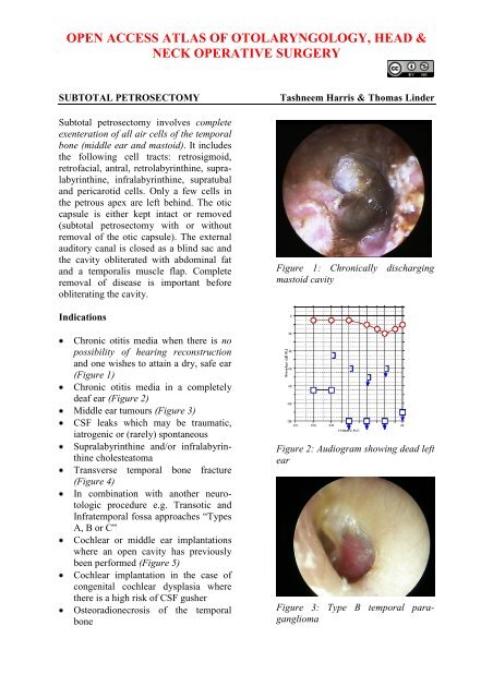
Subtotal petrosectomy - Vula - University of Cape Town
Subtotal petrosectomy - Vula - University of Cape Town
Subtotal petrosectomy - Vula - University of Cape Town
- No tags were found...

Create successful ePaper yourself
Turn your PDF publications into a flip-book with our unique Google optimized e-Paper software.
327Lic. Silvina Alejandra AlzugarayÍndiceTitulo……………………………………………. Pág. 3Resumen………………………………………… Pág.3Palabras clave…………………………………... Pág.3Introducción……………………………………. Pág. 4 a 10.Desarrollo………………………………………. Pág. 10 a 19.Conclusiones…………………………………… Pág. 19 a 21.Bibliografía……………………………………… Pág. 21 a 22.2
transect the anterior canal safely bycutting onto the curved clamp. Thisavoids injury to the facial nerve(Figure 10)Figure 11: Elevating skin <strong>of</strong> externalcanalFigure 10: Safely transecting anteriorwall <strong>of</strong> ear canalEversion <strong>of</strong> auditory canal skinOcular magnifying loupes are usefulfor this stepThe skin <strong>of</strong> the cartilaginous canal iselevated for 1cm from the cut marginwith tympanoplasty scissors t<strong>of</strong>acilitate eversion <strong>of</strong> the canal (Figure11)It is important that the skin <strong>of</strong> theexternal canal is not breached. Toavoid this, direct the curve <strong>of</strong> thetympanoplasty scissors towards thecartilageIt may be difficult to find the correctplane, especially if a previous openmastoidectomy with a widemeatoplasty was performedTwo 2-0 vicryl stay sutures are placedat 6 and 12 0’clock. These are placedas 2 purse string sutures with the freeends on the inside <strong>of</strong> the canal (Figures12, 13)Figure 12: Placing stay suturesFigure 13: Stay suturesA curved artery clamp is passedthrough the canal from externally andapplied to each <strong>of</strong> the free ends <strong>of</strong> thetwo stay sutures. Tension is applied tothe stay sutures to evert the canal skin(Figure 14)4
Chorda tympani is divided with a largeBellucci scissorsThe incudo-stapedial joint is separatedwith a 45° 1,5mm hookThe malleus is cut at its neck with amalleus nipperTensor tympani tendon is cut with alarge Bellucci scissorsThe tympanic membrane (with handle<strong>of</strong> malleus attached) is now beremoved along with the incus and head<strong>of</strong> malleusMastoidectomyIt is important that all mastoid cells areexenterated and no mucosa is leftbehind as this can potentially formmucocoelesStructures like dura, sigmoid sinus andfacial nerve have to be skeletonised,not exposedUsing a mastoid raspatory elevate thes<strong>of</strong>t tissues <strong>of</strong>f the mastoidThe sternocleidomastoid muscle isfreed from its insertion into the mastoidtipPlace a 2-0 silk stay suture from theedge <strong>of</strong> temporalis muscle to its fasciato expose the area above the temporallineNext proceed as for open mastoidoepitympanectomy(see chapter onmastoidectomy)After performing an open mastoidoepitympanectomythe additional cells areremoved to complete the subtotal<strong>petrosectomy</strong>:The stapes superstructure is removedby using crurotomy scissors to cut theanterior and posterior crusThe inferior surface <strong>of</strong> the external earcanal cam be lowered to the level <strong>of</strong>the hypotympanumBefore skeletonising the jugular bulb, itis important to review the CT scan toconfirm once again that the jugularbulb is not high-ridingThe pericarotid cells are exenterated byfirst approaching the anterocarotidcells. The carotid is recognised by itswhitish colour beneath bone. Thecarotid is not infrequently dehiscent atits bend medial to Eustachian tubeThe mastoid tip may be left in place,but is drilled down to the level <strong>of</strong> thedigastric ridgeRetr<strong>of</strong>acial, retrosigmoid, retrolabyrinthine,supralabyrinthine, infralabyrinthine,and supratubal cells are allexenterated. Figure 19 shows themastoid cavity once all the cell trackshave been exenteratedFigure 19: Mastoid cavity with all celltracks have been exenteratedTechnical pointsWhen drilling in the supralabyrinthinesegment a diamond drill is used inreverse when operating on the right earto avoid injuring the facial nerveThe mucosa <strong>of</strong> the middle ear can beremoved by using the microraspatoryand scraping the mucosa with a smallcotton ball, taking care not to subluxthe stapes superstructure if left intactObliteration <strong>of</strong> Eustachian tubeThe internal carotid artery is followedsuperiorly to the medial wall <strong>of</strong> theeustachian tube (watch out for adehiscent carotid at this point!)6
The mucosa <strong>of</strong> the bony eustachiantube is removed as far as the isthmuswith a 2mm/3mm diamond burrAt this point the remaining mucosa iscoagulated with bipolar forceps and theeustachian tube is obliterated with bonewax (Figure 20)Technical pointsThe tensor tympani muscle can bedissected out <strong>of</strong> its semicanal bydrilling its lateral surface and reflectingit anteriorly with a microraspatory intothe protympanum and the eustachiantube orificeBone wax is then placed over this sothat the muscle lies sandwichedbetween two layers <strong>of</strong> bone wax, thusobliterating the eustachian tubeObliteration <strong>of</strong> Operative CavityFigure 20: Eustachian tube obliteratedwith bone waxUsing a cottonoid, the bone wax maybe pushed into the orifice <strong>of</strong> theeustachian tube with a suction tip. Thetip <strong>of</strong> the microraspatory is directedaway from the carotid (Figure 21)The middle ear cleft is obliterated withabdominal fat harvested from the lowerquadrant <strong>of</strong> the abdomen (Figure 22)It is important to achieve meticuloushaemostasis as the most commoncomplication <strong>of</strong> subtotal <strong>petrosectomy</strong>is a haematoma <strong>of</strong> the abdominalwoundA suction drain is placed in theabdominal woundThe abdominal wound closed in layersusing 3-0 vicryl subcutaneously andmonocryl 3-0 or nylon 3-0 to skinWith osteoradionecrosis <strong>of</strong> thetemporal or chronic infection it isadvisable not to use abdominal fat toobliterate the cavity. Rather use onlytemporalis muscleFigure 21: Microraspatory directedaway from carotidA temporalis muscle muscul<strong>of</strong>ascialgraft is used as an additional seal <strong>of</strong>orifice by placing it lateral to the bonewaxFigure 22: Abdominal fat7
Transposition <strong>of</strong> temporalis muscleThe skin incision is extendedsuperiorly above the temporal line toexpose the temporalis muscleRemaining in a plane above thetemporalis fascia, the skin and s<strong>of</strong>ttissue edges are underminedUsing two skin rakes, the assistantretracts the skin edges for adequateexposureThe posterior 2/3 <strong>of</strong> the temporalismuscle is mobilized by creating a flapusing a diathermy knifeTo facilitate mobilisation andtransposition <strong>of</strong> the temporalis flapover the mastoid cavity, a smallinverted v-shaped incision is made atthe base <strong>of</strong> the temporalis muscleThe muscle flap is rotated inferiorlyover the cavity and sutured to thesternocleidomastoid muscle and s<strong>of</strong>ttissues <strong>of</strong> the occiput with 2-0 vicrylsuture (Figure 23)Figure 23: Temporalis flap andabdominal fat in <strong>petrosectomy</strong> defectInevitably the fat used to obliterate thecavity will atrophy, therefore more fatis placed in the cavity once thetemporalis muscle has been sutured tothe s<strong>of</strong>t tissues via the superior pocketcreated by the transposed temporalisflap (Figure 24)Figure 24: Additional fat placed indefectWound ClosureA 3mm suction drain is placed underthe scalp over the squamous part <strong>of</strong> thetemporal bone and not over the mastoidcavityThe wound is closed in layers with 2-0vicryl sutures subcutaneously and skinclipsA compression bandage is appliedPostoperative careAntibiotics (amoxicillin with clavulanicacid) are continued for one weekpostoperativelyThe drain is left in place until thedrainage is less than 10mls/24hrs.If the surgery was done for CSF leak,then the drain is removed on the firstpostoperative dayThe abdominal drain is removed whenthe drainage is less than 10mls/24hrsClips/sutures are removed after 10 daysThe resorbable vicryl sutures withinthe external ear canal are removed at 4weeksLong-term follow-upImagingWhere subtotal <strong>petrosectomy</strong> isperformed for chronic otitis media, CT8
scans are routinely performed after oneyear and then again at 3 yearsIn cases <strong>of</strong> previous cholesteatoma, adiffusion weighted non-EPI MRIwould detect recurrent diseaseAuditory rehabilitationThis depends on the status <strong>of</strong> hearing <strong>of</strong>opposite ear, the degree and type <strong>of</strong>hearing loss and cochlear function. Theoptions are: bone anchored hearing aid(BAHA), if the inner ear function has beenpreserved <strong>of</strong> the ipsilateral or contralateralear; active middle ear implant (e.g. Vibrantsoundbridge) where there is good cochlearreserve; or cochlear implant (bilateraldeafness)References1. Fisch U, Mattox D, eds. Microsurgery<strong>of</strong> the Skull Base. Stuttgart,Germany:Georg Thieme Verlag, 1988.2. Linder T, Schlegel C, DeMin N, vander Westhuizen S.Active Middle EarImplants in Patients Undergoing<strong>Subtotal</strong> Petrosectomy: NewApplication for the VibrantSoundbridge Device and ItsImplication for Lateral Cranium BaseSurgery. Otol Neurotol. 2008;30:41-7AcknowledgementsThis guide is based on the text byPr<strong>of</strong>essor Fisch (Microsurgery <strong>of</strong> the SkullBase) and personal experience <strong>of</strong> Pr<strong>of</strong>essorLinder, as well as course materials for thelateral skull base course conductedannually by Pr<strong>of</strong>essors Fisch and Linder atthe Department <strong>of</strong> Anatomy, <strong>University</strong> <strong>of</strong>Zurich, Switzerland.AuthorTashneem Harris MBChB, FCORL,MMed (Otol), Fisch InstrumentMicrosurgical FellowENT SpecialistDivision <strong>of</strong> Otolaryngology<strong>University</strong> <strong>of</strong> <strong>Cape</strong> <strong>Town</strong><strong>Cape</strong> <strong>Town</strong>, South Africaharristasneem@yahoo.comSenior AuthorThomas Linder, M.D.Pr<strong>of</strong>essor, Chairman and Head <strong>of</strong>Department <strong>of</strong> Otorhinolaryngology,Head, Neck and Facial Plastic SurgeryLucerne Canton Hospital, Switzerlandthomas.linder@ksl.chEditorJohan Fagan MBChB, FCORL, MMedPr<strong>of</strong>essor and ChairmanDivision <strong>of</strong> Otolaryngology<strong>University</strong> <strong>of</strong> <strong>Cape</strong> <strong>Town</strong><strong>Cape</strong> <strong>Town</strong>South Africajohannes.fagan@uct.ac.zaTHE OPEN ACCESS ATLAS OFOTOLARYNGOLOGY, HEAD &NECK OPERATIVE SURGERYwww.entdev.uct.ac.zaThe Open Access Atlas <strong>of</strong> Otolaryngology, Head &Neck Operative Surgery by Johan Fagan (Editor)johannes.fagan@uct.ac.za is licensed under a CreativeCommons Attribution - Non-Commercial 3.0 UnportedLicense9


