Oliguria and acute kidney injury - PACT - ESICM
Oliguria and acute kidney injury - PACT - ESICM
Oliguria and acute kidney injury - PACT - ESICM

Create successful ePaper yourself
Turn your PDF publications into a flip-book with our unique Google optimized e-Paper software.
LEARNING OBJECTIVESp. iiAfter studying this module on <strong>Oliguria</strong> <strong>and</strong> Acute Kidney Injury, you should be able to:1. Recognise <strong>and</strong> resuscitate the oliguric patient2. Determine the diagnosis in an oliguric patient3. Effectively treat the oliguric patient4. Comprehend the pathophysiologic mechanisms of different causes of oliguria/AKIFACULTY DISCLOSURESThe authors of this module have not reported any disclosures.Patrick T. Murray is a consultant to Abbott Laboratories, Argutus Medical, Alere, FASTDiagnostics, <strong>and</strong> Mitsubishi Pharma Europe Ltd.DURATION7 hoursCopyright©2010. European Society of Intensive Care Medicine. All rights reserved.ISBN 978-92-95051-68-3 - Legal deposit D/2005/10.772/15
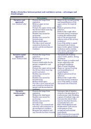
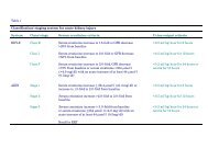
IntroductionINTRODUCTIONThe evolution of the term ‘<strong>acute</strong> renal failure’ dates back to 1802, when WilliamHeberden first described it as Ischuria Renalis. Since then there are over 35 officialdefinitions of the term; these include: <strong>acute</strong> Bright’s disease, war nephritis <strong>and</strong>crush syndrome. It wasn’t until 1951 that Homer W. Smith introduced the term‘<strong>acute</strong> renal failure’.Today, <strong>acute</strong> <strong>kidney</strong> <strong>injury</strong> (AKI) is considered the correct nomenclature for theclinical disorder formerly termed ‘<strong>acute</strong> renal failure’ (ARF). AKI is a commonclinical problem in critically ill patients that is associated with increased morbidity<strong>and</strong> mortality. Even a modest impairment in renal function is an independent riskfactor for mortality, but the onset of AKI is often not recognised.Kellum JA, Levin N, Bouman C, Lameire N. Developing a consensus classificationsystem for <strong>acute</strong> renal failure. Curr Opin Crit Care 2002; 8: 509-514. PMID12454534Link to <strong>ESICM</strong> Flash Conference: Michael Joannidis, Innsbruck, Austria.Definition <strong>and</strong> classification of AKI: Where do we st<strong>and</strong>? Vienna 2009.In order to stage the severity of AKI, a graded classification, known as the RIFLEcriteria (risk, <strong>injury</strong>, failure, loss, ESRD) was established. The RIFLE criteriaincorporate levels of oliguria in addition to incremental serum creatinineelevations. The RIFLE criteria were later modified <strong>and</strong> referred to as the <strong>acute</strong><strong>kidney</strong> <strong>injury</strong> network (AKIN) definition (see Table 1). Compared with the RIFLEclassification, the AKIN definition includes lesser degrees of serum creatinineelevation to diagnose AKI, identical grades of oliguria, <strong>and</strong> a similar severitystaging system. For all practical purposes, RIFLE <strong>and</strong> AKIN criteria are the same.The concept of AKI as defined by RIFLE/AKIN creates a new paradigm. Furthervalidation studies may be required to confirm the RIFLE/AKIN criteria as a meansof classifying patients with AKI but the current evidence is that categorising AKIpatients by either RIFLE or AKIN severity criteria facilitates rational clinicalmanagement, is predictive of clinical outcomes <strong>and</strong> changes the way we view AKI.Link to <strong>ESICM</strong> Flash Conference: Claudio Ronco, Vicenza, Italy. New criteria forrenal failure: The RIFLE system. Berlin 2007.
IntroductionRIFLE <strong>and</strong> AKIN criteria for AKI classification <strong>and</strong> staging. Adapted from Bellomo et al., 1<strong>and</strong> Mehta et al., 2 with permission from the original publisher, BioMed Central. AKI, <strong>acute</strong><strong>kidney</strong> <strong>injury</strong>; AKIN, Acute Kidney Injury Network; GFR, glomerular filtration rate,RIFLE, Risk, Injury, Failure, Loss <strong>and</strong> End-Stage <strong>kidney</strong> disease; RRT, renal replacementtherapy.1Bellomo R, Ronco C, Kellum JA, Mehta RL, Palevsky P; Acute Dialysis QualityInitiative workgroup. Acute renal failure – definition, outcome measures,animal models, fluid therapy <strong>and</strong> information technology needs: the SecondInternational Consensus Conference of the Acute Dialysis Quality Initiative(ADQI) Group. Crit Care 2004; 8(4): R204–212. PMID 153122192Mehta RL, Kellum JA, Shah SV, Molitoris BA, Ronco C, Warnock DG, et al; AcuteKidney Injury Network. Acute Kidney Injury Network: report of an initiativeto improve outcomes in <strong>acute</strong> <strong>kidney</strong> <strong>injury</strong>. Crit Care 2007; 11(2): R31. PMID17331245Bagshaw SM, George C, Bellomo R; ANZICS Database Management Committee. Acomparison of the RIFLE <strong>and</strong> AKIN criteria for <strong>acute</strong> <strong>kidney</strong> <strong>injury</strong> incritically ill patients. Nephrol Dial Transplant 2008; 23(5):1569–1574. PMID18281319Joannidis M, Metnitz B, Bauer P, Schusterschitz N, Moreno R, Druml W, et al. Acute<strong>kidney</strong> <strong>injury</strong> in critically ill patients classified by AKIN versus RIFLE usingthe SAPS 3 database. Intensive Care Med 2009; 35(10): 1692–1702. PMID19547955. Full text (pdf)
Task 1. How do I recognise <strong>and</strong> resuscitate the oliguric patient? p. 5intrinsic renal <strong>and</strong> post-renal) is of real clinical utility. The clinical circumstanceusually suggests the category of renal impairment (see Tasks 3 <strong>and</strong> 4 for treatment<strong>and</strong> aetiology).Pre-renal failure (azotaemia) is most common among hospitalised patients. Prerenalindicates that the cause lies outside the <strong>kidney</strong>, specifically ‘before’ the<strong>kidney</strong>. A history of high output gastrointestinal losses, haemorrhage, sepsis,congestive heart failure (CHF) <strong>and</strong>/or decreased oral intake resulting inhypovolaemia or a combination of these factors associated with hypotension <strong>and</strong>decreased urine output suggests AKI due to pre-renal disease (or ATN ifpersistent). When more than 10-15% of the circulating volume is lost, findings onphysical examination may include: tachycardia, dry mucous membranes,hypotension, low central venous pressure, oliguria, peripheral hypoperfusion withaltered mentation <strong>and</strong> cold clammy skin with delayed capillary return.Causes of ‘intrinsic’ renal failure depend on the clinical setting. In the ICU, prerenalfailure is the most common diagnosis, usually from hypovolaemia or sepsis. Afailure of haemodynamic restoration with a trial of fluid replacement to restoreurine output <strong>and</strong> the exclusion of post-renal pathologies supports the diagnosis.Allergic interstitial nephritis, usually due to antibiotics may also be responsible.The topic of intrinsic renal disease is addressed in the <strong>PACT</strong> module on Acute renalfailure <strong>and</strong> the textbook chapter below.Clarkson MR, Friedewald JJ, Eustace JA, Rabb H. Acute <strong>kidney</strong> <strong>injury</strong>. In: BrennerBM, editor. Brenner & Rector's The Kidney. 8th edition. Philadelphia, Pa:Saunders Elsevier; 2007: chapter 29. pp. 943.<strong>PACT</strong> module on Acute renal failurePost-renal failure is due to urinary tract obstruction <strong>and</strong> accounts for
Task 1. How do I recognise <strong>and</strong> resuscitate the oliguric patient? p. 6with presumed ‘renal’ oliguria have an ischaemic element <strong>and</strong> a trial of expansionof the circulating volume may be warranted, even if only to avoid unnecessaryaggravation of the ‘renal’ insult due to uncorrected hypoperfusion.Joannidis M, Druml W, Forni LG, Groeneveld AB, Honore P, Oudemans-vanStraaten HM, et al; Critical Care Nephrology Working Group of the EuropeanSociety of Intensive Care Medicine. Prevention of <strong>acute</strong> <strong>kidney</strong> <strong>injury</strong> <strong>and</strong>protection of renal function in the intensive care unit. Expert opinion of theWorking Group for Nephrology, <strong>ESICM</strong>. Intensive Care Med 2010; 36(3):392–411. PMID 19921152. Full text (pdf)The ‘fluid challenge’ attempts to identify <strong>and</strong> treat pre-renal failure that canprogress to ATN if not treated promptly. Fluids should be given early <strong>and</strong> targetedto haemodynamic end points, such as increases in mean arterial pressure, pulsepressure variation, cardiac output, central venous pressure (CVP), central venousoxygen saturation, pulmonary artery occlusion pressure (PAOP), urine output <strong>and</strong>improvements in lactic acidosis <strong>and</strong> skin perfusion. In selected cases, acomprehensive haemodynamic assessment (see ‘Diagnostic approach’ Task 2) isindicated.Hinds CJ, Watson JD. Intensive Care: A Concise Textbook. 3rd edition. SaundersLtd; 2008. ISBN: 978-0-7020259-6-9. pp. 108–113; Cardiovascular Support.<strong>PACT</strong> module on Haemodynamic monitoringRivers et al. have shown that early fluid therapy (<strong>and</strong> inotropic support wherenecessary), when titrated to targeted endpoints, achieved a significantly greaterurine flow, CVP <strong>and</strong> arterial pressure <strong>and</strong> a significantly reduced mortality(p=0.009) in patients with severe sepsis or septic shock attending Accident <strong>and</strong>Emergency.Rivers E, Nguyen B, Havstad S, Ressler J, Muzzin A, Knoblich B, et al. Early goaldirectedtherapy in the treatment of severe sepsis <strong>and</strong> septic shock. New EnglJ Med 2001; 345: 1368–1377. PMID 11794169Lin SM, Huang CD, Lin HC, Liu CY, Wang CH, Kuo HP. A modified goal-directedprotocol improves clinical outcomes in intensive care unit patients with septicshock: a r<strong>and</strong>omized controlled trial. Shock 2006; 26(6): 551–557. PMID17117128Concurrent attention to diagnosis is important to confirm or deny the initialassumption <strong>and</strong> to guide subsequent therapy.The choice of the type of fluid used for resuscitation in the critically ill remainscontroversial <strong>and</strong> usually involves a fluid challenge with either natural/artificial
Task 1. How do I recognise <strong>and</strong> resuscitate the oliguric patient? p. 7colloids or crystalloids. These can be given as 10-15 ml/kg of crystalloid e.g. 0.9%saline or compound sodium lactate or non-protein colloid e.g. gelatin (not availablein USA) or hetastarch delivered rapidly via one or two large bore (14G) intravenouscannulae. Fluid replacement should be continued as long as there is ahaemodynamic response; hypervolaemia is avoided.The rate of fluid administration is reduced substantially or even stopped, whencardiac filling pressures (CVP or pulmonary artery occlusion pressure) increase orplateau without concurrent haemodynamic improvement. If haemodynamicendpoints are not reached despite adequate fluid resuscitation,inotropic/vasoactive drugs should be considered (catecholamines, vasopressin,others).As the volume of distribution is much larger for crystalloid balanced salt solutionsthan for colloids, resuscitation with crystalloids requires more fluid (three to fourtimes) to achieve the same end points <strong>and</strong> is likely to result in more oedema.However, current evidence indicates that the choice of fluid does not influenceoutcome. The SAFE study (see below) showed that 4% albumin was safe but notsuperior to saline (in similar infused volumes) in preventing death or need fordialysis. The use of either crystalloid or colloid for haemodynamic support ofpatients in the intensive care unit seems to be associated with equivalent outcomes.Finfer S, Bellomo R, Boyce N, French J, Myburgh J, Norton R; SAFE StudyInvestigators. A comparison of albumin <strong>and</strong> saline for fluid resuscitation inthe intensive care unit. N Engl J Med 2004; 350(22):2247–2256. PMID15163774The potential detrimental effect of hydroxyethyl starch (HES) on <strong>kidney</strong> functionhas become a major concern <strong>and</strong> is not without debate over recent years (Boldt,2009, Hartog <strong>and</strong> Reinhart, 2009). HES preparations have a greater volume effectthan that of albumin. However, their use has been associated with osmoticnephrosis <strong>and</strong> possibly medullary hypoxia. A further problem with HES may betissue deposition <strong>and</strong> associated pruritus, which appears to be dose dependent. Therecent publication of recommendations <strong>and</strong> guidelines by an internationalcollaboration of the Critical Care Nephrology Working Group of the EuropeanSociety of Intensive Care Medicine (<strong>ESICM</strong>) currently recommends avoidinghigher-molecular-weight preparations of HES <strong>and</strong> dextrans in sepsis.Boldt J. PRO: hydroxyethylstarch can be safely used in the intensive care patient–therenal debate. Intensive Care Med 2009; 35(8): 1331–1336. PMID 19533094.Full text (pdf)Hartog C, Reinhart K. CONTRA: Hydroxyethyl starch solutions are unsafe incritically ill patients. Intensive Care Med 2009; 35(8): 1337–1342. PMID19533093. Full text (pdf)
Task 1. How do I recognise <strong>and</strong> resuscitate the oliguric patient? p. 8Joannidis M, Druml W, Forni LG, Groeneveld AB, Honore P, Oudemans-vanStraaten HM, et al; Critical Care Nephrology Working Group of the EuropeanSociety of Intensive Care Medicine. Prevention of <strong>acute</strong> <strong>kidney</strong> <strong>injury</strong> <strong>and</strong>protection of renal function in the intensive care unit. Expert opinion of theWorking Group for Nephrology, <strong>ESICM</strong>. Intensive Care Med 2010; 36(3):392–411. PMID 19921152. Full text (pdf)Wiedermann CJ. Hydroxyethyl starch–can the safety problems be ignored? WienKlin Wochenschr 2004; 116(17–18): 583-594. PMID 15515874A common adverse consequence of fluid resuscitation is ‘fluid overload’ <strong>and</strong>pulmonary oedema with significant reductions in lung function <strong>and</strong> oxygenation. Athreshold may exist beyond which the perceived benefit of additional fluid therapyafter resuscitation may be detrimental. A positive cumulative fluid balance hasbeen shown in several studies to independently predict hospital morbidity <strong>and</strong>mortality.Payen D, de Pont AC, Sakr Y, Spies C, Reinhart K, Vincent JL; Sepsis Occurrence inAcutely Ill Patients (SOAP) Investigators. A positive fluid balance isassociated with a worse outcome in patients with <strong>acute</strong> renal failure. Crit Care2008; 12(3): R74. PMID 18533029Bouchard J, Soroko SB, Chertow GM, Himmelfarb J, Ikizler TA, Paganini EP, et al;Program to Improve Care in Acute Renal Disease (PICARD) Study Group.Fluid accumulation, survival <strong>and</strong> recovery of <strong>kidney</strong> function in critically illpatients with <strong>acute</strong> <strong>kidney</strong> <strong>injury</strong>. Kidney Int 2009; 76(4):422–427. PMID19436332Ventilated ICU patients are relatively protected against the immediateconsequences of ‘fluid overload’, at least in the short term. For example, FiO 2<strong>and</strong>/or PEEP can be increased to counteract the adverse effects of pulmonarycongestion on gas exchange. PEEP is carefully titrated so as not to produce adversehaemodynamic effects which might further reduce renal blood flow.If the neck veins are distended from the outset, a fluid challenge should only beused cautiously, if at all, since the patient is more likely to need inotropic supportcombined with a rapid diagnostic work-up to identify whether the cause iscardiogenic or obstructive (see Tasks 2 <strong>and</strong> 4 for diagnosis <strong>and</strong> aetiology).In the setting of ‘renal’ oliguria, removal of precipitating ischaemic or toxic factorsis the immediate priority. If there is diagnostic doubt or a possibility of concurrentrenal hypoperfusion, a cautious trial of volume loading is usually warranted.Failure to respond to measures aimed at reversing presumed ‘pre-renal’ oliguria issuggestive of ‘renal’ (or ‘post-renal’) oliguria (see diagnosis below).Early recognition <strong>and</strong> treatment of obstruction in ‘post-renal’ oliguria will usuallyresult in some degree of recovery of renal function. Superimposed infection(urosepsis) will require appropriate antibiotic therapy together with early release ofobstruction.
Task 2. How do I reach a diagnosis in the oliguric patient? p. 92. HOW DO I REACH A DIAGNOSIS IN THE OLIGURICPATIENT?Hinds CJ, Watson JD. Intensive Care: A Concise Textbook. 3rd edition. SaundersLtd; 2008. ISBN: 978-0-7020259-6-9. pp. 362–365; Diagnosis <strong>and</strong>investigations.The assessment is primarily clinical using targeted history taking <strong>and</strong> physicalexamination. A sequence of supplementary testing, which includes bloodinvestigations, bedside urinary (macroscopic) visualisation <strong>and</strong> subsequentdipstick, biochemical <strong>and</strong> microscopic examination of urine can assist with makinga firm diagnosis. In cases of AKI where obstruction is suspected or which areunresponsive to a trial of fluid/haemodynamic therapy, an ultrasound examinationof the <strong>kidney</strong>s, ureters <strong>and</strong> bladder is indicated.It is important to be alert to the possibility that oliguria may be ‘renal’ or ‘postrenal’,as identification <strong>and</strong> correction of the cause(s) can be rapidly rewarding <strong>and</strong> avoidswasting time with ineffectual, <strong>and</strong> possibly inappropriate treatments, targeting presumed‘pre-renal’ oliguria. For an overview of causes see Task 4.Clinical assessmentAs mentioned in Task 1, it is the clinical circumstance that suggests the category ofrenal impairment (pre-renal, intrinsic, post-renal), pre-renal failure (azotaemia)<strong>and</strong> ATN being the most important among critically ill patients. A history oftrauma, haemorrhage, hypotension, sepsis or septic shock, congestive heart failure(CHF), anaphylactic shock, fasting, recent surgery, high output gastrointestinallosses, decreased oral intake or patients receiving diuretics suggests AKI due to‘pre-renal’ disease.In the clinical assessment of the oliguric patient, a comprehensive history,study of the observation chart, clinical examination <strong>and</strong> a review of recentinvestigations <strong>and</strong> drug therapies will be necessary.Fluid requirements in trauma patients may be increased due to covert blood orfluid loss from the intravascular space, ‘third-space’ losses associated with majorsurgery or as a result of fever, systemic inflammation or rhabdomyolysis.Vasodilated patients e.g. due to sepsis may become relatively hypovolaemic.<strong>Oliguria</strong> <strong>and</strong> circulatory dysfunction due to either a low cardiac output or tosystemic inflammation with vasodilation <strong>and</strong> hypotension is relatively common inintensive care patients. Conditions such as myocardial infarction, pulmonary
Task 2. How do I reach a diagnosis in the oliguric patient? p. 10embolism, anaphylaxis or perioperative complications in high-risk surgical patientsare also common causes of oliguria <strong>and</strong> hypotension. For further information seeTask 4 <strong>and</strong> the following link:<strong>PACT</strong> module on High-risk surgical patientsA history of sepsis, prolonged hypotension, drug <strong>and</strong> nephrotoxin exposure will inmost cases identify patients with ‘intrinsic’ renal failure <strong>and</strong> ATN. Althoughischaemic <strong>and</strong> nephrotoxic injuries are dominant in the ICU setting, glomerular<strong>and</strong> vascular pathologies, interstitial nephritis <strong>and</strong> autoimmune pulmonary renalsyndromes should be included in the differential diagnosis. See Task 4 <strong>and</strong> thereference below for further information.Clarkson MR, Friedewald JJ, Eustace JA, Rabb H. Acute <strong>kidney</strong> <strong>injury</strong>. In: BrennerBM, editor. Brenner & Rector's The Kidney. 8th edition. Philadelphia, Pa:Saunders Elsevier; 2007: chapter 29. pp. 943.A full clinical history <strong>and</strong> physical examination will often reveal the diagnosis in‘post-renal’ oliguria. Findings may include a misplaced catheter, palpable bladder<strong>and</strong> tenderness of the suprapubic abdominal region. The clinical features of postrenaloliguria may, however, be obscured by abdominal or pelvic injuries, obesity,stupor, intoxication or anaesthesia (see below). Nevertheless their identification<strong>and</strong> rectification can often be relatively straightforward <strong>and</strong> rewarding. A failure torecognise such features may lead to serious mismanagement – see anecdote.At the time when it was believed that high dose frusemide (furosemide) had a rolein converting oliguric to non-oliguric renal failure, an ICU patient whose blockedurethral catheter was not detected clinically, was given frusemide 2 g i.v. Thisresulted in a 16 litre diuresis over the eight hours following relief of the obstruction.In ruling out a ‘post-renal’ aetiology, specific attention should be paid to:ProstatismClassic symptoms are well-known but intermittent or partial urinary tractobstruction, typically from insidious-onset benign prostatic hypertrophy mayproduce a pressure-mediated, nephrogenic, vasopressin-resistant diabetesinsipidus. Polyuria may aggravate symptoms <strong>and</strong> confuse the diagnosis.Agitated patientIn brain injured, encephalopathic, or sedated patients <strong>and</strong> in those with spinal cord<strong>injury</strong> or during residual spinal anaesthesia, urinary retention may be manifested
Task 2. How do I reach a diagnosis in the oliguric patient? p. 11by agitation or heart rate <strong>and</strong> blood pressure instability, including unexplainedhypertensive episodes.Bladder discomfortThis symptom, suggestive of <strong>acute</strong> bladder outlet obstruction, may also be a usefuladjunctive sign when confirming bladder distension by palpation. The bladder maynot be easily palpable, especially, in the obese, the peri-partum patient or in thosewith trauma to the abdomen/pelvis or after laparotomy.If a patient develops oliguria abruptly or if a blocked urinary catheter is suspectede.g. due to clot formation in a patient with recent haematuria, a trial of flushing thecatheter should be performed under sterile conditions. Many doctors have been‘caught out’ by an obstructed urinary catheter at some time in their career – seeanecdote above.Trauma patientsAnuria associated with blood at the urethral meatus, perineal ecchymosis <strong>and</strong> ahigh-riding prostate on rectal examination suggests urethral disruption.Urine diversionBe alert to the possibility that urine formation may be normal but there may bedisruption or diversion of anatomical outlets e.g. ileal conduit, nephrostomy,vesicovaginal or vesicocolic fistula.UrosepsisFlank pain <strong>and</strong> tenderness may signify <strong>acute</strong> ureteric obstruction (renal colic),perhaps with associated infection. Urinary obstruction predisposes to infection.Recurrence of urosepsis, or failure to respond rapidly to st<strong>and</strong>ard antimicrobial<strong>and</strong> supportive measures, should alert the clinician to the possibility of urinarytract obstruction.Further investigationsBloodAccording to the AKIN/RIFLE criteria, the diagnosis of AKI is based on eitherelevation of serum creatinine or the presence of oliguria. Measurements of bloodurea nitrogen <strong>and</strong> serum creatinine to assess glomerular filtration rate (GFR) aredone daily in the ICU but can be monitored more frequently e.g. 12 hourly. A rise inserum creatinine is associated with a parallel decrease in GFR <strong>and</strong> generallyimplies a reduction in <strong>kidney</strong> function, <strong>and</strong> vice versa. The rate of change of urea<strong>and</strong> creatinine blood levels may differ in different pathologic situations <strong>and</strong> thischange (of one relative to the other) can be used diagnostically (see appendix –biochemistry).
Task 2. How do I reach a diagnosis in the oliguric patient? p. 12Where the units (mg/dL) are used e.g. in the US, a simple rule of thumb applieswhereby a blood urea nitrogen (BUN)–creatinine ratio greater than 20 isconsidered suggestive of pre-renal azotaemia <strong>and</strong> less than 10 to 15, reflective ofATN.Where SI units are used however, <strong>and</strong> urea rather than BUN is used, themeasurement units for urea (mmol/L) <strong>and</strong> creatinine (mcmol/L) are different <strong>and</strong>the rule of thumb, if calculated, requires a conversion (see Appendix –biochemistry)According to the AKIN criteria, even small changes in creatinine within 48 hours,defined as an absolute increase in serum creatinine of more than or equal to 26.4micromol/litre (>0.3 mg/dl) per day or a percentage increase in serum creatinineof more than or equal to 50% (1.5-fold from baseline) is an independent predictorof mortality from AKI (see ‘Outcome after <strong>Oliguria</strong>/AKI’ below).Barrantes F, Tian J, Vazquez R, Amoateng-Adjepong Y, Manthous CA. Acute <strong>kidney</strong><strong>injury</strong> criteria predict outcomes of critically ill patients. Crit Care Med 2008;36(5):1397–1403. PMID 18434915Q. What do you underst<strong>and</strong> by the term ‘uraemia’?A. Uraemia is a clinical entity the principal characteristics of which in <strong>acute</strong> practice areencephalopathy, bleeding <strong>and</strong> pericarditis. All are life threatening e.g. the pericarditis maybecome haemorrhagic. Uraemia is an indication for dialysis. An elevated blood urea level iscalled ‘azotaemia’.Q. Other than serum urea, which blood test might be checked readily tosupport a presumed diagnosis of water depletion?A. Elevated serum sodium <strong>and</strong> haemoglobin levels are also indicative of water depletion.Serum osmolality provides more specific information.Q. In which situations might a deceptively low urea or creatinine leveldelay recognition of renal impairment?A. Creatinine may be deceptively low in patients with a small muscle mass e.g. in smallelderly women. Urea may be deceptively low in those whose protein turnover is low e.g. inmalnourished or liver failure patients.Hyperkalaemia is a biochemical indicator of <strong>acute</strong>ly deteriorating renal excretorycapacity. Hyperkalaemia may be life threatening, or subject to a hyper<strong>acute</strong> rise e.g.due to concurrent rhabdomyolysis <strong>and</strong> is one of the recognised indications forurgent dialysis in <strong>acute</strong> renal impairment. Concurrent acidosis can aggravatehyperkalaemia. Hyperphosphataemia may also indicate failing excretory capacity<strong>and</strong>/or cellular destruction.
Task 2. How do I reach a diagnosis in the oliguric patient? p. 13Other specific blood investigations can aid the diagnois of primary glomerulardiseases, these include: complement levels (C3,C4), anti-glomerular basementmembrane antibody (aGBM), antineutrophil cytoplasmic antibodies ( ANCA)levels, anti-streptolysin-O (ASO) titers.Focused investigation based on the clinical evaluation is likely to be themost rewarding.Urine dipstickCertain patterns of bedside urinalysis are associated with intrinsic renal disease.Essential information includes specific gravity, the presence of proteinuria(glomerular <strong>injury</strong>), glycosuria (tubular <strong>injury</strong>), or haematuria (infection,nephrolithiasis, primary glomerular diseases). See appendix.Q. Does the urinary dipstick differentiate between myoglobinuria <strong>and</strong>haemoglobinuria? Explain your answer.A. No. Dipstick reaction is non-discriminatory in this respect. Orthotoluidene used in thedipstick interacts with the globin fragment of both myoglobin <strong>and</strong> haemoglobin. In traumapatients for example, haemolysis may confound attempts to use the urine dipstick toconfirm myoglobinuria.Urine biochemistry <strong>and</strong> sediment analysisIn ‘pre-renal’ oliguria, the urinary macroscopic appearance is concentrated, thedipstick specific gravity (SG) is high (>1.018), as is the osmolality (>350 mosm);the spot urine Na is low (1, urinary osmolality (mosm/l)
Task 2. How do I reach a diagnosis in the oliguric patient? p. 14with coarse granular casts, muddy brown casts <strong>and</strong> tubular epithelial cell casts. Thepresence of red blood cell casts indicates glomerular disease. The urinary sedimentin post-renal failure is often very bl<strong>and</strong> in appearance, without casts .Thediscriminating ability of these findings is of limited practical value, particularly inthe ICU setting. A systematic review of studies describing urinary biochemistryindices, <strong>and</strong> microscopy in AKI demonstrated significant variability <strong>and</strong>inconsistency in these measures. In fact, no single measure of urinarybiochemistry, derived index, or pattern on microscopy can be used reliably todiagnose AKI or classify or predict the clinical course of AKI in septic patients. Onthe contrary, a study by Parazella et al., using a urinary scoring system based on thepresence of casts <strong>and</strong> renal tubular epithelial cells was highly predictive of the finaldiagnosis of ATN. It is recommended that urinary biochemistry <strong>and</strong> microscopy isperformed in patients with AKI, unresponsive to fluid <strong>and</strong>/or haemodynamictherapy, to exclude primary renal disease for which timely therapy is veryimportant for outcome of renal failure.Bagshaw SM, Langenberg C, Bellomo R. Urinary biochemistry <strong>and</strong> microscopy inseptic <strong>acute</strong> renal failure: a systematic review. Am J Kidney Dis 2006; 48(5):695–705. PMID 17059988Perazella MA, Coca SG, Kanbay M, Brewster UC, Parikh CR. Diagnostic value ofurine microscopy for differential diagnosis of <strong>acute</strong> <strong>kidney</strong> <strong>injury</strong> inhospitalized patients. Clin J Am Soc Nephrol 2008; 3(6): 1615-1619. PMID18784207Kanbay M, Kasapoglu B, Perazella MA. Acute tubular necrosis <strong>and</strong> pre-renal <strong>acute</strong><strong>kidney</strong> <strong>injury</strong>: utility of urine microscopy in their evaluation– a systematicreview. Int Urol Nephrol 2010; 42(2): 425-433. PMID 19921458Q. What causes of ‘renal’ oliguria are not associated with a high UrineNa concentration <strong>and</strong> high FeNa?A.Not all causes of renal oliguria result in a high FeNa. Interstitial nephritis, <strong>acute</strong>glomerulonephritis <strong>and</strong> uric acid nephropathy have been reported in association with lowFeNa.Q. What pathologies are associated with ‘Muddy brown casts’, ‘Red cellcasts’, ‘Eosinophils’ or ‘Crystals’ in urinary sediment?A. Muddy brown casts are associated with <strong>acute</strong> tubular necrosis. Red cell casts areassociated with glomerulonephritis. Eosinophils are associated with interstitial nephritisCrystals may be urate crystals associated with tumour lysis syndrome <strong>and</strong> uric acidnephropathy.Urine microscopy, although sometimes termed ‘the poor man's biopsy’ may beespecially useful diagnostically <strong>and</strong> in guiding therapy in ‘renal’ oliguria. Otherinvestigations that may be useful in selected cases are renal biopsy (if notcontraindicated by coagulopathy) <strong>and</strong> renal perfusion scanning. For further
Task 2. How do I reach a diagnosis in the oliguric patient? p. 15information about urinalysis, urine biochemistry <strong>and</strong> urine microscopy see theappendix.Biomarkers of AKISerum creatinine is widely used in the diagnosis of AKI <strong>and</strong> is considered to bespecific but generally an insensitive biomarker of renal dysfunction. With therecognition of the importance of small changes in serum creatinine of >0.3 mg/dL(26.4 mcmol/L), the sensitivity of serum creatinine to detect early renal <strong>injury</strong> hasimproved. However, significant renal tubular <strong>injury</strong> can occur before suchcreatinine increments have had time to develop. Serum creatinine concentration isgreatly influenced by changes in muscle mass <strong>and</strong> tubular secretion, body weight,race, age, sex, total body volume, drugs, muscle metabolism <strong>and</strong> protein intake. Forthese reasons it is generally considered a poor marker of early AKI <strong>and</strong> an evenpoorer reflection of <strong>kidney</strong> function because patients with AKI are not in steadystate <strong>and</strong> serum creatinine therefore lags far behind renal <strong>injury</strong>. The recentdevelopment of novel biomarkers for the early detection of AKI promises to be areal advance in critical care <strong>and</strong> <strong>acute</strong> nephrology. The most promising of theseinclude: NGAL (neutrophil gelatinase-associated lipocalin), IL-18, KIM-1, CystatinC, <strong>and</strong> L-FABP.Parikh CR, Devarajan P. New biomarkers of <strong>acute</strong> <strong>kidney</strong> <strong>injury</strong>. Crit Care Med2008; 36(4 Suppl): S159–165. PMID 18382188Nickolas <strong>and</strong> colleagues examined the diagnostic properties of a single urinaryNGAL level in 635 adults presenting to an inner city emergency department; 5%had AKI. Urinary NGAL distinguished AKI from other forms of <strong>kidney</strong> dysfunction<strong>and</strong> predicted excess morbidity after hospital admission. Logistic regressionanalysis demonstrated that NGAL was a better predictor of nephrologyconsultation, dialysis, ICU admission <strong>and</strong> death than other conventional or novelbiomarkers of <strong>acute</strong> <strong>kidney</strong> <strong>injury</strong>. The AUROC (area under the receiver-operatingcharacteristic) curves was 0.95, sensitivity was 0.900 (95% CI 0.730 to 0.980) <strong>and</strong>specificity was 0.995 (95% CI 0.990 to 1.000) for prediction of AKI using a cutoffvalue of 130 µg/g.Nickolas TL, O’Rourke MJ, Yang J, Sise ME, Canetta PA, Barasch N et al., Sensitivity<strong>and</strong> specificity of a single emergency department measurement of urinaryneutrophil gelatinase-associated lipocalin for diagnosing <strong>acute</strong> <strong>kidney</strong> <strong>injury</strong>.Ann Intern Med 2008; 148(11): 810–819. PMID 18519927Another study by Koyner et al. evaluated both NGAL <strong>and</strong> cystatin C (CyC) as earlybiomarkers of AKI after adult cardiothoracic surgery. Both urinary CyC <strong>and</strong> urinaryNGAL were elevated in those patients who later developed AKI, compared withthose with no <strong>injury</strong>. The urinary NGAL at the time of intensive care unit arrival(AUC 0.700) <strong>and</strong> the urinary CyC level 6 h after ICU admission (AUC 0.724) were
Task 2. How do I reach a diagnosis in the oliguric patient? p. 16most useful for predicting AKI. This study was also notable for the fact that plasmaNGAL was a poor predictor of AKI (AUC 0.536) <strong>and</strong> plasma CyC also failed todiagnose AKI in the early postoperative period (AUC 0.624). Taken together thesetwo studies <strong>and</strong> the studies listed below suggest the utility of urinary NGAL <strong>and</strong>CyC to diagnose AKI earlier than conventional methods.Koyner JL, Bennett MR, Worcester EM, Ma Q, Raman J, Jeevan<strong>and</strong>am V, et al.Urinary cystatin C as an early biomarker of <strong>acute</strong> <strong>kidney</strong> <strong>injury</strong> followingadult cardiothoracic surgery. Kidney Int 2008; 74(8): 1059–1069. PMID18650797Haase M, Bellomo R, Devarajan P, Schlattmann P, Haase-Fielitz A; NGAL MetaanalysisInvestigator Group. Accuracy of neutrophil gelatinase-associatedlipocalin (NGAL) in diagnosis <strong>and</strong> prognosis in <strong>acute</strong> <strong>kidney</strong> <strong>injury</strong>: asystematic review <strong>and</strong> meta-analysis. Am J Kidney Dis 2009; 54(6): 1012–1024. PMID 19850388With the inevitable introduction of the urinary <strong>and</strong> serum AKI biomarkers inclinical practice, the specific use of individual <strong>and</strong> combination biomarkers acrosspatient cohorts e.g. septic patients, patients with pre-existing renal disease <strong>and</strong>other high-risk AKI clinical settings needs further investigation. On the basis ofexisting literature, serum but not urinary NGAL has limited capacity to detect earlyAKI in septic patients. The performance of biomarkers of AKI in patients with preexistingrenal disease with tubular damage has recently been studied. Manybiomarkers, such as urinary NGAL <strong>and</strong> glutathione S-transferases, perform betterin those with no history of chronic <strong>kidney</strong> disease. Continued comparison amongthe different AKI biomarkers across the different patient cohorts is essential for theongoing development of the biomarkers in the evolving field of AKI <strong>and</strong> it ispossible that ultimately an AKI biomarker package will emerge.Bagshaw SM, Bennett M, Haase M, Haase-Fielitz A, Egi M, Morimatsu H, et al.Plasma <strong>and</strong> urine neutrophil gelatinase-associated lipocalin in septic versusnon-septic <strong>acute</strong> <strong>kidney</strong> <strong>injury</strong> in critical illness. Intensive Care Med 2010;36(3):452–461. PMID 19956924. Full text (pdf)Mårtensson J, Bell M, Oldner A, Xu S, Venge P, Martling CR. Neutrophil gelatinaseassociatedlipocalin in adult septic patients with <strong>and</strong> without <strong>acute</strong> <strong>kidney</strong><strong>injury</strong>. Intensive Care Med 2010; 36(8): 1333–1340. PMID 20397003. Fulltext (pdf)Koyner JL, Vaidya VS, Bennett MR, Ma Q, Worcester EM, Akhter SA, et al. UrinaryBiomarkers in the Clinical Prognosis <strong>and</strong> Early Detection of Acute KidneyInjury. Clin J Am Soc Nephrol 2010. [Epub ahead of print] PMID 20798258McIlroy DR, Wagener G, Lee HT. Neutrophil gelatinase-associated lipocalin <strong>and</strong><strong>acute</strong> <strong>kidney</strong> <strong>injury</strong> after cardiac surgery: the effect of baseline renal functionon diagnostic performance. Clin J Am Soc Nephrol 2010; 5(2): 211-219. PMID20056755
Task 2. How do I reach a diagnosis in the oliguric patient? p. 17Q. List some of the roles <strong>and</strong> applications of NGAL in AKI?A. NGAL can be useful for the early diagnosis of AKI in the following settings:cardiopulmonary bypass, contrast-induced nephropathy, sepsis inICU <strong>and</strong> (early) afterrenal transplantation. A single urinary NGAL level was highly predictive for distinguishingAKI from normal renal function, pre-renal azotaemia <strong>and</strong> CKD.Early NGAL measurements can predict the subsequent need for RRT, in-hospitalmortality <strong>and</strong> response to therapy. Coincidentally, it has a role as a safetybiomarker for monitoring drug toxicities in humans during the drug developmentprocess.ImagingUltrasonographyUltrasonography is a bedside, non-invasive investigation which avoids the need foradministration of potentially nephrotoxic contrast media. The main purpose of theinvestigation is to diagnose or rule out an obstructive cause of oliguria. It alsoprovides information on <strong>kidney</strong> size, enlarged <strong>kidney</strong>s being typical for AKI butsmall <strong>kidney</strong>(s) for chronic <strong>kidney</strong> disease. Papillary necrosis can be detected <strong>and</strong>might be useful in the diagnosis of analgesic nephropathy. Duplex sonography maydistinguish between intrinsic <strong>and</strong> pre-renal disease.Darmon M, Schnell D, Zeni F. Doppler-Based Renal Resistive Index: AComprehensive Review. In: Vincent J-L, editor. Yearbook of Intensive Care<strong>and</strong> Emergency Medicine, 2010. Berlin Heidelberg New York: Springer-Verlag; 2010. ISBN 978-3-642-10285-1. pp. 331-338RadiologyPlain films are useful for detecting <strong>kidney</strong> stones <strong>and</strong> calcification <strong>and</strong> fordetermining renal size. Hydroureter in ureterovesical junction obstruction <strong>and</strong>/orhydronephrosis due to obstruction at the pelviureteric junction may be seen. Inbladder outlet obstruction, bilateral ureteric dilatation is seen.CT renalsCT renal study is a useful non-contrast study to diagnose nephrolithiasis orpyelonephritis as a cause of <strong>acute</strong> <strong>kidney</strong> <strong>injury</strong>.Q. What useful diagnostic information, other than the exclusion ofrenal obstruction, can ultrasound provide?A. Ultrasound also provides information on <strong>kidney</strong> size. Small <strong>kidney</strong>s are suggestive oflong-st<strong>and</strong>ing (rather than <strong>acute</strong>) renal disease. Congenital abnormalities e.g. polycystic
Task 2. How do I reach a diagnosis in the oliguric patient? p. 18<strong>kidney</strong> disease <strong>and</strong> <strong>kidney</strong> agenesis may be diagnosed. Assessment of renal parenchymalappearance can provide information on renal cortex pathologies such asglomerulonephritis or cortical infarction. Duplex Doppler images of renal artery canprovide further information on renal artery occlusion by embolus.Intravenous urogramThis carries a risk of nephropathy caused by the intravenous contrast agent,particularly in diabetic patients. In severe obstruction, the nephrogram may bedelayed, see Task 3.Retrograde <strong>and</strong> antegrade contrast studiesWhere the risk of contrast-induced nephropathy is great <strong>and</strong> when consideringsurgical intervention, retrograde or antegrade (urological/percutaneous) contraststudies, may be employed.Other testsHaemodynamic assessmentAs discussed in Task 1 (Resuscitation), assessment of circulatory status may requirecentral venous pressure monitoring (CVP), pulmonary artery catheterisation orother techniques for assessing cardiac output <strong>and</strong> filling pressures e.g. non-invasivemeasurement of cardiac output. Mixed venous oxygen saturation is used as anindirect indication of oxygen balance which can be affected by cardiac output,haemoglobin, arterial saturation <strong>and</strong> tissue oxygen consumption. Central venousoxygen saturation has been shown (by Rivers et al.) to be a useful early guide totargeted resuscitation in septic patients. Transthoracic or transoesophagealechocardiography (TOE) should be considered where greater knowledge of cardiacfunction is required for complete assessment. Targeted therapy entails frequentserial measurement <strong>and</strong> therapeutic adjustment.Rivers E, Nguyen B, Havstad S, Ressler J, Muzzin A, Knoblich B, et al. Early goaldirectedtherapy in the treatment of severe sepsis <strong>and</strong> septic shock. New EnglJ Med 2001; 345: 1368–1377. PMID 11794169<strong>PACT</strong> module on Haemodynamic monitoringBladder pressure measurementOften an overlooked reason for <strong>acute</strong> oliguria is abdominal compartment syndromedefined as intra-abdominal pressure greater than 20 mmHg <strong>and</strong> abdominalperfusion pressure less than 60 mmHg occurring in association with a new <strong>and</strong>attributable organ dysfunction e.g. in a bleeding postoperative abdominal surgicalpatient or in those with severe ascites or other cause of <strong>acute</strong> abdominal distension.Abdominal compartment syndrome causes oliguria <strong>and</strong> AKI mainly by directlyincreasing renal outflow pressure <strong>and</strong> reducing renal perfusion.
Task 2. How do I reach a diagnosis in the oliguric patient? p. 19Hinds CJ, Watson JD. Intensive Care: A Concise Textbook. 3rd edition. SaundersLtd; 2008. ISBN: 978-0-7020259-6-9. pp. 462–464; Abdominalcompartment syndrome.Cheatham ML. Abdominal compartment syndrome. Curr Opin Crit Care 2009; 15(2):154–162. PMID 19276799Intra-abdominal hypertension is transmitted directly to the bladder <strong>and</strong>intravesical pressure also rises. The bladder pressure therefore reflects intraabdominalpressure <strong>and</strong> is the most commonly used mode of measurement.Using the in-dwelling urinary catheter, no more than 25ml of sterile saline isinfused into the bladder, following which intravesical pressure is measured using awater manometer or a pressure transducer connected to a side-port needle orthreeway tap in the catheter system. Appropriately modified catheter systems areavailable. Sugrue has advocated a more st<strong>and</strong>ardised diagnostic approach to intraabdominalpressure measurement <strong>and</strong> the international conference of experts (seereference below) has brought this forward.See also <strong>PACT</strong> module on ‘Abdominal problems’Sugrue M. Intra-abdominal pressure: time for clinical practice guidelines? IntensiveCare Med 2002; 28: 389–391. PMID 11967590. Full text (pdf)Malbrain ML, Cheatham ML, Kirkpatrick A, Sugrue M, Parr M, De Waele J, et al.Results from the International Conference of Experts on Intra-abdominalHypertension <strong>and</strong> Abdominal Compartment Syndrome. I. Definitions.Intensive Care Med 2006; 32(11): 1722–1732. PMID 16967294. Full text (pdf)Intra-abdominal hypertension has a number of deleterious effects. ItsTreatment <strong>and</strong> Underst<strong>and</strong>ing are considered in Task 3 <strong>and</strong> 4, respectively.
3. HOW DO I TREAT THE OLIGURIC PATIENT?Task 3. How do I treat the oliguric patient? p. 20Hinds CJ, Watson JD. Intensive Care: A Concise Textbook. 3rd edition. SaundersLtd; 2008. ISBN: 978-0-7020259-6-9. pp. 365–370; Clinical course <strong>and</strong>management.The mainstay of treatment of oliguria is to ensure adequate renal perfusionthrough optimisation of volume status, cardiac output <strong>and</strong> systemic blood pressure.Unfortunately, despite many advances in medical technology, the mortality <strong>and</strong>morbidity of AKI in the ICU remain high <strong>and</strong> have not improved significantlyduring the past two decades. This however, may reflect the likely increased illnessseverity of today’s cohort of ICU patients.The treatment of oliguric AKI is supportive, requires identification <strong>and</strong> correctionof precipitating factors but has no specific pharmacologic therapies. Supportivemeasures include maintenance of adequate renal perfusion with fluids <strong>and</strong>/orvasoactive drugs, avoidance of nephrotoxic agents, dose adjustment of renallyexcreted drugs <strong>and</strong> institution of renal replacement therapy (RRT) should anindication arise.‘Pre-renal’ oliguriaSupportive treatmentActive supportive management is sometimes termed ‘renal rescue’ therapy.Optimisation of cardiac output <strong>and</strong> of renal perfusion pressure to restore anadequate renal blood flow is the essence of this approach.Fluid therapyAs mentioned in the ‘resuscitation’ section in Task 1, the cause is ‘pre-renal’ in mostoliguric patients <strong>and</strong> intravascular volume loading (using a titrated ‘fluidchallenge’) is fundamental. For an overview of fluid therapy in oliguria see Task 1.Vasoactive <strong>and</strong> inotropic agentsDespite adequate fluid resuscitation, many patients with circulatory shock havepersistent hypotension. Hypotension, associated with shock can be the result of anyof a number of factors e.g. intravascular hypovolaemia, (bi)ventricular dysfunction,vascular effects of the inflammatory response or a combination of these. Underthese circumstances, potent systemic vasopressor agents, such as norepinephrine,epinephrine, inotropic doses of dopamine, phenylephrine or low-dose vasopressinor terlipressin have been used to restore an acceptable mean arterial bloodpressure.
Task 3. How do I treat the oliguric patient? p. 21<strong>PACT</strong> module on HypotensionThe use of vasopressors is not without debate because of a belief that renalvasoconstriction is responsible for AKI <strong>and</strong> that such drugs may make renalvasoconstriction worse <strong>and</strong> induce more AKI. On the basis of currently availableevidence in hypotensive vasodilated patients with AKI, restoring renal perfusionpressure is physiologically sound, especially in septic patients when some of theimportant autoregulatory mechanisms that help preserve GFR in the face offluctuating blood pressure are disrupted.Hinds CJ, Watson JD. Intensive Care: A Concise Textbook. 3rd edition. SaundersLtd; 2008. ISBN: 978-0-7020259-6-9. pp. 113–118; Inotropic <strong>and</strong> vasoactiveagents.Bellomo R, Wan L, May C. Vasoactive drugs <strong>and</strong> <strong>acute</strong> <strong>kidney</strong> <strong>injury</strong>. Crit Care Med2008; 36(4 Suppl): S179–186. PMID 18382191Vasoactive agentsThe choice of catecholamine depends on the clinical circumstance. There is noproven benefit of a particular vasoactive agent over another with regard to renaloutcome; the key is to restore renal perfusion. Both dopamine <strong>and</strong> norepinephrineare widely used as first-line agents; other agents can be incorporated if the patientremains hypotensive <strong>and</strong>/or oliguric. Dopamine can cause tachycardias <strong>and</strong>arrhythmias, which may limit its use. Norepinephrine is as effective at raisingblood pressure as dopamine, but has fewer cardiac side effects: it does not increasecardiac output as much as dopamine <strong>and</strong> causes less tachycardia.Dellinger RP, Levy MM, Carlet JM, Bion J, Parker MM, Jaeschke R, et al. SurvivingSepsis Campaign: international guidelines for management of severe sepsis<strong>and</strong> septic shock: 2008. Intensive Care Med 2008; 34(1): 17–60. PMID18058085. Full text (pdf)ACC/AHA guidelines for the management of patients with ST-elevation myocardialinfarction: a report of the American College of Cardiology/American HeartAssociation Task Force on Practice Guidelines (Committee to Revise the 1999Guidelines for the Management of Patients with Acute MyocardialInfarction). Circulation 2004; 110(9): e82–292.PMID 15339869De Backer et al. conducted a r<strong>and</strong>omised multicentre trial in which patientsreceived either dopamine or norepinephrine as first-line vasopressor therapy totreat circulatory shock (septic shock, cardiogenic shock or hypovolaemic shock).There was no significant difference in the rate of death between patients who weretreated with dopamine <strong>and</strong> those treated with norepinephrine. However,arrhythmias were more frequent in the dopamine group. Among patients with
Task 3. How do I treat the oliguric patient? p. 22cardiogenic shock, the mortality at 28 days was higher in those treated withdopamine than in patients treated with norepinephrine. This study raises concernsabout the safety of dopamine as a first-line therapy for cardiogenic shock.De Backer D, Biston P, Devriendt J, Madl C, Chochrad D, Aldecoa C, et al; SOAP IIInvestigators. Comparison of dopamine <strong>and</strong> norepinephrine in the treatmentof shock. N Engl J Med 2010; 362(9): 779–789. PMID 20200382Epinephrine can be used for hypotension, <strong>and</strong> has been shown to cause a rise inserum lactate levels due to stimulation of pyruvate production. In one r<strong>and</strong>omisedcontrolled study comparing epinephrine <strong>and</strong> norepinephrine in critically illpatients, there was no difference in achieving a MAP goal difference between thetwo agents <strong>and</strong> the 28 <strong>and</strong> 90-day mortality was similar in both groups. However,there was a higher incidence of drug-related side effects with epinephrine <strong>and</strong> thisresulted in the withdrawal of epinephrine by attending clinicians in 12% of casestreated.Myburgh JA, Higgins A, Jovanovska A, Lipman J, Ramakrishnan N, Santamaria J;CAT Study investigators. A comparison of epinephrine <strong>and</strong> norepinephrine incritically ill patients. Intensive Care Med 2008; 34(12): 2226–2234. PMID18654759. Full text (pdf)Vasopressin is commonly used as an adjunct to catecholamines to support bloodpressure in refractory septic shock, but does not reduce mortality rates ascompared with norepinephrine among patients with septic shock (VAASTinvestigators). In a post hoc analysis of the VAAST study, vasopressin wasassociated with a tendency to improved renal function, lower mortality <strong>and</strong>reduced requirement for renal replacement therapy in patients at ‘risk’ of <strong>acute</strong><strong>kidney</strong> <strong>injury</strong>, but not in those who had already sustained significant renal <strong>injury</strong>.Further r<strong>and</strong>omised trials are necessary to validate these observations.Russell JA, Walley KR, Singer J, Gordon AC, Hébert PC, Cooper DJ, et al.; VASSTInvestigators. Vasopressin versus norepinephrine infusion in patients withseptic shock. N Engl J Med 2008; 358(9): 877–887.PMID 18305265Gordon AC, Russell JA, Walley KR, Singer J, Ayers D, Storms MM, et al. The effectsof vasopressin on <strong>acute</strong> <strong>kidney</strong> <strong>injury</strong> in septic shock. Intensive Care Med2010; 36(1): 83–91. PMID 19841897. Full text (pdf)Joannidis M, Druml W, Forni LG, Groeneveld AB, Honore P, Oudemans-vanStraaten HM, et al.; Critical Care Nephrology Working Group of the EuropeanSociety of Intensive Care Medicine. Prevention of <strong>acute</strong> <strong>kidney</strong> <strong>injury</strong> <strong>and</strong>protection of renal function in the intensive care unit. Expert opinion of theWorking Group for Nephrology, <strong>ESICM</strong>. Intensive Care Med 2010; 36(3):392–411. PMID 19921152. Full text (pdf)
Task 3. How do I treat the oliguric patient? p. 23The use of vasopressin <strong>and</strong> its synthetic analogues has other therapeuticindications in the ICU other than septic shock. Both vasopressin <strong>and</strong> terlipressinhave shown beneficial effects in the management of patients with cirrhosis,especially in the context of variceal bleeding, the hepatorenal syndrome or both. Ineither case, current evidence suggest that terlipressin can produce a significantreduction in mortality. In patients with hepatorenal syndrome, systemicvasodilatation, in particular, splanchnic vasodilatation, is important in theactivation of endogenous renal vasoconstrictors. These pathways, in turn, arebelieved to induce functional AKI. Accordingly, vasoconstrictors such asterlipressin may improve renal function by reducing splanchnic vasodilation <strong>and</strong>increasing central circulating blood volume <strong>and</strong> reducing endogenous renalvasoconstriction.Asfar P, Radermacher P, Calès P, Oberti F. The effects of vasopressin <strong>and</strong> itsanalogues on the liver <strong>and</strong> its disorders in the critically ill. Curr Opin CritCare 2009. [Epub ahead of print]. PMID 20019608Taken together, <strong>and</strong> in view of the current lack of convincing evidence of thesuperiority of one vasoactive agent over another, an individualised approach isrecommended.InodilationImproved myocardial performance <strong>and</strong> vasodilatation is usually achieved usingdobutamine, although dopexamine is an alternative but more vasodilatory agent.The classic indication for dobutamine is for the treatment of heart failure when theblood pressure is relatively normal. The accompanying vasodilation mayprecipitate hypotension (especially if the patient is hypovolaemic) but the increasedcardiac output which is often associated with tachycardia usually compensates.Levosimendan is a calcium sensitiser that can be used to treat patients with <strong>acute</strong>decompensated heart failure (ADHF). This drug enhances myocardial contractilitywithout increasing myocardial oxygen use <strong>and</strong> does not appear to beproarrhythmic. In one meta-analysis from 27 r<strong>and</strong>omised controlled studies,levosimendan was associated with a 4.8% reduction in mortality in critically illpatients. However, a large r<strong>and</strong>omised controlled study is warranted to examinethe effects of levosoimendan on renal function in patients with ADHF.L<strong>and</strong>oni G, Mizzi A, Biondi-Zoccai G, Bignami E, Prati P, Ajello V, et al.Levosimendan reduces mortality in critically ill patients. A meta-analysis ofr<strong>and</strong>omized controlled studies. Minerva Anestesiol 2010; 76(4): 276-286.PMID 20332741
Task 3. How do I treat the oliguric patient? p. 24Dopamine receptor agonistsQ. What is meant by a ‘renal dose’ of dopamine? Give reasons as to whythis is (or is not) a valid concept.A. It is not a meaningful clinical entity but the dose is usually quoted as 1– 3 mcg/kg/min(also called ‘low-dose’ dopamine).Low-dose dopamine is defined as the dose that produces preferential dopaminergic(<strong>and</strong> β-adrenergic effects) over α-adrenergic actions (
Task 3. How do I treat the oliguric patient? p. 25L<strong>and</strong>oni G, Biondi-Zoccai GG, Tumlin JA, Bove T, De Luca M, Calabrò MG, et al.Beneficial impact of fenoldopam in critically ill patients with or at risk for<strong>acute</strong> renal failure: a meta-analysis of r<strong>and</strong>omized clinical trials. Am J KidneyDis 2007; 49(1): 56–68. PMID 17185146Q. What is the mechanism of the diuretic action of dopamine? Is thediuresis renoprotective?A. Dopamine causes a natriuresis, the mechanism of which is threefold: improvedrenal blood flow <strong>and</strong> glomerular filtration due to elevated cardiac output <strong>and</strong> bloodpressure (especially in heart failure patients), modification of intrarenalhaemodynamics <strong>and</strong> via dopamine receptors at the level of the tubules. There is noevidence that a dopamine diuresis is renoprotective.ChronotropyVery occasionally chronotropic agents are useful if bradycardia is responsible for alow cardiac output <strong>and</strong> renal hypoperfusion. Cardiac pacing avoidspharmacotherapy <strong>and</strong> may be useful in some cases.Intra-aortic balloon counterpulsationHinds CJ, Watson JD. Intensive Care: A Concise Textbook. 3rd edition. SaundersLtd; 2008. ISBN: 978-0-7020259-6-9. pp. 121–123; Mechanical CirculatoryAssistance.Intra-aortic balloon counterpulsation (IABCP) can be useful in certain specificcircumstances where there is reversible cardiac pathology e.g. <strong>acute</strong> (post-infarct)ventricular septal defect (VSD), mitral incompetence or myocardial dysfunctionpost cardiopulmonary bypass.DiureticsDiuretics are not a treatment for oliguria. They may be important in themanagement of volume overload or hyperkalaemia.Volume overload may complicate the fluid challenge. Despite the lack of convincingevidence supporting their efficacy, the use of diuretic agents in oliguric renal failureis widespread. Numerous studies have evaluated loop diuretics in the treatment ofAKI. The majority have failed to demonstrate clinical benefit. Diuretics do not alterthe outcome of renal <strong>injury</strong>. Diuretics have traditionally been used to ‘convert’ theoliguric state to non-oliguric ATN.
Task 3. How do I treat the oliguric patient? p. 26A meta-analysis by Ho <strong>and</strong> Sheridan has shown that the use of diuretics in oliguricrenal failure does not improve survival. The PICARD study group reported theresults of a large prospective observational study of critically ill patients with AKIfrom 1989 to 1995. The study found that those patients with oliguric renalimpairment who received diuretics had an increased risk of death or non-recoveryof renal function. This increased risk associated with diuretic use was largely borneby those oliguric individuals who were relatively diuretic resistant <strong>and</strong> had a moresevere form of renal failure. The use of diuretics in this setting should therefore berestricted to the treatment of volume overload <strong>and</strong> occasionally hyperkalaemia,<strong>and</strong>, even then, caution is advised as there is reasonable concern that excessivereliance on diuretics might delay initiation of RRT.Ho KM, Sheridan DJ. Meta-analysis of frusemide to prevent or treat <strong>acute</strong> renalfailure. BMJ 2006; 333(7565): 420. PMID 16861256Mehta RL, Pascual MT, Soroko S, Chertow GM; PICARD Study Group. Diuretics,mortality <strong>and</strong> nonrecovery of renal function in <strong>acute</strong> renal failure. JAMA2002; 288: 2547–2553. PMID 12444861As mentioned in the ‘resuscitation’ section of Task 2, a common adverseconsequence of fluid resuscitation is ‘fluid overload’ that may contribute tomorbidity <strong>and</strong> mortality in the critically ill patient.Payen D, de Pont AC, Sakr Y, Spies C, Reinhart K, Vincent JL; Sepsis Occurrence inAcutely Ill Patients (SOAP) Investigators. A positive fluid balance isassociated with a worse outcome in patients with <strong>acute</strong> renal failure. Crit Care2008; 12(3): R74. PMID 18533029While diuretics are useful in managing volume overload, it is equally important toconsider concurrent conservative treatments such as fluid restriction. Equally, incases resistant to diuretic therapy <strong>and</strong> fluid restriction, initiation of renalreplacement therapy for volume control needs to be considered. Onceultrafiltration is commenced, it is advised that diuretic therapy be discontinued toprevent hypovolaemia. For an overview on initiation of renal replacement therapysee <strong>PACT</strong> module on Acute renal failure.Q. What are the commonest adverse effects of frusemide in <strong>acute</strong> caremedicine?A. Hypovolaemia, hypokalaemia <strong>and</strong> hypomagnesaemia; worsening renal insufficiency isalso an <strong>acute</strong> risk. Hypernatraemia <strong>and</strong> contraction metabolic alkalosis may be associatedwith chronic frusemide use.
Task 3. How do I treat the oliguric patient? p. 27‘Renal’ oliguriaSpecific treatmentPrecipitating or aggravating factors are likely, despite supportive measures, to havea major influence on the course of the illness <strong>and</strong> require prompt correction wherepossible. These factors may include concurrent sepsis, rhabdomyolysis, intraabdominalhypertension, hyperuricaemia associated with tumour lysis syndrome<strong>and</strong> drug effects.Specific treatment for intrinsic renal disease e.g. immunosuppressive therapy forsome forms of glomerulonephritis needs to be addressed once diagnosis has beenmade <strong>and</strong> other precipitating factors (‘pre’- <strong>and</strong> ‘post-renal’) have been excluded.For further discussion of these disease processes, see the <strong>PACT</strong> module on Acuterenal failure.Urinary alkalinisation (<strong>and</strong> mannitol, perhaps) have been advocated forrhabdomyolysis <strong>and</strong> myoglobinuria (see Task 4). Specific therapy is also indicatedfor urate nephropathy. Septic nephropathy requires early identification of theseptic source, vigorous supportive treatment as well as eradication of the septicsource with antimicrobials <strong>and</strong> surgery as indicated.Hinds CJ, Watson JD. Intensive Care: A Concise Textbook. 3rd edition. SaundersLtd; 2008. ISBN: 978-0-7020259-6-9. pp. 287–288; Crush Injuries.‘Post-renal’ oliguriaTransurethral catheterisationSpecific treatment for post-renal oliguria includes transurethral catherisation. Thismay be therapeutic, diagnostic or simply facilitative for patient monitoring.Catheterisation should be considered in all oliguric patients, provided there is nocontraindication such as the suspicion of a ruptured urethra in trauma patients. Anin-dwelling urinary catheter may be obstructed with debris or clot <strong>and</strong> requireflushing or changing. In the case of intraluminal obstruction or extrinsic ‘kinking’of the ureters, referral to urology or interventive radiology for uretericcatheterisation may be required.Suprapubic catheterisationIf the bladder is distended <strong>and</strong> urethral catheterisation is not possible or iscontraindicated, suprapubic catheterisation is favoured. This also applies whereurethral damage from any cause or a false passage following unsuccessfulcatheterisation is suspected.
Task 3. How do I treat the oliguric patient? p. 28Percutaneous ultrasound-guided nephrostomyRadiologic placement of a nephrostomy drainage tube may be beneficial, especiallyif the patient is too unstable for anaesthesia <strong>and</strong> urologic instrumentation.Complications include bleeding in the coagulopathic or thrombocytopaenic patient<strong>and</strong> the possibility of bacteraemia/sepsis secondary to instrumentation of aninfected, obstructed <strong>kidney</strong>. If infection in the presence of obstruction is suspected,periprocedure antibiotic prophylaxis is warranted.Abdominal compartment syndromeAs the pathology is partly ‘pre-renal’ in nature (see Task 4 – Underst<strong>and</strong>ingoliguria), initial treatment with an intravenous fluid challenge <strong>and</strong>/or inotropictherapy is indicated. Remember that the CVP may be artefactually raised due totransmission of the elevated IAP (intra-abdominal pressure) to the thoracic cavity.Despite correction of any ‘pre-renal’ element, oliguria <strong>and</strong> renal dysfunction maynot improve <strong>and</strong> release of pressure may be indicated – see below.Release of intra-abdominal pressureIf the development of a tense abdomen is accompanied by oliguria <strong>and</strong> thediagnosis is confirmed by elevated intravesical pressure, timely release of intraabdominalhypertension (see Task 4), usually by (repeat) laparotomy <strong>and</strong>decompression, will often result in haemodynamic improvement <strong>and</strong> promptrestoration of urinary flow – sometimes the patient becomes polyuric. Thisapproach, however, needs to be balanced against the potential adverse effects oflaparotomy <strong>and</strong> alternative approaches e.g. bowel decompression or percutaneousdrainage of infection sites may be appropriate; alternatives such as peritonealcatheter drainage, are being evaluated.Cheatham ML, Malbrain ML, Kirkpatrick A, Sugrue M, Parr M, De Waele J, et al.Results from the International Conference of Experts on Intra-abdominalHypertension <strong>and</strong> Abdominal Compartment Syndrome. II.Recommendations. Intensive Care Med 2007; 33(6): 951–962. PMID17377769. Full text (pdf)Maerz L, Kaplan LJ. Abdominal compartment syndrome. Crit Care Med 2008; 36(4Suppl): S212–215. PMID 18382196Reintam A, Parm P, Kitus R, Kern H, Starkopf J. Primary <strong>and</strong> secondary intraabdominalhypertension–different impact on ICU outcome. Intensive CareMed 2008; 34(9): 1624–1631. PMID 18446319. Full text (pdf)<strong>PACT</strong> module on Abdominal problemsTo avoid recurrence of intra-abdominal hypertension, the laparotomy wound maybe managed with delayed primary or secondary closure utilising, for example, a
Task 3. How do I treat the oliguric patient? p. 29barrier mesh to minimise fluid loss <strong>and</strong> serosal <strong>injury</strong>. A zipper is sometimesincorporated in the mesh to permit inspection, perhaps in the ICU, if no furthersurgical intervention is planned.In a deteriorating intensive care patient with a suspected intra-abdominalcollection, a contrast enhanced abdominal CT scan may be helpful <strong>and</strong> will requireprophylaxis with IV hydration with either sodium bicarbonate or normal saline <strong>and</strong>administration of N-acetylcysteine. In the meta-analyses listed below, combinationprophylaxis with NAC <strong>and</strong> sodium bicarbonate substantially reduced theoccurrence of contrast-induced AKI overall but not dialysis-dependent renalfailure. Combination prophylaxis should be considered for all high-risk patients(emergent cases or patients with chronic <strong>kidney</strong> disease) <strong>and</strong> for all interventionalradiocontrast procedures.Navaneethan SD, Singh S, Appasamy S, Wing RE, Sehgal AR. Sodium bicarbonatetherapy for prevention of contrast-induced nephropathy: a systematic review<strong>and</strong> meta-analysis. Am J Kidney Dis 2009; 53(4): 617–627. PMID 19027212Brown JR, Block CA, Malenka DJ, O’Connor GT, Schoolwerth AC, Thompson CA.Sodium bicarbonate plus N-acetylcysteine prophylaxis: a meta-analysis. JACCCardiovasc Interv 2009; 2(11): 1116–1124. PMID 19926054Ozcan EE, Guneri S, Akdeniz B, Akyildiz IZ, Senaslan O, Baris N, et al. Sodiumbicarbonate, N-acetylcysteine, <strong>and</strong> saline for prevention of radiocontrastinducednephropathy. A comparison of 3 regimens for protecting contrastinducednephropathy in patients undergoing coronary procedures. A singlecenterprospective controlled trial. Am Heart J 2007; 154(3): 539–544. PMID17719303Conservative treatment of <strong>acute</strong> <strong>kidney</strong><strong>injury</strong>Given the effect of AKI on mortality <strong>and</strong> the relative lack of specificpharmacological therapies, it is imperative that every effort be made to preventAKI.Conservative methods to minimise the risk of adding further insult to <strong>injury</strong> inpatients with AKI include avoidance of nephrotoxic doses of antibiotics, use of nonioniccontrast agents or avoidance of a contrast study with non-contrast imagingmodalities such as ultrasonography. Avoid angiotensin-converting enzyme (ACE)inhibitors <strong>and</strong> non-steroidal anti-inflammatory agents in volume depleted orotherwise susceptible patients. Cessation of drugs that may be causing interstitialnephritis e.g. penicillins should be considered as should the potential role forsteroid therapy in this setting.The prescription of drugs with nephrotoxic side effects is sometimes unavoidable inorder to treat the primary disease process. Aminoglycosides, amphotericin <strong>and</strong>
Task 3. How do I treat the oliguric patient? p. 30vancomycin are widely prescribed in the ICU <strong>and</strong> are common causes of druginducedAKI. Sustained elevations in aminoglyoside levels that occur from multipledaily doses seem to correlate with toxicity. Once daily administration ofaminoglycosides in patients without pre-existing renal impairment is as effective asmultiple daily dosing, is associated with a lower risk of nephrotoxicity <strong>and</strong> nogreater risk of ototoxicity. Given the additional convenience <strong>and</strong> reduced cost, oncedaily dosing should be the preferred mode of administration.In some circumstances the interval between administration ofnephrotoxic agents e.g. aminoglycosides may need to be longer than one day forexample in the presence of severe renal impairment. Trough levels guide theappropriate dosing interval.Gilbert B, Robbins P, Livornese LL Jr. Use of antibacterial agents in renal failure.Infect Dis Clin North Am 2009; 23(4): 899–924, viii. PMID 19909890AKI associated with conventional amphotericin B occurs in 25–30% of patients.The risk of AKI from amphotericin B increases with cumulative doses. There areseveral low powered studies to suggest that lipid formulations of amphotericin Bresult in less AKI. In one meta-analysis, involving over 9,000 patients, liposomalamphotericin B was found to be the least nephrotoxic (14.6%), when compared toconventional amphotericin B which was the most nephrotoxic (33.2%).Girois SB, Chapuis F, Decullier E, Revol BG. Adverse effects of antifungal therapiesin invasive fungal infections: review <strong>and</strong> meta-analysis.Eur J Clin MicrobiolInfect Dis 2006; 25(2): 138–149. PMID 16622909Q. What is the mechanism of the renal <strong>injury</strong> associated withamphotericin B <strong>and</strong> why is this ameliorated by liposomal preparations?A. Amphotericin B is associated with afferent arteriolar vasoconstriction, reducedglomerular blood flow <strong>and</strong> direct cytotoxicity. The liposomal preparations have lowernephrotoxicity probably due in part to accumulation in the reticuloendothelial systemrather than the <strong>kidney</strong>.<strong>Oliguria</strong> management algorithmThe algorithm is for guidance only. It does not replace individualised clinicaldecision-making.
Task 4. Underst<strong>and</strong>ing oliguria/AKI: aetiology, pathophysiology <strong>and</strong> outcome p. 324. UNDERSTANDING OLIGURIA/AKI: AETIOLOGY,PATHOPHYSIOLOGY AND OUTCOMEHinds CJ, Watson JD. Intensive Care: A Concise Textbook. 3rd edition. SaundersLtd; 2008. ISBN: 978-0-7020259-6-9. pp. 360–362; Pathogenesis <strong>and</strong>pathophysiology of <strong>acute</strong> renal failure.Background physiologyDiet <strong>and</strong> the limitations of physiological adaptive capacity underlie the definitionsof oliguria <strong>and</strong> anuria outlined in ‘Task 1’.The normal osmolar production of an adult on a st<strong>and</strong>ard diet isapproximately 600 mmol per day.Given that the maximum concentrating ability of urine is four times thatof plasma i.e. 1200 mmol/l, it follows that a minimum of 500 ml of urineper day is required to excrete the normal daily osmolar load.Physiological oliguriaAlthough rare in the intensivecCaru Unit <strong>and</strong> in the <strong>acute</strong> medical setting, oliguriamay occasionally be physiological or can be anticipated as a feature of certainclinical states e.g. post surgery (especially cardiovascular) when ADH levels arehigh (Jochberger et al.) or of occasional therapeutic regimens/clinical managementapproaches. When a patient is being ‘run dry’ after thoracic surgery for example orwhen vasopressin (e.g. DDAVP for neurogenic diabetes insipidus) is being usedtherapeutically, oliguria can be anticipated <strong>and</strong> may not warrant intervention.Jochberger S, Mayr VD, Luckner G, Wenzel V, Ulmer H, Schmid S, et al. Serumvasopressin concentrations in critically ill patients. Crit Care Med 2006;34(2): 293-299. PMID 16424705Careful clinical evaluation <strong>and</strong> confirmation of normal renal function byblood <strong>and</strong> urine biochemistry, particularly serum creatinine measurement, isrequired before attributing oliguria to a benign (physiological) cause.
Task 4. Underst<strong>and</strong>ing oliguria/AKI: aetiology, pathophysiology <strong>and</strong> outcome p. 33‘Pre-renal’ oliguria – aetiology <strong>and</strong>pathophysiologyCirculatory dysfunctionThis follows the general classification used for circulatory shock.HypovolaemicAbsolute e.g. severe blood loss, burns or diarrhoeaRelative e.g. anaphylaxisDistributiveSeptic, anaphylacticCardiogenicVarious aetiologies e.g. cardiac contusion <strong>and</strong> tamponade, myocardial ischaemiaObstructiveTension pneumothorax, pulmonary embolismCardiogenic shock is the extreme of the spectrum of cardiac failure. Cardiacfailure may be absolute or relative – relative being when cardiac output (<strong>and</strong>consequently tissue oxygen delivery) are inadequate to meet augmented tissuedem<strong>and</strong> e.g. in perioperative major surgery <strong>and</strong> trauma patients.For further information about cardiogenic shock, see the <strong>PACT</strong> modules on Heartfailure, Hypotension <strong>and</strong> Acute myocardial ischaemiaOther causesHepatorenal syndrome (HRS) is a potentially reversible form of renal failure thatoccurs in patients with cirrhosis <strong>and</strong> ascites as well as in patients with <strong>acute</strong> liverfailure. In cirrhotic patients with ascites, pre-renal failure (42%) <strong>and</strong> <strong>acute</strong> tubularnecrosis (ATN) (38%) represent the most common forms of <strong>acute</strong> <strong>kidney</strong> <strong>injury</strong>while HRS is somewhat less frequent (20%). HRS is characterised by marked renalvasoconstriction with a consequent reduction in renal plasma flow <strong>and</strong> glomerularfiltration rate <strong>and</strong> the absence of pathological changes in the renal tissue. Inpatients with hepatorenal syndrome, systemic vasodilatation, in particular,splanchnic vasodilatation, is mediated via increased release of nitric oxide, carbonmonoxide, glucagon, prostacyclin <strong>and</strong> adrenomelullin. This leads to an overallreduction in systemic arterial blood pressure <strong>and</strong> thereby activation renalvasoconstrictor pathways (the sympathetic nervous system, the renin-angiotensinsystem, <strong>and</strong> the non-osmotic release of vasopressin). It is the activation of theserenal vasoconstrictor systems that in turn, are believed to induce functional AKI.Accordingly, vasoconstrictors such as terlipressin may improve renal function byreducing splanchnic vasodilation <strong>and</strong> increasing central circulating blood volume<strong>and</strong> reducing endogenous renal vasoconstriction
Task 4. Underst<strong>and</strong>ing oliguria/AKI: aetiology, pathophysiology <strong>and</strong> outcome p. 34Salerno F, Gerbes A, Ginès P, Wong F, Arroyo V. Diagnosis, prevention <strong>and</strong>treatment of hepatorenal syndrome in cirrhosis. Postgrad Med J 2008;84(998): 662-670. PMID 19201943Angeli P, Merkel C. Pathogenesis <strong>and</strong> management of hepatorenal syndrome inpatients with cirrhosis. J Hepatol 2008; 48 Suppl 1: S93–103. PMID18304678Meltzer J, Brentjens TE. Renal failure in patients with cirrhosis: hepatorenalsyndrome <strong>and</strong> renal support strategies. Curr Opin Anaesthesiol 2010; 23(2):139–144. PMID 20124895Pre-renal oliguria may also be caused by direct renal artery compression or beassociated with intra-abdominal hypertension. See Aetiology <strong>and</strong> Pathology of‘Post-renal’ oliguria – Abdominal Compartment Syndrome.PathophysiologyThe ‘pre-renal state’ – physiological response to circulatory dysfunction.Systemic responseThe general vascular response to tissue hypoperfusion or a reduced ‘effective’vascular volume includes baroreceptor mediated sympatho-adrenal activation.Consequent venoconstriction <strong>and</strong> mobilisation of venous capacitance blood to thecentral circulation helps to preserve venous return, cardiac index <strong>and</strong> renalperfusion.Sympatho-adrenal activity also stimulates resistance vessels, thereby increasingrenal perfusion pressure; pre-capillary arteriolar vasoconstriction reduces capillaryhydrostatic pressure <strong>and</strong> facilitates fluid resorption into the intravascularcompartment from the interstitial space.Conservation <strong>and</strong> expansion of ECF <strong>and</strong> intravascular volume is further favouredby thirst <strong>and</strong> ADH induced water retention. This is mediated primarily viavenoatrial volume receptors <strong>and</strong> hypothalamic osmoreceptors.‘Pre-renal’ oliguria is characterised by avid renal sodium <strong>and</strong> waterretention which is physiologically designed to conserve intravascular volume,maintain cardiac index <strong>and</strong> preserve tissue (including renal) perfusion.Renal responseSpecific intrarenal, vasoregulatory, protective mechanisms designed to maintainglomerular filtration in response to reduced perfusion include:Afferent arteriolar myogenic (autoregulatory) vasodilation
Task 4. Underst<strong>and</strong>ing oliguria/AKI: aetiology, pathophysiology <strong>and</strong> outcome p. 35Efferent arteriolar vasoconstriction via juxtaglomerular apparatus (JGA)induced renin-angiotensin mechanismIntrarenal vasoregulatory change: prostagl<strong>and</strong>in induced diversion ofblood flow to favour the juxtamedullary cortexThe <strong>kidney</strong> further contributes to the conservation of ECF <strong>and</strong>intravascular volume via renin-angiotensin induced aldosterone release <strong>and</strong>consequent sodium retention – effected primarily at the proximal tubular level.An effect of these renal responses, in ‘pre-renal’ circumstances, is to favour themaintenance of the glomerular filtration rate (GFR) by increasing the filtrationfraction i.e. the proportion of blood flow that is filtered. The favouring ofjuxtamedullary, cortical flow (see ‘renal response’ above) entails a reduction inmedullary flow <strong>and</strong> thereby augmentation of the concentration gradient generatedby the renal counter-current flow mechanism.The mechanisms outlined promote sodium <strong>and</strong> water retention <strong>and</strong>enhance the concentrating effect on urinary urea <strong>and</strong> creatinine. The urine Naconcentration falls (as a result of avid sodium retention) while the specific gravity<strong>and</strong> osmolality of urine rises, features which are useful diagnostically. See Task 2<strong>and</strong> the appendix.
Task 4. Underst<strong>and</strong>ing oliguria/AKI: aetiology, pathophysiology <strong>and</strong> outcome p. 36‘Renal’ oliguria – aetiology <strong>and</strong>pathophysiologyThere is often significant overlap in the aetiology of oliguria.Multifactorial renal insults are common. For example, in the patient with SIRS,factors such as hypotension, inadequate volume status, hypoperfusion <strong>and</strong> adisturbance of the balance of vasodilator <strong>and</strong> vasoconstrictor influences on theintrarenal microcirculation, may all contribute to oliguria. These changes may beexacerbated by iatrogenic insults such as aminoglycoside toxicity or theadministration of radiographic contrast agents.‘Renal’ oliguria may be classified under the following headings:Hypoperfusion/ischaemiaNephrotoxinsGlomerular <strong>and</strong> vascular disordersInterstitial nephritisAetiologyIschaemiaHypovolaemia/hypotensionSepsis (hypovolaemia, ischaemia, local neurohumoral response)Hepatorenal syndrome (including relative volume depletion)Wan L, Bagshaw SM, Langenberg C, Saotome T, May C, Bellomo R. Pathophysiologyof septic <strong>acute</strong> <strong>kidney</strong> <strong>injury</strong>: what do we really know? Crit Care Med 2008;36(4 Suppl): S198–203. Review. PMID 18382194Devarajan P. Cellular <strong>and</strong> molecular derangements in <strong>acute</strong> tubular necrosis. CurrOpin Pediatr 2005; 17(2): 193–199. PMID 15800411ToxinsAminoglycosides, non-steroidal anti-inflammatory agents (analgesicabuse), radiocontrast media, amphotericin B, chemotherapeutic agentse.g. cisplatinEthylene glycol, heavy metal poisoning, paraquatPigment induced e.g. haemolysis <strong>and</strong> rhabdomyolysisGlomerular <strong>and</strong> vascularGlomerulonephritis, thrombotic thrombocytopaenic purpura (TTP),Haemolytic uraemic syndrome (HUS)Infective endocarditisRenal artery embolism, renal vein thrombosis, malignant hypertension
Task 4. Underst<strong>and</strong>ing oliguria/AKI: aetiology, pathophysiology <strong>and</strong> outcome p. 37Interstitial nephritisDrug induced e.g. penicillins, thiazides, frusemide, quinolones.Infection e.g. leptospirosis, streptococcal infection, Legionnaires’disease.Q. What are the mechanisms of endocarditis-induced glomerular<strong>injury</strong>?A. In endocarditis, glomerular microvascular damage (glomerulnephritis) may be aconsequence of direct antibody mediated toxicity e.g. anti-basement membrane antibodiesor of indirect damage via immune complex disease.THINK of how an underst<strong>and</strong>ing of aetiology may influence clinical practice. Seeexamples in Task 3.THINK of the possible mechanisms of renal <strong>injury</strong> in rhabdomyolysis <strong>and</strong> how theymight be minimised by therapeutic intervention.In addition to volume resuscitation <strong>and</strong> optimisation of renal perfusion,neutralisation of the acidic urine environment by alkalinisation may be achieved byadministering intravenous sodium bicarbonate, titrated to achieve a urinary pH of≥7.0. Acid urine favours the denaturisation <strong>and</strong> hence precipitation, of myoglobin<strong>and</strong> also the crystallisation of urate. Alkalinisation also inhibits myoglobin or haeminduced lipid peroxidation. However, definitive clinical benefit of alkalinisation ofurine has not been established.Bosch X, Poch E, Grau JM. Rhabdomyolysis <strong>and</strong> <strong>acute</strong> <strong>kidney</strong> <strong>injury</strong>. N Engl J Med2009; 361(1): 62–72. PMID 19571284
Task 4. Underst<strong>and</strong>ing oliguria/AKI: aetiology, pathophysiology <strong>and</strong> outcome p. 38Koyner JL, Sher Ali R, Murray PT. Antioxidants. Do they have a place in theprevention or therapy of <strong>acute</strong> <strong>kidney</strong> <strong>injury</strong>? Nephron Exp Nephrol 2008;109(4):e109–17. PMID 18802373Holt SG, Moore KP. Pathogenesis <strong>and</strong> treatment of renal dysfunction inrhabdomyolysis. Intensive Care Med 2001; 27: 803–811. PMID 11430535.Full text (pdf)‘Post-renal’ oliguria – aetiology <strong>and</strong>pathophysiology<strong>Oliguria</strong> may be caused by obstruction occurring anywhere from the renalpelvis to the external urethral meatus. Obstruction may be intraluminal, in the wallor extrinsic to the urinary tract.Renal dysfunction is mediated by increased intraluminal pressure as initially,glomerular filtration continues leading to distension of the ureter, renal pelvis <strong>and</strong>calyces. Despite the initial maintenance of renal blood flow, arteriolarvasoconstriction supervenes <strong>and</strong> GFR then falls. As is the case in many othercauses of oliguria, activation of the renin-angiotensin system is contributory <strong>and</strong>may also be important in the subsequent generation of tubulointerstitial fibrosis.Klahr S. Urinary tract obstruction. Semin Nephrol. 2001 Mar;21(2):133-45. PMID11245776Intrinsic causesIf a patient has a solitary <strong>kidney</strong>, either structurally or functionally, unilateralureteric obstruction may result in <strong>acute</strong> renal failureExtrinsic causesA large retroperitoneal haematoma caused for example by overanticoagulation maycause extrinsic compression <strong>and</strong> deviation of the uretersCancer of pelvic visceraVascular aneurysm
Task 4. Underst<strong>and</strong>ing oliguria/AKI: aetiology, pathophysiology <strong>and</strong> outcome p. 39LymphomaRetroperitoneal haematoma - anticoagulant therapy, traumaTrauma (including iatrogenic ureteric <strong>injury</strong>)Retroperitoneal fibrosisOther causes – Intra-abdominal hypertensionAetiologyHinds CJ, Watson JD. Intensive Care: A Concise Textbook. 3rd edition. SaundersLtd; 2008. ISBN: 978-0-7020259-6-9. pp. 462–464; Abdominalcompartment syndrome.Intra-abdominal hypertension (IAH) may complicate laparotomy <strong>and</strong> variouspathologies. After repair of a ruptured abdominal aortic aneurysm, for example,intra-abdominal pressure may increase due to haematoma (often retroperitoneal)<strong>and</strong> extravasation of fluid into bowel, omentum <strong>and</strong> abdominal wall (‘thirdspacing’). Severe ascites, gastric distension or other causes of significant abdominaldistension e.g. peritonitis or burns <strong>injury</strong> may also cause IAH. If intra-abdominalpressure approaches 20–25 mmHg, the complications of the abdominalcompartment syndrome may arise. In these patients, other factors such as massiveblood loss/massive transfusion <strong>and</strong> hypotension may contribute to oliguria.The <strong>kidney</strong>s are especially vulnerable to IAH-induced dysfunction <strong>and</strong> AKI is oneof the most consistently described organ dysfunctions associated with IAH. Incritically ill patients, intra-abdominal hypertension is an independent predictivefactor of AKI at intra-abdominal pressure levels as low as 12 mmHg.Dalfino L, Tullo L, Donadio I, Malcangi V, Brienza N. Intra-abdominal hypertension<strong>and</strong> <strong>acute</strong> renal failure in critically ill patients. Intensive Care Med 2008;34(4):707–713. PMID 18157662. Full text (pdf)Cheatham ML. Abdominal compartment syndrome: pathophysiology <strong>and</strong>definitions. Sc<strong>and</strong> J Trauma Resusc Emerg Med 2009;17(1): 10. PMID19254364De laet I, Malbrain ML, Jadoul JL, Rogiers P, Sugrue M. Renal implications ofincreased intra-abdominal pressure: are the <strong>kidney</strong>s the canary for abdominalhypertension? Acta Clin Belg Suppl 2007; (1): 119–130. PMID 17469709Balogh Z, McKinley BA, Cocanour CS, Kozar RA, Cox CS, Moore FA.Patients withimpending abdominal compartment syndrome do not respond to earlyvolume loading. Am J Surg. 2003; 186(6): 602–607. PMID 14672765PathophysiologyVarious pathophysiological mechanisms are associated with IAH.
Task 4. Underst<strong>and</strong>ing oliguria/AKI: aetiology, pathophysiology <strong>and</strong> outcome p. 40Abdominal compartment syndrome results from elevated intra-abdominal pressuredue to volume expansion within the confines of the abdominal cavity. Systemichaemodynamic compromise contributes to renal impairment <strong>and</strong> the high IAP mayincrease CVP spuriously, concealing hypovolaemia. However, as detailed byBiancofiore et al. (reference below), local compression of the renal parenchyma <strong>and</strong>veins appears to play a major role by reducing the renal filtration gradient. Therenal response is activation of the renin-angiotensin-aldosterone system <strong>and</strong>upregulation of antidiuretic hormone. A profile consistent with <strong>acute</strong> tubularnecrosis commonly follows relief of the abdominal compartment syndrome <strong>and</strong> isassociated with both renal hypoperfusion <strong>and</strong> an oxygenated reperfusion <strong>injury</strong>.Direct ureteric compression does not appear to be important.Biancofiore G, Bindi ML, Romanelli AM, Boldrini A, Consani G, Bisà M, et al. Intraabdominalpressure monitoring in liver transplant recipients: a prospectivestudy. Intensive Care Med 2003; 29; 30–36. PMID 12528019. Full text (pdf)Outcomes after oliguria/AKIHinds CJ, Watson JD. Intensive Care: A Concise Textbook. 3rd edition. SaundersLtd; 2008. ISBN: 978-0-7020259-6-9. p. 378; Prognosis of Acute RenalFailure.<strong>Oliguria</strong> is a common problem in intensive care units <strong>and</strong> RIFLE <strong>and</strong> AKINclassification criteria of AKI (which incorporates the duration of oliguria) hasfacilitated a much better underst<strong>and</strong>ing of the epidemiology <strong>and</strong> outcomes of thiscondition. One- to two-thirds of ICU patients develop AKI according to thesecriteria, a complication that is associated with worse short-term outcomes such asincreased length of ICU stay, costs, <strong>and</strong> mortality. Nearly half will be at increasedrisk of death because of AKI (as demonstrated in the three studies below).Unfortunately, current general clinical severity scores (APACHE, SOFA, etc.) arenot good predictors of renal recovery but rather the severity grade of RIFLE <strong>and</strong>AKIN. To date, RIFLE criteria have been validated as outcome predictors in over500,000 patients worldwide. A summary of mortality outcomes according to theRIFLE criteria is provided in Table 2.
Task 4. Underst<strong>and</strong>ing oliguria/AKI: aetiology, pathophysiology <strong>and</strong> outcome p. 41Joannidis M, Metnitz B, Bauer P, Schusterschitz N, Moreno R, Druml W, et al. Acute<strong>kidney</strong> <strong>injury</strong> in critically ill patients classified by AKIN versus RIFLE usingthe SAPS 3 database. Intensive Care Med 2009;35(10):1692–1702. PMID19547955. Full text (pdf)Bagshaw SM, George C, Bellomo R; ANZICS Database Management Committee. Acomparison of the RIFLE <strong>and</strong> AKIN criteria for <strong>acute</strong> <strong>kidney</strong> <strong>injury</strong> incritically ill patients. Nephrol Dial Transplant 2008; 23(5): 1569–1574. PMID18281319Ostermann M, Chang RW. Acute <strong>kidney</strong> <strong>injury</strong> in the intensive care unit according toRIFLE. Crit Care Med 2007; 35(8): 1837–1843; quiz 1852. PMID 17581483Uchino S, Bellomo R, Goldsmith D, Bates S, Ronco C. An assessment of the RIFLEcriteria for <strong>acute</strong> renal failure in hospitalized patients. Crit Care Med 2006;34(7): 1913–1917. PMID 16715038Schneider J, Khemani R, Grushkin C, Bart R. Serum creatinine as stratified in theRIFLE score for <strong>acute</strong> <strong>kidney</strong> <strong>injury</strong> is associated with mortality <strong>and</strong> length ofstay for children in the pediatric intensive care unit. Crit Care Med 2010;38(3): 933-939. PMID 20124891Timely diagnosis of the aetiology <strong>and</strong> expeditious management are the maindeterminants of outcome. Recognition of oliguria allows the clinician to amelioratethese insults, for example by switching to an alternative non-nephrotoxic antibioticor reducing the dose of, or stopping, diuretic when renal dysfunction is firstrecognised. Renal insults are heterogenous <strong>and</strong> some, such as rhabdomyolysis, mayrespond well to early resuscitation <strong>and</strong> treatment. If AKI supervenes,rhabdomyolytic ATN has a relatively better outcome. Other insults are lessforgiving, renal failure in septic critically ill patients still being associated with ahigh mortality. Fortunately, several serum <strong>and</strong> urinary biomarkers will have anincreasing role in the early diagnosis of AKI <strong>and</strong> hopefully will permit the clinicianto intervene early to alter outcome.For some e.g. high-risk patients requiring contrast studies, the opportunity to
Task 4. Underst<strong>and</strong>ing oliguria/AKI: aetiology, pathophysiology <strong>and</strong> outcome p. 42provide prophylaxis arises <strong>and</strong> the prophylactic use of IV hydration with eithersodium bicarbonate or normal saline <strong>and</strong> N-acetylcysteine may be helpful inminimising or preventing <strong>acute</strong> renal <strong>injury</strong>.While short-term outcomes of critically ill patients surviving AKI has increasinglybeen documented, long-term, post-discharge information is lacking. It does seem,however, that for critically ill patients who develop AKI, (even those requiringRRT) but survive to hospital discharge the long-term outcome <strong>and</strong> quality of life isreasonable.L<strong>and</strong>oni G, Zangrillo A, Franco A, Aletti G, Roberti A, Calabrò MG, et al. Long-termoutcome of patients who require renal replacement therapy after cardiacsurgery. Eur J Anaesthesiol 2006 23: 17–22. PMID 16390560Luckraz H, Gravenor MB, George R, Taylor S, Williams A, Ashraf S, et al. Long <strong>and</strong>short-term outcomes in patients requiring continous renal replacementtherapy post cardiopulmonary bypass. Eur J Cardiothorac Surg 2005; 27:906–909. PMID 15848334Morgera S, Schneider M, Neumayer HH. Long-term outcomes after <strong>acute</strong> <strong>kidney</strong><strong>injury</strong>. Crit Care Med 2008; 36(4 Suppl): S193–197. PMID 18382193An incompletely answered question is the degree to which renal dysfunctionpersists after hospital discharge. In one study, renal dysfunction persisted in 41% ofcases <strong>and</strong> 10% required maintenance dialysis. Many patients require transfer to arenal service to provide dialysis until recovery ensues. Frequently there is a patternof reducing need for intermittent haemodialysis e.g. from three times to two timesper week while renal recovery takes place. Occasional patients suffering very severerenal ischaemia develop <strong>acute</strong> cortical necrosis after which renal recovery is notanticipated although, anecdotally, this condition appears to be less common now.Delannoy B, Floccard B, Thiolliere F, Kaaki M, Badet M, Rosselli S, et al. Six-monthoutcome in <strong>acute</strong> <strong>kidney</strong> <strong>injury</strong> requiring renal replacement therapy in theICU: a multicentre prospective study. Intensive Care Med 2009; 35(11):1907–1915. PMID 19693486. Full text (pdf)As mentioned earlier, once AKI develops, mortality is high (40–60%) <strong>and</strong> survivalfigures are unchanged over the years, despite the availability of ‘improved’ methodsof renal replacement therapy. Limited progress has been made in the field oftreatments of AKI. Renal replacement therapy, at best, only provides partial renalsupport. Why, despite haemodialysis, does mortality remain high? Rodent AKImodels do not resemble human renal <strong>injury</strong> <strong>and</strong> the absence of renal biopsy datalimits our underst<strong>and</strong>ing of the pathophysiology of human AKI. In most cases, AKIdevelops as part of multiple organ dysfunction but AKI may lead to changes indistant organs including, brain, lungs, heart, liver, gasterointestinal tract, <strong>and</strong> bone
Task 4. Underst<strong>and</strong>ing oliguria/AKI: aetiology, pathophysiology <strong>and</strong> outcome p. 43marrow. Acute lung <strong>injury</strong> (ALI) is currently the better characterised manifestationof distant organ <strong>injury</strong> secondary to AKI as inflammatory cytokines, in particularIL-6, play a role in the pathogenesis of ALI after AKI.Klein CL, Hoke TS, Fang WF, Altmann CJ, Douglas IS, Faubel S. Interleukin-6mediates lung <strong>injury</strong> following ischemic <strong>acute</strong> <strong>kidney</strong> <strong>injury</strong> or bilateralnephrectomy. Kidney Int 2008; 74(7): 901–909. PMID 18596724
Conclusion p. 44CONCLUSION<strong>Oliguria</strong>/AKI is a serious <strong>and</strong> common complication in the critically ill. Theincreasing underst<strong>and</strong>ing of this syndrome has led to revised criteria for itsdefinition <strong>and</strong> staging. A disparate range of conditions can cause oliguric AKI,including functional pre-renal states (the most common), obstructive conditions<strong>and</strong> a spectrum of intrinsic renal diseases including ATN, <strong>acute</strong> interstitialnephritis <strong>and</strong> glomerulonephritis. The aetiology is often multifactorial.The diagnosis of a specific aetiology is usually based on careful assessment ofhistory <strong>and</strong> physical examination aided by urinary diagnostic indices <strong>and</strong>microscopy together with renal ultrasonography. Fortunately, the development ofseveral renal biomarkers is imminent to assist with the early diagnosis of AKI. Theestablishment of RIFLE/AKIN criteria to provide a st<strong>and</strong>ardised categorisation ofthe severity of AKI <strong>and</strong> the corresponding risk of death has assisted the<strong>acute</strong>/critical care management of <strong>kidney</strong> <strong>injury</strong> considerably. Early, activetreatment of oliguria can avert AKI with the potential, thereby of greatly improvingpatient outcome.
5. Appendix p. 455. APPENDIXUrine analysis by the Medieval Itinerant PhysicianSutcliffe J <strong>and</strong> Duin N. A History of Medicine. London: Simon <strong>and</strong> Schuster; 1992.ISBN 0671711326Urine reagent (‘Dipstick’) analysisProvides information on:Urinary pHHaemoglobinMyoglobinProteinGlucoseKetonesNitritesLeucocyte esteraseSpecific gravityAlthough considerable discriminatory information can be gained from this simpletest, caution is required in interpretation of dipstick results.pHNormal range 4.0 to 7.8.An alkaline urine may be seen with infection with urea-splitting organisms, diuretictherapy <strong>and</strong> nasogastric suction.HaemoglobinHaemoglobin <strong>and</strong> myoglobin are detected by dipsticks. Haemoglobin hasmolecular weight of 65 kD <strong>and</strong> is poorly filtered, whereas myoglobin (17 kD) isreadily filtered at the glomerulus.False negatives may occur with high levels of ascorbic acid in the urinary tract.False positives occur with oxidising contaminants <strong>and</strong> povidone-iodine.
5. Appendix p. 46Absence of red blood cells on urine microscopy <strong>and</strong> positive dipstick forhaemoglobin along with red or dark discoloration of the urine is consistent withintravascular haemolysis or rhabdomyolysis.ProteinThe physiological urinary protein excretion is 150 mg/day, including Tamm-Horsfall glycoprotein secreted by tubules <strong>and</strong> trace albumin of 15 mg/day. Cationic,low molecular weight proteins c. 20 kD are filtered at the glomerulus.Nephrotic-range proteinuria of 3 g/day is usually glomerular in origin <strong>and</strong> 1-2g/day tubular in origin.Selective glomerular proteinuria (with IgG/albumin ration of 0.1) is suggestive ofminimal change glomerulonephritis.Early <strong>kidney</strong> transplant rejection is characterised by increased urine alpha2-microglobulin levels due to decreased tubular resorption of the filtered protein.Proteinuria of
5. Appendix p. 47NitritesBacteria convert nitrates to nitrites. False negatives may occur in alkaline urine.Biochemical analysisUrinary biochemical analysis comprises:DensitySpecific gravityOsmolalityTonicityUrine electrolyte concentrationsSpecific gravity measurements are affected by glucose, protein, radiocontrast <strong>and</strong>mannitol.Osmolality is a colligative property due to the number of particles in solution <strong>and</strong> ismeasured using depression of freezing point by an osmometer. Increased glucoseconcentrations may alter osmolality.Reagent strip or ‘dipstick’ estimation of specific gravity is based on ionic strength<strong>and</strong> is affected by pH. ‘Dipstick’ method does not detect glucose-inducedalterations in specific gravity. The normal range is 1.003 to 1.030.Urine electrolyte analysis‘Spot’ urine sodiumSometimes considered ‘the poor man’s’ fractional sodium excretion but isnonetheless useful as an immediate supplementary measurement which is simplerto obtain.Urine Na 40 mmol/l = ‘RENAL’A low urine sodium (40 mmol/l) corresponds with a high fractional excretion<strong>and</strong> suggests that oliguria is ‘'renal’. Intermediate results are of uncertainsignificance.Fractional excretion of Sodium (FeNa)FeNa is a derived variable <strong>and</strong> relates sodium concentrating capacity to creatinineclearance. FeNa is a sensitive diagnostic index which differentiates pre-renaloliguria from renal impairment. FeNa is calculated from UNa x PCr/PNa x UCr *FeNa
5. Appendix p. 48A simpler ‘Renal Failure Index’ is sometimes used on the grounds that PNa doesnot change much <strong>and</strong> may therefore be excluded from the equation viz.BUN: Creatinine ratio:Using the units (mg/dL), the ratios quoted in the Task are derived <strong>and</strong> vary withthe clinical circumstance.[A conversion chart is included below if Urea <strong>and</strong> SI units are being used <strong>and</strong> atable of normal values is also included for reference]The principle behind this ratio is the fact that both BUN (urea) <strong>and</strong> creatinine (Cr)are freely filtered by the glomerulus; however urea reabsorbed by the tubules canbe regulated (increased or decreased) whereas creatinine reabsorption remains thesame (minimal reabsorption).The ratio may be used to determine the cause of the AKI (pre-renal versus ATN).BUN:Cr Urea:Cr>20:1 >100:1 Pre-renal: BUN reabsorption is increased.BUN is disproportionately elevated relativeto creatinine in serum10–20:1 100–40:1 Normal Range
collection. It should not be relegated to batches in the laboratory whereexamination may be delayed. A nephrology service, when available, can beexpected to provide this service expertly <strong>and</strong> consistently.5. Appendix p. 49Patterns indicative of ATN, glomerulonephritis <strong>and</strong> interstitial nephritis may beevident.Formed elementsRed blood cellsA positive dipstick test for haematuria should be followed by urine microscopy forred blood cells. Absence of red blood cells indicates the presence of haemoglobin ormyoglobin in the urine. Dysmorphic RBCs are usually glomerular in origin. Normalurine may contain 5 RBCs <strong>and</strong> 3 WBCs per high-power field.Abnormal numbers of red blood cells may be present in:GlomerulonephritisRenal thrombosisUreteric colicWhite blood cellsThe presence of more than 1-3 WBC per high-power field is abnormal. Usuallyleukocytes in the urine are polymorphonuclear neutrophils. Eosinophiluria istypically associated with allergic interstitial nephritis, or with urinary tractinfection. Atheroembolic renal failure presents with eosinophiluria,hypocomplementaemia <strong>and</strong> livedo reticularis.Other cellsRenal tubular cells, bladder urothelial cells <strong>and</strong> urethral squamous cells may alsobe found in the urine.LipidsLipid may be present in urine in droplet form (oval fat bodies) or as crystals. Lipiddroplets are translucent <strong>and</strong> when they contain cholesterol, have a ‘Maltese cross’appearance in polarised light. Lipiduria is found in heavy proteinuria or nephroticsyndrome but may also be found in fat embolism syndrome.CastsCasts are elongated cylindrical formed elements which originate in the distaltubules or collecting ducts from a matrix of Tamm-Horsfall protein. Hyaline castscontain Tamm-Horsfall protein <strong>and</strong> are considered non-pathologic.They may be present in heart failure or fever without renal dysfunction. Decreasedurine volume <strong>and</strong> acid urine favour cast formation.Granular casts are considered pathologic (Image 1). Sloughed tubular cells form adark, granular cast, the ‘muddy brown’ cast of <strong>acute</strong> tubular necrosis.
5. Appendix p. 50Image 1. Acute tubular necrosis with ‘muddy brown casts’Cellular casts consist of epithelial cells, red blood cells or white blood cells. Redblood cell casts (Image 2) occur in glomerulonephritis.Image 2. Red blood cell castWhite blood cell casts (Image 3) are found in pyelonephritis, interstitial nephritis<strong>and</strong> glomerulonephritis.Image 3. White blood cell castTubular epithelial cell casts occur in nephritic or nephrotic syndrome. Fatty casts(Image 4) may occur in nephrotic syndrome <strong>and</strong> heavy proteinuria.
5. Appendix p. 51Image 4. Nephrotic sediment with fatty castWaxy casts (degenerated cellular casts) are seen in chronic renal disease, as arebroad granular casts.Pigmented casts may be haemoglobin casts, which occur in intravascularhaemolysis, or myoglobin casts, which occur in rhabdomyolysis.CrystalsCrystal precipitation may occur with changes in temperature <strong>and</strong> pH <strong>and</strong> is nonpathologic.Alkaline urine contains phosphate crystals. Acid urine contains uric acid, oxalate,amino acids (cysteine etc) or cholesterol crystals.Uric acid crystals (Image 5) are amber <strong>and</strong> birefringent in polarised light.Image 5. Uric acid crystalsCrytalluria is found in association with:Acute urate nephropathyMethotrexateSulphonamidesAcyclovirRhabdomyolysisEthylene glycol toxicity (oxalate <strong>and</strong> hippurate)Bedside diagnostic renal ultrasonographyBasic principlesThe sonogram image is a digital tomogram with each picture element (pixel)assigned a light intensity (grey shade) proportional to the intensity of the reflectedsound beam. Structural features assessed on renal sonogram are size, echogenicity,corticomedullary differentiation, pelvicalyceal system size <strong>and</strong> other morphologicalabnormalities.
5. Appendix p. 52Obstruction to the collecting system caused, for example by prostatic hypertrophymay be evident. Renal pathology such as a tumour may also be diagnosed.Congenital renal anomalies may be identified e.g. renal agenesis, horseshoe <strong>kidney</strong>,duplex ureter, supernumerary <strong>kidney</strong> or pelvic <strong>kidney</strong>.Colour Doppler allows assessment of vessel patency <strong>and</strong> blood flow velocity in renalartery <strong>and</strong> vein in both native <strong>kidney</strong>s <strong>and</strong> allografts. Renal arterial <strong>and</strong> venousthrombosis, renal artery stenosis, AV fistulae <strong>and</strong> pseudoaneurysms may be seen. Italso provides information on <strong>kidney</strong> size, enlarged <strong>kidney</strong>s being typical for AKIbut small <strong>kidney</strong>(s) for chronic <strong>kidney</strong> disease. Papillary necrosis can be detected<strong>and</strong> might be useful in the diagnosis of analgesic nephropathy. Duplex sonographymay distinguish between intrinsic <strong>and</strong> pre-renal disease.Darmon M, Schnell D, Zeni F. Doppler-Based Renal Resistive Index: AComprehensive Review. In: Vincent J-L, editor. Yearbook of Intensive Care<strong>and</strong> Emergency Medicine, 2010. Berlin Heidelberg New York: Springer-Verlag; 2010. ISBN 978-3-642-10285-1. pp. 331-338HydronephrosisNormal calibre pelvicalyceal system <strong>and</strong> ureters does not rule out obstructiveuropathy. With extrinsic compression (retroperitoneal fibrosis), ureteric calibremay be within normal limits.PyelonephritisMay cause diffuse renal enlargement with increased echogenicity due to theinflammatory infiltrate. Intrarenal abscesses appear cystic with some low-levelechogenic areas consistent with pus. Gas-producing organisms in association withemphysematous cystitis may produce areas of acoustic shadowing. Extension ofinfection may produce a perinephric abscess or a pararenal collection.Kidney transplant evaluationUltrasound evaluation of the graft in the <strong>acute</strong> rejection phase will show loss ofcorticomedullary differentiation <strong>and</strong> small fluid collections in the renal pelvis.Spectral Doppler analysis of flow in the graft artery <strong>and</strong> adjacent external iliacartery will show increased resistance (resistivity index) in <strong>acute</strong> rejection. Thisfinding is sensitive but not specific. Such findings are helpful in differentiatingrejection from immunosuppressive agent toxicity.Renovascular diseaseIn atheromatous disease, atheroma may be visible at the renal ostium. In renalartery stenosis characteristic spectral Doppler flows are present. Renal veinthrombosis, a complication of nephrotic syndrome, has ultrasound appearances ofincreased renal vein size containing clot with perinephric fluid collection.
5. Appendix p. 53Darmon M, Schnell D, Zeni F. Doppler-Based Renal Resistive Index: AComprehensive Review. In: Vincent J-L, editor. Yearbook of Intensive Care<strong>and</strong> Emergency Medicine, 2010. Berlin Heidelberg New York: Springer-Verlag; 2010. ISBN 978-3-642-10285-1. pp. 331-338Links to ‘Bedside ultrasonography’ in the appendix (Task 5).
Self-assessment p. 54SELF-ASSESSMENT QUESTIONSEDIC-style Type K1. The first stage of <strong>acute</strong> <strong>kidney</strong> <strong>injury</strong> (AKI) risk categorisation includes:A. Urine flow less than 0.5 ml/kg/hr for an hourB. Urine flow less than 0.5 ml/kg/hr for two hoursC. Urine flow less than 0.5 ml/kg/hr for six hoursD. Urine flow less than 100 ml for 12 hours2. In most oliguric patients the cause isA. RenalB. Post-renalC. Pre-renalD. Equally distributed3. Uraemia is:A. A clinical syndrome with encephalopathy, bleeding <strong>and</strong> pericarditisB. The elevation of serum ureaC. Acute <strong>kidney</strong> <strong>injury</strong> with oliguriaD. Anaemia in chronic renal failure4. Acute <strong>kidney</strong> <strong>injury</strong> is associated withA. HyperkalaemiaB. AlkalosisC. HyperphosphatemiaD. Hypocalcaemia5. In a patient with suspected <strong>acute</strong> <strong>kidney</strong> <strong>injury</strong> the purpose ofultrasonography for diagnostic work-up isA. To rule out obstructive causes of oliguriaB. To measure glomerular filtration rate (GFR)C. To detect <strong>kidney</strong> sizeD. To estimate blood flow to the <strong>kidney</strong>6. Vasopressors are sometimes used in patients with <strong>acute</strong> <strong>kidney</strong> <strong>injury</strong>(AKI). Which statement(s) of various vasoactive drugs is/are true?A. Epinephrine may induce an increase in lactate productionB. Dopamine may increase urine outputC. Norepinephrine may improve survival compared to dopamineD. Vasopressin is contraindicated in all forms of AKI7. Epidemiology of Acute Kidney Injury (AKI) in the critically ill patientpopulation shows that:A. Approximately 1:10 develops AKIB. The mortality does not increase when going from Risk to Failure in the RIFLEcriteriaC. The ICU length of stay increases in patients with AKI compared to non-AKIpatientsD. In the failure group more than 50% of the patients die in the hospital
Self-assessment p. 55EDIC-style Type A8. In patients with suspected pre-renal oliguria a fluid challenge is oftengiven. Which of the following fluid challenge regimens is INCORRECT?A. 10–15 ml/kg of Ringer’s LactateB. 5 ml/kg albumin 40 mg/ml (4%)C. 25 ml/kg glucose 50 mg/ml (5%)D. 15 ml/kg of mannitol 20%E. 10–15 ml/kg of saline (NaCl 9 mg/ml)9. Which of the solutions have been claimed to increase the risk of <strong>acute</strong>renal failure in patients with sepsis?A. Ringer’s lactateB. Hydroxyethyl starchC. Albumin 200 mg/ml (20%)D. Dextran 70E. Tris buffer (THAM)10. The most common cause of post-renal oliguria in the ICU is:A. ProstatismB. Bladder haematomaC. Retroperitonal bleedingD. Blocked urinary catheterE. Urinary bladder infection11. According to the AKIN criteria, the diagnosis of <strong>acute</strong> <strong>kidney</strong> failure isbased on:A. An elevation of serum creatinine or serum ureaB. An elevation of serum creatinine or oliguriaC. A reduction in glomerular filtration rateD. A 3-fold elevation of serum creatinine <strong>and</strong> anuria for 12hrsE. Urine–creatinine ratio
Self-assessment p. 56Self-assessment answersType KQ1. Q2. Q3. Q4. Q5. Q6. Q7.A. FB. FC. TD. FA. FB. FC. TD. FA. TB. FC. FD. FA. TB. FC. TD. FA. TB. FC. TD. TA. TB. TC. TD. FA. FB. FC. TD. TType A8. Answer D is correct9. Answer B is correct10. Answer D is correct11. Answer D is correct12. Answer D is correct13. Answer B is correct
PATCH p. 57PATIENT CHALLENGESA 62-year-old female, Mrs P. presents with severe shortness of breath at rest<strong>and</strong> chest pain, progressively worse over the past 24 hr. She is known to have a history ofcongestive heart failure (left ventricular ejection fraction is 25%), ischaemiccardiomyopathy <strong>and</strong> type 2 diabetes mellitus. She has had two hospitalisations for <strong>acute</strong>decompensated heart failure in the past 12 months. Her outpatient medications includean ACE inhibitor, beta-blocker, aldosterone antagonist, frusemide <strong>and</strong> gliclazide. Onadmission she was noted to have severely altered mentation with a glascow coma scale of8, pulse rate of 104/min, sinus rhythm, BP 80/60mmHg, CVP 19 mmHg, oxygensaturation of
PATCH p. 58AKI classificationHer creatinine was found to be 270 µmol/L (3.05 mg/dL) <strong>and</strong> she had a baselinecreatinine of 130 µmol/L. A urinary catheter was passed <strong>and</strong> was draining
PATCH p. 59Response to oliguriaEnsuring an adequate volume status in oliguria?urine dipstickQ. List the factors that are likely contributors to oliguria <strong>and</strong> possible AKI in thispatient.A. Hypotension, ADHF with poor renal perfusion, nephrotoxic drugs including ACEinhibitors <strong>and</strong> diuretics, pre-existing CKD, (‘cath lab’) contrast nephropathy.Q. Now that AKI has developed, how would you distinguish between pre-renalazotaemia <strong>and</strong> <strong>acute</strong> tubular necrosis?A. Urine dipstick, urine chemistries, urinalysis/microscopy <strong>and</strong> urine NGAL.Biochemical evaluation of urine in this case demonstrates pre-renal azotaemia: SG is1.020, ‘spot’ urine sodium is 4mmol/L <strong>and</strong> FeNa is0.6. Her urine NGAL measured 746ng/ml (normal range 0.7–9.6 ng/mL). Limited evidence suggests a single emergencydepartment urinary NGAL level was highly predictive of AKI <strong>and</strong> predicts poor patientoutcomes.Identifying pre-renal oliguriaNickolas TL, O’Rourke MJ, Yang J, Sise ME, Canetta PA, Barasch N et al., Sensitivity<strong>and</strong> specificity of a single emergency department measurement of urinaryneutrophil gelatinase-associated lipocalin for diagnosing <strong>acute</strong> <strong>kidney</strong> <strong>injury</strong>.Ann Intern Med 2008; 148(11): 810–819. PMID 18519927Q. There appears to have been a good haemodynamic response to dobutamine (BP110/65, HR 105, CVP 14, peripheral perfusion, ScvO2 <strong>and</strong> lactate improved) but theoliguria persists. How do you ensure the patient’s volume status is adequate?A. An echocardiogram would provide an accurate assessment of the patient’s volumestatus (LVEDV) <strong>and</strong> cardiac function. The use of the already placed pulmonary arterycatheter (PAC) might be reasonable, although evidence in favour of the use of thesedevices is conflicting.
PATCH p. 60Treatment of oliguriaThe cardiac index is 2.6 l/min/m 2 , the PAOP 16 mmHg <strong>and</strong> the SvO 2 is 50%.Q. How do you interpret these findings in this circumstance?A. The low/normal cardiac index <strong>and</strong> the low SvO2 together with the clinicalcircumstances indicate that cardiac output is inadequate. The high PAOP indicates aconcurrent congestive component to the cardiac failure. You decide to increase the doseof dobutamine.Haemodynamic evaluation of pre-renal oliguriaYour colleague who is involved in the management of this patient has suggested that‘renal dopamine’ would be particularly appropriate at this juncture. He is of the opinionthat dopamine improves renal blood flow in experimental conditions <strong>and</strong> should beemployed here. Although he is aware that there is controversy about the drug, he feelsthat dopamine in ‘renal’ or ‘low’ dose (1–3 mcg/kg/min) will do little harm. You areaware of the evidence with regard to ‘renal dopamine’ in human patients <strong>and</strong> mentionthat there has been no renal benefit demonstrated.Q. Do you agree however that low-dose dopamine ‘cannot do much harm’? Explain youranswer?A. No. Its use will contribute to unnecessary polypharmacy <strong>and</strong> may predispose to avariety of complications such as arrhythmia <strong>and</strong> hypoxemia. There is also suggestiveevidence that it causes confusion in ICU patients. In the long term, pituitary suppressionmay become a problem. Several meta-analyses have concluded that ‘renal dose’dopamine is of no benefit in either preventing or ameliorating AKI in the critically ill <strong>and</strong>may even promote AKI. In addition, low-dose dopamine may also interfere with thehypothalamic-thyroid gl<strong>and</strong>s.Lack of efficacy of ‘renal dose’ dopamineFriedrich JO, Adhikari N, Herridge MS, Beyene J. Meta-analysis: low-dose dopamineincreases urine output but does not prevent renal dysfunction or death. AnnIntern Med 2005; 142(7): 510–524. PMID 15809463
Karthik S, Lisbon A. Low-dose dopamine in the intensive care unit. Semin Dial 2006;19(6): 465–471. Review. PMID 17150046Bellomo R, Chapman M, Finfer S, Hickling K, Myburgh J. Low-dose dopamine inpatients with early renal dysfunction: a placebo-controlled r<strong>and</strong>omised trial.Australian <strong>and</strong> New Zeal<strong>and</strong> Intensive Care Society (ANZICS) Clinical TrialsGroup. Lancet 2000; 356(9248): 2139–2143. PMID 11191541PATCH p. 61You decide against dopamine <strong>and</strong> instead increase the dose of dobutamine, which resultsin an increased cardiac index. Mrs P’s urinary output returns to 60 ml/hr after 24–36 hr<strong>and</strong> her renal function settles to baseline after 3–4 days.At day 9, Mrs P is recovering from her renal <strong>and</strong> cardiac failure <strong>and</strong> is being weanedfrom her adrenergic drugs. Mrs P complains of abdominal <strong>and</strong> lower extremity musclepains. Her serum creatinine increases to 220 µmol/L <strong>and</strong> her urinary output is now 10–15ml/hr. Her amylase is elevated at 320 U/L, with a creatinine phosphokinase of 470IU/mL. Her urine specific gravity is 1.012, with 1+ blood <strong>and</strong> 2+ proteinuria by dipstick.Microscopic examination reveals 3 to 5 red blood cells per high-power field, rare whiteblood cells, <strong>and</strong> a moderate number of fine granular casts.Evolving skin changes after coronary angiography – picture taken at day 9.Q. What is the most likely cause of the patient’s recurrence of AKI?A. The diagnosis is most likely atheroembolic disease as suggested by the delayeddevelopment of AKI after an angiographic procedure, accompanied by symptoms ofsystemic atheroembolisation. The lower extremity muscle pains <strong>and</strong> elevated creatinekinase reflect muscle involvement; the abdominal pain <strong>and</strong> elevated serum amylasereflect pancreatic <strong>and</strong>/or intestinal embolisation; the raised creatinine may indicateembolisation to the renal arteries.Athero-embolic disease as a cause of AKI
PATCH p. 62A diagnosis of contrast nephropathy is unlikely given the late (>48hrs) onset of AKI aftercontrast administration.Rhabdomyolysis must be considered in view of the muscle pain <strong>and</strong> dipstick positiveurine. However, there is no <strong>acute</strong> hyperkalaemia or other biochemical markers of thisdiagnosis. The absence of pigmented casts in the urinary sediment with only a mildelevation of creatinine kinase also goes against rhabdomyolysis.A diagnosis of pre-renal azotaemia is less likely given the presence of isothenuric urine(manifested by the specific gravity of 1.012).On clinical examination of her foot, embolic manifestations have become evident (seefigure). In addition, her right calf, which had been ‘doughy’ has now become tense <strong>and</strong>swollen <strong>and</strong> increasingly tender despite i.v. opioid analgesia. The dorsalis pedis <strong>and</strong>posterior tibial pulses are absent both to palpation <strong>and</strong> on Doppler ultrasonography.You measure muscle compartment pressure <strong>and</strong> find it to be 30 mmHg. Your colleaguereminds you that clinical examination is of primary value in assessment of ischaemia butaccepts that measurement of compartment pressure is often helpful diagnostically.Fasciotomy is discussed but capillary refill of the toes <strong>and</strong> foot, along with a Dopplersignal of the posterior tibial artery improve. However, the patient remains oliguric <strong>and</strong>later biochemical testing reveals a further elevation in CPK to 15,200 IU/ml. The serumpotassium is now 6.5mmol/L (quite <strong>acute</strong>ly) <strong>and</strong> hyperphosphataemia <strong>and</strong>hypocalcaemia are evident. Urinary testing reveals myoglobinuria.Quality of clinical outcome for this patient depends on both limb salvage <strong>and</strong>overall survival. One large review noted a substantial increase in mortality (59.6%), inpatients who suffered ischaemic vascular complications e.g. with an intra-aortic balloonpump (IABP) <strong>and</strong> an amputation rate of 3.4%.Q. Why might the patient be deteriorating despite an improvement in clinicalperfusion?A. The clinical scenario, the hyper<strong>acute</strong> rise in serum potassium <strong>and</strong> the otherbiochemical abnormalities now suggest a rhabdomyolytic syndrome.Rhabdomyolysis as a cause of AKIQ. Explain the biochemical abnormalities of rhabdomyolysis?A. Release of intracellular electrolytes from the ischaemic muscles with hyocalcaemiaresulting from the <strong>acute</strong> rise in serum phosphate.
PATCH p. 63<strong>Oliguria</strong> persists despite cardiovascular optimisation bearing in mind this patient’s poormyocardial reserve. A urinary pH of 5.0 is noted by the intensive care nurse who wantsto know if the aciduria is to be corrected.Pathophysiology of rhabdomyolytic oliguriaQ. How does an acid urine exacerbate the pathology?A. A variety of factors including renal vasoconstriction <strong>and</strong> hyperuricaemia are involvedbut the tubular toxicity of myoglobin <strong>and</strong> lipid peroxidation is theoretically aggravatedby an acid urine.The <strong>acute</strong> rise in serum potassium (K+ 5.5 to 6.8 mmol/l) represents a life-threateningprogression <strong>and</strong> occurred despite the therapies already instituted which include volumeexpansion, administration of bicarbonate, insulin/dextrose infusion <strong>and</strong> efforts topromote a kaliuresis.Manipulation of urine pH (alkalinisation) in oliguriaQ. What therapy is required now?A. Dialytic therapy is indicated given the failure of the above measures to controlhyperkalaemia.Indications for dialytic therapy in AKIFollowing institution of continuous haemodiafiltration, the biochemical abnormalities inthis patient were more easily managed. With increasing haemodynamic stability over thenext week, she was changed to intermittent haemodialysis. Following transfer to thehigh dependency unit, she required intermittent dialysis three times weekly for threeweeks while renal function recovered. There was a short polyuric phase during renalrecovery; dialysis frequency decreased <strong>and</strong> stopped after another two weeks. Following afurther two weeks in a convalescent centre, she was discharged home, feeling well, oncardiac medications at ten weeks post surgery. She was scheduled for review in the renalclinic.
PATCH p. 64A 51-yr old man, Mr. L is admitted to ICU with severe abdominal pain of twoweeks duration. He has no past medical history except for epilepsy for which he takessodium valproate. His BP is 95/60mmHg, pulse rate of 120 bpm (sinus rhythm) <strong>and</strong> heis pyrexial at 38.4°C. Physical examination is remarkable for abdominal distention <strong>and</strong>tenderness, his serum amylase <strong>and</strong> lipase are elevated. A urinary catheter is placed <strong>and</strong>although his urine output was previously 50ml/hr it has now decreased to 15–25ml/hrsince his CT abdomen <strong>and</strong> pelvis was performed over 24hrs ago. The CT abdomen <strong>and</strong>pelvis demonstrates extensive pancreatic oedema, a significant amount ofintraperitoneal ascites <strong>and</strong> small number of intra-abdominal collections. His white cellcount (WCC) is 20 x 10 -9 /ul <strong>and</strong> his creatinine is 212 µmol/l. A diagnosis of necrotisingpancreatitis is made.Pancreatitis as a cause of AKIQ. What is the aetiology of Mr. L’s oliguria?A. Pre-renal azotaemia, intrinsic AKI including ATN <strong>and</strong> sepsis, nephrotoxic antibioticuse, contrast-inducted nephropathy <strong>and</strong> intra-abdominal hypertension (IAH) fromabdominal compartment syndrome (ACS) must all be considered here.Aetiological factors in pancreatitis-related AKIQ. Diuretic therapy is controversial for oliguria; do you agree with its use here? Whenmight you use frusemide?A. <strong>Oliguria</strong> per se is not an indication for frusemide. However, frusemide may berequired for volume control in oliguric patients with pulmonary oedema <strong>and</strong>hypervolaemia. Hyperkalaemia is another indicationIndications for frusemideQ. If frusemide achieves a diuresis for Mr L, will this improve his renal outcome?A. No. Neither a reduction in the need for dialysis, or in its duration, has been shown.Lack of efficacy of frusemide in AKI
PATCH p. 65Ho KM, Sheridan DJ. Meta-analysis of frusemide to prevent or treat <strong>acute</strong> renalfailure. BMJ 2006; 333(7565): 420. PMID 16861256Joannidis M, Druml W, Forni LG, Groeneveld AB, Honore P, Oudemans-vanStraaten HM, et al.; Critical Care Nephrology Working Group of the EuropeanSociety of Intensive Care Medicine. Prevention of <strong>acute</strong> <strong>kidney</strong> <strong>injury</strong> <strong>and</strong>protection of renal function in the intensive care unit. Expert opinion of theWorking Group for Nephrology, <strong>ESICM</strong>. Intensive Care Med 2010; 36(3):392–411. PMID 19921152. Full text (pdf)Q. Are there important side effects to frusemide deployment here?A. Hypovolaemia (<strong>and</strong> aggravation of pre-renal AKI) is the most immediate potentialproblem together with observational evidence associating frusemide with a worse renaloutcome. Renal insufficiency, hypernatraemia, metabolic alkalosis, interstitial nephritis<strong>and</strong> ototoxicity (associated with high dose frusemide) are other recognised adverseeffects of frusemide.Side effect of frusemideQ. What are the initial management imperatives to restore urinary output?A. Restoration of the circulation. This will initially consist of fluid replacement tocompensate for the expected large volume losses into the abdomen. The addition of aninotrope <strong>and</strong>/or vasopressor will be dependent upon the response to fluid. Mr. L. isvolume-resuscitated with approximately 8L of crystalloid, <strong>and</strong> his BP improves to135/85 mmHg. If sepsis is suspected, cultures are done <strong>and</strong> broad-spectrum antibiotics(renal doses) are given – prophylactic antibiotics are not recommended.Initial approach to oliguriaLink to Pancreatitis moduleIf the abdomen is distended, consider measurement of intravesical pressureusing an in-dwelling urinary catheter water manometer or a pressure transducer as areflection of intra-abdominal pressure.Intra-abdominal hypertension
PATCH p. 66Q. Mr. L’s intra-abdominal pressure is 30–32 mmHg for two readings. Does he haveIAH or ACS?A. IAH is defined as sustained or repeated pathologic elevation of the intra-abdominalpressure (IAP) above 12 mmHg <strong>and</strong> ACS is the sustained elevation of IAP above 20mmHg in combination with newly developed organ dysfunction. Given that he hasdeveloped AKI, he has ACS as a complication of his IAH.Measurement of intra-abdominal pressureLink to ACS in module on Intra-abdominal problemsQ. What are the treatment options for IAH <strong>and</strong> ACS in our patient?A. Non-surgical measures to reduce IAP should be undertaken first, <strong>and</strong> if ineffective,consider surgical approaches in patients with persistent organ dysfunction. Strategiesaimed at reducing IAP include: nasogastric suction, colonic decompression,percutaneous intra-abdominal abscess drainage or wide-bore tube (or other) imageguideddrainage of necrotic pancreatic tissue.Treatment of IAH/ACSIn patients with large volumes of pancreatic ascites, percutaneous drainage of theintraperitoneal exudates can lead to a significant drop in IAP. The use of neuromuscularblockers <strong>and</strong> in one study the use of continuous venovenous haemodiafiltration(CVVHDF) has been described. However the use of CVVHDF for the treatment of IAHremains to be validated in large prospective studies. Surgical measures to reduce IAPinclude decompressive laparotomy. Because of the significant morbidity associated withsurgical decompression <strong>and</strong> the management of the ensuing open abdomen, moreresearch is needed to better define the appropriate indications <strong>and</strong> techniques forsurgical intervention.Dalfino L, Tullo L, Donadio I, Malcangi V, Brienza N. Intra-abdominal hypertension<strong>and</strong> <strong>acute</strong> renal failure in critically ill patients. Intensive Care Med 2008;34(4): 707–713. PMID 18157662. Full text (pdf)De Waele JJ, Leppäniemi AK. Intra-abdominal hypertension in <strong>acute</strong> pancreatitis.World J Surg. 2009; 33(6): 1128–1133. PMID 19350318<strong>PACT</strong> module on Pancreatitis
PATCH p. 67A 68-year-old, hypertensive, male, with a recurrence of angina pectoriswhich persisted despite medical therapy, underwent revision coronary artery bypassgrafting. Pre-operative echocardiography revealed a left ventricular ejection fraction of35%. He becomes oliguric four hours post operatively. Heart rate is 105/min (SR), BP100/70 mmHg, CVP 7mmHg <strong>and</strong> lactate is 4mmol/L.Lactate as an index of decreased survivalQ. What is your initial management?A. You decide to volume load as a ‘fluid challenge’ using 10–15 ml/kg of crystalloid <strong>and</strong>then you repeat with a colloid solution. After these two challenges, the BP, CVP <strong>and</strong>resuscitation endpoints, other than persisting oliguria, become satisfactory.Initial response to managing the oliguric patientVolume loading therapy in oliguric/AKI patientsRegarding the cause of the oliguria, a review of the notes <strong>and</strong> history from referringanaesthesia/surgical teams show that although anaesthesia was generally uneventful, apre-operative pulmonary artery catheter had been placed to guide therapy given hishigher risk status. Surgical revascularisation was satisfactory but hypotension, due toapparent myocardial stunning, at the time of weaning from cardiopulmonary bypass(CPB) necessitated high dose inotropic therapy <strong>and</strong> placement of an IABP. Perioperativeantibiotic prophylaxis was with vancomycin <strong>and</strong> gentamicin as there was a history ofpenicillin allergy.Diagnosis of cause of AKI – clinical including drug historyQ. List the factors that are likely contributors to oliguria <strong>and</strong> possible AKI at this time.A. Hypotension, <strong>acute</strong> decompensated heart failure, nephrotoxic drugs <strong>and</strong> CPB status.Link to <strong>PACT</strong> module on Heart failureInitial i.v. volume resuscitation to a PAOP of 15 mmHg together with an increase ininotrope therapy (dobutamine 15 µg/kg/min), achieved an improvement in the cardiacindex (2.2 to 2.9 l/min/m2) <strong>and</strong> in SvO 2 from 55 to 65%. Blood lactate has fallen from 5(at peak) to 3mmol/L. However the patient remains oliguric (20–30mls/hr). There isbedside debate as to whether the patient could still be ‘pre-renal’ despite the
PATCH p. 68haemodynamic improvement with presumed corresponding improved renal perfusion. Aurine dipstick test for specific gravity (<strong>and</strong> other pointers to pathology) <strong>and</strong> urinebiochemistry to analyse renal concentrating capacity show a pre-renal pattern – urineSG is 1.020, ‘spot’ urine sodium is 8 mmol/l <strong>and</strong> FeNa is 0.5. These findings areinterpreted as indicating further measures to reverse the pre-renal state.You perform a leg elevation manoeuvre with some apparent responsiveness but a furthertrial of fluid therapy does not improve the oliguria.Physical examination of the oliguric/AKI patientHaemodynamic optimisation for oliguria/AKI.Link to <strong>PACT</strong> module on Haemodynamic monitoring.Although there has been haemodynamic improvement <strong>and</strong> the patient is well volumeloaded (filling pressures in the 12–16 mmHg range <strong>and</strong> no further response to fluid). TheMAP is 70 mmHg. Given the patient’s background of hypertension, a trial of BPaugmentation is initiated using a noradrenaline (norepinephrine) infusion in an initialdose of 0.05–0.5 mcg/kg/min. The dose is titrated to a mean arterial pressure of 85–95mmHg resulting in an improved urine flow (50–60/hr). Dobutamine is also commencedto maintain improve cardiac output <strong>and</strong> perhaps facilitate weaning of the IABP.Monitoring for excessive vasoconstriction is done clinically <strong>and</strong> by serial lactatemeasurement.Inotropic/vasopressor infusion in oliguriaThe patient appeared to stabilise initially with an apparently trivial associated rise inserum creatinine (116 to 150 µmol/L). On day two however, he remained oliguric (
PATCH p. 69Noradrenaline (vasoconstrictor) therapy to increase MAP for oliguria/AKI.Q. What is the pathophysiological basis for the development of AKI postcardiopulmonary bypass (CPB)?A. AKI is an important complication of cardiac surgery/CPB that affects 30–50% ofpatients, 5% require renal replacement therapy (RRT). Risk factors for development ofAKI associated with CPB include: reduced renal perfusion during CPB, increasing age,diabetes, presence of pre-existing CKD, <strong>and</strong> increasing length of time on CPB (longer forre-do CABG <strong>and</strong> valve repairs).Haemolysis is a common consequence of CPB that is caused by mechanical damage inthe perfusion circuit/pump <strong>and</strong> results in the release of haemoglobin from lysederythrocytes. Although several mechanisms of CPB-associated AKI have been proposed,free serum haemoglobin may be a major contributor. Furthermore, CPB-associated AKImay be a form of pigment nephropathy where in an acidic environment, the conversionof haemoglobin to met-haemoglobin forming casts is thought to be an importantpathophysiological step.Serial haemodynamic adjustment to treat oliguriaHaase M, Haase-Fielitz A, Bagshow SM, Ronco C, Bellomo R. Cardiopulmonarybypass-associated <strong>acute</strong> <strong>kidney</strong> <strong>injury</strong>: a pigment nephropathy? ContribNephrol 2007; 156: 340–353. PMID 17464145CPB as cause of AKIThe renal ultrasound confirms normal sized <strong>kidney</strong>s bilaterally with no evidence ofhydronephrosis. Urine SG is 1.020, ‘spot’ urine sodium is 6 mmol/l <strong>and</strong> FeNa is 0.4 <strong>and</strong>thus reflects avid sodium retention in response to a pre-renal status.Q. Why, despite adequate volume loading with haemodynamic improvements, does theurinalysis reveal a pre-renal profile of AKI? What IABP-related cause might there for thepatients AKI?A. A possible cause of the pre-renal failure is the intra-aortic balloon pump (IABP)causing compression of the renal arteries.
PATCH p. 70Renal USS in AKIQ. Give arguments as to why you would remove the IABP?A. Likely improvement in renal perfusion but benefit may be offset by the anticipatedfall in cardiac output.The position of the tip of the IABP was checked on a plain film X-ray <strong>and</strong> was probablyimpairing renal perfusion. The IABP is removed <strong>and</strong> urinary output returns to 50ml/hrover the following 12 hours. Creatinine returns to baseline over the next 3 days.Following a further two weeks in hospital, he was discharged home, feeling well. Theonly complication of his postoperative course was AKI resulting in prolonged ICU <strong>and</strong>hospital stay. He is scheduled for review at renal outpatient clinic.On reflection,Most instances of oliguria are ‘pre-renal’ in nature but still require individual assessment<strong>and</strong> targeted therapy. The ‘pre-renal’ state needs confirmation <strong>and</strong> there is a need foruniversal appreciation of the level of risk (to survival) involved in all levels of AKI – evenat the lower (‘risk’) of the AKI categorisation. Volume loading <strong>and</strong> treatment of cardiacfailure/circulatory dysfunction is the key to successful management. The most commonrisk for AKI among the critically ill is sepsis <strong>and</strong>, like all causes of AKI includingrhabdomyolysis, it requires prompt specific therapy.Circulatory support using fluid <strong>and</strong> vasoactive therapy to improve renal perfusion isoften indicated. Dopamine in ‘renal dose’ or frusemide therapy, although widely useduntil recently, have no ‘renoprotective’ role in human critically ill patients. Occasionally,there is the opportunity to prevent AKI by prophylactic therapy e.g. N-acetylcysteine orbicarbonate before contrast administration or CPB. Failure to avert ARF by prophylaxisor by targeted treatment of septic or other underlying causes is associated with increasedmortality <strong>and</strong>, even in those who do survive, the risk of long-term dialysis dependence ishigher than hitherto appreciated.Joannidis M, Druml W, Forni LG, Groeneveld AB, Honore P, Oudemans-vanStraaten HM, et al.; Critical Care Nephrology Working Group of the EuropeanSociety of Intensive Care Medicine. Prevention of <strong>acute</strong> <strong>kidney</strong> <strong>injury</strong> <strong>and</strong>protection of renal function in the intensive care unit. Expert opinion of theWorking Group for Nephrology, <strong>ESICM</strong>. Intensive Care Med 2010; 36(3):392–411. PMID 19921152. Full text (pdf)