


Look Again - Pitt Med - University of Pittsburgh
Look Again - Pitt Med - University of Pittsburgh
Look Again - Pitt Med - University of Pittsburgh
- No tags were found...

Create successful ePaper yourself
Turn your PDF publications into a flip-book with our unique Google optimized e-Paper software.
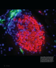
RaUsing neuroimaging, Wayne Drevets isshowing the world marked differences inbrain structure and brain activity amongthose with familial mood disorders.For example, serotonin receptors <strong>of</strong>depressed patients were only about half asprevalent, compared with controls, in thecrucial region known as the midbrain raphe(Ra). The PET scan on the left capturesreceptor binding potential in the raphe,amygdala, and hippocampus. On the right isan MRI, which Drevets coregistered with thePET to pinpoint neuroanatomy.22 PITTMED
FEATURENOW WE CAN SEE—DEPRESSED BRAINS AREBIOLOGICALLY DIFFERENT | BY EDWIN KIESTER JR.LOOKAGAINWhen the cable arrived, my wife and Iboth cried, “How can I tell you thatDavid is gone, never to return?” Themessage read, in words still burned intomy brain: “Sunday night he took his life.”David was 49. He was our closest friend, a backpackingbuddy, a brilliant editor and writer, magna cum laudefrom Harvard, creative in every way, loyal to my wife, anda pal to our son. But we also, painfully, saw David’s otherside—deep, dark, despairing moods that alternated withbursts <strong>of</strong> almost boundless, unstoppable energy. We knewabout the medication he swallowed in an effort at a moreserene, stable life; we also knew about his close relativeswho sought to end their own anguish by poison or a bullet.David chose a noose. His death was devastating, but it wasscarcely a total surprise.NEUROIMAGING | COURTESY OF DREVETSPHOTOGRAPHY | CRAIG THOMPSONOCTOBER 2000 23
Serious depression, <strong>of</strong>ficially known asunipolar or major depressive disorder(MDD), touches more than 10 millionAmerican adults each year. Manic-depressiveillness—bipolar disorder, characterized bydrastic swings <strong>of</strong> mood—affects more thantwo million, according to National Institute<strong>of</strong> Mental Health (NIMH) estimates. Manypeople plunge into darkness again and again.In families like David’s, the illness runsthrough the generations like an ominousblack thread. Counting milder forms <strong>of</strong>depression, NIMH estimates, 10 percent <strong>of</strong>the population is afflicted, and antidepressantsare among the most-prescribed drugs inthe physician’s armamentarium. For all theirprevalence, however, the exact whys andwherefores <strong>of</strong> mood disorders have remaineda tragic and recalcitrant puzzle.At the <strong>University</strong> <strong>of</strong> <strong>Pitt</strong>sburgh School <strong>of</strong><strong>Med</strong>icine, however, Wayne C. Drevets, anMD and associate pr<strong>of</strong>essor <strong>of</strong> psychiatryand radiology, has been fitting illuminatingpieces into that puzzle. Using positron emissiontomography (PET) and magnetic resonanceimaging (MRI), he has demonstratedthat the brains <strong>of</strong> those with familial mooddisorder, both unipolar and bipolar, functionabnormally during depressive episodes, andportions <strong>of</strong> their brains are actually diminishedin size. One <strong>of</strong> the areas where thesedifferences occur is a part <strong>of</strong> the forebrainthe size and shape <strong>of</strong> an index finger. Thisarea, located behind the eyes and betweenthe brain’s two hemispheres, is known as thesubgenual prefrontal cortex (PFC). Thisregion serves as a way station between whatmight loosely be called the “thinking” partA HOLY GRAILOverlaying the two sets <strong>of</strong> images, Drevets could seethat in the bipolar group the subgenual prefrontal cortexwas 39 percent smaller than in the control group,and in the unipolar group it was 48 percent smaller.<strong>of</strong> the brain, where information is receivedand processed, and the “feeling” part, thelower-order centers where inputs are interpretedin emotional terms. Depression, in thewords <strong>of</strong> one researcher, results from “imperfecttraffic” between these two areas. InDrevets’s words, depressed persons lack “aneffective set <strong>of</strong> brakes” for checking persistentand inappropriate emotional responses to agiven experience.That the brains <strong>of</strong> those suffering fromwhat has been called “a malignant sadness” arevisibly different is obviously a key discovery inthe mood-disorder puzzle. Seated undermounted slides <strong>of</strong> brain cross sections in his<strong>of</strong>fice at the UPMC Health System PETFacility, Drevets sums up his pathbreakingfindings in somewhat more technical language:“We have been able to identify circuits <strong>of</strong>the brain that function abnormally in depressionand have also been able to show actualdecreases in volume in a number <strong>of</strong> key structuresinvolved in inhibiting emotional expressionor stress responses. That seems to be theneural model for individuals who have mooddisorders that run in families, so that they getstuck in a persistent pattern <strong>of</strong> negative emotionsthat they find difficult to control andthat the medication we use to treat these illnessesdoes not cure.”In Drevets’s ongoing neuroimaging re-UNEARTHEDIf you want to see depression in action at the neurobiological level, you mightstart at the thalamus and head south a bit. Here, in the midbrain raphe, iswhere serotonin is synthesized. For years, scientists’ ability to measure accuratelyhow serotonin receptors bound in the raphe <strong>of</strong> living persons was, to saythe least, elusive. As Wayne Drevets puts it, this was one <strong>of</strong> clinical neuroscience’sholy grails.However, further developing a novel radioligand (i.e., a radioisotope used totag and measure binding activities), first applied in England, changed all that.Recently, Drevets, Ellen Frank, Chet Mathis, and others in <strong>Pitt</strong>’s Departments <strong>of</strong>Psychiatry and Radiology found that the binding potential <strong>of</strong> serotonin 1A receptorswas reduced by almost half (42 percent) in the raphe <strong>of</strong> depressed personscompared with a control population. The binding potential in limbic and neocorticalareas <strong>of</strong> depressed patients also was less than optimal: 25 and 33 percentlower, respectively. What’s more, those discrepancies were found among patientswith both unipolar (major-depressive) and bipolar (manic-depressive) familialmood disorder—the same subgroups falling under the umbrella <strong>of</strong> illnessesknown as “depression” that Drevets had identified earlier as having smaller subgenualprefrontal cortexes.—ELsearch, adult patient volunteers are selectedfrom those just entering an episode <strong>of</strong> depressionbut not yet receiving antidepressant medication.Two out <strong>of</strong> three are women, reflectingthe caseload in the mood-disorder population.After being infused with a radioactive form <strong>of</strong>oxygen, they travel supine through a humpbacked,tunnel-like machine to trace brainblood flow and glucose metabolism (glucose isthe brain’s fuel). The radioactive oxygen emitspositrons, or positive electrons, which collidewith electrons in the body to give <strong>of</strong>f gammarays. The PET scanner encircling the patientdetects the gamma rays and creates a continuous,multicolored image <strong>of</strong> the region where thereaction is occurring, providing a millisecondby-millisecondvisual record <strong>of</strong> brain activity.A few years ago, Drevets conducted a studywith depressed patients and other persons servingas nondepressive controls. Some patientswere characterized as unipolar, others as bipolar.PET images <strong>of</strong> the two groups showed thatthe depressed patients metabolized glucose inthe subgenual PFC at a rate 12 percent lowerthan the control group did—with the strikingexception <strong>of</strong> bipolar patients in the manicphase, whose rates were actually higher. PETimages were then matched with those obtainedby MRI, which can produce more minutelydetailed images <strong>of</strong> brain anatomy. Overlayingthe two sets <strong>of</strong> images, Drevets could see thatin the bipolar group the subgenual PFC was39 percent smaller on average than in the controlgroup, and in the unipolar group it was 48percent smaller.“The changes were subtle,” Drevets says.“It’s not like traditional radiology for tumor orstroke, where they jump right out at you.” Yetthe results were unmistakable. The depressedperson’s brain was clearly different.Drevets and colleagues published their findingsin Nature (April 24, 1997), immediatelycausing a stir among neuroscientists and bringinghim worldwide television exposure, including,he notes a bit shyly, interviews on theBBC. “Drevets has done first-rate work,”declared Frederick K. Goodwin, formerNIMH director and a leading voice in depressionresearch. Antonio Damasio, <strong>of</strong> the<strong>University</strong> <strong>of</strong> Iowa, another commanding pres-24 PITTMED
Wayne Drevets with the PET scanner. He keeps handing in moreevidence for neurobiological explanations <strong>of</strong> familial mood disorder.OCTOBER 2000 25
In the subgenual prefrontal cortex, Wayne Drevets found that metabolic activityis reduced during depressive episodes. Colors show decreased glucosemetabolism—the red/yellow end <strong>of</strong> the spectrum indicates the highest differencein activity relative to controls. The low activity is at least partly explainedby the smaller subgenual prefrontal cortexes (PFCs) in depressed persons. Thebar graph compares subgenual PFC volume in Drevets’s nondepressed controlgroup with that <strong>of</strong> bipolar (manic-depressive) and unipolar subjects duringdepressed episodes. The reduction is 39 percent in the bipolar and 48 percentin the unipolar.PFC VOLUME (mm 3 )300200100-5.5 -2.8 0CONTROL BIPOLAR UNIPOLARn =21 n =21 n =1726 PITTMED
Experts point to a subtext in the research. The fact thatrecurrent familial bipolar and unipolar disorder bothshow abnormalities in the same part <strong>of</strong> the brain indicatesthey are “close cousins.” Physicians should considerthat relationship when choosing treatment.ence in neuroscience, similarly praisedDrevets’s research in an editorial accompanyingthe Nature paper. Of the PFC discrepanciesreported by Drevets, he wrote, “Drevets et al.have identified a key player in one <strong>of</strong> the severalsystems that underlie emotional processing—avaluable finding indeed.”For 30 years, evidence has been mountingfor a neurobiological explanation <strong>of</strong> familialmood disorder. In other words, “It’s not just amatter <strong>of</strong> how your parents raised you,” toquote Goodwin, the former NIMH directorand author with Kay Redfield Jamison <strong>of</strong>Manic-Depressive Illness. Indeed, the drugsthat are the basis <strong>of</strong> treatment are aimed at correctinga presumed defect in the brain’s chemistryby boosting the available supply <strong>of</strong> theneurotransmitter serotonin, one <strong>of</strong> the chemicalsthat enable brain cells to communicatewith one another.Drevets keeps handing the neuro communitymore evidence. In recent research, hefocused on receptors for serotonin, which allowthe neurotransmitter to be taken up and reusedby cells. Accurately measuring receptor actionhad long been a murky region <strong>of</strong> study—sort<strong>of</strong> a holy grail for clinical neuroscience. Drevetset al.’s PET scans showed marked differences inserotonin receptors for patients with familialmood disorder. Drevets also has conductedpostmortem studies on brain tissue fromdepressed persons who died <strong>of</strong> suicide or othercauses (in collaboration with Joseph Price <strong>of</strong>Washington <strong>University</strong> in St. Louis, Missouri).Sure enough, the subgenual PFCs <strong>of</strong> depressivesturned out to be smaller than the subgenualPFCs <strong>of</strong> controls.Before David J. Kupfer, chair <strong>of</strong> <strong>Pitt</strong>’sDepartment <strong>of</strong> Psychiatry, recruited him fouryears ago from Washington <strong>University</strong>,Drevets, at 39, already had earned a nationalreputation in neuroscience research andreceived a Career Development Award.Meanwhile <strong>Pitt</strong> was seeking to strengthen itsacknowledged leadership in neuroscienceresearch, especially neuroimaging.“It was the premier psychiatry departmentin America,” Drevets notes in describing hisdecision to join <strong>Pitt</strong>’s faculty. “They saw earlyon that receptor imaging was going to be veryimportant in psychiatry, and they had assembledthe radiochemists and physicists toenable them to do it. Plus, it was about threetimes larger than Wash U.”The workings <strong>of</strong> the human brain hadintrigued Drevets since he was a junior atWheaton College volunteering at Elgin StateHospital in Illinois.“I developed this enduring fascination forwhat caused psychiatric illness,” he recalls.Drevets comes from a family <strong>of</strong> internists,and he had planned to practice cardiology.But on psychiatric rotation at the <strong>University</strong><strong>of</strong> Kansas <strong>Med</strong>ical School, he got his firstglimpse <strong>of</strong> the potential <strong>of</strong> brain imaging.“I quickly saw it as a way to understandthe pathophysiology <strong>of</strong> psychiatric disorder,”he says.At “Wash U,” he was mentored byMarcus Raichle, whom he recognizes as “one<strong>of</strong> the true pioneers <strong>of</strong> imaging.” Drevetsspent 10 years in St. Louis, then moved to<strong>Pitt</strong>’s School <strong>of</strong> <strong>Med</strong>icine.Since then, Drevets has done his part tostir up conventional wisdom. His postmortemstudies revealed a striking finding:The depressed brain had lost glial cells, yetthe functioning neuron count was normal.That was big news. Glia have long been consideredscaffolding that supports functioningneurons, yet they appear to do much morethan science had recognized. Researchershave found that among other responsibilities,these numerous cells store glucose andplay a role in neuronal metabolism. Sometypes equipped with serotonin receptors helpto innervate the brain with that importantneurotransmitter. If the brain loses glia, lessserotonin might be available, and thus thebrain might not be as well equipped to rightitself from attacks <strong>of</strong> depression.The link between loss <strong>of</strong> glia anddepression isn’t clear, Drevets says, “butwhat we know is that in depression, thereare too few glia.”All this raises one <strong>of</strong> those chicken-or-eggquestions: Does an episode <strong>of</strong> depressiontrigger changes in the brain, such as lowerglia counts and subgenual PFC volumes; oris depression brought on by existing brainabnormalities? Or rather, is “malignant sadness”caused by an interaction between thetwo? Drevets is now aggressively seekingthose answers, studying the same patientswhen they are depressed and when they haveemerged from depression. Preliminaryresults seem to indicate that some abnormalitiesare persistent and may serve as markersfor the illness. He also has begun to performPET and MRI imaging on “high risk” youngadults (meaning people who have more thanone family member affected) to see whetherabnormalities might predate the firstepisodes <strong>of</strong> depression.Other questions arise, too. Do repeatedepisodes <strong>of</strong> depression cause damage in otherparts <strong>of</strong> the body, the way hypertension or diabetesassaults the blood vessels, the heart, andthe kidneys? Is there a way to target drugsdirectly to the deficient areas where they areneeded? Finally, is there some distinct anatomicalor neurochemical marker detectable witha brain image by which those vulnerable todepression could be picked out in advance?The benefits <strong>of</strong> Drevets’s research to diagnosisand treatment <strong>of</strong> mood disorders willbe played out largely in the future. YetGoodwin, who is now director <strong>of</strong> the psychopharmacologyresearch center at GeorgeWashington <strong>University</strong>, points to a subtext inthe research <strong>of</strong> potential importance to treatmentnow. The fact that recurrent familialbipolar and unipolar disorders both showabnormalities in the same part <strong>of</strong> the brainindicates they are “close cousins.” Physiciansshould consider that relationship whenchoosing treatment. They should also note,Goodwin cautions, that both conditions aredifferent from what happens when a mansuffers from a single bout <strong>of</strong> depression afterlosing his job.“Our work in imaging has led us to clueswe knew nothing about beforehand,”Drevets says.“We don’t yet know what sets what inmotion. We now know the important thingsto study. The kind <strong>of</strong> information we’re gettingmay well account for why treatmentswork. [It] may also lead to an understanding<strong>of</strong> how we should be treating people moreeffectively in the future.”Let’s hope that understanding will savethe lives <strong>of</strong> future Davids.■OCTOBER 2000 27

![entire issue [pdf 2.79 mb] - Pitt Med - University of Pittsburgh](https://img.yumpu.com/50435398/1/190x231/entire-issue-pdf-279-mb-pitt-med-university-of-pittsburgh.jpg?quality=85)
![entire issue [pdf 6.47 mb] - Pitt Med - University of Pittsburgh](https://img.yumpu.com/50360689/1/190x231/entire-issue-pdf-647-mb-pitt-med-university-of-pittsburgh.jpg?quality=85)
![entire issue [pdf 12.7 mb] - Pitt Med - University of Pittsburgh](https://img.yumpu.com/49831615/1/190x231/entire-issue-pdf-127-mb-pitt-med-university-of-pittsburgh.jpg?quality=85)


![entire issue [pdf 11.3 mb] - Pitt Med - University of Pittsburgh](https://img.yumpu.com/46685830/1/190x231/entire-issue-pdf-113-mb-pitt-med-university-of-pittsburgh.jpg?quality=85)
![entire issue [pdf 12.7 mb] - Pitt Med - University of Pittsburgh](https://img.yumpu.com/44997419/1/190x231/entire-issue-pdf-127-mb-pitt-med-university-of-pittsburgh.jpg?quality=85)