BECKER'S NEVUS WITH IPSILATERAL BREAST HYPOPLASIA: A ...
BECKER'S NEVUS WITH IPSILATERAL BREAST HYPOPLASIA: A ...
BECKER'S NEVUS WITH IPSILATERAL BREAST HYPOPLASIA: A ...

Create successful ePaper yourself
Turn your PDF publications into a flip-book with our unique Google optimized e-Paper software.
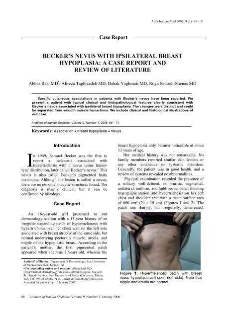
Arch Iranian Med 2006; 9 (1): 68 – 71Case ReportBECKER’S <strong>NEVUS</strong> <strong>WITH</strong> <strong>IPSILATERAL</strong> <strong>BREAST</strong><strong>HYPOPLASIA</strong>: A CASE REPORT ANDREVIEW OF LITERATUREAbbas Rasi MD • , Alireza Taghizadeh MD, Babak Yaghmaii MD, Roya Setareh-Shenas MDSpecific cutaneous associations in patients with Becker’s nevus have been reported. Wepresent a patient with typical clinical and histopathological features clearly consistent withBecker’s nevus associated with ipsilateral breast hypoplasia. The changes were distinct and couldbe separated from smooth muscle hamartoma. We include clinical and histological illustrations ofour case.Archives of Iranian Medicine, Volume 9, Number 1, 2006: 68 – 71.Keywords: Association • breast hypoplasia • nevusIntroductionIn 1949, Samuel Becker was the first toreport a melanosis associated withhypertrichosis with a nevus unius lateristypedistribution, later called Becker’s nevus. 1 Thisnevus is also called Becker’s pigmented hairymelanosis. Although the lesion is called a nevus,there are no nevomelanocytic structures found. Thediagnosis is mainly clinical, but it can beconfirmed by histology.Case ReportAn 18-year-old girl presented to ourdermatology section with a 15-year history of anirregular expanding patch of hypermelanosis withhypertrichosis over her chest wall on the left sideassociated with breast atrophy of the same side, butnormal underlying pectoralis muscle, areola, andnipple of the hypoplastic breast. According to thepatient’s mother, the first pigmented patchappeared when she was 3 years old, whereas theAuthors’ affiliation: Department of Dermatology, Iran Universityof Medical Sciences, Tehran, Iran.•Corresponding author and reprints: Abbas Rasi MD,Department of Dermatology, Rasoul-e-Akram Hospital, NiayeshSt., Sattarkhan Ave., Iran University of Medical Sciences, Tehran,Iran. Tel: +98-21-66516071-6, E-mail: dr_rasi2002@ yahoo.com.Accepted for publication: 16 January 2005breast hypoplasia only became noticeable at about13 years of age.Her medical history was not remarkable. Nofamily members reported similar skin lesions orany other cutaneous or systemic disorders.Generally, the patient was in good health, and areview of systems revealed no abnormalities.Physical examination revealed the presence ofa solitary well-defined, nonpruritic, segmental,unilateral, uniform, and light-brown patch showinghyperpigmentation and hypertrichosis on her leftchest and shoulder area with a mean surface areaof 600 cm² (20 × 30 cm) (Figures 1 and 2). Thepatch was sharply, but irregularly, demarcated.Figure 1. Hypermelanotic patch with breastmass hypoplasia are seen (left side). Note thatnipple and areola are normal.68Archives of Iranian Medicine, Volume 9, Number 1, January 2006
A. Rasi, A. Taghizadeh, B. Yaghmaii, et alrete ridges had flat tips). In the dermis, there wasnot any marked increase in smooth muscle fibers,but only one bundle of pilar smooth muscles waspresent, and no nevus cells were observed (Figure3). Examination with trichrome stain demonstratedno increase in smooth muscle fibers.On the basis of clinical and histopathologicfindings, a diagnosis of Becker’s nevus associatedwith ipsilateral breast hypoplasia was made.DiscussionFigure 2. A hypermelanotic patch with welldefinedand irregular border is seen.There were no pustules, vesicles, perifollicularpapular elevations, or indurations. The lesion didnot show transient elevations on rubbing. Over thetime, hypertrichosis gradually developed within it.There was no associated hypoplasia of underlyingstructures and asymmetry of the limbs.The patient’s blood pressure was 110/80mmHg. Radiographic studies of the entire spineand upper limbs did not show any alterations. Theresults of routine blood tests and urine analysiswere within normal limits. The following hormoneexaminations were all within normal values: freetestosterone, dihydrotestosterone, dehydroepiandrosterone,prolactin, follicular stimulatinghormone, luteinizing hormone, and thyroidhormones. A biopsy specimen of the left chestpigmented lesion showed: hyperkeratosis,increased basal layer pigmentation, regularelongation, and clubbing of rete ridges (elongatedFigure 3. A smooth muscle bundle independentof the hair follicle is seen (arrow).Becker’s nevus is a unilateral (rarely bilateral)solitary, macular light brown hyperpigmentation ofvarying size, with geographic borders. The borderis irregular and sharply demarcated. Occasionally,lesions resembling acne vulgaris, includingpapules, pustules, and cystic nodules, may occurwithin the affected area. 2 Over time, hypertrichosis(which is a characteristic feature of the lesion),develops within the lesion in about 50% of cases. 3Hairs, which are confined to the hyperpigmentedarea, are darker and coarser than normal. Thelesion varies in size and may cover the entire upperarm or shoulder. A typical lesion is about 125 cm 2 ,although lesions up to 500 cm² have been reported.These lesions are usually asymptomatic and areusually localized on the shoulder, anterior chest, orupper arms, but there have been reports in otherareas (e.g., proximal upper extremities, lowerextremities, face, forearm, wrist, neck, and upperand lower back). 2In a study of 19,302 men aged 17 to 26 years, aprevalence of 0.52% was observed. 3 Reports of thiscondition in women are much rarer in theliterature. The male-to-female ratio, approximatedfrom case studies, has been reported to beanywhere from 4:1 to 6:1. 4, 5 There is no racialpredilection, but it is more common in youngpeople with fair skin. 6 Becker’s nevus is usuallyacquired in adolescence (the majority of cases arefirst noticed sharply before, at, or after puberty),but a congenital onset has been recorded, 7 as havefamilial cases in siblings, and in an uncle and anephew.The lesion is a developmental anomaly, butoccasionally, lesions have been said to followsevere sunburn. Lesional tissue has been found tohave an increased level of androgen receptors,suggesting that heightened local androgensensitivity may result in the hypertrichosis.Many abnormalities have been associated withArchives of Iranian Medicine, Volume 9, Number 1, January 2006 69
Becker’s nevus with ipsilateral breast hypoplasiaBecker’s nevus such as:• Smooth muscle hamartoma. This is the mostfrequent finding. In such cases, the area ofBecker’s nevus may exhibit slightperifollicular papular elevations or slightinduration. 8• Unilateral breast hypoplasia. This is possiblyas a result of enhanced androgen sensitivity. 2, 9• Hypoplasia of underlying structures andasymmetry of limbs. 10Other abnormalities include: various skeletalmalformations, connective tissue nevus, 9 aplasia ofipsilateral pectoralis major muscle, ipsilateral limbshortening, localized lipoatrophy, spina bifida,scoliosis pectus carinatum, congenital adrenalhyperplasia, and accessory scrotum. 2Unless associated with a hamartomatousprocess, histologic changes in Becker’s nevus areminimal. The epidermal changes are variable, butusually there is hyperkeratosis, subtle acanthosis,papillomatosis, 8 and basal layer hyperpigmentation.There are regular elongation of reteridges, and elongated rete ridges tend to have flat,rather than pointed tips. Melanocyte proliferationis usually mild and not always obvious in routinesections; this increase is particularly evident whenmelanocytes are stained for dopa-oxidase activityin both involved and uninvolved skin nearby. 8 Thedermis is thickened and contains numerous, butoften inconspicuous bundles of smooth musclefibers, unrelated to hair follicles or blood vessels. 8Controversy exists about the relationship of thesecases to smooth muscle hamartoma. Some authorsbelieve that, in cases associated with smoothmuscle hamartoma, irregularly arranged, thickbundles of smooth muscle are present in thedermis. 8 There may also be an underlying increasein the number and size of hair follicles andsebaceous glands. 11 Some melanophages are seenin the dermis. 1 There should be no problem inmaking the diagnosis in a well-developed Becker’snevus. In early lesions, the age of onset and,generally, the site are perhaps the most helpfuldiagnostic features.Clinically, congenital Becker’s nevus may beconfused with a congenital melanocytic nevus.Histologically, Becker’s nevus does not havenevocellular nevus cells.Albright’s syndrome may be easily confusedwith Becker’s nevus, but the pigmented macule inAlbright’s syndrome is present at birth.Theoretically, Becker’s nevus and smoothmuscle hamartoma are two quite separate entities.In fact, there are cases which could be consideredas intermediate. As a matter of fact, a slightunderlying smooth muscle hyperplasia can be seenin Becker’s nevus; on the other hand,hypermelanosis of basal layer and hypertrichosismay be encountered in smooth muscle hamartoma.Both conditions can be considered polar forms of aspectrum of dermal smooth muscle hyperplasia. 12,13 Becker’s nevus may enlarge slowly a year or twoafter presentation and then stabilizes and appearsto persist indefinitely. The hyperpigmentationusually remains stable, although there have been4, 14reports of fading over many years. Becker’snevus is usually too large to remove and is best leftuntouched. The hair may be shaved or permanentlyremoved. The skin hyperpigmentation mayrespond to therapy with Q-switched ruby laser,although the results are unpredictable andrecurrences are common.Although Becker’s nevus is most common inmales, ipsilateral breast hypoplasia in Becker’snevus occurs infrequently. Cases associated withbreast hypoplasia are more frequently reported inwomen. In fact, 13 cases have been reported in theliterature in whom the onset of Becker’s nevus inthe mammary area during the prepubertal age wasfollowed by breast hypoplasia. 8, 9, 15 – 17 For thisreason, the androgen skin receptors wereexamined, and the results revealed high levels ofthese receptors in the affected skin. 9 This explainsthe mammary hypoplasia of the specific cases, aswell as the frequent hypertrichosis, the possiblepresence of acneiform lesions, and the thick dermiswith which the nevus is associated. We thought itwould be interesting to report this case, not onlybecause of the rare association of breast hypoplasiawith Becker’s nevus, but due to this case’s earlyage of onset and the large size of the lesion.References1 Becker SW. Concurrent melanosis and hypertrichosis indistribution of nevus unius lateris. Arch Dermatol Syph.1949; 60: 155 – 160.2 Rook A, Wilkinson DS, Ebling FJG, Champion RH.Rook/Wilkinson/Ebling Textbook of Dermatology. 6th ed.Oxford; Malden, MA: Blackwell Science; 1998;542 – 543.3 Tymen R, Forestier J-F, Boutel B, Colomb D. Nevus tardifBecker. A propos d, une serie de 100 observations. AnnDermatol Venereol. 1981; 108: 41 – 46.4 Chima KN, Janniger CK, Schwartz RA. Becker’smelanosis. Cutis. 1996; 57: 311 – 314.5 Happle R, Koopman RJ. Becker nevus syndrome. Am J70Archives of Iranian Medicine, Volume 9, Number 1, January 2006
A. Rasi, A. Taghizadeh, B. Yaghmaii, et alMed Genet. 1997; 68: 357 – 361.6 Ballone E, Fazii P, Lappa G, Di Mascio R, Di Mascio C,Schinoppa F. Prevalence of Becker’s nevi in a populationof young men in central Italy. J Am Acad Dermatol. 2003;48: 795.7 Picascia DD, Esterly NB. Congenital Becker’s melanosis.Int J Dermatol. 1989; 28: 127 – 128.8 Freedberg IM, Fitzpatrick TB. Fitzpatrick’s Dermatologyin General Medicine. 5th ed. New York: Mc Graw-Hill,Health Profession Division; 1999: 880 – 881, 882, 903,994 – 995.9 Formigon M, Alsina MM, Mascara JM, Rivera F.Becker’s nevus and ipsilateral breast hypoplasia.Androgen receptor-study in two patients. Arch Dermatol.1992; 128: 992 – 993.10 Lucky AW, Saruk M, Lerner AB. Becker’s nevusassociated with limb asymmetry. Arch Dermatol. 1981;117: 243.11 Chapel TA, Tavafoghi V, Mehregan AH, Gagliardi C.Becker’s melanosis: an organoid hamartoma. Cutis. 1981;27: 405 – 415.12 De la Espriella J, Grossin M, Marinho E, Belaich S.Smooth muscle hamartoma. anatomoclinicalcharacteristics and nosological limits [in French]. AnnDermatol Venereol. 1993; 120: 879 – 883.13 Civatte J, Marinho E, Oliver-Santos R. Smooth musclehamartoma or nevus of Becker? A propos of 4 cases [inSpanish]. Med Cutan Ibero Lat Am.1988; 16: 145 – 148.14 Kopf AW, Yuppa J. Becker’s nevus. Arch Dermatol.1968; 98: 97 – 98.15 Cambiaghi S, Brusasco A, Tadini G, et al. Nevo di Beckercongenito con ipoplasia mammaria ipsilaterale. G ItalDermatol Venereol. 1994; 129: 169 – 172.16 Sharma R, Mishra A. Becker’s nevus with ipsilateralareolar hypoplasia in three males. Br J Dermatol. 1997;136: 471 – 472.17 Hoon-Jung J, Chan-Kim Y, Joon-Park H, Woo-Cinn Y.Becker’s nevus with ipsilateral breast hypoplasia:improvement with spironolactone. J Dermatol. 2003; 30:154 – 156.Archives of Iranian Medicine, Volume 9, Number 1, January 2006 71