59. Infantile Hypertrophic Pyloric Stenosis - Global HELP
59. Infantile Hypertrophic Pyloric Stenosis - Global HELP
59. Infantile Hypertrophic Pyloric Stenosis - Global HELP
- No tags were found...

Create successful ePaper yourself
Turn your PDF publications into a flip-book with our unique Google optimized e-Paper software.
CHAPTER 59<strong>Infantile</strong> <strong>Hypertrophic</strong><strong>Pyloric</strong> <strong>Stenosis</strong>Lohfa B. ChirdanEmmanuel A. AmehAmy Hughes-ThomasIntroduction<strong>Infantile</strong> hypertrophic pyloric stenosis (IHPS) is a common surgicalcause of vomiting in infancy in the Western world. 1,2 Historically, itwas described as a disease entity in 1888 by Harald Hirschsprung. 3Gastrojejunostomy was used to treat this disease until 1912, whenextramucosal muscle-splitting pyloromyotomy was described byRamstedt. 4 This procedure has dramatically changed the outcome ofinfants with IHPS.DemographicsThe reported incidence of IHPS in the Western world is 1–4 per 1,000live births. 5 There is a male-to-female ratio of 4:1, with reported ratiosranging from 2.5:1 to 5.5:1. 6<strong>Pyloric</strong> stenosis appears to be more common in infants of caucasiandescent and is less common in India and among black and Asianpopulations, with a frequency that is one-third to one-fifth that in thewhite population. 7In about 6–33% of infants with IHPS, associated anomalies havebeen described in the central nervous system (CNS), gastrointestinaltract (GIT), and urinary tract. 8AetiologyDespite the frequency of pyloric stenosis, the aetiology remains unclear.Genetic predisposition acting in conjunction with environmental factorsis the most widely accepted explanation; however, debate still continuesas to whether it is a congenital or acquired disease. 9–11 Breast-feeding hasbeen suggested as offering some immunity to the disease. 12First-born children have been noted to be more likely affected, anda familial link is seen with a greater than fivefold increase in the riskin first-degree relatives. The genetics explaining this are likely to bepolygenic, as no single locus has been identified. 6 Male and femalechildren of affected mothers carry a 20% and 7% risk, respectively,of developing the condition, whereas male and female children ofaffected fathers carry a 5% and 2.5% risk, respectively. Furthermore,an association is seen in twins, with concordance among monozygotictwins of 0.25–0.44, and in dizygotic twins of 0.05–0.10. 13Pathophysiology<strong>Pyloric</strong> stenosis is characterized by hypertrophy of the pyloric musculature,leading to a mechanical obstruction of the gastric outlet inthe affected infant. Thus, hypertrophied pyloric antral muscle fibresprotrude distally into the duodenal lumen, producing a reflection ofduodenal mucosa.Infants with a diagnosis of pyloric stenosis will show characteristicallylow chloride and hydrogen ions as measured in the serum. The lossof gastric secretions secondary to protracted vomiting will result indehydration. As a result, through aldosterone-stimulated absorption,potassium is excreted in the urine in an attempt to conserve sodium.As potassium depletion worsens, sodium resorption across the renaltubule is then achieved in exchange for a hydrogen ion, thereby creatingparadoxical aciduria. Classically, this results in the occurrence of ahypochloraemic hypokalaemic metabolic alkalosis. In severe cases withdiagnostic delay, hypoglycaemia and hypoalbuminaemia can be observed.It is known that the pyloric hypertrophy will eventually resolve, butthis takes a long period of time; the infant would usually succumb to theelectrolyte derangement and dehydration before this happened.Clinical PresentationInfants with pyloric stenosis usually present with a gradual onset ofworsening nonbilious vomiting, beginning between 3 to 6 weeks of age.The pattern of vomiting can vary, but often it progresses to the characteristic“projectile” vomiting. Infants may present in the early stages of thedisease and be treated for reflux disease or undergone numerous formulachanges before the diagnosis is made. Delay in diagnosis can result insignificant electrolyte imbalance, weight loss, and failure to thrive.The typical clinical features include the following:• Nonbilious vomiting is usually forceful and postprandrial.• The infant is hungry after vomiting and eager to feed, only to vomitagain.• Weight loss occurs in severe cases.• Signs of dehydration present in cases of repeated vomiting.• Scaphoid abdomen especially noted after recent vomiting.• Visible peristalsis may be observed in the upper abdomen, usuallymoving from the left hypochondrium towards the right side.• A palpable mass is present in the right upper quadrant (90% inexperienced hands); this is best appreciated while the infant is beingfed with clear fluid.Differential DiagnosisThe differential diagnosis of pyloric stenosis includes:• gastro-oesophageal reflux;• viral enteritis;• pylorospasm;• duodenal stenosis/duodenal web; and• raised intracranial pressure.EvaluationClinical DiagnosisDepending of the time to presentation, the clinical picture can vary enormouslyfrom a well-hydrated baby to an emaciated infant. Weight lossand dehydration coupled with an insatiable appetite lead to a characteristicfacies, with a furrowed brow, wrinkled appearance, and prominentsucking pads. In some infants, the distended stomach may be identifiablein the hypochondrium, with active peristaltic activity visible through the
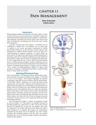
<strong>Infantile</strong> <strong>Hypertrophic</strong> <strong>Pyloric</strong> <strong>Stenosis</strong> 369thin abdominal wall. On examination, a mobile, ovoid mass, commonlyreferred to as an “olive”, is palpable in the epigastrium or the rightupper quadrant.If the pylorus is palpated by an experienced clinician, no furtherimaging is necessary. In some cases, however, other structures may beconfused with hypertrophied pylorus, including the caudate lobe of theliver, the right kidney, the vertebrae, or an orogastric tube in the distalstomach. If there is any doubt, or in the absence of a palpable “olive”,diagnostic imaging can be helpful.UltrasonographyIn situations where doubt exists, examination by ultrasound (US)should be performed. This would normally confirm the presence of apyloric “tumour”. The characteristic appearance of pyloric stenosis onultrasound is that of a “doughnut” or “bull’s eye” on cross section ofthe pyloric channel. <strong>Pyloric</strong> dimensions with positive predictive valuegreater than 90% are muscle thickness greater than 4 mm and a pyloricchannel length greater than 17 mm. 14 These limits may be lower ininfants younger than 30 days of age (Figure <strong>59.</strong>1).An experienced sonographer will recognize periods of relaxation ininfants with pylorospasm, commonly confused with pyloric stenosis atexamination. Pylorospasm has been hypothesized to be an early stageof IHPS, but this has not been proven.Upper Gastrointestinal Contrast StudyIn an occasional case where doubt still persists after US examination,an upper gastrointestinal (UGI) series may be done. The UGI serieswould show a narrow pyloric channel, the so-called “string sign” andthe “shoulder sign”, caused by the impression of the pylorus into thestomach (Figure <strong>59.</strong>2).Figure <strong>59.</strong>1: Ultrasound features of pyrolic stenosis.Figure <strong>59.</strong>2: UGI contrast showing pyloric stenosis.Serum ElectrolytesSerum electrolytes should be measured immediately when the patientarrives in hospital. If vomiting has been ongoing for several days,serum electrolytes are frequently deranged. The nature of derangementis a spectrum, 15,16 ranging from mild to severe hyponatraemia,hypochloraemia, hypokalaemia, and metabolic alkalosis. The degree ofelevation of serum urea is directly related to the severity of dehydration.Haemogram/Full Blood CountInfants presenting late are often malnourished and may have somedegree of anaemia, which may require correction. Therefore, a haemogramand full blood count are warranted.TreatmentCorrection of Electrolyte and Fluid DepletionPatients with pyloric stenosis may have severe electrolyte disturbances,so the serum electrolytes should always be estimated. Mild electrolytedisturbances can be corrected preoperatively with 0.45% normal salinewith 5% dextrose solution. Severe disturbances require correction with0.9% normal saline bolus of 10 to 20 ml/kg, followed by administrationof 0.9% saline in 5% dextrose solution. Potassium can be addedif necessary when adequate urine output (1.5–2 ml/kg per hour) isestablished and under electrocardiogram (ECG) monitor. Fluid shouldbe administered at a rate of 25–50% above maintenance.Following resuscitation and correction of electrolyte imbalance,maintenance IV with 0.45% saline in 5% dextrose with 20 mmolpotassium chloride should be given at 25-50% above the standard rate.Meticulous care and time should always be taken to correct fluid andelectrolyte depletion before any surgical correction. It is important toemphasize that mortalities from pyloric stenosis are attributable to fluidand electrolyte problems.Nasogastric DecompressionOnce diagnosis is made, all feeds are stopped. It is helpful to aspirateall gastric content by nasogastric tube (NGT). Frequently, this contentcomprises milk curds, which may require lavage with saline toadequately evacuate the stomach. Keeping the stomach empty wouldhelp prevent aspiration from vomiting. Once the stomach is emptied,the NGT is either closed off or removed to avoid worsening electrolytedepletion by aspirating gastric content.In the West, gastric lavage is not routinely performed. An NGTis passed, size 8 Fr or above. Gastric losses are monitored andreplaced milliliter for milliliter with 0.9% saline. To avoid iatrogenichyperkalaemia, no potassium is added to the replacement fluid.Surgical CorrectionSurgical correction of pyloric stenosis is not an emergency, and thereforethe electrolyte disturbances can and should be meticulously correctedbefore operation. Occasionally, children with pyloric stenosiswill have jaundice due to a transient impairment of glucuronyl transferaseactivity. This is self-limited once postoperative feeding is initiated.Infants undergoing pyloromyotomy are assumed to have a fullstomach and the anaesthesiologist should keep this in mind. Both theanaesthesiologist and surgeon should be vigilant during the operationto prevent aspiration of gastric juice. The stomach must be evacuatedin the operating room, particularly if NGT had not been inserted earlier.Preoperative antibiotics are controversial; data supporting their usewith the standard right upper quadrant incision are scant. They may beof benefit when performing the operation through the umbilical skinfold.Operative DetailsThe standard operation is the Ramstedt pyloromyotomy. Classically,the operation has been approached through a right upper quadrantmuscle-splitting approach 17 . Alternatively, the approach may be via asupra-umbilical transverse skinfold incision.1. Once the peritoneum is entered, the omentum is retrieved into
370 <strong>Infantile</strong> <strong>Hypertrophic</strong> <strong>Pyloric</strong> <strong>Stenosis</strong>the wound and elevated to lift the transverse colon. This manoeuvreenables the surgeon to identify the antrum of the stomach. The lowerthird of the stomach is then gently elevated using moist gauze todeliver the pyloric mass into the wound (Figure <strong>59.</strong>3).2. A vertical incision is then made into the mid anterior surface throughthe serosa and superficial muscularis, beginning about 1–2 mm fromthe pyloroduodenal junction to a point 0.5 cm into the lower antrum.3. The underlying firm fibres are then divided using blunt dissectionwith a clamp, rounded end of a scalpel blade, or special Benson’spyloromyotomy spreader. Special care is taken to prevent mucosalperforation, especially at the lower end of the incision. Upwardprotrusion of the gastric mucosa indicates relief of the obstruction(Figures <strong>59.</strong>4 and <strong>59.</strong>5).Mucosal perforation usually occurs at the duodenal end and isindicated by the appearance of bilious fluid. When this occurs, repairis done by using interrupted fine monofilament long-term absorbablesutures placed transversely and covered with omentum. If the closure ofthe mucosal perforation compromises the pyloromyotomy, which rarelyhappens, a fresh pyloromyotomy is done at about 45°–90° of the firstincision. Air is then instilled through the NGT to check the integrity ofthe duodenal mucosa.Use of a laparoscopic approach is increasing, with evidencesupporting its benefits emerging. 18,19 A recent study has shown a safealternative with a decreased time to full feeds postoperatively. 20Figure <strong>59.</strong>3: Operative view of pyloric mass.Postoperative ManagementPostoperative nasogastric decompression is not necessary unless themucosa has been entered and repaired. Several feeding schedules havebeen advocated after surgery. Traditional structured feeding regimensas opposed to more rapid initiation and advancing feeding schedules areprobably unnecessary. Feedings are begun 4 to 6 hours after operation,normally with low-volume balanced electrolyte or dextrose solutioninitially, rapidly advanced to full feeds of formula over the next 12- to24-hour period. If the patient vomits, which is common after this procedure,the same volume feed that caused the emesis can be repeated.The patient is usually discharged the day after operation.Surgical ComplicationsIntraoperative risks include bleeding, infection, and mucosal perforation.Postoperative complications include wound infection and dehiscence inabout 1%. Persistent vomiting beyond 48 hours, thought to be due to gastricatony, occurs in about 3%. Unrecognized perforation during pyloromyotomyis a serious but rare problem demanding immediate reoperation.OutcomeThe majority of infants go on to make a full recovery postoperativelyand need no further medical input. After a surgical pyloromyotomy, thepyloric muscle subsides to a normal size and, when viewed during subsequentoperations, is usually visible only as a fine line over the pylorusat the site of the myotomy.Incomplete pyloromyotomy may occur, but it is difficult to diagnose in theearly postoperative phase. Imaging studies done postoperatively are difficultto interpret and usually not helpful. If complete gastric-outlet obstruction ispresent on a contrast study, repeated pyloromyotomy is necessary.Mortality is rare, but when it occurs, it is usually from fluid andelectrolyte depletion in infants presenting late, and inadequatelycorrected electrolyte problems before surgery.Evidence-Based ResearchEvidence on the management of pyloric stenosis in African children israre, so clinicians have to depend on literature from the West, where thedisease is more frequent. Table <strong>59.</strong>1 presents the results of a survey onthe management of IHPS conducted in the United Kingdom and Ireland.Table <strong>59.</strong>1: Evidence-based research.Figure <strong>59.</strong>4: Spreading of the divided pyloric muscle.Figure <strong>59.</strong>5: Myotomy with mucosal bulge.TitleAuthorsInstitutionSurgical practice for infantile hypertrophic pyloric stenosis inthe United Kingdom and Ireland—a survey of members of theBritish Association of Paediatric SurgeonsMullassery D, Perry D, Goyal A, Jesudason EC, Losty PDAcademic Department of Pediatric Surgery, The RoyalLiverpool Children’s Hospital (Alder Hey), University ofLiverpool, United KingdomReference J Pediatr Surg 2008; 43:1227–1229ProblemOutcome/effectHistoricalsignificance/commentsCurrent practice amongst paediatric surgeons on themanagement of infantile hypertrophic pyloric stenosis.More than half of the surgeons surveyed used umbilicalincision for pyloromyotomy, whereas only 15% do thepyloromyotomy laparoscopically. Fewer than 10% of surgeonssurveyed use the classical right upper quadrant incision forpyloromyotomy. The study also showed that about half of thesurgeons do not use antiobiotics; however, 70% of those usingthe umbilical incision use antibiotics. The study concluded thatumbilical incision and laparoscopic incisions are benchmarksfor surgeons caring for children with infantile hypertrophicpyloric stenosis.Acknowledging that IHPS may not be a major workload forthe paediatric surgeon practicing in Africa, patients with thiscondition do come in occasionally, especially in major centres,so paediatric surgeons need to be aware of the currentpractices amongst paediatric surgeons who care frequently forthese patients; hence, the importance of this article. Althoughthere are a lot of variations in the practice, pyloromyotomythrough whatever route remains the gold standard for caringfor these group of patients.
<strong>Infantile</strong> <strong>Hypertrophic</strong> <strong>Pyloric</strong> <strong>Stenosis</strong> 371Key Summary Points1. <strong>Infantile</strong> hypertrophic pyloric stenosis affects infants 2–8 weeksof age, often presenting with repeated vomiting.2. Although the disease may not be common in African children,practitioners may encounter the condition.3. The aetiology is not clear, but pyloric muscle hypertrophyleading to mechanical obstruction of the pylorus is the endpoint.4. The disease can be self-limiting, but the infant would succumbto dehydration and electrolyte imbalance if not treated.5. Care should be taken to correct any fluid and electrolytedepletion before embarking on any surgical correction.6. Extramucosal pyloromyotomy, introduced about a century ago,still remains the gold standard for surgical management of IHPS.References1. Hirschsprung H. Falle von angeborener pyloric stenose. JbKinderheik 1888; 27:61.2. Ramstedt C. Zur operation der angeborenen pylorus-stenose. MedKlin 1912; 8:1702–1705.3. Spicer RD. <strong>Infantile</strong> hypertrophic stenosis: a review. Br J Surg1982; 69:128–135.4. Stringer MD, Brereton RJ. Current management of infantilehypertrophic pyloric stenosis. Br J Hosp Med 1990; 43:266–272.5. To T, Wajja A, Wales PW, et al. Population demographic indicatorsassociated with incidence of pyloric stenosis. Arch Pediatr AdolescMed 2005; 159:520–525.6. Michel LE, Risch N. The genetics of infantile hypertrophic pyloricstenosis: a reanalysis. Am J Dis Child 1993; 147:1203–1211.7. Klein A, Cremin BJ. Racial significance in pyloric stenosis. S AfrMed J 1970; 44:1130–1134.8. Bidair M, Kalota SJ, Kaplan GW. <strong>Infantile</strong> hypertrophic pyloricstenosis and hydronephrosis: is there an association? J Urol 1993;150:153–155.9. Ohshiro K, Puri P. Pathogenesis of infantile pyloric stenosis: recentprogress. Pediatr Surg Int 1998; 13:243–252.10. Abel RM, Bishop AE, Doe CJ, et al. A quantitative study of themorphological and histochemical changes within the nerves andmuscle in infantile hypertrophic pyloric stenosis. J Pediatr Surg1998; 33:682–687.11. Sherwood W, Choudhry M, Lakhoo K. <strong>Infantile</strong> hypertrophic pyloricstenosis: an infectious cause? Pediatr Surg Int 2007; 23:61–63.12. Osifo DO, Evbuomwan I. Does exclusive breastfeeding conferprotection against infantile pyloric stenosis? A 30 year experiencein Benin City, Nigeria. J Trop Pediatr 2009; 55:132–134.13. Carter CO, Evans KA. Inheritance of congenital pyloric stenosis. JMed Genet 1969; 6:233–254.14. Hernanz-Schulman M. <strong>Infantile</strong> hypertrophic pyloric stenosis.Radiology 2003; 227(2):319–331.15. Nmadu PT. Alterations in serum electrolytes in congenitalhypertrophic pyloric stenosis: a study in Nigerian children. Ann TropPaediatr 1992; 12:169–172.16. Touloukian RJ, Higgins E. The spectrum of serum electrolytes inhypertrophic pyloric stenosis. J Pediatr Surg 1983; 18(4):394–397.17. Fonkalsrud EW. <strong>Hypertrophic</strong> pyloric stenosis. In O’Neil Jr JA,Grosfeld JL, Fonkalsrud EW, Coran AG, Caldamone AA (eds).Principles of Pediatric Surgery, 2nd ed. Mosby, 2003, Pp 467-470.18. van der Bilt JD, Kramer WL, van der Zee DC, Bax NM.Laparoscopic pyloromyotomy for hypertrophic pyloric stenosis:impact of experience on the results in 182 cases. Surg Endosc2004; 18(6):907–909. Epub 27 Apr 2004.19. Mullassery D, Perry D, Goyal A, Jesudason EC, Losty PD. Surgicalpractice for infantile hypertrophic pyloric stenosis in the UnitedKingdom and Ireland—a survey of members of the British Associationof Paediatric Surgeons. J Pediatr Surg 2008; 43:1227–1229.20. Hall NJ, Pacilli M, Eaton S, Reblock K, Gaines BA, et al. Recoveryafter open versus laparoscopic pyloromyotomy for pyloric stenosis:a double blind multi centre randomized controlled trial. Lancet2009; 373:390–398.

![Clubfoot: Ponseti Management [Vietnamese] - Global HELP](https://img.yumpu.com/51276842/1/184x260/clubfoot-ponseti-management-vietnamese-global-help.jpg?quality=85)


![Steenbeek Brace For Clubfoot [2nd Edition] - Global HELP](https://img.yumpu.com/46612972/1/190x245/steenbeek-brace-for-clubfoot-2nd-edition-global-help.jpg?quality=85)

![Basics Of Wound Care [Indonesia] - Global HELP](https://img.yumpu.com/41566370/1/190x245/basics-of-wound-care-indonesia-global-help.jpg?quality=85)