Human Heart Chapter - iWorx
Human Heart Chapter - iWorx
Human Heart Chapter - iWorx

Create successful ePaper yourself
Turn your PDF publications into a flip-book with our unique Google optimized e-Paper software.
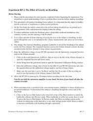
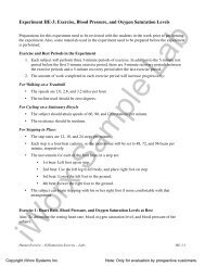
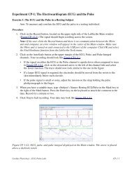
<strong>Human</strong> <strong>Heart</strong> <strong>Chapter</strong>ExperimentsBasic Level Difficulty RatingHH-1: The Electrocardiogram and CirculationHH-2: The Electrocardiogram and <strong>Heart</strong> SoundsHH-3: Exercise, the Electrocardiogram, and CirculationHH-8: <strong>Heart</strong> Sounds/AuscultationHH-9: ECG and the Electronic StethoscopeAdvanced Level Difficulty RatingHH-4: The Six-Lead ElectrocardiogramHH-5: The Diving ReflexHH-6: <strong>Heart</strong> Rate VariabilityHH-7: ECG using Six Chest leadsHH-10: 12 Lead ECG 5OverviewThe heart is a pump that pushes blood around the body. Blood enters the heart at a low pressure andleaves at a higher pressure, and this high pressure provides the force to propel the blood through thecirculatory system. The organization of the human heart and the circulatory system is shown in FigureHH-0-1. Blood returning from the body is sent to the right side of the heart and then to the lungs to pickup oxygen and release carbon dioxide. This oxygenated blood is sent to the left side of the heart andback to the body, where oxygen is released and carbon dioxide is collected. The complete division ofthe heart insures that there is no mixing of deoxygenated blood (in the right side) with oxygenatedblood (in the left side).The mammalian heart is autorhythmic, since it will continue to beat if removed from the body (andkept in an appropriate solution). <strong>Heart</strong> contractions are, therefore, not dependent upon the brain, ratherthe rhythm comes from within the heart itself. The heart is composed almost entirely of large, strongmuscle fibers (myocardium), which are responsible for the pumping action of the heart. Other cardiacmuscle cells are weakly contractile and produce or conduct the rhythm for the rest of the heart. A groupof these weak muscle cells is located in the sinoatrial (SA) node and acts as the pacemaker for the heart(Figure HH-0-2). These cells rhythmically produce action potentials, which spread via gap junctions tofibers of both atria. The resulting contraction pushes blood into the ventricles. While adjacent atrialfibers are connected by gap junctions, the only electrical connection between the atria and theventricles is via the atrioventricular (AV) node. The action potential spreads slowly through the AVnode and then rapidly through the Bundle of His and Purkinje fibers to excite both ventricles.<strong>iWorx</strong> Sample Lab<strong>Human</strong> <strong>Heart</strong> – IntroductionHH-0-1Copyright <strong>iWorx</strong> Systems Inc.Note: Only for evaluation by prospective customers.
Figure HH-0-1: A diagram to show the circulation of blood around the human body and its associationwith the heart, composed of a right atrium (RA), a left atrium (LA), a right ventricle (RV), and a leftventricle (LV).Figure HH-0-2: A diagram of the human heart to show the location of the sinoatrial (SA) andatrioventricular (AV) nodes.The semilunar valves are located between the ventricle and the main artery on each side of the heart. Inthe relaxed heart, the high arterial pressure shuts the semilunar valves and prevents blood flow from theartery back into the ventricle. Ventricular contraction increases the pressure of the blood in theventricle. When the ventricular pressure is greater than the arterial pressure, the semilunar valves openand blood flows into the artery. Then, the myocardium relaxes, the ventricular pressure declines, andthe semilunar valves close.<strong>iWorx</strong> Sample Lab<strong>Human</strong> <strong>Heart</strong> – IntroductionHH-0-2Copyright <strong>iWorx</strong> Systems Inc.Note: Only for evaluation by prospective customers.