A rapid method for sex determination of bovine and buffalo embryos
A rapid method for sex determination of bovine and buffalo embryos
A rapid method for sex determination of bovine and buffalo embryos

You also want an ePaper? Increase the reach of your titles
YUMPU automatically turns print PDFs into web optimized ePapers that Google loves.
Furthermore, selective <strong>sex</strong> pre<strong>determination</strong> together with the multiple ovulation <strong>and</strong> embryotransfer (MOET) procedures (Colleau, 1991) can effectively aid in improvement <strong>of</strong> the geneticgain <strong>of</strong> the herd. Progress in the field <strong>of</strong> in vitro embryo production has opened up new horisons<strong>for</strong> supporting the <strong>bovine</strong> industry on both research <strong>and</strong> economic levels, in terms <strong>of</strong> embryomanipulation, genetic enhancement <strong>and</strong> production <strong>of</strong> <strong>sex</strong>ed <strong>embryos</strong> (Bavister, 2002; Faber etal., 2003). Buffalo species is considered an important farm animal with respect to its productiverole (Suzuki et al., 1992). Improvement <strong>of</strong> reproductive technologies <strong>and</strong> genetic approachesrepresents a significant aim <strong>of</strong> interest with the possibility <strong>of</strong> an integrated wide-spread geneticupgrading in such species (Gasparrini, 2002). Several <strong>method</strong>s, such as cytogenetic analysis <strong>of</strong>chromosomes (Kitiyanant et al., 2000), immunological <strong>method</strong>s (Utsumi <strong>and</strong> Iritani, 1993)immunocytochemical hybridization techniques (Bondioli et al., 1989) <strong>and</strong> X-linked enzymeactivities as a biochemical assay (Monk <strong>and</strong> H<strong>and</strong>yside, 1988) have been presented <strong>for</strong> <strong>sex</strong><strong>determination</strong> <strong>of</strong> preimplantation <strong>embryos</strong>. However, most <strong>of</strong> these approaches were found to betime consuming, little accurate <strong>and</strong> unreliable to cover potentially the daily practice <strong>of</strong> embryotransfer programs. Polymerase chain reaction (PCR) was introduced as a molecular approach <strong>for</strong>embryo <strong>sex</strong>ing using a few biopsied embryonic cells (Peura et al., 1991; Faber et al., 2003;Manna et al., 2003) depending up on amplification <strong>of</strong> Y-chromosome specific DNA sequence asa specific indicator <strong>for</strong> male <strong>embryos</strong> <strong>and</strong> an autosomal repeated fragment common <strong>for</strong> both male<strong>and</strong> female <strong>sex</strong>es. In order to augment the DNA amplification <strong>of</strong> a single copy embryonic biopsy,<strong>sex</strong> <strong>of</strong> <strong>bovine</strong> <strong>embryos</strong> was determined by PCR with some modifications, like the use <strong>of</strong> primerextension pre-amplification (PEP-PCR) using r<strong>and</strong>om oligonucleotides (Hassun et al., 1999), coamplification<strong>of</strong> fragments that are present on both X <strong>and</strong> Y chromosomes using nested <strong>and</strong>allele-specific amplification (Kirkpatrick <strong>and</strong> Monson, 1993) or the use <strong>of</strong> fluorogenic probes in apre-amplification nested PCR (Virta et al., 2002) that does not require post-PCR electrophoresis.Moreover, loop-mediated isothermal amplification (LAMP) was recently used <strong>for</strong> <strong>sex</strong>ing <strong>of</strong><strong>bovine</strong> blastomeres depending on the visual estimation <strong>of</strong> the reaction turbidity, (Hirayama et al.,2004). However, these multi-step molecular <strong>method</strong>s are <strong>of</strong>ten time consuming <strong>and</strong> requiretechnical skill. Beside this, a risk <strong>of</strong> contamination may take place during the duplicate PCRprocedures or post-PCR electrophoresis. Development <strong>of</strong> a <strong>rapid</strong> sensitive molecular <strong>method</strong> <strong>for</strong><strong>sex</strong>ing <strong>bovine</strong> embryo biopsies remains essential. In addition, little is known about the moleculartools <strong>for</strong> <strong>sex</strong>ing <strong>of</strong> <strong>buffalo</strong> <strong>embryos</strong>. There<strong>for</strong>e, the objective <strong>of</strong> this study was to develop a <strong>rapid</strong>2
<strong>and</strong> sensitive <strong>method</strong> <strong>for</strong> <strong>sex</strong> <strong>determination</strong> <strong>of</strong> <strong>bovine</strong> embryo blastomeres using real time PCR.We also established a preliminary <strong>sex</strong>ing <strong>of</strong> single copy female gametes <strong>of</strong> <strong>buffalo</strong> <strong>and</strong> <strong>buffalo</strong><strong>embryos</strong> using one step real time PCR assay.Material <strong>and</strong> <strong>method</strong>sIn vitro embryo productionOvaries <strong>of</strong> slaughtered cows <strong>and</strong> <strong>buffalo</strong>es were aspirated with a 18 gauge needle <strong>and</strong> the oocyteswere matured up to 24 hours in bicarbonate-buffered TCM 199 supplemented with FSH/LH (0.05U.I./ml, Pergovet, Serono, Rome, Italy) <strong>and</strong> 10% FCS, in 5% CO 2 <strong>and</strong> 95% humidified air at38.5°C. Frozen-thawed semen was selected by 40 min centrifugation at 800 × g on discontinuousPercoll gradients (45-90%) <strong>and</strong> then washed by 10 min centrifugation at 500 × g in HEPESbuffered TALP medium. The pellet was resuspended in bicarbonate buffered TALP IVFcontaining penicillamine, hypotaurine, epinephrine (PHE) <strong>and</strong> 1µg/ml heparin, to the desiredconcentration. The sperm-containing medium was put into 4-well plates in 50µl microdropsunder mineral oil (Sigma) <strong>and</strong> 20 matured oocytes per drop were added <strong>and</strong> incubated in 5%CO 2 , 5% O 2 <strong>and</strong> 95% humidified air at 38.5°C. After 18 to 20 hours, the presumptive fertilisedeggs were vortexed in Hepes-buffered h-SOF medium to remove the cumulus cells. Denudedzygotes were cultured in 300µl <strong>of</strong> bicarbonate-buffered SOF medium <strong>and</strong> at the day 4 half <strong>of</strong> themedium was renewed. At day 6 <strong>of</strong> culture, half volume <strong>of</strong> the culture medium was replaced withTCM 199 supplemented with BSA. Blastocysts were evaluated at day 7 <strong>and</strong> day 8.Preparation <strong>of</strong> embryonic cells <strong>for</strong> PCRBovine morulae at 4-8 cells stage were washed in PBS <strong>and</strong> incubated <strong>for</strong> 2-3 min with 0.05%pronase E (P5147, Sigma) <strong>for</strong> degradation <strong>of</strong> zona pellucida. Blastomeres were then separated bymouth pipetting using a fine glass pipette pulled on flame to approximately the size <strong>of</strong> ablastomere. Under a stereomicroscope, each single, double or triple blastomeres from each<strong>bovine</strong> morula (as shown in table 1) were transferred individually in a volume <strong>of</strong> 1µl PBS + 0.1%BSA into 0.2 ml eppendorf tubes <strong>and</strong> stored at –20 °C until PCR analysis. For <strong>buffalo</strong>, matureoocytes <strong>and</strong> single blastocysts were washed <strong>and</strong> stored as described <strong>for</strong> <strong>bovine</strong> <strong>embryos</strong> (eachsingle or two oocytes <strong>and</strong> each individual blastocyst in 0.2 ml micro-tube).3
Table 1. Number <strong>of</strong> <strong>bovine</strong> morulae (M1 to M14) <strong>and</strong> number <strong>of</strong> the separated cells in the <strong>for</strong>m <strong>of</strong> single,double or triple blastomeres.MorulaeM1 M2 M3 M4 M5 M6 M7 M8 M9 M10 M11 M12 M13 M14Number <strong>of</strong> samplesSingle blastomere 4 2 6 9 4 7 7 2 9 9 2 6 6 2Two blastomeres 4 2 2 1 2 5 1 1 4 1Three blastomeres 1 2DNA extraction from the embryonic cellsDNA was extracted from the embryonic cells (<strong>bovine</strong> blastomeres, <strong>buffalo</strong> oocytes <strong>and</strong>blastocysts) according to the <strong>method</strong> described by Peura et al. (1991) with some modifications.Briefly, the total lysis mixture consisted <strong>of</strong> 10 µl <strong>of</strong> 1 X PCR-buffer containing 25 mM MgCl2,0.1 % triton X-100 <strong>and</strong> 150 µg/ml proteinase K (Applied Biosystems). The tubes were incubated<strong>for</strong> 60 min at 37 °C followed by 8 min at 99 °C <strong>and</strong> then stored at – 4 °C.DNA extraction from bloodGenomic DNA was extracted from blood samples <strong>of</strong> male <strong>and</strong> female <strong>bovine</strong> <strong>and</strong> <strong>buffalo</strong> species(positive controls) using DNeasy tissue kit (Qiagen) according to the manufacturer’s instructions.DNA <strong>of</strong> the positive controls was prepared as 10 fold serial dilution (10, 1, 0.1, 0.01, 0.001 ng/µl)in order to test the sensitivity <strong>of</strong> PCR at the lowest target concentration.Primers <strong>and</strong> probes designTwo different sets <strong>of</strong> primers <strong>and</strong> fluorogenic TaqMan probes were designed using PrimerExpress s<strong>of</strong>tware 2.1 (PE Applied Biosystems). The male-specific set (Y) was designed on aconserved region <strong>of</strong> Y chromosome-linked BBYS 1 DNA locus <strong>of</strong> water <strong>buffalo</strong> (GenBankaccession no. X93551). The second set is common <strong>for</strong> both <strong>sex</strong>es <strong>and</strong> used <strong>for</strong> <strong>bovine</strong> <strong>and</strong> <strong>buffalo</strong>species (BOV) was designed on the <strong>bovine</strong> locus 1.715 repeated satellite DNA (GenBankaccession no. V00125). Screening <strong>of</strong> primers <strong>and</strong> probes alignment was verified by Blasts<strong>of</strong>tware (www.ncbi.nlm.gov). Primers <strong>and</strong> probes sequences are shown in table 2.4
Table 2. Oligonucleotide primers <strong>and</strong> probes sequences used in the real time PCR assayAmpliconY-specificBOVPrimers <strong>and</strong> probes sequencesForward: 5’-CCCCTGAGCTTGCCATGA-3’Reverse: 3’-CTGCAGTGCTGGTCCATTTCT-5’Probe (FAM-labelled): 5’-ATGAACATCCTTTGCCTTTTTTCTGAGGTT-3’Forward: 5’-TCTCGAATTGTGACGGGTATCTC-3’Reverse: 3’-GACCAATCTCGCGACCTCTCT-5’Probe (VIC-labelled): 5’-TGGAGCTCACTGGGTGGACTCAAGG-3’Real time PCR <strong>for</strong> <strong>sex</strong> <strong>determination</strong> <strong>of</strong> <strong>bovine</strong> <strong>and</strong> <strong>buffalo</strong> embryonic cellsReal time PCR was carried out by ABI PRISM 7900HT Sequence Detection System (SDS)(Applied Biosystems). The PCR mixture (25 µl total volume) contained 300 nM <strong>for</strong>ward primer,300 nM reverse primer, 200 nM TaqMan probe <strong>for</strong> Y amplicon <strong>and</strong> BOV amplicon, <strong>and</strong> 12.5 µlTaqMan 2 X universal PCR Master Mix (Applied Biosystems). For <strong>bovine</strong> blastomeres, PCRwas conducted in two different wells using 7.5 µl DNA lysate <strong>of</strong> each blastomere <strong>for</strong> Y amplicon<strong>and</strong> 2.5 µl <strong>for</strong> BOV amplicon. For <strong>buffalo</strong> samples (oocytes <strong>and</strong> blastocysts) the reaction wasper<strong>for</strong>med in a multiplex real time PCR utelising all the 10 µl DNA lysate. For the positivecontrols, 1 µl <strong>of</strong> the target DNA <strong>of</strong> each dilution (10, 1, 0.1, 0.01, 0.001 ng/µl) was amplified.The amplification protocol consisted <strong>of</strong> two initial steps, the first at 50 °C <strong>for</strong> 2 min (AmpEraseUNG activation) <strong>and</strong> the second at 95 °C <strong>for</strong> 10 min (Amplitaq Gold DNA Polymeraseactivation) followed by 50 repeated cycles <strong>of</strong> two steps (95 °C <strong>for</strong> 15 sec <strong>and</strong> 60 °C <strong>for</strong> 1 min).The fluorescence emitted from hydrolysis <strong>of</strong> TaqMan probes was detected <strong>and</strong> analysed by SDSs<strong>of</strong>tware (Applied Biosystems). The cycle threshold (Ct) at which the fluorescence emission risesabove a baseline is recorded. For male <strong>embryos</strong>, two amplification plots were detected <strong>for</strong> bothprimer sets, while <strong>for</strong> females, only the BOV plot was detected. Results are shown asamplification plots <strong>for</strong> each target amplicon (Fig.1)Polymerase chain reaction (PCR)For confirming the results obtained by the real time PCR, <strong>bovine</strong> embryonic blastomeres weresubjected to a classical PCR analysis using two different primers (Peura et al., 1991) with somemodifications. The total PCR volume was adjusted to 25 µl containing 10 µl DNA lysate <strong>of</strong> eachblastomere.5
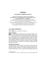
CtFig.1. An example <strong>of</strong> the amplification plots <strong>of</strong> the two primer sets, showing the plot <strong>of</strong> eachtarget amplicon.ResultsSensibility <strong>of</strong> the primers <strong>and</strong> TaqMan probes has been tested through amplifying genomic DNA<strong>of</strong> the male <strong>and</strong> female positive controls (<strong>bovine</strong> <strong>and</strong> <strong>buffalo</strong>) as 10 fold serial dilution (from 10to 0.001 ng/µl). For <strong>bovine</strong> male control, the Ct <strong>of</strong> Y primers ranged from 33.83 to 41.34, while,<strong>for</strong> BOV primers, Ct ranged from 16.79 to 27.00 <strong>and</strong> from 16.46 to 28.33 <strong>for</strong> male <strong>and</strong> female<strong>bovine</strong> control respectively. Ct values <strong>of</strong> <strong>buffalo</strong> male control ranged from 22.31 to 37.53 <strong>for</strong> Yprimers, while <strong>for</strong> BOV primers ranged from 21.11 to 35.49 <strong>and</strong> from 27.75 to 33.23 <strong>for</strong> male<strong>and</strong> female <strong>buffalo</strong> control respectively. A total number <strong>of</strong> 71 blastomeres, originating from 14<strong>bovine</strong> morulae , were analysed. A number <strong>of</strong> 57 blastomeres were analysed using real time PCRassay (one to four replicates from each embryo) <strong>and</strong> 14 blastomeres (one replicate from each <strong>of</strong>the 14 morulae) were analysed by classical PCR. After real time PCR analysis, 5 morulae weredetected as males <strong>and</strong> 9 morulae were detected as females. The analisys <strong>of</strong> the 14 blastomeres byclassical PCR, confirmed the 9 females <strong>and</strong> 4 males to be correctly <strong>sex</strong>ed, while <strong>for</strong> one morula(M 8) the signal was not the same as seen after real time PCR analysis. Overall, 56 out <strong>of</strong> 57<strong>bovine</strong> blastomeres were succesfully <strong>sex</strong>ed by real time PCR <strong>and</strong> the results were confirmed bythe classical PCR analysis <strong>of</strong> the same embryo replicate (98.24 % <strong>of</strong> correct <strong>sex</strong>). Results <strong>of</strong>blastomeres analysis by the real time PCR assay are shown in table 3.6
Table 3. Real time PCR <strong>sex</strong> <strong>determination</strong> results <strong>of</strong> <strong>bovine</strong> one to three embryo blastomeresNumber <strong>of</strong> cells Analysed Females Males Incorrect <strong>sex</strong>1 blastomere 45 30 15 02 blastomeres 9 6 3 13 blastomeres 3 3 0 0Total 57 39 18For <strong>buffalo</strong>, a number <strong>of</strong> 5 blastocysts <strong>and</strong> 23 oocytes (5 single <strong>and</strong> 18 double) were analysed in amultiplex real time PCR. Three out <strong>of</strong> 5 blastocysts were detected as males <strong>and</strong> two blastocystsas females. The analysis <strong>of</strong> the oocytes has shown a correct normal female (XX) amplificationplot <strong>and</strong> no amplification <strong>of</strong> the Y-specific primers was reported in any sample.DiscussionThe connection between the scientific research <strong>and</strong> the economical needs <strong>of</strong> the society remainsan important basic target. In particular, our study supports potentially some <strong>of</strong> the needs <strong>of</strong> theanimal breeding market disciplines <strong>and</strong> can help in drawing specific strategies <strong>for</strong> a dairy or beeflivestock. Over recent years, different <strong>method</strong>s have been proposed <strong>for</strong> <strong>sex</strong> <strong>determination</strong> <strong>of</strong>embryonic blastomeres, but most <strong>of</strong> these <strong>method</strong>s are multi-step, lacking the full accuracy,sensitivity <strong>and</strong> may require a technical skill. In this study, we presented a <strong>rapid</strong> sensitive <strong>method</strong>in one step by real time PCR <strong>for</strong> <strong>sex</strong> <strong>determination</strong> <strong>of</strong> <strong>bovine</strong> embryo blastomeres <strong>and</strong> <strong>buffalo</strong>preimplantation <strong>embryos</strong>. Real time PCR possesses the advantages <strong>of</strong> being sensitive throughdetection <strong>of</strong> the amplification in a real time after release <strong>of</strong> the fluorescence from the specificprobes, <strong>and</strong> because there is no need <strong>for</strong> post-PCR electrophoresis the contamination risk isminimised. Beside this, the reaction was conducted in one step without needing duplicate PCRprocedures or PEP-PCR reaction. Moreover, the reaction could be done in 96-well plate thusallowing the analysis <strong>of</strong> a large number <strong>of</strong> samples within few hours <strong>and</strong> this strongly supportsthe daily need <strong>of</strong> ET industry. Male <strong>and</strong> female genomic DNA (<strong>bovine</strong> <strong>and</strong> <strong>buffalo</strong>) were used aspositive controls (10 fold serial dilution) in order to test the sensibility <strong>of</strong> the assay at lower targetconcentrations. It is obvious that each new primer pair <strong>and</strong> probe combination needs a series <strong>of</strong>careful optimisation steps. For <strong>bovine</strong> blastomeres as well as the controls, the two primes wereused separately due to the unsuccessful amplification when they are used in a multiplex.Moreover, Ct detection <strong>for</strong> <strong>bovine</strong> DNA required a large number <strong>of</strong> amplification cycles <strong>for</strong> theY-specific primers at the lowest concentration (Ct 41.34) that might require an improvement <strong>of</strong>7
the fluorogenic probe design in the future throughout changing its region on the Y-specific locusor a careful optimisation. Sex <strong>determination</strong> was successful in 56 out <strong>of</strong> 57 <strong>bovine</strong> blastomeresdemonstrating an accuracy <strong>of</strong> more then 98% with the same results obtained by the classical PCR<strong>for</strong> the same embryo replicates. Real time PCR has reflected a high efficiency degree in <strong>sex</strong>ing <strong>of</strong>blastomeres as little as a single copy embryonic cell in one step. For <strong>buffalo</strong>, this preliminarystudy shows <strong>for</strong> the first time the usefulness <strong>and</strong> the efficiency <strong>of</strong> a multiplex real time PCRassay <strong>for</strong> <strong>sex</strong>ing in one step. A preliminary <strong>sex</strong>ing <strong>of</strong> some <strong>buffalo</strong> blastocysts as well as <strong>buffalo</strong>oocytes that representing single copy female gametes (XX) was per<strong>for</strong>med successfully. Ct value<strong>of</strong> Y primers in <strong>buffalo</strong> DNA positive controls was earlier than <strong>bovine</strong> DNA at the lowestconcentration (Ct 37.53) that may indicate the more sensitivity <strong>of</strong> the probe to the Y locus <strong>of</strong><strong>buffalo</strong> DNA. Our preliminary <strong>sex</strong>ing <strong>of</strong> <strong>buffalo</strong> blastocysts as well as single copy <strong>buffalo</strong>oocytes will open the gate <strong>for</strong> a future novel <strong>sex</strong>ing assay <strong>of</strong> single <strong>buffalo</strong> blastomeres. In thisstudy, we developed a novel, <strong>rapid</strong> <strong>and</strong> sensitive <strong>method</strong> <strong>for</strong> embryo <strong>sex</strong>ing as a single copyblastomere by using real time PCR. This will provide a powerful tool <strong>for</strong> embryo <strong>sex</strong>ing in theanimal breeding market <strong>and</strong> should be supported by other future studies to enhance <strong>and</strong> toimprove the <strong>sex</strong>ing <strong>and</strong> the genetic efficiency <strong>of</strong> some economically <strong>and</strong> agriculturally importantspecies.References1.2.3.4.5.6.7.8.9.Bavister, B. D. Early history <strong>of</strong> in vitro fertilisation. Reprod. 2002, 124, 181-196.Bondioli, K. R., Ellis, S. B., Pryor, J. H., Williams, M. E., Harpold, M. M. The use <strong>of</strong> malespecificchromosomal DNA fragments to determine the <strong>sex</strong> <strong>of</strong> <strong>bovine</strong> preimplantation <strong>embryos</strong>.Theriog. 1989, 31, 95-104.Colleau, J. J. Using embryo <strong>sex</strong>ing within closed mixed multiple ovulation <strong>and</strong> embryo transferschemes <strong>for</strong> selection on dairy cattle. J Dairy Sci. 1991, 74, 3973-3984.Faber, D. C., Molina, J. A., Ohlrichs, C. L., V<strong>and</strong>er Zwaag, D. F., Ferre, L.B. Commercialization<strong>of</strong> animal biotechnology. Theriog. 2003, 59, 125-138.Gasparrini, B. In vitro embryo production in <strong>buffalo</strong> species: state <strong>of</strong> the art. Theriog. 2002, 57,237-256.Hassun, P. A., Mello, M. R. B., Porto, L. P. C., Garcia, J. F. Bovine embryo <strong>sex</strong>ing by primerextension preamplification polymerase chain reaction (PEP-PCR). Theriog. 1999, 51, 398Herr, C. M., Reed, K. C. Micromanipulation <strong>of</strong> <strong>bovine</strong> <strong>embryos</strong> <strong>for</strong> <strong>sex</strong> <strong>determination</strong>. Theriog.1991, 35, 45-54.Hirayama, H., Kageyama, S., Moriyasu, S., Sawai, K., et al. A. Rapid <strong>sex</strong>ing <strong>of</strong> <strong>bovine</strong>preimplantation <strong>embryos</strong> using loop-mediated isothermal amplification: Theriog. 2004, 62, 887-896.Kirkpatrick, B. W., Monson, R. L. Sensitive <strong>sex</strong> <strong>determination</strong> assay applicable to <strong>bovine</strong><strong>embryos</strong> derivedfrom IVM <strong>and</strong> IVF. J Reprod. Fertil. 1993, 98, 355-340.8
10.11.12.13.14.15.16.Kitiyanant, Y., Saikhun, J., Siriaroonrat, B., Pavasuthipaisit., K. Sex <strong>determination</strong> by polymerasechain reaction <strong>and</strong> karyotyping <strong>of</strong> <strong>bovine</strong> <strong>embryos</strong> at first cleavage in vitro. ScienceAsia 2000, 26,9-13.Manna, L., Neglia, G., Marino, M., Gasparrini, B., et al. Sex <strong>determination</strong> <strong>of</strong> <strong>buffalo</strong> <strong>embryos</strong>(Bubalus bubalis) by polymerase chain reaction. Zygote 2003, 11, 17-22.Monk, M., H<strong>and</strong>yside, A. H. Sexing <strong>of</strong> preimplantation mouse <strong>embryos</strong> by measurement <strong>of</strong> X-linked gene dosage in a single blastomere. J Reprod. Fertil. 1988, 82, 365-368.Peura, T., Hyttinen, J. M., Turunen, M., Janne, J. A reliable <strong>sex</strong> <strong>determination</strong> assay <strong>for</strong> <strong>bovine</strong>preimplantation <strong>embryos</strong> using the polymerase chain reaction. Theriog. 1991, 35, 547-555.Suzuki, T., Singla, S. K., Sujata, J., Madan, M. L. In vitro fertilization <strong>of</strong> water <strong>buffalo</strong> follicularoocytes <strong>and</strong> their ability to cleave in vitro. Theriog. 1992, 38, 1187-1194.Utsumi, K., Iritani, A. Embryo <strong>sex</strong>ing by male specific antibody <strong>and</strong> by PCR using male specific(SRY) primer. Molec. Reprod. Develop. 1993, 36, 238-241.Virta, J., Markola, J., Peippo, J., Markkula, M., Vilkki, J. Sex <strong>determination</strong> <strong>of</strong> <strong>bovine</strong> embryoblastomeres by fluorogenic probes. Theriog. 2002, 57, 2229-2236.9