
You also want an ePaper? Increase the reach of your titles
YUMPU automatically turns print PDFs into web optimized ePapers that Google loves.
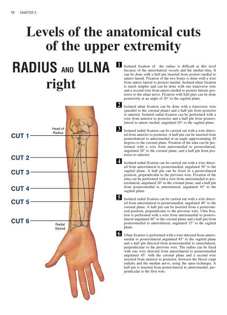
11 CHAPTER 2Levels of the anatomical cutsof the upper extremity<strong>RADIUS</strong> AND <strong>ULNA</strong>rightCUT 1CUT 2CUT 3CUT 4CUT 5CUT 6Head ofRadiusRadialStyloid123456Isolated fixation of the radius is difficult at this levelbecause of the anterolateral vessels <strong>and</strong> the medial ulna. Itcan be done with a half pin inserted from postero medial toantero lateral. Fixation of the two bones is done with a wirefrom antero lateral to postero medial. Isolated ulnar fixationis much simpler <strong>and</strong> can be done with one transverse wire<strong>and</strong> a second wire from antero medial to postero lateral, posteriorto the ulnar nerve. Fixation with half pins can be doneposteriorly at an angle of 20° to the sagittal plane.Isolated ulnar fixation can be done with a transverse wire(parallel to the coronal plane) <strong>and</strong> a half pin from posteriorto anterior. Isolated radial fixation can be performed with awire from anterior to posterior <strong>and</strong> a half pin from posterolateralto antero medial, angulated 20° to the sagittal plane.Isolated radial fixation can be carried out with a wire directedfrom anterior to posterior. A half pin can be inserted fromposterolateral to anteromedial at an angle approximating 20degrees to the coronal plane. Fixation of the ulna can be performedwith a wire from anteromedial to posterolateral,angulated 20° to the coronal plane, <strong>and</strong> a half pin from posteriorto anterior.Isolated radial fixation can be carried out with a wire directedfrom anterolateral to posteromedial, angulated 30° to thesagittal plane. A half pin can be fixed in a posterolateralposition, perpendicular to the previous wire. Fixation of theulna can be performed with a wire from anteromedial to posterolateral,angulated 20° to the coronal plane, <strong>and</strong> a half pinfrom posteromedial to anterolateral, angulated 10° to thesagittal plane.Isolated radial fixation can be carried out with a wire directedfrom anterolateral to posteromedial, angulated 40° to thecoronal plane. A half pin can be inserted from a posterolateralposition, perpendicular to the previous wire. Ulna fixationis performed with a wire from anteromedial to posterolateralangulated 40° to the coronal plane <strong>and</strong> a half pin fromposteromedial to anterolateral, angulated 15° to the sagittalplane.Ulnar fixation is performed with a wire directed from anteromedialto posterolateral angulated 45° to the sagittal plane<strong>and</strong> a half pin directed from posteromedial to anterolateral,perpendicular to the previous wire. The radius can be fixedwith one wire directed from anterolateral to posteromedialangulated 45° with the coronal plane <strong>and</strong> a second wireinserted from anterior to posterior, between the flexor carpiradialis <strong>and</strong> the median nerve, using the open technique. Ahalf pin is inserted from posterolateral to anteromedial, perpendicularto the first wire.
LEVELS OF THE ANATOMICAL CUTS OF THE UPPER EXTREMITY - <strong>RADIUS</strong> <strong>and</strong> <strong>ULNA</strong>12INSERTION WIRES AND HALF-PINS - RIGHTANTERIORCUT 1 CUT 4ANTERIORCUT 2ANTERIORCUT 5ANTERIORCUT 3ANTERIORCUT 6ANTERIOR
13 CHAPTER 2Levels of the anatomical cutsof the upper extremity<strong>RADIUS</strong> AND <strong>ULNA</strong><strong>left</strong>Head ofRadiusRadialStyloidCUT 1CUT 2CUT 3CUT 4CUT 5CUT 6123456Isolated fixation of the radius is difficult at this levelbecause of the anterolateral vessels <strong>and</strong> the medial ulna. Itcan be done with a half pin inserted from postero medial toantero lateral. Fixation of the two bones is done with a wirefrom antero lateral to postero medial. Isolated ulnar fixationis much simpler <strong>and</strong> can be done with one transverse wire<strong>and</strong> a second wire from antero medial to postero lateral, posteriorto the ulnar nerve. Fixation with half pins can be doneposteriorly at an angle of 20° to the sagittal plane.Isolated ulnar fixation can be done with a transverse wire(parallel to the coronal plane) <strong>and</strong> a half pin from posteriorto anterior. Isolated radial fixation can be performed with awire from anterior to posterior <strong>and</strong> a half pin from posterolateralto antero medial, angulated 20° to the sagittal plane.Isolated radial fixation can be carried out with a wire directedfrom anterior to posterior. A half pin can be inserted fromposterolateral to anteromedial at an angle approximating 20degrees to the coronal plane. Fixation of the ulna can be performedwith a wire from anteromedial to posterolateral,angulated 20° to the coronal plane, <strong>and</strong> a half pin from posteriorto anterior.Isolated radial fixation can be carried out with a wire directedfrom anterolateral to posteromedial, angulated 30° to thesagittal plane. A half pin can be fixed in a posterolateralposition, perpendicular to the previous wire. Fixation of theulna can be performed with a wire from anteromedial to posterolateral,angulated 20° to the coronal plane, <strong>and</strong> a half pinfrom posteromedial to anterolateral, angulated 10° to thesagittal plane.Isolated radial fixation can be carried out with a wire directedfrom anterolateral to posteromedial, angulated 40° to thecoronal plane. A half pin can be inserted from a posterolateralposition, perpendicular to the previous wire. Ulna fixationis performed with a wire from anteromedial to posterolateralangulated 40° to the coronal plane <strong>and</strong> a half pin fromposteromedial to anterolateral, angulated 15° to the sagittalplane.Ulnar fixation is performed with a wire directed from anteromedialto posterolateral angulated 45° to the sagittal plane<strong>and</strong> a half pin directed from posteromedial to anterolateral,perpendicular to the previous wire. The radius can be fixedwith one wire directed from anterolateral to posteromedialangulated 45° with the coronal plane <strong>and</strong> a second wireinserted from anterior to posterior, between the flexor carpiradialis <strong>and</strong> the median nerve, using the open technique. Ahalf pin is inserted from posterolateral to anteromedial, perpendicularto the first wire.
LEVELS OF THE ANATOMICAL CUTS OF THE UPPER EXTREMITY - <strong>RADIUS</strong> <strong>and</strong> <strong>ULNA</strong>14INSERTION WIRES AND HALF-PINS - LEFTANTERIORCUT 1ANTERIORCUT 4ANTERIORCUT 2ANTERIORCUT 5ANTERIORCUT 3ANTERIORCUT 6
15 CHAPTER 2<strong>RADIUS</strong> <strong>and</strong> <strong>ULNA</strong> <strong>left</strong>M. Pronator TeresM. BrachialisHUMERUS(TROCHLEA)M. Flexor Carpi UlnarisN. Ulnar<strong>ULNA</strong>ANTERIORCUT 1A.V. BrachialN. MedianBiceps TendonN. RadialM. Brachio Radialis<strong>RADIUS</strong>M. Extensor CarpiRadialis (Longus<strong>and</strong> Brevis)Common Extensor TendonM. AnconeusIn this cross section the ulnar N. runs medial to the ulna at thepoint of confluence of the flexor carpi ulnaris, the flexor digi-torum superficialis <strong>and</strong> the pronator teres. The brachial A. hasmoved laterally <strong>and</strong> now runs along side the median N.Head ofRadiusCUT 1ANTERIORRadialStyloidIsolated fixation of the radius is difficult at this level because of the anterolateral vessels <strong>and</strong> the medial ulna. It can be done witha half pin inserted from postero medial to antero lateral. Fixation of the two bones is done with a wire from antero lateral to posteromedial. Isolated ulnar fixation is much simpler <strong>and</strong> can be done with one transverse wire <strong>and</strong> a second wire from antero medialto postero lateral, posterior to the ulnar nerve. Fixation with half pins can be done posteriorly at an angle of 20° to the sagittalplane.
LEVELS OF THE ANATOMICAL CUTS OF THE UPPER EXTREMITY - <strong>RADIUS</strong> <strong>and</strong> <strong>ULNA</strong>16<strong>RADIUS</strong> <strong>and</strong> <strong>ULNA</strong> <strong>left</strong>M. Flexor Carpi RadialisM. Flexor Digitorum SublimusN. MedianA.V. UlnarN. UlnarM. Flexor Digitorum Profundus<strong>ULNA</strong>M. Flexor Carpi UlnarisANTERIORCUT 2A.V. RadialM. Brachii RadialisN. Radial (superficial)M. Pronator TeresM. Extensor CarpiRadialis (Long)<strong>RADIUS</strong>M. SupinatorM. Extensor Carpi RadialisN. Radial (prof.)M. Extensor DigitorumCommunisM. Extensor Carpi UlnarisThis cross sectional cut is performed distal to the flexor creaseof the elbow. Here bony l<strong>and</strong>marks are restricted to the subcutaneousborder of the ulna as the remainder of the forearm iscovered with muscle.The ulnar neurovascular bundle is positioneddirectly volar to the ulna between the flexor carpi ulnaris<strong>and</strong> the flexor profundus. The median N. is volar to the medialHead ofRadiusportion of the radius, <strong>and</strong> is covered by the flexor digitorumsublimus <strong>and</strong> flexor pollicis longus.The radial A. <strong>and</strong> N. are situated between the flexor carpi radialis<strong>and</strong> the brachioradialis. The lateral cutaneous nerve of theforearm can be found in the subcutaneous plane along theanterolateral portion of the forearm.The ulnar N. runs volar to the ulna at the point of confluence ofthe flexor carpi ulnaris, the superficial flexors <strong>and</strong> the deepflexors. The posterior interosseous N. <strong>and</strong> superficial radial N.run together with the radial A.V.ANTERIORCUT 2RadialStyloidIsolated ulnar fixation can be done with a transverse wire (parallel to the coronal plane) <strong>and</strong> a half pin from posterior to anterior.Isolated radial fixation can be performed with a wire from anterior to posterior <strong>and</strong> a half pin from posterolateral to antero medial,angulated 20° to the sagittal plane.
17 CHAPTER 2<strong>RADIUS</strong> <strong>and</strong> <strong>ULNA</strong> <strong>left</strong>M. Flexor Carpi RadialisM. Flexor Digitorum (superf.)A.V. UlnarN. UlnarM. Flexor Carpi Ulnaris<strong>ULNA</strong>M. Flexor Digitorum (prof.)ANTERIORCUT 3M. Brachii RadialisA.V. RadialN. Radial (superf.)M. Pronator TeresM. Extensor Carpi RadialisN. MedianM. Flex. Carpi Ulnaris<strong>RADIUS</strong>M. Extensor Carpi RadialisN. RadialM. Extensor Com. DigitorumM. Flex. Digiti Min.M. Extensor Carpi UlnarisIn this cross section each of the three major neurovascular elementshave assumed a deep position protected by the overlyingmuscles. The superficial radial N. <strong>and</strong> radial A. are volar <strong>and</strong>lateral beneath the brachioradialis. The median N. is volar <strong>and</strong>central between the superficial <strong>and</strong> deep flexors of the fingers.The ulnar A.V. <strong>and</strong> N. remain covered by the flexor carpiulnaris. The anterior interosseous artery has maintained itsposition on the volar surface of the interosseous membrane.ANTERIORHead ofRadiusCUT 3RadialStyloidIsolated radial fixation can be carried out with a wire directed from anterior to posterior. A half pin can be inserted from posterolateralto anteromedial at an angle approximating 20 degrees to the coronal plane. Fixation of the ulna can be performed with awire from anteromedial to posterolateral, angulated 20° to the coronal plane, <strong>and</strong> a half pin from posterior to anterior.
LEVELS OF THE ANATOMICAL CUTS OF THE UPPER EXTREMITY - <strong>RADIUS</strong> <strong>and</strong> <strong>ULNA</strong>18<strong>RADIUS</strong> <strong>and</strong> <strong>ULNA</strong> <strong>left</strong>M. Flexor Carpi RadialisM. Flexor Digitorum (superf.)N. MedianA.V. UlnarN. UlnarM. Flexor Carpi UlnarisM. Flexor Digitorum (prof.)<strong>ULNA</strong>ANTERIORCUT 4A.V. RadialN. Radial Sup.M. Flex. Pollicis LongusM. Extensor Carpi Radialis<strong>RADIUS</strong>M. Extensor Long. CarpiM. Flex. Digiti Min.M. Extensor Carpis BrevisM. Extensor IndicisM. Extensor Comm. DigitorumM. Extensor Carpi UlnarisThis cross sectional cut is taken at the midpoint between theflexor creases of the elbow <strong>and</strong> wrist. This level represents theapex of radial bowing. The two bones are maximally separatedat this point. The three major neurovascular elements haveassumed a deep position protected by overlying muscles. Thesuperficial radial N. <strong>and</strong> radial A. are volar <strong>and</strong> lateral underneaththe brachioradialis. The median N. is volar <strong>and</strong> centralbetween the superficial <strong>and</strong> deep flexors of the fingers. Theulnar A.V. <strong>and</strong> N. remain under the cover of the flexor carpiulnaris. The anterior interosseous artery has maintained itsposition on the volar surface of the interosseous membrane.Head ofRadiusANTERIORCUT 4RadialStyloidIsolated radial fixation can be carried out with a wire directed from anterolateral to posteromedial, angulated 30° to the sagittalplane. A half pin can be fixed in a posterolateral position, perpendicular to the previous wire. Fixation of the ulna can be performedwith a wire from anteromedial to posterolateral, angulated 20° to the coronal plane, <strong>and</strong> a half pin from posteromedial to anterolateral,angulated 10° to the sagittal plane.
19 CHAPTER 2<strong>RADIUS</strong> <strong>and</strong> <strong>ULNA</strong> <strong>left</strong>CUT 5M. Flexor Digitorum sublimusM. Pronator QuadratusA.V. UlnarN. UlnarM. Flexor Carpi UlnarisM. Flexor Digitorum profundus<strong>ULNA</strong>N. MedianANTERIORA.V. RadialN. Radial sup.M. Flexor Carpi RadialisM. Extensor Carpi RadialisLongus <strong>and</strong> Brevis<strong>RADIUS</strong>M. Abductor PollicisM. Extensor Carpi BrevisM. Extensor IndiciM. Extensor Comm. DigitorumM. Extensor Digiti MinimiIn this cross sectional cut the superficial radial N. lies subcutaneouslyover the abductor pollicis longus. The radial A. liessuperficially <strong>and</strong> can be found slightly lateral to the flexor carpiradialis.The median N. is on the medial side of the radial wrist flexor,between it <strong>and</strong> the flexor digitorum sublimus. The ulnar A.V.<strong>and</strong> N. are volar <strong>and</strong> medial under the increasing mass of theflexor carpi ulnaris. The terminal branches of the anteriorinterosseous N. are deeply located on the ulnar border of theradius.Head ofRadiusANTERIORCUT 5RadialStyloidIsolated radial fixation can be carried out with a wire directed from anterolateral to posteromedial, angulated 40° to the coronalplane. A half pin can be inserted from a posterolateral position, perpendicular to the previous wire. Ulna fixation is performed witha wire from anteromedial to posterolateral angulated 40° to the coronal plane <strong>and</strong> a half pin from posteromedial to anterolateral,angulated 15° to the sagittal plane.
LEVELS OF THE ANATOMICAL CUTS OF THE UPPER EXTREMITY - <strong>RADIUS</strong> <strong>and</strong> <strong>ULNA</strong>20<strong>RADIUS</strong> <strong>and</strong> <strong>ULNA</strong> <strong>left</strong>ANTERIORM. Flexor Digitorum SuperficialisM. Flexor Digitorum ProfundusA.V. UlnarM. Flexor Carpi UlnarisN. UlnarM. Pronator Quadratus<strong>ULNA</strong>M. Pronator QuadratusM. Extensor Digiti MinimiCUT 6N. MedianA.V. RadialM. Flexor Pollicis Longus<strong>RADIUS</strong>N. Radial Sup.M. Extensor Carpi RadialisLongus <strong>and</strong> BrevisM. Extensor IndicisM. Longus Extensor CarpiM. Extensor Digitorum CommunisIn this cross sectional cut most of the arteries <strong>and</strong> tendons canbe accurately localized by palpation as they lie superficially.The ulnar A.V. lie volar <strong>and</strong> medial <strong>and</strong> are protected by theflexor carpi ulnaris T. The median N. is slightly more radial inposition, being situated between the flexor digitorum superficialis<strong>and</strong> the flexor carpi radialis. As in 30% of the normal population,this diagram shows no palmaris longus T. The radial A.is found between the flexor carpi radialis <strong>and</strong> the abductor pollicislongus.Head ofRadiusANTERIORRadialStyloidCUT 6Ulnar fixation is performed with a wire directed from anteromedial to posterolateral angulated 45° to the sagittal plane <strong>and</strong> a halfpin directed from posteromedial to anterolateral, perpendicular to the previous wire. The radius can be fixed with one wire directedfrom anterolateral to posteromedial angulated 45° with the coronal plane <strong>and</strong> a second wire inserted from anterior to posterior,between the flexor carpi radialis <strong>and</strong> the median nerve, using the open technique. A half pin is inserted from posterolateral toanteromedial, perpendicular to the first wire.