Create successful ePaper yourself
Turn your PDF publications into a flip-book with our unique Google optimized e-Paper software.
Scenario…. 45 year-old male Acute onset of progressive four limbs weakness Preceded by URI symptoms Associated with tingling sensation of limbs, Starting from legs with difficulty walking steadily progressed to upper limbs weakness within 10 days NE: intact cranial nerves, MP UE 4/4, LE 4/4,generalized hyporeflexia NCV/EMG: polyneuropathy, demyelinating type
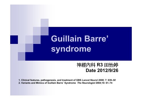
Introduction Before a disease of acute inflammatorydemyelinating polyneuropathy of peripheralnerve Now a term for a group of disease of acuteautoimmune polyneuropathy
Typical GBS: AIDPAntecedent illness before GBSURI and GI symptoms 為 主Days to weeks before disease onsetSuch as infection of Campylobacter jejuni bacteria,cytomegalovirus, Epstein-Barr virus, Mycoplasmapneumoniae, and Haemophilus influenzae.Autoimmune disease, pregnancy, child birth,vaccinationfever (52%), cough (48%), sore throat (39%),nasal discharge (30%), and diarrhea (27%)
Neuromuscular symptoms in 2 weeks after infectionevents Initially symmetric distal leg paresthesia astingling/crawling sensation, ascending, but limited Weakness begins at distal leg region and graduallyascending upward Decreasing or absence of DTR. Respiratory and cranial nerve were involved in about 50% patients Autonomic dysfunction with irregular HR or BP wasnoted in 50 % of patients About 10~30 % patients rapid progression andrespiratory, bulbar involved and need ETT insertion.
Diagnosis Clinical symptoms or sign Progressive symmetric limbs weakness withreduced or absence of DTR Could accompany with sensory impairment,autonomic nerve dysfunction remember the possibility of variant GBS CSF analysis normal WBC, RBC, but elevated protein:50~150mg/dl Albumino-cytological dissociation maybe normal in the first 2 weeks often normal in the first week, but increased in morethan 90% of the patients at the end of the second week
50~150mg/dlUp to 70%Cardiac arrhythmia, labile BP, orthostatic hypotension
NCV/EMGexamination….
Let’s take a look at NCV/EMG….
Sensory and Motor NCSLatency the time difference between the stimulus and the evokedresponse, measures conduction in the fastest fibersNerve conduction velocity (NCV) calculated by latency and measured distance between thestimulating and fixed recording pointsAmplitude reflect the number of fibers conducting the response.(Sensory nerve action potential, SNAP and Compound motoraction potential, CMAP)The shape of the response depends on the differences in velocity between the fastest andslowest fibers
Typical presentation Loss of F waves and H reflexes Temporal dispersion of the compound muscle actionpotential (CMAP) CMAP 變 的 complex and increasing duration, 因 每 條 神 經fiber 受 損 不 一 造 成 傳 遞 latency 不 同 Distal latency prolongation and conductionvelocity slowing more prominent later in the course, may persist for monthsto year Conduction block at any segment of axon = focal segmental demyelination--- NCV/EMG in the initial 2~4 weeks may be normal,need to repeat examination
Antiganglioside antibodyIn about half of patients with GBS, serum antibodies tovarious gangliosides have be found in human peripheralnerves, including LM1, GM1, GM1b, GM2, GD1a,GalNAc-GD1a, GD1b, GD2, GD3, GT1a, and GQ1bAntibodies to GM1, GM1b, GD1a, and GalNAc-GD1aare associated with the pure motor or axonal variants ofGBSAntibodies to GD3, GT1a, and GQ1b are related toophthalmoplegia and MFS
Clinical course a monophasic disease, not fluctuated course peak → in days to 4 weeks, there was a plateauof variant duration Always follow a slower phase of recovery Despite the effect of IVIg or PE treatment about 20% of severely affected patients remainunable to walk after 6 months Death uncommon may follow aspiration pneumonia, pulmonaryembolism, infection, autonomic dysfunction
GBS disability scale Scale 0~6--- scale 0: no symptoms--- scale 1: minor symptoms, capable of running--- scale 2: could walk without assistance, incapable ofrunning--- scale 3: need assistance to walk--- scale 4: wheel-chair bound or bed-ridden status--- scale 5: assisted-ventilation dependent status--- scale 6: death Severe GBS: scale > or = 3 Mild GBS: scale< or = 2 IVIG or PP treatment for severe GBS,bulbar/respiratory involved with pending failure,rapid progressive
Relapse Relapses with increased weakness occurin up to 10 % of patients with GBS. Relapses are usually treated with a partialor complete repeat course of the initialplasma exchange or IVIG treatment.
Atypical type of AIDP Asymmetric presentation Pure motor system involved Sensory impairment was predominantthan motor system Pure sensory or pure autonomicneuropathy Not decreasing or absence of DTRgeneral no decreasing or only some region
Regional variation of GBS Pharyngeal-Cervical-Brachial Paraparetic: only bilateral lower limbsinvolved
Miller fisher<strong>syndrome</strong>
Miller fisher <strong>syndrome</strong> A variant of GBS, account 5 % of GBS more in Asian, Taiwan and Japan: even 10~20 % Monophasic illness with subacute onset Most MFS are benign do not need plasmapheresis Characteristic of cranial nerve demyelination change Most patients had URI(major) or GI symptoms onemonth before MFS Triads of MFS: ophthalmoplegia, ataxia,areflexia.
Symptoms and sign Cranial nerve palsy Ptosis limited EOM, may frozen eye balls symmetric presentation and no diplopia Dilated pupil with sluggishly light reflex absence of doll eye sign dysarthria or nasal speech dysphagia bilateral peripheral type facial palsy absence or reduce of gap reflex
Symptoms and sign Motor of limbs general hypo-reflex or areflex muscle power of limbs are normal or only mildweakness Sensory impairment distal paresthesias of limbs Ataxia: unsure pathogenesis Sensory ataxia Cerebellar ataxia
Diagnosis NCV/EMGloss of SNAP amplitudes in the arm and leg,without significant abnormalities along motornerve trunks.other abnormalities have included loss offacial motor CMAP amplitudes andnonspecific abnormalities of the blinkreflexes
Anti-GQ1b IgG antibody The GQ1b antigenoculomotor nervessensory nervescerebellar neuronscell membranes of C. jejuni Maybe related to C. jejini and H. influenza Antibodies to the GQ1b ganglioside arepresent in the serum of about 95% ofpatients with the Fisher <strong>syndrome</strong>
Therapy for Miller Fisher <strong>syndrome</strong> IVIG, plasmapheresisNot improve clinical course and duration ofsymptoms disappearancemild fast the time of starting recovery
Acute MotorAxonalNeuropathy
Acute Motor AxonalNeuropathyA patient with a presentation similar to AIDP, with rapidlyprogressive weakness including cranio-bulbar andrespiratory function, but without sensory symptoms orsigns. NCV/EMG revealed axon disease loss of CMAP amplitude without demyelinating features normal SNAPs characterizeAntibodies to the gangliosides GD1a and GM1highly correlated with infection with C. jejuni.
Acute Motor andSensory AxonalNeuropathy
Acute Motor and Sensory AxonalNeuropathyA disease like AIDP with rapid progressive muscleweakness and sensory impairment, but poor prognosisand poor recoveryInitially there is difficult to different AMSAN from AIDPUntil NCV/EMG finding: feature of axon disease in motorand sensory neuron, but no feature of demyelination.Patients with AMSAN in general do not respond to IVIGor plasmapheresis.
The EndMade by NEUR R4 董 欣
QuestionsQ1:Which of the descriptions was wrong? (4)1)Patients with AMSAN in general do not respond to IVIG orplasmapheresis2)There was no need for further treatment of Miller Fisher <strong>syndrome</strong>3)The most common variant of GBS was acute inflammatorydemyelinating polyneuropathy4) Antibodies to the GM1 ganglioside are present in the serum of about95% of patients with the Fisher <strong>syndrome</strong>5)All of above was rightQ2: The triad of Miller Fisher <strong>syndrome</strong> described here, except for (3)1)ophthalmoplegia 2)ataxia 3)akinetic mutism 4)areflexia.
QuestionsQ3: Which of the manifestations about acute inflammatorydemyelinating polyneuropathy was wrong? (3)1) Initially symmetric distal leg paresthesia as tingling/crawlingsensation, ascending, but limited2) Weakness begins at distal leg region and gradually ascendingupward3) Increasing of DTR4) Respiratory and cranial nerve were involved in about 50 % patients5) Autonomic dysfunction with irregular HR or BP was noted in 50 % ofpatients
QuestionsQ4: Which one was the variant of regional presentation of GBS? (1)1) Pharyngeal-Cervical-Brachial2) Acute inflammatory demyelinating polyneuropathy3) Acute motor axonal neuropathy4) Acute motor-sensory axonal neuropathy5) Pure sensory neuropathyQ5: The CSF study of GBS list below was right, except for (5)1) normal WBC, RBC2) elevated protein: 50~150mg/dl3) Albumino-cytological dissociation4) often normal in the first week, but increased in more than 90% of thepatients at the end of the second week5) All of above was correct


