Evolution of Use of Special Stains - Dako
Evolution of Use of Special Stains - Dako
Evolution of Use of Special Stains - Dako
- No tags were found...

Create successful ePaper yourself
Turn your PDF publications into a flip-book with our unique Google optimized e-Paper software.
Figure 1. Masson Stained Skeletal Muscle andexample <strong>of</strong> a “general oversight” stain. Thisstain clearly demonstrates muscle, connectivetissue and nerve in contrasting colors.Figure 2. An example <strong>of</strong> a “special stain”,using Victoria blue in a protocol specific forpancreatic islet Beta cells.Connection 2010 | 47
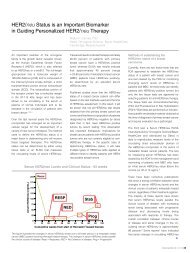
connective tissues including reticular fibers. <strong>Special</strong> stains for specifictissue components (mainly histochemical) are stains for iron, mucinsand glycogen, amyloid, and nucleic acids. IHC stains, which are also a“secondary” stain, just like all other special stains, are not considered“special stains” for reimbursement purposes, and are a separatecategory <strong>of</strong> diagnostic tests (their own unique reimbursement codes).IHC stains have replaced many traditional “special stains” simplybecause they have great specificity. Specificity means that the stain isexceptionally precise in the recognition <strong>of</strong> a specific target or epitope.A primary antibody, which provides the specificity for an IHC stain,can detect an epitope <strong>of</strong> three amino acids. In fact, a series <strong>of</strong> primaryantibodies can be manufactured which can detect the sequentialarrangement <strong>of</strong> three amino acids. While this degree <strong>of</strong> specificityis astonishing, it should be remembered that other special stainsmay also be exquisitely sensitive, for example, the iron stain actuallydetects ions <strong>of</strong> a single element. Not to be forgotten, the PeriodicAcid Schiff stain detects exceptionally small amounts <strong>of</strong> glycogenand mucopolysaccharides. The Feulgen reaction can detect DNAaccurately enough to detect the gain or loss <strong>of</strong> a single one <strong>of</strong> the largerchromosomes (the basis <strong>of</strong> Ploidy measurements).classical microscopic morphology skills may not be as strong as in yearspast. Thus the modern practice <strong>of</strong> pathology has come to depend oncertain <strong>of</strong> the special stains, and on IHC stains. Specimen submissionscontinue to increase, as would be expected in an aging populationwhich tends to live longer. It is quite clear the use <strong>of</strong> special stains andIHC stains will continue to increase, and remain a fundamental element<strong>of</strong> diagnostic pathology.A clear case can be made for increased use <strong>of</strong> “traditional” multicolorstain protocols in research. First, traditional stains provide excellentoverviews <strong>of</strong> specimens, and can easily define tissue organization. Inmany cases, histochemical stains can be combined with specimensurvey stains. As an example, a classic trichrome stain can be replacedwith a combination <strong>of</strong> histochemical stains which provides detailedinformation about the distribution <strong>of</strong> specific tissue constituents, as wellas an overview <strong>of</strong> tissue organization. One such stain combination isthe Blue Feulgen, PAS, Alcian Blue and Naphthol Yellow stain, whichdemonstrates DNA, Glycogen, Acidic muycopolysaccharides andgeneral protein distributions. The individual colors are easily seen, andthe stain is simple to perform.With continued advances in IHC staining (availability <strong>of</strong> new primaryantibodies), it might be expected that IHC staining could eventuallyreplace all traditional special stains. Since IHC staining is based onbiological molecular recognition, it is ill suited for identification <strong>of</strong>elemental inclusions, such as iron. Also, the various special stainsutilized for bacteria and fungi, when combined with clinical laboratorydata, provide sufficient information for diagnosis. While IHC tests canbe used for certain infectious agents, these tests are expensive, and arevulnerable to specimen processing as to reliability. Due to cost factorsalone, IHC staining will most likely not replace all special stains.Microscopic morphology <strong>of</strong> specimens was the driving force behind thedevelopment <strong>of</strong> special stains. For a number <strong>of</strong> reasons, morphology is<strong>of</strong> less concern in the diagnostic histopathology laboratory. There aremany factors which have brought this about, including the explosion <strong>of</strong>knowledge in diagnostic pathology, the increased number <strong>of</strong> specimenssubmitted, and changing emphasis in education <strong>of</strong> pathologists. Withsevere time constraints in medical curricula, microscopic morphologydoes not receive the same emphasis it once did. Pathology residenceprograms still occupy essentially the same amount <strong>of</strong> time as in yearspast, yet the total amount <strong>of</strong> diagnostic information has increased byseveral orders <strong>of</strong> magnitude. The result <strong>of</strong> these time pressures is thatContinuation <strong>of</strong> Change – Where <strong>Special</strong> <strong>Stains</strong> May be HeadedIt is clear that special stains including IHC stains will continue to be akey in accurate diagnosis <strong>of</strong> microscopic specimens. It should also beexpected that changing usage patterns will continue. As an example,consider the case <strong>of</strong> cell proliferation. It is quite simple to identify mitoticfigures in any stain, including H&E. However, in most normal tissues andearly stage tumors, cell division is tied to a circadian rhythm and occursin the time period between 1:00 AM to 6:00 AM. This is not the time whenmost specimens are collected. The introduction <strong>of</strong> a proliferation marker,KI-67 (IHC) made it possible to identify cells preparing for division,even when mitotic figures were not seen. This IHC test is both moreconvenient and gives a better indication <strong>of</strong> proliferation in specimens,and has essentially replaced purely morphological assessments.Another example is seen in breast carcinoma. In this case, it is easyto tell that a malignant process is occurring, using simple morphology.However, with the development <strong>of</strong> targeted drug therapy, it has becomeclear that there are different types <strong>of</strong> malignancies, even though theymay resemble each other based on morphology alone. Thus the use<strong>of</strong> prognostic markers, which are cellular characteristics identified byspecific IHC tests. In specimens positive for (expressing) estrogenreceptor, the patient becomes a candidate for Tamoxifin therapy. Inspecimens positive for Her-2-neu, the patient may respond to treatment48 | Connection 2010
Figure 3. Blue Feulgen, Periodic Acid Schiff,Alcian Blue and Naphthol Yellow S, a combinedhistochemical stain. Note the nuclei in blue,the cell coat stained with PAS, and the distinctgranules <strong>of</strong> Mast cells. The photomicrographon the left shows cross-sectioned smoothmuscle cells in the intestinal wall. The sectionbelow is a higher magnification view <strong>of</strong> crosssectionedblood vessel.Connection 2010 | 49
Procedure: Blue Feulgen, PAS, Naphthol YellowDemonstrates DNA, acid mucopolysaccharides,carbohydrates and proteins.1. Remove paraffin and hydrate slides to water2. Hydrolyze 1 hour in 5 N HCl at room temperature3. Rinse slides well in running water4. Stain 20 minutes in Cresyl violet Schiffwith Herceptin. Clearly, these are the beginning <strong>of</strong> significant changein microscopic diagnosis, and will certainly generate changes in theuse <strong>of</strong> special stains. As IHC stains can be much more specific thanthe empirical multi-color stains <strong>of</strong> the past, it would be expected thatover time, many <strong>of</strong> the general oversight stains, such as the trichromestains, will be <strong>of</strong> less use in routine diagnosis, although this may notbe so in research settings. As the examples given demonstrate, whena targeted therapy becomes available, based on a unique expression<strong>of</strong> the diseased cells or tissues, then targeted visualization tests willalso be required. However, there will always be a need for specialstains for bacteria, fungi, iron, and general tissue architecture.Cresyl Violet Schiff SolutionDissolve 0.02 grams Cresyl Violet Acetate in 1 Liter distilledwater as stock solution. Store in refrigerator.For use, add 3 grams sodium dithionite to 150 ml<strong>of</strong> stock solution. <strong>Use</strong> in covered staining dish.References1. Gomori, G. Observations with Differential <strong>Stains</strong> on Human Islets <strong>of</strong> Langerhans. Amer.J. Pathol. 1941; 17: 395-409.2. Feulgen, R., and H. Rossenbeck. Mikroskopisch-chemischer Nachweis einerNukleinsaure vom Typus Thymusnukleinsaure und die darauf beruhende elekitiveFarbung von Zellkernen in mikroskopischen Praparaten. Z. Physiol. Chem. 1924;135: 203-248.3. Preece, A. A Manual for Histologic Technicians. Little Brown and Company, Boston.1959.4. Code <strong>of</strong> Federal Regulations (CFR) Title 21, sec. 864.1860.5. Rinse well in running water6. Stain in Alcian Blue (0.1% in 0.01 NHCl, pH 2.0) for 10 minutes7. Differentiate in 0.01 N HCl for 2 - 3 minutes8. Wash in running water 1 - 2 minutes9. Hydrolyze 10 minutes in 0.5% periodic acid10. Wash 5 minutes in running water11. Stain 10 minutes in Standard Schiff (Basic Fuchsin Schiff)12. Wash 5 minutes in running water13. Stain 1 minute in Naphthol Yellow S(0.01% ion 1% acetic acid)14. Differentiate in two changes 1% aceticacid, two minutes each15. Dehydrate in tertiary butanol, 2 changes, 3 minutes each16. Clear in xylene and coverslipSteps 6, 7 and 8 may be omitted, if acidicmucopolysaccharides are not <strong>of</strong> interest. If thesesteps are omitted, then final dehydration canbe carried out in a standard ethanol series.50 | Connection 2010