Chapter 7 The Muscular System.pdf
Chapter 7 The Muscular System.pdf
Chapter 7 The Muscular System.pdf
- No tags were found...

You also want an ePaper? Increase the reach of your titles
YUMPU automatically turns print PDFs into web optimized ePapers that Google loves.
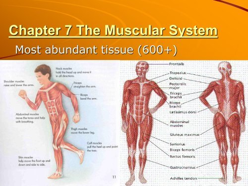
<strong>Chapter</strong> 7 <strong>The</strong> <strong>Muscular</strong> <strong>System</strong>Most abundant tissue (600+)
Sarcolemma:
C) Sarcomere: basic repeating myofibril units1) Z disk: anchors actin; divides sarcomeres2) I band: light zone: actin only; spans Z disk3) A band: dark zone; length of myosin (w/actin)4) H Zone: myosin only; middle5) M line: anchors myosin; center
3) Nerve/Muscle Membrane Potential(polarized): sends message for actionResting: + out (Na + , K + leak/- in (protns - )Depolarization: AP-charge reversd; Na + inRepolarization: K+ outNa/K pump returns resting potential
4) Nerve Supply‣Motor Neurons to MusclesMotor Motor Unit:– 1 neuron to all muscle fibers– Many units per muscle
‣ Neuromuscular Junction or synapse1. AP to presynaptic terminal (Ca ++ in)2. Acetylcholine (Ach): neurotransmitter crossessynaptic cleft (space) open Na + channels3. Depolarization (postsyn) muscle fiber sarcolemmaNa channel closedwithout AchNa channel openedwith Ach
Acetylcholinesterase: brkdwn AchMyasthenia Gravis: antibodies destroy Achreceptors (weakness)Curare binds ACH receptors–flaccid paralysis
5) Muscle ContractionSliding Filament Mechanism:– Myosin cross-bridges pull actin toward H zone
– H/I shorten/A unchanged
1. AP from nerve to muscle2. Ach released; Na channels open3. Na in; AP->sarcolemma->T tubls->s. reticlm4. S. reticulum releases Ca to actin troponin
5. W/Ca ++ Tropomyosin exposes binding sites
6. Myosin form cross-bridge w/actin (rigor)7. Power stroke moves actin w/ADP+P release8. Cross-bridge broken/head rests w/2 ND ATP• Rigor Mortis: No ATP avail for release w/death
9. Contraction continues w/Ca and ATP1. ACH released2. AP moves along membraneand T-tubules3. Ca released from SRvoltage gated Ca channel opens4. Ca binds to Troponin-Cconformation changes favortropomyosin opens actin sites5. myosin cross-bridges attachdetachfrom actins/pullsfilament toward M-line6. Ca removed (uptake by SR)7. tropomyosin blocks actin sitesrelaxation
6) All-or-none contraction: all fibers fullyMotor UnitAll the muscle fibers (cells) innervated by the same motor neuronAll-or-None Principle: All muscle fibers that make up a motor unit contractfully together. This principle holds true at the sarcomere, myofibril, andmuscle fiber levels as well. Partial contraction occurs at the muscle levelbecause all motor units are not contracting at the same time.Recruitment: <strong>The</strong> process by which the number of actively contractingmotor units is increased.
7) Summation: increased contraction forcew/↑ stimulation=tetanus: sustainedcontractn (no relax)Fused or Complete Tetanus: A sustained maximum contraction in whichindividual contractions are not discernable. In fused tetanus, multiplestimuli are occurring so fast, the muscle does not undergo any relaxation.
8) Recruitment:↑ units over time =smooth strength
9) Energy Requirements:• Aerobic Resp: Glucose + O 2 = ↑ ATP• Anaerobic Resp: Glucose = ATP + lactic acid– ATP↓/Creatine P↑: rapidly converted to ATP
Food sourceCreatine level;grams/poundHerring3.0/lbPork2.3/lbSalmon2.0/lbBeef2.0/lbTuna1.8/lbCod1.4/lbCranberries.0009/lb
10) Types of Muscle Contractions:a) Isometric: no shortg w/↑ tensn: posture
) Isotonic: constant tensn w/shortg: movemt• Concentric (tension ↑ w/shortening)• Eccentric (constant tensn w/lengthenin-lower wght)
11) Myofibril Types:a) Slow Twitch:Type I myosin = AerobicDark w/bld (myoglobin↑O 2 ): duck breast/chickn legb) Fast Twitch:Type II myosin = AnaerobicQuick contract/fatigue: chicken breast
c) HumansAve adult: equal I/IISprinters: 80% more II/legDistance: 95% I/legAthletes: balanced I/II# muscle cells somewhat constant after birthundifferentiatedSatellite cells =“new” w/injuryHypertrophy: cell size ↑w/exer- ↑ mito; myofilmtsRelative Distributions of Slow Twitch & Fact Twitch Myosin Isoforms (Type I & Type II)Type I (slow) Type II (fast) Type IIa Type IIxAverage person 50% 50% 40% 10%spinal injury 4% 96% 48% 48%sprinter 20% 80% 45% 35%couch potato 40% 60% 30% 30%marathoner 80% 20% 20% 0%<strong>The</strong>se myosin isoforms are conserved evolutionarily:Comparing myosin isoforms from different mammals reveals remarkably little variationspecies to species. Rat type I is more similar to human type I myosins, than it is to rat typeII's. Thus selective evolution has maintained a functional difference between type I's &type II's over eons of evolution.
II. Smooth Muscle:Unstriated, tapered, 1 nucleus, invol (dig)Slow twitchHormones regulate (autorhythmicity)
III. Cardiac Muscle:Striated, branched, 1 nucleus/cell, involSlow twitchHormones regulateIntercalated disks pass AP
Aging on Skeletal Muscle‣ Decreased muscle mass‣ Increased contraction response time‣ Decreased stamina‣ Increased recovery time‣ Affects of aging slowed/reversedw/exercise
Disorders: <strong>Muscular</strong> Dystrophy; DuchenneMD; Myotonic MD; Myasthenia Gravis;Tendinitis (p189)A patient with this disease producesautoantibodies to the acetylcholine receptorsMUSCLE WEAKNESS CAUSED BY MUSCULAR DYSTROPHYDuchenne muscular dystrophy isinherited in an X-linked recessive pattern
TypesSkeletal: 40% wt,voluntarySmooth: involuntaryCardiac: involuntary