Dr. G K Rath - Aroi.org
Dr. G K Rath - Aroi.org
Dr. G K Rath - Aroi.org
- No tags were found...

You also want an ePaper? Increase the reach of your titles
YUMPU automatically turns print PDFs into web optimized ePapers that Google loves.
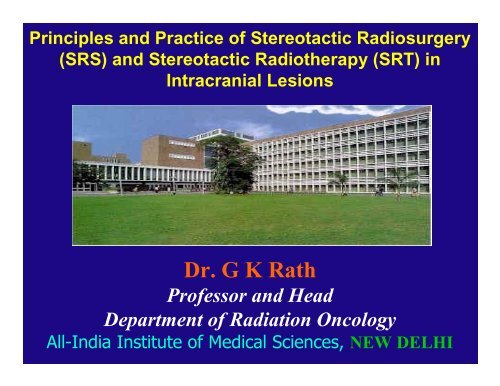
Principles and Practice of Stereotactic Radiosurgery(SRS) and Stereotactic Radiotherapy (SRT) inIntracranial Lesions<strong>Dr</strong>. G K <strong>Rath</strong>Professor and HeadDepartment of Radiation OncologyAll-India Institute of Medical Sciences, NEW DELHI
Stereotactic RadiotherapyThe delivery of multiple fractionated doses ofradiation to a definitive target volume sparingnormal structure (both intra as well as extracranial)
Stereotactic RadiosurgeryThe delivery of asingle, high dose ofirradiation to a smalland critically locatedintracranial volume,sparing normalstructure
Conventional and Stereotactic setup• Conventional– coplanar setup– large volumes– less no. of fields– target volume delineation– positional accuracy ± 5 mm– Optical field, SSD indicator– Marking on patient’s skin• Stereotactic– non-coplanar setup– small volumes– more no. of fields– precise delineation– positional accuracy ± 1 mm– Target volumes preciselydelineated– Margins not necessary– Normal cells within the targetnegligible
Co-planar .vs. Non-coplanar beams• tolerance of normal tissue depends upon both the doseand volume of the tissue irradiated• normal tissue irradiation can be minimized throughstereotactic definition of target and sharply focused,multiple, non-coplanar beamsA. Parallel opposed beamsB. Coplanar arcs (bilateral 100°)C. Non-coplanar beams
Advantages• Enhances clinical outcome• Improves quality of life• Time factor
Quality of Life• Minimally invasive• Less trauma• Faster recovery• Minimal hospitalization• Fewer complications• Documented efficacy
Clinical Outcome• Documented scientific datashows better or equal resultscompared with microsurgery• Fewer complications• Reproducible results• Treatment solution forinoperable patients• Combined treatment with microsurgery andendovascular techniques extend the capabilities
The Time FactorOpenSurgery2-4 daysICU10-16 dayshospitalization4-6 weeksconvalescenceSymptomDiagnosisGammaKnifeSurgery
Treatment techniques & units• charged particle beams - cyclotrons &synchrotrons• gamma ray photons - Gamma Knife & RGS• x-ray photons - modified & dedicated linacs (X-Knife), micro multi leaf collimator• neutrons have been used unsuccessfully
Gamma Knife … the machine• 201 cobalt-60 radioactive sources– cylindrical; 1 mm dia; 2 cm height; 30 Ci activity each• central ray from all the sources focused at a singlepoint, within an accuracy of ± 0.3 mm• dose distribution at this (sec. Collimator) point is in theform of a sphere• helmets can shape the diameter of this ‘radiationsphere’ to 4, 8, 14 and 18 mm• plugging to alter the shape of this sphere• minimal moving part (couch in & out)
Gamma Knife
The man behind the machine ….“The tools used by thesurgeon must be adaptedto the task and where thehuman brain is concernedthey cannot be toorefined.”Late Professor Lars Leksell
X-Knife
The Treatment Procedure• Frame Fixation• Diagnostic Imaging• Image transfer• Treatment planning• Treatment
Why frame?• helps us in defining the images in a coordinatesystem - fiducial points• accurate positioning of the patient at the time oftreatment– SRS frames• Leksell frame in Gamma Knife• BRW (Brown-Roberts-Wells) & CRW (Cosman-Roberts-Wells) inX-Knife– relocatable frames for fractionated stereotactic irradiation(SRT)• GTC (Gill-Thomas-Cosman)
Frame Fixation
Diagnostic ImagingCT scanMRIAngio
Image transfer• Networking– Local area networking (LAN)• PACS– to Gamma plan– to X-plan• Magneto Optical disc• DAC tape• Film scanner• CT, MR film
Treatment Planning• Fast creation of optimaltreatment plan• User- friendly softwarededicated for GammaKnife surgery
Treatment planning• contouring of target and critical structures on imageslices• target contours on image slices constitute an empty sacthree dimensionally• fill up this sac with ‘radiation spheres’ (shots)conformally• x,y,z coordinates of the shots are given in the print out• treatment time for every shot depends on the dose to bedelivered• position the patient for every shot and treat
Conformity of Dose to TargetWith multiple isocenterWith conventional beams
Years of Clinical Experience1968The first prototype of Leksell GammaKnife was installed in Stockholm,Sweden.1999Elekta refines the Art ofradiosurgeryby introducing Leksell Gamma KnifeC.
Treatment Indications• Tumors• Meningioma• Pituitary• Acoustic• Metastatic• Glioma• Vascular• AVM• Fuctional• Trigeminal Neuralgia• Research Areas• Movement Disorders• Intractable Pain• Epilepsy• Macular Degeneration• Uveal Melanoma
Arteriovenous MalformationPre GammaKnife Surgery2 years postGammaKnifeSurgery
AVMPre Dose Plan 13 MonthsPost
Acoustic NeuromaPre6 months post2 years post
MeningiomaDose plan with 6isocenters- minimizing dose to opticchiasm
MeningiomaPre2 years post
Pituitary AdenomaPre54 months post
MetastasisPre2 months post
MetastasisPre10 months post
AstrocytomaPre5 years post
Trigeminal NeuralgiaDose plan6 months post
Clinical SolutionsDoseRadio SurgeryStereotactic GuidedRadiotherapyConformalRadiotherapyConventionalRadiotherapyTargetvolume
SRT/SRS at AIIMS ...• Started on 27 th May, 1997• No. of patients treated till date: 16745%2%3%2% 1%1%12%AVMAcoustic24% 13%MeningiomaPituitaryOther BenignMetastasisGlial TumorOther Malignant37%
X-Knife … the machine• installed on a modified / dedicated linac– Floor Mounted System– Couch Mounted System• collimators are modified– tertiary collimators are added– circular; elliptical– mMLC (gradually replacing cone based radiosurgerysystem- better isodose shaping)
Linac treatment parametersTo be optimized to produce desired isodose.• isocenter location• collimator field size• couch angle• arc rotation interval• weightage of each arc• dose per isocenter
X-Knife planning steps• isocenter selection– place isocenter in target volume center– determine collimator size by coveringtarget volume• arc selection– Through BEV to avoid critical structures– Number of non-coplanar arcs (depends oncollimator size)– couch / gantry restrictions to be considered
QA for linac based SRSBefore every treatment• lasers are aligned to point to isocenter• lasers are then taken as the reference system forpositioning the patient– initial positioning– then, at every couch angle• Laser must fall on the reference line on the framepositional accuracy better than ± 1 mm
Treatment Indications• Cranial– Tumors• Pituitary• Meningioma• Craniopharyngioma• Acoustic• Metastatic• Glioma– Vascular• AVM• Extracranial– Primary lung tumors– Metastatic lung tumors– Liver metastases– Adrenal metastases
Gamma Knife vs. X-Knife• mechanical precision is far superior in Gamma Knife– Accuracy easily achievable (0.1mm- mechanical, 0.3mmintersectionof beams & 0.5mm overall)– only for cranial targets• stereotactic procedures are quite similar, except– CT is a MUST for X-Knife; not so in Gamma Knife– skull contouring (total imaging in X-knife; partial imaging inGamma Knife)
Stereotactic BodyframeStereotactic irradiation concept extended to extra-cranial sites also
In summary...• SRS & SRT are almost the ultimate form of ConformalTherapy (IMRT better for tumors encasing a normalstructure)• The dose distribution accuracy up to +/- 1mm (+/- 0.5 mmfor Gamma Knife) not achieved by any other modality• SRT preferred for targets adjacent to critical structures• Immobilization by frame is the key• SRT is achieved by re-locatable frames (non-invasive)• Both SRT & SRS can be performed with Linac(Only SRS possible with Gamma Knife)