Neven Olivari • Endocrine Ophthalmopathy
Neven Olivari • Endocrine Ophthalmopathy
Neven Olivari • Endocrine Ophthalmopathy

You also want an ePaper? Increase the reach of your titles
YUMPU automatically turns print PDFs into web optimized ePapers that Google loves.
<strong>Neven</strong> <strong>Olivari</strong> <strong>•</strong> <strong>Endocrine</strong> <strong>Ophthalmopathy</strong>
<strong>Endocrine</strong><br />
<strong>Ophthalmopathy</strong><br />
Surgical Treatment<br />
Transpalpebral Decompression by Removal<br />
of Intraorbital Fat<br />
by <strong>Neven</strong> <strong>Olivari</strong><br />
with contributions by<br />
E. Eder, D. Richter, G. Noever, G. Deutsch and B. Stark
Dr. med. <strong>Neven</strong> <strong>Olivari</strong><br />
Professor for Plastic Surgery, University of Cologne (Ret.),<br />
Chief, Department of Plastic Surgery (Ret.)<br />
Dreifaltigkeits Krankenhaus Wesseling<br />
Hohlenberg 26<br />
53332 Bornheim, Germany<br />
Photography: Inge Goldberg, Wesseling<br />
Drawings: <strong>Neven</strong> <strong>Olivari</strong><br />
Translation: Douglas Fear, MA, Heidelberg<br />
Language Editing: Brian D. G. Morgan, MB FRCS, London<br />
Die Deutsche Bibliothek – CIP-Einheitsaufnahme<br />
<strong>Olivari</strong>, <strong>Neven</strong> :<br />
<strong>Endocrine</strong> ophthalmopathy : surgical treatment : transpalpebral decompression by removal of intraorbital fat /<br />
by <strong>Neven</strong> <strong>Olivari</strong>. With contrib. by E. Eder… – Heidelberg : Kaden, 2001<br />
ISBN 3-922777-35-x<br />
© 2001 Kaden Verlag, Heidelberg, Germany<br />
Typesetting: Ch. Molter, Kaden Verlag, Heidelberg, Germany<br />
Printing: Druckhaus Darmstadt, Darmstadt, Germany<br />
Binding: Buchbinderei Schaumann, Darmstadt, Germany<br />
ISBN 3-922777-35-x<br />
This book is protected by copyright. Reprinting, translation, copying of illustrations, copying by means of photomechanical devices or<br />
similar, storage in data processing systems or on electronic data storage media, as well as provision of the content in the Internet or other<br />
systems of communication only with previous written permission from the publisher. Any infringement of these rights, even in the form<br />
of excerpts, is punishable by law.<br />
The advice and recommendations contained in this book have been given by the author and the publisher according to their best knowledge<br />
and have been carefully checked. Nonetheless, any and all responsibility on the part of the author or the publisher is expressly disclaimed.<br />
All liabilities for damage to persons, property or other are categorically excluded.<br />
Insofar as registered trademarks, trade names, and registered names are mentioned or used in this book, these are subject to the corresponding<br />
protective regulations, even when not expressly identified as such.
This book is dedicated to<br />
my wife Anna-Maria<br />
and my sons Alexander and Nicolas
Foreword<br />
<strong>Endocrine</strong> ophthalmopathy, or Graves disease, is a<br />
condition which, over the years, has repeatedly taxed<br />
the ingenuity of ophthalmic surgeons and a small<br />
group of plastic surgeons interested in this condition.<br />
Initially the operations were technically relatively<br />
minor but with the advent of craniofacial surgery more<br />
major procedures were developed and performed.<br />
Unfortunately, these led to only a minimal improvement<br />
in results and sometimes the complications, such<br />
as diplopia, were significant and frequent. The real<br />
problem in terms of treatment was the lack of an adequate<br />
analysis of the problem. This has now been<br />
greatly helped by more sophisticated imaging techniques<br />
such as MRI scanning together with the aid of<br />
three-dimensional CT scanning techniques. It has<br />
now become obvious that it is the increase in conal fat<br />
which seems to be the most significant factor in the<br />
causation of this distressing deformity.<br />
The method described by <strong>Neven</strong> <strong>Olivari</strong>, and so<br />
beautifully presented in this book, is logical.<br />
Unfortunately, when it is presented or demonstrated by<br />
<strong>Neven</strong> <strong>Olivari</strong>, it seems simple to execute. However, at<br />
that point we are watching or listening to a master surgeon<br />
with a huge amount of experience in all aspects<br />
of plastic surgery but particularly in the orbit, demon-<br />
strating a technique which he has pioneered and developed<br />
over many years. To many, the method and its<br />
execution has been something of a mystery. All has<br />
now been revealed in this very clear and well produced<br />
treatise. This is undoubtedly a giant step forward in the<br />
treatment of this condition which affects so many of<br />
our patients. The book must, however, be read<br />
extremely carefully and even after having done this I<br />
would advise for those who are inexperienced to practice<br />
on a cadaver, if possible, using the textbook like a<br />
dissection manual. Those who are experienced in<br />
orbital surgery, of course, will not require this but they<br />
must follow the step-by-step instructions provided by<br />
the author. In this way good results will be obtained<br />
and complications avoided.<br />
We must sincerely thank <strong>Neven</strong> <strong>Olivari</strong> for producing<br />
this publication. He has done a great service to<br />
orbital surgery and to the unfortunate patients who<br />
suffer from the problem of endocrine ophthalmopathy.<br />
I am sure that this will be instrumental in many<br />
patients retaining their sight who otherwise would<br />
not have done so. This may be the greatest complement<br />
that can be applied to this account of his lifelong<br />
study of this distressing problem.<br />
Ian T. Jackson<br />
MD, DSc (Hon), FRCS, FACS, FRACS (Hon)<br />
Institute for Craniofacial and Reconstructive Surgery<br />
Southfield, Michigan, USA
Foreword<br />
<strong>Endocrine</strong> orbitopathy represents one of the main<br />
challenges in the therapy of Graves’ disease, even<br />
today. Although medicational and definitive treatment<br />
of Graves’ hyperthyreosis by means of radioactive<br />
iodine or surgery is effective and can be carried out<br />
with a calculable risk, the benefit for the manifestation<br />
and long-term development of endocrine orbitopathy<br />
has remained uncalculable to a great extent. Improved<br />
diagnosis using ophthalmological examination, determination<br />
of the activity score, and also imaging procedures<br />
has brought progress in the estimation of illness<br />
during the last few years: the severe forms of endocrine<br />
orbitopathy, with their considerable physical and psychological<br />
impairments, remain a serious challenge<br />
nonetheless. The progress in surgical treatment of<br />
endocrine orbitopathy represent a milestone here. A<br />
particularly special aspect is the transpalpebral decompression<br />
by means of resection of the fatty body.<br />
Improved and optimized short- and long-term postoperative<br />
care in prospectively structured investiga-<br />
tions have decisively aided many patients. The cosmetic<br />
results, so important for women in their social<br />
surroundings, are surprisingly favourable. Professor<br />
<strong>Olivari</strong> here summarizes his many years’ experience in<br />
particular of surgical treatment of endocrine orbitopathy,<br />
and in so doing has created a standard work for all<br />
those interested in Graves’ disease. The superb didactic<br />
structure and the clear illustrations explain many<br />
connections much more quickly than ever so many<br />
words would do.<br />
I would like to congratulate the author and the publisher<br />
on the appearance of this extensive work, which<br />
can be recommended without reserve, especially to<br />
those colleagues working in this area.<br />
Prof. Dr. med. Klaus Mann<br />
Director of the Endocrinological Department<br />
University Clinic Essen
Preface<br />
The fate of patients suffering from <strong>Endocrine</strong> <strong>Ophthalmopathy</strong><br />
(EO) is not an easy one. The basic illness,<br />
that is immune hyperthyroidism, can be healed in 95%<br />
of cases by means of either surgery (thyreoidectomy),<br />
thyreostatic medication, or radioactive iodine therapy.<br />
The success of therapy for EO is modest, however.<br />
Conservative therapy in the acute stages improves the<br />
changes in the soft tissue area of the lids, but has<br />
hardly any influence upon the protrusion of the eyeball,<br />
visual acuity, and optic motility. According to<br />
optimistic estimates, exophthalmos reduces spontaneously<br />
in about 10% of cases. The rate of remission is<br />
probably around 5%, but only in the first 6–12 months.<br />
After two years at the latest, the protrusion is stable<br />
and cannot be influenced by medication.<br />
EO is a severe blow for many of the patients. They<br />
are affected not only by reduced visual acuity, double<br />
vision, and other typical symptoms of EO, but also the<br />
protruding eyeballs, often seems to change the facial<br />
expression quite dramatically. The eyes are the centre<br />
of the face. Exophthalmos changes the entire face,<br />
indeed, frequently the entire personality. In a later<br />
phase, patients show a certain helplessness. Once<br />
attractive, outgoing persons are shy and unsure of<br />
themselves. Such a patient develops inferiority complexes<br />
and depressions; social contacts are drastically<br />
reduced. The patient visits his or her endocrinologist,<br />
ophthalmologist, and nuclear medicine specialist<br />
regularly and is constantly asking whether an operation<br />
would help.<br />
The answer of the physicians is objective. Bony<br />
operative decompression is indicated only in cases of<br />
threatening loss of vision, but not in cases of protrusion<br />
without visual problems, or diplopia and certainly not<br />
in cases of aesthetic problems. The operation is complex<br />
and there are many complications. As only about<br />
3–5% of patients have problems with reduced visual<br />
acuity, more than 95% of patients are doomed to live<br />
with exophthalmos.<br />
There are few non-malignant illnesses with a lower<br />
rate of successfull treatment than EO. Probably some<br />
160,000 patients live with this problem in Germany<br />
alone.<br />
In the summer of 1985, a friend who is an internist<br />
and endocrinologist, Dr. Mies from Cologne, asked me<br />
whether I could help his wife, who was suffering from<br />
an endocrine ophthalmopathy. Although at that time I<br />
already had some 20 years’ experience in lid surgery, I<br />
replied that I had never carried out an bony decom-<br />
pression, and I recommended a clinic to him in which<br />
such operations were performed. Dr. Mies was of the<br />
opinion that he had done his homework, and refused an<br />
ossary decompression for his wife. However, he<br />
requested that I treat his wife from the aesthetic<br />
aspects, as she suffered greatly with her appearance.<br />
It was clear to me that the enormous swelling of the<br />
eyelids could be corrected by removing fatty tissue. I<br />
performed a classic plastic surgical blepharoplasty.<br />
Upon opening the orbital septum, a large quantity of<br />
fat prolapsed outward under pressure. I removed as<br />
much as possible from the upper and lower lids, but<br />
also from the deeper regions of the orbits.<br />
To my surprise, not only did the patient’s aesthetic<br />
situation improve, but the Hertel values were reduced.<br />
The well-known nuclear medicine specialist Dr.<br />
Mödder, also from Cologne, heard of this operation<br />
and asked me to operate on some of his many patients<br />
suffering from EO, who had undergone conservative<br />
treatment for years without benefit. He himself has<br />
written numerous books and many papers on thyroid<br />
illnesses.<br />
I let myself be introduced to the complexities of<br />
EO by him, in the course of long, friendly discussions.<br />
After studying the literature concerning EO, anatomy,<br />
and the operative procedures hitherto applied, I decided<br />
to perform individual operations.<br />
During the years 1985–87, a period of three years,<br />
only 8 patients and 15 orbits were operated on. All of<br />
these patients had undergone conservative treatment<br />
for a period of years with no success. All 15 operations<br />
were successful.<br />
From the beginning, the following examinations<br />
were essential prior to advising an operation:<br />
1. Metabolic condition, the patient must be euthyroid<br />
2. Ophthalmologic examination<br />
3. CT or MRI of the orbits to determine the degree of<br />
involvement of the extraocular muscles.<br />
From the second patient onward the volume of fat<br />
removed was measured exactly and all data were<br />
stored. The patients were examined postoperatively<br />
by us as well as by ophthalmologists and endocrinologists<br />
or nuclear medicine specialists.<br />
Orbits were operated on in several cadavers, in<br />
order to attain more accuracy in operating.<br />
I would like to thank Dr. Mies and Dr. Mödder for<br />
moral support. I would also like to express my<br />
gratitude to Dr. Neubauer, the recognized ophthalmo-
logic surgeon from Cologne, for his support. I presented<br />
my results to him at the start of 1991 and he<br />
found the method to be worthy of mention in the book<br />
“Surgical Ophthalmology”, of which he was co-author.<br />
I performed the first one hundred orbital operations<br />
by myself; thereafter I delegated the operation<br />
more and more to my colleagues.<br />
The first short publication was 1988 [106]. The<br />
results following 147 operations were published in<br />
Plastic and Reconstructive Surgery [107].<br />
For this publication I received the “Award for the<br />
Best Clinical Paper 1991” from the American<br />
Association of Maxillofacial Surgeons.<br />
I would like particularly to thank my photographer of<br />
many years’ collaboration, Mrs. Inge Goldberg, for<br />
the excellent photographs of operations and patients,<br />
as well as my colleagues E. Eder, D. Richter, G.<br />
Noever, B. Stark, and G. Deutsch for their clinical and<br />
statistical support during the work on this book.<br />
My wife Anna-Maria patiently kept writing new,<br />
corrected versions on the computer.<br />
I was able to find the necessary peace and quiet<br />
needed for the final version of the book on the balcony<br />
of the Club Coronado, belonging to the Schütte family<br />
on the island of Fuerteventura.<br />
I would like to express my sincere gratitude to Dr.<br />
Brian Morgan, who reviewed the text with care and<br />
friendship to make this a well-edited book.<br />
Prof. Dr. med. <strong>Neven</strong> <strong>Olivari</strong>
Contents<br />
Forewords. . . . . . . . . . . . . . . . . . . . . . . . . . . . . . . . . . . . . . . . . . . . . . . . . . . . . . . . . . . . . . . . . . . . . vii<br />
Preface . . . . . . . . . . . . . . . . . . . . . . . . . . . . . . . . . . . . . . . . . . . . . . . . . . . . . . . . . . . . . . . . . . . . . . . . ix<br />
1. The History of <strong>Endocrine</strong> <strong>Ophthalmopathy</strong> . . . . . . . . . . . . . . . . . . . . . . . . . . . . . . . . . . 1<br />
2. Anatomy . . . . . . . . . . . . . . . . . . . . . . . . . . . . . . . . . . . . . . . . . . . . . . . . . . . . . . . . . . . . . . . . . . . . 3<br />
2.1 The Structure of the Bony Orbits . . . . . . . . . . . . . . . . . . . . . . . . . . . . . . . . . . . . . . . . . . . . . . . 4<br />
2.2 The Extraocular Muscles. . . . . . . . . . . . . . . . . . . . . . . . . . . . . . . . . . . . . . . . . . . . . . . . . . . . . . 4<br />
2.3 The Innervation of the Extraocular Muscles. . . . . . . . . . . . . . . . . . . . . . . . . . . . . . . . . . . . . . . 7<br />
2.4 Vascular Supply of the Orbits . . . . . . . . . . . . . . . . . . . . . . . . . . . . . . . . . . . . . . . . . . . . . . . . . . 7<br />
2.5 The Lacrimal Gland . . . . . . . . . . . . . . . . . . . . . . . . . . . . . . . . . . . . . . . . . . . . . . . . . . . . . . . . . 8<br />
2.6 Intraorbital Fat and Connective Tissue. . . . . . . . . . . . . . . . . . . . . . . . . . . . . . . . . . . . . . . . . . . 8<br />
2.7 Optic Nerve. . . . . . . . . . . . . . . . . . . . . . . . . . . . . . . . . . . . . . . . . . . . . . . . . . . . . . . . . . . . . . . . 9<br />
3. Classification, Diagnosis, and Differential Diagnosis<br />
of <strong>Endocrine</strong> <strong>Ophthalmopathy</strong><br />
3.1 Classification . . . . . . . . . . . . . . . . . . . . . . . . . . . . . . . . . . . . . . . . . . . . . . . . . . . . . . . . . . . . . . 11<br />
3.2 Diagnosis . . . . . . . . . . . . . . . . . . . . . . . . . . . . . . . . . . . . . . . . . . . . . . . . . . . . . . . . . . . . . . . . . 11<br />
3.2.1 Objective Clinical Findings . . . . . . . . . . . . . . . . . . . . . . . . . . . . . . . . . . . . . . . . . . . . . . 11<br />
a. Upper Lid . . . . . . . . . . . . . . . . . . . . . . . . . . . . . . . . . . . . . . . . . . . . . . . . . . . . . . . . . 11<br />
b. Lower Lid . . . . . . . . . . . . . . . . . . . . . . . . . . . . . . . . . . . . . . . . . . . . . . . . . . . . . . . . . 12<br />
c. Infiltrating Lid Symptoms . . . . . . . . . . . . . . . . . . . . . . . . . . . . . . . . . . . . . . . . . . . . 12<br />
d. Proptosis . . . . . . . . . . . . . . . . . . . . . . . . . . . . . . . . . . . . . . . . . . . . . . . . . . . . . . . . . . 13<br />
e. Disturbances of Eye Mobility . . . . . . . . . . . . . . . . . . . . . . . . . . . . . . . . . . . . . . . . . . 13<br />
f. Changes in the Cornea . . . . . . . . . . . . . . . . . . . . . . . . . . . . . . . . . . . . . . . . . . . . . . . 13<br />
g. Visual Disturbances. . . . . . . . . . . . . . . . . . . . . . . . . . . . . . . . . . . . . . . . . . . . . . . . . . 13<br />
h. CT, MRI, and Ultrasound of the Orbit . . . . . . . . . . . . . . . . . . . . . . . . . . . . . . . . . . 14<br />
3.3 Differential Diagnosis . . . . . . . . . . . . . . . . . . . . . . . . . . . . . . . . . . . . . . . . . . . . . . . . . . . . . . . 14<br />
4. Epidemiology of <strong>Endocrine</strong> <strong>Ophthalmopathy</strong> . . . . . . . . . . . . . . . . . . . . . . . . . . . . . . . 15<br />
5. Pathogenesis of <strong>Endocrine</strong> <strong>Ophthalmopathy</strong> . . . . . . . . . . . . . . . . . . . . . . . . . . . . . . . . 17<br />
5.1 Pathophysiological Changes (clinically relevant). . . . . . . . . . . . . . . . . . . . . . . . . . . . . . . . . . . 19<br />
a. Upper Lid . . . . . . . . . . . . . . . . . . . . . . . . . . . . . . . . . . . . . . . . . . . . . . . . . . . . . . . . . 19<br />
b. Lower Lid . . . . . . . . . . . . . . . . . . . . . . . . . . . . . . . . . . . . . . . . . . . . . . . . . . . . . . . . . 20<br />
c. Extraocular Muscles . . . . . . . . . . . . . . . . . . . . . . . . . . . . . . . . . . . . . . . . . . . . . . . . . 20<br />
d. Intraorbital Fatty Tissue . . . . . . . . . . . . . . . . . . . . . . . . . . . . . . . . . . . . . . . . . . . . . . 21<br />
e. Lacrimal Gland . . . . . . . . . . . . . . . . . . . . . . . . . . . . . . . . . . . . . . . . . . . . . . . . . . . . . 22<br />
f. Osseous Changes. . . . . . . . . . . . . . . . . . . . . . . . . . . . . . . . . . . . . . . . . . . . . . . . . . . . 22<br />
g. Pretibial Myxoedema. . . . . . . . . . . . . . . . . . . . . . . . . . . . . . . . . . . . . . . . . . . . . . . . . 22<br />
5.2 Pathophysiological Mechanisms . . . . . . . . . . . . . . . . . . . . . . . . . . . . . . . . . . . . . . . . . . . . . . . 22<br />
6. Forms of Therapy for <strong>Endocrine</strong> <strong>Ophthalmopathy</strong><br />
6.1 Therapy of Immune Hyperthyroidism and EO . . . . . . . . . . . . . . . . . . . . . . . . . . . . . . . . . . . 25<br />
6.1.1 Local Symptomatic Therapy . . . . . . . . . . . . . . . . . . . . . . . . . . . . . . . . . . . . . . . . . . . . . 26<br />
6.1.2 Oral Glucocorticoid Therapy . . . . . . . . . . . . . . . . . . . . . . . . . . . . . . . . . . . . . . . . . . . . . 26<br />
6.1.3 Orbital Irradiation . . . . . . . . . . . . . . . . . . . . . . . . . . . . . . . . . . . . . . . . . . . . . . . . . . . . . 27<br />
6.1.4 Cyclosporin Therapy . . . . . . . . . . . . . . . . . . . . . . . . . . . . . . . . . . . . . . . . . . . . . . . . . . . 28<br />
6.1.5 Other Systemic Medicational Therapies . . . . . . . . . . . . . . . . . . . . . . . . . . . . . . . . . . . . 29
XII<br />
7. Operative Methods of <strong>Endocrine</strong> <strong>Ophthalmopathy</strong><br />
7.1 Temporal Decompression . . . . . . . . . . . . . . . . . . . . . . . . . . . . . . . . . . . . . . . . . . . . . . . . . . . . 31<br />
7.2 Transantral Decompression of the Orbital Floor . . . . . . . . . . . . . . . . . . . . . . . . . . . . . . . . . . 31<br />
7.3 Medial Decompression . . . . . . . . . . . . . . . . . . . . . . . . . . . . . . . . . . . . . . . . . . . . . . . . . . . . . . 32<br />
7.4 Two-Wall Decompression . . . . . . . . . . . . . . . . . . . . . . . . . . . . . . . . . . . . . . . . . . . . . . . . . . . . 32<br />
7.5 Transfrontal Decompression . . . . . . . . . . . . . . . . . . . . . . . . . . . . . . . . . . . . . . . . . . . . . . . . . . 34<br />
7.6 Three-Wall Decompression. . . . . . . . . . . . . . . . . . . . . . . . . . . . . . . . . . . . . . . . . . . . . . . . . . . 34<br />
7.7 Four-Wall Decompression. . . . . . . . . . . . . . . . . . . . . . . . . . . . . . . . . . . . . . . . . . . . . . . . . . . . 34<br />
7.8 Results of Osteotomy Decompression by Different Authors . . . . . . . . . . . . . . . . . . . . . . . . . 34<br />
7.9 Decompression by Removal of Fat . . . . . . . . . . . . . . . . . . . . . . . . . . . . . . . . . . . . . . . . . . . . . 37<br />
8. Transpalpebral Decompression By Means of Fat Removal According to <strong>Olivari</strong><br />
8.1 The Necessity of a New Technique of Surgery . . . . . . . . . . . . . . . . . . . . . . . . . . . . . . . . . . . . 39<br />
8.2 Informing the Patient . . . . . . . . . . . . . . . . . . . . . . . . . . . . . . . . . . . . . . . . . . . . . . . . . . . . . . . 39<br />
8.3 Operational Technique . . . . . . . . . . . . . . . . . . . . . . . . . . . . . . . . . . . . . . . . . . . . . . . . . . . . . . 42<br />
8.3.1 Transpalpebral Access of the Upper Lid . . . . . . . . . . . . . . . . . . . . . . . . . . . . . . . . . . . . 42<br />
8.3.2 Transpalpebral Access of the Lower Lid. . . . . . . . . . . . . . . . . . . . . . . . . . . . . . . . . . . . 64<br />
8.3.3 The Distribution of Intraorbital Fatty Tissue . . . . . . . . . . . . . . . . . . . . . . . . . . . . . . . . 64<br />
8.3.4 Operational Risks . . . . . . . . . . . . . . . . . . . . . . . . . . . . . . . . . . . . . . . . . . . . . . . . . . . . . 65<br />
8.3.5 Discussion of Operative Procedure . . . . . . . . . . . . . . . . . . . . . . . . . . . . . . . . . . . . . . . . 66<br />
8.4 Postoperative Treatment . . . . . . . . . . . . . . . . . . . . . . . . . . . . . . . . . . . . . . . . . . . . . . . . . . . . . 67<br />
8.5 Patients and Methods . . . . . . . . . . . . . . . . . . . . . . . . . . . . . . . . . . . . . . . . . . . . . . . . . . . . . . . 68<br />
8.5.1 Survey . . . . . . . . . . . . . . . . . . . . . . . . . . . . . . . . . . . . . . . . . . . . . . . . . . . . . . . . . . . . . . 68<br />
8.5.2 Pre-operative Therapy . . . . . . . . . . . . . . . . . . . . . . . . . . . . . . . . . . . . . . . . . . . . . . . . . . 69<br />
8.5.3 The Indication for Surgery and Classification . . . . . . . . . . . . . . . . . . . . . . . . . . . . . . . 69<br />
8.5.4 Pre-operative Psychological Situation . . . . . . . . . . . . . . . . . . . . . . . . . . . . . . . . . . . . . . 69<br />
8.6 Results . . . . . . . . . . . . . . . . . . . . . . . . . . . . . . . . . . . . . . . . . . . . . . . . . . . . . . . . . . . . . . . . . . . 97<br />
8.6.1 Removal of Fat Volume and Alteration of Protrusion. . . . . . . . . . . . . . . . . . . . . . . . . . 97<br />
8.6.2 Protrusion . . . . . . . . . . . . . . . . . . . . . . . . . . . . . . . . . . . . . . . . . . . . . . . . . . . . . . . . . . . 97<br />
8.6.2.1Symmetric/Asymmetric Protrusion . . . . . . . . . . . . . . . . . . . . . . . . . . . . . . . . . . 98<br />
8.6.2.2Uni-/Bilateral Protrusion . . . . . . . . . . . . . . . . . . . . . . . . . . . . . . . . . . . . . . . . . . 98<br />
8.6.3 Reduction of Vision . . . . . . . . . . . . . . . . . . . . . . . . . . . . . . . . . . . . . . . . . . . . . . . . . . . 99<br />
8.6.4 Diplopia. . . . . . . . . . . . . . . . . . . . . . . . . . . . . . . . . . . . . . . . . . . . . . . . . . . . . . . . . . . . . 99<br />
8.6.5 Retrobulbar Pressure ‘Burning’ and Headache . . . . . . . . . . . . . . . . . . . . . . . . . . . . . . 101<br />
8.6.6 Photophobia . . . . . . . . . . . . . . . . . . . . . . . . . . . . . . . . . . . . . . . . . . . . . . . . . . . . . . . . 101<br />
8.6.7 Swelling of the Eyelids . . . . . . . . . . . . . . . . . . . . . . . . . . . . . . . . . . . . . . . . . . . . . . . . 102<br />
8.6.7.1Retraction of the Upper Lid . . . . . . . . . . . . . . . . . . . . . . . . . . . . . . . . . . . . . . 102<br />
8.6.8 Glaucoma . . . . . . . . . . . . . . . . . . . . . . . . . . . . . . . . . . . . . . . . . . . . . . . . . . . . . . . . . . 103<br />
8.6.9 Strabismus . . . . . . . . . . . . . . . . . . . . . . . . . . . . . . . . . . . . . . . . . . . . . . . . . . . . . . . . . . 103<br />
8.6.10A Study of Later Results in Operatively Treated Patients (1985–1991),<br />
With Particular Regard to Asymmetric Proptosis (Investigation 1993) . . . . . . . . . . . 104<br />
8.6.11Total Results (Commentary). . . . . . . . . . . . . . . . . . . . . . . . . . . . . . . . . . . . . . . . . . . . 106<br />
8.7 Complications . . . . . . . . . . . . . . . . . . . . . . . . . . . . . . . . . . . . . . . . . . . . . . . . . . . . . . . . . . . . 107<br />
8.7.1 Haematoma . . . . . . . . . . . . . . . . . . . . . . . . . . . . . . . . . . . . . . . . . . . . . . . . . . . . . . . . . 107<br />
8.7.2 Paresis of the Supraorbital Nerve . . . . . . . . . . . . . . . . . . . . . . . . . . . . . . . . . . . . . . . . 107<br />
8.7.3 Diplopia. . . . . . . . . . . . . . . . . . . . . . . . . . . . . . . . . . . . . . . . . . . . . . . . . . . . . . . . . . . . 107<br />
8.7.4 Infection . . . . . . . . . . . . . . . . . . . . . . . . . . . . . . . . . . . . . . . . . . . . . . . . . . . . . . . . . . . 107<br />
8.7.5 Asymmetry of the Upper Lid . . . . . . . . . . . . . . . . . . . . . . . . . . . . . . . . . . . . . . . . . . . 107<br />
8.7.6 Retraction of the Lower Lid . . . . . . . . . . . . . . . . . . . . . . . . . . . . . . . . . . . . . . . . . . . . 107<br />
8.7.7 Other Complications. . . . . . . . . . . . . . . . . . . . . . . . . . . . . . . . . . . . . . . . . . . . . . . . . . 108<br />
9. Summary. . . . . . . . . . . . . . . . . . . . . . . . . . . . . . . . . . . . . . . . . . . . . . . . . . . . . . . . . . . . . . . . . . 109<br />
References. . . . . . . . . . . . . . . . . . . . . . . . . . . . . . . . . . . . . . . . . . . . . . . . . . . . . . . . . . . . . . . . . . . . 115<br />
Index . . . . . . . . . . . . . . . . . . . . . . . . . . . . . . . . . . . . . . . . . . . . . . . . . . . . . . . . . . . . . . . . . . . . . . . . 121
1. The History of<br />
<strong>Endocrine</strong> <strong>Ophthalmopathy</strong><br />
In the English-speaking world (USA, UK, etc.)<br />
immune hyperthyroidism is called “Graves’<br />
Disease”, whilst in parts of continental Europe it is<br />
called “Basedow’s Disease” (in German: “Basedow’sche<br />
Erkrankung”).<br />
Robert James Graves was one of the most ambiguous<br />
personalities in Irish medical history. He was born<br />
in 1796, the seventh of ten children. His father, as also<br />
his maternal grandfather, was a professor of theology<br />
at Dublin University. He enjoyed a good school education<br />
and attended the University at which his father<br />
taught. After completing his medical studies, Graves<br />
travelled throughout Europe, increasing his medical<br />
knowledge at London, Göttingen, Berlin, Vienna,<br />
Copenhagen, and Edinburgh. Thereafter he returned<br />
to Dublin and began his career as a physician. Owing<br />
to his numerous foreign university visits, he possessed<br />
an excellent education and had personal contact with<br />
several well-known European physicians. In Dublin he<br />
rapidly acquired a reputation as a lecturer and clinician,<br />
but also as a writer. An inspired speaker, for years he<br />
held lectures at Meath Hospital, which were regarded<br />
virtually as social events.<br />
In one of these lectures, held in 1834, Graves<br />
described 3 patients, who all showed similar symptoms:<br />
1. tachycardia<br />
2. enlargement of the thyroid gland.<br />
His colleague William Stoke mentioned a fourth patient<br />
with exophthalmos at the conclusion of the lecture.<br />
Graves published this report in 1835 in the London<br />
Medical and Surgical Journal under the title “Lecture N°<br />
12 of the Clinical Lectures” [43]. The journal was not<br />
well known and the title unfortunate. Even in Great<br />
Britain, this report was forgotten after only a few years<br />
[Fig. 1.1].<br />
In 1843 Graves published his medical textbook “A<br />
System of Clinical Medicine”, which enjoyed great<br />
success, being translated into German, French, and<br />
Italian.<br />
Fig. 1.1 Original publication by Robert J. Graves, 1835. He<br />
discovered the connection between tachycardia, goitre, and<br />
exophthalmos. This work was forgotten for years.
2 <strong>Endocrine</strong> <strong>Ophthalmopathy</strong><br />
Nobody overlooked the publication by the German<br />
physician Karl von Basedow from Merseburg an der<br />
Saale. Basedow, three years younger than Graves, was<br />
born in 1799 as the son of the governmental head of<br />
Dessau. He studied in Paris and Halle, where he<br />
obtained his doctorate of medicine and surgery in<br />
1821.<br />
In the year 1840 he published a report on four cases<br />
of exophthalmos, goitre, and tachycardia (Merseburger<br />
Triad) [12]. Basedow had made a special study of the<br />
symptoms of exophthalmos. He recognized that the<br />
eyes themselves are not swollen, but rather the tissue<br />
behind them. He applied iodine and leeches by way of<br />
therapy [Fig. 1.2].<br />
Basedow’s description of the illness became popular<br />
in Europe, and, as it was published three years<br />
before Graves’ textbook appeared, the name<br />
“Basedow’s Disease” became established there.<br />
Only the French scientist and admirer of Graves,<br />
Armand Trousseau, knew that Graves had already recognized<br />
the connexion between the symptoms and<br />
had described this. Trousseau reported this fact to the<br />
Medical Academy in Paris in 1862; this was ignored in<br />
Fig. 1.2 In 1840 Basedow, without knowing about Graves’<br />
discovery, published the so-called Merseburger Triad: tachycardia,<br />
goitre, and exophthalmos. He established that the<br />
globe was not enlarged, but rather the tissue behind the eye.<br />
continental Europe, however.<br />
A further fact was not known to Trousseau. In<br />
1825 the son of the physician Chaleb Parry<br />
(1755–1822) published the reports of his father<br />
posthumously. Chaleb Parry was a practising physician<br />
in the elegant English town of Bath. He had described<br />
the connexion of tachycardia and goitre, with occasional<br />
exophthalmos, as an independent illness. The<br />
publication was, however, ignored for the most part.<br />
Neither Graves nor Basedow knew of the causes of<br />
the disease described by them. 150 years later, we are<br />
not very much further.<br />
It is noteworthy that Bartisch from Königsbrück<br />
had published a lengthy book in 1583 on ophthalmology<br />
[9]. One illustration clearly shows a patient suffering<br />
from exophthalmos. Naturally this was not correctly<br />
interpreted as being connected with the thyroid<br />
(Figs. 1.3, 1.4).<br />
Fig. 1.3 Bartisch<br />
from Königsbrück<br />
published an extensive<br />
book on ophthalmology<br />
in 1583.<br />
Fig. 1.4 In Bartisch’s<br />
book a patient is<br />
shown with exophthalmos.<br />
Naturally the<br />
eye condition was not<br />
associated with the<br />
thyroid function.
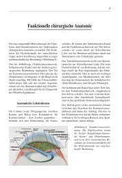
2. Anatomy<br />
The eye lies in the orbit, which is formed by the<br />
skull. The bulbus oculi (eyeball) is covered in the<br />
frontal area by the eyelids, and is otherwise embedded<br />
in the soft orbital fatty tissue. The eye muscles end<br />
with their tendons at the bulbus oculi. Their fibres<br />
converge in the rear part of the orbits, thus forming a<br />
funnel, called the anulus tendineus communis (Zinn’s<br />
ring, tendinous ring), into which the optic nerve, originating<br />
from the eyeball, leads.<br />
Greater wing of the sphenoid<br />
Inferior orbital fissure<br />
Zygomatic bone<br />
Fig. 2.1 The bony orbit<br />
Superior orbital fissure<br />
Lesser wing of sphenoid bone<br />
Nerves and vessels enter the orbit to supply the<br />
structures located there – the globe, the optic nerve,<br />
the exterior eye muscles, and the lacrimal gland, which<br />
lies on the globe in the lateral part of the orbital roof<br />
(Figs. 2.2, 2.6).<br />
Optic foramen<br />
Frontal bone<br />
Maxilla<br />
Lacrimal bone<br />
Maxilla<br />
Infraorbital foramen<br />
Ethmoid bone
4 <strong>Endocrine</strong> <strong>Ophthalmopathy</strong><br />
2.1 The Structure of the Bony Orbit<br />
The bony orbit has the form of a funnel, open towards<br />
the front. It is limited nasally by the orbital plate of the<br />
ethmoid bone, the frontal process of the maxilla, the<br />
lacrimal bone, and the orbital process of the palatine<br />
bone. The orbital floor is formed by the maxilla, the<br />
zygomatic bone, and the orbital process of the palatine<br />
bone. The orbital roof consists of the orbital part of the<br />
frontal bone and the lesser wing of the sphenoid.<br />
Laterally the orbit is limited by the zygomatic bone,<br />
the lesser wing of the sphenoid, and the zygomatic<br />
process of the frontal bone (Fig. 2.1).<br />
Supraorbital nerve<br />
Orbicularis oculi<br />
Orbital septum<br />
Levator palpebrae<br />
superioris (aponeurosis)<br />
Superior tarsal<br />
muscle (Müller)<br />
Superior<br />
tarsus<br />
Inferior tarsus<br />
Orbicularis oculi<br />
Inferior tarsal muscle (retractor)<br />
Orbital septum<br />
Fig. 2.2 Sagittal section through the orbit.<br />
Levator palpebrae sup.<br />
Superior rectus muscle<br />
Periorbita<br />
Infraorbital nerve<br />
Inferior oblique muscle<br />
2.2 The Extraocular Muscles<br />
The extraocular muscles have their common origin in<br />
the depths of the orbits, in front of the optic foramen,<br />
entering from the tendinous ring (Zinn’s ring). They<br />
enclose the optic nerve. The four straight eye muscles<br />
(Superior rectus, inferior rectus, medial rectus, and<br />
lateral rectus) run along the upper, lower, medial, and<br />
lateral walls of the orbit to the bulbus oculi, and radiate<br />
into the sclera. The two obliquely positioned eye<br />
muscles, the superior oblique and the inferior oblique,<br />
run from the front medially to the back laterally along<br />
the globe, and attach to this temporally behind the<br />
coronal equator of the eye (Fig. 2.2, 2.3).<br />
Central retinal artery and vein<br />
Superior ophthalmic vein<br />
Long ciliary nerve<br />
Nasociliary nerve<br />
Lacrimal artery<br />
Frontal nerve<br />
Short ciliary nerve<br />
Oculomotor nerve (superior ramus)<br />
Annular tendon (Zinn)<br />
Dura mater<br />
Nasociliary nerve<br />
Optic nerve<br />
Oculomotor nerve<br />
Ophthalmic artery<br />
Orbital process of the palatine bone<br />
Ciliary ganglion<br />
Oculomotor nerve (inferior ramus)<br />
Inferior rectus muscle<br />
Maxillary sinus Ciliary arteries (artery to the iris)
Anatomy 5<br />
Lacrimal gland<br />
Lateralis rectus<br />
Fig. 2.3 Anatomy of the anterior part of the orbit (coronal<br />
section). The fat compartments and the septi between the<br />
fatty portions are clearly visible.<br />
Lacrimal gland<br />
Lateral rectus<br />
Periorbita<br />
Superior rectus<br />
Levator palpebrae superioris<br />
Levator palpebrae superioris<br />
Fig. 2.4 Coronal section in the middle of the globe.<br />
Superior rectus<br />
Inferior oblique<br />
Supraorbital nerve, artery, vein<br />
Inferior rectus<br />
Supraorbital artery vein<br />
and nerve<br />
Inferior rectus<br />
Superior oblique<br />
Inferior oblique<br />
Supratrochlear nerve,<br />
artery, vein<br />
Medial rectus<br />
Supratrochlear artery vein and nerve<br />
Superior ophthalmic vein<br />
Superior oblique<br />
Medial rectus<br />
Inferior ophthalmic vein
6 <strong>Endocrine</strong> <strong>Ophthalmopathy</strong><br />
Lacrimal nerve artery and vein<br />
Lateral rectus muscle<br />
Abducens nerve<br />
Short and long posterior<br />
ciliary artery, ciliary nerve<br />
Optic nerve, central artery<br />
and vein<br />
Artery to the iris<br />
Periorbita<br />
Oculomotor nerve<br />
(inferior ramus)<br />
Infraorbital nerve<br />
Levator palpebrae superioris<br />
Oculomotor nerve (superior ramus)<br />
Supraorbital nerve, artery, vein<br />
Fig. 2.5 Anatomy of the retrobulbar space. Coronal section 0.5 cm dorsal from the globe.<br />
Frontal nerve<br />
Superior ophthalmic vein<br />
Oculomotor nerve<br />
Lateral rectus muscle<br />
Nasociliary nerve<br />
Abducens nerve<br />
Superior ophthalmic vein<br />
Lacrimal nerve<br />
Infraorbital fissure<br />
Superior orbital fissure<br />
Superior rectus muscle<br />
Trochlear nerve<br />
Levator palpebrae superioris muscle<br />
Infraorbital A.,N.V.<br />
Superior rectus muscle<br />
Supraorbital artery, vein and nerve<br />
Superior oblique muscle<br />
Superior oblique muscle<br />
Supratrochlear nerve,<br />
artery, vein<br />
Medial rectus<br />
Inferior ophthalmic vein<br />
Inferior rectus<br />
Medial rectus muscle<br />
Supratrochlear artery,<br />
vein and nerve<br />
Optic nerve<br />
Optic canal<br />
Ophthalmic artery<br />
Inferior rectus muscle<br />
Oculomotor nerve<br />
Annular tendon (Zinn)<br />
Inferior ophthalmic vein<br />
Oculomotory nerve (inferior ramus)<br />
Fig. 2.6 Topographical view of the apex of the orbit (coronal section). The optic nerve, ophthalmic artery, oculomotor nerves<br />
abducens nerve, and the nasociliary nerve lie within the annular tendon (Zinn’s ring). Other important nerves and<br />
vessels enter through the superior and inorbital fissures.
Anatomy 7<br />
2.3 The Innervation of the<br />
Extraocular Muscles<br />
The extraocular muscles are innervated by three cranial<br />
nerves, the oculomotor nerve, the abducent nerve, and<br />
the trochlear nerve.<br />
The oculomotor nerve (III rd cranial nerve) contains<br />
somatic and autonomic fibres. It innervates the medial<br />
rectus muscle, the inferior rectus, the superior rectus,<br />
the inferior oblique and the levator palpebrae superioris.<br />
The abducent nerve (VI th cranial nerve) is a<br />
somatic nerve, which supplies the lateral muscle. The<br />
trochlear nerve (IV th cranial nerve) is also a purely<br />
somatic nerve, innervating the superior oblique muscle.<br />
All of the above-mentioned nerves enter the orbit<br />
via the superior orbital fissure. There the oculomotor<br />
nerve divides into two branches; the inferior branch<br />
(Inferior ramus) supplies the inferior rectus, the medial<br />
rectus, and the inferior oblique; the superior branch<br />
supplies the superior rectus and the levator palpebrae<br />
superioris. The oculomotor nerve innervates the interior<br />
eye muscles with its autonomic portion.<br />
1<br />
2<br />
Fig. 2.7 Horizontal section through the middle of the right<br />
orbit. The medial rectus and the lateral rectus are attached by<br />
fine thread ligaments (1) to the periorbita. In transpalpebral<br />
decompression with fat removal the muscles remain in their<br />
natural position.<br />
2<br />
1<br />
The branches of the III rd , IV th , and VI th cranial<br />
nerves are inserted into the muscles on the inner side<br />
of the cone. This has a practical consequence: when<br />
removing fat, there is hardly any danger of injury.<br />
2.4 Vascular Supply of the Orbits<br />
The most important artery of the orbit is the ophthalmic<br />
artery, which originates from the internal<br />
carotid artery (A. carotis interna). It runs beneath the<br />
optic nerve through the optic canal. Within the orbit,<br />
it runs within the tendinous ring and describes a helix<br />
around the optic nerve. It turns lateral to and then<br />
above and finally medial to the optic nerve. In this area<br />
the lacrimal artery branches off, running along the<br />
upper edge of the lateral rectus to the lacrimal gland<br />
and the lateral corner of the eye. In the further course<br />
of the ophthalmic artery the ciliary artery and the<br />
central retinal artery branch off, which are important<br />
for the supply of the globe (Fig. 2.2).<br />
Optic nerve<br />
Orbit<br />
Dura mater<br />
Arachnoid<br />
Pia mater<br />
Fig. 2.8 Anatomy of the optic canal. The optic nerve is surrounded<br />
by the dura mater. Every pull on the optic nerve and<br />
thus on the dura mater in a ventral direction in exophthalmos<br />
causes retrobulbar pressure (‘burning’) and headaches.
8 <strong>Endocrine</strong> <strong>Ophthalmopathy</strong><br />
a b<br />
2.5 The Lacrimal Gland<br />
The lacrimal gland lies in the upper temporal portion<br />
of the orbit, in the lacrimal gland fossa of the frontal<br />
bone. It is divided by the tendon of the M. levator<br />
palpebrae into an orbital part, lying on the bone, and<br />
a palpebral part, lying in the eyelid. Approximately ten<br />
small ducts secrete tears above the lateral corner of the<br />
eye into the superior conjunctival fornix. The gland is<br />
supplied by the lacrimal artery and the lacrimal nerve.<br />
2.6 Intraorbital Fat and<br />
Connective Tissue<br />
The space in the eye socket not taken up by muscles,<br />
vessels, and nerves is filled by a fatty tissue interspersed<br />
with connective tissue. A pyramidal cone exists<br />
within the four straight eye muscles. The connective<br />
tissue in the fatty tissue thickens towards the muscles,<br />
but in particular towards the globe, forming a more<br />
solid layer, which offers the eye ball a bed.<br />
The periosteum of the orbit, the periorbit, lines the<br />
bony eye socket. It fades into the dura mater through<br />
the optic canal and the superior orbital fissure. The<br />
orbital septum limits the orbital content in front. It is<br />
a ring-shaped, nearly vertical plate of connective tissue<br />
it runs from the orbit edge to the superior and inferior<br />
tarsus. It is penetrated by several nerves and vessels<br />
(Figs. 2.3–5).<br />
a<br />
b<br />
Fig. 2.9 a,b View of the<br />
orbit from above following<br />
removal of the orbital ceiling.<br />
F = frontal nerve<br />
SO = supraorbital nerve<br />
ST = supratrochlear nerve<br />
L = lacrimal nerve<br />
IV = Branches of the IVth<br />
cranial nerve to the muscles.<br />
Plentiful distribution of<br />
fat is visible.<br />
b View under the above<br />
structures. LEV = levator<br />
palpebrae superioris, SR =<br />
superior rectus muscle.<br />
Here, too, there is plentiful<br />
distribution of fat (more<br />
medially than laterally).<br />
Fig. 2.10 a,b Horizontal section in the upper part of the orbit.<br />
1. Levator palpebrae, 2. Frontal sinus, 3. Frontal llobe of the<br />
brain. b Horizontal section at the height of the middle of the<br />
orbit. 1. Medial rectus, 2. Lateral rectus, 3. Optic nerve – apical<br />
part, 4. Ethmoid sinus, 5. Globe<br />
[We would like to thank Prof. Dr. Koebke of the Anatomical Institute<br />
of the University of Cologne for this illustrative material].
Anatomy 9<br />
a b<br />
Fig. 2.11 a,b The extraocular muscles in the normal orbit.<br />
The drawings follow the 3D-CT display; viewed from beneath.<br />
a All muscles in a relatively relaxed condition, b Opticus nerve<br />
relaxed, with many curves.<br />
2.7 Optic Nerve<br />
The optic nerve runs from the globe dorsally through<br />
the tendinous ring and the optic canal, leads to the chiasma.<br />
The optic nerve is relaxed in the primary position<br />
of the eyeball, curving slightly. This permits corresponding<br />
mobility during the movements of the<br />
globe. The optic nerve is surrounded by the dura<br />
mater, and this in its turn is fixed to the orbital periosteum.<br />
A traction of the optic nerve on the dura mater<br />
(in cases of exophthalmos) causes subjective complaints,<br />
such as retrobulbar “burning” and headaches<br />
(Fig. 2.8).