Pulse contour cardiac output, PiCCO - Intensive Care ...
Pulse contour cardiac output, PiCCO - Intensive Care ...
Pulse contour cardiac output, PiCCO - Intensive Care ...

You also want an ePaper? Increase the reach of your titles
YUMPU automatically turns print PDFs into web optimized ePapers that Google loves.
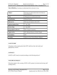
INTENISVE CARE SERVICE<br />
NURSING POLICY& PROCEDURES<br />
NAME OF POLICY: PULSE CONTOUR CARDIAC OUTPUT (<strong>PiCCO</strong>)<br />
GOAL: TO EVALUATE CARDIOPULMONARY AND CIRCULATORY<br />
VARIABLES<br />
Introduction:<br />
The PULSION <strong>PiCCO</strong> is indicated in patients where cardiovascular volume status monitoring is<br />
necessary. Cardiac <strong>output</strong> is determined both continuously through arterial pulse <strong>contour</strong><br />
analysis and intermittently through transpulmonary thermodilution technique. In addition, the<br />
<strong>PiCCO</strong> measures heart rate, systolic and diastolic pressure and derives mean arterial pressure.<br />
Analysis of the thermodilution curve in terms of mean transit time (MTt) and downslope time<br />
(Dst) is used for determination of intra- and extra- vascular fluid volumes. If patient weight and<br />
height are entered the <strong>PiCCO</strong> presents the derived parameters indexed to body surface area<br />
(BSA).<br />
Equipment:<br />
• PV2015L20 PULSIOCATH arterial line 5Fx20cm<br />
• PV8115 Pulsion pressure transducer with PV4046 inline injectate sensor housing<br />
• M1012A#C10 C.O. Module with <strong>PiCCO</strong> capability<br />
• M1643A Cardiac Output cable<br />
• M1006B Pressure module with PMK 206 white Utah pressure cable<br />
• PC80102 Pulsion Inline Injectate Temperature Cable<br />
• 20ml Syringe<br />
• 500ml bag 5%Dextrose<br />
Pre Procedure:<br />
• Inform patient and significant others if applicable<br />
• Adhere to universal precautions<br />
• Insert 500mls preloaded heparin bag, into pressure bag, spike bag with transducer kit, inflate<br />
to 300mmHg and prime the line, ensuring air bubbles are evacuated<br />
• Align transducer to phlebostatic axis (4 th intercostal space mid axilla line)<br />
• ‘zero’ transducer to atmospheric air<br />
Post Procedure:<br />
• Secure arterial line appropriately<br />
• Connect to patient<br />
© ROYAL PRINCE ALFRED HOSPITAL INTENSIVE CARE SERVICE
Connecting the patient:<br />
<strong>PiCCO</strong><br />
• Check that the C.O. module Option C10 is inserted into the rack.<br />
• Make sure that the patient’s height and weight have been entered into the haemodynamic<br />
calculation menu.<br />
• Plug the C.O. interface cable into the C.O. module<br />
• Attach the inline injectate sensor housing (PV4046) to the CVP lumen on central line (if<br />
CVC being inserted follow CVC policy) and attach inline injectate temperature cable<br />
(PULSION PC80102) between the housing and the <strong>cardiac</strong> <strong>output</strong> interface cable (M1643A)<br />
• Attach the blood temperature cable of the CO interface cable (M1643A) to the<br />
PULSIOCATH arterial line thermistor<br />
• Attach the PULSION pressure transducer (PV8115) to the PULSIOCATH arterial line<br />
• Attach the Pressure transducer cable (PMK-206) to the pressure transducer module<br />
(M1006B)<br />
© ROYAL PRINCE ALFRED HOSPITAL INTENSIVE CARE SERVICE
Setup the monitor:<br />
• Enter the Setup CO menu by pressing CO on touch screen<br />
The method = Transpulmonary<br />
The injectate temperature probe = M1646 (PV4046)<br />
Enter CCO<br />
The Source of CCO = same as the name of the transducer connected to the<br />
<strong>PiCCO</strong> arterial line. Must be ABP not ART.<br />
Enter Cardiac <strong>output</strong><br />
The catheter constant = 342 or whatever is on the catheter packet<br />
The volume of injectate = 20mls<br />
If any of the above items need changing select the item on the menu in the<br />
setup CO window and change to the appropriate setting.<br />
Performing Cardiac Output:<br />
• Select Cardiac Output<br />
• In the first C.O. measurement Task Window. A message “…Ready for new measurement”<br />
will appear on the screen.<br />
• Press Start C.O. and wait for the message “…Stable baseline inject now!” to appear on the<br />
screen.<br />
• Inject the 20mls of room temperature injectate as quickly as possible and in a smooth<br />
constant motion<br />
• At the end of the measurement the thermodilution curve, <strong>cardiac</strong> <strong>output</strong>, ITBVI and EVLWI<br />
indexed values<br />
• After waiting for one minute a message will appear “ready for new measurement” repeat<br />
steps 3 – 5<br />
• Continue to repeat this procedure until you have completed all the measurements you want to<br />
perform. If 2 thermodilution curves result in values within 10% of each other then further<br />
curves are unnecessary. You can perform a maximum of 6 measurements before editing<br />
(select which curves should be used for the averaging calculation).<br />
• Select which curve you want to edit.<br />
• Select Accept Reject. The curves you have selected will be green and the curves rejected<br />
red. If there is a question mark before the value it means the result may not be accurate.<br />
• Manual selection of the individual thermodilution curves enables the user to edit the final<br />
C.O. measurement. An average is calculated for the undeleted measurements and stored<br />
when Save CO and calibrate CCO is pressed. The average of multiple thermodilution<br />
measurements should be used for therapy decisions. Averages are also calculated for the<br />
EVLW, ITBV and CFI. The averaged C.O. data is then used to calibrate Continuous Cardiac<br />
Output (CCO).<br />
• If the message “high ETVI, more indicator required” appears cold fluid will need to be used.<br />
The cold fluid must be
Contraindication:<br />
• Patients receiving intra-aortic balloon counter pulsation (IABP) cannot be monitored with the<br />
<strong>PiCCO</strong><br />
Nursing Responsibilities:<br />
• A measurement should be performed at least 8 th hourly to calibrate the PCCO and more<br />
frequently if there is a marked change in haemodynamics<br />
• Other nursing responsibilities see Arterial Line and CVC policies.<br />
Removal of Arterial Line:<br />
• See Arterial Line policy.<br />
• Consider the use of a ‘Femstop’ due to femoral vessel.<br />
•<br />
Parameters:<br />
The following parameters can be derived by transpulmonary thermodilution:<br />
Arterial<br />
Thermodilution<br />
Absolute<br />
Parameters<br />
Indexed<br />
Parameters<br />
Parameter Abbr. Unit Abbr. Unit<br />
Cardiac <strong>output</strong>, arterial C.O.a l/min CIa l/min/m 2<br />
Cardiac function index CFI 1/min<br />
Global end diastolic volume GEDV ml<br />
lntrathoracic blood volume ITBV mI ITBVI mI/m 2<br />
Extravascular lung water<br />
EVLW ml EVLW I ml/kg<br />
After initial calibration the following parameters can continuously derived by pulse <strong>contour</strong><br />
analysis:<br />
<strong>Pulse</strong> Contour<br />
Absolute<br />
Parameters<br />
Indexed<br />
Parameters<br />
Parameter Abbr. Unit Abbr. Unit<br />
<strong>Pulse</strong> <strong>contour</strong> <strong>cardiac</strong> <strong>output</strong> PCCO l/min PCCI l/min/<br />
m 2<br />
Systolic arterial blood pressure APsys mmHg<br />
Diastolic arterial blood pressure APdia mmHg<br />
Mean arterial blood pressure MAP mmHg ITBVI mI/m 2<br />
Heart rate HR 1/min<br />
Stroke volume SV ml SVI ml/m 2<br />
Stroke volume variation SVV %<br />
Systemic vascular resistance SVR dyn/sec/cm -5<br />
SVRI dyn/se<br />
c/cm –<br />
Index of left ventricular contractility DP/dtmax mmHg/s<br />
© ROYAL PRINCE ALFRED HOSPITAL INTENSIVE CARE SERVICE<br />
5 /m 2
INTERMITTENT VOLUMETRIC THERMODILUTION<br />
Cardiac Output (CO) is generally determined using the Stewart-Hamilton method. To<br />
accomplish thermodilution determination, a known volume of cold (at least 10°C lower than<br />
blood temperature) solution is injected intravenously as fast as possible. The recorded<br />
downstream temperature change is dependent on the flow and on the volume through which the<br />
cold indicator has passed. As a result, a thermodilution curve can be constructed. The <strong>PiCCO</strong><br />
detects the indicator in the arterial system (preferably the femoral artery). The transpulmonary<br />
thermodilution curves are much longer and flatter than the respective pulmonary artery<br />
thermodilution curves. Compared to pulmonary artery thermodilution, the arterial thermodilution<br />
measurement has minimal ventilatory variations.<br />
Methods:<br />
The cardiopulmonary system represents a series of different mixing chambers for an indicator<br />
which has been injected central venously.<br />
© ROYAL PRINCE ALFRED HOSPITAL INTENSIVE CARE SERVICE
Principles of Volume Calculation:<br />
Specific volumes can be calculated by multiplying the <strong>cardiac</strong> <strong>output</strong> (C.O.a) by characteristic<br />
transit times determined through the indicator dilution curves.<br />
For this the <strong>PiCCO</strong> calculates the mean transit time (MTt) and the exponential downslope time<br />
(Dst) of the thermodilution curve.<br />
MTt Volume:<br />
The result of the product of C.O. and MTt is the volume through which the relevant indicator has<br />
travelled. ie. the complete volume between the site of injection and the site of detection. For the<br />
cold indicator this is the total intrathoracic thermal volume (ITTV) which is composed of the<br />
global end-diastolic volume (GEDV), pulmonary blood volume (PBV), and extravascular lung<br />
water (EVLW).<br />
ITTV = CO x MTtTDa<br />
DSt Volume:<br />
The result of the product of CO and DSt is the largest individual mixing volume in a series of<br />
indicator dilution mixing chambers. For the cold indicator this is the pulmonary thermal volume<br />
(PTV) which is composed of PBV and EVLW.<br />
PTV = CO x DStTDa<br />
© ROYAL PRINCE ALFRED HOSPITAL INTENSIVE CARE SERVICE<br />
EVL<br />
RAED RVED LAED LVED<br />
PB<br />
EVL<br />
EVL<br />
PB<br />
EVL
Combination:<br />
By subtraction of PTV from ITTV one obtains a volume which represents the global enddiastolic<br />
volume.<br />
GEDV = ITTV - PTV<br />
Global end-diastolic volume is the sum of all end diastolic volumes of the atria and the<br />
ventricles. Thus, GEDV is equivalent to preload volume of the total heart.<br />
Traditionally central venous pressure (CVP) and pulmonary capillary wedge pressure (PCWP)<br />
are used as indicators of <strong>cardiac</strong> preload. However, both CVP and PCWP are dependent on<br />
intravascular filling, intrathoracic pressure, vascular compliance and <strong>cardiac</strong> contractility. In<br />
contrast to pressures, GEDV represents <strong>cardiac</strong> preload volume.<br />
Intrathoracic Blood Volume (ITBV):<br />
ITBV = 1.25 x GEDV<br />
RAED RVED LAED LVED<br />
RAEDV RVEDV PBV<br />
LAEDV LVEDV<br />
Intrathoracic blood volume (ITBV) consists of the global end-diastolic volume (GEDV, is<br />
approximately 4/5 of ITBV) and the pulmonary blood volume (PBV). ITBV is calculated by<br />
GEDV x 1.25.<br />
Three volumes are found in the thorax, the intrathoracic blood volume, the intrathoracic gas<br />
volume and the extravascular lung water. Due to the limited expandability of the thorax, the<br />
volumes interact and change proportionally to each other.<br />
Lichtwarck-Aschoff et al. Were able to show that in intensive care patients on mechanical<br />
ventilation, ITBV reflects the status of the circulating blood volume.<br />
© ROYAL PRINCE ALFRED HOSPITAL INTENSIVE CARE SERVICE
Extravascular Lung Water (EVLW):<br />
EVLW = ITTV - ITBV<br />
Arterial thermodilution results in the direct measurement of pulmonary thermal volume (PTV)<br />
and intrathoracic thermal volume (ITTV). From these values the extravascular lung water<br />
(EVLW) is assessable by the following:<br />
The water content in the lungs increases through left heart failure, pneumonia, ALI, and ARDS.<br />
Increased EVLW can occur through increased fluid transport to the interstitium, resulting from<br />
either high intravascular filtration pressure (left heart failure, volume overload) or an increased<br />
pulmonary vascular permeability for plasma proteins, which drag water along with them,<br />
corresponding to their colloid osmotic pressure.<br />
Relationship between intrathoracic blood volume and extravascular lung<br />
water:<br />
As it is known that the level of EVLW is related to patient outcome any measures to reduce<br />
EVLW are most likely to shorten ventilation days and stay in the ICU and reduce possible<br />
complications (pneumonia, pneumothorax, etc.).<br />
The hydrostatic component of increased EVLW can be reduced by volume restriction. In the<br />
lower diagram it is shown that below the normal range of ITBV one cannot reduce EVLW<br />
anymore. Therefore ITBV representing <strong>cardiac</strong> preload should not be driven below this level in<br />
order to avoid further reduction in <strong>cardiac</strong> <strong>output</strong> and hence oxygen supply to the body, as this<br />
would not result in a further reduction of extravascular lung water.<br />
Cardiac Function Index (CFI):<br />
Cardiac function index (CFI) is derived as the ratio of <strong>cardiac</strong> <strong>output</strong> divided by global enddiastolic<br />
volume.<br />
CFI = CI / GEDVI<br />
© ROYAL PRINCE ALFRED HOSPITAL INTENSIVE CARE SERVICE<br />
EVL<br />
EVL
Principle of Measurement:<br />
PULSE CONTOUR ANALYSIS<br />
During <strong>cardiac</strong> systole blood is ejected into the aorta. Simultaneously, blood flows out of the<br />
aorta into the peripheral vascular system. However, during the time of ejection, the sum of all<br />
blood flowing into the aorta is larger than the blood volume entering the peripheral vascular<br />
system. thus, the volume of the aorta increases. In the subsequent diastole, most of the remaining<br />
blood will empty into the peripheral vasculature. This behaviour is dependent on the ability of<br />
the aorta to expand and contract in response to ejected volumes. The volume change and<br />
subsequent pressure change is described as the compliance function of the aorta.<br />
© ROYAL PRINCE ALFRED HOSPITAL INTENSIVE CARE SERVICE
The relationship between blood flow out of the aorta and pressure measured at the end of the<br />
aorta (femoral artery or other large artery) is determined by the compliance function. The<br />
compliance function can therefore be characterised by measuring blood pressure and blood flow<br />
(<strong>cardiac</strong> <strong>output</strong>) simultaneously. Transpulmonary thermodilution <strong>cardiac</strong> <strong>output</strong> (C.O.)<br />
determined simultaneously with continuous arterial pressure (AP) measurement is utilized to<br />
calibrate the pulse <strong>contour</strong> analysis to each individual patients aortic compliance function.<br />
For the continuous calculation of PCCO the <strong>PiCCO</strong> uses a calibration factor (cal) determined by<br />
the thermodilution <strong>cardiac</strong> <strong>output</strong> measurement and the heart rate (HR), as well as the integrated<br />
values for the area under the systolic part of the pressure curve (P(t)/SVR), the aortic compliance<br />
(C(p)) and the shape of the pressure curve, represented by change of pressure over change of<br />
time (dP/dT)<br />
Calibration of <strong>Pulse</strong> Contour Analysis:<br />
To derive the calibration factor ‘cal’ and the individual compliance function C(p) a reference<br />
transpulmonary thermodilution <strong>cardiac</strong> <strong>output</strong> is necessary.<br />
<strong>Pulse</strong> Contour Cardiac Output (PCCO):<br />
When the calibration factor ‘cal’ and the individual compliance function C(p) is determined,<br />
continuous <strong>cardiac</strong> <strong>output</strong> can be measured using the heart rate and the area under the aortic flow<br />
curve. Displayed PCCO is the mean value of the last 12 seconds.<br />
© ROYAL PRINCE ALFRED HOSPITAL INTENSIVE CARE SERVICE
Arterial Blood Pressure (AP):<br />
Arterial blood pressure is one of the most important diagnostic parameters for patient<br />
management. The <strong>PiCCO</strong> measures the arterial pressure continuously using a special arterial<br />
thermodilution catheter with an additional pressure lumen. The arterial thermodilution and<br />
arterial pressure measurement can be done with the same catheter. The arterial pressure is<br />
registered by a pressure transducer. Displayed AP is the mean value over the last 12 seconds.<br />
Stroke Volume Variation (SVV):<br />
Stroke volume variation is presented as the change in stroke volume (in percent) calculated by<br />
the mean difference between highest and lowest stroke volume divided by a calculated mean<br />
stroke volume over the last 30 seconds.<br />
In mechanically ventilated patients, the SVV mainly depends on the intravascular volume of the<br />
patient. Big variations in stroke volume, induced by mechanical ventilation, is an indication of<br />
insufficient intravascular volume relative to the applied intrathoracic pressures. Thus, SVV<br />
allows a rough estimation of the vascular volume status. When high SVV is detected, it is<br />
recommended to perform a thermodilution measurement to quantify the volume status by<br />
measuring the ITBV.<br />
Systemic Vascular Resistance:<br />
The systemic vascular resistance is the quotient of driving pressure and <strong>cardiac</strong> <strong>output</strong> over the<br />
last 12 seconds. Here, the driving pressure represents the difference between mean arterial<br />
pressure (MAP) and central venous pressure (CVP).<br />
Complications:<br />
• See Arterial Line policy and CVC policy.<br />
SVR = (MAP - CVP)<br />
C.O.a<br />
© ROYAL PRINCE ALFRED HOSPITAL INTENSIVE CARE SERVICE<br />
x80
REFERENCES:<br />
Godje, O. Hoke, K. et al (2002) Reliability of a new algorithm for continuous <strong>cardiac</strong> <strong>output</strong><br />
determination by pulse <strong>contour</strong> analysis during hemodynamic instability. Lippincott<br />
Williams and Wilkins.<br />
PULSION Medical Systems Operators Manual September 2000<br />
PULSION Pacific Information Package March 2000<br />
PULSION Medical Systems Rapid, less invasive continuous <strong>cardiac</strong> <strong>output</strong>, intrathoracic<br />
blood volume, and extravascular lung water monitoring.<br />
Occupational Health and Safety: Universal precautions taken in the preparation, administration of drug and<br />
disposal of equipment and sharps.<br />
Cross Referenced: RPAH Occ. Health & Safety Manual and Infection Control Manual<br />
NSW Infection Control Policy 98/99<br />
Written By: Cheryl Richards (CNE) February 2003.<br />
Authorised by: Richard Totaro (Intensivist) March 2003<br />
Revision due: March 2005<br />
© ROYAL PRINCE ALFRED HOSPITAL INTENSIVE CARE SERVICE