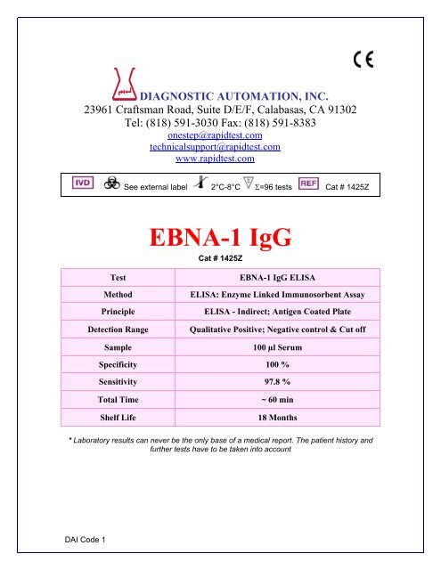
Epstein Barr Virus Nuclear Antigen (EBNA) IgG
Epstein Barr Virus Nuclear Antigen (EBNA) IgG
Epstein Barr Virus Nuclear Antigen (EBNA) IgG
- No tags were found...

You also want an ePaper? Increase the reach of your titles
YUMPU automatically turns print PDFs into web optimized ePapers that Google loves.
INTENDED USEThe DAI <strong>Epstein</strong>-<strong>Barr</strong> <strong>Virus</strong> <strong>Nuclear</strong> <strong>Antigen</strong>-1 (<strong>EBNA</strong>-1) <strong>IgG</strong> Enzyme- LinkedImmunosorbent Assay (ELISA) is intended for the qualitative and semi-quantitativedetermination of <strong>IgG</strong> antibody in human serum to <strong>EBNA</strong>-1 recombinant antgen. The DAI<strong>EBNA</strong>-1 <strong>IgG</strong> assay may be used in conjunction with other <strong>Epstein</strong>-<strong>Barr</strong> serologies (VCA<strong>IgG</strong>, VCA IgM, EA (R&D), and heterophile) as an aid in the diagnosis of infectiousmononucleosis. For in vitro diagnostic use. High complexity test.For in vitro diagnostic use. High complexityINTRODUCTION<strong>Epstein</strong>-<strong>Barr</strong> virus (EBV) is a common human pathogen, affecting 80% of adults in theUS. Since the discovery of <strong>Epstein</strong>-<strong>Barr</strong> virus in 1964, EBV has been etiologicallyimplicated in an increasing number of human diseases, such as infectiousmononucleosis, Burkitt's lymphoma and nasopharyngeal carcinoma.1 EBV has alsobeen associated with B cell lymphomas in immunosuppressed individuals, including bothtransplant patients and patients with AIDS. EBV is classified as a member of theherpesvirus family based upon its characteristic morphology.2,3 All herpesviruses sharethe ability to establish a latent infection in their hosts.4 Although primary infection withEBV during childhood is usually asymptomatic, nearly one-half to two-thirds of primaryinfections with the virus in older adolescents and young adults result in overt clinicaldisease such as infectious mononucleosis (IM). 1 Infectious mononucleosis is an acute,self-limited lympho-proliferative disease caused by EBV. When primary infection isdelayed until young adulthood and adolescence, however, there is about a 50% chancethat it will occur with the classic clinical manifestations associated with IM.5,6Infection of the target cells leads to two forms of viral cycles: 1) latent, nonproductiveand 2) productive, replicative infections.7 In both cycles, one of the earliest antigensexpressed is lymphocyte-detected membrane antigen, a cell-surface antigen recognizedby T-cells. It has been well established that most individuals exposed to EBV develop aheterophile antibody response. Expression of <strong>EBNA</strong>-1 either follows or parallelsmembrane antigen at 12 to 24 hours post infection. <strong>EBNA</strong>-1 is found as nonstructural,intranuclear antigen(s), present in all EBV-transformed cell lines as in tumors fromBurkitt's and nasopharyngeal carcinoma patients. In the fully productive, replicativecycle, the synthesis of antigen follows <strong>EBNA</strong>-1. The viral capsid antigen complex (VCA)appears late in the replicative cycle.It has recently become apparent that <strong>EBNA</strong>-1 is probably not a single antigenic moiety,but a multicomponent antigen complex, on the basis of reactivities of sera from differentclasses of patients. The major component <strong>EBNA</strong>-1 has been purified and sequenced inits entirety.7Antibody levels of <strong>EBNA</strong>-1 <strong>IgG</strong>, are diagnostic in determining acute and convalescentstages in IM. <strong>IgG</strong> antibodies to <strong>EBNA</strong>-1 are rarely present in acute IM and rise duringconvalescence. They will rise to a plateau level in three months to a year and willnormally persist for life.8,9DAI Code 1
The DAI <strong>EBNA</strong>-1 <strong>IgG</strong> kit utilizes the ELISA technology where a purified recombinant<strong>EBNA</strong>-1 antigen is bound to the wells of a microplate. A peroxidase coupled anti-human<strong>IgG</strong> conjugate is used as the detection system.PRINCIPLE OF THE ASSAYEnzyme-Linked Immunosorbent Assays (ELISA) rely on the ability of biological materials(i.e., antigens) to adsorb to plastic surfaces such as polystyrene (solid phase). Whenantigens bound to the solid phase are brought into contact with a patient's serum,antigen specific antibody, if present, will bind to the antigen on the solid phase formingantigen-antibody complexes. Excess antibody is removed by washing. This is followedby the addition of goat antihuman <strong>IgG</strong> conjugated with horseradish peroxidase whichthen binds to the antibody-antigen complexes. The excess conjugate is removed bywashing, followed by the addition of Chromogen/Substrate, tetramethylbenzidine (TMB).If specific antibody to the antigen is present in the patient's serum, a blue color develops.When the enzymatic reaction is stopped with 1N H2SO4, the contents of the wells turnyellow. The color, which is indicative of the concentration of antibody in the serum, canbe read on a suitable spectrophotometer or ELISA microwell plate reader.10,11,12,13The sensitivity, specificity, and reproducibility of ELISAs can be comparable to otherserological tests for antibody, such as immunofluorescence, complement fixation,hemagglutination and radio-immunoassay.14,15,16KIT PRESENTATIONMaterials SuppliedEach kit contains the following components in sufficient quantities to perform the numberof tests indicated on the package label.1. Recombinant <strong>EBNA</strong>-1 antigen (the carboxy-terminal of <strong>EBNA</strong>-1 genomerepresenting ~200 codons) coated microassay plate: 96 wells, configured in twelve1x8 strips, stored in a foil pouch with a desiccant. (96T: one plate; 480T: five plates)2. Serum Diluent Type I: Ready for use. Contains proclin (0.1%) as a preservative.(96T: one bottle, 30 mL, 480T: two bottles, 60 mL each)3. Calibrator: Human serum or defibrinated plasma. Sodium azide (< 0.1%) andpen/strep (0.01%) added as preservatives, with kit specific factor printed on viallabel. The Calibrator is used to calibrate the assay to account for day-to-dayfluctuations in temperature and other testing conditions. (96T: one vial, 0.4 mL;480T: one vial, 0.8 mL) *4. High Positive Control: Human serum or defibrinated plasma. Sodium azide (< 0.1%)and pen/strep (0.01%) added as preservatives, with established range printed onvial label. The High Positive Control is utilized to control the upper dynamic range ofthe assay. (96T: one vial, 0.4 mL; 480T: one vial, 0.8 mL) *5. Low Positive Control: Human serum or defibrinated plasma. Sodium azide (< 0.1%)and pen/strep (0.01%) added as preservatives, with established range printed onvial label. The Low Positive Control is utilized to control the range near the cutoff ofthe assay. (96T: one vial, 0.4 mL; 480T: one vial, 0.8 mL) *6. Negative Control: Human serum or defibrinated plasma. Sodium azide (< 0.1%)and pen/strep (0.01%) added as preservatives, with established range printed onvial label. The Negative Control is utilized to control the negative range of the assay.(96T: one vial, 0.4 mL; 480T: one vial, 0.8 mL) *DAI Code 1
7. Horseradish-peroxidase (HRP) Conjugate: Ready to use. Goat antihuman <strong>IgG</strong>,containing proclin (0.1%) and gentamicin as preservatives. (96T: one bottle, 16 mL;480T: five bottles, 16 mL each)8. Chromogen/Substrate Solution Type I: Tetramethylbenzidine (TMB), ready to use.The reagent should remain closed when not in use. If allowed to evaporate, aprecipitate may form in the reagent wells. (96T: one bottle, 15 mL; 480T: fivebottles, 15 mL each)9. Wash Buffer Type I (20X concentrate): Dilute 1 part concentrate + 19 partsdeionized or distilled water. Contains TBS, Tween 20 and proclin (0.1%) as apreservative. (96T: one bottle, 50 mL; 480T: one bottle, 250 mL)10. Stop Solution: Ready to use, contains a 1N H2SO4 solution. (96T: one bottle, 15mL; 480T: five bottles, 15 mL each)* Note: serum vials may contain excess volume.Additional Requirements• Wash bottle, automated or semi-automated microwell plate washing system.• Micropipettes, including multichannel, capable of accurately delivering 10-200 µLvolumes (less than 3% CV).• One liter graduated cylinder.• Paper towels.• Test tube for serum dilution.• Reagent reservoirs for multichannel pipettes.• Pipette tips.• Distilled or deionized water (dH20), CAP (College of American Pathology) Type 1 orequivalent. 22,23• Timer capable of measuring to an accuracy of +/- 1 second (0 to 60 minutes).• Disposal basins and 0.5% sodium hypochlorite (50 mL bleach in 950 mL dH20).• Single or dual wavelength microplate reader with 450 nm filter. If dual wavelength isused, set the reference filter to 600-650 nm. Read the Operator's Manual or contact theinstrument manufacturer to establish linearity performance specifications of the reader.Note: Use only clean, dry glassware.Storage and Stability1. Store unopened kit between 2° and 8° C. The test kit may be used throughout theexpiration date of the kit. Refer to the package label for the expiration date.2. Unopened microassay plates must be stored between 2° and 8°C. Unused stripsmust be immediately resealed in a sealable bag with desiccant and returned tostorage between 2° and 8° C.3. Store HRP Conjugate between 2° and 8° C.4. Store the Calibrator, High Positive Control, Low Positive Control, and NegativeControl between 2° and 8° C.5. Store Serum Diluent Type I and 20X Wash Buffer Type I between 2° and 8° C.6. Store the Chromogen/Substrate Solution Type I between 2° and 8° C. The reagentshould remain closed when not in use. If allowed to evaporate, a precipitate mayform in the reagent wells.7. Store 1X (diluted) Wash Buffer at room temperature (21° to 25°C) for up to 5 days,or up to one week between 2° and 8° C.DAI Code 1
Note: If constant storage temperature is maintained, reagents and substrate will bestable for the dating period of the kit. Refer to package label for expiration date.Precautions were taken in the manufacture of this product to protect the reagents fromcontamination and bacteriostatic agents have been added to the liquid reagents. Careshould be exercised to protect the reagents in this kit from contamination.Precautions1. For in vitro diagnostic use.2. The human serum components used in the preparation of the Controls andCalibrator in this kit have been tested by an FDA approved method for the presenceof antibodies to human immunodeficiency virus 1 & 2 (HIV 1&2), hepatitis C (HCV)as well as hepatitis B surface antigen and found negative. Because no test methodcan offer complete assurance that HIV, HCV, hepatitis B virus, or other infectiousagents are absent, specimens and human-based reagents should be handled as ifcapable of transmitting infectious agents.3. The Centers for Disease Control & Prevention and the National Institutes of Healthrecommend that potentially infectious agents be handled at the Biosafety Level 2. 174. The components in this kit have been quality control tested as a Master Lot unit. Donot mix components from different lot numbers except Chromogen/SubstrateSolution, Stop Solution, Wash Buffer Type I, and Serum Diluent Type I. Do not mixwith components from other manufacturers.5. Do not use reagents beyond the stated expiration date marked on the packagelabel.6. All reagents must be at room temperature (21° to 25° C) before running assay.Remove only the volume of reagents that is needed. Do not pour reagents backinto vials as reagent contamination may occur.7. Before opening Control and Calibrator vials, tap firmly on the benchtop to ensurethat all liquid is at the bottom of the vial.8. Use only distilled or deionized water and clean glassware.9. Do not let wells dry during assay; add reagents immediately after completing washsteps.10. Avoid cross-contamination of reagents. Wash hands before and after handlingreagents. Cross-contamination of reagents and/or samples could cause falseresults.11. If washing steps are performed manually, wells are to be washed three times. Up tofive wash cycles may be necessary if a washing manifold or automated equipmentis used.12. Sodium azide inhibits Conjugate activity. Clean pipette tips must be used forthe Conjugate addition so that sodium azide is not carried over from otherreagents.13. It has been reported that sodium azide may react with lead and copper in plumbingto form explosive compounds. When disposing, flush drains with water to minimizebuild-up of metal azide compounds.14. Never pipette by mouth or allow reagents or patient sample to come into contactwith skin. Reagents containing proclin, sodium azide, and TMB may be irritating.Avoid contact with skin and eyes. In case of contact, flush with plenty of water.15. If a sodium hypochlorite (bleach) solution is being used as a disinfectant, do notexpose to work area during actual test procedure because of potential interferencewith enzyme activity.DAI Code 1
16. Avoid contact of Stop Solution (1N sulfuric acid) with skin or eyes. If contact occurs,immediately flush area with water.17. Caution: Liquid waste at acid pH must be neutralized prior to adding sodiumhypochlorite (bleach) solution to avoid formation of poisonous gas. Recommenddisposing of reacted, stopped plates in biohazard bags. See Precaution 3.18. The concentrations of anti-<strong>EBNA</strong>-1 in a given specimen determined with assaysfrom different manufacturers can vary due to differences in assay methods andreagent specificity.Specimen Collection and Storage1. Handle all blood and serum as if capable of transmitting infectious agents.172. Optimal performance of the DAI ELISA kit depends upon the use of fresh serumsamples (clear, non-hemolyzed, non-lipemic, nonicteric). A minimum volume of 50µL is recommended, in case repeat testing is required. Specimens should becollected aseptically by venipuncture.20 Early separation from the clot preventshemolysis of serum.3. Store serum between 2° and 8° C if testing will take place within two days. Ifspecimens are to be kept for longer periods, store at -20° C or colder. Do not use afrost-free freezer because it may allow the specimens to go through freeze-thawcycles and degrade antibody. Samples that are improperly stored or are subjectedto multiple freeze/thaw cycles may yield erroneous results.4. If paired sera are to be collected, acute samples should be collected as soon aspossible after the onset of symptoms. The second sample should be collected 14 to21 days after the acute specimen was collected. Both samples must be run induplicate on the same plate to test for a significant rise. If the first specimen isobtained too late during the course of the infection, a significant rise may not bedetectable.5. The NCCLS provides recommendations for storing blood specimens (ApprovedStandard - Procedures for the Handling and Processing of Blood Specimens, H18-A. 1990).20Methods for UsePreparation for the Assay1. All reagents must be removed from refrigeration and allowed to come to roomtemperature before use (21° to 25° C). Return all reagents to refrigerator promptlyafter use.2. All samples and controls should be vortexed before use.3. Dilute 50 mL of the 20X Wash Buffer Type I to 1 L with distilled and/or deionizedH20. Mix well.Assay ProcedureNote: To evaluate paired sera, both serum samples must be tested in duplicateand run in the same plate. It is recommended that the serum pairs be run inadjacent wells.DAI Code 1
1. Place the desired number of strips into a microwell frame. Allow six (6) Control/Calibrator determinations (one Negative Control, three Calibrators and one eachHigh Positive and Low Positive Controls) per run. A reagent blank (RB) should berun on each assay. Check software and reader requirements for the correctControl/Calibrator configuration. Return unused strips to the sealable bag withdesiccant seal and immediately refrigerate.Example Configuration:PlateLocationSampleDescriptionPlateLocationSampleDescription1A RB 2A Patient #21B NC 2B Patient #31C Cal 2C Patient #41D Cal 2D Patient #51E Cal 2E Patient #6 (Acute 1)1F HPC 2F Patient #6 (Acute 2)1G LPC 2G Patient #6 (Convalescent 1)1H Patient #1 2H Patient #6 (Convalescent 2)RB = Reagent Blank - Well without serum addition run with all reagents. Utilized to blankreader.NC = Negative ControlCa = CalibratorHPC= High Positive ControlLPC= Low Positive Control2. Dilute test sera, Calibrator and Control sera 1:21 (e.g., 10 µL + 200 µL) in SerumDiluent. Mix well. (For manual dilutions it is suggested to dispense the SerumDiluent into the test tube first and then add the patient serum).3. To individual wells, add 100 µL of the appropriate diluted Calibrator, Controls andpatient sera. Add 100 µL of Serum Diluent to reagent blank well. Check softwareand reader requirements for the correct reagent blank well configuration.4. Incubate each well at room temperature (21° to 25° C) for 25 minutes +/- 5 minutes.5. Aspirate or shake out liquid from all wells. If using semi-automated or automatedwashing equipment add 250-300 µL of diluted Wash Buffer to each well. Aspirate orshake out to remove all liquid. Repeat the wash procedure two times (for a total ofthree (3) washes) for manual or semi-automated equipment or four times ((for atotal of five (5) washes)) for automated equipment. After the final wash, blot theplate on paper toweling to remove all liquid from the wells.**IMPORTANT NOTE: Regarding steps 5 and 8 - Insufficient or excessive washing willresult in assay variation and will affect validity of results. Therefore, for best results theuse of semi-automated or automated equipment set to deliver a volume to completely filleach well (250-300 µL) is recommended. A total of up to five (5) washes may benecessary with automated equipment.Complete removal of the Wash Buffer after the last wash is critical for the accurateperformance of the test. Also, visually ensure that no bubbles are remaining in thewells.DAI Code 1
6. Add 100 µL Conjugate to each well, including reagent blank well. Avoid bubblesupon addition as they may yield erroneous results.7. Incubate each well at room temperature (21° to 25° C) for 25 minutes +/- 5 minutes.8. Repeat wash as described in Step 5.9. Add 100 µL Chromogen/Substrate Solution (TMB) to each well, including thereagent blank well, maintaining a constant rate of addition across the plate.10. Incubate each well at room temperature (21° to 25° C) for 10-15 minutes.11. Stop reaction by addition of 100 µL of Stop Solution (1N H2SO4) following the sameorder of Chromogen/Substrate addition, including the reagent blank well.Tap theplate gently along the outsides, to mix contents of the wells. The plate may be heldup to 1 hour after addition of the Stop Solution before reading.12. The developed color should be read on an ELISA plate reader equipped with a 450nm filter. If dual wavelength is used, set the reference filter to 600-650 nm. Theinstrument should be blanked on air. The reagent blank must be less than 0.150Absorbance at 450 nm. If the reagent blank is ≥ 0.150 the run must be repeated.Blank the reader on the reagent blank well and then continue to read the entireplate. Dispose of used plates after readings have been obtained.Quality ControlFor the assay to be considered valid the following conditions must be met:1. Calibrator and Controls must be run with each test run.2. Reagent blank (when read against air blank) must be < 0.150 Absorbance (A) at450 nm.3. Negative Control must be ≤ 0.250 A at 450 nm (when read against reagent blank).4. Each Calibrator must be ≥ 0.250 A at 450 nm (when read against reagent blank).5. High Positive Control must be ≥ 0.500 A at 450 nm (when read against reagentblank).6. The ISR (Immune Status Ratio) Values for the High Positive, Low Positive andNegative Controls should be in their respective ranges printed on the vial labels. Ifthe Control values are not within their respective ranges, the test should beconsidered invalid and should be repeated.7. Additional Controls may be tested according to guidelines, or requirements of local,state, and/or federal regulations or accrediting organizations.8. Refer to NCCLS C24-A for guidance on appropriate QC practices. 219. If above criteria are not met upon repeat testing, contact DAI Technical Services.InterpretationCalculations1. Mean Calibrator O.D. (Optical Density) - Calculate the mean O.D. value for theCalibrator from the three Calibrator determinations. If any of the three CalibratorsValues differ by more than 15% from the mean, discard that value and calculate theaverage of the two remaining values.2. Correction Factor - To account for day-to-day fluctuations in assay activity due toroom temperature and timing, a Correction Factor is determined by DAI for each lotof kits. The Correction Factor is printed on the Calibrator vial.3. Cutoff Calibrator Value - The Cutoff Calibrator Value for each assay is determinedby multiplying the Correction Factor by the mean Calibrator O.D. determined in Step1.4. ISR Value - Calculate an Immune Status Ratio (ISR) for each specimen by dividingthe specimen O.D. Value by the Cutoff Calibrator Value determined in Step 3.DAI Code 1
Example:O.D's obtained for Calibrator = 0.38, 0.40, 0.42Mean O.D. for Calibrator = 0.40Correction factor = 0.50Cutoff Calibrator Value = 0.50 x 0.40 = 0.20O.D. obtained for patient sera = 0.60ISR Value = 0.60/0.20 = 3.005. The maximum linearity of the assay is an ISR of 5.40; therefore ISR values of >5.40 should be reported as greater than 5.40.Analysis1. The patients' ISR (Immune Status Ratio) values are interpreted as follows:ISR Results Interpretation≤ 0.90 Negative No detectable <strong>IgG</strong> antibody to <strong>EBNA</strong>-1 by the ELISA test.0.91-1.09 Equivocal Samples that remain equivocal after repeat testing shouldbe retested by an alternate method, e.g.,immunofluorescence assay (IFA). If results remainequivocal upon further testing, an additional sample shouldbe taken≥ 1.10 Positive Indicates presence of detectable <strong>IgG</strong> antibody to <strong>EBNA</strong>-1.2. To determine the cutoff of the assay, thirty-seven (37) negative sera were assayedby the DAI <strong>EBNA</strong>-1 <strong>IgG</strong> test. The negativity and positivity of specimens used todetermine the cutoff for the assay were determined by another ELISA method. Themean and standard deviation of the optical density readings for the sera were0.0262 and 0.0306, respectively. The positive threshold for the assay wasdetermined by adding the mean and four standard deviations (0.0262 + 4(0.0306) =0.15). A positive serum was titrated to give a constant ratio of the threshold value toobtain a Calibrator serum. On all subsequent assays this serum was run and theassay was calibrated by multiplying the O.D. Value for the Calibrator by the ratio tothe cutoff to obtain the Cutoff Calibrator Value. This value was then divided into theO.D. for the patient sera to obtain an Immune Status Ratio (ISR). By definition theCutoff ISR is equal to 1.00. To account for inherent variation in immunoassayvalues of 0.91 - 1.09 were considered equivocal. Therefore values ≤ 0.90 areconsidered negative and values ≥ 1.10 are considered positive.3. The following is a recommended method for reporting the results obtained; "Thefollowing results were obtained with the DAI <strong>EBNA</strong>-1 <strong>IgG</strong> ELISA test. Valuesobtained with different methods may not be used interchangeably. The magnitudeof the reported <strong>IgG</strong> level cannot be correlated to an endpoint titer". When a singlespecimen is assayed the magnitude of the measured result of the cutoff is notindicative of the total amount of antibody present.For the assay to be considered valid the following conditions must be met:1. Calibrator and Controls must be run with each test run.2. Reagent blank (when read against air blank) must be < 0.150 Absorbance (A) at450 nm.DAI Code 1
3. Negative Control must be ≤ 0.250 A at 450 nm (when read against reagent blank).4. Each Calibrator must be ≥ 0.250 A at 450 nm (when read against reagent blank).5. High Positive Control must be ≥ 0.500 A at 450 nm (when read against reagentblank).6. The ISR (Immune Status Ratio) Values for the High Positive, Low Positive andNegative Controls should be in their respective ranges printed on the vial labels. Ifthe Control values are not within their respective ranges, the test should beconsidered invalid and should be repeated.7. Additional Controls may be tested according to guidelines, or requirements of local,state, and/or federal regulations or accrediting organizations.8. Refer to NCCLS C24-A for guidance on appropriate QC practices. 219. If above criteria are not met upon repeat testing, contact DAI Technical Services.10. Four distinctive EBV antibodies are used to provide a comprehensive picture ofEBV infection: these are IgM-viral capsid antibody, <strong>IgG</strong>-viral capsid antibody, <strong>IgG</strong>antibodyto early antigen, and EBV nuclear antibody (<strong>EBNA</strong>). Accurateinterpretation of EBV infection is based on the results from all these antibodies, andusually should not rely on single test results for a diagnosis.11. The performance characteristics for this product have been established using onecalibrator. If a linear dose response curve with the assay is desired, the customershould establish a minimum of two additional calibrators.12. To evaluate paired sera for significant change in antibody level, both samples mustbe tested in duplicate in the same assay. The mean ISR of both (acute andconvalescent) must be greater than 1.00 to evaluate the paired sera for a significantrise in antibody level.13. Additional Quality Control for paired sera (see NOTE under General Procedure). Asa check for acceptable reproducibility of both the acute sera (tested in duplicate)and the convalescent sera (tested in duplicate), the following criteria must be metfor valid results:Acute 1 ISR = 0.8 to 1.2 Convalescent 1 ISR = 0.8 to 1.2Acute 2 ISRConvalescent 2 ISR14. Compare the ISR of the pairs by calculating as follows:Mean ISR (convalescent sample) - Mean ISR (acute sample) X100 = %Rise in ISR LevelMean ISR (acute sample)% Rise in ISR Interpretation< 46.0% No significant change in antibody level. No evidence of recentinfection. If active disease is still suspected, a third sample should becollected and tested in the same assay as the first sample to look forsignificant rise in antibody level.≥ 46.0%Statistically significant change in antibody level detected. This isindicative of acquiringan active primary EBV infection in the past 2 to6 months.15. When evaluating paired serum, it should be determined if samples with highabsorbance values are within linearity specifications of the spectrophotometer. Forreportable results, the acute serum must be ≤ 3.70, due to the maximum linearity ofDAI Code 1
the assay. Read the Operator's Manual or contact the instrument's manufacturer toobtain the established linearity specifications of your spectrophotometer.Expected ValuesAcute PhaseVCA <strong>IgG</strong> and VCA IgM antibodies are normally present. <strong>EBNA</strong>-1 <strong>IgG</strong> antibodies arenormally absent or at very low levels.Transitional PhaseVCA <strong>IgG</strong> antibodies persist and VCA IgM antibodies usually decline. <strong>EBNA</strong>-1 <strong>IgG</strong>antibodies begin to increase.Convalescent PhaseVCA IgM drop to negative or very low. VCA <strong>IgG</strong> and <strong>EBNA</strong>-1 <strong>IgG</strong> antibodies persistusually for life. In the US, about 50% of the population seroconverts before age 5.Another wave of seroconversion occurs midway through the second decade of life. Byadulthood, 90-95% of most populations will have <strong>EBNA</strong>-1 antibodies. 3PrevalenceA group of 204 sera from a healthy population in the northeast portion of the U.S.weretested on the DAI <strong>EBNA</strong>-1 <strong>IgG</strong> assay. The sera were randomized for gender, age, andrace. The distribution of ISR Values from this study is presented in the following chart. Inthis study, 86.4% of the population were positive in the assay.Distribution of ISR Values in a Normal Population (n=204)Limitations of Use1. The user of this kit is advised to carefully read and understand the package insert.Strict adherence to the protocol is necessary to obtain reliable test results. Inparticular, correct sample and reagent pipetting, along with careful washing andtiming of the incubation steps are essential for accurate results.2. This kit is designed to measure <strong>IgG</strong> antibody in patient samples. Positive results inneonates must be interpreted with caution, since maternal <strong>IgG</strong> is transferredpassively from the mother to the fetus before birth. IgM assays are generally moreuseful indicators of infection in children below 6 months of age.3. The performance characteristics have not been established for any matrices otherthan serum.DAI Code 1
4. The values obtained from this assay are intended to be an aid to diagnosis only.Each physician must interpret the results in light of the patient's history, physicalfindings and other diagnostic procedures.5. Results from children should be reviewed with caution. 186. Individuals with chronic active <strong>Epstein</strong>-<strong>Barr</strong> virus infection may not produce anantibody response to <strong>EBNA</strong>-1. 197. Results obtained from immunocompromised individuals should be interpreted withcaution.8. The maximum linearity of this assay is 5.40 ISR.9. There is a possibility of assay cross-reactivity with specimens containing anti-E.coliantibody.10. The performance characteristics have not been established for patients withnasopharyngeal carcinoma, Burkitt's lymphoma, other EBV associatedlymphadenopathies, and other EBV associated diseases other than EBV-relatedmononucleosis.Performance CharacteristicsSensitivity and SpecificityThree different sites compared the DAI <strong>EBNA</strong>-1 <strong>IgG</strong> ELISA kit relative to a commerciallyavailable ELISA test kit. Two of the sites were R&D laboratories at commercialcompanies located in Maryland and New York.The other site was a large commerciallaboratory located in Pennsylvania. The results of the studies are compiled andsummarized in Tables 1 and 1A. None of the performance characteristics wereestablished with specimens from patients having nasopharyngeal carcinoma or Burkitt'slymphoma.Table 1Relative Sensitivity and Specificity of the DAI <strong>EBNA</strong>-1 <strong>IgG</strong> ELISA kitDAI <strong>EBNA</strong>-1 <strong>IgG</strong> ELISA KitPositive> 1.10Equivocal0.91–1.09Negative< .90 TotalAlternate Positive 270 5 6 281<strong>EBNA</strong> <strong>IgG</strong> Equivocal 0 0 1 1ELISA Negative 0 1 74 75Total 270 6 81 357Table 1ASummary of Relative Sensitivity & Specificity DataResults*Results asPercentage95% confidenceintervals**Relative Sensitivity 270/276 97.8% 96.1%-99.6%Relative Specificity 74/74 100% 96.0%-100% ***Relative Agreement 344/350 98.3% 96.9%-99.7%* Equivocal results were not included in the calculations.** The 95% confidence intervals were calculated using the normal method.*** The 95% confidence interval for relative specificity was calculated assumingone false positive.Sensitivity and Specificity based on Serum CharacterizationDAI Code 1
The serum from the first study site were characterized as seronegative (no serologicalevidence of past or present EBV infection), acute (VCA IgM present), or seropositive(presence of VCA <strong>IgG</strong> antibodies, no evidence of VCA IgM, indicative of past infection).The sensitivity, specificity and accuracy of the assay was determined based on thischaracterization. It was assumed that the <strong>EBNA</strong>-1 <strong>IgG</strong> response should be negative forseronegative and acute serum, and positive for seropositive serum. The results aresummarized in Tables 2 and 2A.Table 2SeropositiveVCA <strong>IgG</strong> +VCA IgM -AcuteVCAIgM +SeronegativeVCA <strong>IgG</strong> –VCA IgM -TotalDAI Positive 89 1 1 91<strong>EBNA</strong>-1 Negative 6 23 30 59<strong>IgG</strong>ELISA Total 95 24 31 150Table 2ASummary of Relative Sensitivity & Specificity Data Based On SerumCharacterizationResults*Results asPercentage95% confidenceintervals**Relative Sensitivity 89/95 93.7% 88.7%-98.7%Relative Specificity 53/55 96.4% 91.3%-100%Relative Agreement 142/150 94.7% 91.0%-98.3%*Eight Equivocal results were not included in the calculations.**The 95% confidence intervals were calculated using the normal method.PrecisionFour different sera (High Positive, Mid Positive, Low Positive, and Negative) wereassayed at three different sites to determine the precision of the assay. Each sera wastested three times each, twice a day for twenty days at each of the three study sites. Theinter-site coefficent of variation (CV) for each serum is presented in Table 3.Table 3 Inter Site Precisionn = 360Mean Std Dev CV%High PositiveMid Positive4.952.89 0.381 0.2297.70%7.92%Low Positive 1.41 0.152 10.78%NegativeCalibrator (n=240) HighPositive Control (n=120)0.014.176.900.022 0.1130.419not applicable 2.71%6.07%LinearityThe data in Table 4 illustrate the DAI <strong>EBNA</strong>-1 <strong>IgG</strong> ISR Values for serially two folddiluted sera. The ISR Values are compared to log2 of dilution by standard linearDAI Code 1
egression. The data indicates that the antibody can be semi-quantitated by using asingle serum dilution. The detection of a significant antibody increase may be made onlyby an evaluation of paired specimens, acute and convalescent.Table 4<strong>EBNA</strong>-1 <strong>IgG</strong> LinearitySerum # Neat 1:2 1:4 1:8 1:16 1:32 1:64 Slope Y Intercept r1 8.60 5.96 3.50 1.96 0.87 1.95 -1.66 0.9852 11.4 9.30 6.90 4.10 2.50 1.30 0.42 1.91 -2.49 0.9853 6.90 4.90 2.70 1.50 0.69 1.58 -1.41 0.9834 8.70 6.50 4.07 2.32 1.20 0.55 1.67 -1.95 0.9795 9.40 7.17 4.80 3.20 1.50 0.70 1.77 -1.75 0.989Cross-ReactivitySerum containing <strong>IgG</strong> antibody detectable by ELISA to Herpes Simplex <strong>Virus</strong> 1 & 2,Cytomegalovirus, and Varicella-Zoster <strong>Virus</strong> were assayed. The data summarized inTable 5 indicate that antibodies to these Herpesviruses do not cross-react with the DAI<strong>EBNA</strong>-1 <strong>IgG</strong> ELISA kit.Table 5 Cross-ReactivitySerum DAI <strong>EBNA</strong>-1 <strong>IgG</strong> HSV1 <strong>IgG</strong> HSV2 <strong>IgG</strong> CMV <strong>IgG</strong> VZV <strong>IgG</strong>1 0.67 - 2.43 + 0.60 - 1.69 + 2.28 +2 0.36 - 0.32 - 0.08 - 0.18 - 3.17 +3 0.73 - 3.67 + 0.86 - 1.11 + 2.45 +4 0.29 - 0.30 - 0.39 - 0.18 - 3.21 +5 0.34 - 6.18 + 5.98 + 0.18 - 2.14 +Sera ≥ 1.10 were considered positiveSera ≤ 0.90 were considered negative.Evaluation of paired seraTo validate the sensitivity of paired sera, 20 high positive sera were serially two folddiluted and run on the DAI <strong>EBNA</strong>-1 <strong>IgG</strong> test. From these dilutions, there were 68 pairsthat had a four fold dilution where the acute sera had a value of less than 3.70. All 68pairs demonstrated a > 46% rise in ISR Value, showing a significant rise in antibody.Therefore, the paired sera demonstrated 100% sensitivity in being able to detect a fourfold increase in antibody level when the acute sera has a value of < 3.70.References1. Baltz, M. 1992. Identifying Stages of EBV Infection. American Clinical Lab. pp. 20-22.2. <strong>Epstein</strong>, M.A., Y.M. <strong>Barr</strong>, and B.G. Achong. 1965. Studies with3. Burkitt's Lymphoma. Wistar Inst. Sympos. Monogr. 4:69-82.4. Schooley, R.T. and R. Dolin. 1988. <strong>Epstein</strong>-<strong>Barr</strong> <strong>Virus</strong> (Infectious Mononucleosis),2nd edition. In: Principles and Practices of Infectious Diseases. Mandell, G.L., R.G.Douglas, and J.E. Bennett, eds. John Wiley and Sons, New York. pp.97-982.5. Ambinder, R.F., Mullen, M., Chang,Yung-Nien, Hayward, G.S., and Hayward, S.D.1991. Functional Domains of <strong>Epstein</strong>-<strong>Barr</strong> <strong>Virus</strong> <strong>Nuclear</strong> <strong>Antigen</strong> <strong>EBNA</strong>-1. Journalof Virology, ASM. Vol. 65, No.3, pp. 1466-1478.DAI Code 1
6. Davidsohn, I. 1937. Serologic Testing of Infectious Mononucleosis. Journal ofAmerican Medical Association. 183: 289-295.7. Evans, A.S. 1974. History of Infectious Mononucleosis. American Journal of MedicalScience. 267: 189-195.8. Lennette, E.T. 1988. Herpesviridae: <strong>Epstein</strong>-<strong>Barr</strong> <strong>Virus</strong>. In: Laboratory Diagnosis ofInfectious Diseases, Principles and Practice. Vol. II. Lennette, E.H., Halonen, P.,Murphy, F.A., eds. Springer-Verlag, NY. pp. 230-246.9. Lennette, E.T. and W. Henle. 1987. <strong>Epstein</strong>-<strong>Barr</strong> <strong>Virus</strong> Infections: Clinical andSerological Features. Laboratory Management June: pp. 22-28.10. Henle G.,W. Henle, and C.A. Horwitz. 1974. Antibodies to <strong>Epstein</strong>-<strong>Barr</strong> <strong>Virus</strong>-Associated <strong>Nuclear</strong> <strong>Antigen</strong> in Infection Mononucleosis. Journal of infectiousDiseases. 130:231-239.11. Engvall, E., K. Jonsson, and P. Perlman. 1971. Enzyme-Linked ImmunosorbentAssay, (ELISA) Quantitative Assay of Immunoglobulin G. Immunochemistry. 8:871-874.12. Engvall, E. and P. Perlman. 1971. Enzyme-Linked Immunosorbent Assay, ELISA.In: Protides of the Biological Fluids, H. Peeters, ed., Proceedings of the NineteenthColloquium, Brugge Oxford. Pergamon Press. p. 553-556.13. Engvall, E., K. Jonsson, and P. Perlman. 1971. Enzyme- Linked ImmunosorbentAssay. II. Quantitative Assay of Protein <strong>Antigen</strong>, Immunoglobulin-G, By Means ofEnzyme-Labelled <strong>Antigen</strong> and Antibody-Coated Tubes. Biochem. Biophys. Acta.,251:427-43414. Van Weeman, B. K. and A.H.W.M. Schuurs. 1971. Immunoassay Using <strong>Antigen</strong>-Enzyme Conjugates. FEBS Letter. 15:232-235.15. Bakerman, S. 1980. Enzyme Immunoassays. Lab. Mgmt. August, p. 21-29.16. Voller, A., and D. E. Bidwell. 1975. Brit. J. Exp. Pathology. 56:338-339.17. Engvall, E. and P. Perlman. 1972. Enzyme-Linked Immunosorbent Assay, ELISA.III. Quantitation of Anti Immunoglobulins in <strong>Antigen</strong>-Coated Tubes. J. Immunol. 109:129-135.18. CDC-NIH Manual. 1993. In: Biosafety in Microbiological and BiomedicalLaboratories. 3rd edition. U.S. Dept. of Health and Human Services, Public HealthService. pp. 9-24.19. Sumaya, Ciro. 1992. <strong>Epstein</strong>-<strong>Barr</strong> <strong>Virus</strong>. Textbook of Pediatric Infectious Diseases,3rd ed.. Feigin, R., and H Cherry eds. WB Saunders Co., Philadelphia. p. 1549.20. Miller, C, E. Grogan, et al. 1987. Selective lack of antibody to a component of EBnuclear antigen in patients with chronic active <strong>Epstein</strong>-<strong>Barr</strong> <strong>Virus</strong> Infection. J Inf Dis.156:26-35.21. National Committee for Clinical Laboratory Standards. 1990. Procedures for theCollection of Diagnostic Blood Specimens by Venipuncture Approved Standard.NCCLS Publication H18-A.22. NCCLS. 1991. National Committee for Clinical Laboratory Standard. Internal QualityControl Testing: Principles & Definition. NCCLS Publication C24- A.23. http://www.cap.org/html/ftpdirectory/checklistftp.html. 1998. Laboratory General -CAP (College of American Pathology) Checklist (April 1998). pp 28-32.24. NCCLS. 1997. National Committee for Clinical Laboratory Standard. Preparationand Testing of Reagent Water in the Clinical Laboratory. NCCLS Publication C3-A3.DAI Code 1
DILUTESERUM121ADD TOMICROWELLS 100 µlWASH25 Minutes at RoomTemperatureADDCONJUGATE 100 µlWASH25 Minutes at RoomTemperatureADD TMBSOLUTION 100 µlADD STOP 100 µl1N H2SO410-15 Minutes at RoomTemperatureREADAT450 NMDate Adopted Reference No.2009-10-10 DA-<strong>EBNA</strong> <strong>IgG</strong>-2010DIAGNOSTIC AUTOMATION, INC.23961 Craftsman Road, Suite D/E/F, Calabasas, CA 91302Tel: (818) 591-3030 Fax: (818) 591-8383ISO 13485-2003Revision Date: 07-30-2010DAI Code 1