Boston - American Association for Thoracic Surgery
Boston - American Association for Thoracic Surgery
Boston - American Association for Thoracic Surgery
- No tags were found...

Create successful ePaper yourself
Turn your PDF publications into a flip-book with our unique Google optimized e-Paper software.
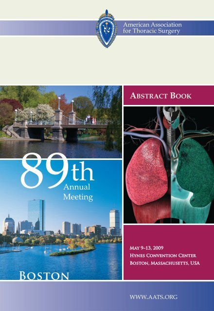
<strong>American</strong> <strong>Association</strong><strong>for</strong> <strong>Thoracic</strong> <strong>Surgery</strong>ABSTRACT BOOK89 AnnualthMeeting<strong>Boston</strong>May 9–13, 2009Hynes Convention Center<strong>Boston</strong>, Massachusetts, USAWWW.AATS.ORG
AMERICAN ASSOCIATIONFOR THORACIC SURGERY89 th ANNUAL MEETING<strong>Boston</strong>, MassachusettsTABLE OF CONTENTSAbstracts .................................................................................................. 54Accreditation..............................................................................................1Adult Cardiac <strong>Surgery</strong> Symposium............................................................ 13Author Index...........................................................................................212Committees............................................................................................ 222Congenital Heart Disease Symposium ....................................................... 18Council...................................................................................................221Developing the Academic Surgeon Symposium ...........................................11Disclosure Policy ........................................................................................5Future Meeting Dates ......................................................... inside back coverGeneral Meeting In<strong>for</strong>mation......................................................................1General <strong>Thoracic</strong> <strong>Surgery</strong> Symposium ....................................................... 16C. Walton Lillehei Resident Forum Session ................................................ 54Scientific Program .................................................................................... 24Speaker and Discussant Guidelines .............................................................6
AMERICAN ASSOCIATION FOR THORACIC SURGERY2009AATS ABSTRACT BOOKGENERAL MEETING INFORMATIONAbout AATSPromoting Scholarship in <strong>Thoracic</strong> and Cardiovascular <strong>Surgery</strong>Founded in 1917 by the earliest pioneers in the field of thoracic surgery,the <strong>American</strong> <strong>Association</strong> <strong>for</strong> <strong>Thoracic</strong> <strong>Surgery</strong> (AATS) is now an internationalorganization of over 1,200 of the world’s <strong>for</strong>emost cardiothoracicsurgeons representing 35 countries.Surgeons must have a proven record of distinction within the cardiothoracicsurgical field and have made meritorious contributions to the extantknowledge base about cardiothoracic disease and its surgical treatment tobe considered <strong>for</strong> membership. The annual meeting, research grantsand awards, educational symposia and courses, and the AATS officialjournal, the Journal of <strong>Thoracic</strong> and Cardiovascular <strong>Surgery</strong>, all strengthenits commitment to science, education and research.The officers and councilors of the AATS welcome you to the 89 thAnnual Meeting in <strong>Boston</strong>, Massachusetts, USA.AATS Annual Meeting AccreditationThe <strong>American</strong> <strong>Association</strong> <strong>for</strong> <strong>Thoracic</strong> <strong>Surgery</strong> is accredited by theAccreditation Council <strong>for</strong> Continuing Medical Education to providecontinuing medical education <strong>for</strong> physicians.The <strong>American</strong> <strong>Association</strong> <strong>for</strong> <strong>Thoracic</strong> <strong>Surgery</strong> designates this educationalactivity <strong>for</strong> a maximum of 35 AMA PRA Category 1 Credit(s).Physicians should only claim credit commensurate with the extent oftheir participation in the activity.1
89 TH ANNUAL MEETING MAY 9–MAY 13, 2009BOSTON, MASSACHUSETTSThe <strong>American</strong> <strong>Association</strong> <strong>for</strong> <strong>Thoracic</strong> <strong>Surgery</strong> designates the followingcredit hours:Saturday, May 9, 2009 – up to 7.2 hours❏ New Technologies and Procedures in Congenital and AcquiredHeart <strong>Surgery</strong>, up to 3.5 hours❏ <strong>Thoracic</strong> Developmental Skills, up to 3.5 hours❏ Developing the Academic Surgeon Symposium, up to 3.7 hoursSunday, May 10, 2009 – up to 7.3 hours❏ AATS/STS Adult Cardiac <strong>Surgery</strong> Symposium, up to 7.25 hours❏ AATS/STS General <strong>Thoracic</strong> <strong>Surgery</strong> Symposium, up to 7 hours❏ AATS/STS Congenital Heart Disease Symposium, up to 7.3 hours❏ C. Walton Lillehei Resident Forum, up to 2 hoursMonday, May 11, 2009 – up to 7.4 hours❏ Plenary Scientific Session, Basic Science Lecture, PresidentialAddress, up to 3.8 hours❏ Simultaneous Scientific Session – Adult Cardiac <strong>Surgery</strong>, up to2.3 hours❏ Simultaneous Scientific Session – General <strong>Thoracic</strong> <strong>Surgery</strong>, upto 2.6 hours❏ Simultaneous Scientific Session – Congenital Heart Disease, upto 2.3 hours❏ NHLBI STICH Trial Debate, up to 1 hourTuesday, May 12, 2009 – up to 7.5 hours❏ Cardiac <strong>Surgery</strong> Forum Session, up to 1.75 hours❏ General <strong>Thoracic</strong> Forum Session, up to 1.75 hours2
AMERICAN ASSOCIATION FOR THORACIC SURGERY❏ Plenary Scientific Session, Simulation Session, Honored SpeakerLecture, up to 3.2 hours❏ Simultaneous Scientific Session – Adult Cardiac <strong>Surgery</strong>, up to2.25 hours❏ Simultaneous Scientific Session – General <strong>Thoracic</strong> <strong>Surgery</strong>, up to2.25 hours❏ Simultaneous Scientific Session – Congenital Heart Disease, up to2.5 hoursWednesday, May 13, 2009 – up to 5 hours❏ Emerging Technologies and Techniques Forum, up to 2 hours❏ Plenary Scientific Session – Controversies, up to 1 hour❏ Ablation vs. <strong>Surgery</strong> <strong>for</strong> Atrial Fibrillation: Antagonism orSynergism?, up to 2 hours❏ Pneumonectomy: A Treatment or a Disease?, up to 2 hoursFor further in<strong>for</strong>mation on the Accreditation Council <strong>for</strong> ContinuingMedical Education (ACCME) Standards of Commercial Support, pleasevisit www.accme.org.CME KiosksAll surgeons looking to obtain their Continuing Medical Educationcredits may do so at the CME Pavilion located on the Second Levelof the Convention Center in the Hall D Foyer adjacent to Registration.The CME Pavilion computers will allow attendees to log on and manageall of their CME credits <strong>for</strong> the Annual Meeting. At the conclusionof the meeting, attendees may print their CME certificate and/or Letter ofAttendance. Following the meeting, attendees will be able to access thismaterial on the AATS website.3
89 TH ANNUAL MEETING MAY 9–MAY 13, 2009BOSTON, MASSACHUSETTSEducational ObjectivesAt the conclusion of the AATS Annual Meeting, through comprehensivelectures and discussions, participants will be able to:❏ Implement the latest techniques and current research specificallyrelated to Adult Cardiac <strong>Surgery</strong>, Congenital Heart Disease,and General <strong>Thoracic</strong> <strong>Surgery</strong>❏ Analyze the pros and cons of each paper presented to implementbest practices in their current practices❏ Select and apply appropriate surgical procedures and other interventions<strong>for</strong> their own patients based upon results presented❏ Utilize basic science developments and emerging technologiesand techniques across the spectrum of Cardiothoracic <strong>Surgery</strong>❏ Apply state-of-the art knowledge into their current practiceTarget AudienceThe AATS Annual Meeting is specifically designed to meet the educationalneeds of:❏ Cardiothoracic Surgeons❏ Physicians in related specialties including CardiothoracicAnesthesia, Cardiology, Pulmonology, Radiology, Gastroenterologyand <strong>Thoracic</strong> Oncology❏ Fellows and Residents in Cardiothoracic and General Surgicaltraining programs❏ Allied Health Professionals involved in the care of cardiothoracicsurgical patients❏ Medical students with an interest in Cardiothoracic <strong>Surgery</strong>4
AMERICAN ASSOCIATION FOR THORACIC SURGERYDisclosure PolicyIt is the policy of the <strong>American</strong> <strong>Association</strong> <strong>for</strong> <strong>Thoracic</strong> <strong>Surgery</strong> thatany individual who makes a presentation or is a co-author on a programdesignated <strong>for</strong> AMA Physician’s Recognition Award Category 1 Creditmust disclose any financial interest or other relationship (grant, researchsupport, consultant, etc.) that individual has with any manufacturer(s)of any commercial product(s) that may be discussed in the individual’spresentation. This policy is established neither to imply any positionregarding the propriety of such relationships nor to prejudice any individualfrom making a presentation but to allow the participants to<strong>for</strong>m their own judgments regarding the presentation.Authors who may have a possible conflict of interest are denoted inthe disclosure index. Authors must disclose any material, financial, orother relationships that may pose conflict of interest at the time ofpresentation.Camera, Recording, Cell Phone and No-Smoking PoliciesDue to privacy issues, it is the policy of AATS that no cameras are permittedin the meeting sessions or exhibit hall. Please refrain from takingphotos in these locations. Audio and videotaping are also prohibited.For the courtesy of all faculty and participants, please ensure that cellphone ringers are silenced during all sessions.Smoking is not permitted in the Convention Center, Hotels or SpecialEvent Venues.5
89 TH ANNUAL MEETING MAY 9–MAY 13, 2009BOSTON, MASSACHUSETTSSPEAKER AND DISCUSSANT GUIDELINESPresentation/Discussion GuidelinesDiscussion of Papers: Members, non-member physicians and invitedspeakers have the privilege of discussing papers. Discussants are limitedto 2 minutes and must limit their discussion to specific questions directlyrelated to the author’s presentation. All discussants should registerwith the Session Moderator prior to the opening of the session duringwhich the paper is to be presented. All discussion will be presentedfrom floor microphones and may not be accompanied by slides.Program Presentation TotalDiscussion*Plenary Sessions 8 minutes 12 minutesSimultaneous Sessions 8 minutes 12 minutesAdult Cardiac &General <strong>Thoracic</strong> ForumEmerging Technologies &Techniques ForumC. Walton LilleheiResident Forum5 minutes 7 minutes5 minutes 7 minutes7 minutes 8 minutesControversies (Debates)8 minutes each × 2 Rebuttals8 minutes each × 2 Rebuttals20 minutes*All discussants are limited to 2 minutes6
AMERICAN ASSOCIATION FOR THORACIC SURGERYIn accordance with the By-Laws of the <strong>Association</strong>:1. Papers which are read at the meeting shall become the property ofthe <strong>Association</strong>. They shall be submitted electronically to the Editorprior to presentation (http://www.editorialmanager.com/jtcvs).The papers submitted <strong>for</strong> consideration <strong>for</strong> publication in theJournal of <strong>Thoracic</strong> and Cardiovascular <strong>Surgery</strong> must bear a closerelationship in length to the paper presented at the meeting.2. Submission and acceptance of an abstract constitutes a commitmentby the Author(s) to present the material at the AATS AnnualMeeting. The work must not have been submitted, presented orpublished in abstract or manuscript <strong>for</strong>m elsewhere prior tothe AATS 89 th Annual Meeting in May 2009. Failure to meet thisrequirement without prior approval of the <strong>Association</strong> will jeopardizea presenter’s further acceptance of abstracts <strong>for</strong> presentationand/or publication. The AATS Council seriously regards andadheres to the submission/presentation policy and will strictlyen<strong>for</strong>ce sanctions upon all authors who fail to meet the policiesoutlined in the rules <strong>for</strong> submission and presentation of abstractsonce submitted. Any questions should be addressed to the Secretaryof the <strong>Association</strong>.7
89 TH ANNUAL MEETING MAY 9–MAY 13, 2009BOSTON, MASSACHUSETTSPROGRAM INFORMATIONSATURDAY, MAY 9, 20098:00 a.m. NEW TECHNOLOGIES AND PROCEDURES INCONGENITAL AND ACQUIRED HEART SURGERYRoom 302–306, Hynes Convention CenterChairman: Mark E. Galantowicz, MDNationwide Children’s Hospital8:00 a.m. – 8:10 a.m. WELCOME AND INTRODUCTION8:10 a.m – 8:30 a.m. Overview of Pediatric Heart AssistDevices in DevelopmentTim Baldwin, PhDNational Heart, Lung and Blood Institute8:30 a.m. – 8:50 a.m. A Decision Matrix to Guide DeviceSelection <strong>for</strong> Pediatric CirculatorySupportBrian W. Duncan, MDCleveland Clinic Foundation8:50 a.m. – 9:10 a.m. A Decision Matrix <strong>for</strong> Adult MechanicalCirculatory SupportBenjamin C. Sun, MDOhio State University9:10 a.m. – 9:30 a.m. Heart Transplantation and HighPulmonary Vascular Resistance –Another Fallen Commandment?Sanjiv K. Gandhi, MDSaint Louis Children’s Hospital8
AMERICAN ASSOCIATION FOR THORACIC SURGERY9:30 a.m. – 10:00 a.m. Pulmonary Assist Devices – Where DoWe Stand?Robert H. Bartlett, MDUniversity of Michigan10:00 a.m. – 10:30 a.m. BREAK10:30 a.m. – 10:50 a.m. Hybrid Approach to HLHSMark E. Galantowicz, MDNationwide Children’s Hospital10:50 a.m. – 11:10 a.m. Hybrid Approach to Other CongenitalHeart DefectsEmile A. Bacha, MDChildren’s Hospital <strong>Boston</strong>11:10 a.m. – 11:30 a.m. Advanced Imaging in Coronary <strong>Surgery</strong>and Hybrid ProceduresJohn G. Byrne, MDVanderbilt Heart Institute11:30 a.m. – 11:50 a.m. 3-D Image Based Surgical Planning ofAortic Valve RepairPedro J. del Nido, MDChildren’s Hospital <strong>Boston</strong>11:50 a.m. –12:00 p.m. DISCUSSION12:00 p.m. ADJOURN9
89 TH ANNUAL MEETING MAY 9–MAY 13, 2009BOSTON, MASSACHUSETTS8:00 a.m. THORACIC DEVELOPMENTAL SKILLSRoom 312, Hynes Convention CenterChairman: Raja M. Flores, MDMemorial Sloan-Kettering Cancer Center8:00 a.m. – 8:05 a.m. WELCOME AND INTRODUCTIONRaja M. Flores, MDMemorial Sloan-Kettering Cancer Center8:05 a.m. – 8:25 a.m. The <strong>Thoracic</strong> Surgeon and EndocbronchialUltrasoundHarvey I. Pass, MDNew York University8:25 a.m. – 8:45 a.m. VATS Lobectomy: Technical Details of AllFive LobesRaja M. Flores, MDMemorial Sloan-Kettering Cancer Center8:45 a.m. – 9:05 a.m. Robotic LobectomyBernard J. Park, MDMemorial Sloan-Kettering Cancer Center9:05 a.m. – 9:25 a.m. Thoracoscopic LVRS and SympathectomyRobert J. McKenna, MDCedars Sinai Medical Center9:25 a.m. – 9:45 a.m. Resection and Reconstruction of MajorIntrathoracic Vascular StructuresErino A.Rendina, MDUniversity La Sapienza9:45 a.m. – 10:15 a.m. BREAK10:15 a.m. – 10:35 a.m. Ivor Lewis Esophagectomy – Refined,Expeditious and Oncologically SoundManjit S. Bains, MDMemorial Sloan-Kettering Cancer Center10
AMERICAN ASSOCIATION FOR THORACIC SURGERY10:35 a.m. – 10:55 a.m. Minimally Invasive EsophagectomyMichael S. Kent, MDBeth Israel Deaconess Medical Center10:55 a.m. – 11:15 a.m. Tracheal Lesions – Initial Managementand Subsequent Surgical TreatmentJoel D. Cooper, MDUniversity of Pennsylvania11:15a.m. – 11:35 a.m.Chest Wall Resection and ReconstructionMichae J. Weyant, MDUniversity of Colorado11:35 a.m. – 12:00 p.m. DISCUSSION12:00 p.m. ADJOURN1:00 p.m. DEVELOPING THE ACADEMIC SURGEONSYMPOSIUMRoom 302-306, Hynes Convention CenterChairman: A. Marc Gillinov, MDCleveland Clinic Foundation1:00 p.m. – 1:10 p.m. INTRODUCTION AND COURSEOVERVIEWA. Marc Gillinov, MDCleveland Clinic Foundation1:10 p.m. – 1:30 p.m. Academic Practice in Cardiothoracic<strong>Surgery</strong>: An Outdated Concept?Irving L. Kron, MDUniversity of Virginia Health System1:30 p.m. – 1:50 p.m. The Cardiothoracic Surgeon as EducatorStephen C. Yang, MDJohns Hopkins Medical Institute1:50 p.m. – 2:10 p.m. Basic Research in <strong>Thoracic</strong> <strong>Surgery</strong>David R. Jones, MDUniversity of Virginia Health System11
89 TH ANNUAL MEETING MAY 9–MAY 13, 2009BOSTON, MASSACHUSETTS2:10 p.m. – 2:30 p.m. Research and Medical Device DevelopmentA. Marc Gillinov, MDCleveland Clinic Foundation2:30 p.m. – 3:00 p.m. Cardiothoracic <strong>Surgery</strong>: A Time ofOpportunityJames L. Cox, MDWashington University3:00 p.m. – 3:20 p.m. BREAK3:20 p.m. – 3:40 p.m. Beyond Clinical Trials: Building anEvidence Basis <strong>for</strong> Cardiothoracic<strong>Surgery</strong> of the FutureEugene H. Blackstone, MDCleveland Clinic Foundation3:40 p.m. – 4:00 p.m. The NIH-Sponsored CardiothoracicSurgical Trials NetworkTimothy J. Gardner, MDChristiana Care Health System4:00 p.m. – 4:20 p.m. Training <strong>for</strong> the FutureMathew R. Williams, MDColumbia University4:20 p.m. – 4:40 p.m. Cardiovascular Disease: An Integrated,Programmatic ApproachBruce W. Lytle, MDCleveland Clinic Foundation4:40 p.m. – 5:00 p.m. DISCUSSION5:00 p.m. ADJOURN12
AMERICAN ASSOCIATION FOR THORACIC SURGERYSUNDAY, MAY 10, 20098:00 a.m. – 5:00 p.m. AATS/STS ADULT CARDIAC SURGERYSYMPOSIUMBallroom A–C, Hynes Convention CenterCo-Chairmen: Michael A. Acker, MDJoseph E. Bavaria, MDUniversity of PennsylvaniaSESSION ITHORACIC AORTA8:00 a.m. – 8:20 a.m. TEVAR: Indications, Current Trends andResultsG. Chad Hughes, MD, Duke University8:20 a.m. – 8:40 a.m. TEVAR in Setting of Aortic Dissection:Type A and BAlberto Pochettino, MD, University of Pennsylvania8:40 a.m. – 9:00 a.m. Hybrid ArchWilson Y. Szeto, MD, University of Pennsylvania9:00 a.m. – 9:15 a.m. DISCUSSIONSESSION IIAORTIC ROOT/AORTIC VALVE9:15 a.m. – 9:35 a.m. Aortic Valve Repair/Retention with AorticRoot DiseaseG. Michael Deeb, MD, University of Michigan9:35 a.m. – 9:55 a.m. Aortic Valve Repair without Root PathologyTricuspid/BicuspidGebrine El Khoury, MD, St-Luc Hospital9:55 a.m. – 10:15 a.m. BREAK10:15 a.m. – 10:35 a.m. Transcatheter Aortic Valve Replacement:TransfemoralJoseph E. Bavaria, MD, University of Pennsylvania13
89 TH ANNUAL MEETING MAY 9–MAY 13, 2009BOSTON, MASSACHUSETTS10:35 a.m. – 10:55 a.m. Transcatheter Aortic Valve Replacement:TransapicalTodd M. Dewey, MD, Medical City Dallas Hospital10:55 a.m. – 11:10 a.m. DISCUSSIONSESSION IIIMITRAL VALVE REPAIR11:10 a.m. – 11:30 a.m. 3-D Echo: Clinical Breakthrough orPretty Pictures?Joseph S. Savino, MD, University of Pennsylvania11:30 a.m. – 11:50 a.m. Degenerative Disease: Complex RepairMade SimpleDavid H. Adams, MD, Mount SinaiMedical Center11:50 a.m. – 12:05 p.m. DISCUSSION12:05 p.m. – 1:05 p.m. LUNCH —Hall A, Plaza LevelSESSION IVSURGICAL DECISION MAKING: WHEN ARE TWOVALVE PROCEDURES BETTER THAN ONE? /ATRIAL FIBRILLATION1:05 p.m. – 1:25 p.m. The Surgical Treatment of Atrial Fibrillation:Are We Ready <strong>for</strong> Prime Time?Niv Ad, MD, Fairfax Hospital1:25 p.m. – 1:45 p.m. Aortic Stenosis/Mitral Regurgitation:When to Repair the Mitral Valve?Thomas G. Gleason, MD, University of Pittsburgh1:45 p.m. – 2:05 p.m. Mitral Stenosis/Mitral Regurgitation andTricuspid Regurgitation: When to Repairthe Tricuspid Valve?Richard J. Shemin, MD, University ofCali<strong>for</strong>nia, Los Angeles2:05 p.m. – 2:20 p.m. DISCUSSION14
AMERICAN ASSOCIATION FOR THORACIC SURGERYSESSION VHEART FAILURE2:20 p.m. – 2:40 p.m. Should Functional Mitral Regurgitationin Heart Failure Patients be Repaired?Patrick M. McCarthy, MD, NorthwesternUniversity2:40 p.m. – 3:00 p.m. Small VADs: Solution <strong>for</strong> Heart FailureMichael A. Acker, MD, University of Pennsylvania3:00 p.m. – 3:20 p.m. Indications <strong>for</strong> Surgical Revascularizationin Heart Failure PatientsY. Joseph Woo, MD, University of Pennsylvania3:20 p.m. – 3:35 p.m. DISCUSSION3:35 p.m. – 3:55 p.m. BREAKSESSION VICORONARY ARTERY BYPASS3:55 p.m. – 4:15 p.m. Drug Eluding Stents: Has the PendulumStarted to Swing Back?David P. Taggart, MD, University of Ox<strong>for</strong>d4:15 p.m. – 4:35 p.m. Debate-Optimal Coronary Revascularization:Off Pump (Puskas) vs. On Pump (Sabik) vs.Hybrid (Byrne)John D. Puskas, MD, Emory UniversityJoseph F. Sabik, MD, Cleveland ClinicJohn G. Byrne, MD, Vanderbilt Heart Institute4:35 p.m. – 4:50 p.m. DISCUSSION4:50 p.m. ADJOURN TO WELCOME RECEPTION —Exhibit Hall, Level 215
89 TH ANNUAL MEETING MAY 9–MAY 13, 2009BOSTON, MASSACHUSETTSSUNDAY, MAY 10, 20098:00 a.m. – 5:00 p.m. AATS/STS GENERAL THORACICSURGERY SYMPOSIUMRoom 302–306, Hynes Convention CenterChairman: David H. Harpole, Jr., MDDuke University8:00 a.m. – 8:10 a.m. INTRODUCTION AND COURSEOVERVIEWDavid H. Harpole, Jr., MD, Duke UniversitySESSION INON-SMALL CELL LUNG CANCER8:10 a.m. – 8:30 a.m. New Lung Cancer Staging SystemJoe B. Putnam, MD, Vanderbilt University8:30 a.m. – 9:00 a.m. Chemotherapy 101Ramaswamy Govindan, MD, WashingtonUniversity9:00 a.m. – 9:30 a.m. Tyrosine Kinase InhibitorsPasi A. Janne, MD, PhD, Dana-FarberCancer Institute9:30 a.m. – 10:00 a.m. Anti-Angiogenesis and Other MolecularTargetsThomas J. Lynch, MD, MassachusettsGeneral Hospital10:00 a.m. – 10:15 a.m. DISCUSSION10:15 a.m. – 10:45 a.m. BREAK10:45 a.m. – 11:15 a.m. Radiotherapy 101Jeffrey Bogart, MD, State University ofNew York Upstate Medical University11:15 a.m. – 11:45 a.m. Current Early Stage Lung Cancer TrialsEric Valleries, MD, Swedish Cancer Institute11:45 a.m. – 12:00 p.m. DISCUSSION12:00 p.m. – 1:00 p.m. LUNCH —Hall A, Plaza Level16
AMERICAN ASSOCIATION FOR THORACIC SURGERYSESSION IICONTROVERSIES IN LUNG CANCER1:00 p.m. – 1:30 p.m. Mediastinscopy or EBUS/EUSBryan F. Meyers, MD, MPH, WashingtonUniversity1:30 p.m. – 2:00 p.m. Who Is Medically Inoperable?Robert J. Cerfolio, MD, University of Alabama2:00 p.m. – 2:30 p.m. Ablative Therapies <strong>for</strong> NodulesJo-Anne O. Shepard, MD, MassachusettsGeneral Hospital2:30 p.m. – 3:00 p.m. Upfront or Outback ChemotherapyRamaswamy Govindan, MD, WashingtonUniversity3:00 p.m. – 3:30 p.m. BREAKSESSION IIIUPDATES OF GENERAL THORACIC SURGERY3:30 p.m. – 4:00 p.m. STS Database Risk AdjustmentCameron D. Wright, MD, MassachusettsGeneral Hospital4:00 p.m. – 4:20 p.m. Pulmonary MetastasectomyThomas A. D’Amico, MD, Duke University4:20 p.m. – 4:40 p.m. Interventions <strong>for</strong> EmphysemaMalcolm M. DeCamp, MD, Beth IsraelDeaconess Medical Center4:40 p.m. – 5:00 p.m. DISCUSSION5:00 p.m. ADJOURN TO WELCOME RECEPTION —Exhibit Hall, Level 217
89 TH ANNUAL MEETING MAY 9–MAY 13, 2009BOSTON, MASSACHUSETTSSUNDAY, MAY 10, 20098:00 a.m. – 5:00 p.m. AATS/STS CONGENITAL HEARTDISEASE SYMPOSIUMRoom 312, Hynes Convention CenterChairman: J. William Gaynor, MDChildren’s Hospital ofPhiladelphiaWELCOME AND INTRODUCTIONJ. William Gaynor, MDChildren’s Hospital of PhiladelphiaSESSION IPRO/CON DEBATE: NIRS IS “STANDARD OF CARE”FOR POST-OPERATIVE MANAGEMENT8:00 a.m. – 8:15 a.m. Pro: James S. Tweddell, MDMedical College of Wisconsin8:15 a.m. – 8:30 a.m. Con: Jennifer C. Hirsch, MDUniversity of Michigan8:30 a.m. – 8:35 a.m. Rebuttal: James S. Tweddell, MD8:35 a.m. – 8:40 a.m. Rebuttal: Jennifer C. Hirsch, MD8:40 a.m. – 9:00 a.m. DISCUSSIONSESSION IISURGICAL TECHNIQUES “HOW I DO IT”9:00 a.m. – 9:20 a.m. Aortic Valvuloplasty <strong>for</strong> Aortic RegurgitationRichard A. Jonas, MDChildren’s National Medical Center9:20 a.m. – 9:30 am DISCUSSION18
AMERICAN ASSOCIATION FOR THORACIC SURGERYSESSION IIIPRO/CON DEBATE: USE OF A FENESTRATION SHOULDBE ROUTINE DURING THE FONTAN PROCEDURE9:30 a.m. – 9:45 a.m. Pro: Scott M. Bradley, MD, Medical Universityof South Carolina9:45 a.m. – 10:00 a.m. Con: Frank L. Hanley, MD, Stan<strong>for</strong>d University10:00 a.m. – 10:05 a.m. Rebuttal: Scott M. Bradley, MD10:05 a.m. – 10:10 a.m. Rebuttal: Frank L. Hanley, MD10:10 a.m. – 10:30 a.m. DISCUSSION10:30 a.m. – 10:50 a.m. BREAKSESSION IVCLINICAL RESEARCH IN PEDIATRIC CARDIACSURGERY: HOW CAN WE DO BETTER?10:50 a.m. – 11:05 a.m. Uses and Limitations of Academic andRegistry DatabasesWilliam G. Williams, MD, The Hospital <strong>for</strong>Sick Children11:05 a.m. – 11:20 a.m. Randomized Treatment Trials: LessonsLearned from the BCAS and Other StudiesJane W. Newburger, MD, MPH, Children’sHospital <strong>Boston</strong>11:20 a.m. – 11:35 a.m. Multi-Institutional Studies: LessonsLearned from the CHSS StudiesChristopher A. Caldarone, MD, The Hospital<strong>for</strong> Sick Children11:35 a.m. – 11:50 a.m. Multi-Institutional Studies: LessonsLearned from the SVR TrialRichard G. Ohye, MD, University of Michigan11:50 a.m. – 12:00 p.m. DISCUSSION12:00 p.m. – 1:00 p.m. LUNCH —Hall A, Plaza Level19
89 TH ANNUAL MEETING MAY 9–MAY 13, 2009BOSTON, MASSACHUSETTSSESSION VVARIABILITY IN OUTCOMES1:00 p.m. – 1:20 p.m. Perioperative Genomics: Why Do “Similar”Patients Have Different Outcomes?Mark F. Newman, MD, Duke University1:20 p.m. – 1:40 p.m. Does Protocol Driven Post-OperativeManagement Improve Outcomes?Peter C. Laussen, MD, Children’s Hospital of<strong>Boston</strong>1:40 p.m. – 2:00 p.m. Difficulties Comparing Outcomes BetweenInstitutionsKarl F. Welke, MD, Oregon Health and ScienceUniversity2:00 p.m. – 2:20 p.m. How Do We Translate Clinical Research toClinical PracticeGil Wernovsky, MD, Children’s Hospital ofPhiladelphia2:20 p.m. – 2:30 p.m. DISCUSSIONSESSION VISURGICAL TECHNIQUES: “HOW I DO IT”2:30 p.m. – 2:50 p.m. Living-Donor Lobar Lung TransplantationVaughn A. Starnes, MD, University of SouthernCali<strong>for</strong>nia2:50 p.m. – 3:00 p.m. DISCUSSION3:00 p.m. – 3:20 p.m. BREAK20
AMERICAN ASSOCIATION FOR THORACIC SURGERYSESSION VIITHE FONTAN/KREUTZER PROCEDURE AT 403:20 p.m. – 3:30 p.m. Surgical Repair of Tricuspid AtresiaFrancis M. Fontan, MD3:30 p.m. – 3:40 p.m. An Operation <strong>for</strong> Correction of TricuspidAtresiaGuillermo O. Kreutzer, MD, Ricardo GutierrezChildren’s Hospital3:40 p.m. – 4:00 p.m. Evolution of the Fontan/Kreutzer ProcedureMarc R. de Leval, MD, International CongenitalCardiac Centre4:00 p.m. – 4:20 p.m. Current Status of Survivors of the Fontan/Kreutzer ProcedureJack Rychik, MD, Children’s Hospital ofPhiladelphia4:20 p.m. – 4:40 p.m. The Fontan/Kreutzer Procedure: FutureDirectionsEdward L. Bove, MD, University of Michigan4:40 p.m. – 5:00 p.m. DISCUSSION5:00 p.m. ADJOURN TO WELCOME RECEPTION —Exhibit Hall, Level 221
89 TH ANNUAL MEETING MAY 9–MAY 13, 2009BOSTON, MASSACHUSETTSSUNDAY AFTERNOONMAY 10, 20093:00 p.m. C. WALTON LILLEHEI RESIDENT FORUM SESSIONRoom 311, Hynes Convention Center(7 minutes presentation, 8 minutes discussion)Moderators: Gus J. Vlahakes, Ara A. VaporciyanL1. In Vivo Structure and Function of Engineered PulmonaryValvesDanielle Gottlieb 1 , Kunal Tandon 1 , Sitaram Emani 1 , Elena Aikawa 2 ,David W. Brown 1 , Andrew J. Powell 1 , Arthur Nedder 1 , Michael S. Sacks 3 ,John E. Mayer 1*1. Children’s Hospital <strong>Boston</strong> and Harvard Medical School, <strong>Boston</strong>, MA, USA;2. Massachusetts General Hospital and Harvard Medical School, <strong>Boston</strong>, MA,USA; 3. University of Pittsburgh, Pittsburgh, PA, USAL2. The Graft Imaging to Improve Patency (GRIIP) Trial ResultsSteve Singh, Nimesh Desai, † Genta Chikazawa, Hiroshi Tsuneyoshi,Visal Pen, Jessica Vincent, Jennifer Ku, Fuad Moussa, Gideon Cohen,George Christakis, * Stephen E. Fremes *Sunnybrook Health Sciences Centre, Toronto, ON, CanadaL3. Tissue Engineered Pro-Angiogenic Fibroblast Matrix ImprovesMyocardial Perfusion and Function and Limits VentricularRemodeling Following InfarctionJ. Raymond Fitzpatrick, John R. Frederick, Ryan C. McCormick,David A. Harris, Ah-Young Kim, Max J. Smith, Carine M. Laporte,Jeffrey R. Muenzer, Alex J. Gambogi, Y. Joseph Woo *Division of Cardiovascular <strong>Surgery</strong>, Hospital of the University of Pennsylvania,Philadelphia, PA, USAL4. Atorvastatin at Reperfusion Reduces Myocardial Infarct Sizein Mice by Activating eNOS of Bone Marrow-Derived CellsZequan Yang, 1 Gorav Ailawadi, 1† Joel Linden, 2 Brent A. French, 3 Irving L. Kron 1*1. <strong>Surgery</strong>, University of Virginia Health System, Charlottesville, VA, USA;2. Medicine, University of Virginia Health System, Charlottesville, VA, USA;3. Biomedical Engineering, University of Virginia Health System, Charlottesville,VA, USA* AATS Member† Resident Traveling Fellowship 200622
AMERICAN ASSOCIATION FOR THORACIC SURGERYL5. Quantitative Assessment of Technical Proficiency of Residentsin Cardiac <strong>Surgery</strong>Hiroo Takayama, Yoshifumi Naka, * Mehmet C. Oz, *† Allan S. Stewart,Mathew R. Williams, Craig R. Smith, * Micheal ArgenzianoColumbia University, New York, NY, USAL6. Divergent Impact of Osteopontin Iso<strong>for</strong>ms on Lung CancerAngiogenesisJustin D. Blasberg, Jessica S. Donington, Chandra M. Goparaju,Harvey I. Pass *New York University Medical Center, New York, NY, USAL7. Temporary Acute Mechanical Circulatory Support <strong>for</strong> AcuteCirculatory Collapse: Experience with 266 PatientsKristopher B. Deatrick, Amit K. Mathur, Ann Schumar, Robert H. Bartlett,Francis D. Pagani, * Jonathan W. HaftCardiac <strong>Surgery</strong>, The University of Michigan, Ann Arbor, MI, USAL8. Age Is an Independent Risk Factor <strong>for</strong> Aspiration FollowingThoracotomy <strong>for</strong> Pulmonary ResectionWilliam B. Keeling 1 , Jonathan M. Hernandez 2 , Vicki Lewis 3 , Melissa Czapla 3 ,Weiwei Zhu 3 , Joseph Garrett 2 , Eric Sommers 21. Emory University, Atlanta, GA, USA; 2. University of South Florida,Tampa, FL, USA; 3. H. Lee Moffitt Cancer Center, Tampa, FL, USA5:00 p.m. ADJOURN TO WELCOME RECEPTIONExhibit Hall, Level 2* AATS Member† Robert E. Gross Research Scholarship 199423
89 TH ANNUAL MEETING MAY 9–MAY 13, 2009BOSTON, MASSACHUSETTSMONDAY MORNINGMAY 11, 20097:30 a.m. BUSINESS SESSION(AATS Members Only)Ballroom A–C, Hynes Convention Center7:45 a.m. PLENARY SCIENTIFIC SESSIONBallroom A–C, Hynes Convention Center(8 minutes presentation, 12 minutes discussion)1. A Formidable Task: Population Analysis Predicts a Deficit of 2,000Cardiothoracic Surgeons by 2030Thomas E. Williams, * Benjamin Sun, Patrick Ross, Andrew M. Thomas<strong>Surgery</strong>, Ohio State University, Columbus, OH, USAInvited Discussant: Irving L. Kron2. Single Center Experience in Treatment of Cardiogenic Shock ofAny Etiology in Children by Pediatric Ventricular Assist DevicesRoland Hetzer, * Evgenij V. Potapov, Oliver Miera, Yu-Guo Weng, Michael Hübler,Felix BergerDHZB, Berlin, GermanyInvited Discussant: Charles Fraser, Jr.3. Long-Term Results of Aortic Valve Sparing Operations in Patientswith Marfan SyndromeTirone E. David, * Susan Armstrong, Manjula Maganti, Jack Colman,Timothy BradleyCardiovascular <strong>Surgery</strong>, Toronto General Hospital, Toronto, ON, CanadaInvited Discussant: Lars G. Svensson4. Outcomes After Laparoscopic Giant Paraesophageal Hernia Repairin 636 PatientsJames D. Luketich, * Katie S. Nason, Rodney J. Landreneau, * Samuel Keeley,Omar Awais, Manisha Shende, Matthew J. Schuchert, Ghulam Abbas,Blair A. Jobe, Arjun PennathurThe Heart, Lung and Esophageal <strong>Surgery</strong> Institute, University of Pittsburgh MedicalCenter, Pittsburgh, PA, USAInvited Discussant: Antoon Lerut* AATS Member24
AMERICAN ASSOCIATION FOR THORACIC SURGERY9:05 a.m. AWARD PRESENTATIONSBallroom A–C, Hynes Convention CenterLifetime Achievement AwardThomas B. Ferguson, MDWashington University School of MedicineC. Walton Lillehei Forum AwardTSRA McGoon AwardTSFRE Report9:20 a.m. INTERMISSION – VISIT EXHIBITSExhibit Hall10:00 a.m. BASIC SCIENCE LECTUREBallroom A–C, Hynes Convention CenterInsights from Developmental and Stem Cell BiologyJonathan A. Epstein, MDWilliam Wikoff Smith Professor of MedicineChairman, Department of Cell and Developmental BiologyScientific Director, Penn Cardiovascular InstituteFounding Co-Director, Penn Institute <strong>for</strong> Regenerative MedicineUniversity of PennsylvaniaIntroduced By: Thomas L. Spray, MD10:40 a.m. PLENARY SCIENTIFIC SESSIONModerators: Alec PattersonThoralf M. Sundt, III5. The Relationship Between Hospital CABG Volume and MultipleDimensions of CABG QualityDavid M. Shahian, 1* Sean O’Brien, 2 Sharon-Lise Normand, 3 Eric Peterson, 2Fred Edwards 4*1. Massachusetts General Hospital, <strong>Boston</strong>, MA, USA; 2. Duke Clinical Research Institute,Durham, NC, USA; 3. Harvard Medical School, <strong>Boston</strong>, MA; USA, 4. University ofFlorida, Jacksonville, FL, USAInvited Discussant: T. Bruce Ferguson, Jr.* AATS Member25
89 TH ANNUAL MEETING MAY 9–MAY 13, 2009BOSTON, MASSACHUSETTS6. Survival After Transapical and Transarterial Aortic ValveImplantation: Talking About Two Different Patient PopulationsSabine Bleiziffer, Hendrik Ruge, Domenico Mazzitelli, Christian Schreiber,Andrea Hutter, Robert Bauernschmitt, Ruediger Lange *Clinic <strong>for</strong> Cardiovascular <strong>Surgery</strong>, German Heart Center Munich, Munich, GermanyInvited Discussant: Michael J. Mack11:25 a.m. PRESIDENTIAL ADDRESSThe Quality ConundrumThomas L. Spray, MD, Philadelphia, PAIntroduced By: Alec Patterson, MD12:15 p.m. LUNCH – VISIT EXHIBITSExhibit HallCARDIOTHORACIC RESIDENTS’ LUNCHEON*Room 311, Hynes Convention Center*Ticketed event* AATS Member26
AMERICAN ASSOCIATION FOR THORACIC SURGERYMONDAY AFTERNOONMAY 11, 20092:00 p.m. SIMULTANEOUS SCIENTIFIC SESSION –ADULT CARDIAC SURGERYBallroom A–C, Hynes Convention Center(8 minutes presentation, 12 minutes discussion)Moderators: R. Duane DavisChuen-Neng Lee7. Outcomes of Reoperative Aortic Valve Replacement FollowingPrevious SternotomyDamien J. LaPar, Zequan Yang, R. Ramesh Singh, T. Brett Reece, † Cory D. Maxwell,Benjamin B. Peeler, John A. Kern, * Irving L. Kron, * Gorav Ailawadi ∞<strong>Surgery</strong>, University of Virginia, Charlottesville, VA, USAInvited Discussant: Leonard N. Girardi8. Apical Myectomy: A New Surgical Technique <strong>for</strong> the Managementof Severely Symptomatic Patients with Apical HypertrophicCardiomyopathyHartzell V. Schaff, 1* Morgan L. Brown, 1 Steve R. Ommen, 1 Joseph A. Dearani, 1Martin D. Abel, 1 A.J. Tajik, 2 Rick A. Nishimura 11. Mayo Clinic, Rochester, MN, USA; 2. Mayo Clinic, Scottsdale, AZ, USAInvited Discussant: Nicholas G. Smedira9. Where Does AF <strong>Surgery</strong> Fail?: Implications <strong>for</strong> Increasing AFSurgical Ablation EffectivenessPatrick M. McCarthy, * Jane Kruse, Shanaz Shalli, Leonard Ilkhanoff,Jeffrey Goldberger, Alan Kadish, Rishi Arora, Richard LeeDivision of Cardiothoracic <strong>Surgery</strong>, Northwestern University; NorthwesternMemorial Hospital, Chicago, IL, USAInvited Discussant: Chuen-Neng Lee3:00 p.m. INTERMISSION – VISIT EXHIBITSExhibit Hall* AATS Member† Resident Traveling Fellowship 2008∞ Resident Traveling Fellowship 200627
89 TH ANNUAL MEETING MAY 9–MAY 13, 2009BOSTON, MASSACHUSETTS3:45 p.m. SIMULTANEOUS SCIENTIFIC SESSION –ADULT CARDIAC SURGERYBallroom A–C, Hynes Convention CenterModerators: R. Duane DavisChuen-Neng Lee10. Have Hybrid Procedures Replaced Open Aortic Arch Reconstructionin High Risk Patients: A Comparative Study of Open Arch Debranchingwith Endovascular Stent Graft Placement and Conventional OpenTotal and Distal Aortic Arch ReconstructionRita K. Milewski, Wilson Y. Szeto, Alberto Pochettino, G. William Moser,Patrick Moeller, Joseph E. BavariaHospital of the University of Pennsylvania, Philadelphia, PA, USAInvited Discussant: Yutaka Okita11. Effect of Partial Thrombosis on Distal Aorta After Repair of AcuteDeBakey Type I Aortic DissectionSuk-Won Song, 1 Byung-Chul Chang, 2*† Bum-Koo Cho, 2*∞ Kyung-Jong Yoo 21. Yondong Severance Hospital, Yonsei University College of Medicine, Seoul, South Korea;2. Severance Hospital, Yonsei University College of Medicine, Seoul, South KoreaInvited Discussant: Anthony L. Estrera12. Staged Repair Significantly Reduces Paraplegia Rate AfterExtensive Thoracoabdominal Aortic Aneurysm RepairChristian D. Etz, Stefano Zoli, Christoph S. Mueller, Carol A. Bodian,Gabriele Di Luozzo, Ricardo Lazalla, Konstadinos A. Plestis, Randall B. Griepp *Mount Sinai School of Medicine, New York, NY, USAInvited Discussant: Joseph S. Coselli* AATS Member† Graham Memorial Traveling Fellowship 1987–1988∞Graham Memorial Traveling Fellowship 1976–197728
AMERICAN ASSOCIATION FOR THORACIC SURGERY13. Preoperative Very Short Term High Dose Erythropoietin AdministrationDiminishes Blood Transfusion Rate in Off Pump Coronary ArteryBypass – A Randomized Blind Controlled StudyLuca Weltert, Stefano D’Alessandro, Saverio Nardella, Fabiana Girola,Alessandro Bellisario, Daniele Maselli, Ruggero De PaulisEuropean Hospital, Rome, ItalyInvited Discussant: Colleen Koch5:05 p.m. ADULT CARDIAC DEBATENHLBI STICH TRIAL:Coronary Bypass with Ventricular Reconstruction DoesNot Improve Survival Compared to Coronary Bypass<strong>Surgery</strong>Ballroom A–C, Hynes Convention CenterModerator: Andrew S. WechslerPro:Robert H. JonesCon:Gerald D. Buckberg6:00 p.m. ADJOURN29
89 TH ANNUAL MEETING MAY 9–MAY 13, 2009BOSTON, MASSACHUSETTSMONDAY AFTERNOONMAY 11, 20092:00 p.m. SIMULTANEOUS SCIENTIFIC SESSION –GENERAL THORACIC SURGERYRoom 302–306, Hynes Convention Center(8 minutes presentation, 12 minutes discussion)Moderators: James D. LuketichBryan F. Meyers14. Thoracoscopic Lobectomy Is Associated with Lower Morbiditythan Open Lobectomy: A Propensity-Matched Analysis from theSTS DatabaseSubroto Paul, 1† Nasser K. Altorki, 1* Shubin Sheng, 2 Paul C. Lee, 1 David H. Harpole, 2*Mark W. Onaitis, 2 Brendon M. Stiles, 1 Jeffrey L. Port, 1 Thomas A. D’Amico 2*1. Cardiothoracic <strong>Surgery</strong>, New York, Presbyterian-Weill Cornell Medical Center,New York, NY, USA; 2. Duke University Medical Center, Durham, NC, USAInvited Discussant: Neil A. Christie15. Learning Curves <strong>for</strong> Video-Assisted <strong>Thoracic</strong> <strong>Surgery</strong> Lobectomyin Non-Small Cell Lung CancerHyun-Sung Lee, Byung-Ho Nam, Jae Ill ZoCenter <strong>for</strong> Lung Cancer, National Cancer Center, Goyang, Gyeonggi, South KoreaInvited Discussant: Bryan F. Meyers16. Propensity Matched Comparison of <strong>Surgery</strong> Versus StereotacticBody Radiation Therapy in Early Stage Lung CancerChadrick Denlinger, Jeffrey D. Bradley, Issam M. El Naqa, Jennifer B. Zoole,Bryan F. Meyers, * Alec Patterson, * Daniel Kreisel, Alexander S. Krupnick, ∞Traves CrabtreeCardiothoracic <strong>Surgery</strong>, Washington University School of Medicine, St. Louis, MO, USAInvited Discussant: James D. Luketich17. NETT REDUX (Accentuating the Positive)Pablo G. Sanchez, John C. Kucharczuk, Stacey Su, Larry R. Kaiser, * Joel D. Cooper *Department of <strong>Surgery</strong>, Division of <strong>Thoracic</strong> <strong>Surgery</strong>, University of Pennsylvania,Philadelphia, PA, USAInvited Discussant: Rodney J. Landreneau3:20 p.m. INTERMISSION – VISIT EXHIBITSExhibit Hall* AATS Member† Resident Traveling Fellowship 2006∞ Norman E. Shumway Research Scholarship 200830
AMERICAN ASSOCIATION FOR THORACIC SURGERY3:55 p.m. SIMULTANEOUS SCIENTIFIC SESSION –GENERAL THORACIC SURGERYRoom 302–306, Hynes Convention CenterModerators: James D. LuketichBryan F. Meyers18. Minimally Invasive Repair of Pectus Excavatum: 10-Year Appraisalwith 1,170 PatientsHyung Joo Park, Jongho Cho, Kwang Taik Kim, Young Ho ChoiKorea University Medical Center, Seoul, South KoreaInvited Discussant: Daniel L. Miller19. Aggressive Surgical Treatment of Multidrug-Resistant Tuberculosis inthe Extensive Drug Resistance EraYuji Shiraishi, Naoya Katsuragi, Hidefumi Kita, Yoshiaki Tominaga, Kota Kariatsumari,Takahito OndaChest <strong>Surgery</strong>, Fukujuji Hospital, Tokyo, JapanInvited Discussant: Alain Chapelier20. Reconstruction of the Pulmonary Artery <strong>for</strong> Lung Cancer: LongTerm ResultsFederico Venuta, 1* Anna Maria Ciccone, 2† Marco Anile, 1 Mohsen Ibrahim, 2Francesco Pugliese, 1 Domenico Massullo, 2 Tiziano De Giacomo, 1Giorgio F. Coloni, 1 Erino A. Rendina 2*1. University Sapienza of Rome – Policlinico Umberto I, Rome, Italy; 2. UniversitySapienza of Rome – Ospedale S. Andrea, Rome, ItalyInvited Discussant: Shaf Keshavjee21. Tracheal Sleeve Pneumonectomy <strong>for</strong> Lung Cancer After InductionChemotherapyDomenico Galetta, Piergiorgio Solli, Giulia Veronesi, Alessandro Borri,Roberto Gasparri, Francesco Petrella, Lorenzo SpaggiariDivision of <strong>Thoracic</strong> <strong>Surgery</strong>, European Institute of Oncology, Milan, ItalyInvited Discussant: Cameron D. Wright5:15 p.m. ADJOURN* AATS Member† Graham Memorial Traveling Fellowship 2001–200231
89 TH ANNUAL MEETING MAY 9–MAY 13, 2009BOSTON, MASSACHUSETTSMONDAY AFTERNOONMAY 11, 20092:00 p.m. SIMULTANEOUS SCIENTIFIC SESSION –CONGENITAL HEART DISEASERoom 312, Hynes Convention Center(8 minutes presentation, 12 minutes discussion)Moderators: James S. TweddellVaughn A. Starnes22. Is Cardiac Diagnosis a Predictor of Neurodevelopmental OutcomeAfter Cardiac <strong>Surgery</strong> in Infancy?J.W. Gaynor, 1* Marsha Gerdes, 1 Alex S. Nord, 2 Judy Bernbaum, 1 Elaine H. Zackai, 1Gil Wernovsky, 1 Robert R. Clancy, 1 Patrick J. Heagerty, 2 Cynthia B. Solot, 1Jo Ann D’Agostino, 1 Nancy B. Burnham, 1 Donna McDonald-McGinn, 1Susan C. Nicolson, 1 Thomas L. Spray, 1* Gail P. Jarvik 21. The Children’s Hospital of Philadelphia, Philadelphia, PA, USA; 2. University ofWashington, Seattle, WA, USAInvited Discussant: Ivan M. Rebeyka23. Endothelial Nitric Oxide Synthase Gene Polymorphism andPulmonary Hypertension in Children with Congenital HeartDiseasesTsvetomir S. Loukanov, 1 Christian Sebening, 1 Nina Hoss, 2 Pencho Tonchev, 2Matthias Karck, Matthias Gorenflo1. Cardiac <strong>Surgery</strong>, University of Heidelberg, Heidelberg, Germany; 2. PediatricCardiology, University of Heidelberg, Heidelberg, GermanyInvited Discussant: Paul M. Kirshbom24. Left Ventricular Rehabilitation Is Effective in MaintainingTwo-Ventricle Physiology in the Borderline Left HeartSitaram Emani, Emile A. Bacha, * Doff McElhinney, Gerald Marx, Wayne Tworetsky,Frank A. Pigula, * Pedro J. del Nido *Childrens Hospital <strong>Boston</strong>, <strong>Boston</strong>, MA, USAInvited Discussant: Frank L. Hanley* AATS Member32
AMERICAN ASSOCIATION FOR THORACIC SURGERY25. A Contemporary Comparison of the Effect of Shunt Type inHypoplastic Left Heart Syndrome on the Hemodynamics andOutcome at Fontan CompletionJean A. Ballweg, 1 Troy E. Dominguez, 1 Chitra Ravishankar, 1 Peter J. Gruber, 1Gil Wernovsky, 1 J.W. Gaynor, 1* Susan C. Nicolson, 1 Thomas L. Spray, 1* Sarah Tabbutt 21. Children’s Hospital of Philadelphia, Philadelphia, PA, USA; 2. University ofCali<strong>for</strong>nia San Francisco, San Francisco, CA, USAInvited Discussant: Christian Pizarro3:20 p.m. INTERMISSION – VISIT EXHIBITSExhibit Hall4:00 p.m. SIMULTANEOUS SCIENTIFIC SESSION –CONGENITAL HEART DISEASERoom 312, Hynes Convention CenterModerators: James S. TweddellVaughn A. Starnes26. Chronological Changes in P-Wave Characteristics After the FontanProcedure: Impact of Surgical ModificationMasahiro Koh, 1 Hideki Uemura, 2 Akiko Kada, 1 Koji Kagisaki, 1 Ikuo Hagino, 1Toshikatsu Yagihara 11. National Cardiovascular Center, Osaka, Japan; 2. Royal Brompton Hospital, London,United KingdomInvited Discussant: Charles B. Huddleston27. Depth of Ventricular Septal Defect and Impact on Reoperation <strong>for</strong>Left Ventricular Outflow Obstruction After Repair of CompleteAtrioventricular Septal Defect: Does Double Patch TechniqueDecrease the Incidence of Left Ventricular Outflow Obstruction?Anatomical and Clinical CorrelationAnastasios C. Polimenakos, 1 Shyam K. Sathanandam, 2 Soraia Bharati, 2Vivian Cui, 2 David Roberson, 2 Mary Jane Barth, 2 Chawki El Zein, 2Robert S.D. Higgins, 1* Michel Ilbawi 21. Center <strong>for</strong> Congenital and Structural Heart Disease/Rush University Medical Center,Chicago, IL, USA; 2. The Heart Institute <strong>for</strong> Children at Hope Christ Hospital, OakLawn, IL, USAInvited Discussant: Carl L. Backer* AATS Member33
89 TH ANNUAL MEETING MAY 9–MAY 13, 2009BOSTON, MASSACHUSETTS28. Fenestration During Fontan Palliation: Now the Exception Insteadof the RuleJorge D. Salazar, Kashif Siddiqui, Farhan Zafar, Ryan Coleman, David L. Morales,Jeffrey Heinle, Charles D. Fraser *Congenital Heart <strong>Surgery</strong>, Texas Children’s Hospital, Houston, TX, USAInvited Discussant: Scott M. Bradley5:00 p.m. ADJOURN* AATS Member34
AMERICAN ASSOCIATION FOR THORACIC SURGERYTUESDAY MORNINGMAY 12, 20097:00 a.m. CARDIAC SURGERY FORUM SESSIONBallroom A–C, Hynes Convention Center(5 minutes presentation, 7 minutes discussion)Moderators: John A. Kern, Bruce R. RosengardF1. Vascularized Patch Used <strong>for</strong> Cardiac Reconstruction StimulatesMyocardial Tissue-Specific RegenerationSerghei Cebotari, 1 Sava Kostin, 2 Igor Tudorache, 1 Matthias Karck, 1Christoph Bara, 1 Omke Teebken, 1 Tanja Meyer, 1 Alexandru Calistru, 1Andres Hilfiker, 1 Axel Haverich 1*1. <strong>Thoracic</strong> and Cardiovascular <strong>Surgery</strong>, Hannover Medical School, Hannover,Germany; 2. Max-Planck-Institute <strong>for</strong> Heart and Lung Research, Bad Nauheim,GermanyInvited Discussant: Bruce R. RosengardF2. Repair of the Right Ventricular Outflow Tract by a MesenchymalStem Cell-Seeded Bioabsorbable Valved Patch: Medium-TermFollow-Up in a Growing Lamb ModelDavid Kalfa, 1 Alain Bel, 2 Annabel Chen-Tournoux, 1 Philippe Rochereau, 1Cyrielle Coz, 1 Valérie Bellamy, 1 Elie Mousseaux, 3 Patrick Bruneval, 4Jérôme Larghero, 5 Philippe Menasché 1*1. INSERM U633, Paris, France; 2. Hôpital Européen Georges Pompidou,Department of Cardiovascular <strong>Surgery</strong>; University Paris Descartes, Paris, France;3. Hôpital Européen Georges Pompidou, Department of Radiology, UniversityParis Descartes, Paris, France; 4. Hôpital Européen Georges Pompidou, Departmentof Pathology, University Paris Descartes, Paris, France; 5. Hôpital Saint-Louis,Laboratory of Cell Therapy; University Paris Diderot, Paris, FranceInvited Discussant: Bret MettlerF3. The Novel Synthetic Serine-Protease Inhibitor CU2010 Dose-Dependently Reduces Postoperative Blood Loss and ImprovesPostischemic Recovery After Cardiac <strong>Surgery</strong>Gábor Szabó, Tamás Radovits, Gábor Veres, Matthias KarckDepartment of Cardiac <strong>Surgery</strong>, University of Heidelberg, Heidelberg, GermanyInvited Discussant: John A. Elefteriades* AATS Member35
89 TH ANNUAL MEETING MAY 9–MAY 13, 2009BOSTON, MASSACHUSETTSF4. 3D Geometry of the Mitral Valve Determines the Success ofSecondary Chordal Cutting in Alleviating Ischemic MitralRegurgitationMuralidhar Padala, 1 Katherine L. Bell, 1 Vinod H. Thourani, 3 David H. Adams, 2*†Ajit P. Yoganathan 11. Biomedical Engineering, Georgia Institute of Technology, Atlanta, GA, USA; 2.Mt. Sinai Hospital, New York, NY, USA; 3. Emory University, Atlanta, GA, USAInvited Discussant: Gus J. VlahakesF5. Successful Resuscitation After Prolonged Periods of CardiacArrest – A New Field in Cardiac <strong>Surgery</strong>Georg Trummer, 1 Katharina Foerster, 1 Gerald D. Buckberg, 2* Christoph Benk, 1Claudia Heilmann, 1 Irina Mader, 1 Friedrich Feuerhake, 1 Oliver Liakopoulos, 2Kerstin Brehm, 1 Friedhelm Beyersdorf 1*1. University Hospital Freiburg, Freiburg, Germany; 2. David Geffen School ofMedicine, University of Cali<strong>for</strong>nia, Los Angeles, CA, USAInvited Discussant: Ani AnyanwuF6. Smooth Muscle Phenotypic Modulation Is an Early Event inMurine Aortic Aneurysms and Human AneurysmsGorav Ailawadi, ∞ Sandra P. Walton, Hong Pei, Chris W. Moehle, ZequanYang, Christine Lau, ‡ Mark C. Mochel, Irving L. Kron, * Gary K. OwensTCV <strong>Surgery</strong>, University of Virginia, Charlottesville, VA, USAInvited Discussant: John S. IkonomidisF7. Biodegradable Synthetic Small-Calibre Vascular Grafts:Long-Term Results After Replacement of the Rat AortaBeat H. Walpoth, 1 Damiano Mugnai, 1 Jean-Christophe Tille, 2Francesco Innocente, 1 Benjamin Nottelet, 3 Corinne Berthonneche, 4Xavier Montet, 5 Sarra de Valence, 3 Michael Moeller, 3 Robert Gurny, 3Afksendiyos Kalangos 11. Department of Cardiovascular <strong>Surgery</strong>, University Hospital of Geneva, Geneva,Switzerland; 2. Department of Pathology, University Hospital of Geneva,Geneva, Switzerland; 3. Department of Pharmaceutics & BiopharmaceuticsEPGL, University of Geneva, Geneva, Switzerland; 4. Department of Medicine,University Hospital of Lausanne, Lausanne, Switzerland; 5. Department ofRadiology, University Hospital of Geneva, Geneva, SwitzerlandInvited Discussant: Gorav Ailawadi* AATS Member† Alton Ochsner Research Scholarship 1992∞ Resident Traveling Fellowship 2006‡ John W. Kirklin Research Scholarship 200636
AMERICAN ASSOCIATION FOR THORACIC SURGERYF8. Optimal Flow Rate <strong>for</strong> Antegrade Cerebral PerfusionTakashi Sasaki, Shoichi Tsuda, Robert K. Riemer, Vadiyala Mohan Reddy, *Frank L. Hanley *Cardiothoracic <strong>Surgery</strong>, Stan<strong>for</strong>d University, Stan<strong>for</strong>d, CA, USAInvited Discussant: Randall B. GrieppF9. Reduced Oxidative Stress Response in the Ascending Aorta ofBicuspid Aortic Valve Patients: Impact on the ExtracellularMatrixJulie A. Phillippi, Michael A. Eskay, Bruce R. Pitt, Thomas G. GleasonDivision of Cardiothoracic <strong>Surgery</strong>, University of Pittsburgh, Pittsburgh, PA, USAInvited Discussant: Frank W. Sellke* AATS Member37
89 TH ANNUAL MEETING MAY 9–MAY 13, 2009BOSTON, MASSACHUSETTSTUESDAY MORNINGMAY 12, 20097:00 a.m. GENERAL THORACIC FORUM SESSIONRoom 302–306, Hynes Convention Center(5 minutes presentation, 7 minutes discussion)Moderators: Yolonda L. Colson, David S. SchrumpF10. MAGE-A3 Expression Is an Independent Determinant ofWorse Survival in Stage IA Non-Small Cell Lung CancerJeffrey L. Port, 1 Sacha Gnjatic, 2 Otavia Caballero, 2 Ramon Chua, 2Achim A. Jungbluth, 2 Gerd Ritter, 2 Cathy A. Ferrara, 1 Paul C. Lee, 1Lloyd J. Old, 2 Nasser K. Altorki 1*1. Weill Cornell Medical College/NY Presbyterian Hospital, New York, NY,USA; 2. Ludwig Institute <strong>for</strong> Cancer Research, New York, NY, USAInvited Discussant: Dao M. NguyenF11. MicroRNA Expression Profiles Predict Recurrence After <strong>Surgery</strong><strong>for</strong> Stage 1 Non-Small Cell Lung CancerSai Yendamuri, 1 Steen Knudsen, 2 Todd L. Demmy, 1* Santosh Patnaik 11. Roswell Park Cancer Institute, Buffalo, NY, USA; 2. Medical PrognosisInstitute, Horsholm, DenmarkInvited Discussant: Virginia R. LitleF12. Seventy-Two Hours Total Respiratory Support with aSingle Double-Lumen Cannula Placed in a VenousvenousPump-Driven Extracorporeal Lung MembraneDavid Sanchez-Lorente, Tetsuhiko Go, Philipp Jungebluth, Irene Rovira,Paolo Macchiarini *General <strong>Thoracic</strong> Surgical Experimental Laboratory, Universitat de Barcelona,Barcelona, SpainInvited Discussant: Jay ZwischenbergerF13. Replacement of the Trachea with Fully Bioengineered Graft in PigsTetsuhiko Go, 1 Philipp Jungebluth, 1 Adelaide Asnaghi, 2 Sara Mantero, 2 Maria-Teresa Conconi, 3 Antony Hollander, 4 Martin Birchall, 4 Paolo Macchiarini 1*1. General <strong>Thoracic</strong> Surgical Experimental Laboratory, Universitat de Barcelona,Barcelona, Spain; 2. Department of Bioengineering, Politecnico di Milano, Milano,Italy; 3. Pharmaceutical Science, University of Padua, Padua, Italy; 4. Departmentof Cellular and Molecular Medicine, School of Medical Sciences, Bristol, UnitedKingdomInvited Discussant: Yolonda L. Colson* AATS Member38
AMERICAN ASSOCIATION FOR THORACIC SURGERYF14. DYRK2, a Dual-Specificity Tyrosine-(Y)-Phosphorylation-Regulated Kinase Gene, Expression can be a Predictive Marker<strong>for</strong> Chemotherapy in Non-small Cell Lung CancerShin-ichi Yamashita, Katsunobu Kawahara<strong>Surgery</strong> II, Oita University Faculty of Medicine, Yufu, JapanInvited Discussant: David JablonsF15. Generation of Epigenetically-Modified Autologous Tumor CellLines <strong>for</strong> Vaccines Targeting Cancer-Testis Antigens in<strong>Thoracic</strong> MalignanciesDavid S. Schrump, * Julie A. Hong, Mary Zhang, Yuwei Zhang, Tricia F. Kunst,Ana Hancox, Leandro Mercedes, King Kwong †<strong>Thoracic</strong> Oncology Section, NCI, Bethesda, MD, USAInvited Discussant: Stephen G. SwisherF16. Atrial Natriuretic Peptide Extends Lung PreservationAttenuating Ischemia-Reperfusion Lung Injury ThroughPhospholipase A2 InhibitionYury A. Bellido Reyes, Prudencio Díaz-Agero, Joaquin García S. Girón<strong>Thoracic</strong> <strong>Surgery</strong>, La Paz Hospital, Madrid, SpainInvited Discussant: Dirk E. Van RaemdonckF17. Comparative Glycomic Profiling in Esophageal AdenocarcinomaZane Hammoud, 1 Yehia Mechref, 2 Ahmed Hussein, 2 Slavka Bekesova, 2Min Zhang, 2 Kenneth Kesler, 3* Robert Hickey, 3 Milos Novotny 21. Cardiothoracic <strong>Surgery</strong>, Henry Ford Health System, Detroit, MI, USA;2. Indiana University, Bloomington, IN, USA; 3. Indiana University Schoolof Medicine, Indianapolis, IN, USAInvited Discussant: Arjun PennathurF18. Matrix Metalloproteinase Expression in Adenocarcinoma andSquamous Cell Carcinoma of the LungSonam A. Shah, 1 John S. Ikonomidis, 2* Robert E. Stroud, 2 Eileen I. Chang, 2Francis G. Spinale, 2* Carolyn E. Reed 2*1. Medical University of South Carolina, College of Medicine, Charleston, SC,USA; 2. Medical University of South Carolina, Department of <strong>Surgery</strong>,Charleston, SC, USAInvited Discussant: David R. Jones* AATS Member† Second John Alexander Research Scholarship 200439
89 TH ANNUAL MEETING MAY 9–MAY 13, 2009BOSTON, MASSACHUSETTSTUESDAY MORNINGMAY 12, 20098:45 a.m. PLENARY SCIENTIFIC SESSIONBallroom A–C, Hynes Convention Center(8 minutes presentation, 12 minutes discussion)Moderators: Thomas L. SprayThoralf M. Sundt, III29. Non Operative <strong>Thoracic</strong> Duct Embolization <strong>for</strong> TraumaticChylothorax: Experience in 103 patientsMaxim Itkin, John C. Kucharczuk, Scott O. Trerotola, Andrew Kwak,Constantin Cope, Larry R. Kaiser *University of Pennsylvania, Philadelphia, PA, USAInvited Discussant: Nasser K. Altorki30. Valve Repair <strong>for</strong> Regurgitant Bicuspid Aortic Valves: A SystematicApproachMunir Boodhwani, † Laurent de Kerchove, David Glineur, Robert Verhelst,Jean Rubay, Christine Watremez, Pasquet Agnes, Philippe Noirhomme,Gebrine El KhouryCardiovascular and <strong>Thoracic</strong> <strong>Surgery</strong>, Cliniques Universitaires Saint Luc, Brussels,BelgiumInvited Discussant: Hartzell V. Schaff31. Ten-Year Experience of Off-Pump Coronary Artery Bypass; LessonsLearned from Early Postoperative AngiogramsKi-Bong Kim, Jun-Sung Kim, Hae-Young Lee, Hyun-Jae Kang, Bon-Kwon Koo,Hyo-Soo Kim, Dae-Won Sohn, Byung-Hee Oh, Young-Bae ParkSeoul National University Hospital, Seoul, South KoreaInvited Discussant: Joseph F. Sabik, III32. Pneumonectomy After Chemo- or Chemoradiotherapy <strong>for</strong> AdvancedNon-Small Cell Lung CancerWalter Weder, 1* Stéphane Collaud, 1 Thomas Krbek, 2 Sven Hillinger, 1 Sylvia Fechner, 2Peter Kestenholz, 1 Rolf Stahel, 1 Georgios Stamatis 21. Zurich University Hospital, Zürich, Switzerland; 2. Ruhrlandklinik, Essen, GermanyInvited Discussant: Robert J. Cerfolio10:05 a.m. INTERMISSION – VISIT EXHIBITSExhibit Hall* AATS Member† Resident Traveling Fellowship 200740
AMERICAN ASSOCIATION FOR THORACIC SURGERY10:40 a.m. PLENARY SCIENTIFIC SESSIONBallroom A–C, Hynes Convention Center(8 minutes presentation, 12 minutes discussion)Moderators: Thomas L. SprayThoralf M. Sundt, III33. Right Ventricle and Tricuspid Valve Function at Mid-TermFollowing the Fontan Operation <strong>for</strong> Hypoplastic Left HeartSyndrome: Impact of Shunt TypeVictor Bautista-Hernandez, Ravi Thiagarajan, Hugo Loyola, Jared Schiff,Joshua Salvin, John E. Mayer, * Mark Scheurer, Frank A. Pigula, *Francis Fynn-Thompson, Pedro J. del Nido, * Emile A. Bacha *Children’s Hospital <strong>Boston</strong>, Harvard Medical School, <strong>Boston</strong>, MA, USAInvited Discussant: Richard G. Ohye34. Four Decades of Experience with Mitral Valve Repair: Analysisof Differential Indications, Technical Evolution and Long-TermOutcomeDaniel J. DiBardino, Andrew W. ElBardissi, Ann Maloney, R. Scott McClure,Oswaldo Razo-Vasquez, Judah A. Askew, Lawrence H. Cohn *Cardiac <strong>Surgery</strong>, Harvard Medical School, <strong>Boston</strong>, MA, USAInvited Discussant: David H. Adams11:20 a.m. The Role of Simulation in Future CardiothoracicSurgical EducationDan Raemer, PhDYolonda L. Colson, MD, PhDGregory S. Couper MDIntroduced By: Edward Verrier, MD11:50 a.m. ADDRESS BY HONORED SPEAKERThe Creation of the Universe, String Theory, andTime TravelProfessor Michio KakuHenry Semat Professor of Theoretical Physics Graduate Center of theCity University of New YorkIntroduced By: Thomas L. Spray, MD12:30 p.m. ADJOURN FOR LUNCH – VISIT EXHIBITSExhibit Hall* AATS Member41
89 TH ANNUAL MEETING MAY 9–MAY 13, 2009BOSTON, MASSACHUSETTSTUESDAY AFTERNOONMAY 12, 20092:00 p.m. SIMULTANEOUS SCIENTIFIC SESSION –ADULT CARDIAC SURGERYBallroom A–C, Hynes Convention Center(8 minutes presentation, 12 minutes discussion)Moderators: Joseph F. SabikDavid H. Adams35. The Papillary Muscle Sling <strong>for</strong> Ischemic Mitral RegurgitationU. Hvass, Thomas JoudinaudHeart <strong>Surgery</strong>, Bichat Hospital, Paris, FranceInvited Discussant: Robert A. Dion36. Surgical Management of Secondary Tricuspid Valve Regurgitation:Anulus, Commissure, or Leaflet Procedure?Jose L. Navia, * Edward R. Nowicki, Eugene H. Blackstone, * Daniel E. Nento,Jeevanantham Rajeswaran, A. Marc Gillinov, * Lars G. Svensson, * Sharif Al-Ruzzeh,Bruce W. Lytle *Cleveland Clinic, Cleveland, OH, USAInvited Discussant: Farzan Filsoufi37. When Is the Ross Procedure a Good Option to Treat Aortic ValveDisease?Tirone E. David, * Anna Woo, Susan Armstrong, Manjula MagantiCardiovascular <strong>Surgery</strong>, Toronto General Hospital, Toronto, ON, CanadaInvited Discussant: Lawrence H. Cohn3:00 p.m. INTERMISSION – VISIT EXHIBITSExhibit Hall* AATS Member42
AMERICAN ASSOCIATION FOR THORACIC SURGERY3:45 p.m. SIMULTANEOUS SCIENTIFIC SESSION –ADULT CARDIAC SURGERYBallroom A–C, Hynes Convention CenterModerators: Joseph F. SabikDavid H. Adams38. Surgical Ventricular Restoration <strong>for</strong> Anteroseptal Scars – Volumeor Shape?Antonio M. Calafiore, 1* Angela L. Iacò, 1 Davide Amata, 1 Cataldo Castello, 1Egidio Varone, 1 Fabio Falconieri, 1 Antonio Bivona, 1 Sabina Gallina, 2Michele Di Mauro 31. Cardiac <strong>Surgery</strong>, University of Catania, Catania, Italy; 2. University of Chieti –Department of Cardiology, Chieti, Italy; 3. University of Catania – Villa BiancaHospital, Catania – Bari, ItalyInvited Discussant: Lorenzo A. Menicanti39. Early and Late Outcome of 517 Consecutive Adult Patients Treatedwith Extracorporeal Membrane Oxygenation <strong>for</strong> RefractoryPostcardiotomy Cardiogenic ShockArdawan J. Rastan, Andreas Dege, Matthias Mohr, Nicolas Doll, Sven Lehmann,Volkmar Falk, Friedrich W. Mohr *Heart <strong>Surgery</strong>, Heart Center Leipzig, Leipzig, GermanyInvited Discussant: R. Duane Davis, Jr.40. Duration of LVAD Support Does Not Impact Post-CardiacTransplant Survival in the Continuous-Flow Pump EraRanjit John, 1 Francis D. Pagani, 2* Yoshifumi Naka, 3* John V. Conte, 4* Charles T. Klodell, 5Carmelo A. Milano, 6*† David Farrar, 7 O. Howard Frazier 8*1. <strong>Surgery</strong>, University of Minnesota, Minneapolis, MN, USA; 2. University ofMichigan, Ann Arbor, MI, USA; 3. Columbia University, New York, NY, USA; 4.Johns Hopkins, Baltimore, MD, USA; 5. University of Florida, Gainsville, FL, USA; 6.Duke University, Durham, NC, USA; 7. Thoratec Corporation, Pleasanton, CA, USA;8. Texas Heart Institute, Houston, TX, USAInvited Discussant: James Kirklin5:00 p.m. EXECUTIVE SESSION(AATS Members Only)Ballroom A–C, Hynes Convention Center* AATS Member† Second John H. Gibbon Jr. Research Scholarship 200143
89 TH ANNUAL MEETING MAY 9–MAY 13, 2009BOSTON, MASSACHUSETTSTUESDAY AFTERNOONMAY 12, 20092:00 p.m. SIMULTANEOUS SCIENTIFIC SESSION –GENERAL THORACIC SURGERYRoom 312, Hynes Convention Center(8 minutes presentation, 12 minutes discussion)Moderators: Nasser K. AltorkiShaf Keshavjee41. Endobronchial Ultrasound-Guided Fine-Needle Aspiration ofMediastinal Lymph Nodes: The <strong>Thoracic</strong> Surgeon’s PerspectiveShawn S. Groth, Natasha M. Rueth, Jonathan D’Cunha, * Michael A. Maddaus, *Rafael S. Andrade<strong>Surgery</strong>, University of Minnesota, Minneapolis, MN, USAInvited Discussant: Hiran C. Fernando42. Extracorporeal Membrane Oxygenation in Pediatric LungTransplantationVarun Puri, 1† Deirdre Epstein, 1 Steven C. Raithal, 1 Sanjiv K. Gandhi, 1*Stuart C. Sweet, 2 Albert Faro, 2 Charles B. Huddleston 1*1. Division of Cardiothoracic <strong>Surgery</strong>, Washington University, St. Louis, MO, USA;2. Department of Pediatrics, Washington University, St. Louis, St. Louis, MO, USAInvited Discussant: Victor Morell43. Lung Transplantation Using Donation After Cardiac DeathDonors: Long-Term Follow-Up in a Single CenterSatoru Osaki, 1 James D. Maloney, 1 Keith C. Meyer, 2 Richard D. Cornwell, 2Holly K. Thomas, 1 Niloo M. Edwards, 1 Nilto C. De Oliveira 11. Division of Cardiothoracic <strong>Surgery</strong>, Department of <strong>Surgery</strong>, University of WisconsinSchool of Medicine and Public Health, Madison, WI, USA; 2. Section of Allergy,Pulmonary, and Critical Care Medicine, Department of Medicine, University ofWisconsin School of Medicine and Public Health, Madison, WI, USAInvited Discussant: Dirk E.M. Van Raemdonck3:00 p.m. INTERMISSION – VISIT EXHIBITSExhibit Hall* AATS Member† Resident Traveling Fellowship 200844
AMERICAN ASSOCIATION FOR THORACIC SURGERY3:45 p.m. SIMULTANEOUS SCIENTIFIC SESSION –GENERAL THORACIC SURGERYRoom 312, Hynes Convention CenterModerators: Nasser K. AltorkiShaf Keshavjee44. Laparoscopic Diaphragm Plication: An Objective Evaluation ofShort-and Mid-Term ResultsShawn S. Groth, 1 Natasha M. Rueth, 1 Amy Klopp, 1 Teri Kast, 1 Jonathan D’Cunha, 1*Rosemary F. Kelly, 2* Michael A. Maddaus, 1* Rafael S. Andrade, 11. <strong>Surgery</strong>, University of Minnesota, Minneapolis, MN, USA;2. Minneapolis Veterans Affairs Medical Center, Minneapolis, MN, USAInvited Discussant: Sudish C. Murthy45. Minimally Invasive Resection of Stage 1 and 2 Thymoma:Comparison with Open ResectionArjun Pennathur, Irfan Qureshi, Matthew Schuchert, Peter Ferson, Neil A. Christie,Sebastien Gilbert, William Gooding, Manisha Shende, Rodney J. Landreneau, *James D. Luketich *University of Pittsburgh Medical Center, Pittsburgh, PA, USAInvited Discussant: David Jablons46. Predictive Factors <strong>for</strong> Survival in Esophageal Cancer Patients withPersistent Lymph Node Metastases Following NeoadjuvantTherapy and <strong>Surgery</strong>Brendon M. Stiles, 1 Subroto Paul, 1† Jeffrey L. Port, 1 Paul C. Lee, 1 Paul Christos, 2Nasser K. Altorki 1*1. Division of <strong>Thoracic</strong> <strong>Surgery</strong>, New York Presbyterian Hospital, Weill CornellMedical College, New York, NY, USA; 2. Department of Biostatistics andEpidemiology, New York Presbyterian Hospital, Weill Cornell Medical College,New York, NY, USAInvited Discussant: Jeffrey Hagen5:00 p.m. EXECUTIVE SESSION(AATS Members Only)Ballroom A–C, Hynes Convention Center* AATS Member† Resident Traveling Fellowship 200645
89 TH ANNUAL MEETING MAY 9–MAY 13, 2009BOSTON, MASSACHUSETTSTUESDAY AFTERNOONMAY 12, 20092:00 p.m. SIMULTANEOUS SCIENTIFIC SESSION –CONGENITAL HEART DISEASERoom 312, Hynes Convention Center(8 minutes presentation, 12 minutes discussion)Moderators: J. William GaynorRichard G. Ohye47. Genetic Factors are Important Determinants of Impaired GrowthFollowing Infant Cardiac <strong>Surgery</strong>Nancy B. Burnham, 1 Richard F. Ittenbach, 2 Virginia A. Stallings, 1 Marsha Gerdes, 1Elaine H. Zackai, 1 Judy Bernbaum, 1 Gil Wernovsky, 1 Robert R. Clancy, 1Jo Ann D’Agostino, 1 Donna McDonald-McGinn, 1 Diane Hartman, 1 Jennifer Raue, 1Jennifer Huf<strong>for</strong>d, 1 Courtney Terrili, 1 Susan C. Nicolson, 1 Thomas L. Spray, 1*J. William Gaynor 1*1. Children’s Hospital of Philadelphia, Philadelphia, PA, USA;2. Cincinnati Children’s Hospital Medical Center, Cincinnati, OH, USAInvited Discussant: Thomas J. Yeh48. Mechanical Mitral Valve Prostheses in Children Don’t DeserveTheir Ill ReputeRoland Henaine, Joseph Nloga, Fabrice Wautot, Jacques Robin,Jean-François L. Obadia, * Jean NinetCadiothoracique <strong>Surgery</strong>, Lyon, FranceInvited Discussant: Christopher A. Caldarone49. Fate of Reconstructed Biventricular Outflow Tracts After Repair<strong>for</strong> Transposition of the Great Arteries with Ventricular SeptalDefect and Left Ventricular Outflow Tract Obstruction: MidtermResults and Future ImplicationsSheng-Shou Hu, * Yan Li, Shoujun Li, Zhigang Liu, Zhe Zheng, Yongqing LiCardiovascular <strong>Surgery</strong>, National Heart Center and Fuwai Hospital, Beijing, ChinaInvited Discussant: Pedro J. del Nido3:00 p.m. INTERMISSION – VISIT EXHIBITSExhibit Hall* AATS Member46
AMERICAN ASSOCIATION FOR THORACIC SURGERY3:30 p.m. SIMULTANEOUS SCIENTIFIC SESSION –CONGENITAL HEART DISEASERoom 312, Hynes Convention CenterModerators: J. William GaynorRichard G. Ohye50. Gene Expression Profiling in the Right Ventricular Myocardium ofNewborns with Hypoplastic Left Heart SyndromeMarco Ricci, 1* Bhagyalaxmi Mohapatra, 2 Arnel Urbiztondo, 1 Matteo Vatta 21. Cardiothoracic <strong>Surgery</strong>, University of Miami Miller School of Medicine, Miami, FL,USA; 2. Texas Children’s Hospital/Baylor College of Medicine, Houston, TX, USAInvited Discussant: Peter Gruber51. Twenty Three Years of One-stage End-to-Side Anastomosis Repairof Interrupted Aortic ArchesYves d’Udekem, 1 Aisyah S. Hussin, 1 Ajay J. Iyengar, 1 Igor E. Konstantinov, 1Suzan M. Donath, 1 Gavin R. Wheaton, 2 Andrew M. Bullock, 3 Leeanne E. Grigg, 4Bryn O. Jones, 1 Christian P. Brizard 11. Cardiac <strong>Surgery</strong>, Royal Children’s Hospital, Parkville, Melbourne, VIC, Australia;2. Women’s and Children’s Hospital, Adelaide, SA, Australia; 3. Princess MargaretHospital, Perth, WA, Australia; 4. Royal Melbourne Hospital, Melbourne,VIC, AustraliaInvited Discussant: V. Mohan Reddy52. Unifocalisation of Major Aortopulmonary Arteries in PulmonaryAtresia with Ventricular Septal Defect Is Essential to AchieveExcellent Outcomes Irrespective of Native Pulmonary ArteryMorphologyBen Davies, 1 Shafi Mussa, 1 Paul Davies, 2 John Stickley, 1 John G. Wright, 1Joseph V. de Giovanni, 1* Oliver Stümper, 1 Rami Dhillon, 1 Timothy J. Jones, 1David J. Barron, 1 William J. Brawn 11. Department of Cardiac <strong>Surgery</strong>, Birmingham Children’s Hospital, Birmingham,United Kingdom; 2. Institute of Child Health, University of Birmingham,Birmingham, United KingdomInvited Discussant: Christian Brizard* AATS Member47
89 TH ANNUAL MEETING MAY 9–MAY 13, 2009BOSTON, MASSACHUSETTS53. Impact of Comprehensive Perioperative and Interstage Monitoringon Survival in High-Risk Infants After Stage 1 Palliation ofUniventricular Heart DiseaseNancy S. Ghanayem, 1 Kathleen A. Mussatto, 2 George M. Hoffman, 1Michael E. Mitchell, 1 Michele A. Frommelt, 1 Joseph R. Cava, 1James S. Tweddell 1*1. Medical College of Wisconsin, Milwaukee, WI, USA; 2. Children’s Hospital ofWisconsin, Milwaukee, WI, USAInvited Discussant: J.W. Gaynor5:00 p.m. EXECUTIVE SESSION(AATS Members Only)Ballroom A–C, Hynes Convention Center* AATS Member48
AMERICAN ASSOCIATION FOR THORACIC SURGERYWEDNESDAY MORNINGMAY 13, 20097:00 a.m. EMERGING TECHNOLOGIES ANDTECHNIQUES FORUMBallroom A–C, Hynes Convention Center(5 Minutes Presentation, 7 Minutes Discussion)Moderators: Robert J. McKenna, Lars G. SvenssonT1. The Direct Flow Valve: First in Man Experience with aRepositionable and Retrievable Pericardial Valve <strong>for</strong>Percutaneous Aortic Valve ReplacementHendrik Treede, 1 Jochen Schofer, 2 Thilo Tuebler, 2 Olaf Franzen, 1Thomas Meinertz, 1 Reginald Low, 3 Steven F. Bolling, 4* Hermann Reichenspurner 1*1. Department of Cardiovascular <strong>Surgery</strong>, University Heart Center Hamburg,Hamburg, Germany; 2. Hamburg University Cardiovascular Center, Hamburg,Germany; 3. University of Cali<strong>for</strong>nia Davis, Davis, CA, USA 4. University ofMichigan Hospital, Ann Arbor, MI, USAInvited Discussant: Tomislav MihaljevicT2. Use of Subclavian-Carotid Bypass and <strong>Thoracic</strong> StentGrafting to Minimize Cerebral Ischemia in Total AorticArch ReconstructionsSteve Xydas, 1 Benjamin Wei, 2 Hiroo Takayama, 1 Mark J. Russo, 1Craig R. Smith, 1* Matthew D. Bacchetta, 1 Allan Stewart 11. NY Presbyterian Hospital-Columbia, Division of Cardiothoracic <strong>Surgery</strong>,New York, NY, USA; 2. NY Presbyterian Hospital-Columbia, Department of<strong>Surgery</strong>, New York, NY, USAInvited Discussant: John A. KernT3. Transcatheter Aortic Valve Replacement in High-Risk Patients:Superior Results Compared to Conventional <strong>Surgery</strong>Robert Bauernschmitt, Domenico Mazzitelli, Christian Schreiber,Hendrik Ruge, Sabine Bleiziffer, Andrea Hutter, Peter Tassani,Ruediger Lange *Clinic <strong>for</strong> Cardiovascular <strong>Surgery</strong>, German Heart Center Munich, Munich, GermanyInvited Discussant: Joseph E. Bavaria* AATS Member49
89 TH ANNUAL MEETING MAY 9–MAY 13, 2009BOSTON, MASSACHUSETTST4. Cavopulmonary Assist Using a Percutaneous, Bi-Conical, SingleImpeller Pump: A New Spin <strong>for</strong> Fontan Circulatory SupportMark D. Rodefeld, 1* Brandon Coats, 2 Travis Fisher, 2 John Brown, 1* Steve Frankel 21. Department of <strong>Surgery</strong>, Indiana University School of Medicine, Indianapolis,IN, USA; 2. Purdue University Department of Mechanical Engineering, WestLafayette, IN, USAInvited Discussant: Glen S. Van ArsdellT5. Tissue Engineered Vascular Grafts in Humans: CorrelatingClinical Outcomes to Vascular Neotissue Formation in MiceNarutoshi Hibino, 1 Edward McGillicuddy, 1 Tai Yi, 1 Goki Matsumura, 2Uji Naito, 2 Hiromi Kurosawa, 2* Christopher Breuer, 1 Toshiharu Shinoka 1*1. Yale University School of Medicine, New Haven, CT, USA; 2. Tokyo Women’sMedical University, Tokyo, JapanInvited Discussant: John E. Mayer, Jr.T6. Abdominal Debranching with <strong>Thoracic</strong> Endografting <strong>for</strong> theTreatment of Thoraco-Abdominal Aneurysm in 21 ConsecutivePatientsJacques Kpodonu, 1 Venkatesh Ramaiah, 2 Grayson H. Wheatley, 2Julio Rodriguez-Lopez, 2 David Caparrelli, 2† Rame Iberdemaj, 2Edward B. Diethrich 21. Hoag Memorial Presbyterian, Newport Beach, CA, USA; 2. ArizonaHeart Institute, Phoenix, AZ, USAInvited Discussant:T7. High Resolution Analysis of Lung Cancer Stem and ProgenitorCells in Primary Non-Small Cell AdenocarcinomaVera S. Donnenberg, 1 Rodney J. Landreneau, 2* James D. Luketich, 2*Albert D. Donnenberg 11. <strong>Surgery</strong>, University of Pittsburgh, Pittsburgh, PA, USA; 2. Hillman Cancer Center,Pittsburgh, PA, USAInvited Discussant: Thomas A. D’AmicoT8. Robotic Lobectomy <strong>for</strong> the Treatment of Early Stage Lung CancerGiulia Veronesi, 1 Franca Melfi, 2 Domenico Galetta, 1 Ralph A. Schmid, 3Patrick Maisonneuve, 1 Nicole Rotmensz, 1 Fernando Vannucci, 1Raffaella Bertolotti, 1 Lorenzo Spaggiari 11. Division of <strong>Thoracic</strong> <strong>Surgery</strong>, European Institute of Oncology, Milan, Italy;2. Department of Cardio-<strong>Thoracic</strong> <strong>Surgery</strong>, University Hospital, Pisa, Italy;3. Division of <strong>Thoracic</strong> <strong>Surgery</strong>, University Hospital, Berne, SwitzerlandInvited Discussant: Kemp Kernstine* AATS Member† Resident Traveling Fellowship 200750
AMERICAN ASSOCIATION FOR THORACIC SURGERY9:00 a.m. CONTROVERSIES IN CARDIOTHORACICSURGERY PLENARY SESSIONBallroom A–C, Hynes Convention CenterModerator: Alec PattersonThe Sole Pathway Leading to ABTS CertificationShould be a Comprehensive IntegratedCardiothoracic <strong>Surgery</strong> Training ProgramBeginning Directly After Medical SchoolPro: Richard H. FeinsCon: David R. Jones51
89 TH ANNUAL MEETING MAY 9–MAY 13, 2009BOSTON, MASSACHUSETTS10:00 a.m. – ABLATION VS. SURGERY FOR ATRIAL12:00 p.m. FIBRILLATION: ANTAGONISM OR SYNERGISM?Jointly Sponsored with the Heart Rhythm SocietyBallroom A–C, Hynes Convention CenterChairmen: Thoralf M. Sundt, III, MDDouglas L. Packer, MD10:00 a.m. The “Classic Maze”: Experimental Origins, SurgicalLesion Sets, Alternative Energy SourcesRalph J. Damiano, Jr., MD, Washington University10:15 a.m. Neurological Approaches to the AF Problem GanglionMappingJames H. McClelland, MD, Oregon Cardiology, PCCervical InterventionsBenjamin J. Scherlag, MD, Cardiac Arrhythmia ResearchInstitute10:35 a.m. Less Invasive Approaches – Critical Step or CriticalMistake?Robotics as Applied to Arrhythmia <strong>Surgery</strong>W. Randolph Chitwood, Jr., MD, East Carolina UniversitySchool of MedicineThoracoscopic Arrythmia <strong>Surgery</strong>Richard Lee, MD, Northwestern UniversityIntravascular ApproachesVivek Y. Reddy, MD, University of Miami Hospital11:25 a.m. Defining SuccessRichard J. Shemin, MD, University of Cali<strong>for</strong>nia, Los Angeles11:40 a.m. Working Together PanelRalph J. Damiano, Jr., MD,Washington UniversityJames H. McClelland, MD, Oregon Cardiology, PCBenjamin J. Scherlag, MD, Cardiac Arrhythmia Research InstituteW. Randoph Chitwood, Jr., MD, East Carolina UniversitySchool of MedicineRichard Lee, MD, Northwestern University12:00 p.m. ADJOURN52
AMERICAN ASSOCIATION FOR THORACIC SURGERY10:00 a.m. – PNEUMONECTOMY: A TREATMENT OR12:00 p.m. A DISEASE?Room 302–306Chairman: Thomas A. D’Amico, MD10:00 a.m. – 10:15 a.m. Patient Selection <strong>for</strong> PneumonectomyJoseph P. Shrager, MD, University ofPennsylvania10:15 a.m. – 10:30 a.m. Role of Thoracoscopic PneumonectomyTodd L. Demmy, MD, Roswell Park CancerInstitute10:30 a.m. – 10:45 a.m. Managing Intraoperative ComplicationsAlec Patterson, MD, Washington University10:45 a.m. – 11:00 a.m. Early Complications After PneumonectomyValerie W. Rusch, MD, MemorialSloan-Kettering Cancer Center11:00 a.m. – 11:15 a.m. Late Complications After PneumonectomyDouglas J. Mathisen, MD, MassachusettsGeneral Hospital11:15 a.m. – 11:30 a.m. Pneumonectomy After Induction TherapyWalter Weder, MD, University Hospital11:30 a.m. – 11:45 a.m. Extrapleural PneumonectomyDavid J. Sugarbaker, MD, Brigham &Women’s Hospital11:45 a.m. – 12:00 p.m. DISCUSSION12:00 p.m. ADJOURN53
89 TH ANNUAL MEETING MAY 9–MAY 13, 2009BOSTON, MASSACHUSETTSSUNDAY AFTERNOONMAY 10, 20093:00 p.m. C. WALTON LILLEHEI RESIDENT FORUM SESSIONRoom 311, Hynes Convention Center(7 minutes presentation, 8 minutes discussion)Moderators: Gus J. Vlahakes, Ara A. VaporciyanL1. In Vivo Structure and Function of Engineered PulmonaryValvesDanielle Gottlieb 1 , Kunal Tandon 1 , Sitaram Emani 1 , Elena Aikawa 2 ,David W. Brown 1 , Andrew J. Powell 1 , Arthur Nedder 1 , Michael S. Sacks 3 ,John E. Mayer 1*1. Children’s Hospital <strong>Boston</strong> and Harvard Medical School, <strong>Boston</strong>, MA, USA;2. Massachusetts General Hospital and Harvard Medical School, <strong>Boston</strong>, MA,USA; 3. University of Pittsburgh, Pittsburgh, PA, USAol, <strong>Boston</strong>, MA, USA; 3 University of Pittsburgh, Pittsburgh, PA, USAOBJECTIVE: Clinical translation of engineered heart valves requires valve competencyin the short and long-term. Early studies of engineered heart valves showedpromise, though lacked complete definition of valve function. Building on priorexperiments, we sought to define a time course of the in vivo changes in structureand function of autologous engineered pulmonary valves (PV).METHODS: Mesenchymal stem cells (MSCs) were isolated from the mononuclearfraction of bone marrow collected from nine neonatal lambs. Cells were characterized,expanded, and seeded onto a 3D heart valve scaffold composed ofpolyglycolic acid (PGA) and poly-L-lactic acid (PLLA). After 4 weeks of culture,sheep underwent autologous PV replacement on cardiopulmonary bypass. Valvefunction was evaluated by epicardial echocardiography at implantation, by MRI atthe experimental midpoint, and by epicardial echocardiography at explant of thevalve at either 6 weeks (n = 3), 12 weeks (n = 3), or 20 weeks (n = 3) post-operatively.Conduit size was measured at the time of implantation and at explantation.Explanted tissues were processed <strong>for</strong> histology.RESULTS: All nine animals survived and were clinically well until valve explant.Evaluation of immediate valve function demonstrated a mean transvalvar gradientof 15.2 mmHg (range 10–20 mmHg), and mean pulmonary regurgitation (PR)score of 0.58 (trivial = 0, mild = 1, moderate = 2, severe = 3). Valve functionremained adequate at 3 and 6 weeks (PR fraction ≤20%), though leaflets appearedincreasingly immobile, resulting in an increasing regurgitant fraction over time.* AATS Member54
AMERICAN ASSOCIATION FOR THORACIC SURGERYSUNDAYAfternoonFigure. Representative short axis epicardialechocardiographic view of an engineeredpulmonary valved conduit at the time ofimplantation.Conduit diameter was unchanged over 20 weeks. Engineered leaflets and conduitwalls underwent dynamic remodeling over the time course, as evidenced by cellproliferation (Ki67), inflammation (CD45), remodeling enzyme expression (MMP-1, -2, -9, -13) and microvessel <strong>for</strong>mation (CD31) at the early stages, and progressiveGAG (versican) and collagen organization (anti-collagen; Masson trichrome) andcomplete endothelization in long-term explants.CONCLUSION: In the largest in vivo series published, we demonstrate reproduciblefabrication and implantation of autologous engineered pulmonary valves whichfunction well at implantation. In vivo valves undergo structural and functionalremodeling resulting in the onset of pulmonary regurgitation after 6 post-operativeweeks. Tissue engineered conduits stayed stable in size after 5 months with no evidenceof conduit stenosis or aneurysm <strong>for</strong>mation.55
89 TH ANNUAL MEETING MAY 9–MAY 13, 2009BOSTON, MASSACHUSETTSL2. The Graft Imaging to Improve Patency (GRIIP) Trial ResultsSteve Singh, Nimesh Desai, † Genta Chikazawa, Hiroshi Tsuneyoshi,Visal Pen, Jessica Vincent, Jennifer Ku, Fuad Moussa, Gideon Cohen,George Christakis, * Stephen E. Fremes *Sunnybrook Health Sciences Centre, Toronto, ON, CanadaOBJECTIVE: The primary objective was to determine if intra-operative graftassessment, with criteria <strong>for</strong> graft revision, can decrease the proportion of patientswith ≥1 total (100%) graft occlusions 1 year post-operatively. Secondary objectiveswere to determine if intra-operative graft flow assessment can decrease: i) the proportionof patients with ≥1 graft stenoses (50–99%); ii) the proportion of patientswith complete graft occlusion or stenosis; and iii) the frequency of perioperativeand 1 year major adverse cardiac events (MACE).METHODS: This a single-centre, randomized, single-blinded controlled clinicaltrial. Patients were randomized to receive intra-operative graft patency assessmentusing indocyanine green fluorescent angiography and transit-time flowmetry andgraft revision according to specific criteria, or serve as controls receiving standardintra-operative management. Patients underwent conventional X-ray or 64 sliceCT angiography post-operatively.* AATS Member† Resident Traveling Fellowship 2006Imaging(n = 43)Controls(n = 41)RR(95% CI) p-valueTotal # grafts 125 120PRIMARY ENDPOINTGraft occlusions, No. (%) 15/125 (12.0) 16/120 (13.3) 0.90 (0.47–1.74) 0.75Saphenous vein grafts, No. (%) 15/59 (25.4) 14/63 (22.2) 1.14 (0.61-2.16) 0.68Arterial grafts, No. (%) 0/66 (0) 2/57 (3.5) 0.19 (0.0–3.90) 0.28Patients with ≥1 graft occlusion, 11/43 (25.6) 13/41 (31.7) 0.81 (0.41–1.59) 0.54No. (%)SECONDARY ENDPOINTSGrafts with >50% stenosis, No. (%) 4/125 (3.2) 5/120 (4.2) 0.77 (0.21–2.79) 0.69Saphenous vein grafts, No. (%) 1/59 (1.7) 4/63 (6.3) 0.27 (0.03–2.32) 0.23Arterial grafts, No. (%) 3/66 (4.5) 1/57 (1.8) 0.86 (0.18–4.11) 0.85Patients with ≥ 1 graft with 3/43 (7.0) 5/41 (12.2) 0.56 (0.14–2.19) 0.40>50% stenosis, No. (%)Grafts with > 50% stenosis or 19/125 (15.2) 21/120 (17.5) 0.87 (0.49–1.53) 0.63occlusion, No. (%)Saphenous vein grafts, No. (%) 16/59 (27.1) 18/63 (28.6) 0.95 (0.54–1.68) 0.86Arterial grafts, No. (%) 3/66 (4.5) 3/57 (5.3) 0.86 (0.18–4.11) 0.85Patients with ≥1 graft with >50% 13/43 (30.2) 17/41 (41.5) 0.73 (0.41–1.30) 0.29stenosis or occlusion, No. (%)56
AMERICAN ASSOCIATION FOR THORACIC SURGERYRESULTS: Between September 2005 and August 2008, 156 patients undergoingisolated CABG surgery were enroled (Imaging n = 76, Control n = 76). The groupswere similar in terms of demographic and angiographic characteristics. On-pumpCABG was per<strong>for</strong>med in all but 12 patients. Operative, cross clamp and cardiopulmonarybypass times were all non-significantly longer in the Imaging patients.The number of grafts constructed in the 2 groups were similar (Imaging: 3.0 ± 0.7grafts/pt; Control: 3.0 ± 0.6 grafts/pt). There were no significant differencesbetween the 2 groups in the incidence of perioperative events. Overall, the 1 yearMACE (death, MI, PCI, redo CABG) was similar in the Imaging (12.7%) and theControl (9.4%) patients (p = 0.55). Post-operative X-ray (n = 23) or CT angiography(n = 61) was per<strong>for</strong>med in 43 Imaging patients at 9.6 ± 8.7 months following surgeryand 41 Control patients at 11.5 ± 8.9 months post-operatively. Graft occlusionresults are presented in the Table. The proportion of patients with ≥1 graft occlusionswas similar between the 2 groups [25.6% in the Imaging group (11/43patients) and 31.7% in the Controls (13/41 patients)] as was the incidence of theother graft patency endpoints. The incidence of saphenous vein graft occlusionwas high in both the Imaging and Control patients.SUNDAYAfternoonCONCLUSION: Routine intra-operative graft assessment is safe, but does notlead to a marked improvement in graft patency 1 year post-CABG. The incidenceof saphenous vein graft failure is high even with routine intra-operative graftsurveillance.57
89 TH ANNUAL MEETING MAY 9–MAY 13, 2009BOSTON, MASSACHUSETTSL3. Tissue Engineered Pro-Angiogenic Fibroblast Matrix ImprovesMyocardial Perfusion and Function and Limits VentricularRemodeling Following InfarctionJ. Raymond Fitzpatrick, John R. Frederick, Ryan C. McCormick,David A. Harris, Ah-Young Kim, Max J. Smith, Carine M. Laporte,Jeffrey R. Muenzer, Alex J. Gambogi, Y. Joseph Woo *Division of Cardiovascular <strong>Surgery</strong>, Hospital of the University of Pennsylvania,Philadelphia, PA, USAOBJECTIVE: Microvascular malperfusion after myocardial infarction createsderangements in cardiomyocyte metabolism, causing infarct expansion, adverseremodeling, and functional impairment. Reparative mechanisms exist but areinsufficient to adequately vascularize the myocardium after severe injury. Wehypothesized that a three-dimensional human fibroblast matrix (3DFM), knownto secrete angiogenic cytokines such as vascular endothelial growth factor (VEGF)and hepatocyte growth factor (HGF), would augment native angiogenesis, limitingadverse effects of microvascular dysfunction in ischemic myocardium.METHODS: Lewis rats (n = 24) underwent LAD ligation to induce heart failure;experimental animals also underwent application of a 3DFM scaffold to the infarctregion. At 4 wks, cardiac function was assessed with echocardiography and pressure-volumeconductance. Peri-infarct tissue was analyzed <strong>for</strong> expression of humanfibroblast surface protein (HFSP), VEGF, HGF, and the angiogenic mediator NFκβ.Hearts were sectioned <strong>for</strong> immunofluorescent analysis of angiogenesis by colocalizationof platelet endothelial cell adhesion molecule (PECAM) and αsmooth muscleactin (αSMA), and digital planimetric analysis of ventricular geometry. Microvascularangiography was per<strong>for</strong>med on a subset of rats with fluorescein-labeled lectin toassess perfusion.RESULTS: See Table. Western blot confirmed presence of HFSP in experimentalrats, indicating survival of human cells. VEGF and HGF upregulation in experimentalrats confirmed elution by the 3DFM. Angiogenic activation was shown byincreased expression of NFκβ. Microvasculature expressing PECAM/αSMA wassignificantly increased in infarct and borderzones of experimental rats. Microvascularperfusion by lectin angiography was significantly greater in experimental ratsin infarct (1.6 ± 0.2 v 0.4 ± 0.1%, P < 0.01) and borderzones (2.3 ± 0.4 v 0.7 ± 0.2%,P = 0.04), while remote perfusion was equivalent (1.9 ± 0.3 v 2.7 ± 0.4%, P = NS).3 DFM rats had increased wall thickness, smaller scar area, shorter scar length,and smaller scar fraction. Cardiac function was preserved in 3DFM rats, withdecreased end-systolic volume and increased ejection fraction, fractional shortening,and contractility.* AATS Member58
AMERICAN ASSOCIATION FOR THORACIC SURGERYControl(n = 9)3D-FM(n = 9) P-ValueHFSP (IU) 28.1 ± 9.0 57.8 ± 7.4 0.022VEGF (IU) 18.2 ± 1.5 30.1 ± 1.4
89 TH ANNUAL MEETING MAY 9–MAY 13, 2009BOSTON, MASSACHUSETTSL4. Atorvastatin at Reperfusion Reduces Myocardial Infarct Sizein Mice by Activating eNOS of Bone Marrow-Derived CellsZequan Yang, 1 Gorav Ailawadi, 1† Joel Linden, 2 Brent A. French, 3 Irving L. Kron 1*1. <strong>Surgery</strong>, University of Virginia Health System, Charlottesville, VA, USA;2. Medicine, University of Virginia Health System, Charlottesville, VA, USA;3. Biomedical Engineering, University of Virginia Health System, Charlottesville,VA, USAOBJECTIVE: Myocardial injury occurs after cardiac surgery despite optimal myocardialprotective strategy. Recently, clinical and experimental studies indicate thatthe advantage of early statin use after acute coronary syndromes is independent ofbaseline levels of cholesterol. We hypothesized that atorvastatin could reduce infarctsize in intact mice by activation of eNOS, specifically the eNOS on bone marrowderivedcells.METHODS: C57BL/6J mice (B6) and congenic eNOS knockout (KO) miceunderwent 45 min LAD occlusion and 60 min reperfusion. Chimeric mice, createdby bone marrow transplantation to post-irradiation mice between B6 and eNOSKO mice, underwent 40 min LAD occlusion and 60 min reperfusion. Mice weretreated either with vehicle or atorvastatin in 5% ethanol at a dose of 10 mg/kg IV 5min be<strong>for</strong>e initiating reperfusion. Infarct size was evaluated by TTC and Phthaloblue staining.RESULTS: In B6 and eNOS KO mice, risk regions (RR, % of LV mass) were comparableamong the four study groups. In vehicle-control B6 mice, post-ischemicreperfusion resulted in an infarct size of 62 ± 2% of RR. Atorvastatin treatmentcaused a 19% decrease in infarct size in B6 mice (vs. vehicle control, p < 0.05). IneNOS KO vehicle-control mice, infarct size was comparable to that of B6 vehiclecontrolmice (65 ± 2 vs. 62 ± 2%, p = NS). Atorvastatin treatment had no effect oninfarct size in eNOS KO mice (vs. eNOS KO vehicle-control, p = NS). In chimeras,Atorvastatin significantly reduced infarct size in B6/B6 (donor/recipient) mice andB6/KO mice, but not in KO/KO mice or KO/B6 mice (see figure).* AATS Member† Resident Traveling Fellowship 200660
AMERICAN ASSOCIATION FOR THORACIC SURGERYCONCLUSION: The results demonstrate that acute administration of atorvastatinsignificantly reduces myocardial ischemia/reperfusion injury in an eNOS-dependentmanner, probably through the post-transcriptional activation of eNOS in bonemarrow-derived cells. The results support further clinical study to test the role ofacute administration of Atorvastatin in patients undergoing cardiac surgery, evenin the absence of coronary artery disease.SUNDAYAfternoon61
89 TH ANNUAL MEETING MAY 9–MAY 13, 2009BOSTON, MASSACHUSETTSL5. Quantitative Assessment of Technical Proficiency of Residentsin Cardiac <strong>Surgery</strong>Hiroo Takayama, Yoshifumi Naka, * Mehmet C. Oz, *† Allan S. Stewart,Mathew R. Williams, Craig R. Smith, * Micheal ArgenzianoColumbia University, New York, NY, USAOBJECTIVE: Board certification in cardiothoracic surgery requires that traineesper<strong>for</strong>m of a minimum of 150 adult cardiac operations as “surgeon.” The aims ofthis study were to identify objective variables that correlated with residents’ technicalcompetence, and to determine the minimum number of operative casesrequired <strong>for</strong> residents to achieve acceptable proficiency.METHODS: The operative records of patients operated on by 12 consecutive residentsand fellows at our institution between 1/2002 and 6/2008 were retrospectivelyreviewed. This analysis included only cases done as “surgeon” by residents intheir final 9 months of training or during a 6 month post-residency fellowship.RESULTS: Over the 6.5 year study period, a total of 2919 cases were analyzed. Thisincluded 1146 isolated CABG, 944 aortic valve procedures (239 AVR+CABG, 220isolated AVR <strong>for</strong> AS, 110 AVR <strong>for</strong> AI, 375 other), 454 mitral valve procedures, 278heart transplants, 185 aortic operations, and 205 other procedures. Isolated AVR <strong>for</strong>AS (n = 220) was selected <strong>for</strong> further analysis due to its standardized operativetechnique and volume. The following variables were evaluated <strong>for</strong> suitability as asurrogate of surgical skill: aortic cross-clamp time (XCL), cardiopulmonary bypasstime, mortality, morbidity, PRBC transfusion requirement, hospital and ICUlength of stay. Among these, only XCL was significantly correlated to the operatingresident’s level of experience, with a progressive decrease in XCL (figure). Comparisonof this data to the XCL <strong>for</strong> isolated AVR <strong>for</strong> AS per<strong>for</strong>med by a senior attendingsurgeon during the same period (57.2 ± 8 min) suggests that a minimum of 200cases would be required to achieve similar proficiency.CONCLUSION: XCL time <strong>for</strong> isolated AVR <strong>for</strong> AS is correlated to a resident’s surgicalexperience, and may be a reasonable surrogate of technical competence. Utilizingthis metric, it appears that more than 150 cases are required <strong>for</strong> residents toapproach the proficiency of an attending cardiac surgeon.* AATS Member† Robert E. Gross Research Scholarship 199462
AMERICAN ASSOCIATION FOR THORACIC SURGERYL6. Divergent Impact of Osteopontin Iso<strong>for</strong>ms on Lung CancerAngiogenesisJustin D. Blasberg, Jessica S. Donington, Chandra M. Goparaju,Harvey I. Pass *New York University Medical Center, New York, NY, USAOBJECTIVE: Osteopontin (OPN) is a multifunctional phosphoprotein with a significantrole in the pathogenesis of many solid tumors including non-small celllung cancer (NSCLC). NSCLC cell lines which express OPN have greater metastaticpotential, but the molecular pathways <strong>for</strong> OPN tumorigenicity and the roleof the three human iso<strong>for</strong>ms (OPNa, OPNb, and OPNc) are incompletely understood.Increased angiogenesis is essential <strong>for</strong> tumor growth and metastasis. Wehypothesize that the individual OPN iso<strong>for</strong>ms play a divergent role in determiningthe angiogenic potential of NSCLC.METHODS: Using RT-PCR primers <strong>for</strong> the three OPN iso<strong>for</strong>ms, we examinedOPN expression in nine lung cancer cell lines and correlated expression with OPNsecretion detected by ELISA of culture media. The angiogenic impact of the individualOPN iso<strong>for</strong>ms were evaluated by transfecting cDNA plasmids specific toeach iso<strong>for</strong>m and empty vector controls into NSCLC cell lines. Conditioned mediawas compared on a bovine capillary endothelial cell (BCE) plat<strong>for</strong>m measuringtubule length, and by ELISA of VEGF concentrations.RESULTS: OPNa mRNA expression correlated with OPN secretion in the experimentalcell lines (r = 0.912, p = 0.0006). OPNa transfection into NCI-H153, aNSCLC cell line with no native OPN expression, resulted in a significant increasein BCE tubule length (1597u) compared to empty vector controls (719u, p
89 TH ANNUAL MEETING MAY 9–MAY 13, 2009BOSTON, MASSACHUSETTSThe inhibitory effect of OPNc was validated in NCI-H460 and A549, NSCLC celllines with high endogenous OPNa expression. OPNc overexpression decreasedtubule length 55% in NCI–H460 (1558u vs 704u, p < 0.0001), and 37% in A549(1707 vs 1070u, p < 0.0003) compared to controls. OPNc overexpression alsoresulted in a significant decrease in VEGF secretion (ug/ml) in all cell lines comparedto controls. In A549, VEGF concentration decreased from 2341 to 347 (p
AMERICAN ASSOCIATION FOR THORACIC SURGERYL7. Temporary Acute Mechanical Circulatory Support <strong>for</strong> AcuteCirculatory Collapse: Experience with 266 PatientsKristopher B. Deatrick, Amit K. Mathur, Ann Schumar, Robert H. Bartlett,Francis D. Pagani, * Jonathan W. HaftCardiac <strong>Surgery</strong>, The University of Michigan, Ann Arbor, MI, USAOBJECTIVE: Temporary mechanical circulatory support can be offered to patients inshock refractory to medical treatment. This report reviews our experience withseveral support systems with respect to early, midterm, and late outcome, andassesses predictors of mortality.METHODS: We systematically reviewed the records of patients 16 years of age andolder who received temporary mechanical support due to acute circulatory collapse.Three modes of support were used: venoarterial extracorporeal membraneoxygenation (ECMO), ABIOMED ventricular assist device (VAD) systems, or theTandemHeart percutaneous VAD. Circulatory support was used <strong>for</strong> circulatorycollapse due one of the following: acute myocardial infarction (AMI) n = 61 (23%),post-cardiotomy failure n = 34 (13%), pulmonary embolism (PE) n = 13 (5%), cardiomyopathy(CM) n = 77 (29%), sepsis n = 22 (8%), other acute heart failure n =28 (11%), or heart or lung transplant graft dysfunction (GD) n = 17 (6%). Mortalitywas confirmed using the Social Security Death Index. Survival was estimated usingthe Kaplan-Meier method. Risk-adjusted in-hospital mortality was determinedusing Cox proportional-hazards models.SUNDAYAfternoonRESULTS: From 1997–2008, 278 patients at our center have received temporarycirculatory support; 266 patients had sufficient data available <strong>for</strong> analysis. Meanage of patients was 47.5 ± 14.1 years. 60% (n = 159) of patients were male. Averageduration on acutely placed support was 5.2 ± 5.6 days. 57% (n = 154) of patientswere successfully weaned. Survival to discharge was 40% (n = 113). Of patientswho survived to discharge, median survival was 235 days. 26% of patients (n = 68)* AATS Member65
89 TH ANNUAL MEETING MAY 9–MAY 13, 2009BOSTON, MASSACHUSETTSreceived a long term VAD, and 18% (n = 47) underwent heart transplantation.Device-specific survival is demonstrated in figure 1. The AMI indication wasindependently associated with increased in-hospital mortality (HR 2.56, 95% CI(1.01–6.50)). Male gender (HR 0.70 95% CI (0.48–0.99)), support greater than 5days (HR 0.62 95%CI (0.43–0.88)), and receiving a long-term VAD (HR 0.55 95%CI(0.34–0.89)) were independently associated with lower mortality.CONCLUSION: Reasonable survival can be expected <strong>for</strong> patients requiring temporarycirculatory support with a variety of devices. Acute MI was an independentpredictor of in-hospital mortality. Multi-center data is needed to better understandpredictors of mortality following acute circulatory support.66
AMERICAN ASSOCIATION FOR THORACIC SURGERYL8. Age Is an Independent Risk Factor <strong>for</strong> Aspiration FollowingThoracotomy <strong>for</strong> Pulmonary ResectionWilliam B. Keeling 1 , Jonathan M. Hernandez 2 , Vicki Lewis 3 , Melissa Czapla 3 ,Weiwei Zhu 3 , Joseph Garrett 2 , Eric Sommers 21. Emory University, Atlanta, GA, USA; 2. University of South Florida,Tampa, FL, USA; 3. H. Lee Moffitt Cancer Center, Tampa, FL, USASUNDAYAfternoonOBJECTIVE: Aspiration is an increasingly recognized complication followingthoracotomy <strong>for</strong> pulmonary resection, but mechanisms of postoperative aspirationare poorly characterized. This study sought to evaluate risk factors to better definepost-thoracotomy aspiration.METHODS: 321 consecutive patients underwent clinical bedside swallowing evaluationsfollowing thoracotomy <strong>for</strong> pulmonary resection on postoperative day one.Videofluoroscopic swallowing studies (VFSS) were independently reviewed by twospeech pathologists and were assigned Aspiration-Penetration (AS-PEN) scores ofeither 1 (normal) or > 1 (abnormal). Operative, demographic and outcomes datawere abstracted <strong>for</strong> each patient and multivariate regression analysis was per<strong>for</strong>med.RESULTS: 73 (22.7%) patients failed bedside evaluation and proceeded toundergo VFSS. Forty-four (60.3%) patients had an abnormal VFSS with a mean AS-PEN score of 3.89 ± .29. Univariate analysis of data comparing patients with normalversus abnormal AS-PEN scores are displayed in Table 1. Multivariate analysisshowed that older age (69.2 versus 53.0) (p = .002), prior or current head and neckcancer (p < .0021) premature spillage (p = .0006), and vallecular residuals (p
89 TH ANNUAL MEETING MAY 9–MAY 13, 2009BOSTON, MASSACHUSETTSCONCLUSION: Postoperative risk of aspiration following thoracotomy <strong>for</strong> pulmonaryresection is characterized by repeatable episodes of pharyngeal dyscoordinationon VFSS. We recommend routine VFSS <strong>for</strong> all patients older than 65 andthose with prior or current head and neck cancer be<strong>for</strong>e the initiation of oral intakein order to diminish the incidence of postoperative aspiration.5:00 p.m. ADJOURN TO WELCOME RECEPTIONExhibit Hall, Level 268
AMERICAN ASSOCIATION FOR THORACIC SURGERYNOTESSUNDAYAfternoon69
89 TH ANNUAL MEETING MAY 9–MAY 13, 2009BOSTON, MASSACHUSETTSMONDAY MORNINGMAY 11, 20097:30 a.m. BUSINESS SESSION(AATS Members Only)Ballroom A–C, Hynes Convention Center7:45 a.m. PLENARY SCIENTIFIC SESSIONBallroom A–C, Hynes Convention Center(8 minutes presentation, 12 minutes discussion)1. A Formidable Task: Population Analysis Predicts a Deficit of 2,000Cardiothoracic Surgeons by 2030Thomas E. Williams, * Benjamin Sun, Patrick Ross, Andrew M. Thomas<strong>Surgery</strong>, Ohio State University, Columbus, OH, USAInvited Discussant: Irving L. KronOBJECTIVE: To estimate the cardiovascular work<strong>for</strong>ce needed by 2030 to meet theneeds of our population and to quantify its costs. Our field is changing. The volumeof surgery and the nature of the surgery are changing. The nation’s populationgrew from 227,000,000 to 282,000,000 between 1980 and 2000, and by 2050 thepopulation will be 420,000,000. At the same time, the applications <strong>for</strong> fellowshipin our specialty are decreasing at an alarming rate. The ABTS has certified 4,500CT surgeons since 1975, but only 1300 in the last ten years. The United StatesDepartment of Health and Human Services predicts only 3,620 full time CT surgeonsin 2020.Will we have enough cardiovascular and thoracic surgeons? While the volume ofcoronary revascularization surgery may or not increase, the volume of lung surgerywill increase. Certainly the volume of heart failure surgery will increase – mitralvalve repairs, ventricular restoration, and VAD’s.POPULATION IN 2030 364,000,000CARDIOTHORACIC SURGEONS NEEDED 5,169CARDIOTHORACIC SURGEONS IN PRACTICE 3,175SHORTAGE 1,994ESTIMATED TO BE CERTIFIED 2011 TO 2030 2,000CERTIFICATION GOAL 2011 TO 2030 3,994RESIDENTS CERTIFIED EACH YEAR 200TOTAL MAN YEARS AT 7 PER RESIDENT 27,958DME COSTS AT \$80,000 PER YEAR OF RESIDENT TRAINING \$2,236,640,000ANNUAL COSTS \$111,832,000* AATS Member70
AMERICAN ASSOCIATION FOR THORACIC SURGERYMETHODS: Retrospective examination of the pertinent literature and with amodification Richard Cooper’s economic trend analysis, a population algorithmwith a ratio of physicians to population of 1.42 /100,000. Each thoracic surgeon willpractice thirty years from Board Certification to retirement. The Balanced BudgetAct will not be revised; there<strong>for</strong>e we will certify 100 graduates from our programsper year. The assumed salaries will be $50,000 with benefits of 30%, and $ 15,000of additional DME costs.RESULTS: The population in 2030 will be 364,000,000 with 5,169 CT surgeonsneeded at that time. Un<strong>for</strong>tunately, there will be only about 3,200 of them in practicewith a shortage of almost 2,000. To maintain our current status per 100,000population from 2011 to 2030, we will have to train 4,000 residents The total manyears would be almost 28,000. The cost <strong>for</strong> this greater than $2,000,000,000. Theannual cost <strong>for</strong> this training prorated over 20 years would be greater than$110,000,000.MONDAYMorningCONCLUSION: 1) We must train almost 4,000 surgeons in our specialty to meetthe needs of the population by 2030, 2) That will cost almost $2,250,000,000, and3) To do this, the Balanced Budget Act of 1997 must be revised to permit moreresidents to be trained in the United States.71
89 TH ANNUAL MEETING MAY 9–MAY 13, 2009BOSTON, MASSACHUSETTS2. Single Center Experience in Treatment of Cardiogenic Shock ofAny Etiology in Children by Pediatric Ventricular Assist DevicesRoland Hetzer, * Evgenij V. Potapov, Oliver Miera, Yu-Guo Weng, Michael Hübler,Felix BergerDHZB, Berlin, GermanyInvited Discussant: Charles Fraser, Jr.OBJECTIVE: Pediatric ventricular assist devices (VAD) are superior to ECMO <strong>for</strong>medium- and long-term support. New devices are in development and will beintroduced into clinical routine soon. We present the development of our clinicalpractice with pulsatile pediatric VAD over almost 20 years.METHODS: Since 1990 and as of October 1, 2008, 95 pediatric Berlin Heart Excorsystems have been implanted in patients below 18 years of age at our institution.The patients were divided into two groups according to the period of treatment:period I – devices implanted between 1990 and 2002 (n = 45) and period II –devices implanted since 2002 (n = 50). We compared our experience during theearlier and later periods.RESULTS: There were no significant differences in the preoperative patient databetween the two periods except <strong>for</strong> time of support (median 10, range 0–111 daysvs. 37, range 1–420 days, p < 0.001). In period I more patients were supported witha biventricular VAD (64% vs. 26%, p < 0.001). In period II more children wereextubated on the VAD (38% vs. 62%, p = 0.018). Discharge from hospital followingeither weaning from the system or heart transplantation was achieved in 49% inperiod I and in 70% in period II (p = 0.035). Whereas in period I 8% of childrenyounger than 1 year old were discharged home, in period II it increased to 44%(p = 0.088). There was a significant improvement in the discharge rate in period IIin patients with postcardiotomy heart failure (17% vs. 80% p = 0.028).CONCLUSION: Earlier implantation of VAD, substantial modifications in cannulaand pump design, improvement in anticoagulation and the coagulation monitoringregime have led to a significant increase in the survival and discharge rate,especially in children under 1 year of age. Now, the pediatric Berlin Heart ExcorVAD is an established treatment <strong>for</strong> children suffering from cardiogenic shock ofany etiology.* AATS Member72
AMERICAN ASSOCIATION FOR THORACIC SURGERY3. Long-Term Results of Aortic Valve Sparing Operations in Patientswith Marfan SyndromeTirone E. David, * Susan Armstrong, Manjula Maganti, Jack Colman,Timothy BradleyCardiovascular <strong>Surgery</strong>, Toronto General Hospital, Toronto, ON, CanadaInvited Discussant: Lars G. SvenssonOBJECTIVE: This study examines the long-term results of aortic valve sparingoperations in patients with Marfan syndrome.METHODS: From 1988 to 2006, 103 consecutive patients with Marfan syndrome(mean age 37 ± 12 years, 72% men) with aortic root aneurysm had aortic valvesparing operations. Emergency surgery was per<strong>for</strong>med in 11 patients: 8 <strong>for</strong> acutetype A dissection and 3 <strong>for</strong> unexplained persistent chest pain. Three patients hadchronic type A dissection and previous ascending aorta replacement. Fifteenpatients had moderate or severe aortic insufficiency (AI) and 14 had mitral insufficiency.Reimplantation of the aortic valve was per<strong>for</strong>med in 77 patients and remodelingof the aortic root in 26. Patients were followed prospectively and had annualechocardiographic studies. The mean follow-up was 7.3 ± 4.2 years, and 100%complete.MONDAYMorningRESULTS: There was one operative death and 5 late deaths, 4 due to complicationsof aortic dissections. Patients’ survival at 15 years was 87.2% and that of thegeneral of population matched <strong>for</strong> age and gender was 95.6%. Three patientsrequired aortic valve replacement: 2 <strong>for</strong> AI and one <strong>for</strong> endocarditis. The freedomfrom reoperation on the aortic valve at 15 years was 87.6 ± 7.7%. The latest echocardiographicstudy be<strong>for</strong>e death or reoperation showed no AI in 33 patients, trivial in35, mild in 27, mild to moderate in 4, moderate in 2, and severe in 1. The freedomfrom AI of mild to moderate or greater grade at 5-, 10- and 15-year was 100%, 94.5± 5.4%, and 88.2 ±1 1.7% respectively. Remodeling of the aortic root was not anindependent predictor of AI. At the most recent follow-up 97 patients were alive:86 were in functional class I and 11 in class II.CONCLUSION: Aortic valve sparing operations provide excellent long-term valvefunction and low rates of valve-related complications in patients with Marfan syndrome.Complications of aortic dissections remain problematic in these patients.* AATS Member73
89 TH ANNUAL MEETING MAY 9–MAY 13, 2009BOSTON, MASSACHUSETTS4. Outcomes After Laparoscopic Giant Paraesophageal Hernia Repairin 636 PatientsJames D. Luketich, * Katie S. Nason, Rodney J. Landreneau, * Samuel Keeley,Omar Awais, Manisha Shende, Matthew J. Schuchert, Ghulam Abbas,Blair A. Jobe, Arjun PennathurThe Heart, Lung and Esophageal <strong>Surgery</strong> Institute, University of Pittsburgh MedicalCenter, Pittsburgh, PA, USAInvited Discussant: Antoon LerutOBJECTIVE: Over the past decade, laparoscopic repair of giant paraesophagealhernias (LRGPEH) has been described but has a high rate of radiographic and/orsymptomatic recurrence in some centers. Our objective was to evaluate our resultswith LRGPEH.METHODS: A retrospective review of patients undergoing elective LRGPEH(1997–2008) was per<strong>for</strong>med. Clinical outcomes, barium swallow (BaSwa) andquality-of-life (QoL) were assessed.RESULTS: LRGPEH was per<strong>for</strong>med in 636 patients (median age 71; range 19–92).The median percent of intrathoracic stomach by BaSwa was 66% (range 30–100%).Hernia reduction, sac resection, crural repair (mesh-rein<strong>for</strong>cement in 13% (84/636)and fundoplication was per<strong>for</strong>med in 98% (407/636) with Collis-gastroplasty in63% (407/636). Open conversion rate was 1.4% (9/636). Nine patients (9/636;1.4%) required re-operation <strong>for</strong> leaks. Median length of stay (LOS) was 3 days(range 1–63). Pleural effusion (54/636; 9%) and pneumonia (27/636; 4%) were themost common major complications. Mortality was 2% at 30-days (11/636). PostoperativeGERD-Health-related QoL scores (30-month median clinical follow-up)were available <strong>for</strong> 470 patients with “Good” to “Excellent” results in 91% (428/470)of patients (excellent = 0–5; good = 6–10). Recurrence requiring re-operationoccurred in 2.5% (16/636). Overall, surgical result was satisfactory in 92% (432/470).CONCLUSION: In the largest series to date, LRGPEH was per<strong>for</strong>med in 636patients with a 1.4% open conversion rate, a 3 day LOS, and 30-day mortality rateof 2%. At 30 months median clinical follow-up, 92% of patients were satisfied withthe surgical result. Re-operation <strong>for</strong> recurrence was required in 2.5%, which iscomparable to open series.* AATS Member74
AMERICAN ASSOCIATION FOR THORACIC SURGERY9:05 a.m. AWARD PRESENTATIONSBallroom A–C, Hynes Convention CenterLifetime Achievement AwardThomas B. Ferguson, MDWashington University School of MedicineC. Walton Lillehei Forum AwardTSRA McGoon AwardTSFRE Report9:20 a.m. INTERMISSION – VISIT EXHIBITSExhibit Hall10:00 a.m. BASIC SCIENCE LECTUREBallroom A–C, Hynes Convention CenterInsights from Developmental and Stem Cell BiologyJonathan A. Epstein, MDWilliam Wikoff Smith Professor of MedicineChairman, Department of Cell and Developmental BiologyScientific Director, Penn Cardiovascular InstituteFounding Co-Director, Penn Institute <strong>for</strong> Regenerative MedicineUniversity of PennsylvaniaIntroduced By: Thomas L. Spray, MDMONDAYMorning75
89 TH ANNUAL MEETING MAY 9–MAY 13, 2009BOSTON, MASSACHUSETTS10:40 a.m. PLENARY SCIENTIFIC SESSIONModerators: Alec PattersonThoralf M. Sundt, III5. The Relationship Between Hospital CABG Volume and MultipleDimensions of CABG QualityDavid M. Shahian, 1* Sean O’Brien, 2 Sharon-Lise Normand, 3 Eric Peterson, 2Fred Edwards 4*1. Massachusetts General Hospital, <strong>Boston</strong>, MA, USA; 2. Duke Clinical Research Institute,Durham, NC, USA; 3. Harvard Medical School, <strong>Boston</strong>, MA; USA, 4. University ofFlorida, Jacksonville, FL, USAInvited Discussant: T. Bruce Ferguson, Jr.OBJECTIVE: Previous research suggests a weak relationship between hospitalCABG volume and risk-adjusted mortality, but the latter is only one dimension ofoverall CABG quality. This study examines the relationship between hospitalCABG volume and each of the four domains of the STS CABG composite score, amultidimensional quality measure consisting of 11 individual NQF-endorsed per<strong>for</strong>mancemetrics.METHODS: The study population consisted of 144, 526 patients who underwentisolated CABG between 1/1/07 and 12/31/07 at one of 733 hospitals participating inthe STS Database. Hospitals were grouped into 6 volume categories based on totalnumber of procedures that included a CABG, while the analysis population consistedonly of isolated CABG procedures.Endpoints included mortality; any major morbidity (stroke, renal failure, sternalinfection, reoperation, and/or prolonged ventilation); failure to receive an IMA;and failure to use all indicated medications. Hierarchical logistic regression modelswere used to assess the association between volume categories and each endpoint,adjusting <strong>for</strong> variables in the 2008 STS CABG risk model.RESULTS: Unadjusted outcomes did not differ significantly across volume categories<strong>for</strong> morbidity or medications. Unadjusted mortality ranged from 2.6% (95%CI 2.2–3.0) <strong>for</strong> hospitals per<strong>for</strong>ming < 100 CABG annually to 1.7% (95% CI 1.5–1.8)<strong>for</strong> hospitals per<strong>for</strong>ming 450+ cases (p < 0.001). Failure to per<strong>for</strong>m an IMA rangedfrom 6.9% (95% CI 5.7, 8.0) <strong>for</strong> hospitals in the 100–149 CABG group to 5.4% (95%CI 4.7, 6.2) <strong>for</strong> hospitals per<strong>for</strong>ming 300–449 procedures (p = 0.0442). The adjustedresults <strong>for</strong> each volume category were compared against the results <strong>for</strong> hospitalsper<strong>for</strong>ming 450+ cases (Table). Only the 95% CI of the odds ratios <strong>for</strong> mortalityexcluded 1.00, and the results were most striking <strong>for</strong> hospitals in the < 100 CABGcategory. When the four endpoints were aggregated into a single composite endpoint,only 1% of the variation in composite score was explained by volume.* AATS Member76
AMERICAN ASSOCIATION FOR THORACIC SURGERYVolumeCategoryNumber ofHospitalsNumber ofPatients≥450 92 47147 reference =1.00300–449 114 31585 1.17(1.01, 1.35)200–299 157 30209 1.31(1.14, 1.51)150–199 108 14789 1.14(0.96, 1.35)100–149 128 12740 1.29(1.08, 1.53)
89 TH ANNUAL MEETING MAY 9–MAY 13, 2009BOSTON, MASSACHUSETTS6. Survival After Transapical and Transarterial Aortic ValveImplantation: Talking About Two Different Patient PopulationsSabine Bleiziffer, Hendrik Ruge, Domenico Mazzitelli, Christian Schreiber,Andrea Hutter, Robert Bauernschmitt, Ruediger Lange *Clinic <strong>for</strong> Cardiovascular <strong>Surgery</strong>, German Heart Center Munich, Munich, GermanyInvited Discussant: Michael J. MackOBJECTIVE: Recently, suspicion rose that survival may be impaired after antegradetransapical valve implantation in high-risk patients with aortic stenosis comparedto the retrograde transarterial access. We analyzed survival in patients undergoingtranscatheter aortic valve implantation with regard to implantation technique.METHODS: Between 06/2007 and 09/2008, 153 high-risk patients (EuroScore24 ± 14%, mean age 81 ± 8 y) underwent transcatheter aortic valve implantationtransapically (n = 27) or transarterially (n = 123 transfemoral, n = 3 via subclavianartery). The transapical implantation technique was chosen only in patients whohad no access through diseased femoral or subclavian arteries.RESULTS: 30-day survival was 89.9% after transarterial vs 79.1% after transapicalimplantation (p = 0.028, see survival curve).The transapical group had a significantlyhigher preoperative BNP value, and a significantly higher incidence ofperipheral vessel and cerebrovascular disease, pulmonary hypertension, and atrioventricularvalve regurgitation. Death was valve-related in 25% (transapical) and29% (transarterial), cardiac in 13% and 10%, and non-cardiac in 63% and 62%,respectively (n.s.). In the transapical group, there were significantly less postoperativevascular complications (4% vs 20%, p = 0.009), and no neurological events (0% vs6.5%, n.s.).* AATS Member78
AMERICAN ASSOCIATION FOR THORACIC SURGERYCONCLUSION: Survival is worse in patients in whom transapical, as opposed totransarterial aortic valve implantation is necessary, because these patients exhibit asignificantly higher incidence of comorbidities. The causes of death were not differentin the two groups, however, more patients in the transapical group succumbduring follow-up. On the other hand, cerebrovascular complications did not occurin patients with transapical access.11:25 a.m. PRESIDENTIAL ADDRESSThe Quality ConundrumThomas L. Spray, MD, Philadelphia, PAIntroduced By: Alec Patterson, MD12:15 p.m. LUNCH – VISIT EXHIBITSExhibit HallCARDIOTHORACIC RESIDENTS’ LUNCHEON*Room 311, Hynes Convention Center*Ticketed eventMONDAYMorning79
89 TH ANNUAL MEETING MAY 9–MAY 13, 2009BOSTON, MASSACHUSETTSNOTES80
AMERICAN ASSOCIATION FOR THORACIC SURGERYMONDAY AFTERNOONMAY 11, 20092:00 p.m. SIMULTANEOUS SCIENTIFIC SESSION –ADULT CARDIAC SURGERYBallroom A–C, Hynes Convention Center(8 minutes presentation, 12 minutes discussion)Moderators: R. Duane DavisChuen-Neng Lee7. Outcomes of Reoperative Aortic Valve Replacement FollowingPrevious SternotomyDamien J. LaPar, Zequan Yang, R. Ramesh Singh, T. Brett Reece, † Cory D. Maxwell,Benjamin B. Peeler, John A. Kern, * Irving L. Kron, * Gorav Ailawadi ∞<strong>Surgery</strong>, University of Virginia, Charlottesville, VA, USAInvited Discussant: Leonard N. GirardiMONDAYAfternoonOBJECTIVE: An increasing number of patients with previous sternotomy requireaortic valve replacement (AVR). We compared the outcomes of reoperative AVRafter previous sternotomy with primary AVR over time. Further, the effect of primaryoperation on reoperative AVR was investigated.METHODS: Between January 1996 and December 2007, 1603 patients undergoingelective AVR were entered prospectively into our clinical database. Patients weredivided into three eras: I: 1996–1999, II: 2000–2003, III: 2004–2000. A total of 191patients (12% [191/1603]) had previous sternotomy <strong>for</strong> CABG (n = 88), CABG withAVR (n = 16), AVR with or without other aortic procedure (n = 30) and other cardiacprocedures (n = 17). The mean age was 66.5 ± 13.1 years in reoperative AVR patientsand 65.5 ± 12.0 years in primary AVR patients.Outcome 1996–1999 2000–2003 2004–2007 P-valuePrimary AVR (n = 1412) 316 (22%) 554 (39%) 542 (38%)
89 TH ANNUAL MEETING MAY 9–MAY 13, 2009BOSTON, MASSACHUSETTSRESULTS: The mortality <strong>for</strong> reoperative AVR patients significantly decreased overtime (I: 15% [6/39], II: 15% [8/53], III: 2% [2/99], p = 0.005) and was equivalent toprimary AVR in the current era (3.5% [19/542] vs. 2.0% [2/99], p = 0.65). Majorcomplication rates also significantly decreased over time in reoperative AVRpatients (I: 15% [6/39], II: 17% [9/53], III: 5% [5/99], p = 0.04) and was similar topatients undergoing primary AVR (12% [23/191] vs. 15% [215/1412], p = 0.30) in thecurrent era. Importantly, patients had more comorbidities including dyslipidemia(26% [10/39], 42% [22/53], 77% [76/99], P < 0.0001), coronary artery disease(31% [12/39], 49% [26/53], 84% [83/99], P < 0.0001) and hypertension (39% [15/39], 53% [28/53], 69% [68/99], P = 0.003) over time while other preoperative riskfactors were similar. In reoperative AVR patients, there were no differences in outcomebased on primary operation. Specifically, mortality at reoperation was similarfollowing primary CABG + AVR (19% [3/16]), CABG (6% [5/88]) and AVR (9%[6/70], p = 0.18). Major complication rates were also not dependent on primaryoperation (CABG + AVR: 25% [4/16], CABG: 15% [13/88], and AVR: 9% [6/70],p = 0.21).CONCLUSION: Reoperative AVR now carries similar morbidity and mortality asprimary AVR. The risk of reoperation is not affected by the primary operation.82
AMERICAN ASSOCIATION FOR THORACIC SURGERY8. Apical Myectomy: A New Surgical Technique <strong>for</strong> the Managementof Severely Symptomatic Patients with Apical HypertrophicCardiomyopathyHartzell V. Schaff, 1* Morgan L. Brown, 1 Steve R. Ommen, 1 Joseph A. Dearani, 1Martin D. Abel, 1 A.J. Tajik, 2 Rick A. Nishimura 11. Mayo Clinic, Rochester, MN, USA; 2. Mayo Clinic, Scottsdale, AZ, USAInvited Discussant: Nicholas G. SmediraOBJECTIVE: Apical hypertrophic cardiomyopathy (ApHCM) is a morphologicvariant in which the hypertrophy is primarily localized to the apex of the leftventricle (LV). A subset of patients develop progressive drug refractory diastolicheart failure with severely limiting symptoms due to a resultant low cardiac output.Heart transplant has been the only therapeutic option available <strong>for</strong> suchpatients. This study analyzes clinical and hemodynamic outcomes of a novel surgicaltechnique to improve diastolic filling by LV cavity enlargement.MONDAYAfternoonMETHODS: From 1993 through May, 2008, 43 symptomatic patients with ApHCMunderwent apical myectomy to augment LV end-diastolic volume (EDV). In<strong>for</strong>mationfrom a prospective database was supplemented by survey in<strong>for</strong>mation, patientcontact, and review of medical records.RESULTS: The mean age was 50 ± 17 yr and 65% were female. All patients wereseverely limited with dyspnea, 63% had angina, and 60% had syncope or presyncope.Ninety-one percent of patients were in New York Heart <strong>Association</strong>(NYHA) class III or IV. Myectomy was per<strong>for</strong>med through an apical incision, andthe LV cavity was augmented by excision of hypertrophic muscle at the apex andmid ventricle; a mean of 16 ± 7 g of muscle was removed. In 14 patients who underwentpre- and postoperative hemodynamic catheterization, the LV end-diastolicpressure decreased from 28 ± 9 to 24 ± 7 mmHg (P = 0.002) and the EDV indexincreased from 55 ± 17 to 68 ± 18 cc/m 2 (P = 0.003). Invasive measurements ofstroke volume increased from of 56 ± 17 cc to 63 ± 19 cc (p = 0.007). Forty of <strong>for</strong>tyonehospital survivors had improvement in symptoms after operation. The meanpeak maximum oxygen consumption on exercise testing (n = 5) increased from13.5 ± 4.4 to 15.8 ± 4.6 mL/kg per minute. Survival at 1, 3, and 5 years was 95%,81%, and 81%, respectively. At an average follow-up of 2.6 ± 3.1 years, 23 patients(74%) were in NYHA class I or II. One patient underwent heart transplant 5 yearsafter apical myectomy.CONCLUSION: For patients with ApHCM who have limiting symptoms despiteoptimal medical treatment, apical myectomy can improve functional status bydecreasing LV end-diastolic pressure, thus improving the effective operative complianceof the LV and increasing stroke volume. This procedure may be of value inother patients with HCM who have severe hypertrophy and small LV end-diastolicvolumes.* AATS Member83
89 TH ANNUAL MEETING MAY 9–MAY 13, 2009BOSTON, MASSACHUSETTS9. Where Does AF <strong>Surgery</strong> Fail?: Implications <strong>for</strong> Increasing AFSurgical Ablation EffectivenessPatrick M. McCarthy, * Jane Kruse, Shanaz Shalli, Leonard Ilkhanoff,Jeffrey Goldberger, Alan Kadish, Rishi Arora, Richard LeeDivision of Cardiothoracic <strong>Surgery</strong>, Northwestern University; NorthwesternMemorial Hospital, Chicago, IL, USAInvited Discussant: Chuen-Neng LeeOBJECTIVE: We sought to identify the location of failure of atrial fibrillation(AF) surgery to determine if a pattern exists that could be used to modify the procedureand increase effectiveness.METHODS: From April 2004 to September 2008, 386 pts (216 male; age 65.8 ±12.4; Table 1) underwent surgical ablation by a single surgeon primarily usingbipolar radiofrequency and cryoablation. This included 339 with other procedures(concomitant group), 47 lone AF [31 Classic; 16 High Intensity Focused Ultrasound(HIFU)]. Operative mortality was 1.8% <strong>for</strong> those with concomitant and 0%<strong>for</strong> lone AF surgery. Since January 2006 pts were prospectively followed, and allpreceding pts were retrospectively followed as well.Table 1Classic HIFU PVI LA only BiatrialMV <strong>Surgery</strong> ± other procedure 21 5 3 159 62AV <strong>Surgery</strong> ± other procedure 1 0 38 8 8Lone AF <strong>Surgery</strong> 31 16 0 0 0CABG 0 3 8 2 4Other combination 11 0 0 3 3RESULTS: At the our center 19 pts who developed AF or Atrial Flutter >3 monthsafter surgery underwent electrophysiology (EP) study with ablation. Of the ClassicMaze pts 3/64 were studied and found to have mitral annular flutter. Of the HIFUpatients 3/24 were studied (an additional 4 pts had ablation elsewhere) and all 7had breakdowns in the pulmonary vein isolation (PVI) lines. Need <strong>for</strong> ablationafter HIFU was much higher (7/24, 29%) than after Classic Maze (3/64, 4.7%, p =0.004). Of the concomitant group the location of arrhythmias was variable andincluded: RA flutter or RA tachycardia (8), left sided macroreentry around the PVor mitral annulus (7), PV (5), and focal mitral annular atrial tachycardia (1).* AATS Member84
AMERICAN ASSOCIATION FOR THORACIC SURGERYCONCLUSION: Failures after HIFU were high and related to breakdown of thePV isolation line. Failures after Classic Maze were infrequent and isolated to themitral isthmus. Failures after concomitant surgery include right side breakthrough(primarily in pts with just LA lesion sets) and incomplete coronary sinus/mitralisthmus lesions. We now per<strong>for</strong>m more extensive biatrial lesions, and wider cyroablationto the mitral valve annulus and coronary sinus. Identifying the location offailures may lead to higher future success and is being prospectively monitored.3:00 p.m. INTERMISSION – VISIT EXHIBITSExhibit HallMONDAYAfternoon85
89 TH ANNUAL MEETING MAY 9–MAY 13, 2009BOSTON, MASSACHUSETTS3:45 p.m. SIMULTANEOUS SCIENTIFIC SESSION –ADULT CARDIAC SURGERYBallroom A–C, Hynes Convention CenterModerators: R. Duane DavisChuen-Neng Lee10. Have Hybrid Procedures Replaced Open Aortic Arch Reconstructionin High Risk Patients: A Comparative Study of Open Arch Debranchingwith Endovascular Stent Graft Placement and Conventional OpenTotal and Distal Aortic Arch ReconstructionRita K. Milewski, Wilson Y. Szeto, Alberto Pochettino, G. William Moser,Patrick Moeller, Joseph E. BavariaHospital of the University of Pennsylvania, Philadelphia, PA, USAInvited Discussant: Yutaka OkitaOBJECTIVE: Open total arch (OTA) and open distal arch plus proximal descendingaortic (ODAD) procedures can be per<strong>for</strong>med electively with adjunct circulatoryand cerebral perfusion management. These procedures have been associated withsignificant, even prohibitive, morbidity and mortality in patients with multiplecomorbidities. Open aortic arch debranching with endovascular stent graft placementas a single stage procedure has emerged as a surgical option in this patientpopulation. This study evaluates the outcomes of a contemporary comparativeseries from one institution of open total arch, open total arch plus descendingaorta, and hybrid surgical procedures <strong>for</strong> extensive aortic arch pathology.METHODS: From July 2000 to September 2008, 1196 open arch procedures wereper<strong>for</strong>med: 694 elective hemiarch, 49 OTA, 42 ODAD and 350 emergent hemiarchand open descending procedures. From 2005 to 2008, 61 open (hybrid) endovascularprocedures were per<strong>for</strong>med: 37 emergent Type A dissections and 24 elective openarch debranchings.86
AMERICAN ASSOCIATION FOR THORACIC SURGERYRESULTS: Primary outcome measures of Mortality, Transient Neurological Deficit(TND), and Permanent Neurological Deficit (PND) were evaluated. Univariateanalysis revealed age > 75 years (y) as a predictor of mortality (p < .06) in the openaortic repair group. For patients >75 y, mortality <strong>for</strong> elective hybrid was 9.09% (1/11),elective OTA 40% (4/10), and elective ODAD 20% (1/5). For patients
89 TH ANNUAL MEETING MAY 9–MAY 13, 2009BOSTON, MASSACHUSETTS11. Effect of Partial Thrombosis on Distal Aorta After Repair of AcuteDeBakey Type I Aortic DissectionSuk-Won Song, 1 Byung-Chul Chang, 2*† Bum-Koo Cho, 2*∞ Kyung-Jong Yoo 21. Yondong Severance Hospital, Yonsei University College of Medicine, Seoul, South Korea;2. Severance Hospital, Yonsei University College of Medicine, Seoul, South KoreaInvited Discussant: Anthony L. EstreraOBJECTIVE: Patency or thrombosis of the residual aorta after repair of acuteDeBakey type I aortic dissection has been found to predict long-term outcome.However, prognostic implications of partial thrombosis of the residual aorta havenot yet been elucidated. We sought to analyze the impact of partial thrombosis onsegmental growth rates, distal reprocedures, and long-term survival.METHODS: One hundred eighteen consecutive patients (55% male; mean age, 60years) with acute DeBakey type I aortic dissection underwent aggressive resectionof the intimal tear and open distal anastomosis (1997–2007). Hospital mortalitywas 17.8%. Survivors had serial computed tomographic scans: digitization yieldeddistal segmental dimensions.Segment-specific average rates of enlargement and factors influencing fastergrowth were analyzed. Distal reprocedures and patient survival were examined.RESULTS: Sixty-six (61%) patients had imaging data sufficient <strong>for</strong> growth ratecalculations. The median diameters after repair were as follows: aortic arch, 3.5 cm;descending aorta, 3.6 cm; and abdominal aorta, 2.4 cm. Subsequent growth rateswere 0.34, 0.51, and 0.35 mm/y, respectively. Partial thrombosis of the residual aortapredicted greater growth in the distal aorta (p = 0.005). There were 13 distal aorticreprocedures (8 stent graft insertions, 5 reoperations) <strong>for</strong> 10 years, and reprocedures-freesurvival was 66%. Partial thrombosis (p = 0.002), or complete patency(p = 0.008) predicted greater risk of aorta-related reprocedures. Cox proportionalhazard analysis revealed eGFR lesser than 60 ml/min/1.73 m2 (p = 0.030), reintubation(p = 0.002), and partial thrombosis (p = 0.023) were independent predictors <strong>for</strong>poor long-term outcome.* AATS Member† Graham Memorial Traveling Fellowship 1987–1988∞Graham Memorial Traveling Fellowship 1976–197788
AMERICAN ASSOCIATION FOR THORACIC SURGERYCONCLUSION: Partial thrombosis of the false lumen after repair of acuteDeBakey type I aortic dissection, as compared with complete patent or thrombosis,is a significant independent predictor of aortic enlargement, aorta-related reprocedures,and poor long-term outcome. Survivors who had partial thrombosis afterrepair of aortic dissection require meticulous and frequent follow-up due to a highrisk of deterioration after discharge.MONDAYAfternoon89
89 TH ANNUAL MEETING MAY 9–MAY 13, 2009BOSTON, MASSACHUSETTS12. Staged Repair Significantly Reduces Paraplegia Rate AfterExtensive Thoracoabdominal Aortic Aneurysm RepairChristian D. Etz, Stefano Zoli, Christoph S. Mueller, Carol A. Bodian,Gabriele Di Luozzo, Ricardo Lazalla, Konstadinos A. Plestis, Randall B. Griepp *Mount Sinai School of Medicine, New York, NY, USAInvited Discussant: Joseph S. CoselliOBJECTIVE: Paraplegia remains a devastating—and still too frequent—complicationafter repair of extensive thoracoabdominal aortic aneurysms (TAAA). Strategiesto prevent ischemic spinal cord damage following extensive segmental artery(SA) sacrifice—or occlusion, essential <strong>for</strong> endovascular repair—are still evolving.METHODS: 90 patients (pts) who underwent extensive SA sacrifice (median:13,range: 9–15; see figure) during open surgical repair from 06/94–12/07 werereviewed retrospectively. 55 pts—most with extensive TAAA/Craw<strong>for</strong>d type II;mean age 65 ± 12 yrs; 49% male—had a single procedure (1-stage group). 35 ptshad two operations (2-stage-group): usually Craw<strong>for</strong>d type III or IV repair afteroperation <strong>for</strong> descending thoracic aneurysm (DTA)/Craw<strong>for</strong>d type I; mean age:62 ± 14 yrs; 57% male. The median interval between the 2-stage procedures was5 yrs (3 months–17 yrs). There were no significant differences between the groupswith regard to age, gender, etiology of the aneurysm, hypertension, COPD, urgency,previous cerebrovascular accidents, year of procedure, or CSF drainage. In 1-stageprocedures, hypothermic circulatory arrest (HCA) was used in 29%; left heartbypass in 40%, and distal aortic perfusion in 27%. Somato-sensory evoked potentials(SSEP) were monitored in all pts, and motor-evoked potentials in 39%. CSF wasdrained in 84%.* AATS Member90
AMERICAN ASSOCIATION FOR THORACIC SURGERYRESULTS: Overall hospital mortality was 11.1%. There were no significant differencesin mortality, stroke, postoperative bleeding, infection, renal failure or pulmonaryinsufficiency between the groups. However, 15% (* in figure) in the 1-stagegroupsuffered permanent spinal cord injury vs. none in the 2-stage-group, p = .02.The significantly lower rate of paraplegia/paraparesis in the 2-stage groupoccurred despite a significantly higher number of SAs sacrificed in this group: amedian of 14 (11–15) vs.12 (9–15), p < .0001. Pts with 1-stage procedures withoutHCA were more likely to develop spinal cord injury than pts with 1-stage procedureswith HCA or 2-stage procedures (p = .02).CONCLUSION: A staged approach to repair of extensive TAAA may dramaticallyreduce the incidence of spinal cord injury: this is of particular importance indesigning strategies involving hybrid or entirely endovascular procedures. If astaged approach is not possible, a single-stage procedure utilizing HCA protectsthe spinal cord better than a 1-stage procedure using other perfusion techniques.MONDAYAfternoon91
89 TH ANNUAL MEETING MAY 9–MAY 13, 2009BOSTON, MASSACHUSETTS13. Preoperative Very Short Term High Dose Erythropoietin AdministrationDiminishes Blood Transfusion Rate in Off Pump Coronary ArteryBypass – A Randomized Blind Controlled StudyLuca Weltert, Stefano D’Alessandro, Saverio Nardella, Fabiana Girola,Alessandro Bellisario, Daniele Maselli, Ruggero De PaulisEuropean Hospital, Rome, ItalyInvited Discussant: Colleen KochOBJECTIVE: Human Recombinant Erythropoietin (HRE) has been used eitheras a single or refracted dose, to obtain rapid increase in red blood cells count a fewdays be<strong>for</strong>e surgery. The shortest preparatory administration interval up to nowwas 4 days. In everyday routine at our Institution only 2,4 days separate averagehospitalization and surgery. We there<strong>for</strong>e propose a randomized blind trial to testthe efficacy of high dose HRE in very short term administration.METHODS: All patients presenting with diagnosis of isolated coronary vesselsdisease were randomized to either HRE administration or control group. Patientswith Creatinin >2 mg/dl, Hb >14.5 g/dl or Hematocrit >44% were excluded.Administration doses were:Day –2, 14.000UI; Day –1, 14.000UI; Day 0 (Day of surgery), 8.000UI; Day 1,8.000UI; Day 2, 8.000UI (average 600UI/Kg/Week). Haemoglobin (Hb) valueswere collected preoperatively, and on postoperative day 1 and day 4. Blood loss andblood transfusion rate were recorded at time of discharge.RESULTS: Three-hundred-twenty consecutive patients were enrolled. All patientsunderwent OFF-pump coronary revascularization. No significant difference werepresent in Age, Ejection Fraction, Euroscore value, incidence of COPD, peripheralvasculopathy, instable angina, recent myocardial infarction, postoperative blood loss.Mean preoperative Hb value were similar between the two groups (p > 0,3). Threepatients required conversion to On-Pump revascularization and were excludedfrom the study. At Day 4 mean Hb was 15,5% higher in the HRE group (10.70 ± 0.72vs 9.26 ± 0.71 g/dl; p < 0,05). The HRE group required 0,33 blood units/per patientswhile the control group required 0,76 blood units/per patient (p = 0,008).CONCLUSION: A significant reduction in transfusion rate and a significantincrease in Hb values in HRE group were observed. No adverse events (thrombosis,allergic reactions) related to HRE administration were recorded. A very shortpreoperative HRE administration seem a safe, easy, and convenient method inreducing the need <strong>for</strong> blood transfusions.92
AMERICAN ASSOCIATION FOR THORACIC SURGERY5:05 p.m. ADULT CARDIAC DEBATENHLBI STICH TRIAL:Coronary Bypass with Ventricular Reconstruction DoesNot Improve Survival Compared to Coronary Bypass<strong>Surgery</strong>Ballroom A–C, Hynes Convention CenterModerator: Andrew S. WechslerPro:Robert H. JonesCon:Gerald D. Buckberg6:00 p.m. ADJOURNMONDAYAfternoon93
89 TH ANNUAL MEETING MAY 9–MAY 13, 2009BOSTON, MASSACHUSETTSNOTES94
AMERICAN ASSOCIATION FOR THORACIC SURGERYMONDAY AFTERNOONMAY 11, 20092:00 p.m. SIMULTANEOUS SCIENTIFIC SESSION –GENERAL THORACIC SURGERYRoom 302–306, Hynes Convention Center(8 minutes presentation, 12 minutes discussion)Moderators: James D. LuketichBryan F. Meyers14. Thoracoscopic Lobectomy Is Associated with Lower Morbiditythan Open Lobectomy: A Propensity-Matched Analysis from theSTS DatabaseSubroto Paul, 1† Nasser K. Altorki, 1* Shubin Sheng, 2 Paul C. Lee, 1 David H. Harpole, 2*Mark W. Onaitis, 2 Brendon M. Stiles, 1 Jeffrey L. Port, 1 Thomas A. D’Amico 2*1. Cardiothoracic <strong>Surgery</strong>, New York, Presbyterian-Weill Cornell Medical Center,New York, NY, USA; 2. Duke University Medical Center, Durham, NC, USAInvited Discussant: Neil A. ChristieMONDAYAfternoonOBJECTIVE: Thoracoscopic lobectomy, compared to thoracotomy, may be associatedwith fewer overall postoperative complications based on several single institutionseries. Propensity matching using a large national database may enable a morepowerful and comprehensive analysis of postoperative outcomes.METHODS: All patients undergoing lobectomy as the primary procedure viathoracoscopy or thoracotomy were identified in the Society of <strong>Thoracic</strong> Surgeons(STS) prospective database from 2002–2007. After excluding patients with priorthoracic surgery, 6434 patients were identified (5134 thoracotomy, 1300 thoracoscopy).A propensity analysis was per<strong>for</strong>med, incorporating preoperative variablesusing a greedy matching algorithm.RESULTS: Propensity scores were calculated based on age, sex, body mass index,functional status, medical co-morbidities, smoking status, pulmonary functiontests, and preoperative therapy. Matching based on propensity scores produced1281 patients in each group <strong>for</strong> analysis of postoperative outcomes. After thoracoscopiclobectomy, 73.8% (n = 945) had no complications, compared to only 65.3%(n = 847) after thoracotomy (p < 0.0001). Compared to thoracotomy, thoracoscopiclobectomy was associated with a lower incidence of arrhythmias [93 (7.3%) v 147(11.5%); p = 0.0004], reintubation [18 (1.4%) v 40 (3.1%); p = 0.0046], and bloodtransfusion [31 (2.4%) v 60 (4.68%); p = 0.0028], as well as a shorter length of stay(4.00 v 6.00 days; p < 0.0001) and chest tube duration (3.00 v 4.00 days; p < 0.0001;Table). Thoracoscopic lobectomy required longer operative time (173 v. 143 minutes;p < 0.05). There was no difference in operative mortality between the 2 groups.* AATS Member† Resident Traveling Fellowship 200695
89 TH ANNUAL MEETING MAY 9–MAY 13, 2009BOSTON, MASSACHUSETTSTablePostoperative ComplicationsThoracotomy(n = 1281)Thoracoscopy(n = 1281) p-Value*Atrial Arrhythmia, n (%) 147 (11.48) 93 (7.26) 0.0004Reintubation, n (%) 40 (3.12) 18 (1.41) 0.0046Blood Transfusion, n (%) 60 (4.68) 31 (2.42) 0.0028No complications 847 (65.3) 945 (73.8) 5 Days, n (%) 111 (8.67) 97 (7.57) 0.3531Pneumonia, n (%) 56 (4.37) 38 (2.97) 0.0758Atelectasis, n (%) 42 (3.28) 27 (2.11) 0.0722Bleeding, n (%) 7 (0.55) 16 (1.25) 0.0931DVT, n (%) 4 (0.31) 2 (0.16) 0.6875Pulmonary Embolus, n (%) 3 (.23) 3 (.23) 1.000Myocardial Infarct, n (%) 1 (0.08) 1 (0.08) 1.000Operative mortality n(%) 12 (0.94) 12 (1.01) 1.000LOS (median), days 6.00 4.00
AMERICAN ASSOCIATION FOR THORACIC SURGERY15. Learning Curves <strong>for</strong> Video-Assisted <strong>Thoracic</strong> <strong>Surgery</strong> Lobectomyin Non-Small Cell Lung CancerHyun-Sung Lee, Byung-Ho Nam, Jae Ill ZoCenter <strong>for</strong> Lung Cancer, National Cancer Center, Goyang, Gyeonggi, South KoreaInvited Discussant: Bryan F. MeyersOBJECTIVE: Video-assisted thoracic surgery (VATS) demands mastery of a steeplearning curve. Defining a learning curve in VATS is useful <strong>for</strong> planning trainingprograms or clinical trials. This study aimed to define the learning curves <strong>for</strong> VATSlobectomy <strong>for</strong> lung cancer by evaluating early surgical outcome data from sing thoracicsurgeon.METHODS: This study analyzed data from 126 consecutive patients undergoingVATS lobectomy <strong>for</strong> lung cancer between 2005 and 2008. VATS lobectomy wasdefined as anatomical resection without rib spreading using utility incision (figure 1).Mediastinal lymph node dissection was routinely per<strong>for</strong>med. The learning curveswere generated using moving average method to assess changes in operation timeand cumulative sum(CUSUM) analysis to assess changes in failure rates [failure =conversion to open surgery, major perioperative complications or mortality, or prolongedoperation time over 6 hours]. Also, the learning curve was generatedaccording to the operation sites.MONDAYAfternoonRESULTS: Mean age was 61 years old and male was 64 patients (51%). Adenocarcinomawas 93 patients (74%) with 2.7 cm in mean size of tumor. The mean numberof harvested lymph nodes was 27 in eight nodal stations. Mean operation timewas 206 minutes.97
89 TH ANNUAL MEETING MAY 9–MAY 13, 2009BOSTON, MASSACHUSETTSFailure occurred in 10 cases. The overall open conversion rate was 3.2%. There wasonly one postoperative mortality (0.8%). Learning curves generated using CUSUManalysis based on a 90% success rate showed that adequate learning occurred after29 cases (Figure 2). Learning curves generated with the moving average methodindicated that the operation time reached a steady state after 45 cases.During right sided VATS lobectomy, learning curves generated using CUSUM analysisshowed that adequate learning occurred after 37 cases. The operation timereached a steady state after 54 cases within 3 hours.During left sided VATS lobectomy, learning curves showed that adequate learningoccurred after 20 cases. The operation time reached a steady state after 42 caseswithin 3 hours (Figure 3).CONCLUSION: Pertinent learning curves <strong>for</strong> VAS lobectomy <strong>for</strong> lung cancer canbe generated using the moving average method and CUSUM analysis. Adequatelearning <strong>for</strong> VATS lobectomy occurs around 30 cases and a steady operation timewithin 3 hours can be achieved around 50 cases. These results are likely to be usefulin designing VATS training programs and clinical trials aimed at investigatingoutcomes of VATS lobectomy <strong>for</strong> lung cancer.98
AMERICAN ASSOCIATION FOR THORACIC SURGERY16. Propensity Matched Comparison of <strong>Surgery</strong> Versus StereotacticBody Radiation Therapy in Early Stage Lung CancerChadrick Denlinger, Jeffrey D. Bradley, Issam M. El Naqa, Jennifer B. Zoole,Bryan F. Meyers, * Alec Patterson, * Daniel Kreisel, Alexander S. Krupnick, †Traves CrabtreeCardiothoracic <strong>Surgery</strong>, Washington University School of Medicine, St. Louis, MO, USAInvited Discussant: James D. LuketichOBJECTIVE: Stereotactic body radiation therapy (SBRT) has been proposed asan alternative local treatment option <strong>for</strong> high-risk patients with early stage lungcancer. A direct comparison of outcomes between SBRT and surgical resection hasnot been reported. This study compares short term outcomes between SBRT andsurgical treatment of non-small cell lung cancer (NSCLC).MONDAYAfternoonMETHODS: We compared all patients treated with surgery (1/2000–12/2006) orSBRT (2/2004–5/2007) with IA/B NSCLC clinically staged by CT and PET.Comorbidity scores were recorded prospectively using the Adult Co-MorbidityEvaluation (ACE-27) scoring system. Charts were reviewed to determine localtumor recurrence, disease-specific and overall survival. A multivariable Cox proportionalhazard model was utilized to adjust estimated treatment hazard ratios <strong>for</strong>confounding effects of patient age, comorbidity index, and clinical stage.Propensity Cox Regression Analysis of Treatment Modality (<strong>Surgery</strong> vs. SBRT)Endpoint<strong>Surgery</strong>EventsSBRTEventsHazard-Ratio*(95% Confidence Interval) P-valueLocal tumor control 22 5 0.479(0.164–1.406)Cause-specific survival 85 12 0.776(0.401–1.482)Overall survival 172 41 0.637(0.433–0.923)0.1820.4480.020*Adjusted <strong>for</strong> Age, Comorbidity score, and T-stage.RESULTS: There were 462 surgery patients and 79 SBRT patients. Overall, surgicalpatients were older (p < 0.001), had lower co-morbidity scores (p < 0.001), andbetter pulmonary function (FEV1 and DLCO) (p < 0.001). Among the surgical andSBRT groups, 62.6% (291/462) and 75.9% (60/79) were clinical stage IA, respectively.Final pathology upstaged 35% (62/462) of the surgery patients. In an unmatchedcomparison, overall 5-year survival was 55% with surgery, and the 3-year survivalwas 32% with SBRT. In clinical stage IA patients, 3-year local tumor control was89% with SBRT and 96% with surgery (p = 0.051). There was no difference in local* AATS Member† Norman E. Shumway Research Scholarship 200899
89 TH ANNUAL MEETING MAY 9–MAY 13, 2009BOSTON, MASSACHUSETTStumor control in IB disease (p = 0.893). In patients < 75 years old, 3-year diseasespecific survival was 85% with surgery and 60% with SBRT (p = 0.013), with nosurvival difference in patients >75 (p = 0.14). In clinical stage IA patients, 3-yeardisease specific survival with surgery was 85% vs. 71% with SBRT (p = 0.04). Nodisease specific survival differences were found in patients with IB disease (p =0.69). Table 1 summarizes the regression analysis comparing local tumor controland survival between surgery and SBRT matched by age, comorbidity score, andtumor stage.CONCLUSION: In an unmatched comparison surgical patients were generallyhealthier and had better local tumor control and disease-specific survival in clinicalstage IA vs. SBRT patients. Propensity regression analysis in clinical stage IA/BNSCLC revealed equivocal local recurrence and disease-specific survival betweensurgery and SBRT.100
AMERICAN ASSOCIATION FOR THORACIC SURGERY17. NETT REDUX (Accentuating the Positive)Pablo G. Sanchez, John C. Kucharczuk, Stacey Su, Larry R. Kaiser, * Joel D. Cooper *Department of <strong>Surgery</strong>, Division of <strong>Thoracic</strong> <strong>Surgery</strong>, University of Pennsylvania,Philadelphia, PA, USAInvited Discussant: Rodney J. LandreneauOBJECTIVE: The National Emphysema Treatment Trial (NETT), a randomizedclinical trial, was designed to determinate whether or not bilateral lung volumereduction surgery (LVRS) was more effective than medical management (controlgroup) <strong>for</strong> selected patients with severe emphysema. A series of arbitrary thresholds<strong>for</strong> improvement were applied equally to both groups and demonstrated staticallysignificant benefit <strong>for</strong> LVRS patients in terms of exercise capacity, increase in FEV1and improvement in health related quality of life (p ≤ .001 <strong>for</strong> each comparison).The established pre-trial hypothesis was that emphysema patients who have “heterogeneouslydistributed emphysema involving the upper lung zones predominantly”would be most likely to benefit from LVRS. However NETT accrualcriteria “were crafted to include all patients who might benefit from LVRS” andthere<strong>for</strong>e included many patients who did not fit the hypothesis. The purpose ofthis paper is to analyze the outcome of LVRS <strong>for</strong> the subgroup of patients who metthe original NETT hypothesis and to determine the magnitude and duration ofbenefit <strong>for</strong> this subgroup.MONDAYAfternoonMETHODS: Under the Freedom of In<strong>for</strong>mation Act, we received and analyzed theNETT data set as of May 2006.RESULTS: Between January 1998 and July 2002, 571 patients received bilateralLVRS and 562 patients received medical therapy within the trial. Two hundred andsixty one of the LVRS patients (46%) and 250 of the medical therapy patients(44%) met the NETT criteria <strong>for</strong> heterogeneously distributed upper lobe predominantdisease. The 90 day mortality rate <strong>for</strong> the LVRS group was 5% (13 patients)and <strong>for</strong> the medical group 1.6% (4 patients). Subsequent mortality at 6 months was1.5% <strong>for</strong> the LVRS group and 1.2% <strong>for</strong> the medical group and at 24 months 7.6% <strong>for</strong>LVRS and 13.6% <strong>for</strong> the medical group. Mortality data was complete but the percentageof LVRS and medical patients with missing objective data was 8% and24% at 6 months; 25% and 42% at 24 months; and 45% and 61% at 36 monthsrespectively. Results in terms of FEV1, Residual Volume (RV), 6 minute walk anddyspnea score are shown in the following table.CONCLUSION: For patients with upper lobe predominant emphysema, LVRSprovided both statistically and functionally significant improvement in exercisetolerance, measured lung function, dyspnea score and quality of life. These resultsconfirm the hypothesis of the NETT trial and also confirm the results of othernon-randomized reports as to the value of LVRS in appropriately selected patients.* AATS Member101
89 TH ANNUAL MEETING MAY 9–MAY 13, 2009BOSTON, MASSACHUSETTSPatients with Heterogeneous Emphysema and UPLP †LVRSMedical TherapyPre-Op(n = 261)6 months(n = 224)24 months(n = 167)Pre-Op(n = 250)6 months(n = 183)24 months(n = 121)FEV1Mean ± SD(L)%pred0.7 ± 0.327%1.0 ± 0.437%*0.9 ± 0.336%*0.7 ± 0.227%0.7 ± 0.227%0.8 ± 0.327%RVMean ± SD(L)%pred4.9 ± 1.2226%3.4 ± 1158%*3.6 ± 1160%*5.0 ± 1.1227%5.0 ± 1.2224%5.0 ± 1.2217%6 min walk(feet)Mean ± SD 1166 ± 315 1351 ± 306* 1329 ± 356* 1121 ± 302 1171 ± 317 1142 ± 362Total score onSt George’sRespiratoryQuestionnaire ‡Mean ± SD 56 ± 13 39 ± 21* 42 ± 17* 57 ± 13 56 ± 14 57 ± 14Total USCDShortness ofBreath Score Mean ± SD 65 ± 19 41 ± 23* 46 ± 24* 66 ± 19 65 ± 20 67 ± 22*p ≤ .001 <strong>for</strong> paired analyses with Pre-Op scores (two tailed t tests) UPLP: upper lobe predominance,when both upper lobes have the highest HRCT scores in terms of parenchyma destruction.† Difference of 2 points in the HRCT, between the areas of at least 1 lung to define heterogeneity.‡ The St. George’s Respiratory Questionnaire is a 51 item questionnaire on the health-relatedquality of life with regard to respiratory symptoms. Total score ranges from 0 to 100, with lowerscores indicating better health related quality of life. The University of Cali<strong>for</strong>nia, San Diego (UCSD), Shortness of Breath Questionnaire is a 24 itemquestionnaire about dyspnea. The total score ranges from 0 to 120, with lower scores indicatingless shortness of breath.3:20 p.m. INTERMISSION – VISIT EXHIBITSExhibit Hall102
AMERICAN ASSOCIATION FOR THORACIC SURGERY3:55 p.m. SIMULTANEOUS SCIENTIFIC SESSION –GENERAL THORACIC SURGERYRoom 302–306, Hynes Convention CenterModerators: James D. LuketichBryan F. Meyers18. Minimally Invasive Repair of Pectus Excavatum: 10-Year Appraisalwith 1,170 PatientsHyung Joo Park, Jongho Cho, Kwang Taik Kim, Young Ho ChoiKorea University Medical Center, Seoul, South KoreaInvited Discussant: Daniel L. MillerOBJECTIVE: The minimally invasive repair of pectus excavatum (MIRPE) wasintroduced by Nuss in 1998. Since then, serious problems associated with lack ofexperience and insufficient surgical techniques have hindered this procedure toprogress. We started this new procedure in 1999, and to overcome these obstacles,the concepts of the repair as well as surgical techniques have been modified continuously.As a result, the morphology-tailored approach with a diverse bar-shaping,bar fixation techniques, and techniques <strong>for</strong> adults were developed. To reset themost current status of the MIRPE, our 10-year experience was appraised.MONDAYAfternoonMETHODS: A single surgeon (HJP) experience with 1,170 consecutive pectusexcavatum patients between August 1999 and September 2008 was analyzed. Allpatients treated with the author’s modifications were enrolled to assess the efficacyof repair techniques and surgical outcomes.RESULTS: The mean age of the patients were 10.3 years (range: 16 months to 51years). Male to female ratio was 4.1. Adult patients (age = />15 years) were331(28.3%). 491 patients (42.0%) had bar removal mean of 2.5 years (range: 3months to 7 years) after the bar placement. To repair the eccentric and unbalancedasymmetry, the asymmetric bar (n = 471, 40.3%), the seagull bar (n = 219, 18.7%),and the crest compression technique (n = 119, 10.2%) were employed. Post-repairsymmetry of the asymmetric types was verified with the asymmetry index (AI)(Pre: 1.10 vs. Post: 1.02, p < 0.001). Techniques <strong>for</strong> the adults were the compound bar(n = 244, 20.9%) and the crane technique (n = 397, 33.9%). Changes of complicationrates between 1999 and 2008 were: total complication (15/51, 29.4% vs. 9/185,4.9%, p < 0.001), pneumothorax (10/51, 19.6% vs. 1/185, 0.5%, p < 0.001), and bardisplacement rate (4/51, 7.8% vs. 0/185, 0%, p = 0.037). Reoperation rate alsodecreased (7/51, 13.7% vs. 1/185, 0.5%, p < 0.001). (Figure1). Satisfaction outcomeswere excellent in 1,085/1170 (92.7%), good in 69/1,170 (5.9%), and fair in 16/1,170(1.4%). After the bar removal, 3 patients (0.6%) had minor recurrence, and two ofthem were undergone reoperation.103
89 TH ANNUAL MEETING MAY 9–MAY 13, 2009BOSTON, MASSACHUSETTSFigure 1. Changes of Complication and Reoperation rates.Analysis of 1,170 patients from 1999 through 2008 revealedthat the Multipoint Bar Fixation technique (MPF) at Period 1(P1), and routine Hemo-vac drainage (HVD) at Period 2 (P2)are attributed to major reductions of complications.CONCLUSION: The morphology-tailored approach and the techniques devised<strong>for</strong> adults seem to be effective in repair of complex pectus excavatum, includingasymmetry and older patients. With the authors’ techniques refined during thepast 10 years, this new minimally invasive procedure can be safely applied to a fullspectrum of pectus excavatum with low morbidity and favorable outcomes.104
AMERICAN ASSOCIATION FOR THORACIC SURGERY19. Aggressive Surgical Treatment of Multidrug-Resistant Tuberculosis inthe Extensive Drug Resistance EraYuji Shiraishi, Naoya Katsuragi, Hidefumi Kita, Yoshiaki Tominaga, Kota Kariatsumari,Takahito OndaChest <strong>Surgery</strong>, Fukujuji Hospital, Tokyo, JapanInvited Discussant: Alain ChapelierOBJECTIVE: Since extensively drug-resistant tuberculosis has emerged, adequatecontrol of drug-resistant tuberculosis is becoming increasingly important. Wereport on our experience in using adjuvant resectional surgery liberally as part ofaggressive treatment of patients with multidrug-resistant tuberculosis.METHODS: Between January 2000 and December 2006, 54 patients underwent59 pulmonary resections <strong>for</strong> multidrug-resistant tuberculosis. Five patients underwentmultiple resections (bilateral 3, ipsilateral 2). There were 41 males and 13females with a mean age of 46 years (range: 22 to 64 years). None of the patientswas HIV-positive. Isolates were resistant to 2 to 10 anti-tuberculosis drugs (mean:5.6 drugs). Multidrug regimens employing 3 to 7 drugs (mean: 4.6 drugs) were initiatedin all patients. Indications <strong>for</strong> surgery were a high risk of relapse in 35patients, persistent positive sputum in 18, and associated empyema in one. Proceduresper<strong>for</strong>med included completion pneumonectomy (3), pneumonectomy (17),bilobectomy (1), lobectomy (32), and segmentectomy (6). Bronchial stump wasrein<strong>for</strong>ced with muscle flap in 52 resections.MONDAYAfternoonRESULTS: There was no operative mortality. Major postoperative complicationsincluded bronchopleural fistula (3) and empyema (2). All patients attainedsputum-negative status after the surgery. Relapse occurred in 5 patients. Three ofthem were converted by the second resection; one responded to resumption ofchemotherapy; and one remained positive. Late death occurred in 2 patients withoutevidence of relapse. Among 52 survivors, 51 (98%) were considered cured.CONCLUSION: Pulmonary resection under cover of state-of-the-art chemotherapyis safe and effective <strong>for</strong> patients with multidrug-resistant tuberculosis. Since acquisitionof resistance to additional drugs will likely be inevitable if relapse occurs, webelieve that liberal use of adjuvant resectional surgery is justified in patients whohave been converted by chemotherapy but are still at high risk of relapse.105
89 TH ANNUAL MEETING MAY 9–MAY 13, 2009BOSTON, MASSACHUSETTS20. Reconstruction of the Pulmonary Artery <strong>for</strong> Lung Cancer: LongTerm ResultsFederico Venuta, 1* Anna Maria Ciccone, 2† Marco Anile, 1 Mohsen Ibrahim, 2Francesco Pugliese, 1 Domenico Massullo, 2 Tiziano De Giacomo, 1Giorgio F. Coloni, 1 Erino A. Rendina 2*1. University Sapienza of Rome – Policlinico Umberto I, Rome, Italy; 2. UniversitySapienza of Rome – Ospedale S. Andrea, Rome, ItalyInvited Discussant: Shaf KeshavjeeOBJECTIVE: Lobectomy with resection and reconstruction of the pulmonaryartery (PA) is technically feasible with low morbidity and mortality; it is a valuablealternative to pneumonectomy with clear functional advantages and oncologicalreliability. In order to assess long term results, we hereby report our 20-year experiencewith 105 consecutive patients.METHODS: Between 1989 and 2008 we per<strong>for</strong>med PA reconstruction in 105patients (87 men, 18 women; mean age 62 ± 10.5 years) with lung cancer; tangentialresections are not included in this series. The mean preoperative FEV1 was 76.1% ±14%. Twenty-seven patients (25.7%) received induction therapy. We per<strong>for</strong>med 47sleeve resections (44.8%), 55 (52.3%) reconstructions by a pericardial patch (3 associatedwith pneumonectomy under cardiopulmonary by pass) and 3 (2.9%) by apericardial conduit. The surgical technique was uni<strong>for</strong>m throughout the studyperiod. In 65 patients (62%) PA reconstruction was associated with bronchialsleeve resection; in 6 cases also Superior Vena Cava reconstruction was required.Sixteen patients were at stage IB, 36 were stage II, 29 IIIA and 24 IIIB. Sixty-onepatients had epidermoid carcinoma and 38 had adenocarcinoma. The mean follow-upwas 42.2 ± 40 months.RESULTS: The procedure-related major complications were 1 PA thrombosisrequiring completion pneumonectomy and one massive hemoptysis leading todeath (28th postoperative day; operative mortality: 1 patient, 0.95%); 28 patientsexperienced other complications; the most frequent (10 patients) was prolongedair leaks. Overall 5-year survival was 44.3%. Five and ten-year survival <strong>for</strong> stage I-IIand III was respectively 57.1% and 27.1%; and 31.1% and 6.2%. At multivariateanalysis induction therapy, stage, histology and patch reconstruction were negativeprognostic factors.CONCLUSION: PA reconstruction is safe and yields excellent long term survival.Our results in a large series of patients support this technique as a viable and effectiveoption <strong>for</strong> patients with lung cancer.* AATS Member† Graham Memorial Traveling Fellowship 2001–2002106
AMERICAN ASSOCIATION FOR THORACIC SURGERY21. Tracheal Sleeve Pneumonectomy <strong>for</strong> Lung Cancer After InductionChemotherapyDomenico Galetta, Piergiorgio Solli, Giulia Veronesi, Alessandro Borri,Roberto Gasparri, Francesco Petrella, Lorenzo SpaggiariDivision of <strong>Thoracic</strong> <strong>Surgery</strong>, European Institute of Oncology, Milan, ItalyInvited Discussant: Cameron D. WrightOBJECTIVE: Non-small cell lung cancer (NSCLC) less than 2 cm from the carinaor invading the tracheo-bronchial angle, <strong>for</strong>merly considered inoperable, may beamenable to an “extended” resection (tracheal sleeve pneumonectomy – TSP). Inthese patients the role of induction chemotherapy (IC) and their effects on morbidityand mortality are unclear. We evaluated the surgical results and the long-termoutcome of patients who underwent TSP <strong>for</strong> locally advanced NSCLC after IC.MONDAYAfternoonMETHODS: From September 1998 to September 2008, 29 patients (19 men;median age of 58 years) with NSCLC of the carinal or tracheo-bronchial anglereceived induction chemotherapy (cisplatin based polichemotherapy) after mediastinoscopy.Patients with disease judged to be resectable at restaging underwentsurgery.RESULTS: All patients were available <strong>for</strong> re-staging. No complete response wasobserved. Twelve patients (41.4%) had a progression disease. Partial response ratewas 41.4% (n = 12), and stable disease 17.2% (n = 5). All patient with partialresponse and stable disease (n = 17, all with pN2) underwent surgery. Superiorvena cava was involved and resected in 11 cases (64.7%). Complete resection wasachieved in 14 patients (82.3%). Thirty-day mortality was 5.8% (n = 1). Major complicationsoccurred in 4 patients (23.5%): 3 bronchopleural fistulas (17.6%), 2ARDS (11.7%), and 1 cardiac hernia (5.8%). Nodal downstaging was diagnosed in 9(53%) patients (all passed from N2 to N1). Median survival was 12 months (range,1 to 90 months). Overall 5-year survival rate was 34%. Overall, 5-year freedomfrom recurrence was 58.2%. Seven patients (41%) had recurrence: 1 local (5.8%)and 6 systemic (35.2%). Patients receiving postoperative radiotherapy (n = 8) andthose with downstaging had a significant 5-year survival rate (50.6% vs 0%;logrank, p = .007, and 63.5% vs 0%; log-rank, p = .04). Patients with squamous cellcarcinoma (n = 9) had a best prognosis in respect of those with adenocarcinoma (n= 8) (76.2% vs 0%; logrank, p = .002). At multivariate analysis, postoperativeradiotherapy influenced long-term survival (p = .04).CONCLUSION: Induction chemotherapy improves patient selection avoidinguseless operation allowing a safety TSP with acceptable morbidity and mortality.In or experience, downstaging and squamous cell carcinoma are associated to abest prognosis. Postoperative radiotherapy improves long-term survival.5:15 p.m. ADJOURN107
89 TH ANNUAL MEETING MAY 9–MAY 13, 2009BOSTON, MASSACHUSETTSNOTES108
AMERICAN ASSOCIATION FOR THORACIC SURGERYMONDAY AFTERNOONMAY 11, 20092:00 p.m. SIMULTANEOUS SCIENTIFIC SESSION –CONGENITAL HEART DISEASERoom 312, Hynes Convention Center(8 minutes presentation, 12 minutes discussion)Moderators: James S. TweddellVaughn A. Starnes22. Is Cardiac Diagnosis a Predictor of Neurodevelopmental OutcomeAfter Cardiac <strong>Surgery</strong> in Infancy?J.W. Gaynor, 1* Marsha Gerdes, 1 Alex S. Nord, 2 Judy Bernbaum, 1 Elaine H. Zackai, 1Gil Wernovsky, 1 Robert R. Clancy, 1 Patrick J. Heagerty, 2 Cynthia B. Solot, 1Jo Ann D’Agostino, 1 Nancy B. Burnham, 1 Donna McDonald-McGinn, 1Susan C. Nicolson, 1 Thomas L. Spray, 1* Gail P. Jarvik 21. The Children’s Hospital of Philadelphia, Philadelphia, PA, USA; 2. University ofWashington, Seattle, WA, USAInvited Discussant: Ivan M. RebeykaMONDAYAfternoonOBJECTIVE: To determine if cardiac diagnosis is a predictor of neurodevelopmental(ND) outcomes after infant cardiac surgery.METHODS: Infants with ventricular septal defect (VSD), tetralogy of Fallot(TOF), transposition of the great arteries (TGA) and hypoplastic left heart syndrome(HLHS) in a study of apolipoprotein E (APOE) polymorphisms and NDoutcome underwent ND and genetic evaluation at 4 years of age. Domains testedincluded: cognition; language; speech; memory; executive function; visual-motor,fine motor and academic skills.RESULTS: Testing was completed in 178 patients with normal genetic evaluations:VSD (26), TOF (44), TGA (41) and HLHS (67). There were no differences in gestationalage, ethnicity, APOE genotype, socioeconomic status or maternal educationamong groups. Age at first operation was significantly lower <strong>for</strong> patients with TGAand HLHS compared to TOF and VSD. Post-operative length of stay (LOS) was significantlylonger <strong>for</strong> HLHS compared to all other groups and <strong>for</strong> TGA compared toTOF and VSD. HLHS was significantly correlated with use of deep hypothermiccirculatory arrest (DHCA) and multiple operations. Mean scores <strong>for</strong> each domainwere within normal limits <strong>for</strong> all groups. (Figure) Compared to HLHS, patients* AATS Member109
89 TH ANNUAL MEETING MAY 9–MAY 13, 2009BOSTON, MASSACHUSETTSwith TGA had significantly higher scores <strong>for</strong> cognition, fine motor skills, executivefunction, and math skills. Compared to HLHS patients with TOF had higherscores <strong>for</strong> cognition and executive function. There were no significant differencesbetween HLHS and VSD patients <strong>for</strong> any domain. Significant impairments in atleast 1 domain were identified in 8% (2/25) of patients with VSD, 20% (8/41) withTOF, 17% (7/41) with TGA and 18% (12/65) with HLHS. After correction <strong>for</strong> demographic,pre-operative, and operative variables; there were no significant differencesamong groups <strong>for</strong> any domain.CONCLUSION: Mean scores <strong>for</strong> ND outcomes are in the normal range <strong>for</strong> preschoolchildren with no recognized genetic syndromes after surgery <strong>for</strong> VSD, TOF,TGA, and HLHS. ND outcomes <strong>for</strong> HLHS are comparable to VSD, TOF and TGAin most domains. The number of children with impairments in at least 1 domain isincreased compared to the general population <strong>for</strong> all groups. Differences do existamong diagnoses <strong>for</strong> unadjusted outcomes <strong>for</strong> some domains. However, because ofthe correlation of diagnosis with factors such as age at surgery, LOS, DHCA, andmultiple operations; it is not possible to determine if cardiac diagnosis is causal inits prediction of outcomes or related secondary to these variables.110
AMERICAN ASSOCIATION FOR THORACIC SURGERY23. Endothelial Nitric Oxide Synthase Gene Polymorphism andPulmonary Hypertension in Children with Congenital HeartDiseasesTsvetomir S. Loukanov, 1 Christian Sebening, 1 Nina Hoss, 2 Pencho Tonchev, 2Matthias Karck, Matthias Gorenflo1. Cardiac <strong>Surgery</strong>, University of Heidelberg, Heidelberg, Germany; 2. PediatricCardiology, University of Heidelberg, Heidelberg, GermanyInvited Discussant: Paul M. KirshbomOBJECTIVE: The operative correction of the congenital heart diseases in childrenwith left-right shunt is often associated with post operative pulmonary hypertension(PH). This paper discusses the correlation between the Glu298Asp polymorphismof the gene eNOS and PH in children with congenital heart diseases.MONDAYAfternoonMETHODS: The study group includes 80 children (m = 41; f = 39) on the averageage 3.8 [0.1–36.2] years (median [range]) with congenital heart diseases and 136children as a control group. Patients presented with significant left-to- right shunt(Qp/Qs of 2.8 [1.4–7.5]). Forty out of 80 patients showed PH with mean pressure(PAP) of 30 [13- 82] mmHg prior to the intra-cardiac repair. Fifteen out of 31 operatedpatients were found to have postoperative persistent PH.RESULTS: The Glu298Asp polymorphism was identified using polymerase-chainreaction (PCR) and Restriction Fragment Length Polymorphism (RFLP). In bothgroups, the control group and the group of 80 patients, the genotypes distributioncorresponded to the Hardy-Weinberg Equilibrium (HWE) – Chi2 = 0.96 and Chi2= 0.25. The gene frequency <strong>for</strong> Glu298Glu, Glu298Asp and Asp298Asp was notdifferent in control group compared with the group of patients (Chi-square = 0.79;2 degree of freedom; p = 0.37477). The Armitage trend test showed however aclearly significant result (53.3% vs 25.0%; p = 0.03799) <strong>for</strong> the correlation of e-NOSpolymorphism and post-operative PH. Significant association between the postoperativePH and the allele frequencies of the Glu298Asp was determined with Fischer’sExact Test (p = 0.0481, one-sided).CONCLUSION: The investigation of the polymorphism concerning postoperativePH after intra-cardiac surgery shows that Asp- carrier patients have morefrequently persistent PH. The Glu-Asp polymorphism of the gene e-NOS would beindicated as genetic marker <strong>for</strong> predisposition <strong>for</strong> the development of persistentpulmonary hypertension.111
89 TH ANNUAL MEETING MAY 9–MAY 13, 2009BOSTON, MASSACHUSETTS24. Left Ventricular Rehabilitation Is Effective in MaintainingTwo-Ventricle Physiology in the Borderline Left HeartSitaram Emani, Emile A. Bacha, * Doff McElhinney, Gerald Marx, Wayne Tworetsky,Frank A. Pigula, * Pedro J. del Nido *Childrens Hospital <strong>Boston</strong>, <strong>Boston</strong>, MA, USAInvited Discussant: Frank L. HanleyOBJECTIVE: In borderline left heart (BLH) disease, there is generally somedegree of endocardial fibroelastosis (EFE), mitral valve dysfunction, and/or aorticstenosis. The multilevel obstruction and impaired left ventricular (LV) systolic anddiastolic function place such patients at high risk <strong>for</strong> biventricular repair. We studiedthe effects of EFE resection with mitral and/or aortic valvuloplasty on LV diastolicand systolic function.METHODS: All patients with BLH structures and EFE who underwent an LVrehabilitation procedure (LV rehab) consisting of EFE resection and mitral valverepair, with or without aortic valvuloplasty, were retrospectively analyzed to determineoperative mortality, reintervention rates, and hemodynamic status. Echocardiographicmeasures obtained pre- and post-operatively included ejection fraction, LVend diastolic volume (EDV), LV mass/volume ratio, and estimated right ventricular(RV) pressure. At cardiac catheterization, left atrial (LAp) and RV/LV pressureratios were obtained. Postoperative LAp was obtained from the LA line early afterLV rehab. Pre- and post-operative values were compared by paired t-test.RESULTS: Between 1999 and 2007, 9 patients with EFE and BLH structuresunderwent LV rehab at a median age of 5.6 months (range 1–38 months). Nonehad associated ventricular septal defects. Interventions prior to LV rehab includedcoarctation repair (4/9) and aortic valve balloon dilation either in utero (5/9) orpostnatally (7/9). LV rehab consisted of mitral valvuloplasty and EFE resection(9/9 patients), aortic valvuloplasty (4/9), and subaortic resection (2/9). There wasno operative mortality, and at a median follow up of 13 months (1 to 95 months),there was one death from non cardiac causes (motor vehicle collision). Twopatients required reoperations, one <strong>for</strong> mitral valve replacement, and another <strong>for</strong>aortic and mitral valve repairs. No patients required single ventricle palliation orheart transplantation. Table 1 summarizes average pre- and postoperative hemodynamicdata. Significant increase in EF and LVEDV were observed, whereas LAp,and RV/LV ratios decreased postoperatively.Table 1Preoperative PostoperativeEjection fraction (%) 36 ± 12 58 ± 10 P < 0.01LVEDV z score –0.17 ± 1.7 2.72 ± 1.8 P < 0.05Mass/Vol ratio z score 0.68 ± 1.15 0.10 ± 2.1LA pressure (mmHg) 27.5 + 6.3 11 + 2.4 P < 0.01RV/LV systolic pressure ratio 0.78 ± 0.36 0.32 ± 0.11 P < 0.05* AATS Member112
AMERICAN ASSOCIATION FOR THORACIC SURGERYCONCLUSION: In patients with BLH disease, LV rehab with surgical EFE resectionand mitral and aortic valvuloplasty results in improved LV systolic and diastolicper<strong>for</strong>mance and decreased RV pressures. This approach may provide analternative to single ventricle management in this difficult patient group.MONDAYAfternoon113
89 TH ANNUAL MEETING MAY 9–MAY 13, 2009BOSTON, MASSACHUSETTS25. A Contemporary Comparison of the Effect of Shunt Type inHypoplastic Left Heart Syndrome on the Hemodynamics andOutcome at Fontan CompletionJean A. Ballweg, 1 Troy E. Dominguez, 1 Chitra Ravishankar, 1 Peter J. Gruber, 1Gil Wernovsky, 1 J.W. Gaynor, 1* Susan C. Nicolson, 1 Thomas L. Spray, 1* Sarah Tabbutt 21. Children’s Hospital of Philadelphia, Philadelphia, PA, USA; 2. University ofCali<strong>for</strong>nia San Francisco, San Francisco, CA, USAInvited Discussant: Christian PizarroOBJECTIVE: We previously reported no difference in morbidity or mortality ininfants undergoing stage 1 and stage 2 reconstruction with either a modified BTshunt (mBTS) or a right ventricular to pulmonary artery conduit (RV-PA). We nowcompare the hemodynamics and peri-operative course at the time of the Fontancompletion and report longer-term survival.METHODS: We retrospectively reviewed the echocardiograms, catheterizationsand hospital records of all patients who previously underwent stage 1 reconstruction(S1R) between January 2002 and May 2005 and subsequent surgical procedures,as well as cross-sectional analysis of hospital survivors.RESULTS: 176 pts with HLHS and variants underwent initial S1R with eithermBTS (n = 114) or RV-PA conduit (n = 62). The median duration of follow-up was53 months (range 1–76). By Kaplan-Meier analysis, shunt type did not influencesurvival or freedom from transplant at 5 years (RV-PA 61%, 95% CL: 47–72% vs.mBTS 70%, 95% CL: 60–77%, p = 0.55). Nintey three pts underwent Fontan (62mBTS and 31 RV-PA) with 98% (91/93) early survival. Pre-Fontan there was a trendtowards higher pulmonary artery pressure (13 ± 8 mmHg vs. 11 ± 3 mmHg, p =0.05) and common atrial pressure (8 ± 2 mmHg vs. 7 ± 2 mmHg, p = 0.06) in ptswith RV-PA conduits. By echo evaluation, there was a trend towards more qualitativemoderate to severe ventricular dysfunction (RV-PA 31% (11/35) vs. mBTS 17%(11/65), p = 0.08) and moderate to severe atrioventricular valve regurgitation (RV-PA38% (13/34) vs. mBTS 17% (11/65), p = 0.07) in the RV-PA group. Use of diuretictherapy, ACE inhibition, reflux medications and tube feedings were no differentbetween groups. There was a trend towards increased digoxin use in the RV-PAgroup (RV-PA 71% (25/35) vs. 65% mBTS (45/69), p = 0.06). Overall 5 pts underwentheart transplantation (RV-PA 4 vs. mBTS 1, p = 0.1) prior to Fontan. Therewas no difference in age or weight at Fontan, bypass time, ICU or hospital lengthof stay, post-operative pleural effusions or need <strong>for</strong> reoperation between groups.CONCLUSION: Interim analyses continue to suggest that there is no advantageof one shunt type over another. Longer term follow-up of a randomized patientpopulation remains of utmost importance.3:20 p.m. INTERMISSION – VISIT EXHIBITSExhibit Hall* AATS Member114
AMERICAN ASSOCIATION FOR THORACIC SURGERY4:00 p.m. SIMULTANEOUS SCIENTIFIC SESSION –CONGENITAL HEART DISEASERoom 312, Hynes Convention CenterModerators: James S. TweddellVaughn A. Starnes26. Chronological Changes in P-Wave Characteristics After the FontanProcedure: Impact of Surgical ModificationMasahiro Koh, 1 Hideki Uemura, 2 Akiko Kada, 1 Koji Kagisaki, 1 Ikuo Hagino, 1Toshikatsu Yagihara 11. National Cardiovascular Center, Osaka, Japan; 2. Royal Brompton Hospital, London,United KingdomInvited Discussant: Charles B. HuddlestonMONDAYAfternoonOBJECTIVE: The Fontan procedure has undergone several modifications, however,the effect of these modifications on the prevalence of atrial arrhythmia is notclearly demonstrated. P-wave characteristics are known as useful markers <strong>for</strong> therisk of atrial tachyarrhythmia. We analyzed chronological changes in P-wave characteristicsafter total cavopulmonary connection including either extracardiac conduit(EC) or intraatrial baffling (IB), in comparison with classic atriopulmonary connectionFontan procedure (APC).METHODS: A retrospective analysis was done on clinical and electrocardiographicdata from 40 patients with tricuspid atresia or tricuspid stenosis whounderwent the Fontan procedure and had follow-up of more than 5 years: 9 hadEC, 13 IB, and 18 APC. Mean age at operation was 1.3 ± 0.4 <strong>for</strong> EC, 3.9 ± 2.5 <strong>for</strong> IB,and 5.3 ± 4.8 years <strong>for</strong> APC. Mean follow-up period was 8.0 ± 1.5 <strong>for</strong> EC, 13.3 ± 1.3<strong>for</strong> IB, and 19.8 ± 4.5 years <strong>for</strong> APC. We measured P-wave duration, dispersion115
89 TH ANNUAL MEETING MAY 9–MAY 13, 2009BOSTON, MASSACHUSETTS(difference between maximum and minimum duration), and amplitude from consecutivepostoperative 12-lead electrocardiograms. Changes in maximum P-waveduration and P-wave dispersion were analyzed using a general linear mixed modelwith years as a fixed effect and patients as a random effect.RESULTS: Atrial tachyarrhythmia was documented during follow-up in 9 APC,but not in any EC or IB patients. Freedom from arrhythmia in APC was 88.5 ±11.5%, 65.0 ± 35.1%, 41.2 ± 51.8% at 10, 15, and 20 years, respectively. Both P-wavemaximum duration and P-wave dispersion slightly decreased over time in EC,while progressively increasing in IB and APC. EC patients had significantly shortermaximum P-wave duration (p < 0.001) and smaller P-wave dispersion (p = 0.004)than APC. IB patients had significantly shorter maximum P-wave duration thanAPC (p = 0.001). P-wave amplitude dropped dramatically immediately after surgeryin IB and EC, but remained unchanged in APC.CONCLUSION: Changes in P-wave characteristics over time were different in ECcompared with those in APC. The IB group showed an intermediate trend. Thesefindings suggest that EC is the most promising modification of the Fontan procedurein terms of rhythm prognosis.116
AMERICAN ASSOCIATION FOR THORACIC SURGERY27. Depth of Ventricular Septal Defect and Impact on Reoperation <strong>for</strong>Left Ventricular Outflow Obstruction After Repair of CompleteAtrioventricular Septal Defect: Does Double Patch TechniqueDecrease the Incidence of Left Ventricular Outflow Obstruction?Anatomical and Clinical CorrelationAnastasios C. Polimenakos, 1 Shyam K. Sathanandam, 2 Soraia Bharati, 2Vivian Cui, 2 David Roberson, 2 Mary Jane Barth, 2 Chawki El Zein, 2Robert S.D. Higgins, 1* Michel Ilbawi 21. Center <strong>for</strong> Congenital and Structural Heart Disease/Rush University Medical Center,Chicago, IL, USA; 2. The Heart Institute <strong>for</strong> Children at Hope Christ Hospital, OakLawn, IL, USAInvited Discussant: Carl L. BackerMONDAYAfternoonOBJECTIVE: In complete atrioventricular septal defect (CAVSD) left ventricularoutflow (LVOT) obstruction is of concern. Modified single patch technique (MSP)has been used as an alternative to double patch technique (DP). Clinical analysis ofCAVSD repairs was conducted. Anatomical comparison between MSP and DP inunoperated specimens was per<strong>for</strong>med and the impact of the depth of ventricularseptal defect on LVOT assessed.METHODS: From September 2002 to August 2008, 77 infants underwent CAVSDrepair. Thirteen had MSP and 64 DP. Seven of 13 had trisomy 21 vs 46 of 64 (p ns).Mean age was 4.6 ± 1.1 months (MSP) vs 4.9 ± 1.3 months (DP) (p ns). LVOT peakgradient (PG) and depth of the ventricular component of the AVSD (dVSD) fromAV valve annulus were measured by echocardiogram and dVSD expressed as aratio to the length of ventricular septum from the apex (D). Sixteen anatomy specimenswere examined. Each had MSP. The repair was, then, taken down followed byDP. Each specimen served as its own control. Measurements of LVOT were taken:1 at the level of the free edge of AV valve anterior leaflet, 3 immediately in the subaorticvalve area, 2 at the mid-distance. A and B indicate DP and MSP respectively.Finally, dVSD and D ratio were measured.RESULTS: Rastelli type A were 47 (10 MSP vs 37 DP), 3 type B (1 MSP vs 2 DP)and 27 type C (2 MSP vs 25 DP). Patients with smaller dVSD (D ratio) preferentiallyhad MSP (0.21 ± 0.07 in MSP vs 0.32 ± 0.07 in DP, p < 0.001). Mean follow-up was36.4 ± 2.3 months. Fifteen patients developed LVOT PG greater than 20 mmHg (4 of13 had MSP, 30.8% vs 11 of 64 had DP, 20.7% – p < 0.05 ). When freedom fromreoperation <strong>for</strong> LVOT obstruction (LVOT PG greater than 50 mmHG) was analyzed3 of 13 (23%) with MSP and 6 of 64 (9.4%) with DP (p < 0.05) required surgicalintervention. Seven had modified Konno and 2 subaortic resection. In anatomicalcomparison, 1A was 20.67 ± 7.05 mm vs 1B 12.33 ± 4.96 mm (p < 0.001). 2A was12.55 ± 3.36 mm vs 2B 8.72 ± 1.71 mm (p < 0.001). 3A was 8.99 ± 2.29 mm vs 3B 7.65± 1.81 mm (p < 0.001). There was direct correlation between reduction of LVOT atlevel 1 and dVSD (D ratio) when the MSPT was used (p 0.025, Pearson’s r 0.557).* AATS Member117
89 TH ANNUAL MEETING MAY 9–MAY 13, 2009BOSTON, MASSACHUSETTSCONCLUSION: MSP is associated with higher incidence of LVOT gradient andlower freedom from reoperation <strong>for</strong> LVOT obstruction. The impact of dVSD (D ratio)on LVOT, especially at level 1 (as shown in anatomical comparison), can be essentialin selecting surgical strategy. Preoperative assessment, as described here, is warranted.118
AMERICAN ASSOCIATION FOR THORACIC SURGERY28. Fenestration During Fontan Palliation: Now the Exception Insteadof the RuleJorge D. Salazar, Kashif Siddiqui, Farhan Zafar, Ryan Coleman, David L. Morales,Jeffrey Heinle, Charles D. Fraser *Congenital Heart <strong>Surgery</strong>, Texas Children’s Hospital, Houston, TX, USAInvited Discussant: Scott M. BradleyOBJECTIVE: Fenestration during Fontan palliation has been employed todecrease surgical morbidity and mortality, particularly in high-risk patients. Thoughfenestration allows <strong>for</strong> maintenance of cardiac output and decompression of theFontan circuit, its limitations include oxygen desaturation, risk of paradoxicalembolism, and potential need <strong>for</strong> later intervention. Recognizing these factors, ourpractice has evolved away from routine fenestration of the Fontan connection. Thepurpose of this study was to review one institution’s experience with Fontan palliationand assess both short and long-term outcomes in the setting of decreased fenestrationutilization.MONDAYAfternoonMETHODS: Between January 2002 and April 2008, 209 patients underwent primaryFontan palliation. Outcomes in this retrospective cohort study were assessedby ICU and hospital length of stay, as surrogates <strong>for</strong> early morbidity (includingpleural effusions), and early and late mortality. No patients were discharged homewith a chest drain in place.RESULTS: Prominent morphologies were hypoplastic left heart syndrome (41;20%), heterotaxy syndrome (38; 18%), tricuspid atresia (37; 18%) and double-inletleft ventricle (35; 17%). A lateral tunnel connection was created in 67 patients(32%), and an extra-cardiac connection was created in 142 patients (68%), withextracardiac connections used increasingly in recent years and exclusively in 2008.Concomitant AV valve repair was per<strong>for</strong>med in 15 (7%) patients. Mean age andweight at time of surgery were 5 ± 5 yrs and 18 ± 11 kg respectively.In 2002, 14 of 16 patients (88%) received a fenestrated Fontan circulation, comparedto 0 patients in 2008. Mean ICU and total hospital length of stay <strong>for</strong> allpatients were 3.5 ± 4.0 and 10.9 ± 9.0 days. Survival to hospital discharge or 30 dayswas 99% (206/209). There have been no late deaths in up to 6 year follow-up. Atotal of 6 patients (3%) required pacemaker insertion, and no strokes occurred.When comparing these selected outcome measures between years, no significantdifferences were noted (p = NS). See Table.* AATS Member119
89 TH ANNUAL MEETING MAY 9–MAY 13, 2009BOSTON, MASSACHUSETTSTable 1: Fenestration Utilization and OutcomesYear 2002 2003 2004 2005 2006 2007 2008 TotalTotal Fontan 16 31 27 39 40 40 16 209casesFenestrated * 14 (87) 20 (65) 12 (44) 18 (46) 14 (35) 14 (35) 0 (0) 92 (44)N (%)ICU LOS 4.3 (4.8) 2.7 (1.8) 3.6 (6.6) 4.4 (6.5) 3.4 (3.2) 3.5 (3.4) 2.8 (3.0) 3.5 (4.5)mean (s.d.)Hospital 11.8 (9.8) 8.4 (3.9) 11.7 (7.8) 12.3 (11.9) 10.6 (6.5) 11.3 (12.4) 9.6 (3.4) 10.9 (9.0)LOS mean (s.d)Early Mortality 0 (0) 1 (3.2) 1 (3.7) 1 (2.6) 0 (0) 0 (0) 0 (0) 3 (1.4)N (%)*p < .05 <strong>for</strong> decreased fenestration rate LOS = length of stay (days)CONCLUSION: Highly selective use of fenestration in patients undergoing Fontanpalliation achieves excellent results with no increase in surgical morbidity ormortality, irrespective of anatomic subtype. The potential hypoxia, systemic embolism,and need <strong>for</strong> later instrumentation that accompany fenestration can beavoided in most patients.5:00 p.m. ADJOURN120
AMERICAN ASSOCIATION FOR THORACIC SURGERYNOTESMONDAYAfternoon121
89 TH ANNUAL MEETING MAY 9–MAY 13, 2009BOSTON, MASSACHUSETTSTUESDAY MORNINGMAY 12, 20097:00 a.m. CARDIAC SURGERY FORUM SESSIONBallroom A–C, Hynes Convention Center(5 minutes presentation, 7 minutes discussion)Moderators: John A. Kern, Bruce R. RosengardF1. Vascularized Patch Used <strong>for</strong> Cardiac Reconstruction StimulatesMyocardial Tissue-Specific RegenerationSerghei Cebotari, 1 Sava Kostin, 2 Igor Tudorache, 1 Matthias Karck, 1Christoph Bara, 1 Omke Teebken, 1 Tanja Meyer, 1 Alexandru Calistru, 1Andres Hilfiker, 1 Axel Haverich 1*1. <strong>Thoracic</strong> and Cardiovascular <strong>Surgery</strong>, Hannover Medical School, Hannover,Germany; 2. Max-Planck-Institute <strong>for</strong> Heart and Lung Research, Bad Nauheim,GermanyInvited Discussant: Bruce R. RosengardOBJECTIVE: Several patch materials are currently used to replace diseased cardiactissue segments in both adults and children. Most of these conduits representeither non-viable materials or bio-artificial grafts with high susceptibility to infection,tissue degeneration and calcification. Hereby, we present our experience ofusing autologous vascularized matrix (AutoVaM) as a viable graft <strong>for</strong> myocardialtissue repair.METHODS: AutoVaM patches based on small bowel segments without mucosawith adjacent jejunal artery and vein were harvested and used <strong>for</strong> the replacementof right atrial defects (2 × 3 cm) in pigs (N = 6). The AutoVaMs were revascularizedby connecting jejunal vessels to the right internal thoracic artery and vein. Autologouspericardium grafts were used as controls (N = 6).RESULTS: Complications such as bleeding, graft rupture or dislodgement did notoccur. Intraoperative angiography revealed regular blood perfusion of the patches withvenous backflow. Histological investigations (up to 6 months) by using Nkx 2.5and myosin heavy chain revealed newly <strong>for</strong>med cardiomyocytes inside of AutoVaMexplants mostly localized in a disseminated pattern in close proximity to mesentericcapillaries. With increasing time these cells showed strong tendency to <strong>for</strong>m isletsand to communicate with each other via Connexin 43 containing gap-junction. Incontrast, the explanted pericardial patches appeared as a fibrotic tissue with no evidenceof myocytes inside the patch. Based on these experimental results, 2 patients* AATS Member122
AMERICAN ASSOCIATION FOR THORACIC SURGERYwith myocardial sarcoma underwent subtotal resection of the right artrium. Theresulting defects were grafted using AutoVaM. Postoperative Echocardiographyrevealed systolic and diastolic motion of the graft along with the left atrium duringthe cardiac cycle. Control angiography per<strong>for</strong>med 1 month after operation revealedpatent internal thoracic-jejunal artery anastomosis and permeable capillary bed ofthe cardiac neo-chamber. No signs of thromboembolic complications or endocarditiswere observed.CONCLUSION: Vascularized intestinal graft is more superior then autologouspericardium in terms of higher regenerative potential by repopulation with myocytes.This represents a promising method <strong>for</strong> cardiac restoration.TUESDAYMorning123
89 TH ANNUAL MEETING MAY 9–MAY 13, 2009BOSTON, MASSACHUSETTSF2. Repair of the Right Ventricular Outflow Tract by a MesenchymalStem Cell-Seeded Bioabsorbable Valved Patch: Medium-TermFollow-Up in a Growing Lamb ModelDavid Kalfa, 1 Alain Bel, 2 Annabel Chen-Tournoux, 1 Philippe Rochereau, 1Cyrielle Coz, 1 Valérie Bellamy, 1 Elie Mousseaux, 3 Patrick Bruneval, 4Jérôme Larghero, 5 Philippe Menasché 1*1. INSERM U633, Paris, France; 2. Hôpital Européen Georges Pompidou,Department of Cardiovascular <strong>Surgery</strong>; University Paris Descartes, Paris, France;3. Hôpital Européen Georges Pompidou, Department of Radiology, UniversityParis Descartes, Paris, France; 4. Hôpital Européen Georges Pompidou, Departmentof Pathology, University Paris Descartes, Paris, France; 5. Hôpital Saint-Louis,Laboratory of Cell Therapy; University Paris Diderot, Paris, FranceInvited Discussant: Bret MettlerOBJECTIVE: A major issue in congenital heart surgery is the lack of viable rightventricular outflow tract (RVOT) replacement materials with a growth potentialavoiding reoperations. We assessed the feasibility of restoring a living, autologousRVOT in a growing lamb model, using autologous mesenchymal stem cells(MSCs) seeded on a polydioxanone (PDO) bioabsorbable valved patch.METHODS: Autologous peripheral blood-derived MSCs were phenotypicallycharacterized, labeled with quantum dots, seeded onto monocusp-fitted PDO bioabsorbablepatches and cultured <strong>for</strong> 6 days. These patches were implanted in atransannular position into the RVOT of 6 growing lambs (group I), with 1, 4, or8 months of follow-up. Unseeded PDO valved patches (group II, n = 2) and autologouspericardial patches fitted with a polytetrafluoroethylene monocusp (group III,n = 2) were used as controls. Morphological and functional data on the RVOTwere evaluated by echocardiography (US) and MRI. Explanted specimens wereassessed by gross examination, histology, immunohistochemistry and calciumcontent assays.RESULTS: US and MRI did not show stenosis (peak gradient: 3.2 ± 1.2 mmHg,mean ± SD) or aneurysm (pulmonary annular dilation: +18% ± 9% (16 mm →18,9 mm) in group I. Gross examination and biochemical assays of cell-seededpatches demonstrated a better tissue growing, less retraction, less fibrosis and lesscalcifications compared to the standard-of-care group III (0.08% ± 0.03% Ca2+ vs.3.6% ± 0.65%). Histology in group I revealed complete biodegradation of the PDOscaffold, a viable, layered, endothelialized tissue (Figure) and an extracellularmatrix (with elastic fibers) comparable to that native ovine tissue. The neo-tissuethat reconstituted the RVOT exhibited environment-dependent differentiation patterns:the proximal portion of the patch harbored cells expressing cardiac myosinwhereas its distal segment harbored α-smooth muscle actin (SMA)-expressingmyofibroblasts. Only group I patches demonstrated cells with an endothelialphenotype (vW factor) on the luminal surface. Quantum dots were found in vWForα-SMA-positive cells at 1 month, thereby suggesting that at least some of themwere donor-derived.* AATS Member124
AMERICAN ASSOCIATION FOR THORACIC SURGERYHematoxylin-Eosin staining of the vascular (pulmonaryartery) segments of a tissue-engineered patch (group I,panel A) and a control pericardial patch (group III,panel B) after 4 months. Panel A: Polydioxanone iscompletely degraded, and a viable layered tissue similarto that of the native pulmonary artery (PA) is restored(with a neo-intima, a neo-media and a neo-adventitia).Panel B: the pericardial patch is calcified, degeneratedand surrounded by a dense inflammatory tissue.Magnification: ×5.TUESDAYMorningCONCLUSION: This study demonstrates that an autologous MSC-seeded PDOvalved transannular patch restores at mid-term a living and functional RVOT, withsynthesis of a viable layered tissue close to that of the native RVOT. Such anapproach may ultimately lead to applications in the treatment of congenital heartdiseases involving the RVOT.125
89 TH ANNUAL MEETING MAY 9–MAY 13, 2009BOSTON, MASSACHUSETTSF3. The Novel Synthetic Serine-Protease Inhibitor CU2010 Dose-Dependently Reduces Postoperative Blood Loss and ImprovesPostischemic Recovery After Cardiac <strong>Surgery</strong>Gábor Szabó, Tamás Radovits, Gábor Veres, Matthias KarckDepartment of Cardiac <strong>Surgery</strong>, University of Heidelberg, Heidelberg, GermanyInvited Discussant: John A. ElefteriadesOBJECTIVE: Serine-protease inhibitors such as aprotinin reduce perioperativeblood loss and may improve post pump cardiac per<strong>for</strong>mance due to their antiinflammatoryproperties. After the “aprotinin era”, we investigated the efficacy ofthe novel synthetic serine-protease inhibitor CU2010 with improved coagulatoryand anti-inflammatory profile on blood loss and reperfusion injuryin a caninemodel.METHODS: 36 dogs were divided into six groups: control, aprotinin (Hammersmithscheme), and CU2010 (0.5; 0.83; 1.25 and 1.66 mg/kg). All animals underwent90-minute cardiopulmonary bypass with 60 minutes of hypothermic cardioplegicarrest. Endpoints were blood loss during the first two hours after application ofprotamin, as well as recovery of myocardial contractility (slope of the end-systolicpressure volume relationship, Ees), coronary blood flow and vascular reactivity.RESULTS: CU2010 dose-dependently reduced blood loss which was comparble toaprotinin at lower doses and even further improved at higher doses (Figure 1, *p
AMERICAN ASSOCIATION FOR THORACIC SURGERYF4. 3D Geometry of the Mitral Valve Determines the Success ofSecondary Chordal Cutting in Alleviating Ischemic MitralRegurgitationMuralidhar Padala, 1 Katherine L. Bell, 1 Vinod H. Thourani, 3 David H. Adams, 2*†Ajit P. Yoganathan 11. Biomedical Engineering, Georgia Institute of Technology, Atlanta, GA, USA; 2.Mt. Sinai Hospital, New York, NY, USA; 3. Emory University, Atlanta, GA, USAInvited Discussant: Gus J. VlahakesOBJECTIVE: Mitral annuloplasty often fails in patients with dilated left ventriclesdue to ischemic heart disease or cardiomyopathies, resulting in recurrence ofmitral regurgitation (MR). Sub-valvular repair using secondary chordal cutting(CT-cut) is proposed as a solution to prevent recurrent MR by relieving leaflet tethering.However, current clinical literature is divided on the efficacy of this techniquewith some studies supporting its efficacy while others challenging it. In thisstudy, we sought to investigate if the 3D geometry of the mitral valve (ie, spatiallocation of the papillary muscles in the ventricle, and extent of leaflet tethering)impacts the outcomes of the chordal cutting technique.METHODS: Eight porcine mitral valves (N = 8) of sizes 28 were studied in an invitropulsatile left heart simulator at 120 mm Hg peak transmitral pressure, 5 L/mincardiac output at 70 bpm. Each valve was first tested with its physiological geometryto obtain the baseline conditions. MR was induced by dilating the annulus (to size34) and selectively displacing the PMs first by 10 mm apically only, followed by 10mm apically, laterally & posteriorly. MR was repaired in both cases by implantingan annuloplasty ring (size 28) first and then by transecting the secondary chordaeon the anterior leaflet. At each step, MR volume (ml/beat), and tenting area (mm2)were measured and compared to the baseline.TUESDAYMorningFigure 1A: depicts the MR volume be<strong>for</strong>e and after chordal cutting <strong>for</strong> the twoPM positions; Figure 1B: depicts the reduction in tenting area with chordal cutting<strong>for</strong> the two PM positions* AATS Member† Alton Ochsner Research Scholarship 1992127
89 TH ANNUAL MEETING MAY 9–MAY 13, 2009BOSTON, MASSACHUSETTSRESULTS: At baseline conditions none of the valves had MR, but annular dilatationand PM displacement induced significant MR (Fig 1A). Annuloplasty alonedecreased MR, but did not eliminate it completely at both PM locations (Fig 1A).CT-cut technique reduced residual MR to trace levels only when the PMs wereapically displaced, but did not have a positive effect when the PMs were apicallylaterally-posteriorlydisplaced from their physiological positions (Fig 1A). Tentingarea was reduced to the baseline conditions after CT-cutting in the apical-displacementcase but not in the apical-lateral-posterior displacement case (Fig 1B).CONCLUSION: This study demonstrates that the location of the PMs and theextent of leaflet tethering impact the outcomes of the secondary chordal cuttingsubvalvular repair technique, explaining the variability seen in clinical studies.There<strong>for</strong>e, pre-operative assessment of the 3D mitral valve geometry is imperative<strong>for</strong> appropriate patient selection <strong>for</strong> the procedure, optimal surgical planning andimproved outcomes of this procedure.128
AMERICAN ASSOCIATION FOR THORACIC SURGERYF5. Successful Resuscitation After Prolonged Periods of CardiacArrest – A New Field in Cardiac <strong>Surgery</strong>Georg Trummer, 1 Katharina Foerster, 1 Gerald D. Buckberg, 2* Christoph Benk, 1Claudia Heilmann, 1 Irina Mader, 1 Friedrich Feuerhake, 1 Oliver Liakopoulos, 2Kerstin Brehm, 1 Friedhelm Beyersdorf 1*1. University Hospital Freiburg, Freiburg, Germany; 2. David Geffen School ofMedicine, University of Cali<strong>for</strong>nia, Los Angeles, CA, USAInvited Discussant: Ani AnyanwuOBJECTIVE: Cardiopulmonary resuscitation (CPR) after cardiac arrest (CA) willrestore normal cerebral and myocardial function only, if it is applied within 3–5mins after CA. CPR attempted later on results in sharply increasing mortality ratesand poor neurolgic recovery. State-of-the-art CPR, which restores circulation withinconsistent blood-flow and pressure, may cause an ischemia-reperfusion injury ofthe whole body and the brain.We assessed the hypothesis that whole-body controlled reperfusion using peripheralextracorporal circulation will limit reperfusion injury after 15 mins of normothermicCA with improved survival and neurologic recovery.METHODS: Eleven pigs (54.9 ± 4.5 Kg BW) were anesthesized and ventilated.Animals were exposed to normothermic ischemia <strong>for</strong> 15 mins after induction ofventricular fibrillation (VF). Thereafter, either conventional CPR-ALS (controlgroup, n = 4) or peripheral extracorporal circulation (ECC) was started (experimentalgroup, n = 7). In the ECC-group, conditions of reperfusion were controlledregarding pressure, flow and the composition of the reperfusate. ECC was stoppedafter 60 mins and the animals were allowed to regain consciousness. Neurologicassessment followed a scoring system (Neurologic Deficit Score (NDS): 0 = normal;500 = brain death) while MRI and brain histology were per<strong>for</strong>med at the end ofthe experiment (day 7).RESULTS: In the experimental group all (n = 7) animals survived. 6/7 had 100%neurological recovery within 48 hours until day 7 (NDS = 0 ± 0), 1 fully consciouspig was not able to walk. This animal showed an incomplete recovery (NDS = 145)and had to be sacrificed after 30 hours. All animals (n = 7) regained full cardiac,kidney, liver and lung recovery, and only mild changes in ischemia-sensitive brainareaswere revealed by MRI and brain histology. All animals in the control group(n = 4) died within 20 min despite continuous CPR-ALS.CONCLUSION: This study demonstrates <strong>for</strong> the first time complete functionalneurologic recovery after a period of 15 mins CA. This is in contrast to currentlyused conventional treatment methods, where successful resuscitation has beenreported only after 3–5 mins of CA. This new surgical technique to limit ischemiareperfusioninjury of the whole body including the brain by controlling the conditionsof reperfusion using ECC is a new approach toward survival and functionalrecovery of patients undergoing sudden death.TUESDAYMorning* AATS Member129
89 TH ANNUAL MEETING MAY 9–MAY 13, 2009BOSTON, MASSACHUSETTSF6. Smooth Muscle Phenotypic Modulation Is an Early Event inMurine Aortic Aneurysms and Human AneurysmsGorav Ailawadi, † Sandra P. Walton, Hong Pei, Chris W. Moehle, ZequanYang, Christine Lau, ∞ Mark C. Mochel, Irving L. Kron, * Gary K. OwensTCV <strong>Surgery</strong>, University of Virginia, Charlottesville, VA, USAInvited Discussant: John S. IkonomidisOBJECTIVE: Vascular smooth muscle cells (SMCs) have the ability to undergoprofound changes in phenotype, defined by coordinated repression of SMC markergenes and production of matrix metalloproteinases (MMPs), in models of atherosclerosis.In aneurysm development, studies have primarily focused on the role ofleukocytes, while little is known of the role of SMCs. We hypothesized that SMCsundergo phenotypic modulation in experimental and human aortic aneurysms(AAs) and that his event is an early event in disease progression.METHODS: Abdominal aortas from wild type C57B6 mice (n = 56) were perfusedwith elastase or saline (control) and harvested at 1, 3, 7 or 14 days. Aorticdiameter was measured using video micrometry pre-perfusion and at harvest.Aortas were analyzed by real time-PCR and immunohistochemistry <strong>for</strong> a numberof smooth muscle marker genes, including SM22α, SMα-actin, SM MHC, as well asMMP-2,-9. In complimentary experiments, human ascending aneurysmal aortas(n = 10) undergoing open repair and control aorta from patients undergoing coronaryartery bypass grafting (n = 10) were harvested and analyzed by immunohistochemistry.RESULTS: Aortic diameter in elastase perfused mice was similar to saline perfusedmice at 7 days (60.0 ± 9.13% versus 53.3 ± 18.3%, P = .49). At 14 days, aorticdiameter was significantly larger following elastase perfusion (100 ± 9.6% versus59.5 ± 18.9%, P = .0002). By 7 days, elastase perfused mice had significant downregulationof SM22α (0.72 ± 2.62 versus 12.19 ± 2.35, P < .0001) and SMα-actin (0.27± 2.84 versus 10.97 ± 1.97, P < .0001) expression compared to saline perfused animalswell be<strong>for</strong>e the aneurysm phenotype was present. At 14 days, SM22α (1.43 ±0.88 versus 3.26 ± 1.54, P = .05) and SMα-actin (3.73 ± 0.20 vs. 6.51 ± 1.74, P = .02)expression remained less in aneurysmal aortas. Immunohistochemistry confirmedmarkedly less SM22α and SMα-actin in experimental aneurysms in concert withincreased MMP2,-9 staining at 7 and 14 days. Similarly, human aneurysms had lessSM22α and SMα-actin and increased MMP-9 staining by immunohistochemistrycompared to control aorta.CONCLUSION: Experimental murine and human aneurysms demonstratesmooth muscle cell phenotypic modulation characterized by downregulation ofsmooth muscle marker genes and upregulation of MMPs. These events in experimentalmodels occur early prior to aneurysm <strong>for</strong>mation. Targeting SMCs to a reparativephenotype may provide a novel therapy in the treatment of aortic aneurysms.* AATS Member† Resident Traveling Fellowship 2006∞ John W. Kirklin Research Scholarship 2006130
AMERICAN ASSOCIATION FOR THORACIC SURGERYF7. Biodegradable Synthetic Small-Calibre Vascular Grafts:Long-Term Results After Replacement of the Rat AortaBeat H. Walpoth, 1 Damiano Mugnai, 1 Jean-Christophe Tille, 2Francesco Innocente, 1 Benjamin Nottelet, 3 Corinne Berthonneche, 4Xavier Montet, 5 Sarra de Valence, 3 Michael Moeller, 3 Robert Gurny, 3Afksendiyos Kalangos 11. Department of Cardiovascular <strong>Surgery</strong>, University Hospital of Geneva, Geneva,Switzerland; 2. Department of Pathology, University Hospital of Geneva,Geneva, Switzerland; 3. Department of Pharmaceutics & BiopharmaceuticsEPGL, University of Geneva, Geneva, Switzerland; 4. Department of Medicine,University Hospital of Lausanne, Lausanne, Switzerland; 5. Department ofRadiology, University Hospital of Geneva, Geneva, SwitzerlandInvited Discussant: Gorav AilawadiOBJECTIVE: Shelf-ready synthetic small calibre grafts are needed <strong>for</strong> coronaryartery bypass grafting. Biodegradable scaffolds resistant to degradation-inducedaneurysm <strong>for</strong>mation in the systemic arterial circulation have been developed <strong>for</strong> invivo vascular tissue engineering. Our aim is to assess the long-term results of synthetic,biodegradable small-calibre vascular grafts compared to ePTFE <strong>for</strong> aorticreplacement in the rat model.TUESDAYMorningMETHODS: Ten anaesthetised Sprague Dawley rats (male, 275g), received aninfrarenal aortic graft (5 biodegradable; 5 ePTFE) replacement (end-to-end; 2 mmID; 20 mm long) and 5 rats served as shame controls. Biodegradable grafts (polycaprolactone= PCL) were produced by random nano-fibre (porosity 80%) electrospinning.After 1-year survival in vivo high resolution ultra-sonography (Visualsonics;see figure) and angiography were per<strong>for</strong>med to assess patency, stenosis,aneurysm <strong>for</strong>mation, intimal hyperplasia and compliance. After explantationmicro CT calcification quantification, histology, immuno-histology, scanning electronmicroscopy (SEM) and morphometry were carried out.RESULTS: All grafts (PCL and ePTFE) showed 100% patency at 12 months. Noaneurysmal dilation or stenoses were found in the PCL group by angiography.Ultra-sonography showed minimal peri-anastomotic intimal hyperplasia in PCL131
89 TH ANNUAL MEETING MAY 9–MAY 13, 2009BOSTON, MASSACHUSETTScompared to ePTFE grafts. In vivo compliance revealed a marked reductionbetween the native abdominal aorta (7–9%) and PCL (3–5%) or ePTFE grafts(1–2%). Micro-pet calcifications were present in both grafts (2–6% of total graftvolume) and absent in the native aorta. Histologically low cellular ingrowth wasfound in ePTFE grafts, whereas PCL grafts showed good homogenous cellularityproducing collagen and extra-cellular matrix replacing the PCL scaffold. SEMrevealed a confluent neoendothelialisation of the PCL grafts, unlike ePTFE.CONCLUSION: Synthetic biodegradable small calibre nano-fibre polycaprolactonegrafts show excellent results after 1-year of aortic replacement and comparefavourably with the clinically used ePTFE grafts. Thus, such novel in situ tissueengineered grafts could become a future option <strong>for</strong> clinical applications such ascoronary artery bypass grafting.132
AMERICAN ASSOCIATION FOR THORACIC SURGERYF8. Optimal Flow Rate <strong>for</strong> Antegrade Cerebral PerfusionTakashi Sasaki, Shoichi Tsuda, Robert K. Riemer, Vadiyala Mohan Reddy, *Frank L. Hanley *Cardiothoracic <strong>Surgery</strong>, Stan<strong>for</strong>d University, Stan<strong>for</strong>d, CA, USAInvited Discussant: Randall B. GrieppOBJECTIVE: Antegrade cerebral perfusion (ACP) is widely used yet perceivedideal flow rates vary significantly among centers and have never been standardized.We compared cerebral blood flow (CBF) at different ACP rates to establish theirrelation.METHODS: Nine 7-day old piglets (3.5–4.4 kg) were anesthetized and total bodycardiopulmonary bypass was established via innominate artery and right atrialcannulation. The piglets were cooled to a nasopharyngeal temperature of 18°Cusing pH stat at an initial perfusion rate of 200 ml/kg/min and hematocrit maintainedbetween 25% and 30%. At the cooling target, total body perfusion rate wasreduced to 100 ml/kg/min (Baseline) <strong>for</strong> 15 minutes, the aorta was cross-clampedand cardioplegia (30 ml/kg) was administered via the aortic root. CBF was thenmeasured under these conditions using 15-micron microspheres injected into thepump outflow line, and this value was used as the standard baseline CBF. Theproximal innominate, left carotid, and left subclavian arteries were then clampedand ACP was initiated at each of three randomly selected perfusion rates (10, 30, or50 ml/kg/min), microspheres of different colors were injected, and perfusion wascontinued <strong>for</strong> 15 minutes be<strong>for</strong>e switching perfusion rate. The piglets were theneuthanized, the brains were dissected and microsphere-derived CBF was expressedas ml flow/100gm tissue/min. CBF at each of the ACP rates was then compared tothe baseline cerebral flow at total body perfusion (100 ml/kg/min). Bihemisphericregional cerebral oxygen saturation (rSO2, NIRS) was monitored.TUESDAYMorningRESULTS: CBF at an ACP rate of 50 matched the CBF achieved during baseline,(73 ± 24 vs 72 ± 24 ml/100gm/min, p = 0.93, n = 9, 8; ANOVA), but ACP at 30 onlyprovides about 60% of baseline CBF (44 ± 11 ml/100gm/min, p = 0.003 vs baseline,n = 9). NIRS data revealed that ACP at 50 produces a higher rSO2 than baseline:90 ± 4 vs 79 ± 13%, n = 9, 8, p = 0.035. However, jugular vein saturation was not differentfrom baseline at ACP rates of 30 or 50. The distribution of CBF and rSO2were equal in each brain hemisphere at all ACP rates.CONCLUSION: This study demonstrates that delivery of oxygen to the brainincreases with ACP rate. We conclude that an optimal ACP rate is about 50 ml/kg/min because it matches baseline CBF rates while an ACP rate of 30 provides only60% of baseline CBF.* AATS Member133
89 TH ANNUAL MEETING MAY 9–MAY 13, 2009BOSTON, MASSACHUSETTSF9. Reduced Oxidative Stress Response in the Ascending Aorta ofBicuspid Aortic Valve Patients: Impact on the ExtracellularMatrixJulie A. Phillippi, Michael A. Eskay, Bruce R. Pitt, Thomas G. GleasonDivision of Cardiothoracic <strong>Surgery</strong>, University of Pittsburgh, Pittsburgh, PA, USAInvited Discussant: Frank W. SellkeOBJECTIVE: Our goal is to reveal the mechanisms that govern extracellularmatrix (ECM) degradation and smooth muscle cell (SMC) apoptosis in theascending aorta of bicuspid aortic valve (BAV) patients. We recently showed thatexpression and induction of metallothionein (MT) is reduced in BAV-associatedaneurysms relative to controls. MT is stimulated by oxidative stresses (OS) andheavy metal exposure and is known to regulate cell survival via vascular endothelialgrowth factor (Vegf) expression in other cell systems. We hypothesize thatreduced OS responses occur among BAV-aortic SMCs that cause dysregulation ofthe ECM leading to aneurysm <strong>for</strong>mation. We sought to characterize the role of MTin the OS response of BAV-aortic SMCs and examine its impact on ECM regulation.METHODS: Ascending aorta was harvested during aortic surgery in BAV and tricuspidaortic valve (TAV) patients and from transplant donors. Aortic samples wereexclusively from males controlled <strong>for</strong> age and comorbidity. Tissue and aortic-SMCswere analyzed <strong>for</strong> ECM and cell survival gene expression at baseline and under OSin vitro. SMCs were cultured in the presence of CdCl 2 to induce MT expression.MT-null mice were used to help delineate the role of MT in ECM regulation in theaorta. Data were compared by ANOVA with Tukey-Kramer post hoc tests. Age waseliminated as a covariance by an analysis of regression.RESULTS: Under OS conditions, BAV-aortic SMCs exhibited significantly lessinducible Vegf than controls or TAV as did MT-null mice relative to wild-type, andaortic SMCs from MT-null mice had significantly lower cell viability. Treatment ofBAV-aortic SMCs with CdCl 2 prior to culture under OS conditions improved cellviability to a significantly less extent than <strong>for</strong> controls or TAV. BAV-aorta andmurine MT-null aorta exhibited significantly greater col I gene expression.CONCLUSION: Limited SMC protection from OS by cadmium further supportsa role <strong>for</strong> MT in regulating OS responses in BAV-aorta. These results are consistentwith our previous report that cadmium-induced MT was lower in BAV than incontrol SMCs. Increased col I is seen in BAV-aorta and MT-null aorta when MTand Vegf expression and induction is reduced, strongly suggesting that OS responsevia MT plays an important role in ECM homeostasis in the ascending aorta. Thesedata continue to support our hypothesis that BAV SMCs lack a sufficient OSresponse to maintain aortic ECM homeostasis which imparts a predisposition toascending aortic aneurysm <strong>for</strong>mation.134
AMERICAN ASSOCIATION FOR THORACIC SURGERYNOTESTUESDAYMorning135
89 TH ANNUAL MEETING MAY 9–MAY 13, 2009BOSTON, MASSACHUSETTSTUESDAY MORNINGMAY 12, 20097:00 a.m. GENERAL THORACIC FORUM SESSIONRoom 302–306, Hynes Convention Center(5 minutes presentation, 7 minutes discussion)Moderators: Yolonda L. Colson, David S. SchrumpF10. MAGE-A3 Expression Is an Independent Determinant ofWorse Survival in Stage IA Non-Small Cell Lung CancerJeffrey L. Port, 1 Sacha Gnjatic, 2 Otavia Caballero, 2 Ramon Chua, 2Achim A. Jungbluth, 2 Gerd Ritter, 2 Cathy A. Ferrara, 1 Paul C. Lee, 1Lloyd J. Old, 2 Nasser K. Altorki 1*1. Weill Cornell Medical College/NY Presbyterian Hospital, New York, NY,USA; 2. Ludwig Institute <strong>for</strong> Cancer Research, New York, NY, USAInvited Discussant: Dao M. NguyenOBJECTIVE: MAGE-A3 is a tumor specific antigen that belongs to the cancer –testis (CT) gene family. MAGE-A3 is expressed in 40–50% of non-small cell lungcancer (NSCLC) and its expression is negatively correlated with survival. Membersof the CT antigen family are considered ideal targets <strong>for</strong> tumor immunotherapyand a randomized trial is currently underway to evaluate the efficacy of MAGE-A3vaccination in the adjuvant setting <strong>for</strong> stage IB-IIIA NSCLC (MAGRIT). However,no in<strong>for</strong>mation is available about the expression of MAGE-A3 in early stage disease.In this study we examined the expression of MAGE-A3 in patients withresected IA disease using tumor tissues from an institutional tissue bank linked toa prospectively established clinical database.METHODS: Fresh tumor tissue was obtained at surgery from stage IA patientswho underwent curative resection (1996–2008) without preoperative therapy. TotalRNA was extracted <strong>for</strong> semiquanitiative RT-PCR. Univariate analysis was per<strong>for</strong>medusing the Wilcoxon rank sum and the chi-square test, as appropriate. The effect ofexpression on overall survival (OS) was evaluated using the Kaplan-Meier methodand differences between groups compared by the log-rank test. The independentimpact of MAGE-A3 expression on survival was calculated using a multivariableCox regression model. In<strong>for</strong>med consent <strong>for</strong> tissue banking was obtained and thecurrent study was approved by the IRB and patient consent was waived.RESULTS: 195 stage IA patients (117 female) with a median age of 69, a mediantumor size of 2.0 cm, and a median follow-up of 3.8 years were analyzed. PositiveMAGE-A3 expression was seen in 56% of patients and was significantly correlated* AATS Member136
AMERICAN ASSOCIATION FOR THORACIC SURGERYwith male gender (p = 0.013), a history of smoking (p = 0.05), and squamous histology(p < 0.0001). 5-year OS <strong>for</strong> the entire group was 75.8%. 5-yr OS <strong>for</strong> MAGE + vsMAGE − patients was 69.1% vs 83.0%, respectively (p = 0.008) (FIGURE). A multivariateCox regression analysis <strong>for</strong> OS determined male gender (hazard ratio [HR]2.13, p = 0.01) and MAGE expression (HR 2.32, p = 0.01) to be significant negativepredictors of survival.TUESDAYMorningP = 0.0080 by log-rank test.CONCLUSION: This study identified MAGE expression as a significant negativeprognostic factor <strong>for</strong> survival among stage IA NSCLC patients. In addition thereappears to be a link between MAGE expression and male gender, squamous histology,and a previous history of smoking. These results provide a rationale <strong>for</strong> immunotherapyin stage IA NSCLC patients where standard cytotoxic therapy is not justified.137
89 TH ANNUAL MEETING MAY 9–MAY 13, 2009BOSTON, MASSACHUSETTSF11. MicroRNA Expression Profiles Predict Recurrence After <strong>Surgery</strong><strong>for</strong> Stage 1 Non-Small Cell Lung CancerSai Yendamuri, 1 Steen Knudsen, 2 Todd L. Demmy, 1* Santosh Patnaik 11. Roswell Park Cancer Institute, Buffalo, NY, USA; 2. Medical PrognosisInstitute, Horsholm, DenmarkInvited Discussant: Virginia R. LitleOBJECTIVE: <strong>Surgery</strong> <strong>for</strong> stage 1 NSCLC has a significant recurrence rate. A tool<strong>for</strong> predicting recurrence in these patients may direct adjuvant therapy to high riskpatients to maximize its risk benefit ratio. We studied the ability of an updatedmicroRNA (miRNA) microarray to predict recurrence in patients with pathologicstage 1 NSCLC.METHODS: Formalin fixed paraffin embedded (FFPE) tissue specimens from 79patients with pathologic stage 1 NSCLC were used <strong>for</strong> analysis. Tissue was deparaffinizedand miRNA extracted. After quality control assessments of the extractedRNA, hybridization was per<strong>for</strong>med to a locked nucleic acid based array plat<strong>for</strong>mcontaining probes <strong>for</strong> all miRs in miRBase version 11. Data from the arrays werebackground corrected and Loess normalized. In a leave-one-out cross validation,miRNAs differentially expressed between patients with recurrence and patientswithout, were selected with a t-test, using a multiple testing correction leaving afalse discovery rate of 1%. The resulting miRNAs were subjected to Principal ComponentAnalysis. The five most important components trained a multivariate classifierusing the classification algorithms: K nearest neighbor, nearest centroid, neuralnetwork and support vector machine. The left out sample was predicted by majorityvote among the classification algorithms into “Good Prognosis” or “Poor Prognosis”.A Kaplan-Meier plot was prepared of the time to recurrence <strong>for</strong> the “Good Prognosis”and “Poor Prognosis” groups. A log-rank test <strong>for</strong> statistical significance of thedifference between the two groups was per<strong>for</strong>med. As a leave one out cross validationwas per<strong>for</strong>med, separate internal training and test sets were not created.* AATS Member138
AMERICAN ASSOCIATION FOR THORACIC SURGERYRESULTS: Of 79 samples, 78 samples passed the quality control conditions <strong>for</strong>hybridization. Data analysis per<strong>for</strong>med as detailed above led to a model containingover 100 miRNA included in the five principal components. This model predictedoutcome in a statistically significant fashion (Figure 1). Median time to recurrencein “Good Prognosis” tumors had not been reached, whereas the median time torecurrence in “Poor Prognosis” tumors was 22 months (p < 0.01).CONCLUSION: This miRNA microarray profile predicts recurrence after surgery<strong>for</strong> stage 1 NSCLC and deserves validation by datasets from other institutions.Furthermore, ease in the handling of input material (avoiding frozen tissue) andstability of miRNA to degradation makes this plat<strong>for</strong>m more practical thanmRNA-based technologies in all clinical environments.TUESDAYMorning139
89 TH ANNUAL MEETING MAY 9–MAY 13, 2009BOSTON, MASSACHUSETTSF12. Seventy-Two Hours Total Respiratory Support with aSingle Double-Lumen Cannula Placed in a VenousvenousPump-Driven Extracorporeal Lung MembraneDavid Sanchez-Lorente, Tetsuhiko Go, Philipp Jungebluth, Irene Rovira,Paolo Macchiarini *General <strong>Thoracic</strong> Surgical Experimental Laboratory, Universitat de Barcelona,Barcelona, SpainInvited Discussant: Jay ZwischenbergerOBJECTIVE: To investigate the safety and feasibility of obtaining total respiratorysupport during 72 hours using a pump-driven (Levitronix Centrimag ® centrifugalpump) venousvenous extracorporeal lung membrane (Novalung GmbH, Hechingen,Germany) attached via a single double-lumen cannula (Novalung GmbH)into the femoral or jugular vein in adult pigs.METHODS: Twelve pigs were initially ventilated <strong>for</strong> 2 hours (respiratory rate, 20–25breaths/min; tidal volume, 10–12 mL/Kg; fraction of inspired oxygen, 1.0; positiveend-expiratory pressure, 5 cm H 2 O). Thereafter, the extracorporeal lung membranewas attached to the right femoral (n = 6, 26 F cannula) or jugular vein (n = 6,22F cannula) using a single double-lumen cannula having one inflow venous andone outflow arterial channel. Ventilatory settings were then reduced to achievenear apneic ventilation (target settings: respiratory rate, 4 breath/min; tidal volume,1–2 mL/Kg; fraction of inspired oxygen, 1.0; positive end-expiratory pressure,10 cm H 2 O) and the pump flow increased hourly until maximal efficacy. Bloodgases and hemodynamics were measured every hour and bronchial lavages andplasmatic cytokines level per<strong>for</strong>med 4 hourly.RESULTS: Mean blood flow through the device was 2.16 ± 0.43 L/min, and permittedan O2 transfer and CO2 removal 203.6 ± 54.6 and 590.3 ± 23.3 mL/min,respectively. Despite static ventilation, all pigs showed optimal respiratory supportduring the study period, being the mean PaO2, PaCO2 and SvO2 226.2 ± 56.4; 59.7± 8.8 and 85.6 ± 5.3 mmHg, respectively. There was no vasoactive drugs requirementto maintain hemodynamic stability (Table 1). Animals did not develop anysignificant changes regarding cytokine release or significant cellular trauma, andcoagulatory and inflammatory response over the 72 hours. The route of cannulation(femoral vs. jugular) and the size of the cannulae did not changed hemodynamicor respiratory parameters significantly.* AATS Member140
AMERICAN ASSOCIATION FOR THORACIC SURGERYTable 1. Pigs Mechanical Ventilatory and Hemodynamic Settings During InitialVentilation (2 Hour) and Apneic Ventilation Under Extracorporeal Support(72 Hours)VariablesInitial Ventilationwithout ExtracorporealSupportApneic Ventilationwith ExtracorporealSupportp-ValueVT (ml) 537 ± 68 115 ± 13 p < 0.05RR(breaths/min) 20 ± 0 4 ± 0 p < 0.05MV (L/min) 10.7± 1.4 0.4 ± 0.05 p < 0.05CI (L/min/m2) 4.8 ± 0.6 5.1 ± 0.9 NSMAP (mmHg) 113 ± 9.9 95.4 ± 12.6 NSMPAP (mmHg) 24 ± 5.7 34.4 ± 3.1 NSSVR (dyne/cm5) 872 ± 252.4 1073± 273.2 NSPVR (dyne/cm5) 120.8 ± 14.3 188 ± 40.6 NSPCWP (mmHg) 15.3 ± 1.75 16.9 ± 2.4 NSCVP (mmHg) 11.5 ± 2.3 12.4 ± 2.5 NSVT, volume tidal; RR, respiratory rate; MV, minute volume; CI, cardiac index; NS, not significant;MAP, mean arterial pressure; MPAP, mean pulmonary arterial pressure; SVR, systemic vascularresistance; PVR, pulmonary vascular resistance; PCWP, pulmonary capillary wedge pressure;CVP, central venous pressure.TUESDAYMorningCONCLUSION: The venousvenous, pump-driven extracorporeal lung membranesingleand double-lumen cannula system is an effective provider of total respiratorysupport over 72 hours and does not induce hemodynamic, coagulatory or inflammatoryinbalances.141
89 TH ANNUAL MEETING MAY 9–MAY 13, 2009BOSTON, MASSACHUSETTSF13. Replacement of the Trachea with Fully Bioengineered Graft in PigsTetsuhiko Go, 1 Philipp Jungebluth, 1 Adelaide Asnaghi, 2 Sara Mantero, 2 Maria-Teresa Conconi, 3 Antony Hollander, 4 Martin Birchall, 4 Paolo Macchiarini 1*1. General <strong>Thoracic</strong> Surgical Experimental Laboratory, Universitat de Barcelona,Barcelona, Spain; 2. Department of Bioengineering, Politecnico di Milano, Milano,Italy; 3. Pharmaceutical Science, University of Padua, Padua, Italy; 4. Departmentof Cellular and Molecular Medicine, School of Medical Sciences, Bristol, UnitedKingdomInvited Discussant: Yolonda L. ColsonOBJECTIVE: Evaluate the outcome of a fully bioengineered tracheal graft in pigs.METHODS: Non-immunogenic tracheal matrices were obtained via detergentenzymaticmethod (DEM) from pig donors. MHC-unmatched animals (weighing65 ± 4 Kg) were divided into four groups (each, n = 5) and 6 cm of their tracheasreplaced with a DEM matrix alone (group I) or seeded with recipients autologouschondrocytes (group II) or epithelial cells (group III), or both (groupIV). Epithelialcells (via bronchial-epithelial biopsies) and stem cells (bone marrow aspiration)were harvested from recipients and in-vitro cultured. Stem cells were differentiatedinto chondrocytes using specific growth factors. Both cell types were seeded simultaneouslyusing a novel bioreactor allowing dynamic and physiological cell culture.Pigs were observed during study period of 60 days via bronchoscopy, blood samplesand biopsies. Grafts were evaluated mechanically and immunohistologicallypre-implantation and post-mortem.RESULTS: Matrices were completely covered with both chondrocytes and epithelialcells within 72 hours using the new device. Extent of seeding affected animalslife time and outcome significantly (p < 0.05) (group I: 11 ± 2days; II: 29 ± 4 days;III: 34 ± 4 days; IV: 60 ± 1 days). Animals died due to severe respiratory disorders(group I), grafts bacteria contamination (group II) or stenosis and anastomoticfailure (group III). Group IV animals showed bronchoscopically healthy and blandcovered graft surface without any collapse of the graft. No rejection signs occurredin this immunosuppression-free model. Grafts strain abilities were equal to nativetracheas (tissue de<strong>for</strong>mation: 211 ± 13 vs 206 ± 12%).CONCLUSION: The obtained bioengineered tracheal graft demonstrated itshigh potential as airway replacement.* AATS Member142
AMERICAN ASSOCIATION FOR THORACIC SURGERYF14. DYRK2, a Dual-Specificity Tyrosine-(Y)-Phosphorylation-Regulated Kinase Gene, Expression can be a Predictive Marker<strong>for</strong> Chemotherapy in Non-small Cell Lung CancerShin-ichi Yamashita, Katsunobu Kawahara<strong>Surgery</strong> II, Oita University Faculty of Medicine, Yufu, JapanInvited Discussant: David JablonsOBJECTIVE: Several predictive markers of treatment and survival benefit werereported such as ERCC1 in NSCLC (non-small cell lung cancer). We report herethe correlation between clinicopathological factors in non-small cell lung cancer(NSCLC) and expression of DYRK2, a dual-specificity tyrosine-(Y)-phosphorylationregulated kinase gene, furthermore, the possibility to predict benefit from chemotherapy<strong>for</strong> patients in recurrent NSCLC.METHODS: DYRK2 expression in 157 patients with NSCLC were evaluated byimmunohistochemistry (IHC) and quantitative RT-PCR. The correlation betweenthe expression levels of this gene and clinicopathological factors were investigated.In the other series, <strong>for</strong>ty patients with recurrent disease after surgery received severalcombinations of platinum-based chemotherapy. Chemotherapy effectiveness wasevaluated according to RECIST criterion and the relationship between clinicaleffectiveness and the expression levels of this gene by IHC were evaluated.TUESDAYMorningRESULTS: We could not find any correlation between age, sex, pathological stage,tumor size, histological type and DYRK2 expression. However, this gene expressionwas significantly related to nodal metastases (P < 0.05). Overall response rate is22.2% (4 out of 18) in DYRK2 positive group compared with 4.5% (1 out of 22) innegative group. On the other hand, 17 PD (progressive disease) is consisted of 3DYRK2 positive patients and 14 DYRK2 negative patients.(p = 0.0086) The mediantime to the progression of disease was 120 days in the DYRK2 negative group, ascompared with 310 days in the DYRK2 positive group(HR = 1.984, 95% CI =[1.039–3.788], p = 0.034).CONCLUSION: Our study showed that DYRK2 has the critical role of nodalmetastases in NSCLC. Furher, patients with recurrent NSCLC and DYRK2-positive tumors derived a substantial benefit from chemotherapy, as comparedwith patients with DYRK2-negative tumors.143
89 TH ANNUAL MEETING MAY 9–MAY 13, 2009BOSTON, MASSACHUSETTSF15. Generation of Epigenetically-Modified Autologous Tumor CellLines <strong>for</strong> Vaccines Targeting Cancer-Testis Antigens in<strong>Thoracic</strong> MalignanciesDavid S. Schrump, * Julie A. Hong, Mary Zhang, Yuwei Zhang, Tricia F. Kunst,Ana Hancox, Leandro Mercedes, King Kwong †<strong>Thoracic</strong> Oncology Section, NCI, Bethesda, MD, USAInvited Discussant: Stephen G. SwisherOBJECTIVE: Cancer-testis antigens (CTA) are highly diverse immunogenic proteinsencoded by germ cell restricted genes, which are aberrantly activated by epigeneticmechanisms in human cancers. One potential strategy to target CTAs in thoracicmalignancies involves utilization of epigenetically-modified autologous tumorlines to immunize patients against multiple CTAs that can be up-regulated in primarycancers by systemic gene induction regimens. The present study was undertakento assess the feasibility of this approach as a prelude to a phase I clinical trial.METHODS: Primary tumor tissues were harvested from 21 patients with thoracicmalignancies including 10 NSCLC, 2 SCLC, 4 EsC, 3 MPM, and 2 sarcomas, andprocessed <strong>for</strong> cell culture. Quantitative RT-PCR, western blot, and immunohistochemistry(IHC) techniques were used to assess BORIS variant, MAGE-A1,-A3,NY-ESO-1, and CT-45 expression in cell lines cultured in normal media with orwithout the DNA demethylating agent, Decitabine (DAC), the histone deacetylaseinhibitor, Depsipeptide (DP), or sequential DAC/DP. Cytokine release assays wereused to assess recognition of tumor lines by MAGE-A3 and NY-ESO-1-specificcytolytic T lymphocytes (CTL) be<strong>for</strong>e and after drug exposure.RESULTS: Primary tumor lines were successfully generated and continuouslypropagated from 12 of 21 individuals (57%), including 3 NSCLC, 2 SCLC, 3 EsC,2 MPM, and 2 sarcoma patients. Quantitative RT-PCR and IHC analysis revealedheterogeneous, time- and dose-dependent gene induction profiles in cell lines followingtreatment with DAC, DP, or sequential DAC/DP under exposure conditionsgreatly exceeding those achievable in clinical settings. Induction levels of cancertestisgenes frequently approximated or exceeded those observed in control testes,as well as thresholds <strong>for</strong> CTL recognition in cultured cancer lines.CONCLUSION: Generation of autologous epigenetically-modified cancer linesfrom thoracic oncology patients is feasible. These data support phase I evaluationof epigenetically-modified autologous tumor cell vaccination as a means to broadlyimmunize thoracic oncology patients against a variety of potentially relevant CTAsthat can be targeted using gene induction protocols.* AATS Member† Second John Alexander Research Scholarship 2004144
AMERICAN ASSOCIATION FOR THORACIC SURGERYF16. Atrial Natriuretic Peptide Extends Lung PreservationAttenuating Ischemia-Reperfusion Lung Injury ThroughPhospholipase A2 InhibitionYury A. Bellido Reyes, Prudencio Díaz-Agero, Joaquin García S. Girón<strong>Thoracic</strong> <strong>Surgery</strong>, La Paz Hospital, Madrid, SpainInvited Discussant: Dirk E. Van RaemdonckOBJECTIVE: Phospholipase A2 (PLA2), a key enzyme in the regulation of thearachidonic acid metabolism, is potentially involved in the physiopathology ofischemia-reperfusion (IR) injury. In the present study, we hypothesized thatsupplementation of low potassium dextram (LPD) solution with atrial natriureticpeptide (ANP) extends lung preservation attenuating IR lung injury through inhibitionof the PLA2 cascade.METHODS: To test the hypothesis, we examined the effects of ANP in an isolatedrat lung model. Three groups were defined (n = 6, each): in the vehicle group,lungs were perfused <strong>for</strong> 2 hours without an ischemic period. In two ischemicgroups, lungs were flushed with low potassium dextram solution (LPD group) orLPD containing 10 nM of ANP (LPD+ANP group), cold-stored 18 hours, andreperfused <strong>for</strong> 2 hours.TUESDAYMorningEdema Formation, Neutrophil Extravasation, and Phospholipase A2 MetabolismAfter Ischemia-ReperfusionVehiclegroupLPDgroupLPD+ANPgroupWet-to-DryRatioProteinsBALFmg/mLMPOActivityOD/mg/mincPLA2Activitynmol/mg/minsPLA2ActivityThromboxan LeukotrieneA2 B4nmol/mg/min pg/mL pg/mL6.22 ± 0.37 § 0.17 ± 0.07 § 0.44 ± 0.06 § 1.15 ± 0.14 § 171.8 ± 38.2 § 203.3 ± 70.9 § 132.4 ± 68.8 §10.97 ± 1.40 1.01 ± 0.15 1.21 ± 0.15 1.63 ± 0.18 350.3 ± 84.3 826.1 ± 213.0 392.3 ± 77.36.62 ± 1.24 § 0.38 ± 0.09 0.62 ± 0.05 § 1.12 ± 0.21 § 239.3 ± 62.0 § 495.5 ± 97.9 §, 253.6 ± 63.0 §,Values are mean ± SEM (n = 6, per group). BALF, bronchoalveolar lavage fluid; MPO, myeloperoxidase;cPLA2, cytosolic phospholipase A2; sPLA2, soluble phospholipase A2. (§) p < 0.01 vs LPD group,() p < 0.05 vs vehicle group.RESULTS: Isquemia-reperfusion reduced PO2 from 615.7 ± 28.5 to 452.1 ± 28.2mmHg (p < 0.001), at the end of reperfusion in the LPD group. Compared to thevehicle group the pulmonary artery pressure, airway pressure, wet-to-dry ratio,proteins in BAL, and myeloperoxidase activity increased significantly in the LPDgroup, (p < 0.05) respectively. In addition, IR increased significantly cytosolic andsoluble phospholipase A2 activity together with thromboxane and leukotriene<strong>for</strong>mation in the LPD group compare to vehicle; while supplementation of the145
89 TH ANNUAL MEETING MAY 9–MAY 13, 2009BOSTON, MASSACHUSETTSpreservation solution with ANP decreased all these maintaining the PO2 at a levelsimilar to the vehicle group throughout reperfusion and decresed significantly thealveolar-capillary leakage, edema <strong>for</strong>mation and neutrophil extravasation.CONCLUSION: Supplementation of the preservation solution with atrial natriureticpeptide extends the preservation properties of LPD solution attenuating IR injurythrough inhibition of the phospholipase A2 cascade.146
AMERICAN ASSOCIATION FOR THORACIC SURGERYF17. Comparative Glycomic Profiling in Esophageal AdenocarcinomaZane Hammoud, 1 Yehia Mechref, 2 Ahmed Hussein, 2 Slavka Bekesova, 2Min Zhang, 2 Kenneth Kesler, 3* Robert Hickey, 3 Milos Novotny 21. Cardiothoracic <strong>Surgery</strong>, Henry Ford Health System, Detroit, MI, USA;2. Indiana University, Bloomington, IN, USA; 3. Indiana University Schoolof Medicine, Indianapolis, IN, USAInvited Discussant: Arjun PennathurOBJECTIVE: Aberrant glycosylation has been implicated in various types of cancersand changes in glycosylation may be associated with signaling pathways duringmalignant trans<strong>for</strong>mation. Cancerous cells with altered glycosylation of their surfaceproteins shed such proteins into the circulating fluids. Glycomic profiling ofsuch fluids would reveal the altered glycosylation. We per<strong>for</strong>med glycomic profilingof serum from patients with no known disease, patients with high grade dysplasia,and patients with esophageal adenocarcinoma in an attempt to delineate distinctdifferences in glycosylation between these groups.METHODS: Serum samples from patients with Barrett’s metaplasia (N = 5), highgradedysplasia (HGD, N = 11) and esophageal adenocarcinoma (EAC, N = 50)were collected; samples from 18 healthy volunteers were used as control. SerumN-glycans were enzymatically released using PNGase F. Samples were then appliedto both C18 Sep-Pak ® cartridges and activated charcoal cartridges. N-glycans werepermethylated and then spotted directly on the MALDI plate and mixed withequal volume of DHB-matrix. Mass spectra were acquired using the AppliedBiosystems 4800 MALDI TOF/TOF Analyzer. The obtained MALDI-MS data wereprocessed using DataExplorer files listing m/z values and intensities.TUESDAYMorning* AATS Member147
89 TH ANNUAL MEETING MAY 9–MAY 13, 2009BOSTON, MASSACHUSETTSRESULTS: The intensities of 98 glycans were significantly different among the 3groups; 26 of these correspond to known glycan structures. Pairwise comparisonsshowed that 8 glycans are significantly different in all three pairwise comparisons.Figure 1 shows the mass spectra plots obtained <strong>for</strong> each category.CONCLUSION: We have demonstrated that comparative glycomic profiling ofEAC reveals a subset of glycans that can be selected as candidate biomarkers.These markers can differentiate normal from HGD, normal from EAC, and HGDfrom EAC. Further validation will be necessary to determine the clinical utility ofthese glycan biomarkers.148
AMERICAN ASSOCIATION FOR THORACIC SURGERYF18. Matrix Metalloproteinase Expression in Adenocarcinoma andSquamous Cell Carcinoma of the LungSonam A. Shah, 1 John S. Ikonomidis, 2* Robert E. Stroud, 2 Eileen I. Chang, 2Francis G. Spinale, 2* Carolyn E. Reed 2*1. Medical University of South Carolina, College of Medicine, Charleston, SC,USA; 2. Medical University of South Carolina, Department of <strong>Surgery</strong>,Charleston, SC, USAInvited Discussant: David R. JonesOBJECTIVE: Non-small cell lung cancer (NSCLC) is the leading cause of cancerdeaths. Matrix metalloproteinases (MMPs) are an endogenous proteinase systemshown to facilitate cancer invasion and metastasis. The purpose of this study wasto evaluate MMP expression in the two most common histologies of NSCLC,squamous cell (SCC) and adenocarcinoma (AC), relative to normal lung tissue.METHODS: A comprehensive MMP multiplex plate analysis was run on homogenatesof 23 SCC and 22 AC surgically resected tumor specimens and compared(pg of MMP/mg tissue) to MMP concentrations in adjacent normal tissue. A subsetanalysis of patients who recurred versus those who did not was per<strong>for</strong>med.TUESDAYMorningMMP Levels: Squamous Cell Carcinoma vs. Adenocarcinoma.MMP-1: SCC: 30.8* ± 9.3, AC: 6.8 ± 2.1; MMP-2: SCC: 128.2* ± 30.1,AC: 52.1 ± 8.1; MMP-3: SCC: 13.9* ± 4.3, AC: 0.9 ± 0.2; MMP-8:SCC: 396.0* ± 93.5, AC: 31.2 ± 8.6; MMP-9: SCC: 209.9* ± 19.5, AC:65.1 ± 16.2; MMP-12: SCC: 24.7* ± 5.7, AC: 4.9 ± 1.2; MMP-13: SCC:3.4* ± 1.9, AC: 1.4 ± 0.4, *p < 0.05. All values in pg of MMP/mg tissue.* AATS Member149
89 TH ANNUAL MEETING MAY 9–MAY 13, 2009BOSTON, MASSACHUSETTSRESULTS: Analysis of the combined tumor groups showed increased MMPabundance compared to normal tissue, with the exception of MMP-9 (MMP-1:tumor: 19.4* ± 5.1, normal: 0.4 ± 0.1; MMP-2: tumor: 103.8* ± 18.3, normal: 39.2 ± 5.0;MMP-3: tumor: 7.7* ± 2.4, normal: 0.4 ± 0.1; MMP-8: tumor: 189.5* ± 41.3, normal:61.0 ± 8.9; MMP-9: tumor: 150.2 ± 16.6, normal: 163.7 ± 18.4; MMP-12: tumor: 16.1*± 3.4, normal: 0.2 ± 0.1; MMP-13: tumor: 2.6* ± 1.0, normal: 0.0 ± 0.0; * p < 0.05) ).Analysis of SCC tumor groups versus AC tumor groups revealed a distinct MMPprofile <strong>for</strong> each histological subtype (data not shown). The profiles of histologicsubtypes were then compared to each other (see figure). The subset analysisshowed that only MMP-9 was significantly elevated in patients whose tumorrecurred (MMP-1: recurred: 14.1 ± 7.5, not: 14.4 ± 3.5; MMP-2: recurred: 86.8 ± 25.7,not: 76.2 ± 11.4; MMP-3: 18.1 ± 13.6, not: 4.4 ± 1.4; MMP-8: recurred: 233.0 ± 78.3,not: 109.3 ± 27.2; MMP-9: recurred: 223.3* ± 31.7, not: 118.9 ± 19.4; MMP-12:recurred: 27.7 ± 11.9, not: 14.7 ± 4.0; MMP-13: recurred: 10.1 ± 7.3, not: 1.0 ± 0.3; *p
AMERICAN ASSOCIATION FOR THORACIC SURGERYNOTESTUESDAYMorning151
89 TH ANNUAL MEETING MAY 9–MAY 13, 2009BOSTON, MASSACHUSETTSTUESDAY MORNINGMAY 12, 20098:45 a.m. PLENARY SCIENTIFIC SESSIONBallroom A–C, Hynes Convention Center(8 minutes presentation, 12 minutes discussion)Moderators: Thomas L. SprayThoralf M. Sundt, III29. Non Operative <strong>Thoracic</strong> Duct Embolization <strong>for</strong> TraumaticChylothorax: Experience in 103 patientsMaxim Itkin, John C. Kucharczuk, Scott O. Trerotola, Andrew Kwak,Constantin Cope, Larry R. Kaiser *University of Pennsylvania, Philadelphia, PA, USAInvited Discussant: Nasser K. AltorkiOBJECTIVE: To demonstrate the efficacy of a minimally invasive, non-operativecatheter based approach to the treatment of traumatic chylothoraxMETHODS: A retrospective review of 103 patients (52 male, 51 female, average age59) was conducted to assess the efficacy of thoracic duct (TD) embolization orinterruption <strong>for</strong> the treatment of high output chyle leak caused by injury to thethoracic duct.RESULTS: Causes of the chyle leak in 103 patients are listed in the Table. 101patients presented with chylothorax (left 46, right 44, bilateral 11), while onepatient had chylopericardium and one had a cervical lymphocele following neckdissection. 17 patients (16%) had previous unsuccessful attempts at thoracic ductligation. In 102/103 patients lymphangiogram was able to be per<strong>for</strong>med successfully.Catheterization of the TD was achieved in 68 (66%) patients. CatheterizationList of the Causes of the Chylous LeaksChest surgery 33Mediastinal surgery 32Cardiac surgery 17Aortic surgery 11Trauma 4Head and Neck 4Spinal surgery 1Radiation 1Total 103* AATS Member152
AMERICAN ASSOCIATION FOR THORACIC SURGERYof the duct is dependent on being able to achieve puncture of the cisterna chyli. In66 of these 68 patients embolization of the TD was per<strong>for</strong>med; in 2 patients it wasnot attempted. Endovascular coils and/or fibrin glue was used to occlude the TD.In 18 of 35 cases where catheterization of the duct was unsuccessful, TD needleinterruption was attempted. Resolution of the chyle leak was observed in 60/66(91%) patients post embolization (3 failed, 2 were lost to follow-up, and 1 diedwithin several days post-procedure from unrelated causes). Needle interruption ofthe TD was successful in 13/18 (72%). patients. In 14 of the 17 patients who hadprevious attempts at TD ligation, embolization or interruption was attempted in14 and was successful in 11 (78%). The overall success rate <strong>for</strong> the entire series was72% (73/103). There were three minor (3%) complications: 1 asymptomatic embolizationof glue into the pulmonary artery and 2 patients developed transient lowerextremity edema.CONCLUSION: Catheter embolization or needle interruption of the thoracicduct was safe, feasible and successful in eliminating a high output chyle leak in themajority (72%) of cases. This minimally invasive, though technically challenging,procedure should be the initial approach employed <strong>for</strong> the treatment of a traumaticchylous.TUESDAYMorning153
89 TH ANNUAL MEETING MAY 9–MAY 13, 2009BOSTON, MASSACHUSETTS30. Valve Repair <strong>for</strong> Regurgitant Bicuspid Aortic Valves: A SystematicApproachMunir Boodhwani, † Laurent de Kerchove, David Glineur, Robert Verhelst,Jean Rubay, Christine Watremez, Pasquet Agnes, Philippe Noirhomme,Gebrine El KhouryCardiovascular and <strong>Thoracic</strong> <strong>Surgery</strong>, Cliniques Universitaires Saint Luc, Brussels,BelgiumInvited Discussant: Hartzell V. SchaffOBJECTIVE: Young patients with bicuspid aortic valves (AV) can present withaortic insufficiency (AI) due to disease of the leaflet or of the aortic root and functionalaortic annulus. Valve repair is emerging as an attractive and feasible alternativeto valve replacement <strong>for</strong> bicuspid aortic valve insufficiency. We present a singlecenter experience with a functional approach to bicuspid aortic valve repair focusingon valve assessment and systematic application of repair techniques (Figure 1).METHODS: Between 1995 and 2008, 121 consecutive patients (mean age: 44 ± 12years) with bicuspid aortic valves underwent non-emergent valve repair <strong>for</strong> isolatedAI (43%), aortic root dilatation (13%), or both (44%). Preoperative echocardiographyidentified aortic dilatation (n = 75), cusp prolapse (n = 96), and cusp restriction(n = 45) as contributory mechanisms of AI which were confirmed on surgicalinspection. Conjoint cusp raphe repair was per<strong>for</strong>med in 97 patients by shaving(22%) or resection of the raphe with primary closure (60%) or pericardial patchaugmentation (18%). Cusp prolapse (n = 80) was repaired by free margin plicationand/or free margin rein<strong>for</strong>cement with PTFE suture. All patients underwent a† Resident Traveling Fellowship 2007154
AMERICAN ASSOCIATION FOR THORACIC SURGERYfunctional aortic annuloplasty using sub-commissural annuloplasty (n = 52),ascending aortic replacement (n = 17) or aortic root replacement (n = 54) using areimplantation (76%) or remodelling technique (24%). Clinical (median: 57months, range [1–147]) and echocardiographic (median: 40 months, range [1–143])follow-up was complete in 99% of patients. Kaplan-Meier and Cox regression analyseswere used.RESULTS: There was no operative mortality. Five patients underwent early aorticvalve reoperation (3 re-repairs, 2 Ross procedure). Post-repair, intraoperativeechocardiography revealed AI grade 0/1 in all patients. On discharge echocardiography,92% of patients had AI grade 0/1 and 8% had grade 2 AI. Three additionalpatients underwent aortic valve replacement during follow-up. Overall survivalwas 97 ± 3% at 8 years. At 5 and 8 years follow-up, freedom from AV reoperationwas 95 ± 4% and 92 ± 7% and freedom from AV replacement was 97 ± 3% and 94± 6%. Freedom from recurrent AI (>2+) was 94 ± 5% and from valve related eventswas 88 ± 4% at 5 years.CONCLUSION: A systematic approach to bicuspid aortic valve repair yields goodearly and mid-term results. Repair of bicuspid valves <strong>for</strong> AI is a feasible and attractivealternative to mechanical valve replacement in young patients.TUESDAYMorning155
89 TH ANNUAL MEETING MAY 9–MAY 13, 2009BOSTON, MASSACHUSETTS31. Ten-Year Experience of Off-Pump Coronary Artery Bypass; LessonsLearned from Early Postoperative AngiogramsKi-Bong Kim, Jun-Sung Kim, Hae-Young Lee, Hyun-Jae Kang, Bon-Kwon Koo,Hyo-Soo Kim, Dae-Won Sohn, Byung-Hee Oh, Young-Bae ParkSeoul National University Hospital, Seoul, South KoreaInvited Discussant: Joseph F. Sabik, IIIOBJECTIVE: We have per<strong>for</strong>med early postoperative angiograms to assess theaccuracy and patency of the anastomosis after off-pump coronary artery bypass(OPCAB).METHODS: One thousand and three hundred <strong>for</strong>ty five patients who underwentOPCAB between January 1998 and December 2007 were studied. The grafts used<strong>for</strong> distal anastomoses were left internal thoracic artery (n = 1278), right internalthoracic artery (n = 677), right gastroepiploic artery (n = 837), radial artery (n = 14),and saphenous vein (n = 190). Early postoperative (1.8 ± 1.7 days) angiographieswere per<strong>for</strong>med in 1306 patients (97.1%). The patients were divided into group I(n = 234), which underwent OPCAB without using intraoperative graft flowmeasurement, and group II (n = 1111), which under went OPCAB with flowmeasurement.RESULTS: Operative mortality was 1.6%. The average number of distal anastomoseswas 3.0 ± 1.0. Early postoperative patency rates were 98.8% (3554/3597) <strong>for</strong> arterialgrafts and 88.2% (285/323) <strong>for</strong> vein graft (p = 0.00). In group II, intraoperativeflowmeter-guided graft revision was per<strong>for</strong>med in 2.6% (84/3239) of anastomoses.Patency rate of arterial grafts was significantly higher in group II than in group I(97.2%, 455/468 vs 99.0%, 3099/3129; p = 0.001); however, patency rates of veingraft was not different between the two groups (86.4%, 184/213 vs 91.8%, 101/110;p = ns). Early postoperative reoperation <strong>for</strong> graft revision was per<strong>for</strong>med in 33patients (6.4%, 15/234 in group I vs 1.6%, 18/1111 in group II; p = 0.001) based onthe angiographic finding.CONCLUSION: The early postoperative patency rate of vein graft after OPCABwas significantly lower than that of arterial grafts. Intraoperative flow measurementsignificantly improved the patency rate of arterial grafts and decreased thereoperation rate <strong>for</strong> graft revision. There were 1.6% of patients requiring reoperationbased on the early angiographic findings in spite of the intraoperative flowmeterguidedrevision.156
AMERICAN ASSOCIATION FOR THORACIC SURGERY32. Pneumonectomy After Chemo- or Chemoradiotherapy <strong>for</strong> AdvancedNon-Small Cell Lung CancerWalter Weder, 1* Stéphane Collaud, 1 Thomas Krbek, 2 Sven Hillinger, 1 Sylvia Fechner, 2Peter Kestenholz, 1 Rolf Stahel, 1 Georgios Stamatis 21. Zurich University Hospital, Zürich, Switzerland; 2. Ruhrlandklinik, Essen, GermanyInvited Discussant: Robert J. CerfolioOBJECTIVE: Pneumonectomy after chemo- or chemoradiotherapy is reported tobe associated with a mortality of up to 20%. We retrospectively reviewed medicalrecords of patients who underwent standard or extended pneumonectomy afterinduction therapy <strong>for</strong> advanced NSCLC.METHODS: 827 patients underwent induction therapy <strong>for</strong> NSCLC after stagingwith CT, PET-CT and/or mediastinoscopy in two different centers from 1998–2007.Induction chemotherapy consisted mainly of 3 cycles of a platin-based regimen.Chemoradiotherapy consisted of an additional radiation of 45 Gy. Re-staging wasper<strong>for</strong>med with CT, PET-CT and/or re-mediastinoscopy prior to surgical resection.Patients who underwent a pneumonectomy were further analyzed.RESULTS: 176 pneumonectomies were per<strong>for</strong>med. 117 (66%) were extendedresections including pericardium in 108 (60%), left atrium in 31 (18%), diaphragmin 10 (6%), chest wall in 8 (5%), superior vena cava in 7 (4%), aorta in 7 (4%) andoesophageal muscle in 5 (3%) patients. R0-resection was achieved in 165 (94%).TUESDAYMorningPre-induction clinical stage was IIB in 8 (5%), IIIA in 96 (54%), IIIB in 71 (40%)and IV in 1 (1%) patient. Post-induction pathological stage was a complete responsein 36 (20%), stage I in 31 (18%), II in 39 (22%), III in 58 (33%) and IV in 12 (7%).There were 6 perioperative deaths (3% mortality) due to pulmonary embolism in3, respiratory failure (pneumonia/ARDS) in 2 and cardiac failure in 1 patient.Within 90 post-operative days, 22 major complications occurred in 19 patients(11%): 6 (27%) broncho-pleural fistulas (BPF), 6 (27%) pneumonias/ARDS, 5(23%) empyemas without BPF, 4 (18%) pulmonary embolism and 1 (5%) gastricherniation due to displacement of the diaphragmatic repair.3- and 5-year survivals <strong>for</strong> the overall population were 55% and 38%, respectively.CONCLUSION: Pneumonectomy after chemo- or chemoradiotherapy as induction<strong>for</strong> advanced NSCLC can be per<strong>for</strong>med with a perioperative mortality rate of 3%and should not exclude patients from surgical resection. The achieved 5-year survivalrate of 38% justifies aggressive surgery within a multimodality concept <strong>for</strong>selected cases.10:05 a.m. INTERMISSION – VISIT EXHIBITSExhibit Hall* AATS Member157
89 TH ANNUAL MEETING MAY 9–MAY 13, 2009BOSTON, MASSACHUSETTS10:40 a.m. PLENARY SCIENTIFIC SESSIONBallroom A–C, Hynes Convention Center(8 minutes presentation, 12 minutes discussion)Moderators: Thomas L. SprayThoralf M. Sundt, III33. Right Ventricle and Tricuspid Valve Function at Mid-TermFollowing the Fontan Operation <strong>for</strong> Hypoplastic Left HeartSyndrome: Impact of Shunt TypeVictor Bautista-Hernandez, Ravi Thiagarajan, Hugo Loyola, Jared Schiff,Joshua Salvin, John E. Mayer, * Mark Scheurer, Frank A. Pigula, *Francis Fynn-Thompson, Pedro J. del Nido, * Emile A. Bacha *Children’s Hospital <strong>Boston</strong>, Harvard Medical School, <strong>Boston</strong>, MA, USAInvited Discussant: Richard G. OhyeOBJECTIVE: Concerns exist about late ventricular dysfunction and tricuspidvalve (TV) function in patients with hypoplastic left heart syndrome (HLHS) palliatedinitially with a right ventricle-pulmonary artery conduit (RV-PA). The aim of thisstudy was to evaluate the mid-term RV, TV and neo-aortic valve (neo-AV) functionand clinical outcomes in patients with HLHS after completion of staged palliationbased on the type of shunt used at stage I reconstruction.METHODS: Retrospective review of records of all patients with HLHS who hadcompleted Fontan palliation between 2000 through 2007. The outcome variableswere: RV function, TV and neo-AV regurgitation (from latest post-Fontan echocardiogram),cardiac index (CI), pulmonary vascular resistance (PVR) and pressureTable 1. Latest Echocardiographic Data in Patients with HLHS After Completionof Staged Palliation Based on the Type of Shunt Used at Stage I ReconstructionShunt TypeRV FunctionNone orTrivial Mild Moderate Severe TotalFisher’sExact TestRV-PA conduit 20 (55.6%) 12 (33.3%) 3 (8.3%) 1 (2.8%) 36 (100%) 0.315BTS 53 (67.1%) 14 (17.7%) 7 (8.9%) 5 (6.3%) 79 (100%)TV regurgitationRV-PA conduit 15 (41.7%) 19 (52.8%) 2 (5.6%) 0 (0%) 36 (100%) 0.271BTS 20 (25.6%) 47 (60.3%) 10 (12.8%) 1 (1.3%) 78 (100%)Neo-aorticregurgitationRV-PA conduit 26 (83.9%) 5 (16.1%) 0 (0%) 0 (0%) 31 (100%) 0.441BTS 54 (75%) 18 (25%) 0 (0%) 0 (0%) 72 (100%)* AATS Member158
AMERICAN ASSOCIATION FOR THORACIC SURGERY(PAp) and right ventricular end diastolic pressure (RVEDp) (from latest post-Fontancatheterization). Clinical status was obtained from medical records and by contactwith the referring cardiologist if necessary.RESULTS: Of 118 HLHS patients (76 males) undergoing a Fontan <strong>for</strong> HLHS, 116had a fenestrated lateral tunnel and 2 an extra-cardiac conduit. At stage I, 36patients had an RV-PA conduit and 82 patients a Blalock-Taussig shunt (BTS). Allpatients survived the Fontan and were discharged home. Three patients were lostto follow-up. At a mean follow-up post Fontan of 27.6 months (range 0.2 to 88.9months), 4 patients had died and 1 had the Fontan circulation taken-down. Nopatient underwent a heart transplant. Most recent follow-up echocardiogramsfrom 115 patients (mean f/u in months of 14.5 <strong>for</strong> RV-PA and 34.8 <strong>for</strong> BTS) andcatheterizations from 66 (mean f/u in months of 18.8 <strong>for</strong> RV-PA and 43.6 <strong>for</strong> BTS)were reviewed. Hemodynamic results <strong>for</strong> RV-PA conduits versus BTS were, CI 3.3 ±0.69 vs 3.4 ± 1.15, PVR 2.0 ± 0.8 vs 1.7 ± 0.8, PAp 13.7 ± 3.1 vs 13.6 ± 4.4, RVEDp 8 ± 4.3vs 9.1 ± 4.8, respectively. No statistically significant differences were found betweenshunt types in terms of survival, degree of RV dysfunction, TV or neo-AV regurgitation,CI, PVR, PAp or RVEDp. Latest echocardiographic data is shown in table I.CONCLUSION: Contemporary results after Fontan palliation <strong>for</strong> HLHS areexcellent. At mid-term after the Fontan, there were no differences in terms of RVfunction, TV or neo-AV function or survival based on type of shunt used at stage Ipalliation.TUESDAYMorning159
89 TH ANNUAL MEETING MAY 9–MAY 13, 2009BOSTON, MASSACHUSETTS34. Four Decades of Experience with Mitral Valve Repair: Analysisof Differential Indications, Technical Evolution and Long-TermOutcomeDaniel J. DiBardino, Andrew W. ElBardissi, Ann Maloney, R. Scott McClure,Oswaldo Razo-Vasquez, Judah A. Askew, Lawrence H. Cohn *Cardiac <strong>Surgery</strong>, Harvard Medical School, <strong>Boston</strong>, MA, USAInvited Discussant: David H. AdamsOBJECTIVE: The objective was to determine the long-term outcome of mitralvalve repair (MVP) in 1,469 patients from 1972 to 2007. We compare per<strong>for</strong>mance ofevolving differential repair strategies among MV disease types.METHODS: Patients having MVP by a single surgeon were retrospectivelyreviewed and current survival and reoperation data were collected. Emphasis wason repair strategy and long-term survival/reoperation status by MV disease etiology.RESULTS: There were 1,469 MV repairs since 2/23/1972; overall mean age was 60yrs and 57% were male. Etiologies included 1,010 myxomatous (mean age 60 ± 13yrs, 66% male), 193 rheumatic (mean 55 ± 15 yrs, 85% female), 129 ischemic (mean70 ± 10 yrs) and 93 functional/cardiomyopathic (FCM, mean 67 ±1 1 yrs). Repairstrategies evolved over four decades and included commissurotomy, papillary musclesplitting, leaflet resection with reconstruction and ring annuloplasty, commissuroplasty,fold-o-plasty, Gortex chord creation and edge-to-edge repair. The 30 daymortality was n = 19/1,469 (1.29%) while overall 10, 20 and 30 year actuarial survivalwas 72%, 47% and 35%. Rheumatic and myxomatous actuarial survival wassimilar at 10, 20 and 30 years (77%, 55%, 38% versus 77%, 55%, 27%, respectively)while Cox proportional hazards modeling determined ischemic [Hazard Ratio(HR) 4.671, p < 0.0001] and FCM etiology [HR 3.298, p < 0.0001] as significantpredictors of poor survival. Combined MVP/CABG had decreased survival versusisolated MVP at all time points (61% versus 33% at 20 years, p < 0.0001). Length ofstay was less <strong>for</strong> right parasternal (5.9 days) and lower mini-sternotomy (6.5 days)than <strong>for</strong> right thoracotomy (10.9 days) and full sternotomy (8.6 days, p < 0.0001).Overall actuarial 10, 20 and 30 year freedom from reoperation was 84%, 60% and18%; 83% of myxomatous valves remained free from reoperation at 20 years (versus32% of rheumatics) while only 9% of rheumatics remained so at 30 years. Coxproportional hazard estimates of freedom from reoperation found rheumatic disease(HR 18.52, p < 0.001, figure 1) and prolonged cardiopulmonary bypass time (HR1.020, p = 0.0004) among significant predictors of reoperation.* AATS Member160
AMERICAN ASSOCIATION FOR THORACIC SURGERYCONCLUSION: These follow-up data up to 36 years support repair as the procedureof choice <strong>for</strong> the majority of MV disease. Disease etiology strongly determinessurvival and durability; rheumatics enjoy the longest survival but require reoperationmore frequently. Myxomatous MVP demonstrates the longest proven durability,approaching 30 years postoperatively.11:20 a.m. The Role of Simulation in Future CardiothoracicSurgical EducationDan Raemer, PhD,Yolonda L. Colson, MD, PhDGregory S. Couper MDIntroduced By: Edward Verrier, MD11:50 a.m. ADDRESS BY HONORED SPEAKERThe Creation of the Universe, String Theory, andTime TravelProfessor Michio KakuHenry Semat Professor of Theoretical Physics Graduate Center of theCity University of New YorkIntroduced By: Thomas L. Spray, MD12:30 p.m. ADJOURN FOR LUNCH – VISIT EXHIBITSExhibit HallTUESDAYMorning161
89 TH ANNUAL MEETING MAY 9–MAY 13, 2009BOSTON, MASSACHUSETTSNOTES162
AMERICAN ASSOCIATION FOR THORACIC SURGERYTUESDAY AFTERNOONMAY 12, 20092:00 p.m. SIMULTANEOUS SCIENTIFIC SESSION –ADULT CARDIAC SURGERYBallroom A–C, Hynes Convention Center(8 minutes presentation, 12 minutes discussion)Moderators: Joseph F. SabikDavid H. Adams35. The Papillary Muscle Sling <strong>for</strong> Ischemic Mitral RegurgitationU. Hvass, Thomas JoudinaudHeart <strong>Surgery</strong>, Bichat Hospital, Paris, FranceInvited Discussant: Robert A. DionOBJECTIVE: Evaluate long-term stability of mitral repair and reverse remodellingin patients with severe ischemic left ventricular dysfunction (LVD) and functionalmitral regurgitation (FMR).TUESDAYAfternoonMETHODS: Since June 2000, thirty-seven patients with ischemic FMR havebenefited from a double-level mitral repair associating an intra-ventricular peripapillarymuscle sling completed by a classical intra-atrial mitral annuloplastyring. (mean age 64 yrs, LVEDD 70 ± 0 mm LVESD 55 ± 5,6 mm, ejection fraction15 to 45%, pulmonary hypertension >60, NYHA III-IV). All patients had bothpapillary muscles (PM) encircled with a 4 mm gore-tex tube, correcting their lateraland downwards displacement. Annuloplasty rings are moderately undersized ornormal. Efficiency was evaluated on mitral stability or recurrence rates of FMR,ventricular parameters and functional status. According to the Leyden algorhythmbased on pre-operative end diastolic and end systolic left ventricular diameters,only a minority of our patients were expected to experience reverse remodelling.RESULTS: Regurgitation is none to trivial in 33, mild to moderate in four. Follow-up,3 to 74 months, mean 53 ± 22 months shows stability of all initially successful doublelevel mitral repairs. Ventricular diameters, ejection fraction, volume, and sphericityindex significantly improve. Two patients died during follow-up and one wastransplanted.CONCLUSION: Re-approximating the PM has an immediate effect on mitralleaflet mobility by suppressing the tethering due to displacement of the PM. It hasan effect in preventing recurrent MR by <strong>for</strong>bidding further PM displacement. Inthis cohort of severely disabled patients, reverse remodelling can be expected.163
89 TH ANNUAL MEETING MAY 9–MAY 13, 2009BOSTON, MASSACHUSETTS36. Surgical Management of Secondary Tricuspid Valve Regurgitation:Anulus, Commissure, or Leaflet Procedure?Jose L. Navia, * Edward R. Nowicki, Eugene H. Blackstone, * Daniel E. Nento,Jeevanantham Rajeswaran, A. Marc Gillinov, * Lars G. Svensson, * Sharif Al-Ruzzeh,Bruce W. Lytle *Cleveland Clinic, Cleveland, OH, USAInvited Discussant: Farzan FilsoufiOBJECTIVE: Anuloplasty has been the main technique used to manage tricuspidvalve (TV) regurgitation (TR) accompanying left-sided heart valve disease, buttechniques at the commissure or leaflet level may also be useful. This study soughtto compare early and long-term success of procedures per<strong>for</strong>med at anular, commissural,leaflet, and combined levels.METHODS: From 1990 to 2008, 2,277 patients underwent TV procedures <strong>for</strong>secondary TR concomitantly with mitral (n = 1,527, 67%), aortic (n = 180, 8.0%), orcombined (n = 570, 25%) valve surgery. These included anulus (rigid prosthesis[n = 584, 26%], flexible prosthesis [n = 1,052, 46%], DeVega suture [129, 5.7%], andPeri-Guard [n = 185, 8.1%] anuloplasty), commissure (Kay [n = 248, 11%]),and leaflet (edge-to-edge suture [n = 79, 3.5%]) procedures. 4,745 postoperativetransthoracic echocardiograms in 1,965 patients were analyzed (median follow-up20 days) and TV reoperations identified at follow-up (median 1.2 years).RESULTS: At 3 months, prevalence of 3+/4+ TR was least <strong>for</strong> combined Kay andleaflet procedures (2.4%) and Peri-Guard anuloplasty (3.8%), and similar (8.7% to11%) <strong>for</strong> other procedures (Figure). However, by 5 years, 3+/4+ TR had increasedonly slightly to 12% <strong>for</strong> isolated rigid prothesis anuloplasty. It was progressivelygreater <strong>for</strong> all other anular procedures (flexible prosthesis [16%], DeVega [24%],and Peri-Guard [44%]), and 19% <strong>for</strong> the Kay procedure. Freedom from TV reoperationwas 98% at 5 years, similar <strong>for</strong> all procedures (P = .3).CONCLUSION: Early success of treatment <strong>for</strong> TR secondary to left-sided heartvalve disease is best sustained over time by rigid prosthesis anuloplasty alone. Theprotracted failure pattern after Peri-Guard anuloplasty suggests abandoning thisprocedure.* AATS Member164
AMERICAN ASSOCIATION FOR THORACIC SURGERY37. When Is the Ross Procedure a Good Option to Treat Aortic ValveDisease?Tirone E. David, * Anna Woo, Susan Armstrong, Manjula MagantiCardiovascular <strong>Surgery</strong>, Toronto General Hospital, Toronto, ON, CanadaInvited Discussant: Lawrence H. CohnOBJECTIVE: To identify suitable patients <strong>for</strong> the Ross procedure.METHODS: A cohort of 212 patients (mean age 34 ± 9 years, 66% men, 82% withbicuspid aortic valve disease = BAV) had the Ross procedure and was prospectivelyfollowed with clinical evaluations and echocardiography from 1 to 19 years, meanof 9.5 ± 3.7 years. In addition to longitudinal outcomes by Kaplan-Meier analysis,numerous perioperative variables were entered into a multivariable analysis toidentify predictors of failure of the procedure.RESULTS: There were one operative and four late deaths, none valve-related. Thesurvival at 15 years was 96.6 ± 1.5% and identical to the general populationmatched <strong>for</strong> age and gender. There were 18 reoperations: 11 in the pulmonaryautograft, 3 in the pulmonary homograft and 4 others. At 15 years the freedomfrom reoperation in the pulmonary autograft was 93.0 ± 2.2%, and the freedomfrom moderate or severe aortic insufficiency (AI) was 90 ± 3%. Cox regressionanalysis identified preoperative AI due to BAV as independent predictors of AI >2+ (H.R. = 3.9; 95% C.I. 2.4–5.4). The technique of implantation of the autografthad no effect on the development of late AI > 2+. There was no reoperation due toAI in patients with aortic stenosis. At 15 years the freedom from moderate to severepulmonary insufficiency and/or peak gradient >40 mmHg was 88.8 ± 2.6%, andthe event-free survival was 87.0 ± 2.8%.TUESDAYAfternoonCONCLUSION: The Ross procedure should not be per<strong>for</strong>med in patients with AIdue to BAV. The long-term results in patients with aortic stenosis with or withoutBAV are excellent. This operation is an option <strong>for</strong> young adults with aortic stenosiswho choose a tissue valve.3:00 p.m. INTERMISSION – VISIT EXHIBITSExhibit Hall* AATS Member165
89 TH ANNUAL MEETING MAY 9–MAY 13, 2009BOSTON, MASSACHUSETTS3:45 p.m. SIMULTANEOUS SCIENTIFIC SESSION –ADULT CARDIAC SURGERYBallroom A–C, Hynes Convention CenterModerators: Joseph F. SabikDavid H. Adams38. Surgical Ventricular Restoration <strong>for</strong> Anteroseptal Scars – Volumeor Shape?Antonio M. Calafiore, 1* Angela L. Iacò, 1 Davide Amata, 1 Cataldo Castello, 1Egidio Varone, 1 Fabio Falconieri, 1 Antonio Bivona, 1 Sabina Gallina, 2Michele Di Mauro 31. Cardiac <strong>Surgery</strong>, University of Catania, Catania, Italy; 2. University of Chieti –Department of Cardiology, Chieti, Italy; 3. University of Catania – Villa BiancaHospital, Catania – Bari, ItalyInvited Discussant: Lorenzo A. MenicantiOBJECTIVE: Surgical ventricular restoration (SVR) has, as a target, reductionof left ventricular (LV) volume. More recently maintaining a conical shape wasconsidered as important as volume reduction. This retrospective analysis comparedthe results of these two strategiesMETHODS: From January 1988 to February 2008, 276 patients with anteroseptalscars underwent elective SVR. Be<strong>for</strong>e 2002 a Dor procedure was per<strong>for</strong>med in 107cases (favoring volume reduction, group A). From 2002, 169 patients underwentSVR to maintain a conical LV chamber (favoring shaping, group B); a Dor procedure(when the scar was septoapical) was used in 29 cases and septal reshaping(when the septum was more involved than the anterior wall) in 140. Preoperativelythe 2 groups were similar but age (A 62 ± 10 vs B 66 ± 10 years, p = 0.001), ejectionfraction (EF) (A 38 ± 10 vs B 33 ± 8, p < 0.001), mitral regurgitation grade (A 0.9 ±0.9 vs B 1.7 ± 1.4, p < 0.001) and mitral valve surgery (MVS) rate (A 22.4% vs B46.2%, p < 0.001). Late events included death any cause, NYHA Class III-IV andheart transplantation; cardiac events included cardiac death instead of death any cause.RESULTS: Early mortality was 7.6%, 11.2% (A) versus 5.3% (B) (p = 0.072). Logisticregression, adjusted <strong>for</strong> age, EF, and MVS showed that the choice of volumereduction (A) more than shape (B) was significantly related to higher early mortality(OR = 5.1, p = 0.002). Four-year freedom from any death was 79.2 ± 2.5 (A 75.7 ±4.1 vs B 81.6 ± 3.2, p = 0.232), from cardiac death was 83.9 ± 2.3 (A 78.3 ± 4.0 vs B87.6 ± 2.8, p = 0.037), from cardiac events was 72.9 ± 2.9 (A 65.8 ± 4.6 vs B 78.3 ± 3.7,p = 0.023) and from any event was 68.8 ± 3.0 (A 63.6 ± 4.7 vs B 72.7 ± 3.8, p = 0.117).Cox analysis, adjusted <strong>for</strong> age, EF and MVS showed that volume reduction ratherthan LV reshaping provided lower survival (HR = 2.1, p = 0.011), cardiac survival(HR = 3.0, p < 0.001), cardiac event-free survival (HR = 2.7, p < 0.001) and event-freesurvival (HR = 2.2, p < 0.001).CONCLUSION: Maintaining a conical ventricular shape provides better resultswhen compared with pure volume reduction.* AATS Member166
AMERICAN ASSOCIATION FOR THORACIC SURGERY39. Early and Late Outcome of 517 Consecutive Adult Patients Treatedwith Extracorporeal Membrane Oxygenation <strong>for</strong> RefractoryPostcardiotomy Cardiogenic ShockArdawan J. Rastan, Andreas Dege, Matthias Mohr, Nicolas Doll, Sven Lehmann,Volkmar Falk, Friedrich W. Mohr *Heart <strong>Surgery</strong>, Heart Center Leipzig, Leipzig, GermanyInvited Discussant: R. Duane Davis, Jr.OBJECTIVE: PCS occurs in 1–2% of adult cardiac surgery patients. Hospital andlong-term results of 517 consecutive patients receiving perioperative ECMOimplantation were analyzed regarding preoperative and procedural risk factors thateffect outcomes.TUESDAYAfternoonOverall cumulative survival after ECMO implantationin adults PCSMETHODS: Between 05/96 and 06/08 517 of 40.538 pts (1.3%) undergoing cardiacsurgery (37.1% elective, 24.4% urgent, 38.5 emergency) received perioperativeECMO support. Data were prospectively recorded. Procedures were isolatedCABG (32.4%), CABG + valve surgery (19.3%), valve surgery (38.1%), thoracicorgan transplantation (6.4%) and others (3.8%). Fifty-four preoperative, 26 proceduraland 37 postoperative risk factors were evaluated by uni- und multivariatelogistic regression analyses to identify risk factors <strong>for</strong> early and late mortality.Cumulative survival was estimated by Kaplan-Meier methods. Mean follow-upwas 2.9 y (0.0–11.4 y).* AATS Member167
89 TH ANNUAL MEETING MAY 9–MAY 13, 2009BOSTON, MASSACHUSETTSRESULTS: Age was 61.3 y, 73.0% were male, ejection fraction was 44.2 ± 17.3%.ECMO implantation was per<strong>for</strong>med through thoracic (56.7%) or extrathoracic(42.3%) cannulation using femoral or axillary arterial and femoral venous cannulation.Additional IABP support was employed in 77.0%. Mean drainage loss was,3.2 and 4.4 liter 24 and 48h, respectively. 52.7% were successfully weaned fromECMO after mean 86h and 24.4% were discharged from the hospital after 41 ± 25 d.Hospital mortality was 75.6%. Neurological complications occurred in 21.3%,renal replacement therapy was indicated in 62.6%. Multivariate risk factors <strong>for</strong>hospital mortality were emergency indication (odds ratio OR 2.4), preoperativecardiogenic shock (OR 1.7), EF < 30% (OR 3.5), preoperative renal dysfunction(OR 4.2) and combined coronary and valve procedure (OR 5.7, p < 0.01 each), whileage >70 y and diabetes were none. Estimated cumulative survival was 18.1 ± 2.9%after 6 months, 16.7 ± 2.7% after one, 15.5 ± 1.6. and 16.1 ± 3.3% after five years. Riskfactors <strong>for</strong> late death were age, combined CABG + MV surgery and diabetes.CONCLUSION: Temporary ECMO support it is an acceptable option <strong>for</strong> patientswith PCS that otherwise would die and justified by the good long-term survival ofhospital survivors. However, because of high morbidity and mortality individualECMO indication has to be made on the specific risk profile.168
AMERICAN ASSOCIATION FOR THORACIC SURGERY40. Duration of LVAD Support Does Not Impact Post-CardiacTransplant Survival in the Continuous-Flow Pump EraRanjit John, 1 Francis D. Pagani, 2* Yoshifumi Naka, 3* John V. Conte, 4* Charles T. Klodell, 5Carmelo A. Milano, 6*† David Farrar, 7 O. Howard Frazier 8*1. <strong>Surgery</strong>, University of Minnesota, Minneapolis, MN, USA; 2. University ofMichigan, Ann Arbor, MI, USA; 3. Columbia University, New York, NY, USA; 4.Johns Hopkins, Baltimore, MD, USA; 5. University of Florida, Gainsville, FL, USA; 6.Duke University, Durham, NC, USA; 7. Thoratec Corporation, Pleasanton, CA, USA;8. Texas Heart Institute, Houston, TX, USAInvited Discussant: James KirklinOBJECTIVE: Previous evaluations of pulsatile left ventricular assist devices(LVADs) have shown that transplantation either early after LVAD implantation(6 months) adversely affected post-cardiac transplant survival.We sought to determine if the post-transplant survival of patients supported withnewer continuous flow LVADs was related to the duration of LVAD support.METHODS: The HeartMate II LVAD was implanted in 459 patients as a bridge-totransplantat 33 centers in a multicenter trial. Patients were divided into 5 groupsbased on duration of LVAD support as shown in the Table. The median age was 55(range 15–77), 45% had ischemic etiology, and 23% were females. Survival wasdetermined at 30 days and 1 year post post-transplantation.TUESDAYAfternoonTransplanted Patients Who Have Reached 30 Days or 1 Year Since TransplantationLVADDuration (Days)LVAD Patients atStart of IntervalPatients Transplantedin IntervalPost-TransplantSurvival30 Days 1-year0–30 459 19 100% 93%30–89 408 60 100% 91%90–179 311 63 95% 88%180–365 249 69 93% 93%>365 109 24 100% 86%Overall 97% 90%RESULTS: Of 459 patients, 236 underwent cardiac transplant after a medianduration of LVAD support of 143 days (longest: 3.2 yr), 87 died (19%), 12 (2.6%)recovered ventricular function and the device was removed, and 121 (26%) are stillon LVAD support. There were no significant differences in baseline demographicsamong the 5 groups. The overall 30 day and 1-year post-TX survival was 97% and90%. As shown in the Table, there were no significant differences in survival basedon the duration of LVAD support.* AATS Member† Second John H. Gibbon Jr. Research Scholarship 2001169
89 TH ANNUAL MEETING MAY 9–MAY 13, 2009BOSTON, MASSACHUSETTSCONCLUSION: Post-cardiac transplant survival is not influenced by the durationof LVAD support with continuous-flow devices. Their improved durability andreduced short and long-term morbidity has reduced the need <strong>for</strong> urgent cardiactransplantation which may have adversely influenced survival in the pulsatileLVAD era. This data may allow <strong>for</strong> better donor selection <strong>for</strong> patients on continuousflowdevices independent of LVAD duration, thereby favoring improved post-transplantoutcomes.5:00 p.m. EXECUTIVE SESSION(AATS Members Only)Ballroom A–C, Hynes Convention Center170
AMERICAN ASSOCIATION FOR THORACIC SURGERYNOTESTUESDAYAfternoon171
89 TH ANNUAL MEETING MAY 9–MAY 13, 2009BOSTON, MASSACHUSETTSTUESDAY AFTERNOONMAY 12, 20092:00 p.m. SIMULTANEOUS SCIENTIFIC SESSION –GENERAL THORACIC SURGERYRoom 312, Hynes Convention Center(8 minutes presentation, 12 minutes discussion)Moderators: Nasser K. AltorkiShaf Keshavjee41. Endobronchial Ultrasound-Guided Fine-Needle Aspiration ofMediastinal Lymph Nodes: The <strong>Thoracic</strong> Surgeon’s PerspectiveShawn S. Groth, Natasha M. Rueth, Jonathan D’Cunha, * Michael A. Maddaus, *Rafael S. Andrade<strong>Surgery</strong>, University of Minnesota, Minneapolis, MN, USAInvited Discussant: Hiran C. FernandoOBJECTIVE: To assess our results with endobronchial ultrasound-guided fineneedleaspiration (EBUS-FNA) of mediastinal lymph nodes (MLNs) and todescribe the number and types of additional procedures we per<strong>for</strong>med in the sameanesthetic setting as EBUS-FNA.METHODS: We per<strong>for</strong>med an Institutional Review Board-approved review of ourprospectively maintained database of all patients who underwent EBUS-FNA ofMLNs by thoracic surgeons at our institution from September 1, 2006 throughSeptember 30, 2008.We included patients in our analysis if (1) EBUS-FNA cytology revealed malignancyor (2) non-malignant cytology (normal lymph node, benign pathology, ornondiagnostic samples) was verified with a confirmatory procedure (i.e., mediastinoscopy,thoracoscopy, or thoracotomy) that sampled the same MLN stations assampled by EBUS-FNA. We excluded the 10 initial procedures required to overcomeour learning curve. These criteria ensured the most accurate representationof our sensitivity, specificity, negative predictive value (NPV), positive predictivevalue (PPV), and accuracy.RESULTS: Over the study period, 166 patients underwent EBUS, 155 with FNA.Of these, 77 met our inclusion criteria. We report a sensitivity of 91.1%, a specificityof 96.8%, a NPV of 88.2%, a PPV of 97.6%, and a diagnostic accuracy of 93.4%.We per<strong>for</strong>med an additional procedure in 59% of patients in the same anestheticsetting as EBUS-FNA: 41% underwent a diagnostic procedure (mediastinoscopy* AATS Member172
AMERICAN ASSOCIATION FOR THORACIC SURGERY[21%], endoscopic ultrasound-guided FNA [11%], thoracoscopy [9%], thoracotomy[0.2%]) and 35% underwent a therapeutic procedure (pulmonary resection[23%], tracheostomy [5%], intravenous port placement [5%], gastrostomy tubeplacement [4%], and pleurodesis [1%]).CONCLUSION: <strong>Thoracic</strong> surgeons can per<strong>for</strong>m EBUS-FNA with excellentresults and have the distinct ability to combine EBUS-FNA with additional diagnosticand therapeutic procedures in a single anesthetic setting. EBUS-FNA addsto the thoracic surgeon’s unique capacity to expedite diagnostic work-up and treatmentthereby streamlining patient care.TUESDAYAfternoon173
89 TH ANNUAL MEETING MAY 9–MAY 13, 2009BOSTON, MASSACHUSETTS42. Extracorporeal Membrane Oxygenation in Pediatric LungTransplantationVarun Puri, 1† Deirdre Epstein, 1 Steven C. Raithal, 1 Sanjiv K. Gandhi, 1*Stuart C. Sweet, 2 Albert Faro, 2 Charles B. Huddleston 1*1. Division of Cardiothoracic <strong>Surgery</strong>, Washington University, St. Louis, MO, USA;2. Department of Pediatrics, Washington University, St. Louis, St. Louis, MO, USAInvited Discussant: Victor MorellOBJECTIVE: To study Extracorporeal Membrane Oxygenation (ECMO) supportin the perioperative period in pediatric lung transplantation (LTx).METHODS: Review of an institutional database of pediatric LTx from 1990 to 2008.RESULTS: Three hundred <strong>for</strong>ty-two patients underwent LTx over the study period.Thirty-three of 342 (9.6%) patients required ECMO support in the perioperative period.Fifteen patients (mean age 2.7 ± 4.4 years) required 16 ECMO runs in the pretransplantperiod (PRE). Their diagnoses were; Pulmonary hypertension n = 4, Surfactantdeficiency n = 3, Graft failure n = 3, others n = 4. The indications <strong>for</strong> ECMO wererespiratory failure 8/16 (50%), severe pulmonary hypertension 5/16 (31%) and cardiopulmonarycollapse 3/16 (19%). Vascular access was V-A (veno-arterial) (16/16,100%) with neck vessels the preferred cannulation site (14/16, 87%). Mean durationof ECMO support was 226 ± 159 hours. All patients survived through LTx and 4/15(27%) required ECMO support postoperatively. The mean time to LTx from institutionof ECMO was 516 ± 631 hours and 6/15 (40%) patients were weaned off ECMOprior to LTx. Six of 15 (40%) PRE patients survived to hospital discharge. Complications(sepsis, reexploration and massive bleeding) were seen in 10/16 (63%)ECMO runs. Survival to discharge was higher in patients weaned off ECMO priorto LTx (4/6, 66%) than patients on ECMO going into LTx (2/9, 22%). All PREpatients requiring ECMO support postoperatively, or undergoing redo LTx died.Twenty-two patients (mean age 8.9 ± 7.5 years) underwent 24 ECMO runs afterLtX (POST). Their diagnoses were; Cystic fibrosis n = 6, Pulmonary hypertension n= 5, Obliterative bronchiolitis n = 4 and others n = 7. The indications <strong>for</strong> ECMOsupport were; Primary graft dysfunction 16/24 (67%), pneumonia 4/24 (16%) andothers 4/24 (16%). The mean time between LTx and institution of ECMO was 222± 312 hours. Access was predominantly V-A (23/24, 96%) and mean duration ofECMO support was 158 ± 125 hours. Four of 22 (18%) patients survived to hospitaldischarge (median survival 5.8 years). Amongst the non-survivors, the causes ofdeath were intractable respiratory failure (13/18, 72%) and infectious complications(3/18, 17%). No specific risk factors were identified to predict poor outcomesin the POST group.CONCLUSION: The need <strong>for</strong> perioperative ECMO support is associated with significantmorbidity and mortality in pediatric LTx. A subset of patients who can beweaned off ECMO in the preoperative setting have greater likelihood of survival.* AATS Member† Resident Traveling Fellowship 2008174
AMERICAN ASSOCIATION FOR THORACIC SURGERY43. Lung Transplantation Using Donation After Cardiac DeathDonors: Long-Term Follow-Up in a Single CenterSatoru Osaki, 1 James D. Maloney, 1 Keith C. Meyer, 2 Richard D. Cornwell, 2Holly K. Thomas, 1 Niloo M. Edwards, 1 Nilto C. De Oliveira 11. Division of Cardiothoracic <strong>Surgery</strong>, Department of <strong>Surgery</strong>, University of WisconsinSchool of Medicine and Public Health, Madison, WI, USA; 2. Section of Allergy,Pulmonary, and Critical Care Medicine, Department of Medicine, University ofWisconsin School of Medicine and Public Health, Madison, WI, USAInvited Discussant: Dirk E.M. Van RaemdonckOBJECTIVE: The shortage of donor organs is the most critical problem in solidorgan transplantation. In an attempt to solve this, donation after cardiac death(DCD) donors have been proposed as an additional source of donor organs.Although short-term outcomes after DCD lung transplantation (LTx) have beendescribed, there are no long-term survival reports and the susceptibility to injuryand post-transplant reliability of DCD lung allograft are unclear. This study examinesour institutional experience in DCD LTx after 1993.METHODS: Between 1993 and 2007, 365 consecutive patients underwent LTx at asingle center. Among these patients, 17 (4.7%) patients had LTx from DCD donors.Patients transplanted from DCD donors (DCD group, n = 17) were compared topatients transplanted from brain dead donors (BDD group, n = 348).TUESDAYAfternoonRESULTS: Patient demographics, donor age, and cold ischemic time did not differbetween the groups: recipient age (DCD: 49 ± 12 yrs vs BDD: 49 ± 12 yrs, p = 0.89),distribution of diagnosis (% of chronic obstructive lung disease; 47% vs 38%, p =0.97), donor age (28 ± 13 yrs vs 31 ± 14 yrs, p = 0.29), bilateral LTx procedure (40%vs 41%, p = 0.55), and cold ischemic time (363 ± 145 min vs 381 ± 106 min, p = 0.70).Warm ischemic time (from withdrawal of support to reperfusion of organs) was33 ± 17 min (range: 18–89 min, 10 DCDs < 30 min). The survival rates in the DCDgroup at 1, 2 and 5 yrs were 88%, 88% and 80%, respectively (median follow-up,1075 days; range, 1–3210). These survival rates were not different from those of theBDD group (Log-rank test; p = 0.81, Figure). In the DCD group, 5 patients died.Causes of death were: small bowel infarction and multiple system organ failure(MSOF) on day 1, bronchiolitis obliterans (BOS) on day 305, metastatic colon cancerafter 2.91 yrs, non-small cell cancer on native lung after 5.59 yrs, and MSOFafter 8.79 yrs. 3 DCD patients required redo LTx (2 <strong>for</strong> BOS on day 91 and 8.55 yrsand 1 <strong>for</strong> bronchial dehiscence on day 22).175
89 TH ANNUAL MEETING MAY 9–MAY 13, 2009BOSTON, MASSACHUSETTSCONCLUSION: Our data shows that the long term patient survival after DCDLTx was equivalent to that after BDD LTx. Although the number is small and furtherevaluation is necessary to confirm our findings, our data substantiated excellentshort-term survival. The use of DCD donors will substantially expand thedonor pool <strong>for</strong> LTx.3:00 p.m. INTERMISSION – VISIT EXHIBITSExhibit Hall176
AMERICAN ASSOCIATION FOR THORACIC SURGERY3:45 p.m. SIMULTANEOUS SCIENTIFIC SESSION –GENERAL THORACIC SURGERYRoom 312, Hynes Convention CenterModerators: Nasser K. AltorkiShaf Keshavjee44. Laparoscopic Diaphragm Plication: An Objective Evaluation ofShort-and Mid-Term ResultsShawn S. Groth, 1 Natasha M. Rueth, 1 Amy Klopp, 1 Teri Kast, 1 Jonathan D’Cunha, 1*Rosemary F. Kelly, 2* Michael A. Maddaus, 1* Rafael S. Andrade, 11. <strong>Surgery</strong>, University of Minnesota, Minneapolis, MN, USA;2. Minneapolis Veterans Affairs Medical Center, Minneapolis, MN, USAInvited Discussant: Sudish C. MurthyOBJECTIVE: To objectively assess laparoscopic diaphragm plication (LDP) <strong>for</strong>hemidiaphragm paralysis or eventration (HPE) using pulmonary function tests(PFTs) and a respiratory quality of life questionnaire.METHODS: We per<strong>for</strong>med an Institutional Review Board-approved prospectivecohort study of symptomatic patients with HPE who underwent LDP from April2005 through September 2008. Patients with primary neuromuscular disorderswere excluded from our analysis. We evaluated patients with pulmonary functionstests (PFT) and the St. George’s Respiratory Questionnaire (SGRQ) preoperativelyand 1 and 12 months postoperatively. The SGRQ is a standardized questionnairethat evaluates the health impairment from respiratory disease; we report the totalscore (range, 0 to 100; normal score, ≤ 6; highest score = maximum impairment). Achange of >4 points after an intervention is considered significant. Matched pairs ttests were utilized to evaluate the changes between preoperative and postoperativePFT results and SGRQ scores. A 2-sided significance level of 0.05 was used <strong>for</strong> allstatistical testing.TUESDAYAfternoonRESULTS: During the study period, 22 patients underwent LDP. We had 1 conversionto open. Two patients developed pleural effusions that required drainage;we found no other complications. Preoperative and 1-month postoperative PFTswere obtained from 20 patients; 11 of these patients also completed 12-month postoperativePFT. As compared with preoperative values, we noted a significantimprovement in the 1-month postoperative % predicted <strong>for</strong>ced vital capacity(FVC%), % predicted <strong>for</strong>ced expiratory volume in 1 second (FEV1%), and maximum<strong>for</strong>ced inspiratory flow (FIFMAX) (Table). The improvement in these PFT parameterspersisted at 12 months.* AATS Member177
89 TH ANNUAL MEETING MAY 9–MAY 13, 2009BOSTON, MASSACHUSETTSPulmonary Function Tests and St. George’s Respiratory Questionnaire ScoresBe<strong>for</strong>e and After Laparoscopic Diaphragm PlicationSGRQTotal Score FVC% FEV1% FIFmax (L)Preoperative 65.2 ± 23.8 58.6 ± 12.4 54.9 ± 13.5 3.6 ± 1.51 Month Postoperative 36.6 ± 15.9* 65.1 ± 10.3* 63.0 ± 12.7* 4.5 ± 1.4*12 Month Postoperative 30.8 ± 18.8* 62.6 ± 10.9* 62.8 ± 10.5* 4.6 ± 1.4*Results are presented as mean ± standard deviation*Postoperative means that are significantly different (p < 0.05) from preoperative means.SGRQ: St. George’s Respiratory Questionnaire FVC%: Percent predicted <strong>for</strong>ced vital capacityFEV1%: Percent predicted <strong>for</strong>ced expiratory volume in 1 second FIFmax: Maximum <strong>for</strong>cedinspiratory flowPreoperative SGRQ and 1 month-postoperative SGRQ were collected from 12patients; 12-month postoperative SGRQ were obtained from 6 patients. The SGRQtotal score improved significantly at 1 month when compared to the preoperativescore (Table). The SGRQ score showed a trend towards further improvement at12 months.CONCLUSION: Our objective evaluation of LDP <strong>for</strong> HPE demonstrates a significantshort- and mid-term improvement in PFTs and quality of life. This novel minimallyinvasive approach represents a potential paradigm shift in the surgical managementof the diaphragmatic paralysis and eventration.178
AMERICAN ASSOCIATION FOR THORACIC SURGERY45. Minimally Invasive Resection of Stage 1 and 2 Thymoma:Comparison with Open ResectionArjun Pennathur, Irfan Qureshi, Matthew Schuchert, Peter Ferson, Neil A. Christie,Sebastien Gilbert, William Gooding, Manisha Shende, Rodney J. Landreneau, *James D. Luketich *University of Pittsburgh Medical Center, Pittsburgh, PA, USAInvited Discussant: David JablonsOBJECTIVE: The minimally invasive thoracoscopic (VATS) approach to resectionof the thymus is practiced in benign disease, but a VATS approach <strong>for</strong> thymomaremains controversial. The objective of this study was to evaluate the results ofVATS thymectomy <strong>for</strong> the treatment of early stage thymoma and compare theseresults with open resectionMETHODS: A retrospective review of patients undergoing surgical resection ofearly stage thymoma over a 9 year period was conducted. Data on complications,recurrence and survival were collected. The primary endpoint studied was overallsurvival.RESULTS: Thymectomy was per<strong>for</strong>med <strong>for</strong> 38 patients with Stage I (n = 14) andStage II (n = 24) thymoma. There were 16 men and 22 women (median age 64;range 35–86 years). Open thymectomy was per<strong>for</strong>med in 22 patients, VATS resectionwas per<strong>for</strong>med in 16. Margins of resection were negative in over 90% in bothgroups and the operative mortality was 0%. Stages were equivalent in both surgicalgroups, and there was no significant difference in the number receiving adjuvantradiotherapy <strong>for</strong> stage II disease. Median length of stay was shorter in the VATSgroup. During follow-up (mean follow-up: 34.8 months) there was one death inthe VATS group at 7. 1 years. Estimated cancer-specific 5-year survival was 100% inboth groups (Table).Comparison of VATS vs. Open Approach <strong>for</strong> Early Stage ThymomaThoravoscopicApproach (n = 16)OpenApproach (n = 22)p-ValueTUESDAYAfternoonStage I 5 (31.3%) 9 (40.9%) 0.4227Stage II 11 (68.7%) 13 (59.1%) 0.8383R0 Resection 15/16 (93.8%) 20/22 (90.9%) 0.8634Median Length of Stay (Days) 2.5 5 0.0057Recurrence 0/16 (0%) 2/22 (9.1%) NSEstimated Overall 5 Year Survival 100% 100% NSCONCLUSION: VATS resection of early stage thymoma appears safe, with ashorter length of stay. Oncologic outcomes were excellent and equivalent in theopen and VATS groups during intermediate term follow-up. Further follow-up isrequired to evaluate the long term results of thoracoscopic thymectomy <strong>for</strong> earlystage thymoma.* AATS Member179
89 TH ANNUAL MEETING MAY 9–MAY 13, 2009BOSTON, MASSACHUSETTS46. Predictive Factors <strong>for</strong> Survival in Esophageal Cancer Patients withPersistent Lymph Node Metastases Following NeoadjuvantTherapy and <strong>Surgery</strong>Brendon M. Stiles, 1 Subroto Paul, 1† Jeffrey L. Port, 1 Paul C. Lee, 1 Paul Christos, 2Nasser K. Altorki 1*1. Division of <strong>Thoracic</strong> <strong>Surgery</strong>, New York Presbyterian Hospital, Weill CornellMedical College, New York, NY, USA; 2. Department of Biostatistics andEpidemiology, New York Presbyterian Hospital, Weill Cornell Medical College,New York, NY, USAInvited Discussant: Jeffrey HagenOBJECTIVE: In esophageal cancer (EC) patients, a complete pathologic responsefollowing neoadjuvant therapy is associated with a favorable survival. Less is knownregarding factors predictive of outcome in patients with persistent nodal diseaseafter induction therapy. The purpose of this study is to determine which variablesaffect survival in this patient population.METHODS: We reviewed a prospectively maintained EC database (1989–2008).Patients with positive lymph nodes after preoperative therapy were selected byreview of surgical pathology. Demographic, surgical, and staging data were reviewed.Overall survival (OS) was determined by the Kaplan Meier method. Predictorsof survival were examined univariately using the log-rank test. Factors identified atp < 0.20 by univariate analysis were selected <strong>for</strong> inclusion in a multivariate cox proportionalhazards regression model.RESULTS: Ninety-six patients (median age 62 yrs; 85% male; 73% adenoCA)with 1 or more positive nodes received preoperative chemotherapy, including 9 whoalso had induction radiation. pT-stage was 0–2 in 25 (26%) and 3 or 4 in 71 (74%)patients. In 28 (30%) patients, nonregional nodal disease was present (M1a). Finalpathologic stages were: IIB in 18 (19%); III in 49 (51%); and IVA in 29 (30%). Postoperatively,44 (46%) patients received additional chemotherapy. OS was 46% at2 years and 30% at 5 years. On univariate analysis, pathologic stage, pathologicT status, and number of positive nodes (range 1–31, median 4) significantlyimpacted OS (Table 1). Patient age, gender, histology, and total lymph nodesresected had no effect on OS. On multivariate analysis, clinical stage (HR 2.43, p = .028),pathologic T status (HR 3.42, p = 0.004) and number of positive nodes (HR 1.047per node, p = 0.029) were significant predictors of OS.CONCLUSION: Long term survival can be achieved in a meaningful proportionof EC patients with persistent nodal disease after neoadjuvant therapy and surgicalresection. Pathological T stage and number of positive nodes resected best predictsurvival. Nonregional nodal disease does not adversely affect outcome. Postoperativechemotherapy appears to confer no additional survival benefit.* AATS Member† Resident Traveling Fellowship 2006180
AMERICAN ASSOCIATION FOR THORACIC SURGERYClinical Stage*Pathologic StagepT StatusNumber of Positive Nodes(by quartile)Total Nodes Resected(by quartile)pM StatusVariable Subgroups 2-Year OS p-ValueI, II (n = 20) 60%III, IV (n = 65) 42%IIB (n = 18) 70%III (n = 49) 31%IVA (n = 29) 55%0,1,2 (n = 25) 74%3,4 (n = 71) 35%1 58%2–3 56%4–7 44%>7 19%42 43%M0 (n = 68) 42%M1a (n = 28) 55%No (n = 52) 41%Adjuvant ChemotherapyYes (n = 44) 50%*In 11 patients clinical stage was not recorded..07.01.001.009.66.25.86TUESDAYAfternoon5:00 p.m. EXECUTIVE SESSION(AATS Members Only)Ballroom A–C, Hynes Convention Center181
89 TH ANNUAL MEETING MAY 9–MAY 13, 2009BOSTON, MASSACHUSETTSNOTES182
AMERICAN ASSOCIATION FOR THORACIC SURGERYTUESDAY AFTERNOONMAY 12, 20092:00 p.m. SIMULTANEOUS SCIENTIFIC SESSION –CONGENITAL HEART DISEASERoom 312, Hynes Convention Center(8 minutes presentation, 12 minutes discussion)Moderators: J. William GaynorRichard G. Ohye47. Genetic Factors are Important Determinants of Impaired GrowthFollowing Infant Cardiac <strong>Surgery</strong>Nancy B. Burnham, 1 Richard F. Ittenbach, 2 Virginia A. Stallings, 1 Marsha Gerdes, 1Elaine H. Zackai, 1 Judy Bernbaum, 1 Gil Wernovsky, 1 Robert R. Clancy, 1Jo Ann D’Agostino, 1 Donna McDonald-McGinn, 1 Diane Hartman, 1 Jennifer Raue, 1Jennifer Huf<strong>for</strong>d, 1 Courtney Terrili, 1 Susan C. Nicolson, 1 Thomas L. Spray, 1*J. William Gaynor 1*1. Children’s Hospital of Philadelphia, Philadelphia, PA, USA;2. Cincinnati Children’s Hospital Medical Center, Cincinnati, OH, USAInvited Discussant: Thomas J. YehTUESDAYAfternoonOBJECTIVE: To estimate the prevalence and identify predictors of impairedgrowth following infant cardiac surgery.METHODS: Secondary analysis of a prospective study of the role of apolipoprotein E(APOE) gene polymorphisms on neurodevelopment in young children followinginfant cardiac surgery. Prevalence estimates <strong>for</strong> growth velocity were derived usinganthropometric measures [weight (WT) and head circumference (HC)] obtainedat birth and at 4 years of age. Growth measure z-scores were calculated using theWorld Health Organization Child Growth Standard. Growth velocity was evaluatedusing two different techniques: first by clustering the children into one ofthree growth velocity subgroups based on z-score differences between birth and4 yrs (impaired growth [>0.5σ], stable [–0.5σ to 0.5σ], growth improving [
89 TH ANNUAL MEETING MAY 9–MAY 13, 2009BOSTON, MASSACHUSETTSgrowth 37% (117/319), stable growth 31% (100/319), and improving growth 32%(102/319). Frequency counts <strong>for</strong> HC categories were: impaired growth 39% (126/319),stable growth 28% (88/319), and improving growth 33% (105/319). Presence of adefinite or suspected genetic syndrome (p = 0.04) was found to be a predictor ofimpaired growth <strong>for</strong> WT, but not HC. When growth z-scores were used as continuousoutcomes, the APOE ε2 allele was found to be predictive of lower z-scores <strong>for</strong> bothWT (p = 0.02) and HC (p = 0.03).CONCLUSION: Impaired growth <strong>for</strong> both WT and HC is common (both > 30%)in this cohort of children following infant cardiac surgery. Both the APOE ε2 alleleand the presence of a definite or suspected genetic syndrome were associated withimpaired WT growth velocity. The APOE ε2 allele was also associated with impairedgrowth velocity <strong>for</strong> HC. Persistent poor growth may have long-term implications<strong>for</strong> the health of children with CHD.184
AMERICAN ASSOCIATION FOR THORACIC SURGERY48. Mechanical Mitral Valve Prostheses in Children Don’t DeserveTheir Ill ReputeRoland Henaine, Joseph Nloga, Fabrice Wautot, Jacques Robin,Jean-François L. Obadia, * Jean NinetCadiothoracique <strong>Surgery</strong>, Lyon, FranceInvited Discussant: Christopher A. CaldaroneOBJECTIVE: There is no doubt that in children with congenital mitral valve(MV) disease reconstructive surgery is the first choice. When repair is not possible,however, MV replacement is an alternative which could benefit from better evaluation.Few data exists in the literature and we here report the long-term MV replacementresults in the largest series ever published: 30 children
89 TH ANNUAL MEETING MAY 9–MAY 13, 2009BOSTON, MASSACHUSETTSCONCLUSION: In contrast to conventional wisdom, MVR gives excellent resultsin children. Postoperative mortality is mainly due to severe and complex congenitalpathologies in children who have often undergone multiple operations. Long-termresults in those who do survive the first month are excellent. Anticoagulants arewell tolerated, with little thrombo-embolic complication.186
AMERICAN ASSOCIATION FOR THORACIC SURGERY49. Fate of Reconstructed Biventricular Outflow Tracts After Repair<strong>for</strong> Transposition of the Great Arteries with Ventricular SeptalDefect and Left Ventricular Outflow Tract Obstruction: MidtermResults and Future ImplicationsSheng-Shou Hu, * Yan Li, Shoujun Li, Zhigang Liu, Zhe Zheng, Yongqing LiCardiovascular <strong>Surgery</strong>, National Heart Center and Fuwai Hospital, Beijing, ChinaInvited Discussant: Pdefro J. del NidoOBJECTIVE: Three techniques have been used as the surgical repair <strong>for</strong> patientswith transposition of the great arteries with ventricular septal defect and left ventricularoutflow tract obstruction (TGA/VSD/LVOTO): Rastelli, Lecompte (REV),and root translocation procedures. This study was designed to compare the midtermresults of these 3 procedures with respect to echocardiographic analysis of thereconstructed biventricular outflow tracts.METHODS: Between 2004 and 2008, 103 consecutive patients with TGA/VSD/LVOTO underwent biventricular repair: Rastelli (n = 48), REV (n = 15), and double(aortic and pulmonary) root translocation (DRT, n = 40). The median age at operationwas 5.2 years (range 0.7 to 19). The operative technique of DRT includes thatboth native aortic and pulmonary roots were excised and translocated. In REV andDRT group, right ventricular outflow tract (RVOT) reconstruction was achievedwith a single-valved bovine jugular vein patch. All these patients were reviewed <strong>for</strong>in-hospital and follow-up echocardiographic assessment of reconstructed biventricularoutflow tracts.TUESDAYAfternoonRESULTS: There were 7 in-hospital deaths (Rastelli: 4, REV: 2, DRT: 1). Within amedian follow-up of 24 months (range 3 to 54 months) there were no late deaths.Concerning neo-LVOT, the follow-up gradient was 4 to 52 mm Hg (median 24) inRastelli group and 2 to 44 mm Hg (median 18) in REV group. In DRT group thefollow-up LVOT gradient was 2 to 20 mm Hg (median 8), unchanged from earlypostoperative condition. Rastelli procedure, VSD/aortic size discrepancy and durationof follow-up time were main precursors of recurrent LVOTO (gradient > 25 mmHg).Aortic regurgitation of 2 or greater developed in 10.9% in Rastelli group, 7.7% inREV group and none in DRT group. Concerning the neo-RVOT, the follow-up gradientwas 9 to 35 mmHg (median 16) in Rastelli group, 4 to 25 mm Hg (median 10)in REV group, and 2 to 24 mmHg (median 10) in DRT group. Moderate or greaterpulmonary regurgitation developed in 15.9% in Rastelli group versus 7.7% in REVgroup and 5.1% in DRT group. Rastelli procedure and duration of follow-up timewere the principal determinant of moderate or greater pulmonary regurgitation.CONCLUSION: Midterm results of DRT procedure, a more anatomic repaircompared with Rastelli or REV procedure, indicate effective relief of LVOTO andbetter hemodynamic per<strong>for</strong>mance of both reconstructed outflow tracts. Because“time” is a principal predictor of the fate of outflow tracts, strict follow-up afteroperation is mandatory.3:00 p.m. INTERMISSION – VISIT EXHIBITSExhibit Hall* AATS Member187
89 TH ANNUAL MEETING MAY 9–MAY 13, 2009BOSTON, MASSACHUSETTS3:30 p.m. SIMULTANEOUS SCIENTIFIC SESSION –CONGENITAL HEART DISEASERoom 312, Hynes Convention CenterModerators: J. William GaynorRichard G. Ohye50. Gene Expression Profiling in the Right Ventricular Myocardium ofNewborns with Hypoplastic Left Heart SyndromeMarco Ricci, 1* Bhagyalaxmi Mohapatra, 2 Arnel Urbiztondo, 1 Matteo Vatta 21. Cardiothoracic <strong>Surgery</strong>, University of Miami Miller School of Medicine, Miami, FL,USA; 2. Texas Children’s Hospital/Baylor College of Medicine, Houston, TX, USAInvited Discussant: Peter GruberOBJECTIVE: Hypoplastic left heart syndrome (HLHS) is characterized by anunderdeveloped left ventricle (LV), which leaves the right ventricle (RV) exposed topressure/volume overload and hypoxemia due to single ventricle physiology. Weinvestigated the molecular plasticity of the neonatal RV in response to the developmental,mechanical, and biochemical stress induced by HLHS.METHODS: In order to test the role of developmental pathways in the neonatalHLHS-RV subsequent to pathophysiological adaptation, we obtained RV tissuefrom 6 neonates undergoing stage 1 Norwood procedure (age 1–7 days; mean 4 days).Quantitative Real-Time PCR (QPCR) was used to compare RV gene expression inHLHS with RV and LV tissue obtained from 5 age-matched human controls (agerange 1–135 days: mean 85 days). A panel of 84 genes involved in TGF/BMP-mediatedcardiac development, cell growth, and differentiation was analyzed.Differences in Gene Expression Profiles Between HLHS-RV VersusControl-RV and Control-LVGene NameSymbolFold Change(RV Control)Fold Change(LV control)Anti Mullerian hormone AMH 2.3 –1.8Anti Mullerian hormone Recptor 2 AMHR2 18.7 3.3Bone morphogenetic protein 5 BMP5 4.5 no changeBMP binding endothelial regulator BMPER 5.6 2.5Growth differentiation factor 3 GDF3 8.5 no changeInhibin, alpha INHA 11.4 5.7Inhibin, beta A INHBA –1.7 no changeInhibin, beta B INHBB –9.7 –7.9Serpin peptidase inhibitor, clade E SERPINE –4.1 no change* AATS Member188
AMERICAN ASSOCIATION FOR THORACIC SURGERYRESULTS: We first compared the gene expression profiles of control LV and RVdue to their physiological differences, and demonstrated significant depression ofTGFβ/BMP signaling in RV compared to LV. Further, we compared HLHS-RV tocontrol RV, and found significant up regulation of anti mullerian hormone (+2.34fold), anti mullerian hormone receptor 2 (+18.79 fold), down regulation of Activingenes (–9.76 fold), and over expression of BMP3 (+2.16 fold) and BMPER (+5.62fold). These genes antagonize Activins, BMP2, BMP4, BMP6 and BMP7, leading toaberrant RV development. Also, we found GDF3 (+8.59 fold) and Nodal (+2.32fold) up regulation, enhancing cell growth in HLHS-RV. Cell survival wasenhanced by CDC25A (+2.18 fold) and CDKN1A (-3.64 fold) changes in HLHS-RV.These differences were less prominent when HLHS-RV was compared to controlLV, suggesting that HLHS induces RV gene expression profiles similar to the axialpatterning and development of control LV.CONCLUSION: Our results suggest that the mechanical/biochemical stressinduced by HLHS causes depression of cardiac development pathways andenhancement of cell growth and differentiation pathways in the neonatal RV. TheRV molecular profiles in HLHS are reminiscent of those observed in normal LVmaturation in the early post-natal period. This work provides the basis <strong>for</strong> futurestudies to understand the molecular mechanisms of RV remodeling and failure inHLHS.TUESDAYAfternoon189
89 TH ANNUAL MEETING MAY 9–MAY 13, 2009BOSTON, MASSACHUSETTS51. Twenty Three Years of One-stage End-to-Side Anastomosis Repairof Interrupted Aortic ArchesYves d’Udekem, 1 Aisyah S. Hussin, 1 Ajay J. Iyengar, 1 Igor E. Konstantinov, 1Suzan M. Donath, 1 Gavin R. Wheaton, 2 Andrew M. Bullock, 3 Leeanne E. Grigg, 4Bryn O. Jones, 1 Christian P. Brizard 11. Cardiac <strong>Surgery</strong>, Royal Children’s Hospital, Parkville, Melbourne, VIC, Australia;2. Women’s and Children’s Hospital, Adelaide, SA, Australia; 3. Princess MargaretHospital, Perth, WA, Australia; 4. Royal Melbourne Hospital, Melbourne,VIC, AustraliaInvited Discussant: V. Mohan ReddyOBJECTIVE: To define the long-term results of a policy of one-stage repair ofinterrupted aortic arches with end-to-side (ETS) anastomosis.METHODS: Records of all pts undergoing interrupted aortic arch repair after theintroduction of the ETS technique were reviewed. From 1985 to 2007, 113 pts (60 males)were operated at a median of 6 days (1 d–2 y). Interruption was type A in 37 pts(33%), type B in 73 (64%), and type C in 3 (3%). Associated conditions were VSD(86), truncus (13), DORV (8), AP window (4), single ventricle (13). Subaortic stenosiswas suspected in 36 pts (31%). Fifty-five pts (49%) required ventilation and 33(30%) inotropic support prior surgery. One-stage repair was per<strong>for</strong>med in 100 pts(89%), 93 having ETS repair. Be<strong>for</strong>e 2000, one-stage repair was per<strong>for</strong>med underdeep hypothermic circulatory arrest, and thereafter with moderate hypothermiaand selective cerebral perfusion.RESULTS: There were 12 hospital deaths (11%). The only predictive factor of hospitalmortality was repair different than ETS (25% (5/20) vs 8% (7/93); p < 0.05).Twelve pts needed arch reintervention during the same hospital stay: 8 <strong>for</strong> residualarch obstruction (5 ETS), and 4 <strong>for</strong> left main bronchus obstruction (3 ETS).Nine pts were lost to follow-up. After a mean of 10 ± 7 years, there were 6 latedeaths <strong>for</strong> a 18 year survival of 94% (95% CI: 84–97%). Pts operated with ETS hadbetter chances of survival (18 year survival 95% (95% CI: 86–98%) vs 77% (95%CI: 44–92%). By multivariate analysis the only predictive factor of late mortalitywas post-operative occurrence of left main bronchus compression (p < 0.005). Followinghospital discharge, 18 pts had to undergo further aortic arch interventionby surgery (5), catheter intervention (7), or both (3). The only factor predictive ofearly or late arch reintervention was initial procedure per<strong>for</strong>med through thoracotomy(p = 0.001). Freedom from arch reintervention after ETS repair was 75% at 18years (95% CI: 56–87%). On echocardiography, an additional 16 pts were identifiedto have a residual gradient higher than 25 mm Hg. The 18 year freedom fromhypertension was 88% (95% CI: 72–95%).190
AMERICAN ASSOCIATION FOR THORACIC SURGERYCONCLUSION: One-stage repair with end-to-side anastomosis seems to be theoptimal approach <strong>for</strong> neonates born with interrupted aortic arch. It provided longlastingrelief of the arch obstruction with low early mortality. After two decades ofexperience with this approach, the incidence of late hypertension seems minimal.The need <strong>for</strong> further arch reintervention warrants close follow-up of these patients.TUESDAYAfternoon191
89 TH ANNUAL MEETING MAY 9–MAY 13, 2009BOSTON, MASSACHUSETTS52. Unifocalisation of Major Aortopulmonary Arteries in PulmonaryAtresia with Ventricular Septal Defect Is Essential to AchieveExcellent Outcomes Irrespective of Native Pulmonary ArteryMorphologyBen Davies, 1 Shafi Mussa, 1 Paul Davies, 2 John Stickley, 1 John G. Wright, 1Joseph V. de Giovanni, 1* Oliver Stümper, 1 Rami Dhillon, 1 Timothy J. Jones, 1David J. Barron, 1 William J. Brawn 11. Department of Cardiac <strong>Surgery</strong>, Birmingham Children’s Hospital, Birmingham,United Kingdom; 2. Institute of Child Health, University of Birmingham,Birmingham, United KingdomInvited Discussant: Christian BrizardOBJECTIVE: Pulmonary atresia with ventricular septal defect and major aortopulmonarycollaterals (MAPCAs) is a complex lesion with a high rate of naturalattrition. We evaluated the outcomes of our strategy of unifocalisation in the managementof these patients.METHODS: From 1989 to 2008, 236 patients (109 male) entered a pathway aiming<strong>for</strong> complete repair by unifocalising major aortopulmonary arteries to an RV-PAconduit with VSD closure. Where ventricular septation was not possible, definitiverepair was considered to include pulmonary artery reconstruction and a limitingRV-PA conduit or systemic shunt. Native pulmonary artery morphology was classifiedinto confluent intrapericardial (n = 154), confluent intrapulmonary (n = 54)and non-confluent intrapulmonary (n = 28).* AATS Member192
AMERICAN ASSOCIATION FOR THORACIC SURGERYRESULTS: Follow-up was 94% complete. 30-day and late mortality after definitivesurgery in all 236 patients was 6% (n = 13) and 6% (n = 14), respectively. Overallsurvival was 89% at 3 years following definitive repair. 203 patients (85%) haddefinitive repair at a median age of 2.0 years. There was no significant difference insurvival following complete repair between patients from any of the three morphologicalpulmonary artery groups (P = 0.18). 132 (56%) patients had complete repairwith VSD closure, as a single or staged procedure in 111 and 21 patients, respectively.Focalisation of MAPCAs with proven long-term patency with the RV wasassociated with a survival benefit compared to 14 patients in whom unifocalisationwas not possible and had only systemic shunts. In the follow-up period, 190patients required 196 catheter and 60 surgical re-interventions.CONCLUSION: Using a strategy of unifocalisation, intrapericardial pulmonaryartery reconstruction and RV-PA conduit, excellent long-term survival can beachieved in this group of patients even in the absence of native intrapericardialpulmonary arteries.TUESDAYAfternoon193
89 TH ANNUAL MEETING MAY 9–MAY 13, 2009BOSTON, MASSACHUSETTS53. Impact of Comprehensive Perioperative and Interstage Monitoringon Survival in High-Risk Infants After Stage 1 Palliation ofUniventricular Heart DiseaseNancy S. Ghanayem, 1 Kathleen A. Mussatto, 2 George M. Hoffman, 1Michael E. Mitchell, 1 Michele A. Frommelt, 1 Joseph R. Cava, 1James S. Tweddell 1*1. Medical College of Wisconsin, Milwaukee, WI, USA; 2. Children’s Hospital ofWisconsin, Milwaukee, WI, USAInvited Discussant: J.W. GaynorOBJECTIVE: Survival after Norwood palliation <strong>for</strong> high-risk patients withuniventricular congenital heart disease is reduced compared to standard-riskpatients. We hypothesized that early goal directed monitoring with venous oximetryand near infrared spectroscopy, and noninvasive interstage monitoring, would offsetthe increased vulnerability of high-risk patients and improve survival.METHODS: An IRB-approved prospective database of patients with univentricularcardiac defects undergoing stage 1 palliation was used to study outcomes sinceincorporation of a comprehensive goal-directed monitoring program. Patients wereconsidered high-risk if ≤35 weeks gestation, birth weight
AMERICAN ASSOCIATION FOR THORACIC SURGERYbetween groups: 97% (38/39) in high-risk versus 97% (119/123) in standard-risk.Survival to stage 2 palliation was 87.2% (34/39) in high-risk versus 93.5% (115/123)in low risk groups, p = 0.2. High-risk patients discharged from ICU were morelikely to require inpatient treatment until stage 2 palliation: 26% (9/34) versus 10%(12/118), p = 0.003, although age at stage 2 palliation was not different (126 ± 33days versus 116 ± 38 days, p = 0.2). High-risk patients had lower 1 year survival(76% versus 93%, p = 0.001) and survival to date (72% versus 92%, p = 0.004).CONCLUSION: With an intensive monitoring strategy, identical high early survivalwas achieved in both patient risk strata. Prolonged interstage hospitalization<strong>for</strong> intensive non-invasive monitoring in high-risk patients until stage 2 palliationconferred similar survival to standard-risk patients monitored at home. Mortalitybeyond stage 2 palliation when level of monitoring is reduced is a relatively uniquefeature of high risk patients. Although mortality is reduced with enhanced monitoring,high resource utilization and late attrition of high-risk patients after stage 2palliation suggests an ongoing need to evaluate our current palliative strategy <strong>for</strong> asubset of patients with univentricular heart disease.5:00 p.m. EXECUTIVE SESSION(AATS Members Only)Ballroom A–C, Hynes Convention CenterTUESDAYAfternoon195
89 TH ANNUAL MEETING MAY 9–MAY 13, 2009BOSTON, MASSACHUSETTSNOTES196
AMERICAN ASSOCIATION FOR THORACIC SURGERYWEDNESDAY MORNINGMAY 13, 20097:00 a.m. EMERGING TECHNOLOGIES ANDTECHNIQUES FORUMBallroom A–C, Hynes Convention Center(5 Minutes Presentation, 7 Minutes Discussion)Moderators: Robert J. McKenna, Lars G. SvenssonT1. The Direct Flow Valve: First in Man Experience with aRepositionable and Retrievable Pericardial Valve <strong>for</strong>Percutaneous Aortic Valve ReplacementHendrik Treede, 1 Jochen Schofer, 2 Thilo Tuebler, 2 Olaf Franzen, 1Thomas Meinertz, 1 Reginald Low, 3 Steven F. Bolling, 4* Hermann Reichenspurner 1*1. Department of Cardiovascular <strong>Surgery</strong>, University Heart Center Hamburg,Hamburg, Germany; 2. Hamburg University Cardiovascular Center, Hamburg,Germany; 3. University of Cali<strong>for</strong>nia Davis, Davis, CA, USA 4. University ofMichigan Hospital, Ann Arbor, MI, USAInvited Discussant: Tomislav MihaljevicOBJECTIVE: Percutaneous aortic valve replacement is a considerable alternative<strong>for</strong> patients carrying a high risk <strong>for</strong> operation. The Direct Flow percutaneous aorticvalve is the first that is not based on stent technology. The stentless tissue valvewith bovine pericardial leaflets is connected to two inflatable rings showing a highflexibility and deliverability. It is immediately competent upon initial inflation.Implantation does not require rapid pacing or cardiac support. The valve is repositionable,retrievable and available in two sizes.METHODS: 31 patients were enrolled in this clinical trial. 9 patients were excludeddue to excessive calcifications or other reasons. A total of 22 patients underwentpercutaneous valve replacement. All patients had a high risk <strong>for</strong> operation (MeanLog. Euroscore 28 ± 7%, mean STS score 24 ± 9%). Mean pre-interventionalgradients were 50 ± 13 mmHg, mean aortic orifice area was 0.55 ± 0.16 cm 2 . Thedevice was placed transfemoral in the left ventricle by a flexible sheath underflouroscopic control. The lower ring was inflated and the valve was positioned inthe LV outflow tract and then pulled against the aortic annulus. After inflation ofthe upper ring valve per<strong>for</strong>mance was controlled and eventual repositioningper<strong>for</strong>med. Polymer media were infused in the rings once correct position wasconfirmed.WEDNESDAYMorning* AATS Member197
89 TH ANNUAL MEETING MAY 9–MAY 13, 2009BOSTON, MASSACHUSETTSRESULTS: Permanent implantation could be achieved in 20 of 22 patients withgood hemodynamic result. Two patients had to be converted to surgical aorticvalve replacement due to increased gradients caused by distortion of the prosthesis.Implanted valves showed a good post-procedural per<strong>for</strong>mance with a mean gradientof 14.7 mmHg (mean) and a mean orifice area of 1.53 cm 2 . 50% of patiens showedsmall paravalvular leaks without hemodynamic influence. 4 patients died due tointraprocdural septal rupture, pulmonary embolism, non device related myocardialinfarction and decompensated congestive heart failure. One patient developed amajor stroke, 3 patients underwent pacemaker implantation due to av-blockage.13 patients showed no peri- or post-procedural complications.CONCLUSION: The Direct Flow aortic valve prosthesis gives the operator unprecedentedfreedom of handling the device during implantation process. Despite thepatients’ high surgical risk profile, implantation without hemodynamic compromiseduring the procedure appears safe. The amount and distribution of leaflet andLVOT calcification impacts procedural outcome, there<strong>for</strong>e sufficient patient selectionis crucial.198
AMERICAN ASSOCIATION FOR THORACIC SURGERYT2. Use of Subclavian-Carotid Bypass and <strong>Thoracic</strong> StentGrafting to Minimize Cerebral Ischemia in Total AorticArch ReconstructionsSteve Xydas, 1 Benjamin Wei, 2 Hiroo Takayama, 1 Mark J. Russo, 1Craig R. Smith, 1* Matthew D. Bacchetta, 1 Allan Stewart 11. NY Presbyterian Hospital-Columbia, Division of Cardiothoracic <strong>Surgery</strong>,New York, NY, USA; 2. NY Presbyterian Hospital-Columbia, Department of<strong>Surgery</strong>, New York, NY, USAInvited Discussant: John A. KernOBJECTIVE: Total aortic arch replacement (TAAR) typically requires either aperiod of hypothermic circulatory arrest (HCA) and/or the use of antegrade selectivecerebral perfusion (SCP), carrying the risks of cerebral ischemia. We recentlyintroduced the use of left subclavian-carotid bypass (SCB) prior to TAAR withstaged thoracic stent grafting to achieve total arch reconstruction with relativelyshort periods of SCP. We compared our institutional experience of TAAR with andwithout SCB.METHODS: From July 2004 to August 2008, 329 patients at our institutionunderwent ascending aorta or arch replacements. Of these, 34 patients (64% male,36% female; mean age 66 years) underwent TAAR. TAAR was per<strong>for</strong>med withcannulation of the right axillary artery to establish SCP after cooling to 28 degreesC and/or HCA at 18 degrees C. In 2008, we began per<strong>for</strong>ming left SCB prior to adebranching procedure of the aortic arch involving use of a bifurcated aortic graftwith aorta to innominate and aorta to left carotid artery bypass. These patientsthen underwent staged thoracic aortic stenting with deployment into the aorticgraft to complete arch reconstruction. 28 patients received TAAR without left SCB(Group I). 6 patients received TAAR with left SCB and aortic stent grafting (Group II).WEDNESDAYMorningRESULTS: Patient characteristics are shown in Table 1. The mean duration of SCBtime in Group I was 34 minutes, compared to 16 minutes in Group II (p = 0.007).50% of the patients in Group I required HCA, compared to 0% in group II. Themean cardiopulmonary bypass (218 min vs 154 min, p = 0.03) and aortic crossclamptimes (109 min vs 76 min, p = 0.04) were longer in Group I than Group II.The incidence of neurological complications, defined as stroke or spinal cordischemia within 48 hours of surgery, was 18% in Group I (5/28), compared to 0%(0/6) in Group II (p = 0.28). There were no significant differences in the mortalityrate or the length of ICU or hospital stay between Groups I and II.CONCLUSION: Left SCB prior to TAAR with staged thoracic stent grafting toachieve total arch reconstruction was associated with a significant decrease in theduration of SCP and eliminated the need <strong>for</strong> HCA. This technique may prove todecrease the risk of neurological complications associated with TAAR and providea viable hybrid approach to patients with aortic arch aneurysms.* AATS Member199
89 TH ANNUAL MEETING MAY 9–MAY 13, 2009BOSTON, MASSACHUSETTSTable 1. Comparison of Preoperative, Intraoperative, and PostoperativeVariables <strong>for</strong> Patients Receiving Taar Only (Group 1) Versus TAAR withSubclavian-Carotid Bypass and <strong>Thoracic</strong> Stent Grafting (Group 2)Group I(n = 28)Group II(n = 6) p-ValuePreoperativeAge (mean), years 69 65 0.57Male, n (%) 17 (61) 5 (83) 0.39Female, n (%) 11 (39) 1 (17) 0.39Hypertension, n (%) 25 (89) 4 (67) 0.21Coronary artery disease, n (%) 11 (39) 3 (50) 0.67Diabetes mellitus, n (%) 6 (21) 2 (17) 0.79Atrial fibrillation, n (%) 3 (11) 2 (33) 0.20Previous stroke, n (%) 2 (7) 2 (17) 0.45COPD, n (%) 4 (14) 0 (0) 0.94Congestive heart failure, n (%) 3 (11) 0 (0) 0.42Reoperative surgery, n (%) 6 (21) 0 (0) 0.22Elective surgery, n (%) 14 (50) 2 (33) 0.67OperativeConcomitant CABG, n (%) 6 (21) 0 (0) 0.22Concomitant valve surgery, n (%) 3 (11) 3 (50) 0.05Descending thoracic stent graft, n (%) 4 (14) 6 (100) n/aUse of HCA, n (%) 14 (50) 0 (0) n/aHCA time (mean), min 19 n/a n/aUse of SCP, n (%) 14 (50) 6 (100) n/aSCP time (mean), min 34 16 0.007CPB time (mean), min 218 154 0.03Aortic cross-clamp time (mean), min 109 76 0.04Packed red blood cells (mean), units 3.1 3.8 0.65PostoperativeICU stay (median), days 3.5 3 0.31Hospital stay (median), days 10 10 0.49Stroke, n (%) 3 (11) 0 (0) 0.42Spinal cord ischemia, n (%) 2 (7) 0 (0) 0.51Neurological complications (stroke or spinal cord 5 (18) 0 (0) 0.28ischemia), n (%)Death, n (%) 5 (18) 1 (17) 0.89CABG – coronary artery bypass graft, CPB – cardiopulmonary bypass, HCA – hypothermiccirculatory arrest, SCP – selective cerebral perfusion, n/a – not applicable200
AMERICAN ASSOCIATION FOR THORACIC SURGERYT3. Transcatheter Aortic Valve Replacement in High-Risk Patients:Superior Results Compared to Conventional <strong>Surgery</strong>Robert Bauernschmitt, Domenico Mazzitelli, Christian Schreiber,Hendrik Ruge, Sabine Bleiziffer, Andrea Hutter, Peter Tassani,Ruediger Lange *Clinic <strong>for</strong> Cardiovascular <strong>Surgery</strong>, German Heart Center Munich, Munich, GermanyInvited Discussant: Joseph E. BavariaOBJECTIVE: To compare the early results of transcatheter aortic valve replacement(THV) in high-risk patients with aortic stenosis to the outcome of conventionalsurgery in a single center.METHODS: In 90 patients (mean age: 81.3 ± 7 y, 48% female, mean logistic Euro-Score 25 ± 15.6%), THV using vascular approach (femoral: 87, subclavian: 3) wasper<strong>for</strong>med between 7/2007 and 5/2008 using the 18-french-CoreValve system.Outcome data were compared to a patient cohort matched according to EuroScorevalues (mean age 78.8 ± 7.7 y, 46% female) treated by conventional surgical aorticvalve replacement (SAVR) with heart-lung machine per<strong>for</strong>med by the same surgicalteam between 2001 and 2008.RESULTS: Procedural success was 98% in patients undergoing THV. Early mortality(30d) was 6.6% in THV-patients vs. 17% in the SAVR-group (p < 0.05), late mortality8.8% vs. 12%. Postoperative stroke rate was comparable in both groups (THV:5.5%, SAVR: 3%). New postoperative dialysis-dependent renal failure occurred in20% of SAVR-patients, but only in 3.3% of THV-patients. Total AV-block requiringpacemaker implantation was more frequent in the THV-group (20.7% vs. 4%), therate of postoperative myocardial infarctions was low in both groups (THV: 0,SAVR 2%).WEDNESDAYMorningCONCLUSION: Except the higher rate of total AV-blocks, the early postoperativemortality and morbidity of THV is lower as compared to SAVR. While long-termresults are still pending, we consider THV the treatment of choice in aged, highriskpatients with aortic stenosis.* AATS Member201
89 TH ANNUAL MEETING MAY 9–MAY 13, 2009BOSTON, MASSACHUSETTST4. Cavopulmonary Assist Using a Percutaneous, Bi-Conical, SingleImpeller Pump: A New Spin <strong>for</strong> Fontan Circulatory SupportMark D. Rodefeld, 1* Brandon Coats, 2 Travis Fisher, 2 John Brown, 1* Steve Frankel 21. Department of <strong>Surgery</strong>, Indiana University School of Medicine, Indianapolis,IN, USA; 2. Purdue University Department of Mechanical Engineering, WestLafayette, IN, USAInvited Discussant: Glen S. Van ArsdellOBJECTIVE: In a univentricular Fontan circulation, a delicate balance exists betweenthe systemic venous and pulmonary arterial circulations. Modest augmentation(2–5 mmHg) of existing cavopulmonary flow would reduce systemic venous pressure,improve ventricular filling, and substantially improve hemodynamic status. Areasonable means of providing high-volume, low-pressure flow in this unique situationdoes not exist. We hypothesized that an expandable single impeller pump,based on the von Karman viscous pump principle, is ideal <strong>for</strong> this function.METHODS: A 3-dimensional computational fluid dynamics (CFD) model of thetotal cavopulmonary connection (TCPC) was created. The impeller was representedby an actuator disk (a 2-sided conical disk) positioned in the center of the TCPCintersection with rotation in the vena caval axis. Flow was modeled under 3 conditions:1) passive flow with no disc present; 2) passive flow with a non-rotating disk,and 3) flow with a rotating disc (1K, 5K, and 10K rpm). Flow patterns, pressuregradient, and flow rate were estimated <strong>for</strong> each case. In vitro per<strong>for</strong>mance of a flexible2-sided conical disk impeller and protective cage was assessed by measuringpressure rise and induced flow rate at 5K, 10K, and 15K rpm.Cavopulmonary assist* AATS Member202
AMERICAN ASSOCIATION FOR THORACIC SURGERYRESULTS: The presence of an actuator disc alone (nonrotating) stabilizes TCPCflow patterns and offsets the hydraulic energy loss which occurs when no disk ispresent at all. Disk rotation (5K, 10K, and 15K rpm) from a dynamic flow of 4.4 L/min(adult Fontan cardiac output) induced significant flow increases (1.1, 1.7, and 1.9 L/min)with a pressure differential of 1.4, 1.8, and 2.0 mmHg across the TCPC. In vitrovideography confirms bidirectional inflow and outflow augmentation. Experimentalflow rates correlate closely to CFD predictions.CONCLUSION: A simple percutaneous rotary pump, comprised of a singleexpandable bi-conical disk impeller and protective cage, is ideal to provide cavopulmonaryassist. With a single impeller, flow is augmented in all 4 axes, in theideal pressure range, with no venous pathway obstruction. It can apply to both the3-way “T” (bidirectional Glenn) and the 4-way “+” (TCPC) conditions. In patientswith established univentricular Fontan circulations, this provides a previouslyunavailable bridge-to-recovery or -transplant option. It can also provide temporaryperioperative support and enable compression of univentricular palliative proceduresat any stage by substantially improving physiologic status after Fontan conversion.WEDNESDAYMorning203
89 TH ANNUAL MEETING MAY 9–MAY 13, 2009BOSTON, MASSACHUSETTST5. Tissue Engineered Vascular Grafts in Humans: CorrelatingClinical Outcomes to Vascular Neotissue Formation in MiceNarutoshi Hibino, 1 Edward McGillicuddy, 1 Tai Yi, 1 Goki Matsumura, 2Uji Naito, 2 Hiromi Kurosawa, 2* Christopher Breuer, 1 Toshiharu Shinoka 1*1. Yale University School of Medicine, New Haven, CT, USA; 2. Tokyo Women’sMedical University, Tokyo, JapanInvited Discussant: John E. Mayer, Jr.OBJECTIVE: The development of a tissue engineered vascular graft (TEVG) thatpossesses the ability to grow holds great promise <strong>for</strong> advancing the field of cardiacsurgery. In 2001, we initiated a human trial evaluating the use of TEVGs as conduitsin patients with single ventricle physiology. Concurrently, we developed andoptimized a murine model to investigate the mechanisms underlying vascular neotissue<strong>for</strong>mation. This study correlates the clinical feasibility and long term outcomesof TEVGs in humans with the basic biology of vascular neotissue <strong>for</strong>mationin mice.METHODS: Human Trial: Autologous bone marrow (BM) mononuclear cellswere seeded onto a biodegradable tubular scaffold fabricated from a polyglycolicacid (PGA) mesh coated with a co-polymer of l-lactide and -caprolactone (P(LA/CL)).Twenty-five TEVGs were implanted as extracardiac total cavopulmonary connections(EC TCPCs) in patients with single ventricle abnormalities. Patient age rangedfrom 1 to 24 years (median: 5.5 years). Post-operatively, patients were followed byserial angiography and/or CT. Mouse Model: Biodegradable PGA-P(LA/CL) scaffolds,0.6 mm in diameter, were implanted into the IVC in mice (N = 12). Six scaffoldswere seeded with murine BM prior to implantation and six scaffolds wereimplanted unseeded. Following implantation, grafts were followed with serialultrasonography. All mice were sacrificed 14 days following implantation <strong>for</strong> histologicalanalysis of the grafts.RESULTS: Human Trial: There was no graft-related mortality during the followupperiod (mean 4.2 years). There was no evidence of aneurysm <strong>for</strong>mation, graftrupture, or ectopic calcification. Five patients (20%) developed silent graft stenoseswhich did not require intervention. Two conduits (8%) developed critical graftstenoses that were successfully treated with balloon angioplasty. Mouse Model:All seeded grafts remained patent, while 5 unseeded grafts (83%) developed significantstenoses. Seeded grafts expressed von Willebrand factor on the luminalaspect, consistent with early endotheliazation. Unseeded grafts, however, demonstrateddense populations of cells expressing smooth muscle actin, trans<strong>for</strong>minggrowth factor-beta, and macrophage-3 antigen consistent with macrophage-driveninflammation.CONCLUSION: In humans undergoing EC TCPC, use of TEVGs is associatedwith acceptable morbidity and mortality. In mice, BM seeded grafts demonstrateincreased patency, early endotheliazation, and an attenuated inflammatoryresponse compared to unseeded grafts.* AATS Member204
AMERICAN ASSOCIATION FOR THORACIC SURGERYT6. Abdominal Debranching with <strong>Thoracic</strong> Endografting <strong>for</strong> theTreatment of Thoraco-Abdominal Aneurysm in 21 ConsecutivePatientsJacques Kpodonu, 1 Venkatesh Ramaiah, 2 Grayson H. Wheatley, 2Julio Rodriguez-Lopez, 2 David Caparrelli, 2† Rame Iberdemaj, 2Edward B. Diethrich 21. Hoag Memorial Presbyterian, Newport Beach, CA, USA; 2. ArizonaHeart Institute, Phoenix, AZ, USAInvited Discussant: Roy K. GreenbergOBJECTIVE: Hybrid revascularization techniques combining visceral debranchingwith endovascular stent graft placement provides a less invasive approach totreat thoracoabdominal aneurysms. We review our clinical experience with thishybrid technique.METHODS: Twenty-one consecutive patients (11 males and 10 females) withmean age 70 years range (35–93) underwent hybrid surgical reconstructions <strong>for</strong>complex thoraco abdominal aneurysms over a 24 month period (March 2005–March 2007). Elective repair was per<strong>for</strong>med on 20 patients with 6 patients havingprior aortic surgery. Mean proximal neck, distal neck and aortic sac diameterwere 30.3 mm, 23 mm and 6.7 cm respectively. Hybrid repair was per<strong>for</strong>med onCraw<strong>for</strong>d type 1 n = 1, Craw<strong>for</strong>d type II n = 3, Craw<strong>for</strong>d type III n = 7, Craw<strong>for</strong>dtype IV n = 4, Craw<strong>for</strong>d Type V n = 6. Endograft deployment was transfemorallyn = 13 and dacron conduit graft n = 8 using standardized endovascular techniques.Inflow conduit was descending thoracic aorta n = 10, aorta bifemoral graft n = 3,tube graft n = 3, right iliac artery n = 4, left iliac artery n = 1. Procedure was stagedin 3 patients. Outcome variables including treatment failures (endoleak, aorticrupture, reintenvention) or aortic related deaths were assessed. Follow-up includedclinical examination, chest and abdominalradiographic, CT scan at discharge, 6months, 1 year and yearly thereafter.WEDNESDAYMorningRESULTS: Patient demographics included hypertension (100%), coronary arterydisease (64%), peripheral vascular disease (100%), diabetes (7%), obesity (21%),chronic obstructive lung disease (78%) renal insufficiency (28.6%). Mean operatingtime and blood loss were 4.25 hours and 0.9L respectively. Debranched vesselsincluded right renal n = 15, left renal n = 16, celiac n = 15 superior mesenteric n = 18.One endograft was deployed in 9 patients and 2 endografts in 12 patients. 30 daymortality was 5.7% (n = 1/21) from complications relating to surgery. At follow up1.5%(n = 1/64) vessel (renal) was lost. Complications included transient leftextremity weakness n = 1, renal insufficiency requiring hemodialysis n = 2, lower* AATS Member† Resident Traveling Fellowship 2007205
89 TH ANNUAL MEETING MAY 9–MAY 13, 2009BOSTON, MASSACHUSETTSlimb ischemia n = 2, mesenteric ischemia n = 1 and respiratory failure n = 2. distaltype I endoleak n = 1, There was no peri operative myocardial infarction, paraplegia,graft migration, graft collapse or aortic rupture.CONCLUSION: Repair of complex thoraco abdominal aneurysms using a hybridtechnique is safe in an elderly and high risk population of patients at short term.Long term data regarding the hybrid techniques remain to be determined.206
AMERICAN ASSOCIATION FOR THORACIC SURGERYT7. High Resolution Analysis of Lung Cancer Stem and ProgenitorCells in Primary Non-Small Cell AdenocarcinomaVera S. Donnenberg, 1 Rodney J. Landreneau, 2* James D. Luketich, 2*Albert D. Donnenberg 11. <strong>Surgery</strong>, University of Pittsburgh, Pittsburgh, PA, USA; 2. Hillman Cancer Center,Pittsburgh, PA, USAInvited Discussant: Thomas A. D’AmicoOBJECTIVE: Recurrence following initial response to therapy can occur after longintervals suggesting that therapy resistant cells can lay dormant and subsequentlyreactivate. Distinguishing cancer and normal stem cells is important from thestandpoint of therapy. Here we characterize normal and cancer stem/progenitor-likecells in non-small cell adenocarcinomas of the lung (NSCLCA).METHODS: We used multiparameter flow cytometry to examine tissue stem cellmarkers CD44, CD90, CD117, and CD133, and epithelial markers cytokeratin andEpCAM (TACSTD1), on freshly isolated from untreated NSCLCA (15 malignanteffusions, 82 tumor and adjacent far tissue, 65 bone marrows, 4 normal BM,) 0.5 to10 million cells were stained (nucleated cells: DAPI; Hematopoietic: CD45-APC.Cy7, CD14+CD33+glycophorin-PE.Cy5; Adhesion molecule CD44-PE; Epithelial:HEA-APC, intracellular pancytokeratin (CK)-FITC; Stem/Progenitor:CD90-PE.TxRed, CD117-PE.Cy7, CD133-PE).RESULTS: We are able to assign these immunophenotypic profiles: Stem cellswere resting (low light scatter, 2N DNA), cytokeratin negative and either CD90dimor CD117+. The major progenitor population was morphologically complex andCD90 positive. Largely non-overlapping populations of cytokeratin dim CD117+and CD133+ progenitor cells were detected. This complex pattern is retained intactin well differentiated adenocarcinoma, but is deranged in poorly differentiated andmetastatic lung cancer; the most common pattern being overexpression of cytokeratinon stem/progenitor populations. Stem and progenitor cells are 10 to 100 timesmore prevalent in lung tumors than in normal lung. Cytokeratin+ stem/progenitorcells were not detected in bone marrow samples isolated from rib fragmentsobtained during lung resection.CONCLUSION: According to the cancer stem cell paradigm, we hypothesize thatamong the minority of tumor cells capable of propagating a tumor, only thosewhich retain the tissue stem cell properties responsible <strong>for</strong> self-renewal and selfprotectionwill survive therapy. Of these, cells with a normal stem-like phenotyperemain in a predominantly resting mode, and can be reactivated to cause late recurrentdisease after apparently successful therapy. There<strong>for</strong>e it is of great importanceto determine phenotypic differences that distinguish tumor stem cells from normaltissue stem cells, both <strong>for</strong> differential targeting and <strong>for</strong> evaluation of clinicalresponses.WEDNESDAYMorning* AATS Member207
89 TH ANNUAL MEETING MAY 9–MAY 13, 2009BOSTON, MASSACHUSETTST8. Robotic Lobectomy <strong>for</strong> the Treatment of Early Stage Lung CancerGiulia Veronesi, 1 Franca Melfi, 2 Domenico Galetta, 1 Ralph A. Schmid, 3Patrick Maisonneuve, 1 Nicole Rotmensz, 1 Fernando Vannucci, 1Raffaella Bertolotti, 1 Lorenzo Spaggiari 11. Division of <strong>Thoracic</strong> <strong>Surgery</strong>, European Institute of Oncology, Milan, Italy;2. Department of Cardio-<strong>Thoracic</strong> <strong>Surgery</strong>, University Hospital, Pisa, Italy;3. Division of <strong>Thoracic</strong> <strong>Surgery</strong>, University Hospital, Berne, SwitzerlandInvited Discussant: Kemp KernstineOBJECTIVE: We analysed the feasibility and safety of robotic approach <strong>for</strong> the treatmentof early stage lung cancer with standard lobectomy and describe the techniqueof robotic assisted lobectomy (RAL) and mediastinal lymph node dissection (MLD).METHODS: During a 21 months period (Dec 2006–Sept 2008), 54 patientsunderwent RAL <strong>for</strong> early stage lung cancer at our Institute. The approach includedthree ports and one utility incision. Dissection and isolation of the hilar structureswas per<strong>for</strong>med using the four arms Da Vinci System. Vascular and bronchial resectionswere done with the use of standard endoscopic staplers. Standard MLD wasper<strong>for</strong>med after completion of the lobectomy. The 54 patients were individuallymatched <strong>for</strong> age (±5 years), sex, stage, nodal status and <strong>for</strong>ced expiratory ventilationin 1 sec with patients who underwent open lobectomy in the same instituteduring the same period and were divided into three series based on the learningcurve according to duration of surgery.RESULTS: In 7 patients (13%) conversion from RAL to open surgery was necessarybecause of absence of fissure in 5, oncological reason and anatomical reason of thechest in each one. The number of overall postoperative complications (20%, p =0.88) and the mean number of lymph nodes removed (18.1 ± 7.9 in open versus16.8 ± 7.5 in RAL, p = 0.43) were similar in both groups. The median time <strong>for</strong> RAL208
AMERICAN ASSOCIATION FOR THORACIC SURGERYdecreased by 52 minutes between the first and the last two series of interventions(p = 0.01). The median length of post-operative stay was significantly shorter afterRAL than after open interventions (4.5 days robotic in the third series vs. 6 daysopen, p = 0.006).CONCLUSION: RAL with MLD is a feasible and safe procedure. It is an acceptabletreatment <strong>for</strong> early stage lung cancer with equal results to open surgery during theearly postoperative course. The benefit in terms of postoperative pain, respiratoryfunction and quality of life are under evaluation in a prospective case control studyand oncological long term results will be evaluated.9:00 a.m. CONTROVERSIES IN CARDIOTHORACICSURGERY PLENARY SESSIONBallroom A–C, Hynes Convention CenterModerator: Alec PattersonThe Sole Pathway Leading to ABTS CertificationShould be a Comprehensive IntegratedCardiothoracic <strong>Surgery</strong> Training ProgramBeginning Directly After Medical SchoolPro: Richard H. FeinsCon: David R. JonesWEDNESDAYMorning209
89 TH ANNUAL MEETING MAY 9–MAY 13, 2009BOSTON, MASSACHUSETTS10:00 a.m. – ABLATION VS. SURGERY FOR ATRIAL12:00 p.m. FIBRILLATION: ANTAGONISM OR SYNERGISM?Jointly Sponsored with the Heart Rhythm SocietyBallroom A–C, Hynes Convention CenterChairmen: Thoralf M. Sundt, III, MDDouglas L. Packer, MD10:00 a.m. The “Classic Maze”: Experimental Origins, SurgicalLesion Sets, Alternative Energy SourcesRalph J. Damiano, Jr., MD, Washington University10:15 a.m. Neurological Approaches to the AF Problem GanglionMappingJames H. McClelland, MD, Oregon Cardiology, PCCervical InterventionsBenjamin J. Scherlag, MD, Cardiac Arrhythmia ResearchInstitute10:35 a.m. Less Invasive Approaches – Critical Step or CriticalMistake?Robotics as Applied to Arrhythmia <strong>Surgery</strong>W. Randolph Chitwood, Jr., MD, East Carolina UniversitySchool of MedicineThoracoscopic Arrythmia <strong>Surgery</strong>Richard Lee, MD, Northwestern UniversityIntravascular ApproachesVivek Y. Reddy, MD, University of Miami Hospital11:25 a.m. Defining SuccessRichard J. Shemin, MD, University of Cali<strong>for</strong>nia, Los Angeles11:40 a.m. Working Together PanelRalph J. Damiano, Jr., MD,Washington UniversityJames H. McClelland, MD, Oregon Cardiology, PCBenjamin J. Scherlag, MD, Cardiac Arrhythmia Research InstituteW. Randoph Chitwood, Jr., MD, East Carolina UniversitySchool of MedicineRichard Lee, MD, Northwestern University12:00 p.m. ADJOURN210
AMERICAN ASSOCIATION FOR THORACIC SURGERY10:00 a.m. – PNEUMONECTOMY: A TREATMENT OR12:00 p.m. A DISEASE?Room 302–306Chairman: Thomas A. D’Amico, MD10:00 a.m. – 10:15 a.m. Patient Selection <strong>for</strong> PneumonectomyJoseph P. Shrager, MD, University ofPennsylvania10:15 a.m. – 10:30 a.m. Role of Thoracoscopic PneumonectomyTodd L. Demmy, MD, Roswell Park CancerInstitute10:30 a.m. – 10:45 a.m. Managing Intraoperative ComplicationsAlec Patterson, MD, Washington University10:45 a.m. – 11:00 a.m. Early Complications After PneumonectomyValerie W. Rusch, MD, MemorialSloan-Kettering Cancer Center11:00 a.m. – 11:15 a.m. Late Complications After PneumonectomyDouglas J. Mathisen, MD, MassachusettsGeneral Hospital11:15 a.m. – 11:30 a.m. Pneumonectomy After Induction TherapyWalter Weder, MD, University HospitalWEDNESDAYMorning11:30 a.m. – 11:45 a.m. Extrapleural PneumonectomyDavid J. Sugarbaker, MD, Brigham &Women’s Hospital11:45 a.m. – 12:00 p.m. DISCUSSION12:00 p.m. ADJOURN211
89 TH ANNUAL MEETING MAY 9–MAY 13, 2009BOSTON, MASSACHUSETTSAUTHOR INDEX(Note: Authors are listed by last name, first name and final ID)AAbbas, Ghulam 4Abel, Martin 8Adams, DavidF4Agnes, Pasquet 30Aikawa, ElenaL1Ailawadi, Gorav7, F6, L4Al-Ruzzeh, Sharif 36Altorki, Nasser14, 46, F10Amata, Davide 38Andrade, Rafael 41, 44Anile, Marco 20Argenziano, MichealL5Armstrong, Susan 3, 37Arora, Rishi 9Askew, Judah 34Asnaghi, AdelaideF13Awais, Omar 4BBacchetta, MatthewT2Bacha, Emile 24, 33Ballweg, Jean 25Bara, ChristophF1Barron, David 52Barth, Mary Jane 27Bartlett, RobertL7Bauernschmitt, Robert 6, T3Bautista-Hernandez, Victor 33Bavaria, Joseph 10Bekesova, SlavkaF17Bel, AlainF2Bell, KatherineF4Bellamy, ValérieF2Bellido, Reyes YuryF16Bellisario, Alessandro 13Benk, ChristophF5Berger, Felix 2Bernbaum, Judy 22, 47Berthonneche, Corinne F7Bertolotti, RaffaellaT8Beyersdorf, FriedhelmF5Bharati, Soraia 27Birchall, MartinF13Bivona, Antonio 38Blackstone, Eugene 36Blasberg, JustinL6Bleiziffer, Sabine6, T3Bodian, Carol 12Bolling, StevenT1Boodhwani, Munir 30Borri, Alessandro 21Bradley, Jeffrey 16Bradley, Timothy 3Brawn, William 52Brehm, KerstinF5Breuer, ChristopherT5Brizard, Christian 51Brown, DavidL1Brown, JohnT4Brown, Morgan 8Bruneval, PatrickF2Buckberg, GeraldF5212
AMERICAN ASSOCIATION FOR THORACIC SURGERYBullock, Andrew 51Burnham, Nancy 22, 47CCaballero, OtaviaF10Calafiore, Antonio 38Calistru, AlexandruF1Caparrelli, DavidT6Castello, Cataldo 38Cava, Joseph 53Cebotari, SergheiF1Chang, Byung-Chul 11Chang, EileenF18Chen-Tournoux, Annabel F2Chikazawa, GentaL2Cho, Bum-Koo 11Cho, Jongho 18Choi, Young 18Christakis, GeorgeL2Christie, Neil 45Christos, Paul 46Chua, RamonF10Ciccone, Anna Maria 20Clancy, Robert 22, 47Coats, BrandonT4Cohen, GideonL2Cohn, Lawrence 34Coleman, Ryan 28Collaud, Stéphane 32Colman, Jack 3Coloni, Giorgio 20Conconi, Maria-Teresa F13Conte, John 40Cooper, Joel 17Cope, Constantin 29Cornwell, Richard 43Coz, CyrielleF2Crabtree, Traves 16Cui, Vivian 27Czapla, MelissaL8DD’Agostino, Jo Ann 22, 47D’Alessandro, Stefano 13D’Amico, Thomas 14David, Tirone 3, 37Davies, Ben 52Davies, Paul 52D’Cunha, Jonathan 41, 44De Giacomo, Tiziano 20de Giovanni, Joseph 52de Kerchove, Laurent 30De Oliveira, Nilto 43De Paulis, Ruggero 13de Valence, SarraF7Dearani, Joseph 8Deatrick, KristopherL7Dege, Andreas 39del Nido, Pedro 24, 33Demmy, ToddF11Denlinger, Chadrick 16Desai, NimeshL2Dhillon, Rami 52Di Luozzo, Gabriele 12Di Mauro, Michele 38Díaz-Agero, Prudencio F16DiBardino, Daniel 34Diethrich, EdwardT6Doll, Nicolas 39Dominguez, Troy 25213
89 TH ANNUAL MEETING MAY 9–MAY 13, 2009BOSTON, MASSACHUSETTSDonath, Suzan 51Donington, JessicaL6Donnenberg, AlbertT7Donnenberg, VeraT7d’Udekem, Yves 51EEdwards, Fred 5Edwards, Niloo 43El Naqa, Issam 16ElBardissi, Andrew 34El Zein, Chawki 27Emani, Sitaram24, L1Epstein, Deirdre 42Eskay, MichaelF9Etz, Christian 12FFalconieri, Fabio 38Falk, Volkmar 39Faro, Albert 42Farrar, David 40Fechner, Sylvia 32Ferrara, CathyF10Ferson, Peter 45Feuerhake, FriedrichF5Fisher, TravisT4Fitzpatrick, Raymond J L3Foerster, KatharinaF5Frankel, SteveT4Franzen, OlafT1Fraser, Charles 28Frazier, O. Howard 40Frederick, JohnL3Fremes, StephenL2French, BrentL4Frommelt, Michele 53Fynn-Thompson, Francis 33GGaletta, Domenico21, T8Gallina, Sabina 38Gambogi, AlexL3Gandhi, Sanjiv 42Garrett, JosephL8Gasparri, Roberto 21Gaynor, J.W. 22, 25, 47Gerdes, Marsha 22, 47Ghanayem, Nancy 53Gilbert, Sebastien 45Gillinov, Marc A 36Girola, Fabiana 13Girón, Joaquin García F16Gleason, ThomasF9Glineur, David 30Gnjatic, SachaF10Go, TetsuhikoF12, F13Goldberger, Jeffrey 9Gooding, William 45Goparaju, ChandraL6Gorenflo, Matthias 23Gottlieb, DanielleL1Griepp, Randall 12Grigg, Leeanne 51Groth, Shawn 41, 44Gruber, Peter 25Gurny, RobertF7214
AMERICAN ASSOCIATION FOR THORACIC SURGERYHHaft, JonathanL7Hagino, Ikuo 26Hammoud, ZaneF17Hancox, AnaF15Hanley, FrankF8Harpole, David 14Harris, DavidL3Hartman, Diane 47Haverich, AxelF1Heagerty, Patrick 22Heilmann, ClaudiaF5Heinle, Jeffrey 28Henaine, Roland 48Hernandez, JonathanL8Hetzer, Roland 2Hibino, NarutoshiT5Hickey, RobertF17Higgins, Robert S.D. 27Hilfiker, AndresF1Hillinger, Sven 32Hoffman, George 53Hollander, AntonyF13Hong, JulieF15Hoss, Nina 23Hu, Sheng-shou 49Hübler, Michael 2Huddleston, Charles 42Huf<strong>for</strong>d, Jennifer 47Hussein, AhmedF17Hussin, Aisyah 51Hutter, Andrea6, T3Hvass, U. 35IIacò, Angela 38Iberdemaj, RameT6Ibrahim, Mohsen 20Ikonomidis, JohnF18Ilbawi, Michel 27Ilkhanoff, Leonard 9Innocente, FrancescoF7Itkin, Maxim 29Ittenbach, Richard 47Iyengar, Ajay 51JJarvik, Gail 22Jobe, Blair 4John, Ranjit 40Jones, Bryn 51Jones, Timothy 52Joudinaud, Thomas 35Jungbluth, AchimF10Jungebluth, Philipp F12, F13KKada, Akiko 26Kadish, Alan 9Kagisaki, Koji 26Kaiser, Larry 17, 29Kalangos, AfksendiyosF7Kalfa, DavidF2Kang, Hyun-Jae 31Karck, Matthias23, F1, F3Kariatsumari, Kota 19Kast, Teri 44215
89 TH ANNUAL MEETING MAY 9–MAY 13, 2009BOSTON, MASSACHUSETTSKatsuragi, Naoya 19Kawahara, Katsunobu F14Keeley, Samuel 4Keeling, WilliamL8Kelly, Rosemary 44Kern, John 7Kesler, KennethF17Kestenholz, Peter 32Khoury, Gebrine 30Kim, Ah-YoungL3Kim, Hyo-Soo 31Kim, Jun-Sung 31Kim, Ki-Bong 31Kim, Kwang 18Kita, Hidefumi 19Klodell, Charles 40Klopp, Amy 44Knudsen, SteenF11Koh, Masahiro 26Konstantinov, Igor 51Koo, Bon-Kwon 31Kostin, SavaF1Kpodonu, JacquesT6Krbek, Thomas 32Kreisel, Daniel 16Kron, Irving7, F6, L4Krupnick, Alexander 16Kruse, Jane 9Ku, JenniferL2Kucharczuk, John 17, 29Kunst, TriciaF15Kurosawa, HiromiT5Kwak, Andrew 29Kwong, KingF15LLandreneau, Rodney 4, 45, T7Lange, Ruediger6, T3LaPar, Damien 7Laporte, CarineL3Larghero, JérômeF2Lau, ChristineF6Lazalla, Ricardo 12Lee, Hae-Young 31Lee, Hyun-Sung 15Lee, Paul14, 46, F10Lee, Richard 9Lehmann, Sven 39Lewis, VickiL8Li, Shoujun 49Li, Yan 49Li, Yongqing 49Liakopoulos, OliverF5Linden, JoelL4Liu, Zhigang 49Loukanov, Tsvetomir 23Low, ReginaldT1Loyola, Hugo 33Luketich, James4, 45, T7Lytle, Bruce 36MMacchiarini, Paolo F12, F13Maddaus, Michael 41, 44Mader, IrinaF5Maganti, Manjula 3, 37Maisonneuve, PatrickT8216
AMERICAN ASSOCIATION FOR THORACIC SURGERYMaloney, Ann 34Maloney, James 43Mantero, SaraF13Marx, Gerald 24Maselli, Daniele 13Massullo, Domenico 20Mathur, AmitL7Matsumura, GokiT5Maxwell, Cory 7Mayer, John33, L1Mazzitelli, Domenico 6, T3McCarthy, Patrick 9McClure, R. Scott 34McCormick, RyanL3McDonald-McGinn, Donna 22, 47McElhinney, Doff 24McGillicuddy, EdwardT5Mechref, YehiaF17Meinertz, ThomasT1Melfi, FrancaT8Menasché, PhilippeF2Mercedes, LeandroF15Meyer, Keith 43Meyer, TanjaF1Meyers, Bryan 16Miera, Oliver 2Milano, Carmelo 40Milewski, Rita 10Mitchell, Michael 53Mochel, MarkF6Moehle, ChrisF6Moeller, MichaelF7Moeller, Patrick 10Mohapatra, Bhagyalaxmi 50Mohr, Friedrich 39Mohr, Matthias 39Montet, XavierF7Morales, David 28Moser, G. William 10Moussa, FuadL2Mousseaux, ElieF2Mrowczynski, Wojciech F7Mueller, Christoph 12Muenzer, JeffreyL3Mugnai, DamianoF7Mussa, Shafi 52Mussatto, Kathleen 53NNaito, UjiT5Naka, Yoshifumi40, L5Nam, Byung-Ho 15Nardella, Saverio 13Nason, Katie 4Navia, Jose 36Nedder, ArthurL1Nento, Daniel 36Nicolson, Susan 22, 25, 47Ninet, Jean 48Nishimura, Rick 8Nloga, Joseph 48Noirhomme, Philippe 30Nord, Alex 22Normand, Sharon-Lise 5Nottelet, BenjaminF7Novotny, MilosF17Nowicki, Edward 36217
89 TH ANNUAL MEETING MAY 9–MAY 13, 2009BOSTON, MASSACHUSETTSOObadia, Jean-François 48O’Brien, Sean 5Oh, Byung-Hee 31Old, LloydF10Ommen, Steve 8Onaitis, Mark 14Onda, Takahito 19Osaki, Satoru 43Owens, GaryF6Oz, MehmetL5PPadala, MuralidharF4Pagani, Francis40, L7Park, Hyung Joo 18Park, Young-Bae 31Pass, HarveyL6Patnaik, SantoshF11Patterson, Alec 16Paul, Subroto 14, 46Peeler, Benjamin 7Pei, HongF6Pen, VisalL2Pennathur, Arjun 4, 45Peterson, Eric 5Petrella, Francesco 21Phillippi, JulieF9Pigula, Frank 24, 33Pitt, BruceF9Plestis, Konstadinos 12Pochettino, Alberto 10Polimenakos, Anastasios 27Port, Jeffrey14, 46, F10Potapov, Evgenij 2Powell, AndrewL1Pugliese, Francesco 20Puri, Varun 42QQureshi, Irfan 45RRadovits, TamásF3Raithal, Steven 42Rajeswaran, Jeevanantham 36Ramaiah, VenkateshT6Rastan, Ardawan 39Raue, Jennifer 47Ravishankar, Chitra 25Razo-Vasquez, Oswaldo 34Reddy, Vadiyala Mohan F8Reece, T. Brett 7Reed, CarolynF18Reichenspurner, Hermann T1Rendina, Erino 20Ricci, Marco 50Riemer, RobertF8Ritter, GerdF10Roberson, David 27Robin, Jacques 48Rochereau, PhilippeF2Rodefeld, MarkT4Rodriguez-Lopez, Julio T6Ross, Patrick 1Rotmensz, NicoleT8Rovira, IreneF12Rubay, Jean 30218
AMERICAN ASSOCIATION FOR THORACIC SURGERYRueth, Natasha 41, 44Ruge, Hendrik6, T3Russo, MarkT2SSacks, MichaelL1Salazar, Jorge 28Salvin, Joshua 33Sanchez, Pablo 17Sanchez-Lorente, David F12Sasaki, TakashiF8Sathanandam, Shyam 27Schaff, Hartzell 8Scheurer, Mark 33Schiff, Jared 33Schmid, RalphT8Schofer, JochenT1Schreiber, Christian6, T3Schrump, DavidF15Schuchert, Matthew 4, 45Schumar, AnnL7Sebening, Christian 23Shah, SonamF18Shahian, David 5Shalli, Shanaz 9Shende, Manisha 4, 45Sheng, Shubin 14Shinoka, ToshiharuT5Shiraishi, Yuji 19Siddiqui, Kashif 28Singh, Ramesh R 7Singh, SteveL2Smith, CraigL5, T2Smith, MaxL3Sohn, Dae-Won 31Solli, Piergiorgio 21Solot, Cynthia 22Sommers, EricL8Song, Suk-Won 11Spaggiari, Lorenzo21, T8Spinale, FrancisF18Spray, Thomas 22, 25, 47Stahel, Rolf 32Stallings, Virginia 47Stamatis, Georgios 32Stewart, AllanL5, T2Stickley, John 52Stiles, Brendon 14, 46Stroud, RobertF18Stümper, Oliver 52Su, Stacey 17Sun, Benjamin 1Svensson, Lars 36Sweet, Stuart 42Szabó, GáborF3Szeto, Wilson 10TTabbutt, Sarah 25Tajik, A 8Takayama, HirooL5, T2Tandon, KunalL1Tassani, PeterT3Teebken, OmkeF1Terrili, Courtney 47Thiagarajan, Ravi 33Thomas, Andrew 1Thomas, Holly 43Thourani, VinodF4Tille, Jean-ChristopheF7219
89 TH ANNUAL MEETING MAY 9–MAY 13, 2009BOSTON, MASSACHUSETTSTominaga, Yoshiaki 19Tonchev, Pencho 23Treede, HendrikT1Trerotola, Scott 29Trummer, GeorgF5Tsuda, ShoichiF8Tsuneyoshi, HiroshiL2Tudorache, IgorF1Tuebler, ThiloT1Tweddell, James 53Tworetsky, Wayne 24Weng, Yu-Guo 2Wernovsky, Gil 22, 25, 47Wheatley, GraysonT6Wheaton, Gavin 51Williams, MathewL5Williams, Thomas 1Woo, Anna 37Woo, Y. JosephL3Wright, John 52XUXydas, SteveT2Uemura, Hideki 26Urbiztondo, Arnel 50VVannucci, FernandoT8Varone, Egidio 38Vatta, Matteo 50Venuta, Federico 20Veres, GáborF3Verhelst, Robert 30Veronesi, Giulia21, T8Vincent, JessicaL2WWalpoth, BeatF7Walton, SandraF6Watremez, Christine 30Wautot, Fabrice 48Weder, Walter 32Wei, BenjaminT2Weltert, Luca 13YYagihara, Toshikatsu 26Yamashita, Shin-ichiF14Yang, Zequan7, F6, L4Yendamuri, SaiF11, F14Yi, TaiT5Yoganathan, AjitF4Yoo, Kyung-Jong 11ZZackai, Elaine 22, 47Zafar, Farhan 28Zhang, MaryF15Zhang, MinF17Zhang, YuweiF15Zheng, Zhe 49Zhu, WeiweiL8Zo, Jae 15Zoli, Stefano 12Zoole, Jennifer 16220
AMERICAN ASSOCIATION FOR THORACIC SURGERY2008–2009COUNCILPresidentThomas L. Spray, Philadelphia, PAPresident-ElectAlec Patterson, St. Louis, MOVice PresidentIrving L. Kron, Charlottesville, VASecretaryThoralf M. Sundt, III, Rochester, MNTreasurerDavid J. Sugarbaker, <strong>Boston</strong>, MAEditorLawrence H. Cohn, <strong>Boston</strong>, MACouncilorsWalter Klepetko, Vienna, AustriaD. Craig Miller, Stan<strong>for</strong>d, CAJohn D. Puskas, Atlanta, GAValerie W. Rusch, New York, NYCraig R. Smith, New York, NYVaughn A. Starnes, Los Angeles, CAHistorianTirone E. David, Toronto, ON, Canada221
89 TH ANNUAL MEETING MAY 9–MAY 13, 2009BOSTON, MASSACHUSETTS2008–2009COMMITTEESAnnual MeetingProgram CommitteeThomas L. Spray, Chair Philadelphia, PADavid H. Adams New York, NYRobert J. Cerfolio Birmingham, ALLawrence H. Cohn <strong>Boston</strong>, MAYolonda L. Colson, <strong>Boston</strong>, MAR. Duane Davis, Jr. Durham, NCJ. William Gaynor Philadelphia, PAIrving L. Kron Charlottesville, VAChuen-Neng LeeTokyo, JapanJames D. Luketich Pittsburgh, PARobert J. McKenna, Jr. Philadelphia, PAAlec PattersonSt. Louis, MOJoseph F. Sabik, III Cleveland, OHVaughn A. Starnes Los Angeles, CAThoralf M. Sundt, III Rochester, MNLars G. Svensson Cleveland, OHJames S. Tweddell Milwaukee, WILudwig K. Von Segesser Lausanne,SwitzerlandAd Hoc ProgramCommittee ReviewersJames S. Allan<strong>Boston</strong>, MANasser K. Altorki New York, NYEmile A. Bacha<strong>Boston</strong>, MAJohn A. Elefteriades New Haven, CTT. Bruce Ferguson, Jr. St. Louis, MORaja M. FloresNew York, NYJames S. Gammie Baltimore, MDEugene A. Grossi New York, NYDavid H. Harpole, Jr. Durham, NCRobert S.D. Higgins Chicago, ILJohn S. Ikonomidis Charleston, SCMichael E. JessenDallas, TXDavid R. Jones Charlottesville, VAJohn A. Kern Charlottesville, VAKemp KernstineDuarte, CAShaf Keshavjee Toronto, ON, CanadaRobert L. Kormos Pittsburgh, PAPatrick M. McCarthy Chicago, ILBryan F. MeyersSt. Louis, MOSudish C. Murthy Cleveland, OHRichard G. Ohye Ann Arbor, MIFrank A. Pigula<strong>Boston</strong>, MAJoe B. PutnamNashville, TNVadiyala Mohan Reddy Stan<strong>for</strong>d, CAJohn StulakRochester, MNGlen S. Van Arsdell Toronto, ON, CanadaAra VaporciyanHouston, TXGus Vlahakes<strong>Boston</strong>, MAJoseph Y. Woo Philadelphia, PA222
AATS FUTURE MEETINGSMay 1–5, 2010Metro Toronto Convention CentreToronto, ON CanadaMay 7–11, 2011Pennsylvania Convention CenterPhiladelphia, PAApril 28–May 2, 2012Moscone West Convention CenterSan Francisco, CAMay 4–8, 2013Minneapolis Convention CenterMinneapolis, MNApril 26–30, 2014Metro Toronto Convention CentreToronto, ON CanadaApril 25–29, 2015Washington State Convention and Trade CenterSeattle, WA
SCHEDULE AT A GLANCE(All scientific sessions and exhibits will take place at the Hynes Convention Center)FRIDAY, May 8, 20091:00 p.m.–5:00 p.m. Registration OpenSATURDAY, May 9, 2009 | Skills Courses and Symposium7:00 a.m.–5:00 p.m. Registration Open8:00 a.m.–12:00 p.m. New Technologies and Procedures in Congenital and Acquired Heart <strong>Surgery</strong>8:00 a.m.–12:00 p.m. <strong>Thoracic</strong> Developmental Skills1:00 p.m.–5:00 p.m. Developing the Academic Surgeon SymposiumSUNDAY, May 10, 2009 | AATS/STS Postgraduate Symposia6:30 a.m.–6:00 p.m. Registration Open8:00 a.m.–5:00 p.m. Adult Cardiac <strong>Surgery</strong> Symposium8:00 a.m.–5:00 p.m. General <strong>Thoracic</strong> <strong>Surgery</strong> Symposium8:00 a.m.–5:00 p.m. Congenital Heart Disease Symposium3:00 p.m.–5:00 p.m. 12 th Annual C. Walton Lillehei Resident Forum5:00 p.m.–7:00 p.m. Welcome Reception—Exhibit Hall7:00 p.m. Various Satellite Post-Activity SymposiaMONDAY, May 11, 2009 | Annual Meeting7:00 a.m.–5:00 p.m. Registration Open9:00 a.m.–4:30 p.m. Exhibits Open7:30 a.m.–7:45 a.m. Business Session (AATS Members Only)7:45 a.m.–12:15 p.m. Plenary Scientific SessionBasic Science Lecture— Jonathan A. Epstein, MD, University of PennsylvaniaPresidential Address—Thomas L. Spray, MD, Children’s Hospital of Philadelphia12:15 p.m.–2:00 p.m. Lunch—Exhibit Hall12:15 p.m.–2:00 p.m. Cardiothoracic Residents’ Luncheon2:00 p.m.–5:15 p.m. Simultaneous Scientific Sessions5:05 p.m.–6:00 p.m. Adult Cardiac DebateEveningVarious Satellite Post-Activity SymposiaTUESDAY, May 12, 2009 | Annual Meeting6:30 a.m.–5:00 p.m. Registration Open9:00 a.m.–4:00 p.m. Exhibits Open7:00 a.m.–8:45 a.m. Cardiac <strong>Surgery</strong> ForumGeneral <strong>Thoracic</strong> <strong>Surgery</strong> Forum8:45 a.m.–12:30 p.m. Plenary Scientific Session“The Role of Simulation in Future Education”Honored Speaker Lecture—Dr. Michio Kaku, City University of New York12:30 p.m.–2:00 p.m. Lunch—Exhibit Hall2:00 p.m.–5:00 p.m. Simultaneous Scientific Sessions5:00 p.m.–5:45 p.m. Executive Session (AATS Members Only)7:00 p.m.–10:00 p.m. Attendee Reception—The Institute of Contemporary Art (ticketed event)WEDNESDAY, May 13, 2009 | Annual Meeting6:30 a.m.–12:00 p.m. Registration Open7:00 a.m.–8:45 a.m. Emerging Technologies and Techniques Forum9:00 a.m.–10:00 a.m. Controversies in Cardiothoracic <strong>Surgery</strong> Plenary Session10:00 a.m.–12:00 p.m. Ablation vs. <strong>Surgery</strong> <strong>for</strong> Atrial Fibrillation: Antagonism or Synergism?10:00 a.m.–12:00 p.m. Pneumonectomy: A Treatment or a Disease?<strong>American</strong> <strong>Association</strong> <strong>for</strong> <strong>Thoracic</strong> <strong>Surgery</strong>900 Cummings Center, Suite 221-U, Beverly, Massachusetts 01915Phone: (978) 927-8330 | Fax: (978) 524-0498 | www.aats.org | Email: meetings@aats.org