Pancreatitis Updated April 2010.pdf - PACT - ESICM
Pancreatitis Updated April 2010.pdf - PACT - ESICM
Pancreatitis Updated April 2010.pdf - PACT - ESICM
- No tags were found...

Create successful ePaper yourself
Turn your PDF publications into a flip-book with our unique Google optimized e-Paper software.
ContentsFACULTY DISCLOSURESThe authors of this module have not reported any disclosures.DURATION7 hoursLearning ObjectivesAfter studying this module on <strong>Pancreatitis</strong>, you should be able to:1. Recognise the patient at risk. Diagnose acute pancreatitis and determine the severity, aetiologicalfactors and complications.2. Manage severe acute pancreatitis with appropriate use of supportive therapy for organ function,antibiotics and surgery.3. Feed the patient with acute pancreatitis. Determine the nutritional needs of patients with acutepancreatitis and the optimum mode of delivery.4. Identify and manage local and systemic complications of acute pancreatitis.
IntroductionINTRODUCTIONEpidemiology of acute pancreatitisSevere acute pancreatitis is still an alarming condition, which presents manychallenges for Critical Care staff.Relevant epidemiological data are:• Reported incidence ranges from 21 to 900 cases per million, per year.Although fluctuating due to environmental and diagnostic differencesthe true incidence of acute pancreatitis is steadily increasing. Thisreflects increased alcohol intake amongst the young but also an increasein gallstone disease in some areas.• Overall mortality rate ranges from 2 to 10% 10.7–14% (see Elandreference, below) but reaches 10 to 40% in necrotising forms. Thefatality rate has decreased during the past two decades. This is ascribedin part to an increase in the number of mild attacks, but also reflectsadvances in patient care, particularly in the area of intensive care.• Those greater than 60 years old are at the highest risk of death as aconsequence of comorbidity.• The male/female ratio ranges from 1/1.2 to 1/1.5. There is a definitiveprevalence amongst females for biliary pancreatitis, and amongst malesfor acute pancreatitis secondary to alcohol abuse.Eland IA, Sturkenboom MJ, Wilson JH, Stricker BH.Incidence and mortality of acutepancreatitis between 1985 and 1995. Scand J Gastroenterol 2000; 35(10):1110-1116. PMID 11099067Acute pancreatitis covers a wide clinical spectrum that includes the systemicinflammatory response syndrome (SIRS), sepsis, multiple organ failure (MOF) anddeath. Patients with acute pancreatitis are amongst the most labour and resourceintensive in intensive care medicine. They are typically ill for an extended periodand require numerous medical, surgical, radiological and nutritional interventionsif they are to survive.Diagnosis may at times be difficult. There is no other abdominal problem wherepatient outcome is so unpredictable at onset, as severe local and subsequentremote complications may arise unexpectedly. In addition the pathogenesis isincompletely understood so that specific treatment is still lacking.All through the course of an attack it is essential that one adopts a multidisciplinaryapproach. Throughout the diagnostic work-up, severity stratification andsubsequent management, intensivists, surgeons, gastroenterologists skilled inendoscopy and interventional radiologists work as a team to ensure the bestpossible patient outcome.
IntroductionThis disease is so variable that it cannot be effectively managed by blindlyfollowing any given set of recommendations. Nevertheless, in your daily practice,you should refer to the consensus statements that focus on the management of thecritically ill patient with severe acute pancreatitis.Nathens AB, Curtis JR, Beale RJ, Cook DJ, Moreno RP, Romand JA, et al.Management of the critically ill patient with severe acute pancreatitis CritCare Med 2004 (12): 2524-2536. Review. PMID 15599161Greer SE, Burchard KW. Acute pancreatitis and critical illness: a pancreatic tale ofhypoperfusion and inflammation. Chest 2009; 136(5): 1413-1419. PMID19892682
Task 1. How to recognise the at risk patient p 4Diagnosis of acute pancreatitisDiagnosis can be straightforward when the following key signs are present:• Acute upper abdominal pain and tenderness• Nausea and vomiting• Increase of serum amylase or lipase more than three times the upperlimit of normal.These clinical and biochemical signs are however non-specific. Several abdominalproblems, such as hollow viscus perforation or mesenteric infarction may presentwith similar diagnostic features. The difficulty in making a diagnosis is illustratedby the finding that, in some patients, the diagnosis is not established until autopsy.For further information see the following reference.Mann DV, Hershman MJ, Hittinger R, Glazer G. Multicentre audit of death fromacute pancreatitis. Br J Surg 1994; 81: 890-893. PMID 8044613Be aware of the clinical and biochemical pitfalls in making a diagnosis of acutepancreatitis and be familiar with biochemical assays and imaging procedures that help youto overcome diagnostic uncertainties.Diagnostic pitfalls<strong>Pancreatitis</strong> without pain is particularly misleading. Lack of a major symptom isusually attributed to a postoperative situation where analgesics/sedatives are inuse. Diabetic coma, severe hypothermia, or remote organ failures such as shock,severe gastrointestinal bleeding and respiratory distress may however at times bethe presenting feature of acute pancreatitis and conceal abdominal pain.Diagnostic cluesSerum amylase levelmay be normal onadmission in 20% ofattacks• Use the lipase assay alone or in combination with amylase. It is moresensitive and specific and remains elevated longer than the serumamylase. (Note: in patients with renal failure, an elevated amylase maybe a false-positive.)• A plain abdominal radiograph may aid diagnosis in the case of hollowviscus perforation, and may in addition show local ileus in cases ofpancreatitis.• When the diagnosis is in doubt, use ultrasound-guided peritonealaspiration if fluid is present in the peritoneal cavity: Gram staining may
ImagingTask 1. How to recognise the at risk patient p 5suggest peritoneal infection which is unusual in acute pancreatitis andnecessitates urgent laparotomy. Fluid rich in amylase is of diagnosticvalue in acute pancreatitis.A computed tomography (CT) scan is the most accurate diagnostictool and should be done whenever there is doubt and in any casewhen a patient fails to improve after three days. Before interpretingabdominal CT images, consider the normal anatomy of theretroperitoneal area.Abdominalultrasound isunreliable fordiagnosticpurposes due tooverlying bowelgas in 30-40% ofcasesUseful websites; in the third you will find images of the retroperitoneal area.http://www.instantanatomy.net/http://www.medicdirect.co.uk/virtual_body/default.ihtmlhttp://rad.usuhs.mil/medpix/On a CT (preferably with nonionic intra-venous radiocontrast) the normal pancreasis seen as an area of homogeneous density with smooth contours and with regularcontrast enhancement of the gland.Diagnostic clues to acute pancreatitis on a CT scan include:• Pancreatic swelling (arrow 1)• Areas of spontaneous attenuation in the gland• Areas which lack enhancement during bolus IV contrast administration(arrow 2)• (Peri)pancreatic fluid collection(s) (arrow 3)
Task 1. How to recognise the at risk patient p 6These signs usually persist for weeks and are almost 100% diagnostic. Areas ofnecrosis (recognisable as lack of contrast enhancement) may increase with time. Inaddition CT may provide diagnostic assistance where enzyme levels aremisleadingly elevated or coexistent extra-pancreatic disease is present.If you want to obtain more information on the use of CT in acute pancreatitis referto:Balthazar EJ. : Contrast-enhanced computed tomography in severe acutepancreatitis. In: Bradley EL III, editor. Acute <strong>Pancreatitis</strong>: diagnosis andtherapy. New York: Raven Press; 1993. p.57-68. ISBN 0781700914Munoz-Bongrand N, Panis Y, Soyer P, Riché F, Laisné MJ, Boudiaf M, et al. Serialcomputed tomography is rarely necessary in patients with acute pancreatitis:a prospective study in 102 patients J Am Coll Surg. 2001; 193(2): 146-152.PMID 11491444In your daily practice identify those patients who present with an abdominalemergency. Using clinical history and physical examination, see how many have symptomsthat are consistent with acute pancreatitis both clinically and biochemically. Build adifferential diagnosis based on clinical, biochemical and radiological findings.Have a high index of suspicion for the diagnosis of acutepancreatitis whenever there is unexplained remote organ failure especially inunconscious patients and in patients with a history of gallstones or alcoholism.You will find useful reviews of diagnostic tools in:Yadav D, Agarwal N, Pitchumoni CS. A critical evaluation of laboratory tests in acutepancreatitis. Am J Gastroenterol 2002; 97(6):1309-1318. PMID 12094843Agarwal N, Kwon R, Banks P. How should acute pancreatitis be diagnosed in clinicalpractice? In: Dominguez-Munoz J, ed. Clinical Pancreatology for PractisingGastroenterologists and Surgeons. Malden, MA: Blackwell Publishing; 2005.ISBN 978-1-4051-2276-4A timely diagnosis of acute pancreatitis is essential in order to ensureappropriate therapeutic intervention.
Task 1. How to recognise the at risk patient p 7Early assessment of severitySeverity is defined by the emergence oflocal complications and/or distant organdysfunction: this occurs in about 10-20% ofattacksTogether with early diagnosis,assessment of disease severity at admission is central to appropriate clinicalmanagement. Prognostic criteria can be classified into four categories: clinicalsigns, biochemical indicators, multi-factor grading systems and imagingprocedures.Clinical assessmentDuring the early phase of acute pancreatitis many patients look clinically betterthan they actually are. Clinical assessment of severity is therefore unreliable on admissionand misclassifies around 50% of patients. However in most cases severity becomesclinically obvious within the first 48h.Within 48h of admission, clinical assessment is as accurate, if not betterthan any other means of assessing the prognosis of acute pancreatitis. Neverunderestimate the importance of clinical evaluation. Keep returning to assess thepatient every few hours and look systematically for the clinical signs shown in thetable below.
Task 1. How to recognise the at risk patient p 8Most of these clinical signs are a manifestation of an exaggerated local andsystemic inflammatory response, which typically manifests in the first few days andpotentially have a high mortality. A careful and continuing clinical assessmenttherefore remains invaluable to implement preventive and therapeutic measures.A thorough discussion on how to integrate clinical assessment with otherprognostic indicators can be found in:Mofidi R, Patil PV, Suttie SA, Parks RW. Risk assessment in acute pancreatitis. Br JSurg 2009; 96: 137-150. PMID 19125435
Task 1. How to recognise the at risk patient p 9Flank EcchymosisMultiple prognostic criteriaSeveral multi-factor grading systems have been devised to identify patients athigher risk. The two most popular, both of which were specifically developed foracute pancreatitis, are the Ranson and Glasgow (Imrie) scoring systems.Glasgow (Imrie) scoring systemThe basic principles of the Ranson score are similar to the Glasgow scoring system.These disease specific scores are cumbersome to use, as they need 48 hours to bedetermined and are flawed by a high false-positive rate. You can find further detailsin the following references.Ranson JH, Rifkind KM, Turner JW. Prognostic signs and nonoperative peritoneallavage in acute pancreatitis. Surg Gynecol Obstet 1976; 143: 209-219. PMID941075De Bernardinis M, Violi V, Roncoroni L, Boselli AS, Giunta A, Peracchia A.Discriminant power and information content of Ranson's prognostic signs inacute pancreatitis: a meta-analytic study. Crit Care Med 1999; 27(10): 2272-2283. PMID 10548220See also the <strong>PACT</strong> module on Clinical outcomeQ. Why are the Ranson and Glasgow scores of limited value in the laterstages of the disease?
Task 1. How to recognise the at risk patient p 10A. The Ranson and Glasgow scores only include clinical and laboratory data from the first48 hours. They primarily determine the risk of early complications and death. Monitoringof pancreatic necrosis and accurate prediction of late septic complications are impossible.In addition they do not take account of the effects of treatment nor the influence ofinappropriate therapy upon measured parameters.It may be better to use the Acute Physiological and Chronic HealthEvaluation (APACHE II) scoring system. Although not originally designed for acutepancreatitis, a score of at least 8 points classifies patients as being in the severecategory. Unlike Ranson and Glasgow scores, an APACHE II score can be obtainedat the outset.Individual outcome relates closely to theseverity of remote organ dysfunctions: early(on admission), multiple, persistent or evolvingorgan dysfunctions carry the worst prognosisBe aware that the scoring systems are not 100% accurate. It is easy to identify theextremely sick patient, and also the very mild attack, but the majority between the twoends of the spectrum are far more difficult to classify.See the following for a review of the scoring systems in acute pancreatitis:Toouli J, Brooke-Smith M, Bassi C, Carr-Locke D, Telford J, Freeny P, et al; WorkingParty of the Program Committee of the Bangkok World Congress ofGastroenterology 2002.Guidelines for the management of acute pancreatitis.J Gastroenterol Hepatol. 2002; 17 Suppl:S15-39. No abstract available. PMID12000591Hinds CJ, Watson JD. Intensive Care: A Concise Textbook. 3rd edition. SaundersLtd; 2008. ISBN: 978-0-7020259-6-9. p. 21–31<strong>PACT</strong> module on Clinical outcome
Task 1. How to recognise the at risk patient p 11Single biochemical indicatorsSeveral biochemical assays of products released by the pancreas,or resulting from the inflammatory reaction, have beendeveloped to identify severe disease and/or pancreatic necrosisbefore the occurrence of multiple organ dysfunction.None are regularly used in clinical practice, except the serumCRP (C-reactive protein): a value >150 mg/dl indicates severedisease and necrosis, but the rise in CRP is often delayed for 48-72 hours after onset of disease.Peak and time courseof serumamylase/lipase levelsdo not correlate withseveritySo far, no singlebiochemical indicatorperforms better thanclinical assessment.Do not rely solely onthem for clinicaldecision making.Frossard JL, Hadengue A, Pastor CM. New serum markers for the detection of severeacute pancreatitis in humans. Am J Respir Crit Care Med 2001; 164: 162-170.PMID 11435255Imaging proceduresContrast-enhanced CT is the most popular imaging method for quantifying(peri)pancreatic necrosis. Necrosis can be detected by CT as a focal or diffuse areaof diminished pancreatic parenchymal contrast enhancement (
Task 1. How to recognise the at risk patient p 12CT Severity index of acute pancreatitisLook at the CT scans that are presented with the first two 'patient challenges' andevaluate the CT Severity Index according to the table above.Balthazar EJ, Robinson DL, Megibow AJ, Ranson JH. Acute pancreatitis: value of CTin establishing prognosis Radiology. 1990; 174(2): 331-336. PMID 2296641Balthazar EJ. Acute pancreatitis: assessment of severity with clinical and CTevaluation Radiology. 2002; 223(3): 603-613. Review. PMID 12034923Arvanitakis M, Koustiani G, Gantzarou A, Grollios G, Tsitouridis I, Haritandi-Kouridou A, et al. Staging of severity and prognosis of acute pancreatitis bycomputed tomography and magnetic resonance imaging-a comparative studyDig Liver Dis. 2007; 39(5): 473-482. PMID 17363349There is no clear relationship between the development of multiple organfailure and the extent of (peri)pancreatic necrosis. However, the latter is closelyrelated to the incidence of local complications, particularly infection, and outcome.Severity = necrosis … but thereverse does not hold true!CT is not an early (first 3 days) indicator ofseverity unless you focus on extrapancreaticsigns of inflammation
Task 1. How to recognise the at risk patient p 13Unless there is doubt about the diagnosis in a critically ill patient, postpone theinitial CT scan until 72h after onset of symptoms in order to facilitate identification ofnecrotic areas and avoid underestimating severity. On the other hand, you may use CTwithin 24h for prognostic purposes if you restrict your evaluation to the extent ofextrapancreatic inflammatory fluid collections.You will find the description of the EPIC score in:De Waele JJ, Delrue L, Hoste EA, De Vos M, Duyck P, Colardyn FA. Extrapancreaticinflammation on abdominal computed tomography as an early predictor ofdisease severity in acute pancreatitis: evaluation of a new scoring systemPancreas. 2007; 34(2): 185-90. PMID 17312456Q. What are the two main indications for the early use of a CT scan inpatients with acute pancreatitis?A.Patients with clinically persistent severe acute pancreatitis after 72 hours of conservativemedical treatment.Patients who demonstrate clinical improvement initially but then manifest an acute changein clinical status suggesting the development of a complication.Prognostic stratificationEarly prognostic assessment is best achieved by a combination of repeated clinicalexamination, APACHE II score and CT. The primary goal of this approach is toidentify those patients who are likely to need intensive or high dependency caregiven the risk for early organ dysfunction(s), life-threatening local complicationsand the need to prevent or reverse organ injury (see Tasks 2 and 4). Accurate andprompt stratification will become more important when specific agents orstrategies are introduced for treatment of the inflammatory necrotising process.In the next five patients admitted to the emergency unit with acute pancreatitis,assess the severity of the attack using the prognostic tools discussed. Follow up the patientuntil discharge from hospital and reflect on the accuracy of each prognostic marker.
Task 1. How to recognise the at risk patient p 14The presence of organ dysfunction is not truly a predictive system butcompared to the extent of necrosis, it is a reliable marker of severe disease. Onceestablished, use dynamic scores of organ dysfunction to provide a dailyassessment as changes in organ function are a valuable predictor of outcome. Forfurther information on how to assess the dynamics of organ dysfunction and itsprognostic importance in acute pancreatitis, see:Isenmann R, Rau B, Beger HG. Early severe acute pancreatitis: characteristics of anew subgroup Pancreas 2001; 22(3): 274-278. PMID 11291929Johnson CD, Abu-Hilal M. Persistent organ failure during the first week as a markerof fatal outcome in acute pancreatitis Gut 2004; 53(9): 1340-1344. PMID15306596<strong>PACT</strong> module on Clinical outcomeAetiological assessmentIn Western industrialised societies, alcohol abuse andWhatever thecholedocolithiasis account for approximately 45% and 35% ofprecipitating factorthe clinical picture iscases, respectively. Numerous other aetiological factors,similaralbeit rare, have been identified. Some are obvious (posttrauma, surgery, endoscopic retrogradecholangiopancreatography (ERCP) and antiretroviraltherapy) but sometimes a detailed history and work-up is requiredto identify the cause.A biliary aetiology should be suspected in females over 40 years of age with a serumalanine aminotransferase level >3 times the upper reference limit. The mostsensitive diagnostic tool is an ultrasound study, which should be performed in allpatients on admission. Gallstones can be detected with 70-90% accuracy. Bile ductstones are more difficult to see, and an invasive procedure is usually required:endoscopic ultrasound, ERCP or Magnetic resonance cholangiopancreatography(MRCP) (see below).You should be aware of these aetiological factors as some of themimpact significantly either on the severity of the attack or on the risk of recurrence.Rapid identification of the cause is required so as not to delay specific therapeuticinterventions.You will find details of aetiological factors and the associateddiagnostic work-up in the following reference.Persistent stone impactionin the common bile ductaggravates remote organdysfunction and isassociated withcholangitis
Task 1. How to recognise the at risk patient p 15Bank S, Indaram A. Causes of acute and recurrent pancreatitis. Clinicalconsiderations and clues to diagnosis Gastroenterol Clin North Am 1999;28(3): 571-89, viii. Review. PMID 10503137Lévy P, Boruchowicz A, Hastier P, Pariente A, Thévenot T, Frossard JL, et al.Diagnostic criteria in predicting a biliary origin of acute pancreatitis in the eraof endoscopic ultrasound: multicentre prospective evaluation of 213 patientsPancreatology 2005; 5(4-5): 450-456. PMID 15985771Q. What are the commonest causes of acute pancreatitis?A. In Western industrialised societies, choledocolithiasis and alcohol abuse, account forapproximately 45% and 35% of cases, respectively. However these percentages will varyfrom place to place according to economic and cultural factors.Q. Do you think aetiological factors impact on prognosis?A. There may be a weak association. Postoperative, idiopathic, ERCP and antiretroviralagent-induced acute pancreatitis have been associated with a higher mortality rate, in partdue to co-morbidity and delay before diagnosis and institution of specific therapeuticinterventions. However once acute pancreatitis has been initiated, it is unlikely that theunderlying aetiological factor has a further influence on the disease itself, provided specifictherapy is timely.Q. Which operative procedures carry the greatest risk forpostoperative acute pancreatitis?A. Operations on the upper part of the abdomen, particularly those involving thegallbladder and stomach. Among operations remote from the pancreatic area cardiacsurgery is a recognised precipitating cause. These patients are notoriously difficult toassess.Management of biliary pancreatitisThe ductsAcute biliary pancreatitis is due to the impaction – albeit often transient – of bileduct stones in the sphincter of Oddi. This has led endoscopists to propose earlydecompression of the common bile duct by endoscopic sphincterotomy (ES), inpatients with clear evidence of cholestasis.
Task 1. How to recognise the at risk patient p 16Sharma VK, Howden CW. Metaanalysis of randomized controlled trials ofendoscopic retrograde cholangiography and endoscopic sphincterotomy forthe treatment of acute biliary pancreatitis Am J Gastroenterol 1999; 94(11):3211-4. PMID 10566716Raised pressure in the pancreatic duct triggers acute pancreatitis at thecellular levelEarly (within 24–48h after admission): Consider endoscopicsphincterotomy (ES) in severe biliary pancreatitis associated with eithercholangitis or persistent distant organ dysfunction. Endoscopic clearance of bileduct stones has benefits in terms of morbidity/mortality in these cases. Broadspectrumantibiotic prophylaxis should be prescribed, assuming the presence ofcholangitis, otherwise pancreatic infection may result from biliary reflux.In patients with biliary pancreatitis:within the first 48 hours 75% show stonesin the common bile duct … but 90% passspontaneously into the duodenumComplications of ES are rare whenperformed by an of experienced operatorsERCP and ES may aggravate the inflammatory process and carry the risk ofbacterial contamination of necrosis, duodenal perforation and haemorrhage. In the patientwith remote organ failures including coagulopathy, ES is a technical challenge. It may bewise to withhold such a procedure as an alternative drainage mechanism may be possibleeither via endoscopically inserted biliary catheter which bypasses (or even dislodges) theimpacted stone by percutaneous hepato-biliary drainage.Investigations directed at the common bile duct should be expeditiously carried outin patients with persistent organ dysfunction in the setting of acute biliarypancreatitis and in those with increasing jaundice, persistent dilatation of thecommon bile duct, biliary pain and high fever. Due to the transient nature of stone
Task 1. How to recognise the at risk patient p 17impaction and the potential morbidity of endoscopic interventions, persistentimpaction or at least the presence of retained common bile duct stone(s) should beconfirmed prior to ES. Endoscopic ultrasound is highly accurate and safer thanERCP to detect stones in the common bile duct as it does not require injection ofdye into the pancreatic duct. Magnetic resonance cholangiopancreatography(MRCP) offers comparable accuracy and also has the potential to limit the use ofinvasive transpapillary imaging for interventional use. Limited availability, absenceof immediate curative possibility and practical difficulties are serious limitations tothe wider use of MRCP in severe, acute, biliary pancreatitis.Stone impactionThe gallbladderRemoval of the gallbladder should be scheduled early to avoid recurrence (of biliarypancreatitis). Recurrence rates are around 30% after 3 months. In severe attacks, itis recommended to wait 4–6 weeks in order to allow inflammation to subside. Thelaparoscopic approach has proved to be feasible and safe, even in cases wheresurgical debridement is required.Q. Is sphincterotomy a safe procedure that should be performed in allcases of biliary pancreatitis?A. Endoscopic sphincterotomy is a difficult and risky procedure when performed in severeacute pancreatitis, due to the local inflammation. Only expert interventional endoscopistsshould undertake this. The procedure has proven beneficial only in severe attacks withpersistent common bile duct stones and in those associated with frank cholangitis.Q. After endoscopic sphincterotomy, is cholecystectomy stillnecessary?A. Endoscopic sphincterotomy avoids recurrence of biliary pancreatitis but predisposes tocholecystitis or biliary colic.Therefore (interval) cholecystectomy is recommended, exceptin patients with a short life expectancy or in those who are not fit for surgery.
Task 1. How to recognise the at risk patient p 18Q. If surgical debridement is required in biliary pancreatitis, should abiliary procedure be associated?A. Usually, surgical debridement is carried out 21–40 days after the onset of acutepancreatitis, and where there is no remaining problem with retained bile duct stones. Atsurgery, the gallbladder should be removed if technically possible and intraoperativecholangiography performed if there is any doubt about the patency of the bile duct.You may wish to extend your knowledge on this subject, see:Mergener K, Baillie J. Endoscopic treatment for acute biliary pancreatitis. When andin whom? Gastroenterol Clin North Am 1999; 28(3): 601-613, ix. Review.PMID 10503139Uhl W, Müller CA, Krähenbühl L, Schmid SW, Schölzel S, Büchler MW. Acutegallstone pancreatitis: timing of laparoscopic cholecystectomy in mild andsevere disease Surg Endosc 1999; 13(11): 1070-1076. Review. PMID 10556440Ongoing assessmentThe clinical course of severe acute pancreatitis can be divided intoan early 'toxaemic' phase (0-15 days) characterised by theemergence of remote organ dysfunction and a later 'necrotic' phasewhen local complications in particular infection, prevail. What setsacute pancreatitis apart from other non-infective diseases of thegastrointestinal tract is its propensity to amplify the localnecrotising process through the induction of a systemicinflammatory response. It is this amplification that dictates most ofthe remote organ injury. Release into the systemic circulation ofactivated enzymes, vasoactive substances, and proinflammatorymediators generated retroperitoneally, as well as secondaryactivation of immune effector cells in distant organs, lead to earlywidespread organ damage.If necrosis isinfected earlyon, the twophases aresuperimposedThe concepts of the systemic inflammatory response (SIRS) and multiple organdysfunction syndrome (MODS) are covered in:Hinds CJ, Watson JD. Intensive Care: A Concise Textbook. 3rd edition. SaundersLtd; 2008. ISBN: 978-0-7020259-6-9. p. 101–103 Systemic InflammatoryResponse Syndrome and p.123–124 Multiple Organ Failure<strong>PACT</strong> modules on Sepsis and MODS and PyrexiaAll patients with severe acute pancreatitis require ongoing assessment at least on adaily basis to monitor progress during the course of the attack, to allow earlydiagnosis of local and/or systemic complications and to guide appropriate therapy.
Task 1. How to recognise the at risk patient p 19This ongoing assessment is complementary to the early classification of severityand consists of clinical, biochemical and radiological investigations.Clinical evaluationThe most useful signals of impending remote organ dysfunction are the earlyclinical signs used to assess severity. The ICU environment offers additionalmonitoring tools. Local complications are dominated by infection. Clinical featuresalone cannot differentiate between sterile and infected necrosis but you shouldsuspect infection in the presence of persisting or worsening distant organ failure,high fever, a prolonged ileus, abdominal distension and tenderness. Appropriateinvestigations e.g. repeat CT scan should then be organised.Biochemical assessmentThere are limitations to biochemical assessment of remote organ dysfunction. Anincrease in the leukocyte count, the APACHE II score and CRP are indicators ofboth sepsis and inflammation and are therefore non-specific.Radiological assessmentCT scanning is the most useful investigation for detecting and monitoring localcomplications and in particular pancreatic infection. In severe acute pancreatitis,this investigation should be repeated on a regular basis, usually every week or atshorter intervals if dictated by a sudden deterioration in clinical or biochemicalstatus. Beyond its diagnostic value, CT scanning provides invaluable guidance forinterventional (either percutaneous or surgical) procedures directed at necrosis.Early diagnosis of pancreatic infection is crucial: antimicrobialtherapy (after blood and other cultures taken) and resuscitative measures may berequired and urgent operative intervention or drainage may be warranted. Refer toTask 4.Q. What are the three major determinants of outcome from acutepancreatitis? You may find the following reference helpful.A.The intensity of the early amplification of the inflammatory responseThe development and extent of local necrosisSecondary microbial contamination/infection of necrosis.Beger HG, Bittner R, Block S, Büchler M. Bacterial contamination of pancreaticnecrosis. A prospective clinical study Gastroenterology 1986; 91(2): 433-438.PMID 3522342
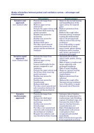
Task 2. Management of severe acute pancreatitis p 202. MANAGEMENT OF SEVERE ACUTE PANCREATITISHinds CJ, Watson JD. Intensive Care: A Concise Textbook. 3rd edition. SaundersLtd; 2008. ISBN: 978-0-7020259-6-9. p. 468–471 ManagementOver the last two decades advances in supportive therapy of failingorgans have enabled most patients to survive the early toxaemicphase of pancreatitis and have decreased the overall mortality rateassociated with this disease. Death from single or multiple organfailure, such as cardiovascular collapse, is now rare except in thosewith co-morbidities. Although early multiple organ dysfunctionsyndrome (MODS) still carries significant morbidity, patients'outcome is now primarily influenced by the volume of necroticareas and their secondary infection.If necrosis remainssterile, mostpatients will survivewith modernintensive caremanagementLink to <strong>ESICM</strong> Flash Conference: Jacques-Andres Romand, ‘<strong>Pancreatitis</strong> – generalmanagement’, <strong>ESICM</strong> congress, Berlin 2007Link to <strong>ESICM</strong> Flash Conference: Graham Ramsay, ‘Acute abdominal problems’,<strong>ESICM</strong> congress, Vienna 2009Implement supportive therapy early for remote organ dysfunctionand start interventions aimed at modulating SIRS, e.g. nutritionaltherapy (see Task 3). Early diagnosis and treatment of pancreaticinfection is key. Indications for surgery are usually restricted tolocal complications and in particular infected pancreatic necrosis.Nowadaysinfection ofpancreatic necrosisaccounts for 50-80% of the deathsSo far there is no specific therapy for acute pancreatitis thatinterferes with the inflammatory necrotising process and hence lessens theemergence of remote organ failure and the volume of necrotic areas.In this task we will cover general intensive care and various therapeutic modalitiesand conclude with a brief discussion on the indications for surgery.General intensive careSupportive therapy of vital organsCardiovascular systemYou should establish sufficient oxygen delivery as soon as possible.Tissue perfusion, in particular in the splanchnic area, may bedrastically diminished in these patients. Rapid restoration ofintravascular fluid volume is the priority as most of these patientsSplanchnicischaemia is a 2ndlocal hit:retroperitonealnecrosis, gut barrierdysfunction, andsecondarypancreatic infectionmay ensue
Task 2. Management of severe acute pancreatitis p 21are severely hypovolaemic. Use inotropes or vasoactive agents afterensuring adequate intravascular volume and allowing for ongoingcontinuing losses which may be substantial – a characteristicincorporated in the Ranson score.Local splanchnic perfusion may be worsened by abdominalcompartment syndrome- increased pressure due to intra-abdominal oedema,fluid sequestration and excessive fluid resuscitation.Hinds CJ, Watson JD. Intensive Care: A Concise Textbook. 3rd edition. SaundersLtd; 2008. ISBN: 978-0-7020259-6-9. p. 462–464 Abdominal CompartmentSyndromeUnfortunately the cytotoxic-related mechanisms responsible for the impairedpancreatic microcirculation are usually not addressed by manipulation of thesystemic circulation aimed at increasing global oxygen delivery.THINK why gastric intramucosal pH may be a better marker of adequacy ofresuscitation than the commonly used haemodynamic parameters, in critically illpatients. Why is this of particular concern in severe acute pancreatitis?For further information:Hinds CJ, Watson JD. Intensive Care: A Concise Textbook. 3rd edition. SaundersLtd; 2008. ISBN: 978-0-7020259-6-9. p. 108–119 and 76–78 (Assessment ofTissue Perfusion and Oxygenation)See also the <strong>PACT</strong> modules on Hypotension, Haemodynamic Monitoring andSepsis and MODSRespiratory systemConsider:• Prevention/correction of hypoxia.• Early physiotherapy and adequate analgesia (perhaps using epiduralanalgesia) to ensure free airways and to prevent atelectasis, preventpulmonary aspiration by nasogastric decompression.• Evacuation of pleural effusion by fine needle puncture ultrasoundguided drainage.
Task 2. Management of severe acute pancreatitis p 22• Continuous positive airway pressure (CPAP) ventilation and/or bilevelpositive airway pressure (BiPAP) ventilation by face or nasal mask incooperative patients.• Early tracheal intubation and mechanical ventilation, particularly inthose with acute lung injury. Apply the general principles of mechanicalventilation as in other causes of acute respiratory distress syndrome(ARDS).Further information on the therapeutic strategy for acute lung injury can be foundin:Hinds CJ, Watson JD. Intensive Care: A Concise Textbook. 3rd edition. SaundersLtd; 2008. ISBN: 978-0-7020259-6-9. p.228–233 ManagementSee <strong>PACT</strong> module on Mechanical ventilation Respiratory failureRenal system• Prevent and/or minimise renal injury by rapid correction ofhypovolaemia.• If acute renal failure develops, start renal replacement therapy withoutdelay to ensure optimal fluid and metabolic control and to enablenutritional support without haemodynamic instability.• Although not evidence based, consider Continuous veno-venoushaemofiltration (CVVH) as it tends to cause less haemodynamicinstability compared with intermittent haemodialysis withultrafiltration.For further information, seeHinds CJ, Watson JD. Intensive Care: A Concise Textbook. 3rd edition. SaundersLtd; 2008. ISBN: 978-0-7020259-6-9. p. 370-376 Blood PurificationUchino S, Kellum JA, Bellomo R, Doig GS, Morimatsu H, Morgera S, et al; Beginningand Ending Supportive Therapy for the Kidney (BEST Kidney) Investigators.Acute renal failure in critically ill patients: a multinational, multicenter study.JAMA 2005 ;294 (7): 813-818. PMID 16106006<strong>PACT</strong> modules on Acute renal failure and Oliguria and anuriaAcute Kidney Injury Initiative: http://www.akinet.orgGastrointestinal systemBeware of intra-abdominal hypertension and assess the patient for thiscomplication regularly. If abdominal compartment syndrome occurs, considerdecompression either surgically or in cases of colonic distension with a wide boretube inserted via the rectum. Abdominal compartment syndrome should besuspected whenever there is evidence of new or worsening organ dysfunction.
Task 2. Management of severe acute pancreatitis p 23You will find more information on abdominal compartment syndrome in thefollowing references.Cheatham ML, Malbrain ML, Kirkpatrick A, Sugrue M, Parr M, De Waele J, et al.Results from the International Conference of Experts on Intra-abdominalHypertension and Abdominal Compartment Syndrome. II.Recommendations. Intensive Care Med 2007 Jun;33(6):951-62. PMID17377769Sugrue M. Abdominal compartment syndrome Curr Opin Crit Care. 2005; 11(4):333-338. Review. PMID 16015111See the <strong>PACT</strong> module on Abdominal problemsQ. What are the most important consequences of abdominalcompartment syndrome in acute pancreatitis?A. Increased airway pressures during mechanical ventilation due to elevated diaphragmassociated with haemodynamic compromise associated with decreased venous return tothe heart, hypotension, renal compromise and possible exacerbation of splanchnicischaemia.Supportive therapy may appear basic. Nonetheless it is an essentialpart of the management of these patients: delayed diagnosis and insufficientresuscitation may exacerbate MODS and the risk of mortality.Pain reliefProvide effective pain relief. First try conventional analgesics bythe i.v. route. Morphine is not contraindicated but the use ofcontinuous thoracic epidural analgesia with a mixture of dilutedlocal anaesthetic solution (bupivacaine) and opiates offersadvantages. You can find further information about epiduralanalgesia and pancreatitis in the following reference.The use of epiduralblockade variesbetween institutionsNiesel HC, Klimpel L, Kaiser H, Bernhardt A, al-Rafai S, Lang U. [Epidural blockadefor analgesia and treatment of acute pancreatitis]. Reg Anaesth. 1991; 14(6):97–100. [German]. PMID 1780489Q. What are the possible advantages and potentialcomplications of epidural analgesia in acute pancreatitis?
Task 2. Management of severe acute pancreatitis p 24A.Epidural analgesia exposes patients to the risk of epidural infection and haematoma, andof circulatory instability, particularly in hypovolaemic patients and is not universally usedin this circumstance.The main advantage of this technique is that effective pain relief is almost guaranteed. Thedose of systemic opiates can therefore be reduced so that the patient is able to breathespontaneously or in an assist-mode if mechanically ventilated. The consequent avoidanceof systemic narcotics may improve bowel motility, facilitate enteral nutrition and mayreduce the risk of secondary pancreatic infection.MiscellaneousIn this high-risk population, routine stress ulcer prophylaxis as well as thromboembolicprevention, usually by low-molecular weight heparin, would be standard.Specific therapeutic modalitiesAntibioticsPrevention of secondary bacterial contamination of necrotic areas is of the utmostimportance in view of the mortality/morbidity attributed to infected pancreaticnecrosis. As the nidus of infection, e.g. the volume of necrosis, cannot be influencedby any therapeutic means except for prompt restoration of splanchnic perfusionyou have to rely on alternative methods of prevention.Randomised controlled trials of intravenous prophylacticantimicrobial therapy have failed to demonstrate a benefit in terms of incidenceof pancreatic infection, surgical intervention and outcome.Bai Y, Gao J, Zou DW, Li ZS. Prophylactic antibiotics cannot reduce infectedpancreatic necrosis and mortality in acute necrotizing pancreatitis: evidencefrom a meta-analysis of randomized controlled trials Am J Gastroenterol.2008; 103(1): 104-110. Review. PMID 17925000Another option is the topical administration of prophylaxis byusing selective decontamination of the digestive system (SDD).Non-absorbed antibiotics are used to reduce the number of aerobicGram-negative bacteria and yeasts in the gastrointestinal tract,whilst retaining the normally predominant anaerobic flora and somodulating colonisation in the digestive tract. Current evidence isnot conclusive enough to allow a firm recommendation in Severe<strong>Pancreatitis</strong> patients.Unlike i.v.prophylaxis,SelectiveDecontamination ofthe Digestive tract(SDD) addressesbacterialtranslocation fromthe gut to thepancreas
Task 2. Management of severe acute pancreatitis p 25Hinds CJ, Watson JD. Intensive Care: A Concise Textbook. 3rd edition. SaundersLtd; 2008. ISBN: 978-0-7020259-6-9. p. 332–333 Selective decontaminationof the digestive tractLuiten EJ, Hop WC, Lange JF, Bruining HA. Controlled clinical trial of selectivedecontamination for the treatment of severe acute pancreatitis Ann Surg.1995; 222(1): 57-65. PMID 7618970Use antibiotics on demand for sepsisrather than prophylactically!The policy of routine systemic antimicrobial prophylaxis isdiscouraged. It may promote the selection of particular strains, notably Staphylococci andEnterococci and may promote the development of fungal overgrowth, as well asmultiresistant Gram-negative organisms.Antibiotic therapyPatients with severe acute pancreatitis are prone to infections early in the course ofthe disease. Antibiotics are required in those with infection and empirical therapyshould be started without delay once infection in the pancreas or elsewhere issuspected on clinical grounds. Continuation of antibiotics and the ultimate choiceof drugs should be based on a thorough investigation for a source of infection andon the results of specimen culture obtained by fine needle aspiration, initialaspirate taken at time of wide bore drainage or during surgery in cases of suspectedpancreatic infection. Once the presence of infection is established and the patientsuffers from severe sepsis or septic shock, additional interventions should beconsidered according to current sepsis therapeutic guidelines.Dellinger RP, Levy MM, Carlet JM, Bion J, Parker MM, Jaeschke R, et al.SurvivingSepsis Campaign: international guidelines for management of severe sepsisand septic shock: 2008. Intensive Care Med 2008; 34(1): 17-60. PMID18058085Antibiotics are merely an adjuvant therapy in infected pancreatic necrosis.Drainage is mandatory for most if not all pancreatic infections.Nutritional therapyThis aspect of management is discussed extensively in Task 3.
Task 2. Management of severe acute pancreatitis p 26Indications for surgeryImproved intensive care in regionalised, multidisciplinary centres, advances ininterventional radiology and endoscopic techniques as well as implementation ofspecific therapeutic modalities that address the pathophysiology of the disease havereduced the indications for surgery, during the toxaemic phase of acute pancreatitisand later.Early complications that might prompt the surgeon to intervene urgently areoutlined below.Undisputed indications• Infected pancreatic necrosis when percutaneous/other techniques notindicated• Severe retroperitoneal haemorrhage• Acute abdomen – peritonitis• Biliary obstruction in case of failure of Endoscopic Sphincterotomy• Abdominal compartment syndrome where percutaneous/other drainagetechniques not successful.Controversial indications: The case for sterile necrosisExtensive (>50%) sterile pancreatic necrosis: Early ‘routine’ debridement ofnecrosis irrespective of its bacteriological status in order to prevent remote organdysfunction and pancreatic infection fails to achieve these goals and may even beharmful. The development of local complications, postoperative infection ofnecrosis, problems related to poor demarcation of necrotic areas and long-termsequelae in survivors have led most surgeons to abandon this indication.Persisting multiple organ failure despite intensive care therapy: earlyand repeated removal of necrotic tissue combined with continuous drainage/lavagehave been advocated to overcome systemic effects. This is based on the assumptionthat ongoing pancreatic inflammation is the sole culprit and that removal of toxicmediators released by the gland will abort the process of distant organ injury onceit has been initiated. A prohibitive mortality has led most surgeons to also abandonthis indication.Neither the extent of sterile pancreatic necrosis, the clinicalseverity of the disease or the duration of intensive supportive therapy should beregarded as indications for surgery.
Task 3. Feeding the patient with acute pancreatitis p 273. FEEDING THE PATIENT WITH ACUTE PANCREATITISArtificial nutrition is increasingly considered to be a key aspect ofthe specific management of pancreatitis patients and not only as anadjuvant therapeutic modality. In this task we shall discuss theindications, modalities and potential complications of nutritionaltherapy during acute pancreatitis.Artificial nutrition: Why?Although no prospective randomised controlled trials havedemonstrated that nutritional support lessens the severity of acutepancreatitis or improves outcome, there are several theoreticalreasons to provide some of these patients with nutritional therapy.You will find details of controlled trials into the effects of nutritionin acute pancreatitis in the following references.Feeding duringsevere acutepancreatitis may bechallenging…See the PatientChallenges in the'Nutrition' moduleRecovery resultsfrom the successfulapplication of acombination oftherapeuticmeasures, includingnutritionalcare/therapyMcClave SA, Chang WK, Dhaliwal R, Heyland DK. Nutrition support in acutepancreatitis: a systematic review of the literature JPEN J Parenter EnteralNutr 2006; 30(2): 143-56. Review. PMID 16517959Marik PE. What is the best way to feed patients with pancreatitis? Curr Opin CritCare 2009; 15(2): 131-138. Review. PMID 19300086Al-Omran M, Albalawi ZH, Tashkandi MF, Al-Ansary LA. Enteral versus parenteralnutrition for acute pancreatitis Cochrane Database Syst Rev. 2010; (1):CD002837. Review. PMID 20091534• Acute pancreatitis is a catabolic, hypermetabolic disease process thatincreases protein and calorie requirements. The following may beexacerbating factors:• Oral intake may be impossible for prolonged periods due to the presenceof pain, gastric atony, ileus, or partial duodenal obstruction frompancreatic enlargement.• There are increased protein losses across inflamed retroperitonealsurfaces and through pancreatic fistulas.• In a subset of patients there are pre-existing protein-caloriemalnutrition and micronutrient deficiencies, usually as a result ofalcohol abuse.
Task 3. Feeding the patient with acute pancreatitis p 28The risk for a net negative energy and protein balance and hence fordeterioration of nutritional status is very high in severe cases.Early assessment of severity dictates the need for nutritionaltherapy: the patients at risk of developing organ failure and pancreatic infectionand those with preexisting malnutrition should be considered for early nutritionaltherapy.There is a rationale for feedingFor further information on the rationale for feeding the critically ill patient see the<strong>PACT</strong> module on Nutrition.Q. List the metabolic hallmarks of severe acute pancreatitisA. Patients with severe acute pancreatitis have, among other features, a number ofmetabolic similarities with septic patients. They share many of the same inflammatorymediators and the subsequent hormonal response to injury is virtually identical in bothgroups. Metabolic manifestations are:• Increased energy expenditure, oxygen consumption and a hyperdynamiccirculation• High protein catabolism and ureagenesis• Increased amino acid oxidation• Increased and unsuppressible gluconeogenesis while glucose clearance andoxidation are diminished (insulin resistance)• Increased rate of lipolysis and free fatty-acid oxidationThese metabolic alterations account for some of the potential complications that may beexacerbated by nutritional therapy in acute pancreatitis.For further information on the stress-related metabolic disturbances in acute pancreatitisand in the critically ill patient see:Shaw JH, Wolfe RR. Glucose, fatty acid, and urea kinetics in patients with severepancreatitis. The response to substrate infusion and total parenteral nutritionAnn Surg 1986; 204(6): 665-72. PMID 3098198Q. Indicate the main consequences of malnutrition in acutepancreatitisA. Protein-calorie malnutrition may result in:
Task 3. Feeding the patient with acute pancreatitis p 29• Poor pancreatic healing• Impaired immune, gut, and lung function• Increased risk for nosocomial and pancreatic infection• Worsened outcomeThere is some evidence that cell-mediated immunity is already compromised in severeacute pancreatitis, regardless of the nutritional status.Nutritional therapy: How, what and when?Route of nutrient delivery: Enteral versus parenteralThe concept of ‘resting’ the pancreas should not be applied toostrictly. This approach implies strict avoidance of all stimuli toexocrine secretion from the pancreas in order to negate theperpetuation of premature enzymatic activation through whichpancreatitis is initiated. This principle explains why starvation waspreviously regarded as the most physiologically appropriateresponse to pancreatic injury. In the past, if nutritional supportwas deemed necessary, parenterally infused nutrients were thepreferred option as they had the least likelihood of stimulating aninflamed pancreatic gland.The more distallythat nutrients areinfused in the gut,the less theystimulatepancreaticsecretionTHINK about how you could combine early enteral feeding therapy with thetherapeutic principle of pancreatic rest? Refer to the regulation of exocrine secretion atthe gut level in the following references.Pandol SJ. Pancreatic physiology. In: Sleisenger MH, Fordtran JS, editors.Gastrointestinal disease-pathophysiology, diagnosis, management.Philadelphia: WB Saunders; 1993. p.1585-1600. ISBN-13 978-0-7216-8973-9O'Keefe SJ, Lee RB, Li J, Stevens S, Abou-Assi S, Zhou W. Trypsin secretion andturnover in patients with acute pancreatitis Am J Physiol Gastrointest LiverPhysiol 2005 ; 289(2): G181-7. PMID 15705659The efficacy of pancreatic rest has never been validated -mechanical, pharmacological and hormonal interventions devisedto block exocrine pancreatic secretion have all failed to influencethe local inflammatory process and patient outcome.Moreover, although introduction of oral feeding may be associatedat times with resurgence of pancreatitis, nutrients infused distallyinto the jejunum have minimal effect on exocrine secretion.The enteral routeis safe in acutepancreatitis, sowheneverpossible, use it!Q. What is the level of basal exocrine pancreatic secretion early afteronset of acute pancreatitis?
Task 3. Feeding the patient with acute pancreatitis p 30A. Baseline exocrine secretory capacity is spontaneously diminished during acutepancreatitis.The preferred route of nutritional support is no longercontroversial: the benefits of enteral delivery in terms of outcome depend mainlyon the avoidance of CVC -related infections, better glycaemic control, improvementin gut blood flow, maintenance of gut structural and immune barrier function,reduction in microbial translocation and pancreatic infection and possibly onimmunomodulation.A detailed discussion of the advantages of the enteral over the parenteral route offeeding can be found in the <strong>PACT</strong> module on Nutrition and in the following references.Jolliet P, Pichard C, Biolo G, Chioléro R, Grimble G, Leverve X, et al. Enteralnutrition in intensive care patients: a practical approach. Working Group onNutrition and Metabolism, <strong>ESICM</strong>. European Society of Intensive CareMedicine Intensive Care Med 1998; 24(8): 848-59. Review. PMID 9757932.Full text (pdf)Petrov MS, van Santvoort HC, Besselink MG, van der Heijden GJ, Windsor JA,Gooszen HG. Enteral nutrition and the risk of mortality and infectiouscomplications in patients with severe acute pancreatitis: a meta-analysis ofrandomized trials Arch Surg. 2008; 143(11): 1111-7. Review. PMID 19015471Link to <strong>ESICM</strong> Flash Conference: Pierre Singer, ‘<strong>Pancreatitis</strong>–early enteralnutrition’, Berlin 2007In order to maximise clinical benefit, enteral feeding should be initiated assoon as possible after admission in all attacks predicted to be severe. Patients inwhom enteral access cannot be achieved or in whom clear-cut contraindications(intestinal rupture, obstruction, or necrosis), intolerance, or exacerbation of thedisease occurs should be considered for partial or total parenteral nutrition (TPN).Enteral accessRepeated plain abdominal X-ray enables detection of tube migrationEarly feeding is usually undertaken through a silicone or polyurethane tube with aninner stylet that is positioned (under fluoroscopic guidance) beyond the firstjejunal loop.
Task 3. Feeding the patient with acute pancreatitis p 31Right-lateral positioning of the patient as well as the use of a prokinetic (e.g.erythromycin 250 mg IV bolus) may assist the passage of the tube through thepylorus so that fluoroscopic guidance is unnecessary in >50% of the cases. Analternative method is to pass the feeding tube over an endoscopically placed guidewire. The correct positioning of the tube should be ascertained regularly byradiography.A blind bedside insertion technique should be used with caution as theduodenum may be distorted and injured by the retroperitoneal inflammatory process.Continuous gastric decompression by gravity should be achieved by a separate tube inpatients with gastro-paresis or post-pyloric obstruction. This prevents the risk of massivepulmonary aspiration and may also help to detect proximal migration of the feeding tube.Preliminary data indicate that early nasogastric tube feeding is safe andwell tolerated in patients with predicted severe acute pancreatitis and the ease ofinsertion facilitates the earlier commencement of enteral nutrition. Althoughduodenal secretion of pancreatic enzymes is inversely related to the severity ofacute pancreatitis, this route of feeding may be associated with a risk ofexacerbating the disease process and of pulmonary aspiration, in particular inthose with gastric outlet compression.Patients who undergo surgery for local complications later in thecourse of the attack can have a catheter jejunostomy performed.This offers an alternative to the nasojejunal tube.Energy and protein requirementsCatheter jejunostomyrequires a hole in theintestine: this practicevaries from site to siteResting energy expenditure, as measured by indirect calorimetry, varies widely inacute pancreatitis, depending upon the magnitude of the regional inflammatory
Task 3. Feeding the patient with acute pancreatitis p 32process and the presence of superimposed infection. The latter raises energyexpenditure by 5 to 20% above resting values, but overfeeding should be avoided.Further information on the use of indirect calorimetry in nutrition and on energy,protein, and micronutrient requirements during acute pancreatitis can be found inthe <strong>PACT</strong> 'Nutrition' module as well as in the following references.McClave SA, Snider HL. Use of indirect calorimetry in clinical nutrition Nutr ClinPract 1992; 7(5): 207-21. Review. PMID 1289691Dickerson RN, Vehe KL, Mullen JL, Feurer ID. Resting energy expenditure inpatients with pancreatitis Crit Care Med 1991; 19(4): 484-90. PMID 2019133Composition of the dietParenteral nutrition should be administered as indicated in othercritically ill patients and should be supplemented with glutamine.Plasma lipid clearance should be regularly monitored to avoidhypertriglyceridaemia. Infused nutrients do not exacerbate thedisease.Do not useprobiotics insevere acutepancreatitis: itworsens outcomeFor further information see:<strong>PACT</strong> module on NutritionBesselink MG, van Santvoort HC, Buskens E, Boermeester MA, van Goor H,Timmerman HM, et al; Dutch Acute <strong>Pancreatitis</strong> Study Group. Probioticprophylaxis in predicted severe acute pancreatitis: a randomised, doubleblind,placebo-controlled trial. Lancet 2008; 371(9613):651-659. PMID18279948There are no data on the optimal enteral formula in severe acute pancreatitis.Polymeric solutions are cheaper, well tolerated and maintain gut structure/function.Immune-enhancing diets have not been specifically tested in this disease.Q. Indicate the theoretical advantages of medium-chain triglycerides inacute pancreatitis in both enteral (EN) and parenteral nutrition?A.• If used enterally: better assimilation by direct absorption into the portal vein inan environment deficient in lipase.• If administered parenterally as a lipid emulsion: rapid clearance and morecomplete mitochondrial oxidation so that the risk of hypertriglyceridaemia istheoretically reduced.
Task 3. Feeding the patient with acute pancreatitis p 33Prescription and timing of nutrient administrationYou should administer enteral solutions as a continuous 24 hourspump driven infusion. Increase the diet gradually (250-500ml/day), starting with 500 ml/day until the patient's targetedcalorie needs are tolerated. Jejunal residual volumes should be
Task 3. Feeding the patient with acute pancreatitis p 34overfeeding and improve glucose tolerance by supplyingsome calories as lipids.• Hypertriglyceridaemia is usually attributed to overfeedingwith inadequate supply of carbohydrate/triglycerides,insulin resistance and poor nutrient utilisation. Monitorserum turbidity and triglyceride level, and titrate fat contentto keep the serum triglyceride level below 400 mg/dl (4.5mmol/l).• Exacerbation of the disease process: Relevant causes arehypertriglyceridaemia, nasogastric feeding, proximaldislodging of the jejunal feeding tube or too early return tooral diet. Monitor serum amylase level and performabdominal CT.• Gut intolerance: monitor bowel distension, abdominalpressure, residual jejunal volume, and diarrhoea. Titrateenteral infusion rate accordingly and/or use prokinetics andenemas.Jejunal and i.v.lipids are safe aslong as youmonitor tubepositioning andserum lipaemia,respectivelyUse parenteralnutrition tomeetnutritionaltarget in thosewith major gutintoleranceSignificant intolerance to appropriate nutritional therapy by either theenteral or the parenteral route usually denotes ongoing local complications(e.g. infection, mesenteric ischaemia) and requires appropriate investigation (referto Task 1 and Task 4).
Task 3. Feeding the patient with acute pancreatitis p 35Algorithm of nutritional therapy in acute pancreatitisUseful review articles on the issues covered in this Task are:Meier R, Ockenga J, Pertkiewicz M, Pap A, Milinic N, Macfie J; DGEM (GermanSociety for Nutritional Medicine), Löser C, Keim V; ESPEN (EuropeanSociety for Parenteral and Enteral Nutrition). ESPEN Guidelines on EnteralNutrition: Pancreas Clin Nutr 2006; 25(2): 275-284. PMID 16678943Gianotti L, Meier R, Lobo DN, Bassi C, Dejong CH, Ockenga J, Irtun O, MacFie J;ESPEN. ESPEN Guidelines on Parenteral Nutrition: pancreas Clin Nutr2009; 28(4): 428-35. Epub 2009 May 22. PMID 19464771Martindale RG, McClave SA, Vanek VW, McCarthy M, Roberts P, Taylor B, et al;American College of Critical Care Medicine; A.S.P.E.N. Board of Directors.Guidelines for the provision and assessment of nutrition support therapy inthe adult critically ill patient: Society of Critical Care Medicine and AmericanSociety for Parenteral and Enteral Nutrition: Executive Summary. Crit CareMed 2009; 37(5): 1757-1761. Review. No abstract available. PMID 19373044In the next five patients you see, assess the timing and success of enteral feeding.
Task 4. How to identify and manage local complications of acute pancreatitis p 364. HOW TO IDENTIFY AND MANAGE LOCALCOMPLICATIONS OF ACUTE PANCREATITISApproximately 50% of patients with severe acute pancreatitisprogress to a spontaneous and uneventful resolution of theregional inflammatory process. In the remainder localcomplications will emerge usually weeks or even months after theonset of the attack. Therefore these patients must be kept underreview for deterioration since timely operative, endoscopic orradiological interventions are essential.Localcomplications areclosely related tothe extent ofnecrosisPathophysiology of regional necrosisThree mechanisms act in concert to promote regional necrosis andwidespread organ damagePremature activation of lipolytic and proteolytic enzymes, inparticular trypsinogen, is the key trigger of acinar cell necrosis. Asthe natural safeguards that prevent autodigestion are overwhelmedin acute pancreatitis, trypsin is able to activate the cascade ofproteases.Secondary overactivation of immune effector cells results in thelocal accumulation of various proinflammatory and cytotoxicsubstances including proteases, reactive oxygen species, cytokinesand lipid mediators. These cellular components and humoralmediators play a pivotal role in acinar cell injury.IntraductalhypertensiontriggersprematureenzymaticactivationAbnormalenzymatic activityin the interstitiumis a key feature
Task 4. How to identify and manage local complications of acute pancreatitis p 37Local microcirculatory disturbances soon amplify regionalnecrosis. They are primarily ascribed to endothelial cell damageresulting from the combined effect of activated leucocytes andproteases.Early on there is amassive migrationof leukocytes intothe glandThe pancreas is the source of damage and surrounding tissues are theprimary targetThe enzyme-rich exudates diffuse from the pancreas into the surrounding tissuesand often form fluid collections. Tissue necrosis involves primarily the peripancreatictissue; the core of the gland being more resistant. However at timesnecrosis may affect the main pancreatic duct, often in the isthmic area, causingrupture.A deeper insight into this topic can be found in:Steer ML. Early events in acute pancreatitis. Baillieres Best Pract Res ClinGastroenterol 1999; 13(2): 213-225. Review. PMID 11030602Gorelick FS, Otani T. Mechanisms of intracellular zymogen activation. Baillieres BestPract Res Clin Gastroenterol 1999; 13(2): 227-240. Review. PMID 11030603Brady M, Christmas S, Sutton R, Neoptolemos J, Slavin J. Cytokines and acutepancreatitis. Baillieres Best Pract Res Clin Gastroenterol 1999; 13(2): 265-289. Review. PMID 11030606Menger MD, Vollmar B. Microcirculation: initiating or aggravating factor. In:Büchler MW, Uhl W, Friess H, Malfertheiner P, editors. Acute pancreatitis -Novel concepts in biology and therapy. Berlin: Blackwell Science; 1999. p.63–70 ISBN 978-0632053391THINK about how biochemical markers relate to the severity of necrotisingpancreatitis.
Task 4. How to identify and manage local complications of acute pancreatitis p 38Pancreatic infectionPathophysiologyNecrosis creates an excellent culture medium for micro-organisms. Accordingly therisk of infection is proportional to the extent and duration of necrosis. Theincidence of infection peaks in the third week after onset, but in up to 25% of thesepatients infection is documented in the first seven days. The earlier infectionoccurs, the higher the mortality, as the combination of infection with theinflammatory process generates a highly toxaemic course. Cultures yieldpredominantly common enteric bacteria. Although not proven in humans, bacterialtranslocation from the gut lumen either transmurally, via lymphatics, ascites orblood is probably the leading mechanism of infection.THINK On the basis of the previous information, think about how we can influence therisk of infection.Q. What are the other potential routes resulting in infection of necrosisduring acute pancreatitis?A.• Intra-abdominal spread: digestive fistula – hollow viscus perforation (colon)• Biliary reflux (cholangitis)• Duodenal reflux (post-ERCP)• Haematogenous: beware of secondary bacterial contamination of necrosis byextra-pancreatic infectious foci (nosocomial infection)Q. List the factors that may promote bacterial translocation duringacute pancreatitisA.• Ileus and secondary aerobic Gram-negative bacterial overgrowth• Disruption of the normal gut microflora with loss of colonisation resistance(antibiotics)• Gut barrier failure (both structural and immunologic) as a result of splanchnicischaemia and the spread of the nearby inflammatory process• Bowel rest (Total Parenteral Nutrition)Luiten EJ, Hop WC, Endtz HP, Bruining HA. Prognostic importance of gramnegativeintestinal colonization preceding pancreatic infection in severe acutepancreatitis. Results of a controlled clinical trial of selective decontaminationIntensive Care Med 1998; 24(5): 438-445. PMID 9660258. Full text (pdf)
Task 4. How to identify and manage local complications of acute pancreatitis p 39Types of infection: infected necrosis or abscess?Because of differences in anatomy, prognosis and management it is crucial that youdifferentiate between infected pancreatic necrosis and pancreatic abscess.Infected pancreatic necrosisDifferentiation between the two forms of pancreatic infection
Task 4. How to identify and manage local complications of acute pancreatitis p 40Pancreatic abscess results from the secondary infection of an acutefluid collection or a liquefied necrotic areaQ. Do you think drainage of the pancreatic abscess should bepercutaneously attempted in this particular patient and why?A. Percutaneous drainage is likely to fail as the collection is large, central and multiloculated:given the appearances on CT scan, surgery may be the preferred option in thiscase.Pancreatic infection may be suspected from clinical signs of sepsis and/or generaldeterioration, but confirmation of the diagnosis requires CT imaging andappropriate culture (aerobes, anaerobes, fungi) of material obtained by ultrasoundorCT-guided fine needle aspiration (FNA).Occasionally infection may be diagnosed by CT alone when gas bubbles are presentwithin a necrotic area. Otherwise, FNA should be directed to all sites of necrosis orat least to those showing the most obvious changes on repeated CT. Monitoring ofserum procalcitonin, the propeptide of calcitonin, is a potential marker for the noninvasiveidentification of infected pancreatic necrosis as well as for the selection ofpatients with persisting sepsis after drainage.Mofidi R, Suttie SA, Patil PV, Ogston S, Parks RW. The value of procalcitonin atpredicting the severity of acute pancreatitis and development of infectedpancreatic necrosis: systematic review. Surgery 2009; 146(1): 72-81. PMID19541012
Task 4. How to identify and manage local complications of acute pancreatitis p 41Microbiological analysis of material harvested by FNA is not 100% accurate: allnecrotic areas may not be sampled or antibiotic prophylaxis, if used, may affect cultureresults. FNA(if negative) should be repeated every 5–7 days in patients with persistentsigns of sepsis. Gram-staining should be used to guide early treatment.Use empirical antimicrobial therapy while awaiting theresults of culture, deescalate or discontinue based on theresultsNegative sampling should not delay surgical exploration and/or drainagewhen the clinical course is strongly suggestive of infection.Schmid SW, Uhl W, Friess H, Malfertheiner P, Büchler MW. The role of infection inacute pancreatitis. Gut 1999; 45(2): 311-6. Review. No abstract available.PMID 10403749Treatment of pancreatic infectionAntimicrobial therapy alone isusually ineffectiveEndoscopic transgastric or duodenal drainageis an alternative to the percutaneous route inspecialised centresWhile pancreatic abscess is usually easily drained, often by CT guidedaspiration, infected necrosis is difficult to evacuate as it is not a wellcircumscribedcollection of pus but rather a diffuse area of bacterial proliferationwithin ill defined necrotic tissues. Therefore drainage is not straightforward andthe surgeon can only remove pus and solid debris of necrosis easily detached bysuction and manual debridement. Surgical intervention will leave in place adherentareas of necrotic tissue that allow proliferating bacteria to persist. Moreoverproduction of necrosis is a dynamic process and continues after the surgicaldebridement, thus perpetuating infection. This accounts for the high incidence ofsepsis recurrence after surgery and the development of specific techniques aimed atevacuating pus and necrotic tissues throughout the evolution of the attack, e.g. anaverage of eight weeks.A percutaneous approach is tempting as it does not injure disease-free areas.It is particularly attractive in pancreatic abscess where the infected material ismostly liquefied or as a temporising measure in the desperately ill patient untilmore formal surgical debridement may be tolerated.
Task 4. How to identify and manage local complications of acute pancreatitis p 42Percutaneous drainage has been associated with a high incidence of sepsisrecurrence, even in the case of well-circumscribed abscesses and despite insertion ofmultiple catheters and prolonged drainage. These results may be due to the inability ofpercutaneous drainage to remove solid debris lying at the periphery of the abscess orinside infected necrosis.Percutaneous drainage is classically reserved for non-loculated,peripheral, and well-defined collections that usually appear late in the course of theattack. Remember that it is a lengthy and resource-intensive approach.Observe the placement of drainage catheters with the radiologist and assist thenursing staff in maintaining their patency and the drainage.The surgical approach remains the gold standard. Exploration of the abdomenand dissection are guided by the CT scan that provides a 'roadmap' to the surgeon.All collections and all pockets of tissue necrosis and infection are opened andevacuated as completely as possible. Overall morbidity averages 70%. Patientsrequire re-operation in 25% of cases, mainly for bleeding, hollow viscus perforationor necrosis, fistula and recurrent sepsis.The ongoing inflammatory, necrotising process necessitates a prolongedcontinuous drainage procedure postoperatively in order to prevent sepsis recurrence.The precise nature of the operative procedure varies according to the surgeons'expertise. Whatever the surgical technique (see illustration below), thoroughdebridement and continuous postoperative drainage are the key features ofinfection management.Depending on the circumstances some surgeons prefer a transverse sub-costal orflank incision for exploration.If the clinical situation permits, operative necrosectomy and/ordrainage should be postponed at least until four weeks after onset. Over time,infected necrotic areas demarcate from viable tissues, undergo liquefaction andcoalesce into an encapsulated entity. This organised necrosis leads to an easierand safer debridement, with sparing of viable pancreatic tissue. It might be evenmore amenable to percutaneous, endoscopic or minimally-invasive (laparoscopic)operative drainage alone or in combination. Thus, the optimal type of interventiondepends on the clinical course and the timing of the procedure.
Task 4. How to identify and manage local complications of acute pancreatitis p 43You will find details on surgical indications in the first of the following referencesand details on surgical techniques and other types of drainage in the otherreferences.Büchler P, Reber HA. Surgical approach in patients with acute pancreatitis. Isinfected or sterile necrosis an indication–in whom should this be done, when,and why? Gastroenterol Clin North Am 1999; 28(3): 661-71. Review. PMID10503142Besselink MG, de Bruijn MT, Rutten JP, Boermeester MA, Hofker HS, Gooszen HG;Dutch Acute <strong>Pancreatitis</strong> Study Group. Surgical intervention in patients withnecrotizing pancreatitis Br J Surg 2006; 93(5): 593-599. PMID 16521173Freeny PC, Hauptmann E, Althaus SJ, Traverso LW, Sinanan M. Percutaneous CTguidedcatheter drainage of infected acute necrotizing pancreatitis:techniques and results AJR Am J Roentgenol 1998; 170(4): 969-975. PMID9530046Seewald S, Groth S, Omar S, Imazu H, Seitz U, de Weerth A, Soetikno R, Zhong Y,Sriram PV, Ponnudurai R, Sikka S, Thonke F, Soehendra N. Aggressiveendoscopic therapy for pancreatic necrosis and pancreatic abscess: a new safeand effective treatment algorithm (videos) Gastrointest Endosc. 2005; 62(1):92-100. PMID 15990825Connor S, Raraty MG, Howes N, Evans J, Ghaneh P, Sutton R, Neoptolemos JP.Surgery in the treatment of acute pancreatitis--minimal access pancreaticnecrosectomy Scand J Surg. 2005; 94(2): 135-42. Review. PMID 16111096Haemorrhage and perforationSignificant spontaneous haemorrhage is rare but carries a high mortality. It isusually due to erosion of the vessel wall by proteases and pseudoaneurysmformation of the pancreatic arteries within a pseudocyst (see below) or an area ofnecrosis. In case of massive retroperitoneal bleeding the CT scan can identify theapproximate location of the haemorrhage. However angiography is often necessaryto localise precisely the source of bleeding and at times to achieve haemostasis withembolisation. Surgery is required if angiographic occlusive techniques fail.
Task 4. How to identify and manage local complications of acute pancreatitis p 44de Perrot M, Berney T, Bühler L, Delgadillo X, Mentha G, Morel P. Management ofbleeding pseudoaneurysms in patients with pancreatitis Br J Surg. 1999;86(1): 29-32. PMID 10027355Massive haemorrhage often heralds infection: surgical drainage ismandatory in these casesSpontaneous perforation occurs usually at the level of the left and transversecolon. The mechanism is predominantly ischaemic as a consequence of extensionof necrosis in the mesocolon and secondary vessel thrombosis. The diagnosisshould be suspected in the presence of septic signs, diarrhoea, rectal bleeding, orhigh-pitched bowel sounds. CT scan shows thickening of the colonic wall and extraluminal gas bubbles or contrast media. At times colonic necrosis is an incidentaloperative finding.During surgery, it is difficult to evaluate colonic viability. Colectomy is indicated inpatients with perforation or obvious irreversible necrosis. If there is doubtregarding the viability of the colon, it is safer to perform a loop ileostomy and toleave the colon in situ. This procedure prevents further deterioration of the coloniclesions, improves tolerance to enteral feeding and is easily reversed.Pseudocysts and pancreatic fistulasA pseudocyst is a collection of pancreatic juice enclosed by a wall of granulationtissue and is formed near an area of tissue necrosis associated with rupture of apancreatic duct (see illustration below).
Task 4. How to identify and manage local complications of acute pancreatitis p 45PseudocystIt can result from the rupture of the main pancreatic duct itself, but in the majorityof instances, the leaking duct is located at the periphery of the gland. Formation ofpseudocysts takes at least four weeks. Fluid collections apparent at an earlier stagelack a defined wall and are named 'acute fluid collections'. Drainage, eitherpercutaneous, surgical, or endoscopic, is required in those with large and/orsymptomatic pseudocysts. Endoscopic treatment, utilising endoscopic ultrasoundguidance, is the primary procedure for pseudocysts with amenable anatomy.Complications for which drainage should be considered:• Compression of adjacent structures.• Rupture of pseudocysts is observed in less than 5% ofcases and may present either as an acute episode or asilent and progressive process. It may lead to a reactiveeffusion (ascites, pleural effusion or pericardialeffusion) or rupture into a hollow viscus, usually thestomach or the colon.• Bleeding is seen in 5% of pseudocysts and is the mostserious complication.• Infection: approximately 10% of all large pseudocystsbecome infected, either spontaneously or after aninappropriate attempt at drainage, usuallypercutaneous.Most pseudocystsare small (
Task 4. How to identify and manage local complications of acute pancreatitis p 46Pitchumoni CS, Agarwal N. Pancreatic pseudocysts. When and how should drainagebe performed? Gastroenterol Clin North Am 1999; 28(3): 615-39. Review.PMID 10503140Baron TH, Harewood GC, Morgan DE, Yates MR. Outcome differences afterendoscopic drainage of pancreatic necrosis, acute pancreatic pseudocysts, andchronic pancreatic pseudocysts Gastrointest Endosc 2002; 56(1): 7-17. PMID12085029Keep a record of the differences in timing, diagnosis and treatment of acute fluidcollections, pseudocysts and pancreatic abscess. Refer to the following reference:Bollen TL, van Santvoort HC, Besselink MG, van Leeuwen MS, Horvath KD, FreenyPC, Gooszen HG; Dutch Acute <strong>Pancreatitis</strong> Study Group. The AtlantaClassification of acute pancreatitis revisited Br J Surg. 2008; 95(1): 6-21.Review. PMID 17985333CONCLUSIONSevere acute pancreatitis is a challenging disease for the ICU clinician: thepathophysiology at the cellular level remains obscure, the outcome isunpredictable, and the long-lasting course of the attack is characterised by theemergence of life threatening local complications and multiple distant organfailures.So far, specific treatment is lacking and the modern management of these patientsrequires treatment in, or referral to, a specialist centre. Here, clinical experience,technical skills and a comprehensive implementation of all facets of Intensive CareMedicine are effected. A consistent and concerted approach, close cooperation aswell as detailed communication between the intensivist, gastroenterologist skilledin endoscopy, surgeon, interventional radiologist and the microbiology service isrequired to optimise patient outcome.
Self-assessment p 47SELF-ASSESSMENTEDIC-style Type K1. Type K. Usual imaging techniques to aid in the diagnosis of acutepancreatitis in the initial phase (first 1–2 days after hospitalisation) include:A. Plain abdominal X-rayB. Endoscopic retrograde cholangiopancreatography (ERCP)C. Abdominal CT with or without contrastD. Abdominal ultrasound2. Type K. Prognostic scores often used in the assessment of acutepancreatitis include:A. APACHE II scoreB. Ranson scoreC. Murray scoreD. Imrie score3. Type K. Regarding infectious complications related to severe acutepancreatitis, which of the following statements are correct:A. Infection is usually present from the startB. A common complication is infected pancreatic necrosisC. Infections most often come from haematogenous spread of bacteriaD. Anaerobic bacteria is the cause of most frequent clinical infection4. Type K. Systemic inflammatory response syndrome (SIRS) is frequentlyfound in patients with acute pancreatitis. Which of the following is/are trueregarding the development of SIRS in this condition?A. SIRS is only present in the severe forms of acute pancreatitisB. SIRS seldom develops prior to the second week of the diseaseC. Includes CRP rise >180 mg/dlD. Includes LPK elevation >12.000/ l5. Type K. Symptomatic treatment of acute pancreatitis include thefollowing options:A. Analgesia with opioids intravenouslyB. Epidural analgesiaC. Fluid restrictionD. Peritoneal lavage6. Type K. Usual complications in severe acute pancreatitis includes:A. Intra-abdominal hypertensionB. Intestinal ischaemiaC. Acute respiratory failureD. Increased intracranial pressure
Self-assessment p 487. Type K. Which of the following statements is/are true regardingnutritional therapies in severe acute pancreatitis?A. Enteral nutrition is associated with fewer complications than parenteral nutritionB. A probiotic should usually be included in the nutritional regimenC. Specific immune-enhancing diets are superior to standard enteral nutritionD. Nutrition direct into the duodenum is not advocated8. Type K. Hyperglycaemia is common in acute severe pancreatitis and canbe a consequence of:A. Alpha cell necrosis in the pancreatic tissueB. Beta cell necrosis in the pancreatic tissueC. Insulin resistance in the CNSD. The use of parenteral rather than enteral nutritionEDIC-style Type A9. Type A. The severity of acute pancreatitis may be evaluated by thefollowing tests and signs EXCEPTA. Development of new organ dysfunctions(s)B. C-Reactive Protein (CRP)C. S-amylaseD. Sequential Organ Dysfunction Score (SOFA score)E. Procalcitonin10. Type A. Which of the following is NOT a cause of acute pancreatitis:A. HyperthyreosisB. Abdominal traumaC. Endoscopic retrograde cholangiopancreatography (ERCP)D. HyperlipidaemiaE. Mumps11. Type A. Prognostic criteria in patients with acute severe pancreatitisinclude the following EXCEPT:A. Age >80B. The extent of pancreatic necrosisC. Co-morbidityD. Circulatory failureE. Development of acute renal failure
Self-assessment p 49Self-assessment Answers1.2.3.4.A. TB. FC. TD. TA. TB. TC. FD. TA. FB. TC. FD. FA. FB. FC. FD. T5.6.A. TB. TC. FD. FA. TB. FC. TD. F7. Answer: TFFFA. TB. FC. FD. F8. Answer: FTFTA. FB. TC. FD. T9. Answer C is correct10. Answer A is correct11. Answer D is correct
PATCH p 50<strong>Pancreatitis</strong>PATIENT CHALLENGESYou are called to the Gastroenterology unit to attend to a 56-year-old obeseman who is in cardio-respiratory distress. While reviewing the patient's record you seethat he has a four-year history of alcohol abuse and that he was admitted to the hospitalvia the emergency room 36h previously with a two-day history of epigastric pain andvomiting. On admission vital signs indicated a blood pressure of 95/30 mmHg, a pulserate of 110 beats/min, a respiratory rate of 28 breaths/min and an axillary temperatureof 38.6 ºC. The abdomen was distended and diffusely tender on palpation withoutguarding. No bowel sounds were heard.Epidemiology of acute pancreatitisAetiology of acute pancreatitisDiagnosis of acute pancreatitisDiagnostic cluesDiagnostic pitfallsLaboratory data included:Haematocrit 51%White blood cell count 18 000/mlBlood glucose 11.6 mmol/lCalcium 1.95 mmol/lCreatinine 195 µmol/lLactate dehydrogenase 980 IU/lC-reactive protein 15 mg/dlAmylase 180 IU/lLipase 1540 IU/lThe serum was lipaemic.Early assessment of severityClinical assessmentScoring systemsBiochemical indicatorsRole of CT
PATCH p 51Arterial blood gas analysis without supplementary oxygen showed a PaO2 of 7.7 kPa andlactate 2.4 mmol /l. A chest X-ray demonstrated a moderate right pleural effusion andelevation of the diaphragm. Abdominal ultrasound examination showed a slightlydilated common bile duct without evidence of stones. Incremental dynamic bolusabdominal CT revealed a moderate enlargement of the pancreas without enhancementdefect and small fluid collections around the head and the tail of the gland.The patient was on the ward, having ‘nil by mouth’ and had received non-opioidanalgesics, oxygen by nasal cannulae and crystalloids 3000 ml/day.Imaging techniques in pancreatitisQ. Considering the data on admission would you have promptly referred the patient tothe intensive care unit (ICU) and if so why?A. Immediate referral to ICU is appropriate. There are signs of hypovolaemia, renaldysfunction, major risks of respiratory failure as well as biochemical indicators of anongoing inflammatory and necrotising process in the pancreatic area. Early multipleorgan dysfunction syndrome is a likely event and this warrants at least close observationand preventive measures in a Critical Care area.Task 1 Early identification of severe and/or necrotising forms of pancreatitis is thepriority.
PATCH p 52Q. What therapeutic measures would you have taken from the outset?A. Firstly, aggressive fluid resuscitation in an effort to protect or restore themicrocirculation, especially in the splanchnic area and at the same time respiratorysupport. The latter should include chest physiotherapy, relief of pain, nasogastric suctionand supplemental oxygen. Consideration could be given to non-invasive positivepressure ventilation. Aspiration and atelectasis with secondary bacterial infection are themain causes of early respiratory failure in acute pancreatitis, especially in an alcoholicpatient.Task 2 General intensive careThe patient's respiratory condition deteriorates and you decide to initiatemechanical ventilation and circulatory support before moving him to the ICU. Duringthe next 72h, his condition remains precarious.A chest X-ray indicates widespread and bilateral pulmonary infiltrates.Pathophysiology of pancreatitisRemote organ dysfunctionsClinical features of pancreatitisToxaemic phaseNecrotic phaseMonitoring the course of pancreatitisCentral venous cannulation, titrated fluid infusion and a high dose of norepinephrine arerequired to maintain a mean arterial pressure of 70 mmHg. Lactate levels range between1.5 and 3 mmol/l. Haemodynamic assessment demonstrates a hyperdynamic circulation
PATCH p 53and increased extravascular lung water. Mechanical ventilation is carried out with lowtidal volume and an adjusted positive end-expiratory pressure (PEEP) between 10–15cm H 2 O. The PaO 2 /FiO 2 ratio is 60–100 mmHg.The patient is anuric and is started on continuous veno-venous haemodiafiltration(CVVH). Core temperature ranges from 37.4 ºC to 39.5 ºC. Ecchymotic spots appear onthe flanks. There is 1000 ml/day of bile-stained nasogastric reflux.See <strong>PACT</strong> modules on Oliguria and anuria and Acute renal failure.Initial management of pancreatitisSupportive therapyRelevant laboratory data on ICU admission are:White blood-cell count 23 000/mlPlatelets 40 000/mlC-reactive protein 30 mg/dlCreatinine 362 µmol/lTotal bilirubin 68 µmol/lOn admission to the ICU Ranson and APACHE II scores are 9 and 24, respectively.On the fourth day FiO2 was decreased to 0.6.Scoring systemsQ. Which procedure is now urgently required for this patient?A. You should recommend a contrast-enhanced abdominal CT scan in order to identifyand localise the presence of (peri)pancreatic necrosis, to appreciate its extent and toassess bacteriologic status by CT-guided percutaneous sampling, Gram stain andappropriate cultures.Q. Assuming sterile necrosis would you now consider surgery?A. Surgeons are increasingly reluctant to operate on these patients early after onset ofdisease because of the poor demarcation between viable and necrotic tissues and theprohibitive mortality/morbidity associated with early debridement and drainage. Thereis also fair evidence of an improved overall prognosis with conservative (critical care)management, even in the face of multiple organ dysfunction, and delayed(necrosectomy) surgery.
PATCH p 54Nathens AB, Curtis JR, Beale RJ, Cook DJ, Moreno RP, Romand JA, et al.Management of the critically ill patient with severe acute pancreatitis CritCare Med 2004 (12): 2524-2536. Review. PMID 15599161Dellinger RP, Levy MM, Carlet JM, Bion J, Parker MM, Jaeschke R, et al.SurvivingSepsis Campaign: international guidelines for management of severe sepsisand septic shock: 2008. Intensive Care Med 2008; 34(1): 17-60. PMID18058085Task 2 Indications for surgeryThis policy of conservative therapy is now less controversial. However, onsite practice may still vary from centre to centre depending on available surgicaland other expertise and management policyQ. Assuming sterile necrosis which specific therapeutic measures would you implementother than continuing intensive support of failing organs?A. You should consider nutritional support and refrain from systemic antibioticprophylaxis.Task 2 Indications for antibiotic prophylaxis in acute pancreatitisYou order an abdominal CT which indicates the presence of necrosis of morethan 50% of the pancreas as well as widespread fluid collections.The Balthazar CT severity index is 10. No gas is detected outside the digestive tract andcultures of percutaneous aspirates yield no micro-organisms. Selective digestive
PATCH p 55decontamination is undertaken via the feeding tube which was endoscopically placedbeyond the first jejunal loop and also by daily enema.Pathophysiology of pancreatitisPancreatic necrosisEarly identification of local complicationsNon operative treatmentSpecific therapeutic issuesBalthazar CT severity indexQ. Would you have recommended an alternative approach to the patient's managementgiven that multiple organ failure persisted in spite of intensive therapy?A. Surgical debridement and drainage of devitalised areas can be an option in anattempt to limit the release of mediators of remote organ failures and to prevent regionalinfection. This remains highly controversial considering the natural prognosis of sterilenecrosis and the morbidity associated with surgery.Determinants of outcomeSurgical treatmentIndications and timingAssociated morbidityQ. Why was the bacteriologic status of both the colon and the necrotic areas regularlyassessed?A. Bacterial translocation from the colon is thought to be the predominant mechanismof pancreatic infection. Pancreatic infection is an indication for antibiotics anddebridement and drainage and is usually preceded by colonisation by aerobic Gramnegativebacteria.Pathophysiology of pancreatitisLocal complications
PATCH p 56The patient is started on an isocaloric semi-elemental diet administered as a continuous24h pump driven infusion. Resting energy expenditure as measured by indirectcalorimetry averages 120% of predicted values. Within four days the target of 2000 kcaland 14 g nitrogen/day is reached exclusively by the enteral route without any clinical,biochemical or radiological exacerbation of the inflammatory process nor signs ofdigestive intolerance. The serum is no longer lipaemic.Nutritional support in pancreatitisQ. What are the two main metabolic issues relating to early feeding in this kind ofpatient?A. The patient should be regularly assessed for hyperglycaemia andhypertriglyceridaemia.Task 3 Nutritional support<strong>PACT</strong> module on Nutrition (Patient Challenges)Intensive support of failing organs is continued. Multiple organ dysfunction syndromepersists for the next two weeks. As serial CT-guided samples of necrotic areas fail togrow significant micro-organisms, conservative management is continued. The patientmakes a slow but uneventful recovery: vasopressor support and mechanical ventilationwere withdrawn 3 and 4 weeks respectively after ICU admission. On day 36 the patientwas discharged to the ward while resuming a low-fat oral diet. Selective digestive tractdecontamination, which had been utilised in the management, was discontinued at thattime.Q. On the ward and during the following months which complications should you lookfor?A. The emergence of pancreatic endocrine insufficiency and local complicationsincluding acute pseudocyst and late infection (pancreatic abscess).Task 4 Abscess v infected pancreatic necrosis
PATCH p 57The patient left the hospital on day 50. At the follow-up visit one year laterabdominal CT indicated complete resolution of fluid collections and atrophy of thepancreas.However the patient had developed insulin dependent diabetes and clinical signs ofmalabsorption suggestive of pancreatic exocrine insufficiency.A 61-year-old male, with a history of severe Parkinson's disease andhypertension, is admitted to the local hospital with acute pancreatitis of moderateseverity.Management of biliary pancreatitisRole of early sphincterotomyDiagnosing the biliary origin of acute pancreatitisDetection of choledocholithiasis in acute pancreatitisUltrasound shows gallbladder stones. On day 2, endoscopic ultrasound is performed,showing a stone in the main bile duct.On day 4, endoscopic sphincterotomy removes the stone, but this is followed by clinicaldeterioration with fever, chills and blood culture positive for E. Coli. Antibiotic
PATCH p 58treatment is ineffective and the patient is referred to the University Hospital on day 13.The appearances on the initial CT scan are dramatic, with necrosis of the peripheralareas of the pancreas, associated with a heterogeneous collection, while the core of thegland remains well perfused.Bile and pancreatic ducts are pathways for bacterial contamination ofpancreatic necrosisFine needle aspiration under CT guidance produces a purulent exudate containingGram-negative rods (E. Coli). The patient undergoes surgery on day 30. Debridement ofthe necrotic tissue situated around the gland results in large cavities, which are thenpacked. A jejunostomy and an ileostomy (with a view to preventing coloniccomplications) are undertaken. No biliary drainage is possible. Packing is subsequentlyreplaced by drains.Pancreatic infection: pathophysiology, diagnosis, and management of pancreaticinfection
PATCH p 59Despite the development of a pancreatic fistula the general condition of the patientimproves and he is discharged at day 75.One year later the patient is admitted for recurring episodes of abdominal pain, vomitingand increased pancreatic enzymes.CT scan shows a large pseudocyst.Hollow viscus perforation during acute pancreatitisPseudocysts: symptoms, diagnosis and managementSurgical drainage (Roux-en-Y cystojejunostomy) is performed, leading to resolution ofthe cyst with no resulting diabetes.Fernández-Cruz L, Navarro S, Castells A, Sáenz A. Late outcome after acutepancreatitis: functional impairment and gastrointestinal tract complicationsWorld J Surg 1997; 21(2): 169-72. Review. PMID 8995073
PATCH p 60Rupture of the pancreatic duct: a frequent event in a case of severe necrosisQ. Can you indicate two possible mechanisms of infected pancreatic necrosis in thispatient?A. Cholangitis and endoscopic transpapillary interventions. Immediate decompressionof the common bile duct is warranted in case of associated cholangitis, but thisprocedure carries the risk of introducing micro-organisms into necrotic areas via thepancreatic duct.Ultrasound is the best diagnostic test for gallstones.Endoscopic retrograde cholangiopancreatography (ERCP) and endoscopicsphincterotomy: useful but potentially dangerous. Peri-procedure antibioticcoverage is required.Q. Why was antibiotic treatment ineffective in spite of extraction of the stone?A. Assuming adequate drainage of the common bile duct it is likely that the necroticpancreas was already infected at this stage. Surgical debridement and drainage werenecessary as antibiotics alone usually cannot cure this kind of infection. Retrospectivelypercutaneous sampling of necrotic areas was unduly delayed in this patient. As thepatient's condition remained stable on antibiotics, surgery was delayed for 4 weeks toallow a clear demarcation between necrotic and viable tissues and facilitate completeevacuation of infected necrosis.A 45-year-old alcoholic male, is admitted to a local hospital with acutepancreatitis. Severity criteria are not available, therefore the Ranson score is unknown.The general symptoms recede but pain recurs following every attempt at feeding. A CTscan is performed on day 19 after onset, showing a rupture of the pancreas and a fluidcollection at the level of the neck of the gland.Food intolerance is a sign of complications or of persisting inflammation.CT-scan allows direct observation of the local evolution of the disease. Organisationof a diffuse infiltrate into an organised collection is frequentPancreatic fistulas: pathophysiology and managementRole of percutaneous/endoscopic drainage in acute pancreatitisThe postoperative course in acute pancreatitis
PATCH p 61The patient is referred to the University Hospital on day 23. A CT scan is performed onday 27 showing that the fluid collection is thick walled.The patient is in a good general condition, but still unable to tolerate any food.Q. What approach would you suggest at this stage: Operate, continue medicaltreatment, or establish percutaneous or endoscopic drainage?A. You should establish percutaneous, endoscopic, or surgical drainage. Local expertisemay play a decisive role in the choice of the therapeutic approach.Persistent illness is the only remaining (late) surgical indication for sterile‘organised’ necrosisQ. Name two major complications associated with percutaneous drainage of acutepseudocysts?
PATCH p 62A. Secondary infection,particularly when organised necrosis is mistaken as a pseudocystand cutaneous fistula, especially if the pseudocyst communicates with the pancreaticduct.A percutaneous drainage of the collection is performed. The fluid is sterile.Despite effective drainage (200 ml/24h) and treatment with octreotide (300 mcg/24hiv), intolerance of enteral feeding persists, leading to a decision to operate.On day 50, the patient finally undergoes surgery. Operation confirms rupture of thegland and shows the presence of solid necrotic debris in the collection. A drain isinserted, together with a feeding jejunostomy and a cholecystotomy is performed.The post-operative course is unremarkable. There is a persisting fistula (50 ml/24h)from the rupture site. The patient is discharged one month after operation with a wellfittedappliance to collect the fistula fluid.Three months after operation, the pancreatic fistula is still open. An ERCP shows theduct in the head of the gland and a fistulography outlines the remaining, glandular duct.Gullo L, Barbara L. Treatment of pancreatic pseudocysts with octreotide Lancet 1991;338(8766): 540-541. PMID 1678802D'Agostino HB, vanSonnenberg E, Sanchez RB, Goodacre BW, Villaveiran RG, LycheK. Treatment of pancreatic pseudocysts with percutaneous drainage andoctreotide. Work in progress. Radiology 1993; 187(3): 685-688. PMID8497614A pancreatic prosthesis is inserted endoscopically in an attempt to bridge the ductaldefect, but this fails. Surgery is then performed and a Roux-en-Y pancreaticojejunostomyis constructed leading to cure of the fistula. The patient is seen one yearafter the operation and is in good condition, with no diabetes.
PATCH p 63Fistula is the expected sequel of a rupture of the gland treated by surgical drainageOn reflection, you were presented with three challenging cases of acutepancreatitis that highlighted the often complex and protracted nature of the condition.Q. Do you, as an intensivist, envisage having a continuing role in the long-term care ofsuch patients? Give your reasons.A. As a general rule in Intensive Care Medicine patients' outcome should be assessednot only on the basis of survival at discharge from the unit. Quality of life is anincreasingly important issue. This applies particularly to patients with severe acutepancreatitis. Even if they survive the early phase of the attack and are transferred to theward a substantial number have a protracted course.Even after discharge from hospital it is useful to keep up to date with your patients'conditions since many of them may face long-term sequelae, particularly diabetesmellitus, that may be a late consequence of the disease and your management of theinitial necrotising process. Malabsorption and chronic or relapsing pancreatitis are alsopossible. This awareness may influence further therapeutic interventions and is anintegral part of quality enhancement.Fernández-Cruz L, Navarro S, Castells A, Sáenz A. Late outcome after acutepancreatitis: functional impairment and gastrointestinal tract complicationsWorld J Surg 1997; 21(2): 169-172. Review. PMID 8995073Halonen KI, Pettilä V, Leppäniemi AK, Kemppainen EA, Puolakkainen PA,Haapiainen RK. Long-term health-related quality of life in survivors of severeacute pancreatitis Intensive Care Med 2003; 29(5): 782-786. PMID 12684744