LORNE LABORATORIES LTD - Balmed
LORNE LABORATORIES LTD - Balmed
LORNE LABORATORIES LTD - Balmed

Create successful ePaper yourself
Turn your PDF publications into a flip-book with our unique Google optimized e-Paper software.
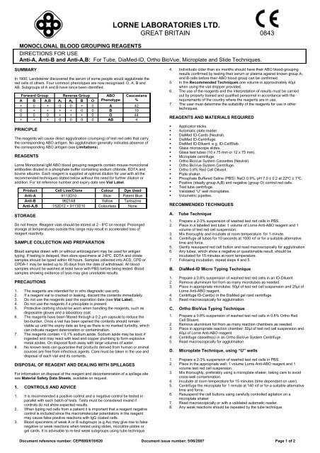
<strong>LORNE</strong> <strong>LABORATORIES</strong> <strong>LTD</strong>.GREAT BRITAIN 0843MONOCLONAL BLOOD GROUPING REAGENTSDIRECTIONS FOR USEAnti-A, Anti-B and Anti-A,B: For Tube, DiaMed-ID, Ortho BioVue, Microplate and Slide Techniques.SUMMARYIn 1900, Landsteiner discovered the serum of some people would agglutinate thered cells of others. Four common phenotypes are now recognised: O, A, B andAB. Subgroups of A and B have since been identified.Forward Group Reverse Group ABO CaucasiansA B A,B A 1 A 2 B O Phenotype %+ 0 + 0 0 + 0 A 420 + + + + 0 0 B 100 0 0 + + + 0 O 44+ + + 0 0 0 0 AB 4PRINCIPLEThe reagents will cause direct agglutination (clumping) of test red cells that carrythe corresponding ABO antigen. No agglutination generally indicates absence ofthe corresponding ABO antigen (see Limitations).REAGENTSLorne Monoclonal IgM ABO blood grouping reagents contain mouse monoclonalantibodies diluted in a phosphate buffer containing sodium chloride, EDTA andbovine albumin. Each reagent is supplied at optimal dilution for use with all therecommended techniques stated below without the need for further dilution oraddition. For lot reference number and expiry date see Vial Label.Product Cell Line/Clone Colour Dye UsedAnti-A 9113D10 Blue Patent BlueAnti-B 9621A8 Yellow TartrazineAnti-A,B 152D12 + 9113D10 Colourless NoneSTORAGEDo not freeze. Reagent vials should be stored at 2 - 8ºC on receipt. Prolongedstorage at temperatures outside this range may result in accelerated loss ofreagent reactivity.SAMPLE COLLECTION AND PREPARATIONBlood samples drawn with or without anticoagulant may be used for antigentyping. If testing is delayed, then store specimens at 2-8ºC. EDTA and citratesamples should be typed within 48 hours. Samples collected into ACD, CPD orCPDA-1 may be tested up to 35 days from the date of withdrawal. All bloodsamples should be washed at least twice with PBS before being tested. Bloodsamples showing evidence of lysis may give unreliable results.PRECAUTIONS1. The reagents are intended for in vitro diagnostic use only.2. If a reagent vial is cracked or leaking, discard the contents immediately.3. Do not use the reagents past the expiration date (see Vial Label).4. Do not use the reagents if a precipitate is present.5. Protective clothing should be worn when handling the reagents, such asdisposable gloves and a laboratory coat.6. The reagents have been filtered through a 0.2 µm capsule to reduce thebio-burden. Once a vial has been opened the contents should remainviable up until the expiry date as long as there is no marked turbidity, whichcan indicate reagent deterioration or contamination.7. The reagents contain < 0.1% sodium azide. Sodium azide may be toxic ifingested and may react with lead and copper plumbing to form explosivemetal azides. On disposal flush away with large volumes of water.8. No known tests can guarantee that products derived from human or animalsources are free from infectious agents. Care must be taken in the use anddisposal of each vial and its contents.DISPOSAL OF REAGENT AND DEALING WITH SPILLAGESFor information on disposal of the reagent and decontamination of a spillage sitesee Material Safety Data Sheets, available on request.1. CONTROLS AND ADVICE1. It is recommended a positive control and a negative control be tested inparallel with each batch of tests. Tests must be considered invalid ifcontrols do not show expected results.2. When typing red cells from a patient it is important that a reagent negativecontrol is included since the macromolecular potentiators in the reagentmay cause false positive reactions with IgG coated cells.3. Blood specimens of weak A or B subgroups (e.g Ax) may give rise to falsenegative or weak reactions when tested using slides, microtitre plates orgel cards. It is advisable to re-test weak subgroups using tube technique.4. Individuals older than six months should have their ABO blood-groupingresults confirmed by testing their serum or plasma against known group A 1and B cells before their ABO blood group can be confirmed.5. In the Recommended Techniques one volume is approximately 40µlwhen using the vial dropper provided.6. The use of the reagents and the interpretation of results must be carriedout by properly trained and qualified personnel in accordance with therequirements of the country where the reagents are in use.7. The user must determine the suitability of the reagents for use in othertechniques.REAGENTS AND MATERIALS REQUIRED• Applicator sticks.• Automatic plate reader.• DiaMed ID-Cards (Neutral).• DiaMed ID-Centrifuge.• DiaMed ID-Diluent: e.g. ID-CellStab.• Glass microscope slides.• Glass test tubes (10 x 75 mm or 12 x 75 mm).• Microplate centrifuge.• Ortho BioVue System Cassettes (Neutral).• Ortho BioVue System Centrifuge.• Ortho 0.8% Red Cell Diluent.• Plate shaker.• Phosphate Buffered Saline (PBS): NaCl 0.9%, pH 7.0 ± 0.2 at 22ºC ± 1ºC.• Positive (ideally group A 2 B) and negative (group O) control red cells.• Test tube centrifuge.• Validated “U” well microplates.• Volumetric pipettes.RECOMMENDED TECHNIQUESA. Tube Technique1. Prepare a 2-3% suspension of washed test red cells in PBS.2. Place in a labelled test tube: 1 volume of Lorne Anti-ABO reagent and 1volume of test red cell suspension.3. Mix thoroughly and incubate at room temperature for 1 minute.4. Centrifuge all tubes for 10 seconds at 1000 rcf or for a suitable alternativetime and force.5. Gently resuspend red cell button and read macroscopically for agglutination6. Any tubes, which show a negative or questionable result, should beincubated for 15 minutes at room temperature.7. Following incubation, repeat steps 4 and 5.B. DiaMed-ID Micro Typing Technique1. Prepare a 0.8% suspension of washed test red cells in an ID-Diluent.2. Remove aluminium foil from as many microtubes as needed.3. Place in appropriate microtube: 50µl of test red cell suspension and 25µl ofLorne Anti-ABO reagent.4. Centrifuge ID-Card(s) in the DiaMed gel card centrifuge.5. Read macroscopically for agglutination.C. Ortho BioVue Typing Technique1. Prepare a 0.8% suspension of washed test red cells in 0.8% Ortho RedCell Diluent.2. Remove aluminium foil from as many reaction chambers as needed.3. Place in appropriate reaction chamber: 50µl of test red cell suspension and40µl of Lorne Anti-ABO reagent.4. Centrifuge cassette(s) in an Ortho BioVue System Centrifuge.5. Read macroscopically for agglutination.D. Microplate Technique, using “U” wells1. Prepare a 2-3% suspension of washed test red cells in PBS.2. Place in the appropriate well: 1 volume Lorne Anti-ABO reagent and 1volume test red cell suspension.3. Mix thoroughly, preferably using a microplate shaker, taking care to avoidcross-well contamination.4. Incubate at room temperature for 15 minutes (time dependant on user).5. Centrifuge the microplate for 1 minute at 140 rcf or for a suitable alternativetime and force.6. Resuspend the cell buttons using carefully controlled agitation on amicroplate shaker7. Read macroscopically or with a validated automatic reader.8. Any weak reactions should be repeated by the tube technique.Document reference number: CEPI600/610/620 Document issue number: 5/06/2007 Page 1 of 2
E. Slide Technique1. Prepare a 35-45% suspension of test red cells in serum, plasma or PBS.2. Place on a labelled glass slide: 1 volume of Lorne Anti-ABO reagent and 1volume of test red cell suspension.3. Using a clean applicator stick, mix reagent and cells over an area of about20 x 40 mm.4. Slowly tilt the slide back and forth for 30 seconds, with occasional furthermixing during the 2-minute period, maintaining slide at room temperature.5. Read macroscopically after 2 minutes over a diffuse light and do notmistake fibrin strands as agglutination.6. Any weak reactions should be repeated by the tube technique.INTERPRETATION OF TEST RESULTS1. Positive: Agglutination of the test red cells constitutes a positive test resultand within accepted limitations of test procedure, indicates the presence ofthe appropriate ABO antigen on the test red cells.2. Negative: No agglutination of the test red cells constitutes a negative resultand within the accepted limitations of the test procedure, indicates theabsence of the appropriate ABO antigen on the test red cells.3. Discrepancies: If the results obtained with reverse group don’t correlatewith forward group, further investigation is required.4. Test results of cells that are agglutinated using the reagent negative controlshall be excluded, as the agglutination is most probably caused by theeffect of the macromolecular potentiators in the reagent on sensitised cells.STABILITY OF THE REACTIONS1. Read all tube and microplate tests straight after centrifugation.2. Slide tests should be interpreted within two minutes to ensure specificityand to avoid the possibility a negative result may be incorrectly interpretedas positive due to drying of the reagent.3. Caution should be exercised in the interpretation of results of testsperformed at temperatures other than those recommended.LIMITATIONS1. ABO antigens are not fully developed at birth and so weaker reactions maytherefore occur with cord or neonatal specimens.2. When using Monoclonal Anti-A,B, blood specimens of weak A or Bsubgroups (e.g Ax) may give rise to false negative or weak reactions whentested using slides, microtitre plates or gel cards. It is advisable to re-testweak subgroups using the tube technique.3. Lorne monoclonal Anti-A and monoclonal Anti-B are not validated to detectAx and A3 or Bx and B3 antigens resp and we therefore do not claimreactivity of the monoclonal Anti-A or Anti-B reagent against these weak Aand B sub-groups.4. Stored blood may give weaker reactions than fresh blood.5. False positive or false negative results may also occur due to:• Contamination of test materials• Improper storage, cell concentration, incubation time or temperature• Improper or excessive centrifugation• Deviation from the recommended techniques• Cord samples contaminated with Wharton’s jelly5. Issitt PD. Applied Blood Group Serology, 3 rd Edition. Montgomery Scientific,Miami 1985; Chapter 66. BSCH Blood Transfusion Task Force. Guidelines for microplate techniquesin liquid-phase blood grouping and antibody screening, Clinical LaboratoryHaematology 1990; 12, 437-460.7. Guidelines for the Blood Transfusion Service in the United Kingdom.H.M.S.O. Current Edition.8. British Committee for Standards in Haematology, Blood Transfusion TaskForce. Recommendations for evaluation, validation and implementation ofnew techniques for blood grouping, antibody screening and crossmatching. Transfusion Medicine, 1995, 5, 145-150.AVAILABLE REAGENT SIZESLorne Monoclonal Anti-ALorne Monoclonal Anti-BLorne Monoclonal Anti-A,BLorne Laboratories LimitedUnit 1 DanehillCutbush Park Industrial EstateLower Earley, Reading,Berkshire, RG6 4UTEnglandTel: +44 (0) 118 921 2264Fax: +44 (0) 118 986 4518E-mail: info@lornelabs.comTABLE OF SYMBOLSVial Size Catalogue Number5 ml 60000510 ml 6000101000 ml 6000005000 ml 6000005 ml 61000510 ml 6100101000 ml 6100005000 ml 6100005 ml 62000510 ml 6200101000 ml 6200005000 ml 620000SPECIFIC PERFORMANCE CHARACTERISTICS1. The reagents have been characterised by all the procedures mentioned inthe Recommended Techniques.2. Prior to release, each lot of Lorne Monoclonal Anti-A, Anti-B and Anti-A,B istested by the Recommended Techniques against a panel of antigenpositivered cells to ensure suitable reactivity.3. Specificity of source monoclonal antibodies is demonstrated using a panelof antigen-negative cells.4. The potency of the reagents has been tested against the followingminimum potency reference standards obtained from National Institute ofBiological Standards and Controls (NIBSC):• Anti-A reference standard 03/188 And / Or• Anti-B reference standard 03/1645. Lorne Anti-B does not react with “Acquired-B” red cells.6. Lorne Monoclonal ABO reagents do not detect crypt antigens such as T, Tnor Cad.7. The Quality Control of the reagents was performed using red cells that hadbeen washed at least twice with PBS prior to use.8. The reagents comply with the recommendations contained in the latestissue of the Guidelines for the UK Blood Transfusion Services.DISCLAIMER1. The user is responsible for the performance of the reagents by any methodother than those mentioned in the Recommended Techniques.2. Any deviations from the Recommended Techniques should be validatedprior to use 8 .BIBLIOGRAPHY1. Kholer G, Milstein C. Continuous culture of fused cells secreting antibody ofpredefined specificity. Nature 1975; 256, 495-4972. Messeter L et al. Mouse monoclonal antibodies with Anti-A, Anti-B andAnti-A,B specificities, some superior to human polyclonal ABO reagents.Vox Sang 1984; 46, 185-1943. Race RR, Sanger R. Blood Groups in Man, 6 th Edition. Blackwell Scientific,Oxford 1975; Chapter 2.4. Mollison PL. Blood Transfusion in Clinical Medicine, 8 th Edition, BlackwellScientific, Oxford 1987; Chapter 7.Document reference number: CEPI600/610/620 Document issue number: 5/06/2007 Page 2 of 2