Case 9-2011: A 37-Year-Old Man with Flushing and ... - AInotes
Case 9-2011: A 37-Year-Old Man with Flushing and ... - AInotes
Case 9-2011: A 37-Year-Old Man with Flushing and ... - AInotes
- No tags were found...
Create successful ePaper yourself
Turn your PDF publications into a flip-book with our unique Google optimized e-Paper software.
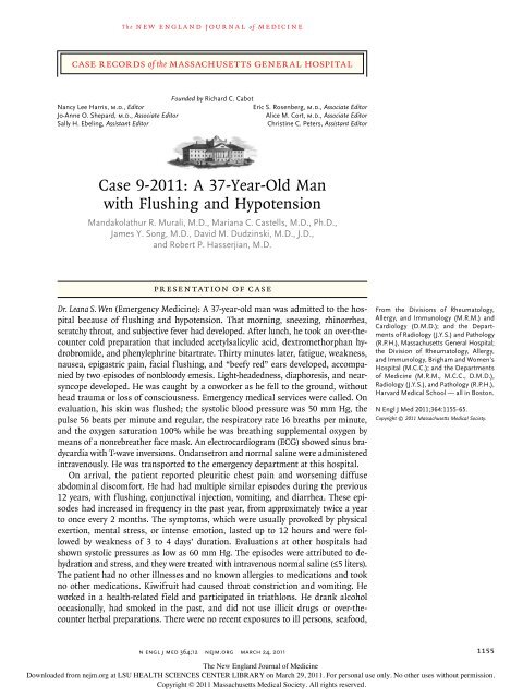
The new engl<strong>and</strong> journal of medicineABCDFigure 1. Findings on Examination of Bone Marrow Aspirate.The bone marrow–biopsy specimen contains multiple aggregates of pale cells that are often located adjacent tobone trabeculae surrounding a central core of mature lymphocytes (Panel A, hematoxylin <strong>and</strong> eosin). The pale cellshave oval nuclei <strong>and</strong> abundant cytoplasm <strong>and</strong> are admixed <strong>with</strong> numerous eosinophils (Panel B, hematoxylin <strong>and</strong>eosin); on the aspirate smear, the rare mast cells are abnormally spindled <strong>with</strong> uneven cytoplasmic granulation (inset,Wright–Giemsa stain). Immunohistochemical analysis shows that the mast cells express the lineage-specificmarker tryptase (Panel C) <strong>and</strong> aberrantly express CD25 (Panel D).20% of the overall marrow cellularity (Fig. 1A <strong>and</strong>1B). The remaining marrow was markedly hypercellular<strong>and</strong> showed trilineage maturing hematopoiesis<strong>with</strong> increased eosinophils. Immunohistochemicalstudies revealed that the abnormalcells were mast cells expressing mast-cell tryptase<strong>and</strong> CD117 (the product of the KIT gene) (Fig. 1C).Unlike normal mast cells, these cells expressedCD25 (Fig. 1D) <strong>and</strong> CD2. The bone marrow aspirateshowed normal hematopoiesis <strong>with</strong> no dysplasiaor increased blasts. Rare abnormal spindleshapedmast cells were present (Fig. 1B, inset).Flow cytometry showed no abnormal cells. Cytogeneticanalysis revealed a normal karyotype (46,XY).Fluorescence in situ hybridization (FISH) showedno rearrangement of the Fip1-like 1 gene (FIP1L1)or the gene that encodes platelet-derived growthfactor receptor α (PDGFRA). Subsequently, polymerase-chain-reactionassay of a peripheral-bloodsample revealed a point mutation at codon 816(Asp-816→Val) of the KIT gene.The diagnosis of systemic mastocytosis isbased on both clinical <strong>and</strong> pathological features(Table 3). 15 The presence of multiple large mastcellaggregates in the bone marrow of this patientconstitutes a major criterion for the diagnosisof systemic mastocytosis. In addition, there wasprominent mast-cell atypia (spindle-shaped cells<strong>with</strong> hypogranulation), aberrant expression ofCD2 <strong>and</strong> CD25, a KIT codon 816 mutation, <strong>and</strong>elevated serum tryptase levels, fulfilling all diagnosticcriteria for systemic mastocytosis.Bone marrow biopsy is an important diagnostictest for systemic mastocytosis, since the large1162n engl j med 364;12 nejm.org march 24, <strong>2011</strong>The New Engl<strong>and</strong> Journal of MedicineDownloaded from nejm.org at LSU HEALTH SCIENCES CENTER LIBRARY on March 29, <strong>2011</strong>. For personal use only. No other uses <strong>with</strong>out permission.Copyright © <strong>2011</strong> Massachusetts Medical Society. All rights reserved.
case records of the massachusetts general hospitalmast-cell aggregates are highly characteristic ofthe disease. As in this case, mast cells may berare in the aspirate smears because of fibrosisassociated <strong>with</strong> the mast-cell aggregates. 15 Whenthe diagnosis of systemic mastocytosis was suggestedon the basis of the bone marrow examinationin this patient, additional considerationsarose in the differential diagnosis.Systemic mastocytosis is associated <strong>with</strong> aclonal non–mast-cell hematologic neoplasm (e.g.,a myelodysplastic syndrome or chronic myelomonocyticleukemia) or acute myeloid leukemiain 30 to 40% of cases (Table 3). 16,17 In thesecases, the prognosis is typically determined bythat of the associated non–mast-cell neoplasm.<strong>Man</strong>y of these myeloid neoplasms have beenshown to share KIT mutation <strong>and</strong> cytogeneticabnormalities <strong>with</strong> the mast-cell proliferation,indicating an origin from a common precursorcell. 18,19 In this patient, there was no morphologicevidence of an associated myelodysplasticsyndrome, myeloproliferative neoplasm, or acuteleukemia, <strong>and</strong> the karyotype was normal.Another important consideration in the diagnosisof systemic mastocytosis is the exclusion ofa myeloid neoplasm <strong>with</strong> FIP1L1–PDGFRA rearrangement.This neoplasm is characterized bydiffusely increased bone marrow mast cells, increasedserum tryptase levels, <strong>and</strong> eosinophilia,<strong>and</strong> it may mimic systemic mastocytosis. <strong>Man</strong>ypatients <strong>with</strong> this neoplasm have a response tospecific targeted therapies, such as imatinib mesylate,that are ineffective in systemic mastocytosis.16 The presence of aggregated (rather th<strong>and</strong>iffusely scattered) mast cells, a KIT mutation,<strong>and</strong> a negative FISH study ruled out a myeloidneoplasm <strong>with</strong> FIP1L1–PDGFRA rearrangement.Dr. Dudzinski: The prominent yet transient STsegmentchanges observed on the admission ECGmay be a manifestation of epicardial coronaryvasospasm due to supraphysiologic concentrationsof histamine from mast-cell degranulation.This mechanism, variously termed hypersensitivitycoronary syndrome, allergic angina, or theKounis syndrome, has been described in responseto a host of allergic stimuli <strong>and</strong> can be manifestedas ECG changes <strong>and</strong> chest pain. 20-22Dr. Vassallo: Upper gastrointestinal endoscopyon the sixth day showed duodenal ulcers. Thepatient remained in stable condition <strong>and</strong> wasdischarged on the eighth day. He was prescribedranitidine <strong>and</strong> cetirizine for prophylactic managementof flushing <strong>and</strong> pruritus <strong>and</strong> epinephrineautoinjectors for use during acute reactions.Two days later, CT of the chest was performed.Dr. James Y. Song: CT of the chest performedafter the administration of intravenous contrastmaterial for evaluation of pulmonary embolism(Fig. 2A) shows loss of corticomedullary differentiation<strong>and</strong> coarse trabeculae in the thoracicABCFigure 2. Imaging Studies of the Chest.An axial CT scan of the chest (Panel A) obtained afterthe administration of contrast material shows loss ofcorticomedullary differentiation <strong>and</strong> heterogeneousosteosclerosis <strong>with</strong>in the 10th thoracic vertebral body(arrow). The bilateral fifth ribs show patchy bone sclerosis(Panel B, arrows) <strong>and</strong> there is a focal lytic lesion(≤7 mm in diameter) <strong>with</strong>in the left posterior eighthrib (Panel C, arrow). An additional lytic lesion was describedin the right third rib (not shown). The diffusesclerotic <strong>and</strong> lytic appearance is characteristic of mastocytosis.n engl j med 364;12 nejm.org march 24, <strong>2011</strong> 1163The New Engl<strong>and</strong> Journal of MedicineDownloaded from nejm.org at LSU HEALTH SCIENCES CENTER LIBRARY on March 29, <strong>2011</strong>. For personal use only. No other uses <strong>with</strong>out permission.Copyright © <strong>2011</strong> Massachusetts Medical Society. All rights reserved.
The new engl<strong>and</strong> journal of medicinespine. The ribs show scattered regions of patchysclerosis (Fig. 2B) alternating <strong>with</strong> normal marrowattenuation, <strong>and</strong> there is a focal lytic lesion<strong>with</strong>in the left eighth rib (Fig. 2C). This mixedlytic <strong>and</strong> sclerotic infiltrative appearance in theaxial skeleton is consistent <strong>with</strong> mastocytosis. Inretrospect, similar subtle changes were probablypresent in the spine on the abdominal <strong>and</strong> pelvicCT scan obtained on admission.Dr. Rosenberg: Dr. Castells, can you discuss thecare of this patient <strong>with</strong> systemic mastocytosis?Discussion of <strong>Man</strong>agementDr. Mariana C. Castells: With its variable clinicalphenotypes, 23 systemic mastocytosis is a greatmasquerader, <strong>and</strong> as in this patient, the onset ofsymptoms can precede the diagnosis by manyyears — a median of 9.5 years in one study. 24Exercise, alcohol, trauma, infections, contrastdyes, medications including nonsteroidal antiinflammatorydrugs (NSAIDs) <strong>and</strong> antibiotics, anesthesia,<strong>and</strong> surgery can induce release of mastcellmediators, triggering symptomatic episodes.When I questioned this patient, he said that histriggers were alcohol, stress, emotions, infections,foods <strong>with</strong> high fat content, <strong>and</strong> NSAIDs. He alsohad chronic symptoms, including daily flushing,chronic fatigue, depression, anxiety, bone pain,fractures, <strong>and</strong> chest pain that had led to multiplecardiac evaluations.Urticaria pigmentosa is associated <strong>with</strong> indolentsystemic mastocytosis in more than 80% ofall cases; scratching of these reddish brown macules,which are scattered over the body exceptfor the palms, soles, <strong>and</strong> scalp, triggers urtication<strong>and</strong> erythema, known as Darier’s sign. Ifskin findings are overlooked or subtle, the otherwisenonspecific symptoms may lead to a fruitlesssearch for other causes. When I examinedthis patient after discharge, I noted several brownlesions on the anterior chest, <strong>with</strong> a positiveDarier’s sign, findings compatible <strong>with</strong> urticariapigmentosa.The management of mastocytosis depends onthe clinicopathological subtype. Reduction of themast-cell burden is indicated in cases of aggressivemastocytosis <strong>and</strong> mast-cell leukemia, <strong>and</strong>treatment of the non–mast-cell hematologic disorderis indicated in cases of mastocytosis <strong>with</strong>such a disorder. This patient has indolent systemicmastocytosis, <strong>and</strong> treatment in these patientsis aimed at avoiding triggers, limiting releaseof mediators, <strong>and</strong> blocking their actions.For this patient, I recommended H 1 - <strong>and</strong> H 2 -histamine–receptor blockade (cetirizine <strong>and</strong> ranitidine);oral disodium cromoglycate, whichblocks the release of mediators from mast cells 25 ;leukotriene-receptor blockade (montelukast); <strong>and</strong>a proton-pump inhibitor (omeprazole). 26 He carriestwo epinephrine self-injectable devices totreat hypotensive events. Imatinib would not beuseful, since its site of action is abrogated by theD816V mutation in this patient. Unfortunately,the patient has been noncompliant <strong>with</strong> his medications,despite extensive counseling, <strong>and</strong> he hashad two further episodes of flushing <strong>and</strong> hypotensionduring the 15 months since the diagnosiswas established. Both episodes were treated inthe emergency department <strong>with</strong> intravenous fluids<strong>and</strong> antihistamines.Dr. Steven E. Goldfinger (Gastroenterology): I notethat the patient was once given epinephrine forhis flushing. The fact that profound hypotensiondid not develop tends to rule out carcinoid tumors,because epinephrine can precipitate a carcinoidcrisis, which could result in death.Anatomical DiagnosisIndolent systemic mastocytosis.This case was presented at the Medicine Gr<strong>and</strong> Rounds.Dr. Castells reports receiving grant support from Ovations forthe Cure <strong>and</strong> consulting <strong>and</strong> other fees from Schering-Plough;Dr. Hasserjian, consulting fees from Genzyme; <strong>and</strong> Dr. Dudzinski,honoraria from Lippincott Williams & Wilkins. No otherpotential conflict of interest relevant to this article was reported.Disclosure forms provided by the authors are available <strong>with</strong>the full text of this article at NEJM.org.References1. Habibzadeh MR, Ewy GA. ST-segmentelevation in lead aVR greater thanthat in lead V(1) secondary to diffusemyocardial ischemia from prolonged hypotension.Clin Cardiol 2007;30:251-4.2. Weyman AE, Hurwitz RA, Girod DA,Dillon JC, Feigenbaum H, Green D. Crosssectionalechocardiographic visualizationof the stenotic pulmonary valve. Circulation1977;56:769-74.3. Pellikka PA, Tajik AJ, Kh<strong>and</strong>heria BK,et al. Carcinoid heart disease: clinical <strong>and</strong>echocardiographic spectrum in 74 patients.Circulation 1993;87:1188-96.4. Izikson L, English JC III, Zirwas MJ.The flushing patient: differential diagnosis,workup, <strong>and</strong> treatment. J Am AcadDermatol 2006;55:193-208.5. Wilkin JK. <strong>Flushing</strong> reactions: consequences<strong>and</strong> mechanisms. Ann InternMed 1981;95:468-76.1164n engl j med 364;12 nejm.org march 24, <strong>2011</strong>The New Engl<strong>and</strong> Journal of MedicineDownloaded from nejm.org at LSU HEALTH SCIENCES CENTER LIBRARY on March 29, <strong>2011</strong>. For personal use only. No other uses <strong>with</strong>out permission.Copyright © <strong>2011</strong> Massachusetts Medical Society. All rights reserved.
case records of the massachusetts general hospital6. Kaliner M, Sigler R, Summers R, ShelhamerJH. Effects of infused histamine:analysis of the effects of H-1 <strong>and</strong> H-2 histaminereceptor antagonists on cardiovascular<strong>and</strong> pulmonary responses. J AllergyClin Immunol 1981;68:365-71.7. Lieberman PL. Anaphylaxis. In: AdkinsonNF Jr, Bochner BS, Busse WW,Holgate ST, Lemanske RF Jr, Simons FER,eds. Middleton’s allergy: principles &practice. 7th ed. Vol. 2. Philadelphia:Mosby, 2009:1027-49.8. Ditto AM, Harris KE, Krasnick J, MillerMA, Patterson R. Idiopathic anaphylaxis:a series of 335 cases. Ann AllergyAsthma Immunol 1996;77:285-91.9. Bone RC. Immunologic dissonance: acontinuing evolution in our underst<strong>and</strong>ingof the systemic inflammatory responsesyndrome (SIRS) <strong>and</strong> the multipleorgan dysfunction syndrome (MODS).Ann Intern Med 1996;125:680-7.10. Modlin IM, Kidd M, Latich I, ZikusokaMN, Shapiro MD. Current status of gastrointestinalcarcinoids. Gastroenterology2005;128:1717-51.11. Dhir V, Arya V, Malav IC, SuryanarayananBS, Gupta R, Dey AB. Idiopathicsystemic capillary leak syndrome (SCLS):case report <strong>and</strong> systematic review of casesreported in the last 16 years. Intern Med2007;46:899-904.12. Horny H-P, Metcalfe DD, Bennett JM,et al. Mastocytosis. In: Swerdlow S, CampoE, Harris NL, et al., eds. WHO classificationof tumours of haematopoietic <strong>and</strong>lymphoid tissues. Lyon, France: IARCPress, 2008:53-63.13. Fukuoka Y, Schwartz LB. Activemonomers of human beta-tryptase haveexp<strong>and</strong>ed substrate specificities. Int Immunopharmacol2007;7:1900-8.14. Schwartz LB, Kawahara MS, HugliTE, Vik D, Fearon DT, Austen KF. Generationof C3a anaphylatoxin from human C3by human mast cell tryptase. J Immunol1983;130:1891-5.15. Schwartz LB. Diagnostic value oftryptase in anaphylaxis <strong>and</strong> mastocytosis.Immunol Allergy Clin North Am 2006;26:451-63.16. Pardanani A, Lim KH, Lasho TL, et al.Prognostically relevant breakdown of 123patients <strong>with</strong> systemic mastocytosis associated<strong>with</strong> other myeloid malignancies.Blood 2009;114:<strong>37</strong>69-72.17. Travis WD, Li CY, Yam LT, BergstralhEJ, Swee RG. Significance of systemicmast cell disease <strong>with</strong> associated hematologicdisorders. Cancer 1988;62:965-72.18. Mir<strong>and</strong>a RM, Tang G, Jones DM, HuhYO, Medeiros LJ, Wang SA. Systemic mastocytosis<strong>with</strong> associated clonal hematologicalnon-mast cell lineage disorder(SM-AHNMD) shows distinct hematologicfeatures <strong>and</strong> reveals a common clonalorigin of myeloid <strong>and</strong> mast cells. ModPathol 2010;23:Suppl:311A. abstract.19. Sotlar K, Colak S, Bache A, et al. Variablepresence of KITD816V in clonal haematologicalnon-mast cell lineage diseasesassociated <strong>with</strong> systemic mastocytosis(SM-AHNMD). J Pathol 2010;220:586-95.20. Kounis NG. Kounis syndrome (allergicangina <strong>and</strong> allergic myocardial infarction):a natural paradigm? Int J Cardiol2006;110:7-14.21. Kounis NG, Kounis GN, Kouni SN,Soufras GD, Niarchos C, Mazarakis A. Allergicreactions following implantation ofdrug-eluting stents: a manifestation ofKounis syndrome? J Am Coll Cardiol2006;48:592-3.22. Rohr SM, Rich MW, Silver KH. Shortnessof breath, syncope, <strong>and</strong> cardiac arrestcaused by systemic mastocytosis.Ann Emerg Med 2005;45:592-4.23. Castells M, Austen KF. Mastocytosis:mediator-related signs <strong>and</strong> symptoms. IntArch Allergy Immunol 2002;127:147-52.24. Horan RF, Austen KF. Systemic mastocytosis:retrospective review of a decade’sclinical experience at the Brigham<strong>and</strong> Women’s Hospital. J Invest Dermatol1991;96:Suppl:5S-14S, 60S-65S.25. Soter NA, Austen KF, Wasserman SI.Oral disodium cromoglycate in the treatmentof systemic mastocytosis. N Engl JMed 1979;301:465-9.26. Escribano L, Akin C, Castells M,Schwartz LB. Current options in the treatmentof mast cell mediator-related symptomsin mastocytosis. Inflamm AllergyDrug Targets 2006;5:61-77.Copyright © <strong>2011</strong> Massachusetts Medical Society.Lantern Slides Updated: Complete PowerPoint Slide Sets from the Clinicopathological ConferencesAny reader of the Journal who uses the <strong>Case</strong> Records of the Massachusetts General Hospital as a teaching exercise or referencematerial is now eligible to receive a complete set of PowerPoint slides, including digital images, <strong>with</strong> identifying legends,shown at the live Clinicopathological Conference (CPC) that is the basis of the <strong>Case</strong> Record. This slide set contains all of theimages from the CPC, not only those published in the Journal. Radiographic, neurologic, <strong>and</strong> cardiac studies, gross specimens,<strong>and</strong> photomicrographs, as well as unpublished text slides, tables, <strong>and</strong> diagrams, are included. Every year 40 sets are produced,averaging 50-60 slides per set. Each set is supplied on a compact disc <strong>and</strong> is mailed to coincide <strong>with</strong> the publication of the<strong>Case</strong> Record.The cost of an annual subscription is $600, or individual sets may be purchased for $50 each. Application forms for the currentsubscription year, which began in January, may be obtained from the Lantern Slides Service, Department of Pathology,Massachusetts General Hospital, Boston, MA 02114 (telephone 617-726-2974) or e-mail Pathphotoslides@partners.org.n engl j med 364;12 nejm.org march 24, <strong>2011</strong> 1165The New Engl<strong>and</strong> Journal of MedicineDownloaded from nejm.org at LSU HEALTH SCIENCES CENTER LIBRARY on March 29, <strong>2011</strong>. For personal use only. No other uses <strong>with</strong>out permission.Copyright © <strong>2011</strong> Massachusetts Medical Society. All rights reserved.


