Rare Case of Pleomorphic Adenoma: A Case Report - IJMD
Rare Case of Pleomorphic Adenoma: A Case Report - IJMD
Rare Case of Pleomorphic Adenoma: A Case Report - IJMD

Create successful ePaper yourself
Turn your PDF publications into a flip-book with our unique Google optimized e-Paper software.
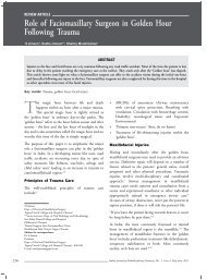
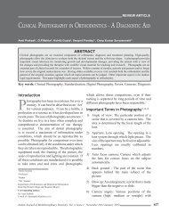
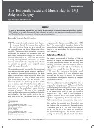
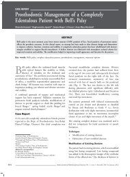
case report<strong>Rare</strong> <strong>Case</strong> <strong>of</strong> <strong>Pleomorphic</strong> <strong>Adenoma</strong>: A <strong>Case</strong> <strong>Report</strong>Srinivasan H*, Loganathan Selvaraj**Abstract<strong>Pleomorphic</strong> adenoma is a benign tumor originating from the mature salivary tissue. Ectopic pleomorphic adenoma is definedas an unusual presentation <strong>of</strong> pleomorphic adenoma in other sites except the major salivary gland and the minor salivary gland.Ectopic pleomorphic adenoma has been found in the s<strong>of</strong>t tissue <strong>of</strong> the neck, in the skin <strong>of</strong> the external auditory canal, in thesubcutaneous layer <strong>of</strong> the nose, and in the lymph node <strong>of</strong> the mediastinum. Ectopic pleomorphic adenoma could appear dueto the metastasis, the neoplastic degeneration <strong>of</strong> the ectopic salivary tissue or the implantation after the surgery <strong>of</strong> a salivarygland tumor. We present a case <strong>of</strong> pleomorphic adenoma found in the superficial subcutaneous layer above the superficialmusculoaponeurotic system (SMAS) <strong>of</strong> the anterior border <strong>of</strong> masseter muscle over the body <strong>of</strong> the mandible area. The patienthad no history <strong>of</strong> trauma or surgery <strong>of</strong> the salivary tumor and no specific physical finding. No case <strong>of</strong> the ectopic salivarytissue or ectopic pleomorphic adenoma in the superficial subcutaneous layer <strong>of</strong> the anterior border <strong>of</strong> masseter muscle overthe body <strong>of</strong> the mandible area has been reported. This case could be a rare presentation <strong>of</strong> the ectopic pleomorphic adenomafrom the ectopic salivary tissue arising from the division <strong>of</strong> the parotid gland in early embryonic period.Key words: <strong>Pleomorphic</strong> adenoma, SMAS, salivary gland<strong>Pleomorphic</strong> adenomas account for 40-70% <strong>of</strong>minor salivary gland tumors. They may arisein unusual locations along the cheek, alongthe Stenson’s duct and in accessory parotid tissue,which is separated from the body <strong>of</strong> the gland, 4 in theparapharyngeal space arising either de novo or fromthe deep lobe <strong>of</strong> the parotid gland and extendingthrough the stylomandibular tunnel into theparapharyngeal space. 5 It may also develop in salivarygland inclusions within the lymph nodes in the neck. 4This is the first report in the English languageliterature <strong>of</strong> a pleomorphic adenoma presenting inthe superficial subcutaneous layer above the superficialmusculoaponeurotic system (SMAS) <strong>of</strong> the anteriorborder <strong>of</strong> masseter muscle over the body <strong>of</strong> themandible area. 2 A brief description <strong>of</strong> the surgicaltechnique is presented.*Reader**Senior LecturerDept. <strong>of</strong> Oral SurgeryPriyadarshini Dental College and Hospital, ChennaiAddress for correspondenceDr Loganathan SelvarajSenior Lecturer, Dept. <strong>of</strong> Oral SurgeryPriyadarshini Dental College and Hospital, ChennaiE-mail: drloganathans@gmail.com<strong>Case</strong> <strong>Report</strong>A 33-year-old Asian-Indian female patient presentedwith swelling on the left side <strong>of</strong> the mandibleregion, first noted approximately one month prior topresentation. She also experienced occasional pain inthe region for the past two weeks. The patient’s pastmedical history was noncontributory.Extraoral examination revealed a mild facial asymmetryin the region <strong>of</strong> the left body <strong>of</strong> the mandible (Fig. 1).The skin over the region was not fixed to any underlyingstructures and was freely movable in all planes. Therewas no evidence <strong>of</strong> locoregional lymphadenopathy.Her mouth opening was within the normal range(30 mm).Intraoral examination revealed no abnormalities. Onpalpation, the swelling was tender. The overlying skinwas free <strong>of</strong> any surface change, ulceration, purulentdischarge or sinus tract. The swelling measured 40 mmin circumference.Radiological finding (Fig. 2) showed no destructionor erosion <strong>of</strong> adjacent mandible. Based on the clinicaland radiologic findings, the provisional diagnosis givenby the clinicians was lipoma or fibroma or vascularlesion. A 10 ml syringe with a wide bore needle was236 Indian Journal <strong>of</strong> Multidisciplinary Dentistry, Vol. 1, Issue 4, May-June 2011
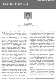
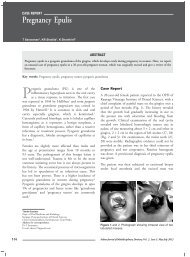
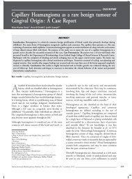
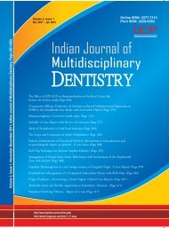
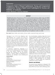
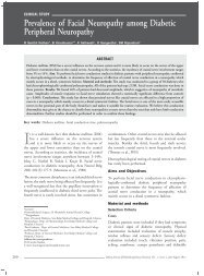
<strong>Case</strong> <strong>Report</strong>Figure 1 . Extraoral picture showing mild facial asymmetryon the left mandibular region.Figure 4. Picture showing the tumor dissected out.Figure 2. Radiological finding showing no destruction orerosion <strong>of</strong> adjacent mandible.Figure 5. Histopathological picture showing an encapsulatedlesion comprising <strong>of</strong> duct like spaces with eosinophiliccoagulum (10×, H&E stain)Figure 3. Picture showing the modified excision <strong>of</strong> thelesion.used to aspirate the contents <strong>of</strong> the swelling to assessif the swelling was a vascular lesion. The aspirationwas found to be negative.An excisional biopsy was performed through an extraoralincision under local infiltration anesthesia. Bluntdissection and excision <strong>of</strong> the lobulated mass was done(Figs. 3 and 4). The total tumor mass was dissectedout and wound was closed in layers. The excised tumormass fixed in formalin was sent for histopathologicalevaluation. On gross examination, the tumor massconsisted <strong>of</strong> eight pieces <strong>of</strong> tissue which measured5-20 mm 2 in size, with a whitish, faintly lobulatedand focally glistening cut surface. Histopathologyrevealed an encapsulated lesion comprising <strong>of</strong> ductlike spaces with an eosinophilic coagulum, surroundedby spindle-shaped and plasmacytoid cells in a dense,hyalinized connective tissue stroma. An area resemblingchondroid tissue was also evident. The histopathologicalfeatures were suggestive <strong>of</strong> pleomorphic adenoma.(Figs. 5 and 6). The patient is on a regular follow-upand is disease free till date.Indian Journal <strong>of</strong> Multidisciplinary Dentistry, Vol. 1, Issue 4, May-June 2011237
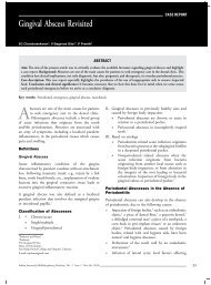
<strong>Case</strong> <strong>Report</strong>Figure 6. Histopathological picture showing an arearesembling chondroid tissue (10×, H&E stain).Discussion<strong>Pleomorphic</strong> adenoma is a benign tumor originatingfrom the mature salivary tissue. Ectopic pleomorphicadenoma is defined as an unusual presentation <strong>of</strong>pleomorphic adenoma in other sites except the majorsalivary gland and the minor salivary gland. Ectopicpleomorphic adenoma was found in the s<strong>of</strong>t tissue <strong>of</strong>the neck, in the skin <strong>of</strong> the external auditory canal,in the subcutaneous layer <strong>of</strong> the nose, and in thelymph node <strong>of</strong> the mediastinum. Ectopic pleomorphicadenoma could appear due to metastasis, the neoplasticdegeneration <strong>of</strong> the ectopic salivary tissue, or theimplantation after the surgery <strong>of</strong> a salivary glandtumor. We discussed the previous cases <strong>of</strong> ectopicsalivary tissues and ectopic pleomorphic adenomas.This case could be a rare presentation <strong>of</strong> the ectopicpleomorphic adenoma from the ectopic salivary tissuearising from the division <strong>of</strong> the parotid gland in earlyembryonic period. 1 If a vascular tumor is suspected,an magnetic resonance imaging (MRI) angiography orconventional arteriography should be performed. If thepossibility <strong>of</strong> a vascular lesion is ruled out, a histologicstudy could be performed. 6 The histopathology <strong>of</strong> ourcase proved to be pleomorphic adenoma. Though, thereis an anatomic relationship between the deep lobe <strong>of</strong>the parotid gland and the mandible, the probability <strong>of</strong>deep lobe tumors spreading to the infratemporal fossaseems to be rare. 2 All these findings indicate that thetumor has arisen from an ectopic minor salivary glandwithin the subcutaneous layer. The most commonsite <strong>of</strong> a PA <strong>of</strong> the minor salivary glands is the palatefollowed by lip, buccal mucosa and floor <strong>of</strong> the mouth,tongue, tonsil, pharynx, retromolar area and nasalcavity. 6,7 <strong>Pleomorphic</strong> adenoma in the parapharyngealspace can arise either de novo or may arise in thedeep lobe <strong>of</strong> the parotid gland and extend throughthe stylomandibular tunnel into the parapharyngealspace. Tumors in the parapharyngeal space are rare andconstitute less than 0.5% <strong>of</strong> head and neck neoplasms.Of these, PA is the commonest benign tumor (40%). 8The elective treatment <strong>of</strong> a pleomorphic adenomais surgery. There are numerous surgical approaches,indicating the difficulty <strong>of</strong> access, accentuated bythe communications with the neighboring regions.The prognosis <strong>of</strong> a pleomorphic adenoma isgood. The patient is remaining disease free aftersurgical excision and is on a regular follow-up.ConclusionIn summary, we are reporting the first case <strong>of</strong>pleomorphic adenoma presenting in the superficialsubcutaneous layer above the SMAS <strong>of</strong> the anteriorborder <strong>of</strong> masseter muscle over the body <strong>of</strong> themandible area.References1.2.3.4.5.6.7.8.Tsukuno M, Nakamura A, Takai S, Kurihara K.Subcutaneous pleomorphic adenomas in two differentareas <strong>of</strong> the face. Scand J Plast Reconst Surg Hand Surg2002;36(2):109-11.Chung JH, Burm JS, Oh SJ. An ectopic pleomorphicadenoma in the superficial subcutaneous layer <strong>of</strong> thepreauricular area. J Korean Soc Plast ReconstrSurg32002;29(2):115-7.Shafer WG, Hine MK, Levy BM. Spread <strong>of</strong> oralinfection. In: a Textbook <strong>of</strong> Oral Pathology. 4th editionWB Saunders Co. 1997:513.Ellis GL, Auclaire PL, Gnepp DR. SurgicalPathology <strong>of</strong> Salivary Gland. WB Saunders Co:Philadelphia 1991:166.Rodriguez-Ciurana J, Rodado C, Saez M, Bassas C.Giant parotid pleomorphic adenoma involving theparapharyngeal space: report <strong>of</strong> a case. J Oral Maxill<strong>of</strong>acSurg 2000;58(10):1184-7.Eveson JW, Cawson RA. Tumour <strong>of</strong> the minor(oropharyngeal) salivary glands: a demographic study <strong>of</strong>336 cases. J Oral Pathol 1985;14(6):500-9.Cohen MA. <strong>Pleomorphic</strong> adenoma <strong>of</strong> the cheek. IntJ Oral Maxill<strong>of</strong>ac Surg 1986;15(6):777-9.Batsakis JG, Sneige N. Pathology consultation:parapharyngeal and retropharyngeal space diseases. AnnOtol Rhinol Laryngol 1989;98(4 Pt 1):320-1.238Indian Journal <strong>of</strong> Multidisciplinary Dentistry, Vol. 1, Issue 4, May-June 2011