Acute Myeloid Leukemia - The Leukemia & Lymphoma Society
Acute Myeloid Leukemia - The Leukemia & Lymphoma Society
Acute Myeloid Leukemia - The Leukemia & Lymphoma Society
- No tags were found...

Create successful ePaper yourself
Turn your PDF publications into a flip-book with our unique Google optimized e-Paper software.
A Message From John WalterPresident and CEO of <strong>The</strong> <strong>Leukemia</strong> & <strong>Lymphoma</strong> <strong>Society</strong><strong>The</strong> <strong>Leukemia</strong> & <strong>Lymphoma</strong> <strong>Society</strong> (LLS) is committed to bringingyou the most up-to-date blood cancer information. We know howimportant it is for you to have an accurate understanding of yourdiagnosis, treatment and support options. With this knowledge, youcan work with members of your oncology team to move forwardwith the hope of remission and recovery. Our vision is that one daythe great majority of people who have been diagnosed with <strong>Acute</strong><strong>Myeloid</strong> <strong>Leukemia</strong> (AML) will be cured or will be able to managetheir disease with a good quality of life. We hope that the informationin this booklet will help you along your journey.LLS is the world’s largest voluntary health organization dedicated tofunding blood cancer research, education and patient services. Sincethe first funding in 1954, LLS has invested more than $814 millionin research specifically targeting blood cancers. We will continue toinvest in research for cures and in programs and services that improvethe quality of life of people who have AML and their families.We wish you well.John WalterPresident and CEO
IntroductionThis booklet provides information about acute myeloid leukemia (AML) forpatients and their families. Brief descriptions of normal blood and marrow anddefinitions of medical terms are included at the end of the booklet to help readersbetter understand the information about AML.AML may be called by other names, including acute myelogenous leukemia, acutemyelocytic leukemia, acute myeloblastic leukemia and acute granulocytic leukemia.About 12,950 new cases of AML were expected to be diagnosed in the UnitedStates in 2011. As of January 2008 an estimated 30,993 people were living with(or were in remission from) AML. Although AML can occur at any age, adults aged60 years and older are more likely to develop the disease than younger people. 1Advances in AML testing and treatment are resulting in improved remissionand cure rates, but much work remains to be done. For example, the vitamin Aderivative all-trans retinoic acid (ATRA) has greatly improved survival rates forpatients with acute promyelocytic leukemia (APL), a subtype of AML. A numberof new therapies are under study in clinical trials.1Howlader N, Noone AM, Krapcho M, Neyman N, Aminou R, Waldron W, Altekruse SF, Kosary CL, Ruhl J,Tatalovich Z, Cho H, Mariotto A, Eisner MP, Lewis DR, Chen HS, Feuer EJ, Cronin KA, Edwards BK (eds).SEER Cancer Statistics Review, 1975-2008, National Cancer Institute. Bethesda, MD, www.seer.cancer.gov/csr/1975_2008/, based on November 2010 SEER data submission, posted to the SEER website, 2011.Here to HelpThis booklet will help you talk to your doctor about the tests and treatment youneed. We encourage you to take the lead in asking questions and discussing yourfears and concerns. <strong>The</strong>se actions will give members of your healthcare team theopportunity to answer your questions, extend emotional support and provide anyneeded referrals.A diagnosis of AML is often a shock to the patient, family members and friends.Denial, depression, hopelessness and fear are some of the reactions people mayhave. Keep in mind that◦{Many people are better able to cope once they begin treatment and can lookforward to recovery.{ ◦ <strong>The</strong> outlook for people with AML is continuing to improve. New approachesto therapy are being studied in clinical trials for patients of all ages and at everystage of treatment.page 2 I 800.955.4572 I www.LLS.org
LLS Has Ways to Help. Treatment for AML will affect your daily life, at least fora time. You may have questions about your treatment and want to have friends,family members or caregivers help you get information.Making treatment choices, paying for medical care, communicating with healthcareproviders, family members and friends—these are some of the stressors that goalong with a cancer diagnosis. LLS offers free information and patient services forindividuals and families touched by blood cancers.Speak to an Information Specialist. Information Specialists are master’s leveloncology professionals. <strong>The</strong>y provide accurate up-to-date disease and treatmentinformation and are available to speak with callers Monday through Friday, 9 a.m.to 6 p.m. ET at (800) 955-4572. You can email infocenter@LLS.org or chat livewith a Specialist at www.LLS.org.Language Services. Free language services are available when you speak with anInformation Specialist. Let your doctor know if you want a professional healthcareinterpreter who speaks your native language or uses sign language to be presentduring your visit. Many times, this is a free service.Información en Español. LLS has a number of resources available in Spanish forpatients, caregivers and healthcare professionals. You can read and download theseresources online at www.LLS.org/espanol or order printed copies by mail or phone.Other Helpful Organizations. Our website, www.LLS.org/resourcedirectory,offers an extensive list of resources for patients and families about financialassistance, counseling, transportation, summer camps and other needs.Chapter Programs and Services. LLS chapter offices around the United Statesand Canada offer support and education. Your chapter can arrange for peer-to-peersupport through the Patti Robinson Kaufmann First Connection Program.<strong>The</strong> Patient Financial Aid program offers a limited amount of financial aid forqualified patients. Find your local chapter by calling (800) 955-4572 or by visitingwww.LLS.org/chapterfind.Clinical Trials. Our Information Specialists help patients work with their doctorsto find out about specific clinical trials. Information Specialists conduct clinical-trialsearches for patients, family members and healthcare professionals. You can alsouse TrialCheck®, an online clinical-trial search service supported by LLS that offerspatients and caregivers immediate access to listings of blood cancer clinical trials.Please visit www.LLS.org/clinicaltrials.Free Materials. LLS publishes many free education and support materials forpatients and healthcare professionals. PDF files can be read online or downloaded.Free print versions can be ordered. Visit www.LLS.org/resourcecenter.<strong>Acute</strong> <strong>Myeloid</strong> <strong>Leukemia</strong> I page 3
Telephone/Web Education Programs. LLS provides a number of free, livetelephone and web education programs presented by experts for patients, caregiversand healthcare professionals. For more information, visit www.LLS.org/programs.Suggestions From Other People Living With Cancer◦{Get information about choosing a cancer specialist or treatment center.◦{Find out about financial matters: What does your insurance cover?What financial assistance is available to you?◦{Learn about the most current tests and treatments for your type of AML.◦{Keep all appointments with the doctor and talk openly about your fears orconcerns or any side effects you experience.◦{Talk with family and friends about how you feel and how they can help.◦{Contact your doctor if you have fatigue, fever, pain or sleep problems sothat any issues can be addressed early on.◦{Get medical advice if you have experienced changes in mood, feelingsof sadness or depression.<strong>The</strong> Trish Greene Back to School Program for Children With Cancer.This program is designed to increase communication among parents, children,adolescents, young adults, healthcare professionals and school personnel.Informative materials, videos and a wealth of literature are available through LLSchapters to help ensure a smooth transition back to school. For more information,please visit www.LLS.org/backtoschool. For practical guidance on how to supportyour child, yourself and other family members, see the free LLS booklet CopingWith Childhood <strong>Leukemia</strong> and <strong>Lymphoma</strong>.Reach Out. You and your loved ones can reach out for support in several ways.For example:◦{LLS offers online Blood Cancer Discussion Boards as well as online chats atwww.LLS.org/getinfo.◦{Local or Internet support groups and blogs can provide forums for support.{ ◦ Patients with cancer often become acquainted with one another, and thesefriendships provide support.page 4 I 800.955.4572 I www.LLS.org
Depression. Treatment for depression has proven benefits for people livingwith cancer. Depression is an illness that should be treated even when a person isundergoing AML treatment. Seek medical advice if your mood does not improveover time—for example, if you feel depressed every day for a 2-week period.Contact LLS or ask your healthcare team for guidance and referrals to othersources of help, such as counseling services or community programs. For moreinformation you can contact the National Institute of Mental Health (NIMH) atwww.nimh.nih.gov and enter “depression” in the search box at the top of the webpage, or call the NIMH toll-free at (866) 615-6464.We’d Like to Hear From You. We hope this booklet helps you. Please tell uswhat you think at www.LLS.org/publicationfeedback. Click on “LLS Disease &Treatment Publications—Survey for Patients, Family and Friends.”<strong>Leukemia</strong><strong>Leukemia</strong> is a cancer of the marrow and blood. <strong>The</strong> four major types of leukemiaare acute myeloid leukemia, chronic myeloid leukemia, acute lymphoblasticleukemia and chronic lymphocytic leukemia.<strong>Acute</strong> leukemias are rapidly progressing diseases that affect cells that are not fullydeveloped. <strong>The</strong>se cells cannot carry out their normal functions. Chronic leukemiasusually progress more slowly, and patients have greater numbers of mature cells.In general, these more mature cells can carry out some of their normal functions(see Normal Blood and Marrow on page 31).With myeloid leukemia, a cancerous change begins in a marrow cell that normallyforms certain blood cells—that is, red cells, some types of white cells and platelets.With lymphocytic (lymphoblastic) leukemia, the cancerous change begins in amarrow cell that normally forms lymphocytes (another type of white cell).<strong>The</strong> four main types of leukemia are further classified into subtypes. Knowing thesubtype of your disease is important because the treatment approach may be basedon the subtype (see AML Subtypes on page 11).More general information about leukemia is given in the free LLS publicationUnderstanding <strong>Leukemia</strong>.<strong>Acute</strong> <strong>Myeloid</strong> <strong>Leukemia</strong> I page 5
<strong>Acute</strong> <strong>Myeloid</strong> <strong>Leukemia</strong>How AML Develops. AML results from acquired changes in the DNA (geneticmaterial) of a developing marrow cell. Once the marrow cell becomes a leukemiccell, it multiplies into 11 billion or more cells. <strong>The</strong>se cells, called “leukemic blasts,”do not function normally. However, they grow and survive better than normal cells.<strong>The</strong> presence of the leukemic blasts blocks the production of normal cells. As aresult, when AML is diagnosed, the number of healthy blood cells (red cells, whitecells and platelets) is usually lower than normal.<strong>The</strong> medical term forLow red cell countI IsI AnemiaLow platelet count I Thrombocytopenia(“thrombocyte” is another word for platelet)Low neutrophil countI Neutropenia (a neutrophil is a type of white cell)Causes and Risk Factors. Most patients diagnosed with AML have no clear-cuttriggering event.Repeated exposure to the chemical benzene can be a factor in AML development.Benzene damages the DNA of normal marrow cells. According to the Agency forToxic Substances and Disease Registry, despite the fact that petroleum productscontribute to the majority of benzene in the atmosphere, half of the total nationalpersonal exposure to benzene comes from cigarette smoke. Benzene is also foundin certain industrial settings; however, the strict regulation of its use has decreasedbenzene exposure in the workplace.A small but increasing percentage of AML cases arise following treatment withchemotherapy (especially with alkylating agents or topoisomerase II inhibitors) orradiation therapy for other cancers, such as lymphoma, myeloma and breast cancer.But only a small proportion of people exposed to chemotherapy, radiation therapyand/or benzene develop AML. A theory about why AML develops in some people isthat they have inherited genes that limit their ability to detoxify the causative agents.Genetic disorders, such as Fanconi’s anemia, Shwachman syndrome, Diamond-Blackfan syndrome and Down syndrome, are associated with an increased riskof AML. Very rarely, an unexpectedly high number of cases of AML may bediagnosed within the same family. Clusters of AML in unrelated people within acommunity are uncommon. AML is not contagious.page 6 I 800.955.4572 I www.LLS.org
AML may develop from the progression of other blood cancers, includingpolycythemia vera, primary myelofibrosis, essential thrombocythemia andmyelodysplastic syndromes (MDS).Incidence. AML is the most common acute leukemia affecting adults. Olderpeople are more likely to develop AML than younger adults or children. However,AML is the most common type of leukemia diagnosed during infancy. About 15to 20 percent of cases of acute childhood leukemia and 80 percent of cases of acuteadult leukemia are AML.<strong>The</strong> risk for developing AML increases about 10-fold from ages 30 to 34 years(about 1 case per 100,000 people) to ages 65 to 69 years (about 10 cases per100,000 people). For people over 70, the incidence rate continues to increase,peaking between the ages of 80 and 84 (see Figure 1).<strong>Acute</strong> <strong>Myeloid</strong> <strong>Leukemia</strong>: Age-Specific Incidence Rates (2004-2008)2222.522.2201919.0171514.3Incidence (per 100,000)131199.576.254.4311.60.9 0.90.7 0.80.41.1 1.3 1.31.72.33.20
Signs and Symptoms. A person with signs or symptoms that suggest thepossibility of leukemia is usually referred to a specialist. This may be a hematologistor an oncologist. <strong>The</strong> doctor will order additional tests to make a diagnosis (seepage 9). <strong>The</strong> signs and symptoms of AML are associated with a number of other,less serious diseases.It is common for people with AML to feel a loss of well-being because of theunderproduction of normal bone marrow cells. <strong>The</strong> person may tire more easilyand have shortness of breath during normal physical activities.People with AML may also have◦{A pale complexion from anemia◦{Signs of bleeding caused by a very low platelet count, including Black-and-blue marks or bruises occurring for no reason or because of aminor injury <strong>The</strong> appearance of pinhead-sized red spots on the skin, called “petechiae” Prolonged bleeding from minor cuts◦{Mild fever◦{Swollen gums◦{Frequent minor infections, such as perianal sores◦{Loss of appetite and weight loss◦{Discomfort in bones or joints◦{Enlarged spleen◦{Enlarged liver.Bleeding. A low platelet count predisposes patients to bleeding. Bleeding in thebrain or lung is serious and can be fatal. However, such bleeding is usually precededby minor bleeding, such as nose bleeds, blood in the urine or bruises (see Diseaseand Treatment Side Effects on page 23).Infection. Severe infection can occur at the time of diagnosis but becomes morecommon and often more serious during treatment, when the bone marrow iscompletely suppressed. If the neutrophil count becomes or remains low because ofAML or its treatment, serious infection almost invariably occurs and is a leadingcause of death from AML (see Disease and Treatment Side Effects on page 23).<strong>Myeloid</strong> Sarcoma. Rarely, a collection of AML cells, called a “myeloid sarcoma,”forms outside the marrow. A myeloid sarcoma may occur in almost any part ofthe body. Other signs of AML may not appear in the blood and marrow untilweeks or months after the initial myeloid sarcoma diagnosis. A myeloid sarcomadiagnosis is equivalent to a diagnosis of AML and is treated with chemotherapyrather than local therapy. Treatment may also include allogeneic or autologous stemcell transplant. Other names for a myeloid sarcoma are “chloroma,” “granulocyticsarcoma,” “myeloblastoma” or “monocytoma.”page 8 I 800.955.4572 I www.LLS.org
DiagnosisAn accurate diagnosis of the type of leukemia is important. <strong>The</strong> exact diagnosishelps the doctor to◦{Estimate how the disease will progress◦{Determine the appropriate treatment.Talk to yourdoctor about<strong>The</strong> diagnostic tests that are being doneWhat the results meanGetting copies of the test results.Some of these tests may be repeated during and after therapy to measure the effectsof treatment.Blood and Bone Marrow Tests. Blood and bone marrow tests are used todiagnose AML and the AML subtype. A change in the number and appearance ofblood cells helps to make the diagnosis. AML cells look similar to normal immaturewhite cells. However, their development is incomplete (see Figure 2).Normal Marrow Cells and AML Blast CellsPanel APanel BFigure 2. I Panel A shows normal marrow cells seen through a microscope. <strong>The</strong> darker shapes are the nuclei ofthe cells. Some of the nuclei are circular and some are horseshoe shaped, reflecting the different developmentalstages and the different types of cells. Panel B shows AML blast cells seen through a microscope. <strong>The</strong>se cells are“arrested” in an early stage of development. <strong>The</strong> AML cells in panel B all have a similar appearance, in contrastto the varied appearance of the normal cells in panel A.<strong>Acute</strong> <strong>Myeloid</strong> <strong>Leukemia</strong> I page 9
Blood and Marrow Samples. To do the tests, blood samples are generally takenfrom a vein in the patient’s arm. Samples of marrow cells are obtained by bonemarrow aspiration and biopsy (see page 36). <strong>The</strong> cells from the blood and marrowsamples are examined under a microscope.Most patients with AML have I Blood tests usedLower-than-expectedred cell and platelet countsToo many immature white cellsand too few mature white cellsI CBC – Blood cell counts are determined by ablood test called a “complete blood count (CBC).I Peripheral Blood Smear – A test called a“peripheral white cells blood smear” (anexamination of the stained [dyed] blood cellswith a microscope) usually shows the presenceof leukemic blast cells (myeloblasts). <strong>The</strong>seimmature cells do not function like normal,mature white blood cells.Confirmation of Diagnosis. In addition to looking at the number and appearanceof the cells in the blood samples, your doctor will also order other tests to◦{Confirm the diagnosis◦{Identify the AML subtype◦{Develop a treatment plan.Your doctor will work with a hematopathologist to confirm the diagnosis. Ahematopathologist is a specialist who studies blood cell diseases by looking atsamples of blood and marrow cells and other tissues. <strong>The</strong> diagnosis of AML isconfirmed by identifying◦{Leukemic blast cells in bone marrow samples◦{<strong>The</strong> percentage of blast cells. Blasts are normally 1 to 5 percent of marrow cells.Having at least 20 percent blasts is generally required for a diagnosis of AML. ButAML can also be diagnosed if the blasts have a chromosome change that occursin a specific type of AML, even if the blast percentage is less than 20 percent.◦{Specific chemical activity in blast cells◦{Characteristic markers (antigens) on the surface of blast cells, such as CD13 orCD33 (CD is an abbreviation for “cluster designation”).{ ◦ Cells based on the types of markers (antigens) on the cell surface, a process called“immunophenotyping.” “Flow cytometry” is the name of one test that may beused to do immunophenotyping.page 10 I 800.955.4572 I www.LLS.org
Other Tests. “Karyotyping” and “cytogenetic analysis” are processes used toidentify certain changes in chromosomes and genes. A laboratory test called“polymerase chain reaction (PCR)” may be done, in which cells in a sample ofblood or marrow are studied to look for certain changes in the structure or functionof genes, such as FLT3 and NPM1.AML Subtypes. Most people who are diagnosed with AML have one of the eightAML subtypes shown in Table 1. This table is based on the French, American,British (FAB) classification system that is used by many doctors. Treatment issimilar for most of these subtypes, with the exception of M3—acute promyelocyticleukemia (APL). Treatment for APL is described on page 20.Table 1. AML Subtypes 1Cell SubtypeMyeloblasticMyeloblastic,with minimal maturationMyeloblastic,with maturationPromyelocyticMyelomonocyticMonocyticErythroleukemicMegakaryocyticI DescriptionI M0 – minimally differentiated AMLI M1 – myeloblasts are the dominant leukemic cells inthe marrow at the time of diagnosis.I M2 – many myeloblasts, but some cells aredeveloping toward fully formed blood cells.I M3 – leukemic cells have a translocation betweenchromosomes 15 and 17.I M4 – leukemic cells often have a translocation oran inversion of chromosome 16.I M5 – leukemic cells have features of developingmonocytes (white cells).I M6 – leukemic cells have features of developingred cells.I M7 – leukemic cells have features ofdeveloping platelets.1Based on the FAB classification.Table 1. I AML cells may have features of red cells, platelets or white cells (monocytes, eosinophils or, rarely,basophils or mast cells) in addition to myeloblasts or promyelocytes. When one cell line is dominant, the disease maybe referred to as acute erythroid leukemia, acute megakaryocytic leukemia, acute monocytic leukemia and so forth.<strong>Acute</strong> <strong>Myeloid</strong> <strong>Leukemia</strong> I page 11
<strong>The</strong> World Health Organization (WHO) classification system for AML is usedby some doctors. It divides AML into several broad groups based on expectedoutcomes. <strong>The</strong> WHO classifications for AML include◦{AML with recurrent genetic abnormalities◦{AML with myelodysplasia-related changes◦{<strong>The</strong>rapy-related AML◦{AML not otherwise specified◦{AML with a translocation between chromosomes 8 and 21◦{AML with a translocation or inversion in chromosome 16◦{AML with changes in chromosome 11◦{<strong>Acute</strong> promyelocytic leukemia (APL, M3), which usually has a translocationbetween chromosomes 15 and 17.TreatmentA diagnosis of AML is associated with a wide range of outcomes.Treatment Planning. A number of factors affect the choice and outcome oftreatment, including◦{Your AML subtype◦{<strong>The</strong> results of cytogenetic analysis◦{Whether you have received chemotherapy in the past to treat another type of cancer◦{Whether you have had myelodysplastic syndrome (MDS) or another blood cancer◦{Whether the AML is in your central nervous system◦{Whether your AML has not responded to treatment or has relapsed◦{<strong>The</strong> presence of systemic infection at diagnosis◦{Your age and general health.Changes to Chromosomes and Genes. A bone marrow examination of thecytogenetic pattern and the status of the molecular markers, for example FLT3 andNPM1, is important. Certain changes to the chromosomes and genes can provideimportant information for treatment planning. Normal human cells contain 23pairs of chromosomes (22 numbered pairs and either XX for female or XY for male).About 60 percent of people with AML have abnormal chromosomes (numberand/or structure). In some cases of AML, the cells have chromosome changes thatcan be seen under a microscope. Not all chromosome changes can be seen undera microscope. Other laboratory tests may be used to detect chromosome changes.Common AML chromosome changes include trisomy 8, trisomy 21, monosomy7, monosomy 21 and loss of an X or Y chromosome. Genetic changes may occurin patients with normal chromosomes, so it is important for your doctor to do amolecular analysis (see Table 2 on page 13).page 12 I 800.955.4572 I www.LLS.org
Table 2. Some AML Risk FactorsCertain chromosome and gene abnormalities, alone or in combination, mayaffect a patient’s response to treatment. Researchers continue to work ondeveloping better treatments for all patients.Risk Group I Chromosomes 1 (Cytogenetic Analysis) I Genes (Molecular Analysis)Most favorable I 8;21 translocation (M2 subtype) I RUNX1-RUNX1T115;17 translocation (M3 subtype, APL) I PML-RARa (APL)Intermediate16;16 translocation or inversion 16 I CBF-ßMYH11(M4 subtype)No chromosome changesI NPM1 or CEBPAmutation, withoutFLT3-ITDI No chromosome changes9;11 translocation I MLLT3-MLLOther nondefined chromosomechanges (fewer than 3 changes)Trisomy 8 2, 3Least favorable I Deletion of all or part ofchromosomes 5 and 7b6;9 translocation I DEK-NUP214Inversion 3 or 3;3 translocation I RPN1-EVI1v;11q23 translocationI MLL-rearrangedMonosomy 5, del(5q), monosomy 7 3 or more chromosome changes withoutone of the recurring translocations or inversionsNo chromosome changesI FLT3-ITD with or withoutNPM1 mutation ERG andBAALC overexpression1Cytogenetic changes are sometimes abbreviated. For example: A translocation may be written as t(8;21) An inversion may be written as inv(16) A deletion may be written as del(7) or -7 <strong>The</strong> letter “v” is an abbreviation used to indicate a variable chromosome. For example, an 11q23 translocationsometimes involves genes other than MLL.See pages 33 to 50 for definitions of terms.2Gene association not defined.3 Trisomy 8 is equally distributed among risk subgroups and does not affect risk in the absence of genetic changes.Impact of other genes such as IDH1, IDH2 and WT1 is under continued study.<strong>Acute</strong> <strong>Myeloid</strong> <strong>Leukemia</strong> I page 13
High White Cell Count. About 5 percent of AML patients develop signs orsymptoms attributable to a very high blood blast cell count. A white cell countgreater than 100,000 at the time of diagnosis is associated with unfavorable risk.Fast Facts About AML Treatment◦{For some patients, AML is curable with current therapies.◦{A person who has AML should (or must) be evaluated and treated by ahematologist or an oncologist.◦{It is essential to seek treatment in a center where doctors are experienced inthe care of patients with acute leukemia.◦{Many patients with AML, particularly those with high white blood cellcounts, need treatment as soon as possible after diagnosis. <strong>The</strong> approachfor treating each patient is based on an individual’s subtype, risk factors andtreatment goals.◦{Achieving a remission is important because it is associated with prolongingsurvival. <strong>The</strong> initial goal of treatment is usually to bring about a remission,in which◦ { <strong>The</strong>re is no evidence of leukemic blast cells in the blood or marrow.◦{Normal blood cell production is restored and blood cell counts return tonormal levels.◦{Variations on standard approaches to treatment are undergoing intensivestudy throughout the world. A patient may receive a different numberof drugs, a different sequence of drugs, or drugs different from thosedescribed in this booklet, and still be receiving appropriate and effectivetreatment.◦{In most patients, intensive chemotherapy is required to achieve completeremission. At least two drugs are combined to treat patients initially.◦{More treatment is needed once a remission is achieved to help preventa relapse.◦{Postremission treatment may consist of chemotherapy, stem celltransplantation or low-dose maintenance chemotherapy.◦{If relapse occurs, treatment options may include different chemotherapyregimens, allogeneic stem cell transplantation or other investigationaltherapies.◦{For older AML patients, age alone is not a contraindication to treatment.Fit patients in their 70s and 80s can enter remission.◦{Patients who have the M3 subtype, acute promyelocytic leukemia (APL),are treated with all-trans retinoic acid, arsenic trioxide and an anthracycline(see page 20).page 14 I 800.955.4572 I www.LLS.org
Talk to yourdoctor aboutYour treatment options and the results you can expectfrom treatment. It is important to be informed about theresults you might expect with standard therapy and todiscuss the possibility of participating in a clinical trial.Chemotherapy. <strong>The</strong> initial phase of chemotherapy is called “induction therapy.”Induction may involve the simultaneous use of multiple drugs or a planned sequenceof treatments. For most AML subtypes, patients are treated with an anthracycline,such as daunorubicin, doxorubicin or idarubicin, combined with cytarabine (alsocalled “cytosine arabinoside” or “ara-C”). Other drugs may be added or substitutedfor higher-risk, refractory or relapsed patients. Autologous or allogeneic stem celltransplantation may be added to the treatment plan for patients with relapsed AMLor patients at high risk of relapse after chemotherapy (see page 18).<strong>The</strong> anthracycline and cytarabine act in different ways to stop AML cell growthand lead to AML cell death. <strong>The</strong> anthracycline is usually given in the first 3 daysof treatment. Cytarabine is started at the same time but is given for 7 to 10 daysof treatment. This treatment is also called “7 plus 3.” Both drugs are dissolvedin fluids and given to the patient via an indwelling catheter (central line) orport. While “7 plus 3” is considered to be a standard, there are several clinicaltrials looking at ways to improve both the rate and duration of remission byadding specific molecularly-targeted drugs, increasing the doses of cytarabineand/or anthracyclines, or using a new drug that combines the cytarabineand anthracycline in a very specific ratio and delivers them together in anencapsulated form.<strong>The</strong> central line is placed surgically in a vein in the upper chest. <strong>The</strong> catheter istunneled under the skin of the chest so that it stays firmly in place. <strong>The</strong> external endof the port can be used to administer medications, fluids or blood products, or towithdraw blood samples for cell counts and chemical tests. See the free LLS bookletUnderstanding Drug <strong>The</strong>rapy and Managing Side Effects for additional informationabout drug administration.Typically, the severity of the disease and the side effects of this initial therapy resultin an initial hospital stay of 4 to 6 weeks. Some patients who live with a caregiverand near the medical facility may be safely discharged sooner. This depends on thepolicies of the treatment center and the status of the patient.<strong>The</strong> goal of induction therapy is to rid the blood and marrow of visible leukemicblast cells. Generally, if blast cells are still evident after the first course of inductionchemotherapy, a second course of the same chemotherapy is given. Table 3, onpage 16, lists some of the standard drugs used to treat AML patients, as well assome of the drugs under study in AML clinical trials. Please check www.LLS.org orcall our Information Specialists at (800) 955-4572 for updates to this information.<strong>Acute</strong> <strong>Myeloid</strong> <strong>Leukemia</strong> I page 15
Table 3. Some Drugs Used to Treat <strong>Acute</strong> <strong>Myeloid</strong> <strong>Leukemia</strong>Most antileukemic drugs interact with the cell’s genetic material (the DNA).Anthracyclines (Antitumor Antibiotics)◦{daunorubicin (Cerubidine®)◦{doxorubicin (Adriamycin®)◦{idarubicin (Idamycin®)◦{mitoxantrone (Novantrone®)Antimetabolites◦{cladribine (2-CdA; Leustatin®)◦{clofarabine (Clolar®)◦{cytarabine (cytosine arabinoside, ara-C; Cytosar-U®)◦{fludarabine (Fludara®)◦{hydroxyurea (Hydrea®)◦{methotrexate◦{6-mercaptopurine (Purinethol®)◦{6-thioguanine (Thioguanine Tabloid®)Topoisomerase Inhibitors◦{etoposide (VP-16; VePesid®, Etopophos®)◦{topotecan (Hycamtin®)DNA Damaging (Alkylating) Agents◦{cyclophosphamide (Cytoxan®)◦{carboplatin (Paraplatin®)◦{temozolomide (Temodar®)Cell-Maturing Agents◦{all-trans retinoic acid (ATRA, tretinoin; Vesanoid®)◦{arsenic trioxide (Trisenox®)Hypomethylating Agents◦{azacitidine (Vidaza®)◦{decitabine (Dacogen®)Table 3. I This table lists some of the standard drugs and some of the drugs under study in clinical trials to treatAML patients. Various approaches to AML treatment are undergoing study in clinical trials. A patient may betreated with drugs that are not listed in this table and still be receiving appropriate and effective treatment. Fora description of standard chemotherapy combinations, see page 15. It is essential to seek treatment in a centerwhere doctors are experienced in the care of patients with acute leukemia.page 16 I 800.955.4572 I www.LLS.org
Postremission <strong>The</strong>rapy. Normal blood cell production will return in manypatients several weeks after initial treatment is completed. Blood cell countsgradually approach normal, well-being returns and any remaining AML cells cannotbe detected in blood or marrow. This is called a “remission.” A small number ofresidual AML cells will not interfere with normal blood cell development, but thenumber of cells has the potential to grow and cause a relapse of the AML.Postremission therapy, also called “consolidation therapy,” is needed to killremaining AML cells and prevent relapse. Some of the main factors that influencethe approach used include◦{Patient age◦{Ability to tolerate intensive treatment◦{Cytogenetic and molecular characteristics of the AML cells◦{Availability of an HLA-matched related or unrelated stem cell donor.AML postremission treatment consists of additional intensive chemotherapy afterremission has been achieved, with or without autologous or allogeneic stem celltransplantation. Patients are hospitalized for postremission therapy. <strong>The</strong> length ofstay varies depending on the treatment and other factors.Patients who do not have a transplant generally are given four cycles ofchemotherapy. If chemotherapy alone is used, the best results occur if intensivetreatment is applied. Intensive chemotherapy can be given with high dosages ofcytarabine or other drugs.Some patients may benefit from intensive chemotherapy alone followed by one ofthree types of stem cell transplantation:◦{Autologous◦{Allogeneic◦{Reduced-intensity allogeneic (under study in clinical trials).<strong>The</strong> question of which patients are likely to benefit from transplantation after theirfirst complete remission is under study in clinical trials. Studies show that allogeneicstem cell transplantation may benefit poor- and intermediate-risk patients whoare younger than 60 and have a sibling match. <strong>The</strong>re does not seem to be anyclear advantage for patients considered favorable or chemo-sensitive. Autologoustransplant is being used in some centers as an alternative to multiple cycles ofchemotherapy. Treating with a reduced-intensity transplant has shown some benefitfor healthier older patients, up to age 75.Clinical trials are examining several different approaches: modulating the activityof the immune system (e.g. vaccines or cytokines) or giving new drugs that differfrom standard chemotherapy (e.g. tipifarnib [Zarnestra®], sorafenib [Nexavar®],<strong>Acute</strong> <strong>Myeloid</strong> <strong>Leukemia</strong> I page 17
azacitidine [Vidaza®], lenalidomide [Revlimid®]). Various forms of less intensivemaintenance treatment such as the role of hypomethylating agents or azacitidineand decitabine, after completion of postremission chemotherapy, are also understudy in clinical trials.Autologous Stem Cell Transplantation. Autologous transplantation is relativelysafe for many patients, including older patients. For some AML patients who donot have an HLA-matched stem cell donor, therapy can be further intensified withvery-high-dose chemotherapy followed by an autologous transplant. This procedureuses the patient’s own stem cells to restore blood cell production after intensivechemotherapy.Talk to yourdoctor about<strong>The</strong> potential benefits and risks of this procedure.For more information, see page 34 and the free LLS publication Blood and MarrowStem Cell Transplantation.Allogeneic Stem Cell Transplantation. Allogeneic stem cell transplantation isused to treat certain AML patients. It is a curative treatment option for some AMLpatients in first remission.<strong>The</strong> upper age limit for transplantation varies by treatment center; many centers useage 60 or 65 years for allogeneic transplantation and 70 years for reduced-intensityallogeneic transplantation.Patients in these age ranges who are in remission and have an HLA-matched stemcell donor may be candidates for this procedure. Umbilical cord blood, like bonemarrow and peripheral blood, is a rich source of stem cells for transplantation. Itis an alternative source for donor stem cells if an appropriate sibling or unrelateddonor is not available.Allogeneic transplantation is associated with a higher rate of side effects andmortality than autologous transplant. However, it may be considered for patientswith worse-risk AML, based on cytogenetic and molecular test results. <strong>The</strong> decisionto perform an allogeneic transplant also depends on the age of the patient and thepatient’s (or his or her family’s) understanding of the potential benefits and risks.As one example, a younger patient with cytogenetic and molecular findings thatare associated with a high probability of relapse would be a candidate for allogeneicstem cell transplantation early in treatment if he or she had a stem cell donor.Alternative therapies include intensive consolidation chemotherapy, reducedintensitytransplantation or autologous transplantation.Reduced-Intensity Stem Cell Transplantation. Reduced-intensity allogeneicstem cell transplantation may be a treatment option for patients who are too old orpage 18 I 800.955.4572 I www.LLS.org
ill to have an allogeneic stem cell transplant (based upon other medical conditionsor general health status), if a suitable donor is available. <strong>The</strong> conditioning therapyused for a reduced-intensity transplant is of lower intensity than that for a standardstem cell transplant; it does not completely inactivate the patient’s immune systemor treat the AML as intensively.Reduced-intensity allogeneic stem cell transplantation is based on twoconsiderations:◦{Much-improved immunosuppressive therapy prevents the patient from rejectingthe donor’s stem cells, even though the patient’s immune system has not beenfully suppressed by the lower-intensity conditioning therapy◦{<strong>The</strong> anticipated attack of the donor’s immune cells successfully suppresses thepatient’s leukemia cells. This attack is referred to as "graft-versus-tumor effect"(graft-versus-leukemia effect or GVL). Over time, if the transplant is successful, thedonor’s stem cells replace the patient’s immune cells. <strong>The</strong> engrafted donor immunecells recognize minor tissue antigens on the patient’s leukemia cells and continue tosuppress their growth.<strong>The</strong> risks and benefits of this treatment have not yet been clearly established. As isthe case with allogeneic stem cell transplantation, the risk of graft-versus-host disease(GVHD) is an important consideration and a potentially disabling side effect.Talk to yourdoctor aboutWhether a reduced-intensity transplant is a potentialoption for you.See the free LLS publications Blood and Marrow Stem Cell Transplantation andCord Blood Stem Cell Transplantation for comprehensive information aboutallogeneic stem cell transplantation.Central Nervous System (CNS) AML. CNS disease occurs in approximately1 in 50 cases at the time of diagnosis. Preventive therapy is usually not indicatedfor CNS AML, but examination of the spinal fluid after remission should beconsidered for patients with◦{Monocytic subtypes◦{Masses of AML cells outside the marrow◦{Inversion 16 and 8;21 translocation◦{CD7- and CD56-positive (neural-cell adhesion molecule) immunophenotypes◦{Very high blood blast-cell counts at diagnosis.Refractory <strong>Leukemia</strong> and Relapsed <strong>Leukemia</strong>. Most patients achieve an initialremission. However, some patients have residual leukemic cells in their marrow even<strong>Acute</strong> <strong>Myeloid</strong> <strong>Leukemia</strong> I page 19
after intensive treatment. This is referred to as “refractory leukemia.” <strong>The</strong>re are otherpatients who have a return of leukemia cells in the marrow and a decrease in normalblood cells after achieving a remission. This is referred to as “relapsed leukemia.”With refractory leukemia, approaches such as using drugs not used in the firstcourse of treatment may be taken in an effort to induce remission. Stem celltransplantation may be used when remission is achieved, which may result in amore durable remission. In patients who relapse, the duration of the remission,the patient’s age and the cytogenetic findings in the leukemia cells influence theapproach to therapy. Drugs similar to those administered initially, different drugs orstem cell transplantation may be used to treat the leukemia.<strong>The</strong> Information Specialists at LLS offer guidance on how patients can workwith their doctors to find out if a specific clinical trial is an appropriate treatmentoption. Information Specialists conduct clinical-trial searches for patients, familymembers and healthcare professionals. You can use the LLS-supported online toolTrialCheck® at www.LLS.org/clinicaltrials, a clinical-trial search service that offerspatients and caregivers immediate access to listings of blood cancer clinical trials.Transplantation in Relapsed Patients. Some form of allogeneic transplantationmay be recommended for patients in early first relapse or second remission. Forpatients who lack a sibling donor, matched-unrelated donor transplants can beeffective, although this is a high-risk procedure. Patients with AML who relapseafter allogeneic stem cell transplantation may have a long-term remission if theyhave a second transplant. Donor leukocyte infusion is sometimes used to treatpatients with AML relapse after transplant. This therapy is most effective in earlyrelapses and in the absence of extensive chronic graft-versus-host disease (GVHD).Talk to yourdoctor about<strong>The</strong>rapies under study in clinical trials if you haverefractory or relapsed AML.Several drugs and drug combinations that can be used to treat AML are beingstudied in clinical trials. For more information about specific clinical trials forrelapsed and refractory leukemia, go to www.LLS.org/clinicaltrials or contact ourInformation Specialists.<strong>Acute</strong> Promyelocytic <strong>Leukemia</strong> (APL) Treatment. APL is the M3 subtype ofAML (see Table 1 on page 11). Patients with APL are among the most frequently cured.APL treatment differs from the other AML treatments described in this booklet.With APL, the cells that accumulate in the marrow can be identified aspromyelocytes, the step in blood cell formation that comes after the developmentof myeloblasts. <strong>The</strong>se cells also have a specific chromosome abnormality involvingchromosome 15, usually in conjunction with chromosome 17.page 20 I 800.955.4572 I www.LLS.org
All-trans retinoic acid (ATRA), a vitamin A derivative, is a standard component ofinduction therapy for APL. ATRA is also known as tretinoin (Vesanoid®). Retinoicacid is capable of inducing the leukemic promyelocytes to develop into mature cells(neutrophils). It causes a marked decrease in the concentration of leukemic blastcells in the marrow, and a remission frequently follows.Used alone, ATRA can induce a short-term remission in at least 80 percent ofpatients. Treatment with ATRA must be followed by or given with chemotherapyin order for the remission to be long-lasting. ATRA often minimizes the side effectsof chemotherapy because blood cell counts may be improved and the number ofleukemic cells may be decreased at the time that chemotherapy is started.<strong>The</strong> remission rate of APL patients treated with ATRA and an anthracycline, suchas idarubicin, is about 70 to 80 percent. Nevertheless, problems with hemorrhageduring the initial phases of treatment, resistance to treatment and relapse occurin a proportion of patients, as they do in some patients with other types of AML.<strong>The</strong>refore, long-term follow-up of patients in remission is required to identify thosewho are cured and those who may require further therapy.For APL patients with a white cell count of 10,000/μL or greater at diagnosis,cytarabine may be added to induction or consolidation regimens.<strong>The</strong> ideal duration of maintenance therapy is also being investigated. Currently, itconsists of 2 years of 6-mercaptopurine (6-MP), methotrexate, and ATRA.A small number of APL patients have persistent minimal residual disease (MRD) atthe end of consolidation therapy. <strong>The</strong>se patients may benefit from arsenic trioxide(Trisenox®), followed by allogeneic stem cell transplantation, if an HLA-matcheddonor is available.Patients who do not have a donor, or cannot have an allogeneic stem cell transplantfor other reasons, may be candidates for an autologous stem cell transplantation.Arsenic trioxide is approved to treat APL patients who have relapsed or are resistantto treatment with chemotherapy and ATRA.See page 30 for an example of a treatment under study in clinical trials.<strong>Acute</strong> Monocytic <strong>Leukemia</strong> Treatment. In some types of leukemia, includingthe subtype of monocytic leukemia (M5; see Table 1 on page 11), the leukemic blastcells sometimes invade the lining of the spinal cord or brain. This does not usuallyoccur with other types of acute myeloid leukemia. When the lining of the spinalcord or brain is involved, chemotherapy is injected into the spinal fluid. A lumbarpuncture (also known as a “spinal tap”) is a commonly used medical procedure,performed under local anesthesia or with heavy sedation. During a lumbarpuncture, a needle is placed into the spinal canal and the spinal fluid is removedand examined for leukemia cells. <strong>The</strong> extracted fluid volume is then replaced withfluid containing appropriate drugs, usually cytarabine or methotrexate.<strong>Acute</strong> <strong>Myeloid</strong> <strong>Leukemia</strong> I page 21
AML Treatment in Older Adults. <strong>Acute</strong> myeloid leukemia occurs morefrequently with advancing age. At least half of patients are older than 65 years ofage when the disease is diagnosed. Today there are curative options available forsome older patients, including those who may have other significant health issues.For AML patients older than 60 years, patient performance status, other healthissues and AML risk features are all considered in developing a treatment plan. Agealone is not a contraindication to treatment, and fit patients in their 70s and 80scan enter remission. Standardized measures of strength and reaction time are usedto determine physiological age, which is a better indicator of tolerance for therapy.However, older patients may have a poorer response to therapy because◦{<strong>The</strong> leukemic cells of older AML patients have a higher occurrence ofunfavorable cytogenetic and molecular abnormalities.◦{Older patients may have other medical problems (called “comorbidities”),including heart, lung or kidney disease or diabetes mellitus. <strong>The</strong> doctor mayhave to select less toxic AML drugs or decrease the dosage and frequencyof treatment.It is important to know that even in otherwise healthy patients aged 75 years orolder, the principal cause of treatment failure is not toxicity, but failure of thetreatment to eliminate the AML cells.Treatment for older adults can be tailored to decreased tolerance if needed.Azacitidine (Vidaza®) and decitabine (Dacogen®) are low-intensity treatmentoptions. Vidaza and Dacogen are approved to treat patients with certain types ofmyelodysplastic syndromes (MDS) and are being studied in clinical trials for thetreatment of patients with AML. <strong>The</strong>re are diverse clinical trials looking at noveldrugs and combinations for the treatment of AML in the elderly. Examples includetipifarnib (Zarnestra®), CPX-351, bortezomib (Velcade®), lenalidomide (Revlimid®),clofarabine (Clolar®) and the combination of azacitidine (Vidaza®) or decitabine(Dacogen®) with other “gene-expression modifying” agents (entinostat, vorinostat[Zolinza®], valproic acid [Depakene®; Stavzor®]).Occasionally, very elderly patients refuse treatment or are so ill from unrelatedillnesses that treatment may be unreasonable.Talk to yourdoctor aboutWhether treatment in a clinical trial is right for you.AML Treatment in Children. Most children who are diagnosed with leukemiahave acute lymphoblastic (lymphocytic) leukemia. <strong>Acute</strong> myeloid leukemiaaccounts for about 15 to 20 percent of cases of acute childhood leukemia.page 22 I 800.955.4572 I www.LLS.org
Children who have AML are treated with an induction therapy similar to that foradults with AML: cytarabine and drugs such as doxorubicin or daunomycin, or athird drug, such as mitoxantrone. This treatment is followed by a complex multidrugprogram that results in about an 80 percent remission rate and a nearly 50 percent5-year, relapse-free remission rate. Slightly more than half of the children in relapsefreeremission are considered cured. Infants are usually treated with the same therapy.Children less than 2 years of age who have AML have a decreased rate of remissionand cure. In addition, the AML subtype acute monocytic leukemia (see page 11 andpage 21) and a very-high-blast-count leukemia called “hyperleukocytic leukemia”are variants of AML that are much more difficult to treat, with lower remission andcure rates than the average results noted above.Allogeneic stem cell transplantation (see page 18) may be used to treat childrenwho have◦{Worse risk, based on cytogenetic and molecular test results◦{Primary induction failure◦{Relapse after intensive multidrug therapy.Clinical Trials for Childhood AML. AML is one of the most challengingchildhood cancers to treat. Multi-institution clinical trials are under way todetermine the best treatments for worse-risk patients. <strong>The</strong> expected outcomesfor children who have AML with cytogenetic or molecular abnormalities may bedifferent from those for adults who have the same abnormalities.Chemotherapy has been used in different combinations and dosages over the pastseveral decades, leading to improved childhood AML cure rates, but more researchis needed to further improve cure rates and decrease the side effects and long-termand late effects of chemotherapy.Researchers have identified cell targets that appear to be the key to treatment withthe new generation of chemotherapy agents. <strong>The</strong>se new targeted agents are beingstudied in conjunction with chemotherapy to examine their impact upon cure ratesand their effect on toxic complications associated with traditional chemotherapy.Researchers are also studying risk factors and treatments for AML chemotherapycomplications, especially infections, to make AML therapy safer for children.See the free LLS booklet Learning & Living with Cancer: Advocating for your child’seducational needs for information about planning for the child’s entry or return toschool following diagnosis and treatment.Disease and Treatment Side Effects. Most AML side effects are temporary andsubside once the body adjusts to therapy or when therapy is completed. During thecourse of therapy and after therapy is completed, healthy new cells begin to growand develop. Severe side effects are treated on an inpatient basis.<strong>Acute</strong> <strong>Myeloid</strong> <strong>Leukemia</strong> I page 23
Low Blood Cell Counts. AML decreases the production of normal blood cells.In addition, chemotherapy is toxic to both normal blood cells and AML cells. <strong>The</strong>normal blood cells are eliminated from the marrow along with AML cells. For thepatient, this results in a severe deficiency in the◦{Red cells (anemia)◦{Platelets (thrombocytopenia)◦{White cells called “neutrophils” and “monocytes” (neutropenia and monocytopenia).Transfusion of red cells and platelets is almost always needed for a period ofseveral weeks during treatment. After that, the blood cell counts usually returntoward normal.Infection. During treatment for AML, the deficiency of neutrophils andmonocytes (types of white cells) can lead to infection from bacteria and funginormally present in the environment, on the skin and in the nose, mouth or colon.<strong>The</strong> risk of infection may be increased because chemotherapy damages the liningof the mouth and intestines, making it easier for bacteria to enter the blood. Whenthe white cell count is low and infection risk is increased, antibiotics are given toprevent or treat infection. Transfusion is not generally used for patients with a lowneutrophil count, but can be used in patients with high fever, infection that isunresponsive to antibiotics, blood fungal infections or septic shock.Growth factors may be given to the patient to stimulate the marrow to make newwhite cells. <strong>The</strong> growth factors used most frequently are G-CSF (granulocytecolony-stimulating factor; filgrastim [Neupogen®] and pegfilgrastim [Neulasta®])and GM-CSF (granulocyte-macrophage colony-stimulating factor; sargramostim[Leukine®]). <strong>The</strong>se agents are used in children only in special circumstances.Because the patient has an increased risk of developing an infection, the medicalstaff and family and friends need to practice frequent and vigorous hand washingand take other precautions to avoid exposing patients to bacteria, viruses and otherinfection-causing agents. Caregivers for patients with central lines or ports need tobe meticulous in the cleaning of catheters.Patients at home should not delay in seeking medical attention if any signs ofinfection develop. A rise in temperature to 101°F or higher, or the onset of chills,may be the only sign of infection in a patient with a very low white cell count.Other signs of infection may include persistent coughing; tenderness at a site proneto infection, such as the area surrounding the anus or the facial sinuses; sore throat;pain on urination; or frequent loose stools.page 24 I 800.955.4572 I www.LLS.org
Other Side Effects. Chemotherapy affects tissues that normally have a high rateof cell turnover. Thus, the lining of the mouth, the lining of the intestines, the skinand the hair follicles may be affected. Common side effects may include◦{Mouth ulcers◦{Diarrhea◦{Temporary hair loss◦{Rashes◦{Nausea and vomiting◦{Fatigue.Some AML patients may build up uric acid in their blood as a result of a very highwhite cell count. <strong>The</strong> use of chemotherapy may also increase uric acid, which is achemical in the cell. Uric acid enters the blood and is excreted in the urine. If manycells are killed simultaneously by therapy, the amount of uric acid in the urine canbe so high that kidney stones can form. This may seriously interfere with the flowof urine. Drugs such as allopurinol (Zyloprim®) or rasburicase (Elitek®) can be givento minimize the buildup of uric acid in the blood.<strong>The</strong>re are drugs and other supportive therapies to prevent or manage many sideeffects. For more information see the free LLS publications Blood Transfusion,Cancer-Related Fatigue Facts and Understanding Drug <strong>The</strong>rapy and ManagingSide Effects.Sometimes, a drug or a drug combination causes effects that continue for a periodof time after treatment ends. Some effects may be long-lasting (see Long-TermEffects of Treatment on page 26).Talk to yourdoctor aboutPossible side effects and follow-up care.Follow-up Care. Some of the tests that were done to diagnose AML may berepeated to◦{Follow the effects of treatment◦{Make decisions about whether to continue, intensify, change or stop treatment.After treatment, patients who are in remission and have completed postremissiontherapy continue to be examined regularly by their doctors. Careful periodic<strong>Acute</strong> <strong>Myeloid</strong> <strong>Leukemia</strong> I page 25
assessment of the patient’s health, blood cell counts and, if indicated, marrow isrequired. As time progresses, the length of time between assessments may grow, butassessments should continue indefinitely.Long-Term Effects of Treatment. Children and young adults who have beentreated for AML may be at increased risk for heart damage, other cancers andneurologic or cognitive problems. Patients should be seen by a primary carephysician for general health examinations at least once a year. <strong>The</strong>y should also beexamined regularly by an oncologist.It is important to know about the potential for long-term effects of treatment sothat any problems can be identified early and managed. Treatment for individualswho have AML sometimes causes effects that continue after treatment ends (longtermeffects) or develop much later in life (late effects). Various factors can influencethe risk of developing long-term or late effects, including◦{Type and duration of treatment◦{Age at the time of treatment◦{Gender and overall health.Most AML patients are treated with an anthracycline, such as daunorubicin.Anthracyclines have been associated with increased risk for heart muscle injury orchronic heart failure. Heart disease may not become apparent until many years aftertherapy ends.Stem cell transplantation is used to treat some patients with AML. It has beenassociated with long-term or late effects, including infertility, thyroid dysfunction,chronic fatigue and risk for developing a second cancer (lymphoma; melanoma ofthe skin; or cancer of the tongue and salivary glands, central nervous system, bone,soft tissue and thyroid gland). <strong>The</strong> number of patients who develop secondarycancers is small.<strong>The</strong>se and other possible long-term and late effects can be managed. For moreinformation see the free LLS fact sheets Long-Term and Late Effects of Treatment forChildhood <strong>Leukemia</strong> or <strong>Lymphoma</strong> and Long-Term and Late Effects of Treatment in Adults.Talk to yourdoctor aboutPossible long-term effects and follow-up care.page 26 I 800.955.4572 I www.LLS.org
Treatment Outcomes. Patients with AML have a difficult disease to cure.However, a few decades ago almost no adults with AML were cured. Today,advances in AML treatment have resulted in improved remission and cure rates.Terms for AML Treatment OutcomesActive diseaseMinimal residual diseaseI AML is still present during treatment or aftertreatment (refractory) or AML has come backafter treatment (relapsed).A patient with AML that has relapsed has morethan 5 percent blast cells present in the marrow.I No AML cells are detected in bone marrowusing standard tests, such as looking at cellsunder a microscope. But more sensitive tests,such as flow cytometry, or very sensitive tests,such as polymerase chain reaction (PCR), detectremaining AML cells in the marrow.Complete molecular remission I No evidence of AML cells in the marrow whenusing very sensitive tests such as PCR.RemissionI No evidence of disease after treatment,(complete based on remission)◦{Less than 5 percent blast cells inthe marrow◦{Blood cell counts within normal limits◦{No signs or symptoms of the disease.Sensitive molecular techniques permit the identification of small amounts of cells(minimal residual disease [MRD]) that cannot be detected by standard tests of thepatient’s blood and marrow. This approach can be used if the leukemia cells have adetectable molecular abnormality. This feature can permit more sensitive follow-up ofpatients who are in remission and can help determine whether additional treatmentis necessary. It is worth noting that, after treatment, a finding that 1 to 5 percent ofthe white cells in a patient’s marrow are blast cells is not an indication of MRD. Thispercentage of blast cells may be found in persons who do not have leukemia.Age is one of the main determinants of AML cure rate. Children with the diseasehave a cure rate just below 50 percent. Younger adults and patients with certaincytogenetic patterns and with certain subtypes, such as APL, have a greaterpossibility of cure. Allogeneic stem cell transplantation can cure some patients.<strong>Acute</strong> <strong>Myeloid</strong> <strong>Leukemia</strong> I page 27
Relative survival compares the survival rate of a person diagnosed with a disease tothat of a person without the disease. Based on data posted to the SEER website inApril 2011, the overall AML 5-year relative survival rates for 2001-2007, by age atdiagnosis, are as follows:◦{Patients diagnosed with AML before age 65 have a 5-year relative survival rateof 39.6 percent◦{Children less than 15 years of age have a total averaged 5-year relative survivalrate of 60.9 percent◦{Patients diagnosed at age 65 and older have an overall 5-year relative survivalrate of 5.2 percent.Figure 3 shows additional 5-year relative survival by age data. Note that thesenumbers do not take into account differences by gender, race and subtype of AML;the patient’s risk based on cytogenetic and molecular test results; or the most recentadvances in therapy and supportive care.<strong>Acute</strong> <strong>Myeloid</strong> <strong>Leukemia</strong>: 5-Year Survival Rates (2001-2007)50%53.940%35.4AML 5-year survival rates30%20%10%21.410.11.8075Age (Years)Figure 3. I Source: SEER Cancer Statistics Review, National Cancer Institute. 2011.For more information about survivorship, including follow-up care, contact ourInformation Specialists at LLS.page 28 I 800.955.4572 I www.LLS.org
Research and Clinical Trials<strong>The</strong> proportion of patients with AML who enter remission, stay in remission foryears or are cured has increased during the last 30 years. However, AML is still one ofthe most difficult cancers to treat. <strong>The</strong> challenge remains to develop treatments thatcure patients of all ages and with all subtypes of AML. LLS invests research funds inboth basic and applied-research programs to improve the cure rate for AML patients.Fast Facts About Clinical Trials◦{Studies of new treatments in clinical trials are conducted under rigorousguidelines to help doctors find out if new cancer treatments are safe andeffective or better than the standard treatment.◦{Patients in cancer clinical trials usually receive either the study treatmentor the best standard treatment.◦{Clinical trials take place throughout the United States and Canada andaround the world.◦{Many of today’s standard treatments for cancer are based on earlierclinical trials.◦{Taking part in a clinical trial may be the best treatment choice for someAML patients.◦{<strong>The</strong>re are some clinical trials for patients at every stage of treatment and forpatients in remission.◦{Our Information Specialists at LLS offer guidance on how patientscan work with their doctors to find out about specific clinical trials.This service can be accessed by calling (800) 955-4572 or visitingwww.LLS.org/clinicaltrials.◦{To learn more about clinical trials, read the free LLS bookletUnderstanding Clinical Trials for Blood Cancers and visit www.LLS.org.Clinical Trials. Every new drug or treatment regimen goes through a series ofstudies called “clinical trials” before it becomes part of standard therapy. Clinicaltrials are carefully designed and rigorously reviewed by expert clinicians andresearchers to ensure as much safety and scientific accuracy as possible. Participationin a carefully conducted clinical trial may be the “best available” therapy.LLS Information Specialists, at (800) 955-4572, can offer guidance on howpatients can work with their doctors to determine if a specific clinical trial is anappropriate treatment option. Information Specialists will conduct individualized<strong>Acute</strong> <strong>Myeloid</strong> <strong>Leukemia</strong> I page 29
clinical-trial searches for patients, family members and healthcare professionals.This service is also available at www.LLS.org/clinicaltrialsResearch Approaches. <strong>The</strong>re are clinical trials for newly diagnosed patients andpatients with relapsed or refractory disease. A number of approaches are understudy in clinical trials for the treatment of patients with AML, as follows:◦{A concept called “epigenetics” is based on the idea that certain genes becomesilenced (or turned off), which contributes to causing or maintaining cancer.Drugs that can reverse the silencing process are being studied in clinical trials,either alone or in combination with other drugs.◦{One process that leads to gene silencing is called “methylation,” and thereare two drugs that inhibit the process: azacitidine (Vidaza®) and decitabine(Dacogen®).◦{Another mechanism of gene silencing is called “histone deacetylaseinhibition.” Histone deacetylases attack silenced genes differently thanmethylation. Histone deacetylase inhibitors under study in clinical trialsinclude valproic acid, suberoylanilide hydroxamic acid (SAHA) andentinostat. <strong>The</strong>se drugs are being studied in combination with Vidazaor Dacogen.◦{<strong>The</strong>re are novel drugs that kill cells by triggering new pathways that cause celldeath and thereby overcome resistance. <strong>The</strong> novel drugs may be combinedwith standard AML drugs such as ara-C and daunorubicin. Some noveldrug examples are clofarabine (Clolar®), which is approved to treat acutelymphoblastic leukemia; vosaroxin (which is being studied in combination withcytarabine for relapsed/refractory AML); tipifarnib (Zarnestra®); flavopiridol;interleukin-2 (IL-2) with histamine dihydrochloride (Ceplene®); and a class ofdrugs called “antisense molecules.” New drugs that target FLT-3-ITD includemidostaurin, sorafenib (Nexavar®) and AC220. <strong>The</strong>se drugs are being combinedwith other chemotherapy drugs. CPX-351 is a treatment currently being studiedin newly diagnosed older adults and in relapsed/refractory adults.◦{Another concept called “differentiation therapy” involves studying the use ofall-trans retinoic acid (ATRA), which is approved to treat APL, and some typesof histone deacetylase inhibitor drugs to promote the growth and differentiationof immature leukemic blast cells.◦{Donor lymphocyte infusion after transplantation, vaccine therapy and otherimmunotherapies.We encourage you to contact our Information Specialists and visit www.LLS.orgfor more information about specific treatments under study in clinical trials.page 30 I 800.955.4572 I www.LLS.org
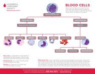
Normal Blood and MarrowBlood is composed of plasma and cells suspended in plasma. Plasma is largely madeup of water in which many chemicals are dissolved. <strong>The</strong>se chemicals include◦{Proteins◦{Albumin, the most common protein in blood◦{Blood-clotting proteins, made by the liver◦{Erythropoietin, a protein made by the kidneys that stimulates red cell production◦{Immunoglobulins, antibodies made by plasma cells in response to infectionsincluding those we develop from our vaccinations (such as poliovirusantibodies, which are made by normal plasma cells in the bone marrow)◦{Hormones (such as thyroid hormone and cortisol)◦{Minerals (such as iron and magnesium)◦{Vitamins (such as folate and vitamin B 12)◦{Electrolytes (such as calcium, potassium and sodium).<strong>The</strong> cells suspended in plasma include red cells, platelets and white cells(neutrophils, monocytes, eosinophils, basophils and lymphocytes).◦{<strong>The</strong> red cells make up a little less than half the volume of the blood. <strong>The</strong>y arefilled with hemoglobin, the protein that picks up oxygen in the lungs anddelivers it to the cells all around the body; hemoglobin then picks up carbondioxide from the body’s cells and delivers it back to the lungs, where it isremoved when we exhale.◦{<strong>The</strong> platelets are small cells (one-tenth the size of red cells) that help stopbleeding at the site of an injury in the body. For example, when a person has acut, the vessels that carry blood are torn open. Platelets stick to the torn surfaceof the vessel, clump together and plug up the bleeding site with the help ofblood-clotting proteins such as fibrin and electrolytes such as calcium. Later, afirm clot forms. <strong>The</strong> vessel wall then heals at the site of the clot and returns to itsnormal state.◦{<strong>The</strong> neutrophils and monocytes are white cells. <strong>The</strong>y are called “phagocytes”(eating cells) because they can ingest bacteria or fungi and kill them. Unlikethe red cells and platelets, the monocytes can leave the blood and enterthe tissue, where they can attack the invading organisms and help combatinfection. Eosinophils and basophils are types of white cells that respond toallergens or parasites.{ ◦ Most lymphocytes, another type of white cell, are found in the lymph nodes, thespleen and the lymphatic channels, but some enter the blood. <strong>The</strong>re are threemajor types of lymphocytes: T lymphocytes (T cells), B lymphocytes (B cells) andnatural killer (NK) cells. Each of these cells is a key part of the immune system.<strong>Acute</strong> <strong>Myeloid</strong> <strong>Leukemia</strong> I page 31
Blood Cell & Lymphocyte DevelopmentStem CellsMultipotentialHematopoietic CellsMultipotentialLymphoid CellsDifferentiate & mature intosix types of blood cellsDifferentiate & mature intothree types of lymphocytesRed CellsNeutrophilsEosinophilsBasophilsMonocytesPlateletsT LymphocytesB LymphocytesNatural Killer CellsFigure 4. I Stem cells develop into blood cells (hematopoiesis) and lymphocytic cells.Marrow is a spongy tissue where blood cell development takes place. It occupiesthe central cavity of bones. In newborns, all bones have active marrow. By the timea person reaches young adulthood, the bones of the hands, feet, arms and legs nolonger have functioning marrow. In adults, the spine (vertebrae), hip and shoulderbones, ribs, breastbone and skull contain the marrow that makes blood cells. <strong>The</strong>process of blood cell formation is called “hematopoiesis.” A small group of cells,the stem cells, develop into all the blood cells in the marrow by the process ofdifferentiation (see Figure 4).In healthy individuals, there are enough stem cells to keep producing new blood cellscontinuously. Blood passes through the marrow and picks up the fully developedand functional red and white cells and platelets for circulation in the blood.Some stem cells enter the blood and circulate. <strong>The</strong>y are present in such smallnumbers that they cannot be counted or identified by standard blood count tests.<strong>The</strong>ir presence in the blood is important because they can be collected by a specialtechnique. <strong>The</strong>re are also methods to induce more stem cells to leave their home inthe marrow and circulate in the blood, allowing a greater number of stem cells to becollected. If enough stem cells are harvested from a compatible donor, they can betransplanted into a recipient.Stem cell circulation, from marrow to blood and back, also occurs in the fetus.After birth, placental and umbilical cord blood can be collected, stored and used asa source of stem cells for transplantation.page 32 I 800.955.4572 I www.LLS.org
Medical TermsAbsolute Neutrophil Count (ANC). <strong>The</strong> number of neutrophils (a type ofwhite cell) that a person has to fight infection. It is calculated by multiplying thetotal number of white cells by the percentage of neutrophils. People who haveAML may have a low or normal absolute neutrophil count, depending on thetotal white cell count.Alkylating Agent. A type of chemotherapy used to kill cancer cells by interferingwith cancer cell division. Alkylating agents cause side effects because they alsointerfere with cell division in certain healthy tissues where cell division is frequent,such as the gastrointestinal tract. Cyclophosphamide is one of several types ofalkylating agents.Allogeneic Stem Cell Transplantation. A treatment that uses donor stem cellsto restore a patient’s marrow and blood cells. First, the patient is given conditioningtherapy (high-dose chemotherapy or high-dose chemotherapy with total bodyradiation) to treat the blood cancer and to “turn off” the patient’s immune systemso that the donor stem cells will not be rejected. A type of transplant called a“reduced-intensity” or “nonmyeloablative” transplant is under study. It uses lowerdoses of conditioning therapy and may be safer, especially for older patients.For more information see the free LLS booklet Blood and Marrow Stem CellTransplantation.Anemia. A decrease in the number of red cells and, therefore, the hemoglobinconcentration of the blood. <strong>The</strong> blood is less able to carry oxygen as a result. Ifsevere, anemia can cause a pale complexion, weakness, fatigue and shortness ofbreath on exertion.Anthracyclines (Antitumor Antibiotics). Chemotherapy agents that interactdirectly with the DNA in the nucleus of cells, thus interfering with cell survival.Antibodies. Proteins released by plasma cells (derived from B lymphocytes) thatrecognize and bind to specific foreign substances, called “antigens.” Antibodiescoat, mark for destruction or inactivate foreign particles such as bacteria, virusesand harmful toxins. Antibodies can also be made in the laboratory in two ways.<strong>The</strong> first way takes advantage of the fact that if material is injected from one speciesinto a different species, the latter will recognize it as foreign and make antibodiesto attack it. <strong>The</strong>se antibodies are usually polyclonal antibodies; that is, they react tomultiple targets (antigens). <strong>The</strong> second way involves monoclonal antibodies, whichreact to only one target (antigen) and can be used in several important ways. <strong>The</strong>ycan be used to identify and classify types of blood cancers or be altered so as tobecome useful in antibody-mediated immunotherapy.<strong>Acute</strong> <strong>Myeloid</strong> <strong>Leukemia</strong> I page 33
Antigen. A foreign substance, usually a protein, that stimulates an immuneresponse when it is ingested, inhaled or comes into contact with the skin or mucousmembranes. Examples of antigens are bacteria, viruses or allergens. Antigensstimulate plasma cells to produce antibodies.Antimetabolites. Chemotherapy agents that are generally similar to naturalbuilding blocks of DNA, RNA or some vitamins. However, they are changedfrom the natural chemical. When they substitute for the DNA or RNA buildingblocks within a leukemic cell, the cell is unable to form normal DNA or RNA. Thisprevents the cell from growing.Antioncogene. See Tumor Suppressor Gene.Apheresis. <strong>The</strong> process of removing components of a donor’s blood and returningthe unneeded parts to the donor. <strong>The</strong> process, also called “hemapheresis,” circulatesblood from a donor through a filter-type apparatus, and then back to the donor.Apheresis makes it possible to remove desired elements from large volumes ofblood. Platelets, red cells, white cells and plasma can be removed separately. Forexample, this technique permits the harvest of enough platelets for transfusion fromone donor (rather than six to eight separate donors). In this way, the recipient of theplatelets is exposed to fewer donors or can be given HLA-matched platelets from asingle related donor. This technique is also used to remove circulating blood stemcells, which can be frozen and stored for later use in transplantation.Autologous Stem Cell Transplantation. A technique used to delay theprogression of certain blood cancers. <strong>The</strong> autologous transplantation process(or autotransplant) takes place after the patient achieves a complete response(remission), or a good partial response, to induction drug therapy. <strong>The</strong> process is asfollows: 1) the patient’s stem cells are harvested, usually from the blood; 2) the stemcells are frozen for later use and the patient receives conditioning drug therapy;3) the stem cells are thawed and infused back to the patient through an indwellingcatheter (central line). <strong>The</strong> main adverse side effects of the transplant are the resultsof the conditioning therapy; these include mouth sores, hair loss, nausea, vomiting,diarrhea and risk of infections. Patients receive supportive care to help prevent and/or manage the side effects. Generally, after 10 to 14 days, blood counts begin tonormalize and the side effects of the conditioning therapy begin to resolve.Autosomes. See Karyotype.Basophil. A type of white cell that participates in certain allergic reactions.Biomarkers. Chemicals or structures present either on the surface of or withincells or in the serum. <strong>The</strong>y may aid doctors in determining when treatment (andwhich type of treatment) is needed by identifying disease that will progress morerapidly and/or have a better or worse response to certain treatments. Examplesof biomarkers are gene expression, serum protein levels and chromosomepage 34 I 800.955.4572 I www.LLS.org
abnormalities in cancer cells. No single feature can accurately predict diseaseprogression in a patient; therefore, doctors use a combination of factors to make adiagnosis and a treatment plan. Biomarkers are also known as “cancer cell markers”and “tumor markers.”Biopsy. A procedure to obtain tissue for diagnosis. In many cases, a specialneedle can be used to obtain the tissue. In some cases, a larger piece of tissue maybe surgically removed. Since the appearance of a lymph node is important incategorizing the type of lymphoma that may be present, surgical removal of oneor more entire, swollen lymph nodes may be necessary (lymph node biopsy). <strong>The</strong>tissue is placed in preservative, stained with dyes and examined under a microscopeby a pathologist.Blast Cells. <strong>The</strong> earliest marrow cells identified by the light microscope.Blasts represent about 1 percent of normally developing marrow cells. <strong>The</strong>y arelargely myeloblasts, which are cells that will develop into neutrophils. In normallymph nodes, blasts are lymphoblasts; that is, cells that are part of lymphocytedevelopment. In the acute types of leukemia, blast cells similar in appearance tonormal blast cells accumulate in large numbers, constituting up to 80 percentof all marrow cells. In myelodysplastic syndromes and acute myeloid leukemia,myeloblasts accumulate, and in acute lymphoblastic leukemia, lymphoblastsaccumulate. Normal myeloblasts give rise to granulocytes (neutrophils, eosinophilsand basophils). With myelodysplastic syndromes, abnormal myeloblasts displace orotherwise interfere with the production of normal red cells, white cells and plateletsin the marrow. Sometimes the distinction between myeloblasts and lymphoblastscan be made by examination of stained marrow cells through the microscope.Often, immunophenotyping or use of specially stained marrow cells is required tobe sure of the distinction.Blood Cell Count. A laboratory test requiring a small blood sample which can beused to measure the number and types of cells circulating in the blood. <strong>The</strong> term“complete blood count” or “CBC” is often used to refer to this test.Blood Cells. Any of the three main types of cells in the blood: red cells, whichcarry oxygen; white cells, which principally prevent or combat infections; andplatelets, which help prevent bleeding. <strong>The</strong>re are several types of white cells inthe blood. Each cell type is represented in blood in the numbers that meet thefunctions it serves. One fluid ounce of blood contains about 150 billion red cells,8 billion platelets, and 20 million white cells. Red cells live for months, platelets livefor a week or two, and white cells live for a few days. <strong>The</strong> marrow must replace over500 billion cells from the blood each day.Blood Plasma. See Plasma.Bone Marrow. A spongy tissue in the hollow central cavity of the bones that is thesite of blood cell formation. By puberty, the marrow in the spine, ribs, breastbone,<strong>Acute</strong> <strong>Myeloid</strong> <strong>Leukemia</strong> I page 35
hips, shoulders and skull is most active in blood cell formation. In adults, thebones of the hands, feet, legs and arms do not contain blood-forming marrow. Inthese sites the marrow is filled with fat cells. When marrow cells have matured intoblood cells, they enter the blood that passes through the marrow and are carriedthroughout the body.Bone Marrow Aspiration. A test to examine marrow cells to detect cellabnormalities. A marrow sample is usually taken from the patient’s hip bone. Aftermedication is given to numb the area, the liquid sample is removed using a specialneedle inserted through the bone and into the bone marrow. <strong>The</strong> sample is lookedat under a microscope for abnormal cells. <strong>The</strong> cells obtained can also be used forcytogenetic analysis and other tests.Bone Marrow Biopsy. A test to examine marrow cells to detect cell abnormalities.This test differs from a bone marrow aspiration in that a small amount of bonefilled with marrow is removed, usually from the hip bone. After medication isgiven to numb the area, a special biopsy needle is used to remove a core of bonecontaining marrow. <strong>The</strong> marrow is examined under a microscope to determine ifabnormal cells are present. Bone marrow aspiration and biopsy may be done in thedoctor’s office or in a hospital. <strong>The</strong> two tests are almost always done together. Bothtests are also done after treatment to determine the proportion of blood cancer cellsthat have been killed by therapy.CBC. See Blood Cell Count.Central Line. A special tube inserted into a large vein in the upper chest. <strong>The</strong>central line, sometimes referred to as an “indwelling catheter,” is tunneled underthe skin of the chest to keep it firmly in place. <strong>The</strong> external end of the catheter canbe used to administer medications, fluids or blood products or to withdraw bloodsamples. With meticulous care, central lines can remain in place for long periodsof time (many months) if necessary. <strong>The</strong>y can be capped and remain in place inpatients after they leave the hospital, and be used for outpatient chemotherapy orblood product administration. Several types of catheters (for example, Groshong®,Hickman®, and Broviac®) can be used for patients receiving intensive chemotherapyor nutritional support.Central Nervous System (CNS) Prophylaxis. Postremission treatment in whichchemotherapy is placed in the fluid that bathes the spinal cord and brain. In certaintypes of leukemia, particularly acute lymphocytic (lymphoblastic) leukemia andacute monocytic leukemia with high blood cell counts, the leukemic cells havea propensity to enter the covering of the spinal cord and brain (the meninges).This process is often not apparent until months or years after remission when theleukemia returns, first in the coverings of the CNS, then in the marrow and blood.To prevent this type of relapse (meningeal leukemia), virtually all children andadults with acute lymphocytic leukemia who enter remission are treated with CNSpage 36 I 800.955.4572 I www.LLS.org
Colony-Stimulating Factor. See Growth Factor.Complete Blood Count. See Blood Cell Count.Conditioning Treatment. Intensive therapy of a patient with cytotoxic drugsor drugs and total body radiation just before receiving a stem cell transplant. <strong>The</strong>therapy serves three purposes. First, it severely depresses the lymphocytes thatare the key cells in the recipient’s immune system. This action helps prevent therejection of the graft (donor tissue). Second, it markedly decreases the number ofmarrow cells, which may be important to open up the special niches where thetransplanted stem cells must lodge to engraft (survive). Third, if the patient is beingtransplanted for a malignancy, this intensive therapy greatly decreases the numbersof any remaining tumor cells.Cord Blood Stem Cells. Stem cells that are present in blood drained from theplacenta and umbilical cord. <strong>The</strong>se stem cells have the capability to repopulate themarrow of a compatible recipient and produce blood cells. Frozen cord blood is asource of donor stem cells for transplantation to HLA-matched recipients. Mostcord-blood transplants are given by matched or nearly matched unrelated donors.Cycle of Treatment. An intensive, clustered period of chemotherapy and/orradiation therapy. <strong>The</strong> therapy may be given for several days or weeks, and this timeperiod represents one cycle of treatment. <strong>The</strong> treatment plan may call for two, threeor more cycles of treatment.Cytogenetic Analysis. <strong>The</strong> process of analyzing the number and size of thechromosomes of cells. In addition to detecting chromosome alterations, insome cases it is possible to identify the actual genes that have been affected.<strong>The</strong>se findings are very helpful in diagnosing specific types of blood cancers, indetermining treatment approaches and in following the response to treatment.<strong>The</strong> individual who prepares and examines the chromosomes and interprets theresults is called a “cytogeneticist.”Cytopenia. A reduction in the number of cells circulating in the blood.Cytotoxic Drugs. Anticancer drugs that act by killing cells or preventing themfrom dividing. See Chemotherapy.Deletion. A chromosomal abnormality in which part or all of a single chromosomehas been lost.Differentiation. <strong>The</strong> process by which stem cells give rise to functional cells ofa single blood cell line. Differentiation of stem cells forms red cells, platelets andwhite cells (neutrophils, monocytes, eosinophils, basophils and lymphocytes).page 38 I 800.955.4572 I www.LLS.org
DNA. <strong>The</strong> abbreviation for deoxyribonucleic acid, the genetic material in the cell.DNA is made up of a sugar-phosphate backbone with ladderlike “steps” composedof purines and pyrimidines (building blocks of nucleic acids). <strong>The</strong> sequence of thepurines and pyrimidines in the DNA is responsible for passing genetic informationto new cells during the process of cell division; for passing genetic informationfrom one generation to the next during reproduction; and for providing theinstructions for building proteins, which in turn carry out the major functions ofa cell. A mutation is generally a change in or loss of the sequence of the purines orpyrimidines of the DNA. Mutations can lead to cell death, to changes in the way acell functions or, in some cases, to cancer.Donor Lymphocyte Infusion (DLI). A therapy that involves giving lymphocytesfrom the original stem cell donor to a patient who has had an allogeneic bonemarrow transplant with a relapse of disease. DLI may induce an immune reactionagainst the patient’s cancer cells. This therapy has been most effective in patientswith chronic myeloid leukemia who relapse after transplantation but this therapy isbeing studied to treat patients with other blood cancers.Eosinophil. A type of white cell that participates in allergic reactions and helpsfight certain parasitic infections.Epigenetic Change. Any change that alters gene activity without changing theDNA sequence. Many types of epigenetic changes have been identified. Whileepigenetic changes are natural and essential to many of the body’s functions,certain epigenetic changes can cause major adverse health effects, including cancer.Drugs that target specific epigenetic changes—for example, the histone deacetylase(HDAC) inhibitor vorinostat (Zolinza®)—are approved to treat some blood cancersand are being studied in clinical trials for treatment of other blood cancers.Erythrocytes. See Red Cells.Erythrocyte Sedimentation Rate (ESR). See Sedimentation Rate.Erythropoietin (EPO). A hormone required for the normal production of redblood cells. It is produced mainly by the kidneys and is released into the bloodin response to decreased levels of oxygen in the blood. Epoetin alfa (Procrit®or Epogen®) and darbepoetin alfa (Aranesp®) are laboratory-made forms of thehuman hormone erythropoietin that can be used to treat anemia. In oncology,these drugs are used to assist in the recovery from chemotherapy-induced anemiaor to treat chronic diseases in which anemia is a troublesome finding, such aslower-risk myelodysplastic syndromes. <strong>The</strong>se drugs stimulate red cell productionby the same mechanism as EPO; that is, by interacting with the EPO receptor onred cell progenitors.ESR. See Sedimentation Rate.<strong>Acute</strong> <strong>Myeloid</strong> <strong>Leukemia</strong> I page 39
Extramedullary Myeloblastoma. See Chloroma.Farnesyl Transferase Inhibitor (FTI). A drug that has the potential to kill cancercells by inhibiting or reversing the effect of farnesyl transferase, an enzyme neededto activate oncogenes (cancer-causing genes). FTIs, including tipifarnib (Zarnestra®)and lonafarnib (Sarasar®), are being studied to treat some blood cancers.FDA. <strong>The</strong> short name for the United States Food and Drug Administration. Part ofthe FDA’s job is to assure the safety and security of drugs, medical devices and theUS food supply.FISH. See Fluorescent In Situ Hybridization.Flow Cytometry. A test that permits the identification of specific cell types withina sample of cells. <strong>The</strong> test may be used to examine blood cells, marrow cells orcells from a biopsy. A diluted suspension of cells from one of these sources can betagged with an antibody specific for a site on the cell surface. <strong>The</strong> antibody has achemical attached that will emit light when activated by a laser beam. <strong>The</strong> cells flowthrough the instrument called a “flow cytometer”; when the cells pass through itslaser beam, those with the antibody-specific surface feature light up and then canbe counted. One use of flow cytometry is to determine whether a sample of cellsis composed of T cells or B cells. This permits the physician to determine if theleukemia or lymphoma is of the B- or T-cell type. Flow cytometry is also used toselect stem cells from a mixed-cell population so that they can be used later in astem cell transplant.FLT3. An abbreviation for the Fms-like tyrosine kinase 3 gene. FLT3 is expressedon blood-forming stem cells and plays a role in cell development. FLT3 mutationscan be detected in about one-third of AML patients. <strong>The</strong>se mutations have beenidentified as part of the AML disease process and may become the basis for newtargeted therapies.Fluorescent In Situ Hybridization (FISH). A technique in which DNA probestagged with fluorescent molecules that emit light of different wavelengths (anddifferent colors) are used on tissue. <strong>The</strong> probes match to the chromosomes withinthe cells, and the chromosomes fluoresce in color. FISH is a means of studyingchromosomes in tissue.Fungus. A microbe often referred to as a “mold” or “yeast.” <strong>The</strong>re are manyspecies of fungi, and some, while relatively harmless in people with healthyimmune systems, are prone to produce serious infections in people who areimmunosuppressed, as after stem cell transplantation or multiple treatments withhigh-dose chemotherapy for progressive leukemia or lymphoma. Fungi belong tothe genera Candida, Aspergillus and Histoplasma, among others.G-CSF (Granulocyte Colony-Stimulating Factor). See Growth Factor.page 40 I 800.955.4572 I www.LLS.org
GM-CSF (Granulocyte-Macrophage Colony-Stimulating Factor). See Growth Factor.Graft-Versus-Host Disease (GVHD). <strong>The</strong> immune attack by lymphocytes ina donor’s marrow or blood cell suspension (the graft) against the tissues of therecipient (the host). <strong>The</strong> immune cells most engaged in this reaction are donor Tlymphocytes, which are present in the donor’s blood or marrow, the source of thestem cells. <strong>The</strong> principal sites of injury are the skin, the liver and the gastrointestinaltract. <strong>The</strong> reaction does not occur in identical twin transplants. <strong>The</strong> reaction may beminimal in closely matched individuals or severe in less well-matched individuals.<strong>The</strong>se reactions are mediated in part by antigens that are not in the major HLAsystem and cannot be matched prior to transplantation. For example, in the caseof a female stem cell donor and a male recipient, factors that are produced bygenes on the male recipient’s Y chromosome may be seen as foreign by the femaledonor’s cells, which do not share the genes on the Y chromosome. This fact doesnot prohibit female donors and male recipients, but it makes the risk of immunereaction higher.Graft-Versus-Tumor Effect (Graft-Versus-<strong>Leukemia</strong> Effect). <strong>The</strong> potentialimmune reaction of transplanted (donor) T lymphocytes to recognize andattack the malignant cells of the recipient. This effect was noted when1) disease recurrence after transplant was seen to be more likely if the donorand recipient were identical twins than if they were nonidentical siblings;2) disease recurrence was less likely the more pronounced the graft-versus-hostdisease (GVHD) was; and 3) the removal of donor T lymphocytes decreasedthe incidence of GVHD but also resulted in a higher frequency of diseaserelapse. Each of these observations could be explained best as an immuneattack by donor lymphocytes against recipient tumor cells that, along with theintensive conditioning treatment, serves to keep the disease in check. This effectseems to be most active in types of myeloid leukemia, although it may alsooccur in patients with other blood cancers.Granulocyte. A type of white cell that has a large number of granules in the cellbody. Neutrophils, eosinophils and basophils are types of granulocytes.Granulocytic Sarcoma. See Chloroma.Growth Factor. A chemical used to stimulate the production of neutrophils andshorten the period of low neutrophil counts in the blood after chemotherapy.Granulocyte colony-stimulating factor (G-CSF) and granulocyte-macrophagecolony-stimulating factor (GM-CSF) are examples of growth factors that are madecommercially. GM-CSF can also stimulate monocytes.Hematocrit. <strong>The</strong> proportion of the blood occupied by the red cells. Normal valuesare 40 to 54 percent in males and 35 to 47 percent in females. If the hematocrit isbelow normal, the condition is called “anemia.” If the hematocrit is above normal,the condition is called “erythrocytosis.”<strong>Acute</strong> <strong>Myeloid</strong> <strong>Leukemia</strong> I page 41
Hematologist. A medical doctor who specializes in the treatment of blood celldiseases. This person is either an internist who treats adults or a pediatrician whotreats children.Hematopathologist. A type of pathologist who studies diseases of blood cellsby looking at peripheral blood smears, bone marrow aspirates and biopsies, andlymph nodes and other tissues. <strong>The</strong> hematopathologist uses his or her expertiseto identify diseases such as blood cancers. In addition to using a microscope, ahematopathologist also uses laboratory values, flow cytometry and moleculardiagnostic tests to make the most accurate diagnosis. <strong>The</strong> hematopathologist worksclosely with the hematologist/oncologist who sees the patient and decides on thebest treatment based upon the diagnosis.Hematopoiesis. <strong>The</strong> process of blood cell development in the marrow. <strong>The</strong> mostundeveloped cells in the marrow are stem cells. <strong>The</strong>y start the process of blood celldevelopment. <strong>The</strong> stem cells begin to develop into young or immature blood cellssuch as red cells or white cells of various types. This process is called “differentiation.”<strong>The</strong> young or immature blood cells then further develop into fully functional bloodcells. This process is called “maturation.” <strong>The</strong> mature cells leave the marrow, enter theblood and circulate throughout the body. Hematopoiesis is a continuous process thatis active normally throughout life. <strong>The</strong> reason for this activity is that most blood cellslive for short periods and must be continually replaced. Red cells live for months,platelets live for a week or two, and white cells live for a few days. About 500 billionblood cells are made each day. When the marrow is invaded with cancer cells, itcannot produce enough normal blood cells to meet the constant demand for them,and the numbers in the blood cell counts become severely depleted.Hemoglobin. <strong>The</strong> iron-containing pigment in red cells that carries oxygen tothe tissue cells. A reduction in the number of red cells decreases the amount ofhemoglobin in the blood. A decreased blood hemoglobin concentration is called“anemia.” A low hemoglobin concentration decreases the oxygen-carrying capacityof blood. If severe, this decreased capacity may limit a person’s ability to exerthimself or herself. Normal values of blood hemoglobin are 12 to 16 grams perdeciliter (g/dL). Compared to men, healthy women have, on average, about 10percent less hemoglobin in their blood.HLA. <strong>The</strong> abbreviation for human leukocyte antigen(s). <strong>The</strong>se are proteins on thesurface of most tissue cells, and they give an individual his or her unique tissuetype. HLA factors are inherited from mother and father, and the greatest chanceof having the same HLA type is between siblings. On average, one in four siblingsis expected to share the same HLA type. <strong>The</strong> testing for HLA factors is referred toas “tissue typing.” <strong>The</strong>re are six major groups of HLA: A, B, C, D, Dr, and Dq.<strong>The</strong>se proteins on the surface of cells act as antigens when donated (transplanted)to another individual, the recipient. If the antigens on the donor cells are identical(as in identical twins) or very similar (as in HLA-matched siblings), the transplantpage 42 I 800.955.4572 I www.LLS.org
(donated stem cells) is more likely to survive (engraft) in the recipient. In addition,the recipient’s body cells are less likely to be attacked by the donated immune cells(a result called “graft-versus-host disease”).Immune System. Cells and proteins that defend the body against infection.Lymphocytes, lymph nodes and the spleen are parts of the body’s immune system.Immunity. <strong>The</strong> ability to resist infection.Immunophenotyping. A method that uses the reaction of antibodies with cellantigens to determine a specific type of cell in a sample of blood cells, marrow cellsor lymph node cells. <strong>The</strong> antibodies react with specific antigens on the cell. A tagis attached to an antibody so that it can be detected. <strong>The</strong> tag can be identified bythe laboratory detector used for the test. As cells carrying their array of antigensare tagged with specific antibodies, they can be identified; for example, myeloidleukemic cells can be distinguished from lymphocytic leukemic cells. Normallymphocytes may be distinguished from leukemic lymphocytes. This method alsohelps subclassify cell types, information that may, in turn, help in deciding on thebest treatment to apply in that type of leukemia or lymphoma. <strong>The</strong> antigen on acell is referred to as a “cluster designation” or “CD,” with an associated number.For example, CD10, also referred to as “CALLA” (common acute lymphoblasticleukemia antigen), may be present on leukemic lymphoblasts, and CD33, may bepresent on leukemic myeloblasts.Immunosuppression. A state in which the immune system does not functionproperly and its protective functions are inadequate. <strong>The</strong> patient is more susceptibleto infections, including those from microbes that are usually not highly infectious.This can occur as a result of intensive chemotherapy and radiation therapy,especially when used in high doses to condition a patient for transplantation.It can also occur because of disease states. Human immunodeficiency virus(HIV) infection is one such disease. Graft-versus-host disease (GVHD) createsan immunosuppressive state in which immune protection against infection isinadequate. In the transplant patient the conditioning regimen and severe GVHDcan result in overwhelming infection. See Graft-Versus-Host Disease.Immunotoxins. See Monoclonal Antibody <strong>The</strong>rapy.Indwelling Catheter. See Central Line.Intrathecal. <strong>The</strong> designation for the space between the covering or lining ofthe central nervous system (CNS) and the brain or spinal cord. This lining iscalled the “meninges.” In some situations, drugs have to be administered directlyinto the spinal canal when leukemia cells are in the meninges. This is called“intrathecal therapy.”<strong>Acute</strong> <strong>Myeloid</strong> <strong>Leukemia</strong> I page 43
Inversion. An abnormality of chromosomes that occurs when a section of achromosome breaks and turns upside down, so that its genetic material is in reverseorder but the inverted piece remains attached to the chromosome.Karyotype. <strong>The</strong> systematic arrangement, using images, of the 46 chromosomes inthe human cell in 22 matched pairs (maternal and paternal member of each pair) bylength from longest to shortest and other features, with the sex chromosomes shownas a separate pair (either XX or XY). <strong>The</strong> 22 pairs are referred to as “autosomes.”Leukocytes. See White Cells.Leukocytosis. An increase above the upper limit of normal in the concentrationof blood leukocytes (white cells).Leukopenia. A decrease below normal in the concentration of blood leukocytes(white cells).Lumbar Puncture. A procedure to remove spinal fluid from the spacesurrounding the spinal cord or to administer anticancer drugs to prevent or treatleukemia or lymphoma of the coverings of the central nervous system (CNS). <strong>The</strong>doctor first injects a local anesthetic, then inserts a needle between two vertebrae inthe lower part of the back. Fluid samples are collected in sterile tubes and examinedfor evidence of leukemia or lymphoma. A lumbar puncture is not often used to testfor AML, but may be used if the patient is having symptoms that could be causedby the spread of leukemia cells into the CNS. Another term for lumbar puncture is“spinal tap.”Lymph Nodes. Small structures (the size of beans) that contain large numbersof lymphocytes and are connected with each other by small channels called“lymphatics.” <strong>The</strong>se nodes are distributed throughout the body. Enlarged lymphnodes can be seen, felt or measured by computed tomography (CT) scan or magneticresonance imaging (MRI) depending on their location and the degree of enlargement.Lymphocyte. A type of white cell that is the essential cell type in the body’simmune system. <strong>The</strong>re are three major types of lymphocytes: B lymphocytes, whichproduce antibodies to help combat infectious agents like bacteria, viruses and fungi;T lymphocytes, which have several functions, including assisting B lymphocytes tomake antibodies; and natural killer (NK) cells, which can attack virus-infected cellsor tumor cells.Macrophage. See Monocyte.Marrow. See Bone Marrow.Microliter (μL) of Blood. A measurement used for some blood test results. Onemicroliter (μL) is an amount equal to one one-millionth of a liter, which is almostequal to a quart of blood.page 44 I 800.955.4572 I www.LLS.org
Minimal Residual Disease (MRD). <strong>The</strong> small amounts of cancer cells that mayremain after treatment, even when blood and marrow may appear to be normal.<strong>The</strong>se residual cells can only be identified by sensitive molecular techniques.Monoclonal. See Clonal.Monoclonal Antibodies. Antibodies made by cells belonging to a singleclone. <strong>The</strong>se highly specific antibodies can be produced in the laboratory.<strong>The</strong>y are very important reagents for identifying and classifying disease by theimmunophenotyping of cells. <strong>The</strong>y also have clinical applications for targeteddelivery of drugs to cancer cells and can be used to purify cells used for stem celltransplants.Monoclonal Antibody <strong>The</strong>rapy. <strong>The</strong>rapy using proteins made in the laboratorythat either react with or attach to antigens on the cancer cells to which theyare targeted. <strong>The</strong> antibodies are used therapeutically in three ways: as “naked”antibodies (monoclonal antibodies); as antibodies to which radioactive isotopes areattached (radioimmunotherapies); and as antibodies to which toxins are attached(immunotoxins).Monocyte/Macrophage. A type of white cell that represents about 5 to 10percent of the cells in normal human blood. <strong>The</strong> monocyte and the neutrophilare the two major microbe-eating and microbe-killing cells in the blood. Whenmonocytes leave the blood and enter the tissue, they are converted to macrophages.<strong>The</strong> macrophage is the monocyte-in-action: it can combat infection in thetissues, ingest dead cells (in this function it is called a “scavenger cell”) and assistlymphocytes in their immune functions.Multidrug Resistance (MDR). A characteristic of cells that makes them resistantto the effects of several different classes of drugs. <strong>The</strong>re are several forms of drugresistance. One type of MDR involves the ability to force several drugs out of thecell. <strong>The</strong> outer wall, or membrane, of the cell contains a pump that ejects chemicals,preventing them from reaching a toxic concentration. <strong>The</strong> resistance to drugs canbe traced to the expression of genes that direct the formation of high amounts ofa protein that prevents the drugs from affecting the malignant cells. If the gene orgenes involved are not expressed or are weakly expressed, the cells are more sensitiveto the drug’s effect. If the genes are highly expressed, the cells are less sensitive to thedrug’s effect.Mutation. An alteration in a gene that results from a change to the part of the DNAthat represents that gene. A “germ cell mutation” is present in the egg or the spermand can be transmitted from parent(s) to offspring. A “somatic cell mutation” occursin a specific tissue cell and can result in the growth of that tissue cell into a tumor.Most cancers start after a somatic mutation. In leukemia, lymphoma or myeloma,a primitive marrow (blood-forming) or lymph node cell undergoes one or more<strong>Acute</strong> <strong>Myeloid</strong> <strong>Leukemia</strong> I page 45
somatic mutations, leading to the formation of a tumor. If the mutation results froma major abnormality of chromosomes, such as a translocation, it can be detected bycytogenetic examination. Sometimes the alteration in the gene is subtler and requiresmore sensitive tests to identify the oncogene.Myeloblasts. See Blast Cells.Myelocyte. A cell of the marrow that is a precursor of the mature granulocytes ofthe blood. Myelocytes are not present in the blood of healthy individuals.Neutropenia. A decrease below normal in the concentration of neutrophils, a typeof white cell.Neutrophil. <strong>The</strong> principal phagocyte (microbe-eating cell) in the blood. Thisblood cell is the main cell that combats infections. Often, it is not present insufficient quantities in patients with acute leukemia or after chemotherapy. Asevere deficiency of neutrophils increases the patient’s susceptibility to infection.A neutrophil may be called a “poly” (polymorphonuclear neutrophil) or “seg”(segmented neutrophil) because its nucleus has several lobes.Oncogene. A mutated gene that is the cause of a cancer. Several subtypes of acutemyeloid leukemia, acute lymphocytic leukemia and lymphoma, and nearly all casesof chronic myeloid leukemia are associated with an oncogene.Oncologist. A medical doctor who diagnoses and treats patients with cancer.Oncologists are usually internists who treat adults or pediatricians who treatchildren. Radiation oncologists specialize in the use of radiation to treat cancer, andsurgical oncologists specialize in the use of surgical procedures to diagnose and treatcancer. <strong>The</strong>se doctors cooperate and collaborate to provide the best treatment plan(surgery, radiation therapy, chemotherapy or immunotherapy) for the patient.Pancytopenia. A decrease below normal in the concentration of the three majorblood cell types: red cells, white cells and platelets.Pathologist. A medical doctor who identifies disease by studying tissues under amicroscope. See Hematopathologist.Peripheral Blood Smear. A sample of blood placed on a slide and dyed so thatthe cells can be examined under a microscope.Peripherally Inserted Central Catheter (PICC or PIC Line). A long, thin,flexible tube that is inserted into the body and used to administer medications,antibiotics, fluids and nutrition for an extended period of time. It can also be usedto obtain blood samples. Prior to insertion of the PICC, the patient is given a localanesthetic to numb the arm between the elbow and the shoulder. <strong>The</strong> PICC isinserted through the skin into a vein in the arm and advanced until it reaches thesuperior vena cava just above the heart. <strong>The</strong> superior vena cava is one of the veinspage 46 I 800.955.4572 I www.LLS.org
in the central venous system. <strong>The</strong> PICC can be maintained for several weeks tomonths, eliminating the need for standard intravenous (IV) administration.Petechiae. Pinhead-sized sites of bleeding in the skin. This type of bleeding resultsfrom a very low platelet count. <strong>The</strong> small punctate hemorrhages are frequently seenon the legs, feet, trunk and arms. <strong>The</strong>y evolve from red to brown and eventuallydisappear. <strong>The</strong>y stop developing when the platelet count increases.Phagocytes. Cells that readily eat (ingest) microorganisms such as bacteriaand fungi and kill them as a means of protecting the body against infection. <strong>The</strong>two principal phagocytes are neutrophils and monocytes. <strong>The</strong>y leave the bloodand enter tissues in which an infection has developed. A severe decrease in theconcentration of these cells is the principal cause of susceptibility to infection inpatients treated with intensive radiation therapy and/or chemotherapy. Treatmentmay suppress blood cell production in the marrow, resulting in deficiencies of thesephagocytic cells.PIC/PICC Line. See Peripherally Inserted Central Catheter.Plasma. <strong>The</strong> liquid portion of the blood, in which the blood cells, platelets,proteins and various other components are suspended. It is also referred to as“blood plasma.”Platelets. Small blood cells (about one-tenth the volume of red cells) that stick tothe site of blood vessel injury, aggregate and then seal off the injured blood vesselto stop bleeding. “Thrombocyte” is a synonym for platelet and is often used as theprefix in terms describing disorders of platelets, such as thrombocytopenia (too few)or thrombocythemia (too many).Platelet Transfusion. Transfusion of donor platelets, which may be needed tosupport some patients treated for blood cancer. <strong>The</strong> platelets can be gathered fromseveral unrelated donors and given as pooled, random-donor platelets. <strong>The</strong> plateletsfrom about six single-unit blood donors are required to significantly raise theplatelet count in a recipient. Sufficient platelets can be obtained from one donorby a procedure known as “apheresis.” This technique skims the platelets from largevolumes of blood as it passes through the apheresis machine. <strong>The</strong> red cells andplasma are returned to the donor. <strong>The</strong> advantage of single-donor platelets is thatthe patient is not exposed to the different antigens on platelets from many differentpeople and thus is less likely to develop antibodies against donor platelets. HLAmatchedplatelet transfusion can be given from a related donor who has an identicalor very similar HLA tissue type.Polymerase Chain Reaction (PCR). A technique to expand trace amounts of DNAor RNA so that the specific type of the DNA or RNA can be determined or studied.This technique has become useful in detecting a very low concentration of residualblood cancer cells, too few to be seen using a microscope. PCR can detect the presence<strong>Acute</strong> <strong>Myeloid</strong> <strong>Leukemia</strong> I page 47
of one blood cancer cell among 500,000 to 1 million blood cells. PCR requires aspecific DNA (or RNA) abnormality or marker, like an oncogene, in the leukemia orlymphoma cells in order to be used for identifying residual abnormal cells.Port. A small device that is used with a central line to access a vein. <strong>The</strong> port isplaced under the skin of the chest. To take blood samples (or to give medicinesor nutrition) the doctor or nurse puts a needle through the skin into the port. Anumbing cream can be put on the skin before the port is used.Promyelocyte. A cell of the marrow that is very early in development along thepathway to myeloid cells. It represents the next stage after the blast cell stage.Radiation <strong>The</strong>rapy. <strong>The</strong> use of x-rays and other forms of radiation in treatment.Radiation therapy may be useful in the treatment of some localized bloodcancers. Radiation therapy can be an important adjunct to therapy when there areparticularly large masses in a localized area or when local large lymph nodes arecompressing or invading normal organs or structures and chemotherapy cannotcontrol the problem.Radioimmunotherapies. See Monoclonal Antibody <strong>The</strong>rapy.Recurrence/Relapse. <strong>The</strong> return of a disease after it has been in remissionfollowing treatment.Red Cells. Blood cells (erythrocytes) that carry hemoglobin, which binds oxygenand carries it to the tissues of the body. <strong>The</strong> red cells make up about 40 to 45percent of the volume of the blood in healthy individuals.Reduced-Intensity Stem Cell Transplantation. A form of allogeneictransplantation, now in clinical trials. In a reduced-intensity transplant, patients receivelower doses of chemotherapy drugs and/or radiation in preparation for the transplant.Immunosuppressive drugs are used to prevent rejection of the graft (donor tissue),and the engraftment of donor immune cells may allow these cells to attack the disease(graft-versus-leukemia effect). More study is needed to determine the effectivenessof this treatment for non-Hodgkin lymphoma patients. Studies to determine theusefulness of reduced-intensity stem cell transplantation in older patients are alsounder way. For more information about all types of stem cell transplantation, see thefree LLS publication Blood and Marrow Stem Cell Transplantation.Refractory Disease. Disease that does not go into remission or improvesubstantially after treatment with standard therapy for the disease. Newly diagnosedpatients or relapsed patients may have refractory disease. See Resistance to Treatment.Relapsed Disease. Disease that initially responded to therapy but has begunto progress.page 48 I 800.955.4572 I www.LLS.org
Remission. <strong>The</strong> disappearance of evidence of a disease, usually as a result oftreatment. <strong>The</strong> terms “complete” and “partial” are sometimes used to modify theterm “remission.” Complete remission means that all evidence of the disease isgone. Partial remission means that the disease is markedly improved by treatment,but residual evidence of the disease is present. Long-term benefit usually requires acomplete remission, especially in acute leukemia or progressive lymphomas.Resistance to Treatment. <strong>The</strong> ability of cells to live and divide despite theirexposure to a chemical that ordinarily kills cells or inhibits their growth. Refractoryleukemia is the condition in which a proportion of malignant cells resists thedamaging effects of a drug or drugs. Cells have several ways to develop drugresistance. See Multidrug Resistance.Risk Factor. A factor that is scientifically established to increase a person’s chanceof getting a disease. Risk factors can be classified as either genetic (inherited),lifestyle related, or environmental. <strong>The</strong> presence of one or more risk factors does notmean that a person will necessarily develop the disease. In the case of environmentalexposure, the extent of exposure and its duration are important considerations indetermining if risk is increased.RNA. Abbreviation for ribonucleic acid, a molecule in cells that carries out DNA’sinstructions for making proteins.Sedimentation Rate. A blood test that measures how quickly red cells(erythrocytes) settle in a test tube in 1 hour. A sedimentation rate test is done tofind out if inflammation is present in the body, to check on the progress of a diseaseor to see how well a treatment is working. This test is also called a “sed rate” or“erythrocyte sedimentation rate (ESR).”Serum. <strong>The</strong> liquid portion of the blood in which no cells are present.Somatic Cell Mutation. See Mutation.Spinal Tap. See Lumbar Puncture.Spleen. An organ located in the left upper portion of the abdomen just under theleft side of the diaphragm. It contains clusters of lymphocytes and also filters oldor worn-out cells from the blood. It is often affected in lymphocytic leukemia andlymphoma. Enlargement of the spleen is called “splenomegaly.” Surgical removalof the spleen is known as “splenectomy.” Certain diseases are treated by removingthe spleen. Most of the functions of the spleen can be performed by other organs,such as the lymph nodes and liver, but a person whose spleen has been removed isat higher risk for infection. He or she is given antibiotic therapy immediately at thefirst sign of infection, such as a fever.<strong>Acute</strong> <strong>Myeloid</strong> <strong>Leukemia</strong> I page 49
Stem Cells. Primitive cells in marrow that develop into red cells, white cells andplatelets. Stem cells are largely found in the marrow, but some leave the marrowand circulate in the blood. Using special techniques, the stem cells in the blood canbe collected, preserved by freezing and later thawed and used for stem cell therapy.See Hematopoiesis.Stem Cell Transplantation. See Allogeneic Stem Cell Transplantation;Autologous Stem Cell Transplantation.Thrombocythemia. An above-normal concentration of platelets in the blood.Thrombocytopenia. A below-normal concentration of platelets in the blood.Toxin. A naturally derived substance that is poisonous to cells. A toxin can be attachedto antibodies that then attach to cancer cells. <strong>The</strong> toxin may kill the cancer cells.Translocation. An abnormality of chromosomes in marrow or lymph node cellsthat occurs when a piece of one chromosome breaks off and attaches to the end ofanother chromosome. In a balanced translocation, genetic material is exchangedbetween two different chromosomes with no gain or loss of genetic information.When a translocation occurs, the gene at which the break occurs is altered. Thisis one form of somatic mutation that may transform the gene into an oncogene(cancer-causing gene). See Mutation.Transplantation. See Allogeneic Stem Cell Transplantation; Autologous Stem CellTransplantation.Tumor Suppressor Gene. A gene that acts to prevent cell growth. If a mutationoccurs in this gene that “turns off” the gene and causes loss of function, it maymake the individual more susceptible to the development of cancer in the tissuein which the mutation occurred. Another term for tumor suppressor gene is“antioncogene.”White Cells. Any of the five major types of infection-fighting cells in the blood:neutrophils, eosinophils, basophils, monocytes and lymphocytes. White cells arealso called “leukocytes.”page 50 I 800.955.4572 I www.LLS.org
More InformationFree LLS publications includeReferencesBlood TransfusionCancer-Related Fatigue FactsChoosing a Blood Cancer Specialist or Treatment Center<strong>The</strong> AML Guide—Information for Patients and CaregiversUnderstanding Clinical Trials for Blood CancersUnderstanding Drug <strong>The</strong>rapy and Managing Side EffectsUnderstanding Lab and Imaging TestsAgency for Toxic Substances and Disease Registry (ASTDR). Toxicological Profilefor Benzene: Atlanta, GA: US Department of Health and Human Resources PublicHealth Service. August 2007.Andrews A. ASH 2010: CPX-351 improves response in elderly patients withnewly diagnosed acute myeloid leukemia. OncLive web site. Available atwww.onclive.com/publications/obtn/2010/December-2010/ash-2010-cpx-351-improves-response-in-elder-patients-with-newly-diagnosed-acute-myeloid-leukemia.Accessed October 25, 2011.Boissel N, Terré C, Cornillet-Lefebvre P, et al. Trisomy 8 in the light of recentcytogenetic classification advances. A study from the French <strong>Acute</strong> <strong>Myeloid</strong><strong>Leukemia</strong> Intergroup. Presented at: 51st ASH Annual Meeting and Exposition;December 6, 2009; New Orleans, LA. Abstract 2594.www.ash.confex.com/ ash/2009/webprogram/Paper17541.html.Accessed October 25, 2011.Dombret H, Gardin C. An old AML drug revisited. New England Journal ofMedicine. 2009;361(13):1301-1303.Dunphy, CH. <strong>Myeloid</strong> sarcoma pathology. Medscape Reference web site 2011.Available at www.emedicine.medscape.com/article/1644141-overview.Accessed October 31, 2011.Feldman, EJ. AML Update: Treatment and Side Effects. Teleconferenceof the <strong>Leukemia</strong> & <strong>Lymphoma</strong> <strong>Society</strong>, Past Patient Education Programs,<strong>Leukemia</strong> January 11, 2011.www.lls.org/resourcecenter/pastprograms/leukemia/011111_amlupdate.Accessed October 25, 2011.<strong>Acute</strong> <strong>Myeloid</strong> <strong>Leukemia</strong> I page 51
Howlader N, Noone AM, Krapcho M, Neyman N, Aminou R, Waldron W,Altekruse SF, Kosary CL, Ruhl J, Tatalovich Z, Cho H, Mariotto A, Eisner MP,Lewis DR, Chen HS, Feuer EJ, Cronin KA, Edwards BK (eds). SEER Cancer.Statistics Review, 1975-2008, National Cancer Institute. Bethesda, MD,www.seer.cancer.gov/csr/1975_2008/, based on November 2010 SEER datasubmission, posted to the SEER website, 2011.Koreth J, Schlenk R, Kopecky KJ, et al. Allogeneic stem cell transplantation foracute myeloid leukemia in first complete remission: systematic review and metaanalysisof prospective clinical trials. Journal of the American Medical Association.2009;301(22):2349-2361.Kotiah SD. <strong>Acute</strong> Promyelocytic <strong>Leukemia</strong> Treatment & Management.Medscape Reference Web site 2011. Available at www.emedicine.medscape.com/article/1495306-treatment. Accessed October 25, 2011.Liesveld JL, Lichtman MA. Chapter 89. <strong>Acute</strong> myelogenous leukemia. KaushanskyK, Lichtman MA, Beutler E, Kipps TJ, Seligsohn U, Prchal J, eds. WilliamsHematology. 8th ed. Available from: AccessMedicine. Accessed October 25, 2011.Manola KN. Cytogenetics of pediatric acute myeloid leukemia. European Journalof Haematology. 2009;83(5):391-405.<strong>The</strong> <strong>Leukemia</strong> & <strong>Lymphoma</strong> <strong>Society</strong>’sLight <strong>The</strong> Night WalkTaking Steps toCure CancerTMLIGHTTHENIGHT.ORG877.LTN.WALKpage 52 I 800.955.4572 I www.LLS.org
REACH OUT TO OURINFORMATION SPECIALISTS<strong>The</strong> <strong>Leukemia</strong> & <strong>Lymphoma</strong><strong>Society</strong>’s (LLS) InformationSpecialists provide patients,families and healthcareprofessionals with the latestinformation on leukemia,lymphoma and myeloma.Our team consists of master’slevel oncology professionals whoare available by phone Mondaythrough Friday, 9 am to 6 pm (ET).Co-Pay AssistanceLLS’s Co-Pay Assistance Programhelps blood cancer patients coverthe costs of private and publichealth insurance premiums,including Medicare and Medicaid,and co-pay obligations. Supportfor this program is based on theavailability of funds by disease.For more information,call 877.557.2672 orvisit www.LLS.org/copay.For a complete directory of our patient services programs, contact us at800.955.4572 or www.LLS.org(Callers may request a language interpreter.)
For more information, please contact:or:National Office1311 Mamaroneck Avenue, Suite 310, White Plains, NY 10605Contact our Information Specialists 800.955.4572 (Language interpreters available upon request)www.LLS.orgOur Mission:Cure leukemia, lymphoma, Hodgkin’s disease and myeloma, and improve thequality of life of patients and their families.LLS is a nonprofit organization that relies on the generosity of individual,foundation and corporate contributions to advance its mission.PS32 25M 12/11