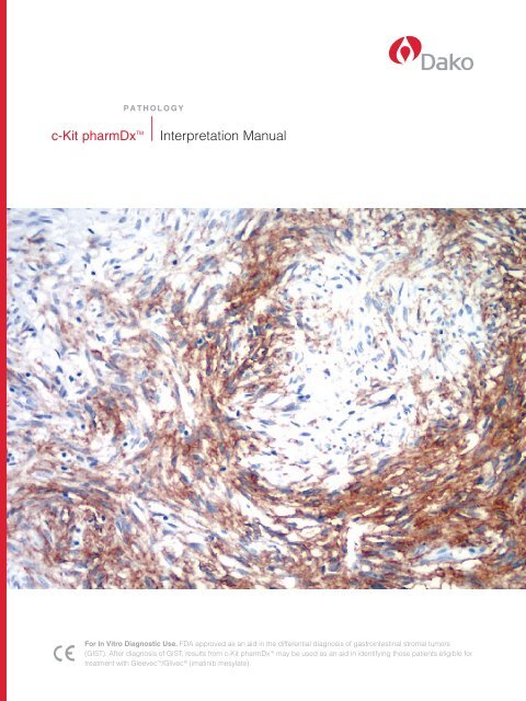
c-Kit pharmDx⢠Interpretation Manual - Dako
c-Kit pharmDx⢠Interpretation Manual - Dako
c-Kit pharmDx⢠Interpretation Manual - Dako
- No tags were found...

Create successful ePaper yourself
Turn your PDF publications into a flip-book with our unique Google optimized e-Paper software.
c-kit OverviewThe proto-oncogene c-kit, otherwise known as CD117 antigen or Stem Cell Factor Receptor, is a 145 kD type IIItransmembrane tyrosine kinase receptor. The c-kit gene encodes a transmembrane tyrosine kinase receptor, structurallysimilar to platelet derived growth factor receptors A and B, as well as the colony stimulating factor 1 receptor. It isthought to play an important role in hematopoiesis, spermatogenesis, and melanogenesis. The c-kit protein containsextracellular domains with 5 Ig-like loops, a highly hydrophobic transmembrane domain, and an intracellular domainwith tyrosine kinase activity split by a kinase insert in an ATP-binding region and in a phosphotransferase domain.Receptor activation is accompanied by receptor dimerization, substrate phosphorylation and autophosphorylation,receptor internalization, activation of protein kinases and phospholipases, and transcription of different protooncogenes.1 The c-kit tyrosine kinase receptor pathway has been shown to be important for tumor growth andprogression in several cancers. 2 Mutations in the c-kit gene may lead to ligand independent phosphorylation(activation) of the c-kit tyrosine kinase, which are believed to have a central pathogenic role in e.g. gastrointestinalstromal tumors. 3Figure 1 Schematic representation of c-kit structure and mechanism of actionc-<strong>Kit</strong> pharmDx SpecificityRabbit polyclonal c-kit antibodies were obtained by subcutaneous injection of a 14 mer peptide (positions 963-976 of theC’ terminus of the c-kit protein) coupled to thyroglobulin. The antisera was fractionated through antigen bound affinitychromatography.A number of soft tissue sarcoma specimens (e.g. leiomyosarcoma) were tested for c-kit expression. This study involved acomparison between the c-<strong>Kit</strong> pharmDx assay and the Novartis Clinical Trial Protocol (NCTP) as used for the selection ofpatients for treatment with Gleevec. Two of a total of twenty eight specimens tested displayed positive staining. The resultwas consistent using the two protocols. All positive immunostaining was abolished after absorption of the primary antibodywith a synthetic peptide (16 amino acid C-terminal end of the c-kit protein). It was therefore concluded that the primary antibodyused in the c-<strong>Kit</strong> pharmDx assay reacted specifically with the c-kit protein in the two positively stained soft tissue sarcomas.The c-kit pharmDx assay has been tested for reactivity against cell lines expressing c-kit protein. In Western blotting of alysate of the small cell lung carcinoma cell line SY that over-expresses c-kit mRNA, the antibody reagent labeled a band of145 kD corresponding to the c-kit protein. The labeled band is rather broad, from 120 to 155 kD. An additional band of 100 kDwas also labeled. 5 Further, applying a second anti-c-kit antibody, a 100 kD protein was, likewise, labeled. This labeling wasabolished when the antibody reagent was incubated with the synthetic c-kit peptide antigen used for immunization. 6 In additionalstudies, the c-kit antibody reagent was tested using Western blotting for cross-reactivity to proteins that share structuralhomology, such as; Platelet Derived Growth Factor Receptor (PDGFRa), macrophage colony stimulating factor receptor (c-FMS); and FMS-like tyrosine kinase 3 (FLT-3). No cross-reactivity was observed with these homologous proteins. Furthermore,in Western blotting the <strong>Dako</strong> antibody reagent did not react with a lysate of the adenocarcinoma cell line HS, which does notexpress the c-kit gene. 5c-<strong>Kit</strong> pharmDx <strong>Interpretation</strong> <strong>Manual</strong>
c-kit Expression in Normal TissuesThe c-kit protein is also expressed on a variety of normal cells including many epithelial cell types.Non-epithelial cells types that express c-kit protein include mast cells and macrophages.Some examples of normal tissue stained with c-<strong>Kit</strong> pharmDx and recommended reagents are summarizedin the table below. All tissues were formalin-fixed and paraffin-embedded. Staining was performed on the<strong>Dako</strong> Autostainer.Table 1: Evaluation of normal tissue staining by <strong>Dako</strong> c-<strong>Kit</strong> pharmDxTISSUE TYPE (# Tested)Adrenal (3)Bone Marrow (3)Breast (3) +Brain/Cerebellum (3)Brain/Cerebrum (3)Cervix (3)Colon (3) +Esophagus (3)Heart (3) –Kidney (3)Liver (3)Lung (3)Mesothelial Cells (3)Ovary (3)Pancreas (3)Parathyroid (3)Peripheral Nerve (3)Pituitary (3)Prostate (3)Salivary Gland (3)Skeletal Muscle (3) –Skin (3) +Small Intestine (3)Spleen (3)Stomach (3)Testis (3)Thymus (3)Thyroid (3)Tonsil (3)Uterus (3)POSITIVE TISSUE ELEMENT STAINING AND STAINING PATTERNNoneRare mast cells (2+): cytoplasmicEpithelial cells (3+): membrane and cytoplasmicMyoepithelial cells: (1+): cytoplasmicPurkinje cell processes (1+): cytoplasmicNoneNoneLamina propria and submucosal mast cells (2+): cytoplasmicSubmucosal mast cells (2+): cytoplasmicNoneTubular epithelium (2+): cytoplasmicNoneMast cells and macrophages (2+): cytoplasmicNoneNoneRare large duct epithelial cells (3+): Mast cells (2+): cytoplasmicNoneMast cells in soft tissue (2+): cytoplasmicNoneNoneNoneMast cells (2+): cytoplasmicBasal epidermal melanocytes (1+): cytoplasmicGlandular myoepithelial cells (1+): cytoplasmicNeuronal cells (1+): cytoplasmicMast cells (2+): cytoplasmicMast cells (2+): cytoplasmicMast cells in lamina propria (2+): cytoplasmicNoneNoneNoneNoneNone+ Recommended for positive control tissue– Recommended for negative control tissuec-<strong>Kit</strong> pharmDx <strong>Interpretation</strong> <strong>Manual</strong>
The c-<strong>Kit</strong> pharmDx <strong>Kit</strong>The c-<strong>Kit</strong> pharmDx kit contains polyclonal antibodies and control slides sufficient to complete an IHC stainingprocedure on formalin-fixed, paraffin-embedded specimens.Following incubation with the primary polyclonal antibodies to human c-kit protein, this kit is optimized for use witha ready-to-use visualization reagent based on dextran technology. This reagent consists of both secondary goat antirabbitantibody molecules and horseradish peroxidase molecules linked to a common dextran polymer backbone,thus eliminating the need for sequential application of link antibody and peroxidase conjugate. The enzymaticconversion of the subsequently added chromogen results in formation of a visible reaction product at the antigensite. The specimen may then be counterstained and coverslipped. Results are interpreted using a light microscope.Control slides containing two formalin-fixed paraffin-embedded mouse (+) and human (–) cell lines with stainingintensity scores of 2+ and 0 respectively are provided for quality control of the kit reagent performance.Two c-<strong>Kit</strong> pharmDx kit configurations are available:K1906 c-<strong>Kit</strong> pharmDx kit for manual use 25 testK1907 c-<strong>Kit</strong> pharmDx kit for <strong>Dako</strong> Autostainer 35 testThe c-<strong>Kit</strong> pharmDx kits include:n c-<strong>Kit</strong> pharmDx Polyclonal Rabbit IgGn Rabbit IgG Negative Control Reagentn c-<strong>Kit</strong> pharmDx Control SlidesMaterials required, but not supplied include:n Wash Buffer (code S3006)n Dual Endogenous Enzyme Block (DEEB) (code S2003)n Target Retrieval Solution (code S1699 or S1700)n EnVision+ HRP, Rabbit (code K4002 or K4003)n DAB+ (code K3467 or K3468)n Hematoxylin (S3301 or S3302)Figure 2 <strong>Kit</strong> ProcedureIncubate 30 secondsat 125°C.ORIncubate 20 minutesat 95–99°C.antigenc-kit proteinDual EndogenousEnzyme Block(DEEB)c-kit antibodysecondary antibodyHRP enzymedextran backboneDABSTEP 1STEP 2STEP 3STEP 4STEP 5Target Retrieval Solutionin either calibratedpressure cooker orwater bath.Application of DEEB.Incubate 5 minutes.Application of PrimaryAntibody. Incubate30 minutes.Application of EnVision+HRP, Rabbit. Incubate30 minutes.Application ofSubstrate-Chromogen.Incubate 10 minutes.c-<strong>Kit</strong> pharmDx <strong>Interpretation</strong> <strong>Manual</strong>
c-<strong>Kit</strong> pharmDx <strong>Kit</strong> Clinical Studies and WorkflowEfficacy and safety testing of imatinib mesylate (Gleevec) for anti-tumor activity in gastrointestinal stromal tumors(GISTs) was performed under the sponsorship of Novartis. Adults with a histologically confirmed, unresectable ormetastatic GIST that expressed CD117 antigen (c-kit) as assessed with a home brew immunohistochemical assayusing <strong>Dako</strong> c-kit antibody reagent were eligible for the study. Subjects were randomly assigned to a treatmentregimen of 400 or 600 mg Gleevec in capsular form administered daily. 7 A total of 147 subjects were enrolled in thetrial at 4 study centers. Confirmation of CD117 antigen (c-kit) positive gastrointestinal stromal tumor was completedfor 135/137 cases. In 10 cases, pathologic material was not available for central review.With follow-up of more than 9 months, partial responses were documented for 53.7 percent of the subjects.An additional 27.9 percent of subjects had stable disease, while disease progression was found for 13.8 percentof patients within 3 months after study entry. 7Figure 3 Recommended WorkflowTumor from GI TractMorphologyPredominantlyspindle cellsPredominantlyepitheliod cellsCombination ofspindle and epithelial cellsTest with c-<strong>Kit</strong> pharmDx for GIST diagnosisRule OutLeiomyosarcomaLeiomyomaMalignant MelanomaSchwannomaFibromatosisMalignant Peripheral Nerve Sheath TumorInflammatory Myofibroblastic TumorSarcomatoid CarcinomaApproximately 95% positivefor c-kit expressionApproximately 5% negativefor c-kit expression Test for tumor genotype 8c-<strong>Kit</strong> pharmDx <strong>Interpretation</strong> <strong>Manual</strong>
Technical Tips for Optimal c-<strong>Kit</strong> pharmDx PerformanceFor accurate and consistent results adherence to recommended c-<strong>Kit</strong> pharmDx procedure is essential.High quality results can be achieved in any laboratory by following these guidelines.Technical problems may arise in two areas, those involving sample collection and preparation of tissue in thepre-analytical processing of the specimen and those involving the actual performance of the c-<strong>Kit</strong> pharmDx assayitself. Technical problems related to performance of the assay are generally caused by planned and unplannedprocedural deviations from the c-<strong>Kit</strong> pharmDx protocol. Quality control slides are provided to detect false resultsfrom technical problems.Tissue Fixation and VariablesSuboptimal sample handling and processing can affect the c-<strong>Kit</strong> pharmDx results.Some of the variables that may affect results are as follows:n Specimens drying prior to fixation.n Fixation with alternative fixatives (neutral buffered formalin is recommended)n Age, pH, and storage conditions of fixative.n Length of fixation.c-<strong>Kit</strong> pharmDx Protocol Recommendationsn Specimens preserved in neutral buffered formalin fixative are suitable for testing with c-<strong>Kit</strong> pharmDx.Alternative fixatives have not been validated and may give erroneous results.n Automated Staining <strong>Dako</strong> recommends the use of c-<strong>Kit</strong> pharmDx on a <strong>Dako</strong> Autostaineror Autostainer Plus. Use of c-<strong>Kit</strong> pharmDx on alternative automated platforms has not been validated andmay give erroneous results.n Wash Buffer Use only wash buffer S3006 (TBST Wash Buffer). Dilute the recommended wash buffer 1:10using distilled or deionized water. Store unused solution at 2-8°C for no more than 7 days. Discard dilutedsolution if cloudy in appearance.n Storage of Reagents Reagents and control slides should be stored at 2-8 °C. Do not use the kit after theexpiration date printed on the outside of the kit box.n Companion Reagents To ensure consistent, reproducible results, use specified companion reagents to performc-<strong>Kit</strong> pharmDx assay.n Proper Incubations All incubation times must be performed according to the package insert. Stay withinthe tolerance indicated in the package insert for all incubation times to avoid erroneous false-negative orfalse-positive results.n For high quality results, review the c-<strong>Kit</strong> pharmDx Training Checklist (Table 2) prior to beginning yourstaining run.c-<strong>Kit</strong> pharmDx <strong>Interpretation</strong> <strong>Manual</strong>
Table 2: c-<strong>Kit</strong> pharmDx Training ChecklistInstitutionTrained byPerson Trained/Title<strong>Manual</strong> or Automated Staining Run?Autostainer Software VersionDateSerial Numberc-<strong>Kit</strong> pharmDx is a modular assay system requiring specified companion reagents and controls to ensure reproducible results.Companion REAGENts YES NOWash Buffer (S3006)Target Retrieval Solution (S1699 or S1700)Dual Endogenous Enzyme Block (S2003)EnVision+ HRP, Rabbit (K4002 or K4003)DAB+ (K3467 or K3468)<strong>Manual</strong> or Automated Procedure Yes NoControl slides and kit stored at 2-8°C?Cell Line control slides and all reagentswarmed to room temperature (20-25°C)prior to starting assay?Tissues fixed in 10% neutral buffered formalin?Specimens stained within 2 months oftissue mounting on slides when storedat room temperature?Xylene and alcohol solutions changedafter 40 slides?Deparaffinization and rehydrationprotocol followed?Wash buffer prepared properly?Prepare sufficient quantity of wash buffer10X,1:10 in reagent-quality water, deionized or distilled water.Distilled or deionized water (not tap water)used for water washes after last alcoholbath in deparaffinization?Temperature and time incubations programmedfor Target Retrieval Solution and specimens heatedfor 30 seconds at 125°C in pressure cooker orfor 20 minutes at 95-99°C in the water bath?Regressive hematoxylin counterstainsare not used?<strong>Manual</strong> Procedure Yes NoAfter heat retrieval, Target Retrieval Solutiondecanted and slides rinsed in Wash Buffer?Distilled or deionized water (not tap water)used for water bath after DAB+ Substrate-Chromogen Solution step?Buffer bath(s) changed between each step?Humid chamber used for Primary Antibody/Negative Control Reagent and EnVision+ HRP,Rabbit incubations?Slides placed in 5 (+/-1) minute buffer bathbetween Dual Endogenous Enzyme Blockand Primary Antibody/Negative Control Reagentsteps; Primary Antibody/Negative Control Reagentand EnVision+ HRP, Rabbit steps; and EnVision+HRP, Rabbit and DAB+ Substrate-ChromogenSolution steps?<strong>Manual</strong> Procedure continued Yes NoSpecimen fully covered with Dual EndogenousEnzyme Block for 5 minutes?Specimen fully covered with Primary Antibodyfor 30 minutes?Specimen fully covered with EnVision+ HRP,Rabbit for 30 minutes?DAB+ Substrate-Chromogen prepared properly?1 drop DAB+ Chromogen to 1 mL DAB+ Substrate Buffer.Specimen fully covered with Substrate-Chromogen solution for 10 minutes?Automated Procedure Yes NoSlides placed in buffer bath 5 (+/-1) minutesbefore loading onto the <strong>Dako</strong> Autostainer?Appropriate protocol template used?Was the Autostainer programmingreviewed for accuracy?Slides rinsed with buffer between steps anddouble rinsed after the EnVision+ HRP, Rabbitstep with an additional 5-minute rinse hold?Specimen fully covered with Dual EndogenousEnzyme Block for 5 minutes?Specimen fully covered with Primary Antibodyfor 30 minutes?Specimen fully covered with EnVision+ HRP,Rabbit for 30 minutes?DAB+ Substrate-Chromogen prepared properly?Add 1 drop of Liquid DAB+ Chromogen toone mL of DAB+ Substrate Buffer and mix.Specimen fully covered with Substrate-Chromogen solution for 10 minutes?Instrumentation/Equipment Yes NoIs regular preventative maintenanceperformed on the pressure cooker orwater bath and the <strong>Dako</strong> Autostainer?Is the necessary equipment availableto perform the c-<strong>Kit</strong> pharmDx assayaccording to protocol?If not, specify in comments below.If you answered No to any of the above, you have deviated fromprotocol and should consult with your <strong>Dako</strong> TechnicalSupport Representative for assistance (800 424 0021).Additional observations or comments:10 c-<strong>Kit</strong> pharmDx <strong>Interpretation</strong> <strong>Manual</strong>
Quality Controlc-<strong>Kit</strong> pharmDx is an IHC assay system requiring controls to ensure reproducible results.The first quality control step for interpretation is the evaluation of the control cell slides. Positive and negative cell lines are includedin each c-<strong>Kit</strong> pharmDx kit (Figures 4 and 5) for the purpose of verification of staining runs, every time the assay is performed.Appropriate staining of the control cell lines provides evidence that the c-<strong>Kit</strong> pharmDx assay is functioning properly. They shouldnot be used as an aid in interpretation of patient results. No staining of the HT-29 control cell line (0) and moderate brown cytoplasmicor paranuclear staining in the P815 control cell line (2+) indicates that the staining run is valid. If the intensity of the positivecontrol cell line is too weak or too strong a false negative or false positive result can occur. If either of the control cell lines havestaining results outside of these criteria, results from all of the test slides stained simultaneously within the same run shouldbe considered invalid and repeated.The second quality control step is the interpretation of positive control tissue (Figure 6). Use a positive control tissue (known to bec-kit positive) fixed, processed and embedded in a manner similar to the patient sample(s) with each staining run to verify thespecificity of the primary antibody and in conjunction with the corresponding negative reagent control tissue provides an indicationof specific background staining. This validation is performed to ascertain proper tissue preparation and staining techniques. Thepresence of a moderate brown reaction in the cell membrane or cytoplasm is indicative of positive reactivity. One positive tissue slidefor each of the test conditions should be included in each staining run. Verify that the negative tissue control slide demonstratesno specific reactivity. Known positive tissue controls should be utilized for monitoring the correct performance of reagents andprocessed tissue, NOT as an aid in formulating a specific diagnosis of patient samples. There are also positive tissue elementsthat can very often serve as intrinsic controls in patient specimens in addition to positive and negative control tissues. Specifically,c-kit positive tissue elements such as Interstitial Cells of Cajal (ICC’s) are frequently present in GIST and adjacent normal tissues(Figure 7). Furthermore, mast cells (Figure 8) are commonly found in a variety of tissue types. If the positive tissue controls failto demonstrate appropriate positive staining, results with the test specimens should be considered invalid.The third quality control step involves the interpretation of the negative control tissue (Figure 9). Use a negative control tissue (knownto be c-kit negative) fixed, processed and embedded in a manner similar to the patient sample(s) with each staining run to verify thespecificity of the primary antibody and in conjunction with the corresponding negative reagent control to provide an indicationof specific background staining. The variety of different cell types present in most tissue sections offers an internal negative control.If specific staining occurs in the negative control tissue, results with the test specimens should be considered invalid.Figure 4 P815 positive cell line controlstained with c-<strong>Kit</strong> pharmDx, 2+ moderateintensity, 40x.Figure 5 HT-29 negative control cell linestained with c-<strong>Kit</strong> pharmDx, 0 intensity, 40x.Figure 6 Breast positive control tissue.Ductal epithelial cells stained positively withc-<strong>Kit</strong> pharmDx, 2+ moderate intensity, 40x.Figure 7 Interstitial Cells of Cajal (ICC’s)staining in GIST adjacent normal tissue,2+ moderate intensity, 20x.Figure 8 Mast cell staining, 2+ moderateintensity, 10x.Figure 9 Skeletal muscle negative controltissue stained with c-<strong>Kit</strong> pharmDx,0 intensity, 20x.c-<strong>Kit</strong> pharmDx <strong>Interpretation</strong> <strong>Manual</strong>11
c-<strong>Kit</strong> pharmDx Evaluation and ReportingSlide evaluation should be performed by a pathologist using a light microscope.The c-<strong>Kit</strong> pharmDx assay stains a variety of normal and neoplastic tissues. Observed c-kit staining patterns may beheterogeneous or homogeneous depending on the tissue and/or tumor type. Heterogeneity includes various stainingintensities, ranging from 0–3+, within a single neoplasm. Positive cell staining patterns may also be heterogeneousincluding membrane, cytoplasmic staining, or both.The performance characteristics of c-<strong>Kit</strong> pharmDx make possible the visualization of four intensity levels from 0–3+.c-kit staining at the cellular level has been observed on both the membrane and the cytoplasm (including paranuclear)at all intensity levels.<strong>Dako</strong> recommends the following interpretation guideline in the assessment of c-kit immunostaining.All assessments are to be made on the tumor region of the specimen.Steps to c-<strong>Kit</strong> pharmDx <strong>Interpretation</strong>1. Evaluate the control cell lines to validate the assay staining run.Examples of acceptable and unacceptable P815 positive cell line staining with c-<strong>Kit</strong> pharmDx.Unacceptable weak staining intensity (too light);40x. Weak staining intensity of cell line mayresult in false-negative results.Acceptable staining (moderate intensity); 40x.Unacceptable excessive staining intensity(too dark); 40x. Excessive staining intensityof cell line may result in false-positive results.2. Evaluate the positive and negative tissue control slides to validate proper tissue preparation, staining techniquesand assay performance.3. An H&E stained slide of the test tissue is useful for comparison. The neoplasm may not be easily appreciable onthe c-<strong>Kit</strong> pharmDx stained slide.4. Evaluate the c-<strong>Kit</strong> pharmDx stained section for identification of neoplasm at low power, 4x magnification.3+ intensity staining of the tumor cells is easily identified at 4x magnification.5. Observe cells that stain brown, move to a higher power (10x magnification) to confirm the staining. In general,most cases should be observed at 10x magnification.6. If the staining pattern is an artifact, it should be disregarded for evaluation. Find another representative area(s).7. Use 20x or 40x magnification to assess staining pattern (cytoplasmic versus membrane, including paranuclear).Confirmation of c-<strong>Kit</strong> pharmDx staining at 20x magnification may be useful in those neoplasms with abundantcytoplasmic staining. Some tumor cells have several populations of cells with different intensities of c-kitmembrane staining.8. If there is no tumor staining and normal elements are not staining, review control slides of that staining runto confirm levels of c-kit expression that are observed. If positive elements in the control slides are negative,repeat the staining run.12 c-<strong>Kit</strong> pharmDx <strong>Interpretation</strong> <strong>Manual</strong>
<strong>Interpretation</strong> of c-kit protein expression must be made within the general morphological and clinical context ofthe tumor. There are three morphologic categories of GIST: spindle cell (70%), epithelioid (20%), or mixed types. 8Regardless of morphology, the majority of GISTs express c-kit protein in a significant proportion of the tumor cells.Positive homogeneous immunostaining with c-<strong>Kit</strong> pharmDx is primarily seen in the cytoplasm, with or without a golgi/paranuclear (dot-like) pattern, and in the cell membranes, with either complete or incomplete circumferential staining.Some cases show a heterogeneous staining pattern in combination with the staining patterns previously listed.Artifactual staining has also been observed in the extracellular spaces.Positive and negative cell lines are included in each c-<strong>Kit</strong> pharmDx kit to validate staining runs, every time the assayis performed. Appropriate staining of the control slides provides evidence that the c-<strong>Kit</strong> pharmDx assay is functioningproperly. No staining of the HT-29 control cell line (0) and moderate brown membrane, cytoplasmic and paranuclearstaining in the P815 control cell line (2+) indicates that the staining run is valid. If the intensity of the positive controlcell line is too weak or too strong a false negative or false positive result may be obtained and the test should berepeated.Slide evaluation should be performed by a pathologist using a light microscope. All assessments are to be made onthe tumor region of the specimen. For evaluation of the immunocytochemical staining and interpretation, an objectiveof 10x or 20x magnification is appropriate. Use intact cells for interpretation of staining results; necrotic or degeneratedcells often stain non-specifically. 9c-<strong>Kit</strong> pharmDx test results should be reported as positive or negative, using cytoplasmic and membrane staining asthe evaluable structure (Table 4). Positivity for c-kit protein expression is defined as any specific cytoplasmic and / ormembrane staining in the tumor cells. It should also be noted that a small percentage of GISTs do not express c-kitprotein. Therefore, a small percentage or absence of c-kit staining does not exclude the diagnosis of GIST.Depending on the incubation length and potency of the hematoxylin used, counterstaining will result in a pale to darkblue coloration of the cell nuclei. Excessive or incomplete counterstaining may compromise interpretation of results.Diagnosis and Immunohistochemical StainingIf necessary, use a panel of antibodies to aid in the identification of false negative reactions. When staining is focallypositive, Report additional to Treating testing Physician should be considered to confirm a GIST diagnosis. Definition Genetic testing as well as the stainingpatterns specified in Table 3 aid in the differentiation between GIST and other mesenchymal sarcomas. Refer to c-<strong>Kit</strong>pharmDx Package Insert sections “Summary and Explanations” and “Limitations and Performance characteristics”,for specific information regarding immunoreactivity.Table 3 Immunohistochemical schema for the Differential Diagnosis of Spindle Cell Tumors of the GI Tract. 10c-kit CD34 SMA S-100 DesminGIST≥95%60–70%30–40%≤5%≤2%Smooth muscle tumor–10–15%+Rare+Schwannoma–Usually–+–FibromatosisDisputed*Rare+–Rare* Most, but not all authors report that fibromatoses are negative for c-kit.c-<strong>Kit</strong> pharmDx <strong>Interpretation</strong> <strong>Manual</strong>13
c-kit Growth Factor Receptor Report (c-<strong>Kit</strong> pharmDx)Patient Name________________________________________________ Collection Date _______________________________Ordering Physician_ __________________________________________ Received Date _______________________________PLEASE PHOTOCOPY FOR YOUR USEOrdering Facility______________________________________________ Report Date __________________________________Medical Record#_____________________________________________ Lab Reference # ______________________________Specimen ID#________________________________________________ Tumor Source ________________________________Date of Birth_ ________________________________________________ Patient Gender _ ______________________________DescriptionDeparaffinized tissue and appropriate control tissue sections are stained using the FDA-approved <strong>Dako</strong>c-<strong>Kit</strong> pharmDx immunohistochemistry kit.A positive result is based on any specific cytoplasmic and/or membrane staining within the tumor cells.PATIENT RESULT POSITIVE NEGATIVEc-kit proteinThe c-<strong>Kit</strong> pharmDx is indicated as an aid in the differential diagnosis of gastrointestinal stromal tumors (GIST).After diagnosis of GIST, results from c-<strong>Kit</strong> pharmDx may be used as an aid in identifying those patients eligiblefor treatment with Gleevec TM /Glivec ® (imatinib mesylate).All subjects in the Novartis Gleevec/Glivec clinical trials were selected using an investigational Novartis ClinicalTrial Protocol (NCTP). The primary anti-c-kit rabbit polyclonal antibody reagent used in the NCTP was purchased from<strong>Dako</strong>. The c-<strong>Kit</strong> pharmDx primary polyclonal antibody reagent underwent the same method of production, purificationand quality control as did the NCTP polyclonal anti-c-kit reagent.Report to Treating Physicianc-kit protein NegativeAbsence of staining in tumor cells.Definitionc-kit protein PositivePositive staining is defined as any IHC staining of tumor cellcytoplasm and/or membranes above background level.Staining Intensity 1+ 2+ 3+Absence of staining in the cells of stromal tumors should be reported as negative for c-kit expression. A smallpercentage of GISTs do not express c-kit protein. Therefore, absence of c-kit staining does not exclude the diagnosisof GIST. Additional Immunohistochemical markers or genetic testing should be considered to aid in the differentialdiagnosis of these cases and other mesenchymal sarcomas.2005 <strong>Dako</strong>14 c-<strong>Kit</strong> pharmDx <strong>Interpretation</strong> <strong>Manual</strong>
Image Guide for <strong>Interpretation</strong>c-<strong>Kit</strong> pharmDx <strong>Interpretation</strong> Guidelines<strong>Dako</strong> emphasizes that interpretation of c-<strong>Kit</strong> pharmDx be performed within the context of the pathologist’s pastexperience and best medical judgment. This guide will highlight examples of c-<strong>Kit</strong> pharmDx positivity and negativity,different staining intensities and areas of interpretation that are potentially problematic for c-<strong>Kit</strong> pharmDx users.Figure 10 Examples of tissues stained with c-<strong>Kit</strong> pharmDx.negative resultGIST, no cytoplasmic or membrane staining (0) 20x.positive resultsGIST, cytoplasmic staining (1+) 20x.GIST, cytoplasmic and membrane staining (2+) 20x.GIST, cytoplasmic and membrane staining (3+) 20x.c-<strong>Kit</strong> pharmDx <strong>Interpretation</strong> <strong>Manual</strong>15
Staining PatternsThe <strong>Dako</strong> c-<strong>Kit</strong> pharmDx stains a variety of normal and neoplastic tissues. Observed c-kit protein staining patternsare homogeneous or heterogeneous depending on the tissue and/or tumor type. The staining can showa range of 0-3+ staining intensity. Positive cell staining patterns can be membrane and / or cytoplasmic.Heterogeneous StainingHeterogeneity includes various staining intensities within a single neoplasm.Figure 11 GIST, example of moderate 2+ heterogeneous staining;positive,10x.Figure 12 GIST, example of moderate 2+ heterogeneous staining;positive, 20x.Homogeneous StainingHomogeneous c-kit protein expression exhibits homogeneous staining patterns.Figure 13 GIST, example of moderate 2+ homogeneous staining;positive,10x.Figure 14 GIST, example of strong 3+ homogeneous staining;positive,10x.Dot-like Paranuclear Staining PatternHomogeneous c-kit protein expression can exhibit paranuclear staining patterns.Figure 15 GIST example of moderate 2+ dot-like paranuclearstaining; positive, 20x.16 c-<strong>Kit</strong> pharmDx <strong>Interpretation</strong> <strong>Manual</strong>
Membrane StainingCancers with c-kit protein expression can exhibit membrane staining patterns.Figure 16 Example of GIST with strong 3+ membrane staining;positive,20x.c-kit pharmDx Staining of Normal and Benign TissuesNormal and benign tissues, some of which are summarized in Table 1, exhibit specific c-kit staining. These can serveas useful internal controls. Staining of normal tissue elements should be excluded from the c-kit interpretation.c-kit is expressed in a variety of normal cells. These cells include but are not limited to:n Ductal and myoepithelial cells of the breastnnnnnnPurkinje cell processesLamina propria cells of the colonTubular epithelium of the kidneyMelanocytes and glandular myoepithelial cells of the skinInterstitial Cells of Cajal of the small intestineMast cellsMast cells can be strongly reactive with the c-<strong>Kit</strong> pharmDx assay. Reactivity in granulocytes is caused byendogenous peroxidase activity. The staining pattern is usually cytoplasmic, not membrane.Figure 17 Strong 3+ mast cell staining; 20x (2+ staining was observed in the normal tissue testing).Non-evaluable Areas of TissueAreas on stained slides that should not be evaluated include dissociated, free floating groups or aggregates ofneoplastic cells, necrotic cells, and damaged areas of the tissue section (torn sections, folded or wrinkled areas, etc.)c-<strong>Kit</strong> pharmDx <strong>Interpretation</strong> <strong>Manual</strong>17
Factors to Consider in Evaluating c-<strong>Kit</strong> pharmDx StainsNon-Specific Background StainingNon-specific background staining is defined as diffuse, non-specific staining of tissue elements. It may be causedby a variety of factors including both biologic activity and technological processes.Biologic Activityn Pseudoperoxidase activity (erythrocytes) and endogenous peroxidase activity (granulocytes) may resultin non-specific background staining due to decomposition of H 2O 2within the substrate.Technological Processesn Use of fixatives other than the recommended 10% neutral buffered formalinn Incomplete deparaffinization of tissue sections prior to stainingn Use of alternative buffers (recommended wash buffer (Code S3006)n Incomplete rinsing of reagents from slidesn Inappropriate drying of slides during staining procedure (use a humid chamber when assayis performed manually)The level of non-specific background staining may be ascertained by incubation of test specimen with thenegative control reagent. If non-specific background staining is significant, the specific staining must beinterpreted with caution.Improper Target Retrievalc-<strong>Kit</strong> pharmDx includes pretreatment by means of heat induced target retrieval. Tissue sections may be overretrieved, causing disruption of cell morphology, overall tissue architecture or tissue removal from the slide.Run the assay with careful attention to the duration of the target retrieval step.Note If tissue removal from the slide is a persistent problem, tissue sections may be mounted on slides whichprovide better section adherence (charged, silanized, and poly-L-lysine coated slides).18 c-<strong>Kit</strong> pharmDx <strong>Interpretation</strong> <strong>Manual</strong>
Interpreting ArtifactsEdge ArtifactFrequently, increased staining is observed around the periphery of the tissue specimen. It is known as the “edgeeffect.” Edge artifacts are commonly the result of inappropriate pre-analytic handling of the tissue. The edge effectrepresents fixation artifact or tissue drying prior to fixation. Usually the staining artifact is limited to a thin rim ofstained cells with an abrupt termination to the staining reaction. Often the method of surgical extraction is thecause (see Crush Artifact section).Tissue section edge staining artifacts are common if there are significant tissue section irregularities. Thick tissuesections may mimic edge artifacts and can be corrected by recutting the tissue block to produce a uniform, thin,3-5 μm thick section that is devoid of folds and wrinkles.When the positive reaction is only at the edge of the tissue section, evaluation of c-kit staining should exclude tumorcells within the region’s edge artifact.Crush ArtifactCompression of tissue around the periphery of specimens, especially biopsy specimens, can produce non-specificstaining of the tissue components in addition to the membranes of neoplastic cells. The appearance of this stainingartifact is similar to those produced by tissue edge-staining artifacts.The staining intensity of cell membranes within compressed tissue is frequently greater than similar appearing cellsin regions of normal architectural tissue arrangements.Retraction ArtifactStromal retraction around tumor cell glands can create clefts where pooled antibody can nonspecifically stain.Thorough washing after the primary antibody reagent incubation step may prevent this reaction. Retraction spacestaining usually is a hemi-circumferential reaction around the periphery of the gland.Thermal ArtifactThermal electrocautery may alter nuclei and cell membranes. <strong>Dako</strong> recommends that the evaluationof c-<strong>Kit</strong> pharmDx staining be performed on tissue with no or minimal thermal electrocautery artifacts.c-<strong>Kit</strong> pharmDx <strong>Interpretation</strong> <strong>Manual</strong>19
Examples of GIST Stained with c-<strong>Kit</strong> pharmDxNegative c-<strong>Kit</strong> pharmDxImmunostainingPositive Homogeneous c-<strong>Kit</strong> pharmDxImmunostainingFigure 18 GIST, example of H&E;10x.Figure 20 GIST, example of H&E; 10x.Figure 19 GIST, example of negative staining; 10x.Figure 21 GIST, example of weak 1+ positivehomogeneous staining; 10x.Figure 22 GIST, example of weak 1+ positivehomogeneous staining; 20x.20 c-<strong>Kit</strong> pharmDx <strong>Interpretation</strong> <strong>Manual</strong>
Positive Homogeneous c-<strong>Kit</strong> pharmDxImmunostaining (moderate)Positive Homogeneous c-<strong>Kit</strong> pharmDxImmunostaining (strong)Figure 23 GIST, example of moderate 2+ positivehomogeneous staining; 20x.Figure 26 GIST, example of strong 3+ positivehomogeneous staining; 10x.Figure 24 GIST, example of moderate 2+ positivehomogeneous staining; 20x.Figure 27 GIST, example of strong 3+ positivehomogeneous staining; 10x.Figure 25 GIST, example of moderate 2+ positivehomogeneous staining; 20x.Figure 28 GIST, example of strong 3+ positivehomogeneous staining; 40x.c-<strong>Kit</strong> pharmDx <strong>Interpretation</strong> <strong>Manual</strong>21
Differential Immunostaining of LeiomyomaFigure 29 Leiomyoma, example of H&E; 10X. Figure 31 Leiomyoma, example of c-kit negative 0 staining; 10X.Figure 30 Leiomyoma, example of CD34 strong 3+ staining; 10X. Figure 32 Leiomyoma, example of SMA moderate 2+ staining; 10X.22 c-<strong>Kit</strong> pharmDx <strong>Interpretation</strong> <strong>Manual</strong>
Differential Immunostaining of LeiomyosarcomaFigure 33 Leiomyosarcoma, example of H&E; 10X.Figure 35 Leiomyosarcoma, example of c-kit negative0 staining; 10X.Figure 34 Leiomyosarcoma, example of CD34 moderate2+ staining; 10X.Figure 36 Leiomyosarcoma, example of Desmin weak1+ staining; 10X.Figure 37 Leiomyosarcoma, example of SMA strong3+ staining; 10X.c-<strong>Kit</strong> pharmDx <strong>Interpretation</strong> <strong>Manual</strong>23
Differential Immunostaining of SchwanomaFigure 38 Schwanoma, example of H&E; 10X.Figure 39 Schwanoma, example of c-kit negative0 staining; 10XFigure 40 Schwanoma, example of CD34 moderate2+ staining; 10XFigure 41 Schwanoma, example of S100 moderate2+ staining; 10X24 c-<strong>Kit</strong> pharmDx <strong>Interpretation</strong> <strong>Manual</strong>
References1. Bühring H-J, Ashman LK, Gattei V, Kniep B, Larregina A, Pinto A, et al. CR2.7 Stem-cell factor receptor (p145(c-kit)) summary report (CD117).Schlossman SF, Boumsell L, Gilks W, Harlan JM, Kishimoto T, Morimoto C, et al., editors. Leukocyte typing V. White cell differentiation antigens.Proceedings of the 5th International Workshop and Conference; 1993 Nov 3–7; Boston, USA. Oxford, New York, Tokyo: Oxford UniversityPress 1995; 18822. Smithey BE, Pappo AS, Hill DA. C-<strong>Kit</strong> expression in pediatric tumors: a comparative immunohistochemical study. Am J Surg Pathol 2002; 26:4863. Mietten M, Lasota J. Gastrointestinal stromal tumors–definition, clinical, histological, immunohistochemical, and molecular genetic features anddifferential diagnosis (review). Virchows Arch 2001; 438:14. Biosource5. Tsura Y, Hiraki H, Watanabe K, Igarashi S, Shimamura K, Fukuda T, et al. Preferential localization of c-kit product in tissue mast cells, basal cellsof skin, epithelial cells of breast, small cell lung carcinoma and seminoma/dysgerminoma in human: immunohistochemical study on formalin-fixed,paraffin-embedded tissues. Virchows Arch 1994; 424:1356. Yardern Y, Kuang W-J, Yang-Feng T, Coussens L, Munemitsu S, Dull TJ, et al. Human proto-oncogene c-kit: a new cell surface receptor tyrosinekinase for an unidentified ligand. EMBO J 1987; 6:33417. Demetri GD, von Mehren M, Blanke CD, Van den Abbeele AD, Eisenberg B, Roberts PJ, Heinrich MC, Tuveson DA, Singer S, Janicek M, FletcherJA, Silverman SG, Silberman SL, Capdeville R, Kiese B, Peng B, Dimitrijevic S, Druker BJ, Corless C, Fletcher CD, Joensuu H. Efficacy and safetyof imatinib mesylate in advanced gastrointestinal stromal tumors. NEJM 2002; 347, 7:4728. Demtri GD, Benjamin R, Blanke CD, Choi H, Corless C, DeMatteo RP, Eisenberg BL, Fletcher CDM, Maki RG, Rubin BP, Van den Abbeele AD,and Margaret von Mehren. NCCN Task Force Report: Optimal Management of Patients with Gastrointestinal Stromal Tumor (GIST)–Expansionand Update of NCCN Clinical Practice Guideline. Journal of the National Comprehensive Cancer Network 2004, Volume 1: Supplement 19. CLIS (formerly National Committee for Clinical Laboratory Standards, NCCLS). Quality Assurance for Immunocytochemistry; Approved guideline.Villanova, PA 1999. Order code MM4-A10. Fletcher CDM, Berman J, Corless C, Gorstein F, Lasota J, Longley JB, Miettinen M, O’Leary TJ, Remotti H, Rubin BP, Shmookler B, Sobin LH,and Weiss SW. Diagnosis of Gastrointestinal Stromal Tumors: A Consensus Approach. Human Pathology May 2002; 33:5; 459Additional c-kit ResourcesnnnDemtri GD, Benjamin R, Blanke CD, Choi H, Corless C, DeMatteo RP, Eisenberg BL, Fletcher CDM, Maki RG, Rubin BP, Van den Abbeele AD,and Margaret von Mehren. NCCN Task Force Report: Optimal Management of Patients with Gastrointestinal Stromal Tumor (GIST)–Expansionand Update of NCCN Clinical Practice Guideline. Journal of the National Comprehensive Cancer Network 2004, Volume 1: Supplement 1Demmatteo RP, Heinrich MC, El-Rifai WM, and Demetri G. Clinical Management of Gastrointestinal Stromal Tumors: Before and After STI-571.Human Pathology, May 2002; 33:5; 466Graadt van Roggen JF, van Velthuysen ML, Hogendoorn PC. The histopathological differential diagnosis of gastrointestinal stomal tumours.Journal of Clinical Pathology 2001, 54; 96n Joensuu H, Kindblom LG. Gastrointestinal Stromal Tumors: a review. Acta orthop Scand Suppl 2004, 75; 62nnnnYamaguchi U, Hasegawa T, Masuda T, Sekine S, Kawai A, Chuman H and Shimoda T. Differential diagnosis of gastrointestinal stromal tumorand other spindle cell tumors in the gastrointestinal tract based on immunohistochemical analysis. Virchow Arch 2004, 445; 142Medeiros F, Corless CL, Duensing A, Hornick JL, Oliviera AM, Heinrich MC, Fletcher JA, and Fletcher CDM. KIT-Negative GastrointestinalStromal Tumors. Am J Surg Pathol July 2004, 28:7; 889Gleevec /Glivec ® (imatinib mesylate) Package Insertc-<strong>Kit</strong> pharmDx Package Insertc-<strong>Kit</strong> pharmDx <strong>Interpretation</strong> <strong>Manual</strong>25
Notes26 c-<strong>Kit</strong> pharmDx <strong>Interpretation</strong> <strong>Manual</strong>
c-<strong>Kit</strong> pharmDx <strong>Interpretation</strong> <strong>Manual</strong>27
Corporate HeadquartersDenmarkTel +45 44 85 95 00Australia+61 2 9316 4633Austria+43 1 408 4334 50Czech Republic+420 541 42 37 10Denmark+45 44 85 97 56Ireland+353 91 768150Italy+39 02 58 078 1Norway+47 23 14 05 40Poland+48 58-661 1879Switzerland+41 41 760 11 66United Kingdom+44 (0)1 353 66 99 11www.dako.comDistributors in morethan 50 countriesBelgium+32 016 38 72 20Canada+1 905 858 8510France+33 1 30 50 00 50Germany+49 40 69 69 470Japan+81 75 211 3655The Netherlands+31 20 42 11 100Spain+34 93 499 05 06Sweden+46 08 556 20 600United States of America+1 805 566 665508070 30NOV0728 c-<strong>Kit</strong> pharmDx <strong>Interpretation</strong> <strong>Manual</strong>