Prezentace aplikace PowerPoint - Univerzita Karlova
Prezentace aplikace PowerPoint - Univerzita Karlova
Prezentace aplikace PowerPoint - Univerzita Karlova
- No tags were found...

Create successful ePaper yourself
Turn your PDF publications into a flip-book with our unique Google optimized e-Paper software.
<strong>Univerzita</strong> <strong>Karlova</strong> v Praze - 1. Lékařská fakultaRespiratory SystemInstitute of AnatomyAuthor: David SedmeraSubject: Anatomy 1Date: 2013
Division of respiratory system• Upper respiratory tract:- external nose- nasal cavity & paranasal sinuses- pharynx• Lower respiratory tract:- larynx- trachea (windpipe)- bronchi (down to respiratorybronchioli)- lungs• Significance: ENT vs. pneumologyURTI vs. pneumonia
Nasal passages and paranasal sinuses
Nasal cavity and paranasalsinuses• Common colds are common• Spatial relationships (syntopies) areimportant for spread of infection• Complications: meningitis, teeth, orbit,mediootitis, mastoiditis, sinusitis…
Nasal cavity and paranasalsinuses• Why do we have them (other than tokeep ENT doctors in business)• Maxillary, frontal, sphenoidal,ethmoidal• Surface projections, visible and X-rayexamination - sinusitis
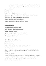
RESUMEN DE ORIENTACIÓNENFERMEDADES CON LA MAYOR CONTRIBUCIÓN CAUSAL DEL MEDIO AMBIENTELa barra verde oscura más la barra verde clara del diagrama representan la carga de morbilidad total.Retraso mental (causado por el plomo), tal como se define en la lista de enfermedades de la OMS correspondiente a 2002,accesible en Internet (www.who.int/evidence).AVAD: una medida ponderada de la mortalidad, la morbilidad y la discapacidad.función pulmonar, es atribuible a factores de riesgo ambientales. La exposición profesional al polvo y asustancias químicas, así como la contaminación del aire en locales cerrados por utilización de combustiblessólidos en los hogares, parecen ser dos de los principales factores que contribuyen a aumentar la fracciónde la carga de morbilidad relacionada con el medio ambiente. Sin embargo, otras formas de contaminacióndel aire en locales cerrados y del aire exterior, que van desde la producida por los medios de transportehasta el humo del tabaco de segunda mano, también ejercen una influencia importante.En el gráphico de arriba figura una lista de las 24 enfermedades con la mayor carga de morbilidad globalatribuible al medio ambiente. En el informe completo se proporciona una descripción detallada de losfactores ambientales y las repercusiones en todas las enfermedades que se examinan así como cuadros yanexos estadísticos que cubren la carga de morbilidad mundial y regional, así como la de subgruposespeciales, como los niños.7
Frontal CT
Horizontal CT
Postnatal growthof paranasalsinuses(pneumatization)
Histology of airwaysTaken from and see more in: Junquiera’s Histology!
Laryngeal cartilagesThyroid cartilage – left + right lamina, superior + inferior notch,superior + inferior horn, oblique line, cricoid articular surfaceCricoid cartilage - arch, lamina, arytenoid + cricoid articular surfaceArytenoid cartilage - base, apex, muscular + vocal process, cricoidarticular surface,Epiglottis, Cuneiform + Corniculate + Triticeal cartilages
The Larynx - development
The Larynx - sagittal view
The Larynx - vocal cordmovementsanteriorposterior
The Larynx - frontal view
The Larynx - examination(laryngoscopy)indirectdirect
The Trachea - cross section
Histology of Trachea Epithelium (cylindricalwith cilia and goblet cells) Connective tissue Glands in lamina propria Hyaline cartilage coveredby perichondrium Smooth (trachealis) muscle
The trachea and segmental bronchiStarts at C6Bifurcation at Th4-5Length: 13 cmDiameter: 2.5 cm
Coniotomy and tracheotomy
Coniotomy and tracheotomyconiotomySuperior tracheotomyInferior tracheotomy
Syntopy of cervical part of trachea
Blood supply• Nasal cavity - ethmoidal and sphenopalatineartery• Larynx -superior and inferior laryngealartery• Trachea - branches from thyroid arteries orthoracic aorta
Innervation• Nasal cavity - I, V1, V2; parasympatheticfibers from VII• Larynx -superior and recurrent laryngealnerve (from X)• Trachea - X, cervical sympathetics
Lymphatic drainage
The Lungs & Pleura - projections
Borders of Lungs & Pleura
Borders of Lungs & Pleura
The Lungs & Pleura - projections
Pleural recesses• Costodiphragmatic recess -accumulation of fluids• recessus phrenicomediastinalis• recessus costomediastinalis
The Pleural Cavity
• Penetration of thepleural cavity equalizespressure• This results in thecollapse of the affectedlung• Could be classified asopen, closed, or tension• Treatment is bydrainage that facilitatesair resorptionPneumothorax
Pneumothorax - X-ray
The lungs
The lungs
Syntopies of trachea and main bronchi
The bronchopulnonary segments
Histology of the Bronchi Epithelium (cylindricalwith cilia and goblet cells) Connective tissue Glands in lamina propria Hyaline cartilage(discontinuous) Smooth muscle
Histology of the Bronchi
Bronchography
Blood supply & innervation• Pulmonary artery and branches - functional• Rr. Bronchiales from thoracic aorta orintercostal arteries - nutritive• Pulmonary vein, anastomoses• Parasympathetic: left and right vagus• Sympathetic: inferior and middle cervicalganglia, rami from the first four thoracicganglia; almost no pain (only parietal pleura)
Lymphatic drainage
The Bronchioli No cartilage, just smooth muscle => bronchocostriction inasthma!
The Lung Lobes - projections
Terminal bronchioli and Clara cells
Histology of lungtissue - respiratorybronchioli, alveolarducts, alveoli
Branching ~23 bifurcations,300-400 mil alveolisurface area: 40-80 sq. m., air-blood barrier 0.2-0.5 µm
Alveolar wall: Capillaries, type I & II alveolar cells, macrophages
The muscles of respirationDiaphragma (C3-C5)Intercostal mm. - bucket handle actionAccessory respiratory muscles
• Piston&syringeMechanism of breathing
Respiratory movements of the diaphragm
Abdominal press• Simultaneous contraction ofdiaphragm and abdominalmuscles• Increased abdominal pressureuseful during miction,defecation, parturition• If the pelvic diaphragm iscontracted as well, supports thelumbar spine (muscular corset)
Pleural cavity dx., sin.Parietal pleuraCostal partMediastinal partDiaphragmatic partPleural cupula (dome))Pleural recesses:costodiaphragmaticcostomediastinalphrenicomediastinalPulmonary lig.bronchopericardialmembraneMediastinumSuperior, Inferior –anterior, middle,posterior
The PleuraLined by mesothelium (M) secreting pleural fluid(WHY this is NOT an epithelium?)The connective tissue is rich in both collagen and elastic fibers andcontains both blood vessels (V) and lymphatics (L).
Mediastinum (interpleural space)superius, inferius – anterius, medium,posterius
Space in thorax between the leftand right pleural cavities,filled by vessels, organs,fatty tissueBorders:• cranial – apertura thoracissuperior• caudal – diaphragm• ventral – sternum and ribs• dorsal – vertebral columnMediastinum
Posterior mediastinum• esophagus• n. vagus dexter et sinister(plexus oesophageus)• Aortic arch (end)• aorta thoracica• ductus thoracicus• v. azygos• v hemiazygos ethemiazygos accessoria• truncus sympaticus dexteret sinister• Lymph nodes
Anterior mediastinumAnterior superior mediastinum• thymus• Venous layer – vv.brachiocephalicae, v. cava sup.,plexus thyroideus impar• Arterial layer – aortic arch andits branches• Trachea, bronchi, recurrentlaryngeal nerveAnterior inferiror (middle)mediastinum• Heart in pericardium• n. phrenicus
What is that?
What is that?
• Lympho-epithelial organ• Primary lymphatic organ• Left and right lobe• Lobules, cortex & medulla• (accessory lobules)• Fibrous capsule• Proportionally large at birth(12-14g)• With ageing undergoesinvolution and replacement byfatty tissue• Residues still discernible atthe old age (watch out duringdissections when opening thechest cavity!)The thymus
Located in the superiormediastinum behind thesternum30-40 g• Involution after puberty• Replaced by fat after 50 years• Possible site of thymoma (cancer from whiteblood cells)
Development of thymus
Development of thymus◗ ventral process of 3rdbranchial pouch◗ mediocaudal descensus◗ endodermal proliferation◗ stem cell colonization in10th week /lymphocytes/derived from blood island,liver, bone marow◗ ingrowth of mesenchymalsepts (fibrous tissue)
P a ra thyro id tis s uein m e dia s tinumc a n bee ve ryw he rethym us c ould beinc ludingm e dia s tina l fa tResidual thymus tissueafter snadard thymectomy,based upon 50 clinicoanatomicalstudies
SUPERIOR VENA CAVA• Formed by the confluence of thebrachiocephalic veins• tributaries:– v. thyroidea inf.– v. vertebralis- v. intercostalis suprema, intercosalis sup. sin.• v. azygos• v. thoracica interna• Visceral branches of the mediastinal organs
Cranial tributaries ofthe superior vena cava
References• Cihak: Anatomie 2: Splanchnologia• Netter’s Atlas of Human Anatomy• Sobotta: Anatomy• Junquiera’s Histology• www.netanatomy.com