Subdural Hematoma - Radiology
Subdural Hematoma - Radiology
Subdural Hematoma - Radiology
- No tags were found...

Create successful ePaper yourself
Turn your PDF publications into a flip-book with our unique Google optimized e-Paper software.
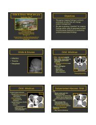
CNS Trauma: Radiologic-Pathologic CorrelationJames G. Smirniotopoulos, M.D.Professor of <strong>Radiology</strong>, Neurology,and Biomedical InformaticsChair, Department of <strong>Radiology</strong>Uniformed Services UniversityBethesda, MDUSUHS - Smirniotopouloshttp://rad.usuhs.mil
http://rad.usuhs.milUniformed Services UniversityBethesda, MD
CNS TRAUMAhttp://rad.usuhs.mil– IMPACT– CONTACT INJURY– scalp/skull Abnormal– INERTIAL• NON-CONTACT INJURY• acceleration/deceleration• scalp/skull Normal
Types of Injuryhttp://rad.usuhs.mil• Primary Lesions– Contusions– Shearing Injury• Secondary Lesions– Mass Effect– Increased ICP– Herniation– Infarction
Recreation Injuryhttp://rad.usuhs.mil<strong>Radiology</strong>Resident
Roller Coaster Headachehttp://rad.usuhs.mil• Roller Coasters can create 2.5 – 3 G’s• Grandpa rides with Grandaughter– She’s screaming with excitement– He’s subdued by <strong>Subdural</strong>• Reference:Fukutake T, Mine S, Yamakami I, Yamaura A, Hattori T.Roller coaster headache and subdural hematoma.Neurology. 2000 Jan 11;54(1):264.PMID: 10636168; UI: 20100123
http://rad.usuhs.milWorkplace Injury<strong>Radiology</strong> Attending
Relative Sensitivityhttp://rad.usuhs.mil• MR Spectroscopy (decreased NAA)• Magnetization Transfer Ratio• Apparent Diffusion Coefficient• Diffusion Weighted Imaging• FLAIR• Convention SE MR (T2W > T1W)• CT (ECT > NCT)• Skull Radiogram
Reasons for Getting an MRhttp://rad.usuhs.mil• CT fails to explain Pt’s Condition• CT fails to explain Pt’s Condition• CT fails to explain Pt’s Condition• CT fails to explain Pt’s Condition• CT fails to explain Pt’s Condition
CENTRIPETAL APPROACHhttp://rad.usuhs.mil– SCALP– CALVARIUM– EPIDURAL– SUBDURAL– SUBARACHNOID– INTRA-PARENCHYMAL– INTRA-VENTRICULAR
CNS TRAUMA --SUBGALEALhttp://rad.usuhs.mil• Between periosteum of OUTER table andthe GALEA (under scalp fat)• In CHILD, significant blood loss• Spontaneous decompression of intracranial(Epidural) hematoma
Subgaleal <strong>Hematoma</strong>http://rad.usuhs.mil
http://rad.usuhs.mil
Linear Skull Fxhttp://rad.usuhs.mil
Depressed Skull Fxhttp://rad.usuhs.mil
Depressed Skull Fxhttp://rad.usuhs.mil
Ballpeen Hammerhttp://rad.usuhs.mil
http://rad.usuhs.mil
MEMBRANE HEMATOMAS:http://rad.usuhs.mil– SUBGALEAL– SUBPERIOSTEAL OUTER TABLE• (CEPHALOHEMATOMA)– SUBPERIOSTEAL INNER TABLE• EPI (EXTRA) DURAL– <strong>Subdural</strong> (‘epi-arachnoid’)– SUBARACHNOID– PARENCHYMAL HEMATOMA– INTRA-VENTRICULAR
CNS TRAUMA EPIDURALHEMATOMAhttp://rad.usuhs.mil• Young men (20-40's)• Acute presentation• Skull fracture (90%)• Bi-convex, hyperdense- limited by sutures
EPIDURAL HEMATOMA -Source of Bleedinghttp://rad.usuhs.mil• MENINGEAL VESSELS• Arterial (high pressure)• Venous (low pressure)• DURAL SINUS• OTHER• High flow, low pressure• Diploic veins (Fx)• Marrow sinusoids
EPIDURAL HEMATOMAhttp://rad.usuhs.mil– Significant trauma– Fracture & concussion (l.o.c.)– Wakes up (lucid interval - 40% pts.)– Delayed neurologic Sx (hrs. Later)– Herniation, coma and death
EPIDURALHEMATOMAhttp://rad.usuhs.mil– Trauma -> fracture & concussion– Tearing/stripping of both layersfrom inner table– Laceration of outer periosteallayer– Laceration of meningeal vessels– Inner (meningeal dura) intact– Blood between naked bone anddura
Epidural <strong>Hematoma</strong>http://rad.usuhs.mil
Epidural <strong>Hematoma</strong>http://rad.usuhs.mil
Epidural <strong>Hematoma</strong>http://rad.usuhs.mil
http://rad.usuhs.mil
Epidural <strong>Hematoma</strong>http://rad.usuhs.mil
Epidural<strong>Hematoma</strong>http://rad.usuhs.mil
Epidural <strong>Hematoma</strong>http://rad.usuhs.mil
Progressive EDH10 AM8 PM
SUBPERIOSTEALHEMATOMAhttp://rad.usuhs.mil– CEPHALOHEMATOMA– (Birth trauma)– (outer table, sub-periosteal)– EPIDURAL HEMATOMA– (Inner table, "sub-periosteal")
http://rad.usuhs.milCephalohematomaBirth Trauma
SUBDURAL HEMATOMAhttp://rad.usuhs.mil• Extremes of age• Infants/elderly• Subacute presentation• Days or weeks after trauma• Fracture not needed• Crescentic• Not hyperdense• Crosses sutures commonly• Interhemispheric fissure (kids)• Epi - Arachnoid
SUBDURAL HEMATOMA -Source of Bloodhttp://rad.usuhs.mil• LACERATION OF CORTICAL AA. AND VV• (Direct: penetrating injury)• LARGE CONTUSIONS• (Direct/indirect: "pulped brain")• TORN BRIDGING (CORTICAL) VEINS• (Indirect: acceleration-deceleration)
<strong>Subdural</strong> <strong>Hematoma</strong>http://rad.usuhs.mil
SUBDURAL HEMATOMAhttp://rad.usuhs.mil• ACCELERATION-DECELERATION– Causes OSCILLATION OF BRAIN– Movement of Brain LAGS behind Skull• BRIDGING VEINS STRETCH & TEAR• Venous bleeding (slow)• DISSECTION OF SUBDURAL SPACE• Under Dura - Over Arachnoid• <strong>Hematoma</strong> spreads around convexity• Into the interhemispheric fissure (child)
Bridging Veins - Slackhttp://rad.usuhs.mil
Bridging Veins - Tensionhttp://rad.usuhs.mil
Bridging Veins - Tensionhttp://rad.usuhs.mil
Bridging Veins - Tearhttp://rad.usuhs.mil
<strong>Subdural</strong> <strong>Hematoma</strong>http://rad.usuhs.mil
<strong>Subdural</strong> <strong>Hematoma</strong>http://rad.usuhs.mil
Hyperdense SDH
<strong>Subdural</strong> <strong>Hematoma</strong>http://rad.usuhs.mil
<strong>Subdural</strong> <strong>Hematoma</strong>http://rad.usuhs.mil
SUBDURAL HEMATOMAhttp://rad.usuhs.mil• ACUTE (0-7 days)• HYPERDENSE (65-90 Hu)• SUBACUTE (7-22 days)• ISODENSE (20-40 Hu)• CHRONIC (>22 days)• HYPODENSE (0-20 Hu)
Hemorrhage: change over timeHyperdenseAcute BloodIsodenseBrainHypodense1 wk 3 wksAcute Subacute Chronic
Isodense subduralSulci
Hypodense SDHhttp://rad.usuhs.mil
SUBDURAL COLLECTIONShttp://rad.usuhs.mil• Acute SDH - hyperdense• Subacute SDH - isodense• Chronic SDH - hypodense• Hygromas• Hypodense, isointense to CSF• CSF leak from arachnoid tears• Effusions• Hypodense(meningo-encephalitis, esp. H.flu)
<strong>Subdural</strong> <strong>Hematoma</strong>http://rad.usuhs.mil
Shaking Injuryhttp://rad.usuhs.mil
Interhemispheric <strong>Subdural</strong>http://rad.usuhs.mil
Interhemispheric <strong>Subdural</strong>http://rad.usuhs.mil
INTERHEMISPHERICFISSURE and the FALXSIGNhttp://rad.usuhs.mil• Normal Falx:• thin white line, may see CSF parallel• Subarachnoid Blood:• anterior, zig-zag, reaches the corpus• <strong>Subdural</strong> <strong>Hematoma</strong>:• posterior, straight, doesn't touch thecorpus callosum
<strong>Subdural</strong> <strong>Hematoma</strong>http://rad.usuhs.mil
<strong>Subdural</strong> = Epi - Arachnoidhttp://rad.usuhs.mil
SUBDURAL HEMATOMAhttp://rad.usuhs.mil• 2-3 wks. to develop fully• develop from outer (dural) margin• not from arachnoid side– inner (arachnoid) border intact• fibroblasts, and new immature capillariesthat are fragile
Neomembrane from Durahttp://rad.usuhs.mil
SUBDURAL HEMATOMA -Source of Re-bleeding• NEO-MEMBRANES– fragile capillarieshttp://rad.usuhs.mil• BRIDGING VEINS– stretching across SDH– constant tension
Chronic Loculated SDHhttp://rad.usuhs.mil
Acute-subacute<strong>Subdural</strong> <strong>Hematoma</strong>• Acute blood-bright• Alternating bands– rebleeding• Mass effect– Subfalcial herniation– “Trapped” ventricle
<strong>Subdural</strong> <strong>Hematoma</strong>http://rad.usuhs.mil
<strong>Subdural</strong> <strong>Hematoma</strong>http://rad.usuhs.mil• Under the Dura– “Sub-Dural”• Over the Arachnoid– “Epi-Arachnoid”• Actually between the “Dural Border Cells”the “Arachnoid Barrier Layer”Schachenmayer and Friede, Am J Path (1978); 92: 53 - 68
CEREBRAL CONTUSIONhttp://rad.usuhs.mil• Traumatic/mechanical disruption of small(capillary) vessels• Extravasation of whole blood, plasma(edema) and RBC's• Admixture of blood mixed with nativetissue (petechial hemorrhage)• Mottled / speckled density ("salt andpepper" on CT)
CEREBRAL CONTUSION(naming conventions)http://rad.usuhs.mil– COUP (SAME SIDE AS IMPACT)– (w/fractures, small area of impact)– INTERMEDIATE (CENTRAL)– (DAI/Shearing Injury)– CONTRE - COUP (OPPOSITE IMPACT)– (w/falls, broad surface of impact)
Coup vs. Contrecouphttp://rad.usuhs.mil
Coup vs. Contrecouphttp://rad.usuhs.mil
CEREBRAL CONTUSIONhttp://rad.usuhs.mil• MECHANICAL INJURY TO VESSELS• Extravasation of whole blood• PETECHIAL / PERIVASCULARHEMORRHAGE• Admixture of tissue and blood• average density may NOT be high• SWELLING/MASS EFFECT• MAY PROGRESS TO HEMATOMA• If larger vessels are damaged• large confluent mass of blood
http://rad.usuhs.milTemporal TipsOrbitofrontal Gyri
Cerebral Cortical Contusionhttp://rad.usuhs.mil•Crowns of Gyri•Linear or flame shape•NOT in depths of Sulci
Cerebral Contusionhttp://rad.usuhs.mil
Cerebral Contusionhttp://rad.usuhs.mil
CEREBRAL CONTUSIONhttp://rad.usuhs.mil• CT Hypodense (EDEMA)• Isodense (mass)• Hyperdense(mottled, speckled)• MR Variable Intensity– (GRE) - Hypointense– Hyperacute Blood• COUP CONTUSION– ASSOCIATED w/Fx
Cerebral Contusionhttp://rad.usuhs.mil
Deep Lesions - Terminologyhttp://rad.usuhs.mil• Intermediate Contusions• Shearing Injury• Diffuse White-matter Injury (DWI)• Diffuse Axonal Injury (DAI)
Mechanism of Axon Injuryhttp://rad.usuhs.mil
SHEARING INJURIEShttp://rad.usuhs.mil– Deep lesions– High Velocity (MVA) Trauma– Acceleration/Deceleration– Do not require an impact or Fx– SHEARING OF AXONS• Breaks connections• Actual force may be tension– SHEARING of Small WM VESSELS– Small (petechial) hemorrhages
Deep Lesionshttp://rad.usuhs.mil• Subcortical and Hemispheric WM• Corpus Callosum– posterior body– splenium• Brain stem– Dorsolateral Quadrant of Upper BS– Deep BS– Ventral BS
DIFFUSE AXONAL INJURYhttp://rad.usuhs.mil– Immediate L.O.C. Persistent VegetativeState– Pathology:– foci of hemorrhage in callosum,brainstem, etc.– axon retraction balls (ARB)– Long-Term Survivors:– microglial clusters– foci of demyelination
http://rad.usuhs.mil
http://rad.usuhs.mil
http://rad.usuhs.mil
http://rad.usuhs.mil
Pontomedullary Tearhttp://rad.usuhs.mil
Brainstem Hemorrhagehttp://rad.usuhs.mil
Dorsolateral Brainstemhttp://rad.usuhs.mil
CT vs. MR (GRE)http://rad.usuhs.mil
Corpus Callosum -> Ventriclehttp://rad.usuhs.mil
Corpus Callosum and BGhttp://rad.usuhs.mil
T2W vs.. GRE (Gradient Recalled Echo)http://rad.usuhs.milT2WGRE
http://rad.usuhs.mil
http://rad.usuhs.milShearing Injury - DeepLesions
Shearing Injury vs. Contusionhttp://rad.usuhs.mil
CNS TRAUMA - Summaryhttp://rad.usuhs.mil– Epidural <strong>Hematoma</strong> (subperiosteal)• acute, convex, white– <strong>Subdural</strong> hematoma (epi-arachnoid)• variable shape, density, age– Contusion (petechial)• Surface - cortex. coup/contra• Dark on GRE - CT "speckled"– Shearing injury (DAI)• Deep - subcortical WM, corpuscallosum and & basal ganglia
The EndMil Gracias !USUHS - Smirniotopoulos
Trauma Quizhttp://rad.usuhs.mil• 1. This image is most consistent with:– A. Acute SDH– B. Chronic SDH– C. Rebleeding SDH– D. Epidural– E. Shearing injury
Trauma Quizhttp://rad.usuhs.mil• 2. Deep lesions like this are due to:– A. Contusions– B. <strong>Hematoma</strong>s– C. Shearing– D. Both A and B– E. A, B, and C
Trauma Quizhttp://rad.usuhs.mil• Which of the following are TRUE?• 3. This is a depressed skull Fx• 4. This is a pattern injury• 5. The skull Fx in Compression
Trauma Quizhttp://rad.usuhs.mil
Trauma Quizhttp://rad.usuhs.mil
Trauma Quizhttp://rad.usuhs.mil
http://rad.usuhs.mil
http://rad.usuhs.mil
http://rad.usuhs.mil
Slides to Addhttp://rad.usuhs.mil• Penetrating Trauma• Ax Slides• Eyeball Slide• Golf Club Slide• Spaghetti Slide• SDH vs. SAH Coronal MRI