ENZ-51002 User Manual - Enzo Life Sciences
ENZ-51002 User Manual - Enzo Life Sciences
ENZ-51002 User Manual - Enzo Life Sciences
- No tags were found...

You also want an ePaper? Increase the reach of your titles
YUMPU automatically turns print PDFs into web optimized ePapers that Google loves.
I. IntroductionThe transition from apoptosis to necrosis is a loosely defined continuumthat necessitates recognition of the various stages of the process.The display of phosphatidylserine (PS) on the extracellular face of theplasma membrane remains a unifying hallmark of early apoptosis.Phospholipid-binding proteins such as Annexin V bind with a highaffinity to PS, in the presence of Ca 2+ . Given that Annexin V is not cellpermeable, the binding of externalized PS is selective for earlyapoptotic cells. Similarly, loss of plasma membrane integrity, asdemonstrated by the ability of a membrane-impermeable DNA intercalatingdye to label the nucleus, represents a straightforward approachto demonstrate late stage apoptosis and necrosis.Considering that Annexin V is commonly conjugated to fluorescein(a.k.a. FITC), apoptosis detection in green fluorescent protein (GFP)-expressing cells can be problematic. The spectral overlap betweenthe emission profiles of these two fluorophores makes differentiationbetween signals difficult or impossible, thus compromising data qualityoverall. While useful for analysis of apoptosis and necrosis in cellsthat do not express any fluorescent proteins, the GFP-CertifiedApoptosis/Necrosis Detection Kit was specifically designed for usewith GFP-expressing cell lines and cells expressing blue or cyan fluorescentproteins (BFPs, CFPs). Additionally, the kit is suitable for usewith live or post-fixed cells in conjunction with probes, such as labeledantibodies, or other fluorescent conjugates displaying similar spectralproperties as fluorescein or coumarin.Each GFP-Certified Apoptosis/Necrosis Detection Kit includes all thenecessary reagents for determination of early and late stages of apoptosisas well as necrosis. An Annexin V-<strong>Enzo</strong>Gold (enhanced Cyanine-3) conjugate enables detection of apoptosis distinct from fluoresceinor GFP. The Necrosis Detection Reagent (Red) similar to the redemittingdye 7-AAD, facilitates late apoptosis and necrosis detection.This kit also includes an Apoptosis Inducer (Staurosporine) for use asa positive control. Detection reagents can be used separately or incombination for either single or multiplexed applications respectively.This kit also provides quick and accurate results via flow cytometryand fluorescence/confocal microscopy.1
II.Reagents Provided and StorageAll reagents are shipped on wet ice. Upon receipt, store all reagents at4°C, protected from light. The reconstituted Apoptosis Inducer should bestored at –20°C.Reagent 100 Reactions 25 ReactionsApoptosis Detection Reagent(Annexin V-<strong>Enzo</strong>Gold)550 µL 140 µLNecrosis Detection Reagent (Red) 600 µL 150 µLApoptosis Inducer (Staurosporine) 50 nmoles 50 nmolesBinding Buffer (10X) 6 mL 1.5 mLIII. Additional Materials Required Standard fluorescence microscope or a flow cytometer that is equippedwith a 488 nm laser for fluorescent dye excitation Phosphate buffered saline (PBS) Calibrated, adjustable precision pipetters, preferably with disposableplastic tips 5 ml tubes appropriate for holding cells during induction of apoptosisand necrosis Adjustable speed centrifuge with swinging buckets Glass microscope slides (for microscope analysis only) Glass cover slips (for microscope analysis only) Deionized water Anhydrous DMSOIV. Safety Warnings and Precautions This product is for research use only and is not intended for diagnosticpurposes. Some components of this kit contain hazardous substances. Reagentscan be harmful if ingested or absorbed through the skin and may causeirritation to the eyes. Reagents should be treated as possible mutagensand should be handled with care and disposed of properly. Observe good laboratory practices. Gloves, lab coat, and protectiveeyewear should always be worn. Never pipet by mouth. Do not eat,drink or smoke in the laboratory areas. All blood components andbiological materials should be treated as potentially hazardous andhandled as such. They should be disposed of in accordance withestablished safety procedures. To avoid photobleaching, perform all manipulations in low light environments,in amber microcentrifuge tubes or protected from light byother means.2
V. Methods and ProceduresA. REAGENT PREPARATION1. Positive ControlThe Apoptosis Inducer (Staurosporine) is supplied as a lyophilizedpowder (50 nmoles) and should be reconstituted in 50 L DMSOfor a 1 mM stock solution. It is recommended that induction occurat the effective concentration (EC 50 ) and that the final percentDMSO in the assay not exceed 0.2%.2. Cell PreparationCells should be maintained via standard tissue culture practices.Positive Control cells should be pretreated with the ApoptosisInducer for 1-16 hours. Induction of apoptosis and necrosis istime and concentration dependent and may also vary significantlydepending among cell types and cell lines. Post treatment cellsshould be harvested and diluted to a working concentration of2.0x10 5 /mL in media. Negative control cells should be treatedwith vehicle (DMSO, media or other solvent used to reconstituteor dilute an inducer or inhibitor) for an equal length of time undersimilar conditions.3. 1X Binding BufferGenerate 500 L of 1X Binding Buffer per sample by diluting50 L of 10X Binding Buffer into 450 L of deionized water. Batchpreparation is recommended for large numbers of samples.B. DUAL DETECTION REAGENT PREPARATIONPrepare the Dual Detection Reagent according to Table 1. Thevolumes recommended in the table are sufficient for one assayand must be scaled accordingly.Table 1. Dual Detection ReagentReagentAmount1X Binding Buffer 500 µLApoptosis Detection Reagent(Annexin V-<strong>Enzo</strong>Gold)5 µLNecrosis Detection Reagent (Red) 5 µLTotal Volume 510 µL3
C. FLOW CYTOMETRY PROTOCOL (Non-Adherent Cells)1. The recommended sample size should be between 2 x 10 5 cells/sample and 1 x 10 6 cells/sample during staining and analysis.2. Treat cells with compound of interest and Negative Control cellswith vehicle.3. Centrifuge cells for 5 minutes at 400 x g at room temperature(RT).4. Carefully remove supernatant by aspiration and gently resuspendand wash the pellet in 5.0 mL cold PBS.5. Centrifuge cells for 5 minutes at 400 x g at room temperature(RT).6. Carefully remove supernatant and gently resuspend the pellet in500 L Dual Detection Reagent. (see Table 1 on section B. p3)7. Protect samples form light and incubate for 15 minutes at roomtemperature.8. Samples should be analyzed via flow cytometry using a 488 nmlaser with the FL 1 channel for GFP or FITC detection, the FL 2channel for the Apoptosis Detection Reagent and the FL 3channel for the Necrosis Detection Reagent. It is recommendedthat compensation corrections be performed using single stainedinduced cells to avoid any overlap between the Apoptosis DetectionReagent and Necrosis Detection Reagent (Red) fluorescentsignals.9. Positive Control Samples: It is recommended that positive controlsamples be pretreated with the Apoptosis Inducer (Staurosporine)at a final concentration of 2 M for 4 hours. Apoptosis may varysignificantly and is dependent upon the length of induction,inducer concentration, cell types, cell lines, inducer concentrationand exposure time. Follow steps 2-8 post treatment.Suggested Compensation Controls for Flow Cytometry:a. Unstained cellsb. Cells stained with Apoptosis Detection Reagent (withoutNecrosis Detection Reagent)c. Cells Stained with Necrosis Detection Reagent (withoutApoptosis Detection Reagent)Note: Samples and controls should be kept on ice before theassay is run and analyzed via flow cytometry within onehour of staining.4
E. WIDE FIELD FLUORESCENCE/CONFOCALMICROSCOPY1. Grow cells directly onto glass slides or polystyrene tissue cultureplates until ~80% confluent.2. Treat cells with compound of interest and Negative Control cellswith vehicle.3. Carefully wash cells twice with 1x PBS in a volume sufficient forcovering the cell monolayer.4. Carefully remove supernatant and dispense the Dual DetectionReagent in a volume sufficient for covering the cell monolayer (asper section B)5. Protect samples from light and incubate for 15 minutes at roomtemperature.6. Flick the staining solution onto a paper towel and if necessary adda few drops of Binding Buffer to prevent the cells for drying out.7. Cover cells and observe under a fluorescence/confocal microscopewith a dual filter set for Cyanine-3 (Ex/Em: 550/570 nm)and 7-AAD (Ex/Em: 546/647 nm) and a GFP/FITC (Ex/Em:488/514 nm) filter should be used if either is imaged simultaneously.8. Positive Control Samples: It is recommended that Positive Controlsamples be pretreated with the Apoptosis Inducer at a finalconcentration of 2 M for 4 hours and may vary significantlyamong cell types and cell lines. Follow steps 3-7 post treatment.VI. APPENDICESA. FILTER SELECTIONThe selection of optimal filter sets for a fluorescence microscopyapplication requires matching the optical filter specifications to thespectral characteristics of the dyes employed in the analysis. Pleaseconsult your microscope or filter set manufacturer for assistance inselecting optimal filter sets for your microscope.B. CELL FIXATION IMPORTANT! The fixation process can disrupt cellularmembranes and should be performed following incubation with theDetection Reagent. 2% formaldehyde is the recommended fixative. Standard immunofluorescence staining protocols using afluorescein- or coumarin-based antibody conjugates, or equivalentshould be administered post-fixation according to manufacturerinstructions.6
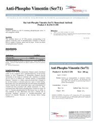
Figure 1B: AnV-8Relative Cell Number: Necrosis DetectionAnV-<strong>Enzo</strong>Gold: AnV-AnV-<strong>Enzo</strong>GoldRelative Cell Num-: Necrosis DetectionFigure 1A
VIII. Troubleshooting GuideProblem Potential Cause SuggestionsLittle or noapoptosisobserved ordetectedNo or few apoptoticcells present insampleIncorrect buffer usedduring sample processingUse the Apoptosis Inducer, providedin the kit, as a positive control to induceapoptosis and optimize the Inducerconcentration and time oftreatment.Calcium and magnesium ions arerequired for binding of the ApoptosisDetection Reagent to PS. Binding isreversible, so divalent cations mustbe present throughout the entire assay.Use the Binding Buffer provided inthe kit.Completely remove wash buffer byaspiration as to not dilute the bindingbuffer or inducer.Assay is inappropriatefor the cells or cellline used.Some cells (i.e., megakaryocytes,platelets, some myeloid lineage cells)are known to display large amountsof PS on their cell surfaces. PeripheralBlood Mononuclear Cells(PBMCs) coated with platelets, therefore,may be positive for the ApoptosisDetection Reagent. Use a differentmethod to monitor apoptosis.Significantapoptosisdetected innegative controlcells.Cells were damagedor permeabilizedduring the assayAssay measurementor analysis wasdelayed.Permeable cells (advanced apoptoticor necrotic), yield an increase inapoptosis detection via cytoplasmicleaflet of the plasma membrane.Trypsinization or scraping or disaggregationof adherent cells may resultin false positives due to cell damage,arising from disruption of the PScontainingplasma membrane. Usegentler cell preparation methods andoptimize the Inducer concentrationand time of treatment.Analysis must be performed quickly,following dye labeling in order toavoid post-label apoptosis. Stainedsamples should be kept on ice untilan assay is run.9
Problem Potential Cause SuggestionsSmall apoptoticpopulationdetected andhigh necroticpopulationdetectedCells are no longerapoptotic and haveprogressed towardsnecroticHigh doses of an agent can oftenlead to necrosis, while lower dosesof the same agent may lead toapoptosis. Often times, apoptosisand necrosis simply represent twoextremes of biochemically overlappingcell death pathways. Lower thedose of the test agent.High variabilityof results observedbetweenand amongsamples on thesame day or indifferent experimentsfrom dayto dayInconsistencies in celltreatment, labelingand FCM data analysiswere introduced.All reagents should be prepared inbatches.An untreated negative control and ifpossible an independent positivecontrol should be included. Propercontrols should be included in eachexperiment and gating of samplesshould be performed uniformlyespecially the gating out of debris.Spectral emissionoverlap orsignal bleedamong differentchannels oremission filtersis observedNo or incorrectcompensation wasperformed for flowcytometryInappropriate filter setused with fluorescencemicroscopyUse correct compensation controlsfor flow cytometry and set up spectralcompensation according to themanufacturer’s instructions.Use correct filters for fluorophoresbeing measured. Refer to the spectralchart in this manual.Decrease band width of theemission filters to prevent spectraloverlap.10