New Frontiers in Laser Surgery - Alma Lasers
New Frontiers in Laser Surgery - Alma Lasers
New Frontiers in Laser Surgery - Alma Lasers

Create successful ePaper yourself
Turn your PDF publications into a flip-book with our unique Google optimized e-Paper software.
<strong>New</strong> <strong>Frontiers</strong> <strong>in</strong> <strong>Laser</strong> <strong>Surgery</strong>Doru T. Alexandrescu, MD,* and Edward V. Ross, MD †The simultaneous advances <strong>in</strong> eng<strong>in</strong>eer<strong>in</strong>g, medic<strong>in</strong>e, and molecular biology have acceleratedthe pace of <strong>in</strong>troductions of new light-based technologies <strong>in</strong> dermatology. In thisreview, the authors exam<strong>in</strong>e recent advances <strong>in</strong> laser surgery as well as peer <strong>in</strong>to the futureof energy-based cutaneous medic<strong>in</strong>e. The future landscape of dermatology will almostundoubtedly <strong>in</strong>clude (1) non<strong>in</strong>vasive imag<strong>in</strong>g technologies and (2) improved “destructive”modalities based on real-time feedback from the sk<strong>in</strong> surface.Sem<strong>in</strong> Cutan Med Surg 31:88-97 © 2012 Elsevier Inc. All rights reserved.KEYWORDS laser, novel, sk<strong>in</strong>, applications, technology, light, radiofrequencyMost commercially viable laser and light-based technologiesstart out <strong>in</strong> the “lab” <strong>in</strong>cubator. After ref<strong>in</strong>ementand determ<strong>in</strong>ation of “practicality,” novel lasers and accessoriesmake their way to the exhibitors’ booths and f<strong>in</strong>ally theprocedure room. What many cl<strong>in</strong>icians do not realize is thatfew laser <strong>in</strong>novations that seem reasonably effective and safe<strong>in</strong> the laboratory are commercialized. Some <strong>in</strong>novations arecost prohibitive. In other cases, the translation from bench tobedside is derailed by chang<strong>in</strong>g cl<strong>in</strong>ical needs or supplantedby newer “even better” technologies. In this brief review, wepresent a potpourri of already established recent devices andpr<strong>in</strong>ciples that might be <strong>in</strong>tegrated <strong>in</strong>to future products.In identify<strong>in</strong>g new frontiers <strong>in</strong> cutaneous laser surgery, theauthors reviewed recently submitted abstracts at nationalmeet<strong>in</strong>gs, exam<strong>in</strong>ed their own research projects, and lookedahead at promis<strong>in</strong>g potential applications that might enhanceour laser arsenal <strong>in</strong> the future.Alexandrite <strong>Laser</strong>for Vascular Lesions*Division of Dermatology, San Diego Medical Center, University of California,San Diego, CA.†Division of Dermatology, Scripps Cl<strong>in</strong>ic Carmel Valley, San Diego, CA.Conflict of Interest Disclosures: Dr Ross has received honoraria, consult<strong>in</strong>gfees, and research support from Palomar Medical Technologies, Syneron-CandelaCorporation, Cutera, Lumenis, and <strong>Alma</strong> <strong>Laser</strong>s. Dr Alexandrescuhas no conflicts to report.Address repr<strong>in</strong>t requests to Edward V. Ross, MD, Scripps Health, 3811Valley Centre Dr S99, San Diego, CA 92130. E-mail: Ross.Edward@scrippshealth.orgDevelopment of the pulsed dye laser (PDL) created a betterapproach for the treatment of port w<strong>in</strong>e sta<strong>in</strong>s (PWS) and avariety of other vascular lesions, but it has significant limitations.PWS seldom clear completely, and deeper lesions haveproved difficult to clear. One recent advance <strong>in</strong> the vasculararena is the newfound use of the alexandrite laser for “medium”-depthlesions (ie, venous lakes), PWS, and even telangiectasia.As this laser is already part of many providers’ laserl<strong>in</strong>eups, its expanded use is logical. The alexandrite lasershows hemoglob<strong>in</strong> (HgB) absorption about 2 times that ofthe neodymium-doped yttrium alum<strong>in</strong>um garnet (Nd:YAG)laser and melan<strong>in</strong> absorption about 3 times that of the Nd:YAG laser. Water absorption is less compared with Nd:YAGlaser; however, the laser’s optical penetration depth is lessthan the Nd:YAG laser <strong>in</strong> bloodless dermis (about one-half).We have noted that the alexandrite laser performs well <strong>in</strong>pigmented and vascular lesions. 1,2 One concern is the potentialfor scarr<strong>in</strong>g, particularly when the laser is used <strong>in</strong> darkersk<strong>in</strong> or with <strong>in</strong>adequate cool<strong>in</strong>g. Because of deeper penetration,the laser creates bulk heat<strong>in</strong>g that might require secondsto cool (like the Nd:YAG laser). On the other hand, despitethe lower vascular to melan<strong>in</strong> absorption ratio vs the Nd:YAGlaser, we have overall found the alexandrite laser less likely tocause ulcers and atrophic/hypertrophic scar than its1064-nm counterpart. Part of these less destructive effectsmight be related to its lower arteriole to venous blood absorptionratio versus the Nd:YAG laser. 3 Kamel 4 reported his useof a long-pulse alexandrite laser to treat bulky vascular malformations.He treated 14 patients with resistant PWS on theface. The alexandrite laser was applied with a fluence of50-70 J/cm 2 , 1.5 ms, and 8-mm spot diameter with the dynamiccool<strong>in</strong>g device. Two cases of scars were observed <strong>in</strong>patients after 1 session. Mild-to-moderate PWS lighten<strong>in</strong>gwas associated <strong>in</strong> 6 patients and hyperpigmentation <strong>in</strong> 1 case.The rema<strong>in</strong>der of the patients’ PWS was largely unchanged.Alexandrite lasers have been used for the treatment ofsmall venous malformations. 5 Venous vascular malforma-88 1085-5629/12/$-see front matter © 2012 Elsevier Inc. All rights reserved.http://dx.doi.org/10.1016/j.sder.2012.04.002
90 D.T. Alexandrescu and E.V. Rossachieved for vessels 0.4-1.6 mm <strong>in</strong> diameter. A limitation oftreatment of telangiectasia with this laser consists of its biastoward melan<strong>in</strong> over vascular heat<strong>in</strong>g.Selective <strong>Laser</strong>Target<strong>in</strong>g of Sk<strong>in</strong> CancersTheoretic advantages of us<strong>in</strong>g lasers <strong>in</strong> sk<strong>in</strong> cancer consist ofthe follow<strong>in</strong>g: less <strong>in</strong>vasiveness, use <strong>in</strong> locations where surgerycan result <strong>in</strong> anatomic distortion or functional impairment,potential for an excellent cosmetic result, and the possibilityof comb<strong>in</strong><strong>in</strong>g targeted laser irradiation or us<strong>in</strong>g it asan adjunct to photodynamic therapy (PDT) <strong>in</strong> patients athigh risk of develop<strong>in</strong>g sk<strong>in</strong> cancers. 12The use of the 585-nm PDL for basal cell carc<strong>in</strong>omas(BCCs) was driven by their histology, as many BCCs have afeed<strong>in</strong>g microvasculature <strong>in</strong> the shape of a basketlike capillaryplexus consist<strong>in</strong>g of vessels with a lum<strong>in</strong>al diameter of 20m or more, 13 correspond<strong>in</strong>g on the surface to the visibletelangiectasia. PDL pulsed at 0.45-1.5 ms represents the standardtreatment for PWS, telangiectasia, and hemangiomas.14-16As the microvasculature plays an important role <strong>in</strong> thenutrient support and BCC tumorigenesis, 17 target<strong>in</strong>g the vascularsupply of this tumor is analogous to that obta<strong>in</strong>ed byselective vascular microembolization, a practice that is common<strong>in</strong> treat<strong>in</strong>g various tumors. 18 Destruction of the bloodvessels with<strong>in</strong> the BCC structure is made on the pr<strong>in</strong>ciple ofselective photothermolysis, analogous to the mechanismused by PDL <strong>in</strong> vascular lesions. Correlat<strong>in</strong>g with fluence,PDL penetrance <strong>in</strong>to sk<strong>in</strong> varies from 0.7 to 1.3 mm, which is<strong>in</strong> the range of the vasculature that feeds a BCC. 19In a recent study by Shah et al, 20 20 biopsy-proven BCCswere treated with 4 595-nm PDL treatments (energy of 15J/cm 2 , 3 ms, 7-mm spot size with 10% overlap of pulses, andno cool<strong>in</strong>g). Of these, 91.7% (11/12) of BCCs smaller than1.5 cm <strong>in</strong> diameter had a complete response (P .0003),compared with only 16.7% of controls. BCCs larger than 1.5cm <strong>in</strong> diameter showed a complete response rate of 25%,compared with 0% <strong>in</strong> controls (P .2). Tumor histologictypes of the complete responders <strong>in</strong>cluded superficial, nodular,micronodular, and kerat<strong>in</strong>iz<strong>in</strong>g. Incompletely respond<strong>in</strong>gBCCs still demonstrated a significant reduction <strong>in</strong> tumorburden after PDL treatment, with residual tumor burden onhistologic exam<strong>in</strong>ation rang<strong>in</strong>g from 1% to 29% of theorig<strong>in</strong>al cl<strong>in</strong>ical tumor size, compared with 13%-68% residualtumor burden for the correspond<strong>in</strong>g controls (P .05).None of the patients developed tumor ulceration or scarr<strong>in</strong>gafter treatment. The most frequent reaction to treatment waspurpura followed by a gray discoloration of the sk<strong>in</strong>, <strong>in</strong>frequentlyassociat<strong>in</strong>g hemorrhagic scal<strong>in</strong>g and crust<strong>in</strong>g, butwith no deep sk<strong>in</strong> damage. Smaller BCCs may respond betterthan the larger lesions secondary to a more-fragile vasculaturethat is more immature and, therefore, more susceptibleto destruction by laser. The conclusion of this study was thatPDL is an effective means of reduc<strong>in</strong>g tumor burden <strong>in</strong> patientswith large BCCs and may be considered an alternativetherapy <strong>in</strong> BCCs 1.5 cm <strong>in</strong> diameter.The alexandrite laser has also been used for nonmelanomask<strong>in</strong> cancer, where high fluences (100 J/cm 2 ) and an 8-mmspot were applied to “bulk” heat the tissue. Biopsies showedthat vascular structures served as the <strong>in</strong>itial targets fromwhich heat diffused to surround<strong>in</strong>g localized micronodulesof BCC. 21<strong>New</strong> Topics <strong>in</strong>Photodynamic TherapyAn attempt to determ<strong>in</strong>e whether PDL or IPL can activateprotoporphyr<strong>in</strong> IX (PpIX) <strong>in</strong> the absence of ambient light andto compare it with the standard therapeutic CW light at adose lower than that required for cl<strong>in</strong>ical AK was made byStrasswimer and Grande. 22 In this study, IPL and flashlamppumpedPDL were capable of activat<strong>in</strong>g PpIX, but pulsedlight <strong>in</strong>duced a lesser reaction than a standard CW blue-lightsource.The efficacy and safety of PDL as the light source for photoactivationof PpIX to treat Bowen’s disease us<strong>in</strong>g PDT wereassessed by Britton et al 23 <strong>in</strong> 17 lesions of Bowen’s disease.Treatment was performed with 20% 5-am<strong>in</strong>olevul<strong>in</strong>ic acid(ALA) under occlusion for 4 hours followed by irradiationwith a 585-nm PDL Candela SPLTL-1b device (Candela Corporation,Wayland, MA) with a 7-mm diameter spot at afluence of 10 J/cm 2 . At 2 months, 8 treatment sites could notbe assessed because of loose overly<strong>in</strong>g crusts; removal ofthese revealed superficial erosions <strong>in</strong> 7 patients. Of the 17lesions treated, on follow-up at 1 year, 14 patches (82%)demonstrated complete cl<strong>in</strong>ical response, although 1 had requireda second treatment. In addition, 2 patients with 3lesions that needed further therapy refused a second treatment.Prolonged crust<strong>in</strong>g last<strong>in</strong>g 8 weeks occurred <strong>in</strong> 8patches, and prolonged discomfort last<strong>in</strong>g 6 weeks occurred<strong>in</strong> 4 patients. PDL appears to be an efficient light source forALA-PDT treatment of Bowen’s disease.CelluliteTreatment of cellulite has largely relied on various “wraps”and massage technologies, sometimes coupled with low-levellight and lasers. Results have been modest <strong>in</strong> most cases. Thepathophysiology of cellulite is unclear, and therapies haveaddressed lymphatic flow, strengthen<strong>in</strong>g of the dermis, fatdestruction, and lysis of fibrous septae. A recently <strong>in</strong>troduceddevice uses side-fir<strong>in</strong>g laser fibers to lyse fibrous bands thatare a contributor to cellulite. DiBernardo 24 evaluated the efficacy,safety, and duration of the cl<strong>in</strong>ical benefit associatedwith a s<strong>in</strong>gle-treatment pulsed laser that delivered 1440 nmenergy to the dermal–hypodermal <strong>in</strong>terface. Subjects receiveda s<strong>in</strong>gle treatment with a 1440-nm pulsed laser. Energywas delivered to the subdermal tissue through a fiberdesigned for targeted delivery of laser energy and enclosed <strong>in</strong>a cannula. Treatment addressed the th<strong>in</strong>n<strong>in</strong>g dermal layer,hypodermal fat lobules that extend upward <strong>in</strong>to the dermis,and fibrous septae by thermal subcision. Ultrasonography,
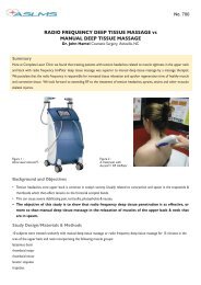
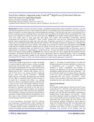
<strong>New</strong> frontiers <strong>in</strong> laser surgery 91sk<strong>in</strong> elasticity measurements, and photographs were conductedat basel<strong>in</strong>e, 1, 3, 6, and 12 months. Mean sk<strong>in</strong> thickness(as shown by ultrasonography) and sk<strong>in</strong> elasticityshowed (P .001) <strong>in</strong>creases at each time po<strong>in</strong>t. Subjectivephysician and subject evaluations <strong>in</strong>dicated improvement,high subject satisfaction, and m<strong>in</strong>imal adverse effects.<strong>Laser</strong> Lipolysis and Fat RemovalSelective fat destruction with laser has proven difficult,largely secondary to the depth of fat relative to the penetrationdepth of the laser. Three wavelengths, 915 nm, 1210nm, and 1720 nm, show favorable ratios of light absorptionrelative to water; however, the ratios are less than 1.5, suchthat selectivity is marg<strong>in</strong>al. Surface cool<strong>in</strong>g improves the likelihoodof achiev<strong>in</strong>g a temperature gradient between superficialwater (dermis) and subcutaneous fat. 25,26 Articles report<strong>in</strong>gfat destruction showed that long (several seconds)irradiation times are required for adequate fat heat<strong>in</strong>g; thelong heat<strong>in</strong>g times and <strong>in</strong>evitable heat<strong>in</strong>g of tissue water areassociated with considerable pa<strong>in</strong>. 27The Food and Drug Adm<strong>in</strong>istration approval <strong>in</strong> October2006 of a 1064-nm Nd:YAG laser lipolysis system (Deka,Cynosure, Westford, MA) triggered an <strong>in</strong>flux of manufacturersto market Nd:YAG and diode devices emitt<strong>in</strong>g laser radiationbetween 920 nm and 1320 nm, as advantages wereclaimed for each of the frequencies. A comb<strong>in</strong>ation of frequencies(980-nm diode and 1064-nm Nd:YAG laser) wasused by Mordon et al 28 <strong>in</strong> 2007 <strong>in</strong> a mathematical model thatsuggested for the first time that heat generated by the <strong>in</strong>teractionof laser light energy with the tissue rather than thebiophysical effects of a particular wavelength produced fatdestruction, which facilitated a focused approach <strong>in</strong> develop<strong>in</strong>gthe efficiency of lipolysis devices. Sk<strong>in</strong> tighten<strong>in</strong>g appearsto occur at dermal temperatures of 48°C-50°C without <strong>in</strong>duc<strong>in</strong>gburn<strong>in</strong>g <strong>in</strong>jury to the epidermis. Correspond<strong>in</strong>g externaltemperatures that balance safety and a good lipolyticeffect were found to be <strong>in</strong> the range of 38°C-41°C. However,a search for the optimal laser frequency <strong>in</strong> <strong>in</strong>duc<strong>in</strong>g lipolysisand/or sk<strong>in</strong> tighten<strong>in</strong>g, <strong>in</strong>clud<strong>in</strong>g 924, 968, 980, 1064, 1319,1320, 1344, and 1440 nm, has not identified a s<strong>in</strong>gle optimalfrequency that would achieve simultaneously both goals andwould satisfy both efficiency and safety criteria. For example,irradiation of fat tissue with 924 nm has the highest selectivityfor melt<strong>in</strong>g of adipose cells, but not as good of a sk<strong>in</strong>tighten<strong>in</strong>geffect. 29AcneDestruction of the sebaceous glands should deprive the acnepronefollicle of the fuel that feeds the acne fire. Like fat,sebum enjoys preferential light absorption at 915 nm, 1210nm, and 1720 nm. The more superficial location of theglands compared with the subcutaneous fat layer permits amore favorable scenario for selective heat<strong>in</strong>g. With optimizedparameters, selective sebaceous gland heat<strong>in</strong>g can beachieved. Future trials should uncover those sett<strong>in</strong>gs thatcreate a practical device for “sebum”-based acne clear<strong>in</strong>g.PDT <strong>in</strong> acne cont<strong>in</strong>ues to be improved. Drawbacks to therapy<strong>in</strong>clude long <strong>in</strong>cubation times and phototoxicity. Variousmethods have been used to decrease these side effects, manyof which are from epidermal PpIX formation. Cool<strong>in</strong>g theepidermis can suppress both s<strong>in</strong>glet O 2 formation as well assuppress the conversion of ALA to PpIX.Another way to suppress epidermal damage is to <strong>in</strong>hibitsurface fluorescence by photobleach<strong>in</strong>g the epidermis us<strong>in</strong>glow-power density blue light. In this way, PpIX is convertedto photoproducts. Sakamoto et al 30 <strong>in</strong>troduced this new conceptwith ALA to enhance the ratio of sebaceous gland toepidermal fluorescence and phototoxicity. They discoveredthat low-level light exposure dur<strong>in</strong>g the period of ALA metabolismprevents accumulation of porphyr<strong>in</strong>s and can beused to selectively <strong>in</strong>hibit epidermal porphyr<strong>in</strong>s. Becauselight prevents accumulation of the photosensitizer, theycalled this phenomenon “photo<strong>in</strong>hibition” of PDT or “i-PDT.” They studied PpIX <strong>in</strong> cultured human kerat<strong>in</strong>ocytes,and <strong>in</strong> vivo <strong>in</strong> Yorkshire sw<strong>in</strong>e, by measur<strong>in</strong>g porphyr<strong>in</strong> accumulation,cell lethality, and <strong>in</strong>flammatory responses when<strong>in</strong>hibitory red (635 nm) or blue (420 nm) light was delivereddur<strong>in</strong>g metabolism after topical ALA application. Subsequentexposure to high-fluence red light was then used to activate aPDT reaction. i-PDT was compared split-face with conventionalPDT <strong>in</strong> a patient with recalcitrant acne. They found, <strong>in</strong>an irradiance-dependent manner, that low threshold levels ofblue light (60-100 W/cm 2 ) suppressed porphyr<strong>in</strong> accumulation<strong>in</strong> vitro and <strong>in</strong> vivo, with a correspond<strong>in</strong>g decrease <strong>in</strong><strong>in</strong>flammation after PDT. i-PDT produced much less <strong>in</strong>flammation,yet a similar benefit <strong>in</strong> deeper targets compared withconventional PDT.Pixilated RadiofrequencyThe <strong>in</strong>creas<strong>in</strong>g use of radiofrequency (RF) devices as laser“surrogates” is expected. RF devices are typically less expensiveto manufacture than their laser counterparts and canperform some tasks as well as light. They can only act assubstitutes for laser where water is the target, as RF devicesoffer none of the selective absorption for HgB and melan<strong>in</strong> asdo wavelength-specific lasers. We recently studied a pixilatedRF device that creates plasmas at the sk<strong>in</strong> surface (Fig. 1)(Pixel RF, <strong>Alma</strong> lasers, Buffalo Grove, IL). The microwoundsare similar to that produced by fractional CO 2 lasers (Fig. 2).The device is equipped with a roller that delivers wounds at aspecific pitch. Multiple passes are delivered to achieve a f<strong>in</strong>alwound density sufficient to improve acne scarr<strong>in</strong>g, wr<strong>in</strong>kles,and/or striae (Fig. 3).<strong>Laser</strong>s for InfectionsRecent evidence supports a role for lasers <strong>in</strong> certa<strong>in</strong> <strong>in</strong>fections,most notably onychomycosis. 31 Studies have shownthat 42.5°C for 2 m<strong>in</strong>utes is sufficient to <strong>in</strong>activate Trubrum and other pathologic fungi. However, much of thiswork was done <strong>in</strong> culture plates and might not be extrapolatedto a thickened dystrophic nail. Both long-pulsedand Q switch Nd:YAG lasers, as well as 1320-nm lasers,have been applied to nails. 32 Typically, sessions are carriedout every 1-4 months, and nails are treated to a po<strong>in</strong>t
92 D.T. Alexandrescu and E.V. RossFigure 1 Roller RF device.where the patient reports a “warm” sensation. Anotherlaser uses 870 nm and 930 nm light. 33 They found thatafter 4- and 2-m<strong>in</strong>ute exposures (<strong>in</strong> the same office visit)which were carried out 4 times at roughly monthly <strong>in</strong>tervals,75% of the nail cultures had cleared. They used <strong>in</strong>frared(IR) thermometers to ma<strong>in</strong>ta<strong>in</strong> temperature (T) at102°F (39°C) at a laser power density of 1 W/cm 2 . Otherlasers have been applied, but no published peer-reviewedstudies compare laser with conventional medical approaches.One popular system is the P<strong>in</strong>Po<strong>in</strong>te laser, adevice that uses 1064 nm to gently heat the nails. In theFigure 3 (A) Pretreatment RF pixel acne scar. (B) 3 months after 1treatment.future, expect to see other light-based technologies that<strong>in</strong>activate fungus and bacteria through photothermal orphotochemical means.Figure 2 Patient just after treatment with roller RF device—notemicrowounds about 200 m <strong>in</strong> diameter.Comb<strong>in</strong><strong>in</strong>g Medications and <strong>Laser</strong>Nelson et al 34 have exam<strong>in</strong>ed the role of angiogenesis<strong>in</strong>hibit<strong>in</strong>gdrugs. They noted that PDL is the treatment ofchoice for PWS, but that <strong>in</strong> many cases, lesions rema<strong>in</strong>resistant to laser treatment. They theorized that low therapeuticefficacy might be caused by revascularization ofphotocoagulated blood vessels ow<strong>in</strong>g to angiogenesis associatedwith the normal wound-heal<strong>in</strong>g response. Rapamyc<strong>in</strong>,an <strong>in</strong>hibitor of mammalian target of rapamyc<strong>in</strong>,effectively <strong>in</strong>hibits the growth of pathologic blood vessels.They developed a transgenic mouse model of pathologicangiogenesis with <strong>in</strong>ducible overexpression of activated
<strong>New</strong> frontiers <strong>in</strong> laser surgery 93prote<strong>in</strong> k<strong>in</strong>ase B (Akt) <strong>in</strong> endothelial cells (myrAkt mice).Pathologic vessels <strong>in</strong> the sk<strong>in</strong> of myrAkt mice were photocoagulatedwith laser pulses. One day after laser treatment,animals were treated once a day with 5% topicaldimethyl sulfoxide or 5% topical rapamyc<strong>in</strong> for up to 22days, at which time they were sacrificed for analysis byhistology and immunofluorescent sta<strong>in</strong>s for CD31 (bloodvessels) and CD45 (leukocytes). Treatment of myrAkt<strong>in</strong>ducedpathologic blood vessels with laser pulses plustopical dimethyl sulfoxide resulted <strong>in</strong> the reformation ofabnormal vessels 22 days after laser treatment. However,treatment with laser plus topical rapamyc<strong>in</strong> completelyblocked the reformation of the abnormal vessels after laserablation, as seen on H&E (hematoxyl<strong>in</strong> and eos<strong>in</strong>) andCD31 sta<strong>in</strong>s.Enhanced Drug Deliverywith Fractional TechnologiesThe stratum corneum poses a formidable barrier to drugpenetration through the sk<strong>in</strong>. Techniques to enhance drugdelivery <strong>in</strong>clude occlusion, iontophoresis, barrier disrupters,tape stripp<strong>in</strong>g, Q-switched laser with a black dye, and microdermabrasiontechnologies. Recently a group reported enhanceddrug delivery through fractional wounds with PDT.Present <strong>in</strong>cubation times, particularly for nonmelanoma sk<strong>in</strong>cancer, range from 3 to 12 hours for pan fluorescence of PpIXafter ALA. On the other hand, after fractional wound<strong>in</strong>g, evenwith surface densities of as little as 5%, studies showed rapiddrug uptake and enhanced drug penetration. They showedorders of magnitude <strong>in</strong>creases <strong>in</strong> fluorescence as deep as 2mm below the surface. 35-37 Likewise, we have observed asimilar dramatic <strong>in</strong>crease <strong>in</strong> drug uptake and PpIX formationafter fractional prim<strong>in</strong>g of the sk<strong>in</strong> before ALA application(Fig. 4).Real-time Temperatureand Pigment MeasurementsObjective tools to exam<strong>in</strong>e the sk<strong>in</strong> condition are other additionsto the laser and IPL arsenal. These measurementsshould aid <strong>in</strong> select<strong>in</strong>g optimal sett<strong>in</strong>gs for wavelength rangeswhere the epidermis is heated. For example, most laser surgeonsconcede that “eyeball” assessment of background epidermalmelan<strong>in</strong> content is <strong>in</strong>consistent and often <strong>in</strong>accurate.Overestimation leads to unnecessarily conservative sett<strong>in</strong>gsand disappo<strong>in</strong>t<strong>in</strong>g results; underestimation leads to excessivesett<strong>in</strong>gs that can result <strong>in</strong> vesiculation and even scarr<strong>in</strong>g. Inour daily practice, we evaluated the predictive value of realtimeMelan<strong>in</strong> Index (MI) measurements (Sk<strong>in</strong>tel, PalomarMedical, Burl<strong>in</strong>gton, MA) for determ<strong>in</strong><strong>in</strong>g appropriate IPLtreatment sett<strong>in</strong>gs across 5 treatment centers. The goal was toidentify a start<strong>in</strong>g po<strong>in</strong>t fluence sett<strong>in</strong>g that was at or justbelow the optimal treatment sett<strong>in</strong>gs. Because sk<strong>in</strong> toleranceis predicted to be related to melan<strong>in</strong> content, then real-timemeasurement of melan<strong>in</strong> should make it possible to accuratelypredict appropriate treatment sett<strong>in</strong>gs. Pulse width andFigure 4 Sk<strong>in</strong>tel pigment meter <strong>in</strong> action—the device reports a valuebetween 10 and 100.fluence sett<strong>in</strong>gs (selected based on eyeball assessment of experiencedoperators) collected dur<strong>in</strong>g patient treatmentswere compared with MI-determ<strong>in</strong>ed presets that were derivedfrom maximum tolerated fluence measured on morethan 100 patients. A 3-wavelength backscatter<strong>in</strong>g reflectometerwas used for MI values, which transmitted sett<strong>in</strong>gs tothe base and which then provided preset fluence values basedon selection of pulse width (Fig. 5). Additionally, <strong>in</strong> morethan 30 patients, test spots were performed <strong>in</strong> affected areasat the MI-determ<strong>in</strong>ed presets as well as at fluences 2 J/cm 2below and above the preset. The spots were evaluated 1 hourafter irradiation for optimized visual end po<strong>in</strong>ts (darken<strong>in</strong>g ofpigmented lesions and vessel clear<strong>in</strong>g). We found that 70%-90% of the patient treatment sett<strong>in</strong>gs (n 85) were at or justabove (0-4 J/cm 2 ) the suggested start<strong>in</strong>g test spot fluencesett<strong>in</strong>g, as predicted based on MI measured with<strong>in</strong> the treatmentarea. Only 4% of the cases had the start<strong>in</strong>g po<strong>in</strong>t sett<strong>in</strong>gsabove the treatment sett<strong>in</strong>gs (all with<strong>in</strong> 4 J/cm 2 ). Testspots <strong>in</strong> affected sk<strong>in</strong> likewise showed that across the range ofreflectometer-guided sett<strong>in</strong>gs, no sett<strong>in</strong>gs were determ<strong>in</strong>ed tobe unsafe; moreover, the sett<strong>in</strong>gs were with<strong>in</strong> 20% of sett<strong>in</strong>gsthe provider would have chosen without reflectometer guidance.Individuals with different Fitzpatrick sk<strong>in</strong> type, butwith similar melan<strong>in</strong> content, were found to have similar sk<strong>in</strong>tolerance to IPL treatment (Figs. 6A and B). We concludedthat when used appropriately, MI-based guidance has thepotential to reduce risk of complications, particularly <strong>in</strong> lessexperiencedoperators.Another device that might be commercially available <strong>in</strong> thefuture is a real-time surface T-measur<strong>in</strong>g device to guide safesett<strong>in</strong>gs. Unlike the <strong>in</strong>expensive “hardware store” IR camerasthat can measure T only over long times (seconds), this de-
94 D.T. Alexandrescu and E.V. Rosscryogen spray precool<strong>in</strong>g at constant sett<strong>in</strong>gs. The result<strong>in</strong>gadverse effects were quantified by bl<strong>in</strong>d scor<strong>in</strong>g andcorrelated with various characteristics of the correspond<strong>in</strong>gPPTR temperature profiles. The area under the epidermalpart of the reconstructed temperature profiles (represent<strong>in</strong>gthe surface density of the laser energy deposited <strong>in</strong>the epidermis) allowed for a prediction of <strong>in</strong>dividual epidermaldamage threshold across a wide range of testedsk<strong>in</strong> phototypes (I–IV). 39Because the surface T <strong>in</strong>crease and epidermal risks havebeen shown to <strong>in</strong>crease roughly proportionally to the epidermalmelan<strong>in</strong> content, the need for a real-time T assessment(over a less-expensive simple melan<strong>in</strong> measurement) is unclear.Future studies might nonetheless show that built-<strong>in</strong> Tmeters might not only improve safety but also report T endpo<strong>in</strong>ts that are associated with efficacy. Non<strong>in</strong>vasive imag<strong>in</strong>gtechniques might also report subsurface changes (ie, vascularocclusion) not observable from the surface (such as <strong>in</strong> thetreatment of PWS). Increas<strong>in</strong>gly we might see “bedside” technologiesthat enhance our ability to treat patients with fewervisits. In other words, rather than treat by recipe, we wouldtreat exclusively end po<strong>in</strong>t. 40 Many other non<strong>in</strong>vasive technologieshave entered this arena, <strong>in</strong>clud<strong>in</strong>g confocal microscopy,multiphoton microscopy, and optical coherence tomography.41Figure 5 (A) Untanned type V patient with meter read<strong>in</strong>g of 30. (B)Tanned type III patient with same read<strong>in</strong>g.Emergence ofHome Use DevicesThe <strong>in</strong>creas<strong>in</strong>g m<strong>in</strong>iaturization of devices has allowed forhome use <strong>in</strong> a range of applications. Multiple home hairremovaldevices are available as are light-emitt<strong>in</strong>g diode panelsfor acne. Any home-based therapy must overcome eyesafety issues. Typically this is achieved by not allow<strong>in</strong>g thelaser to fire unless <strong>in</strong> contact with the sk<strong>in</strong>. With “home”fractional lasers, overtreatment risks are decreased by timelimit<strong>in</strong>g the number of total pulses <strong>in</strong> any given time period.Leyden et al 42 evaluated the safety and efficacy of a novelvice can measure T with response times as short as 1 s,allow<strong>in</strong>g one to accurately show the peak T even for pulsedlasers. This type of device has been studied by Majaron et al. 38They found that despite the application of dynamic cool<strong>in</strong>g,the efficacy and safety of cutaneous laser treatments are oftencompromised by nonselective absorption of epidermal melan<strong>in</strong>.The authors exam<strong>in</strong>ed pulsed photothermal radiometric(PPTR) temperature depth profil<strong>in</strong>g for prediction of maximalsafe radiant exposure for human sk<strong>in</strong> on an <strong>in</strong>dividualbasis.Diagnostic PPTR measurements were performed on 326dist<strong>in</strong>ct spots on the extremities of 13 healthy volunteersus<strong>in</strong>g 3-ms laser pulses at 755 nm and 6 J/cm 2 . From theseradiometric signals, the respective laser-<strong>in</strong>duced temperaturedepth profiles <strong>in</strong> sk<strong>in</strong> were reconstructed us<strong>in</strong>g acustom iterative algorithm with adaptive regularization.The same test spots were irradiated with the same laser atradiant exposures from 10 to 90 J/cm 2 with application ofFigure 6 Patient 5 days after ALA PDT after pretreatment with fractionalerbium laser. The reaction occurred after only a 30-m<strong>in</strong>ute<strong>in</strong>cubation period—note the circled area was not pretreated withthe fractional laser and shows little response.
<strong>New</strong> frontiers <strong>in</strong> laser surgery 95nonablative fractional laser device designed for self-use athome (Palo Via, Palomar Medical). The trials comprised 2studies (Pilot and Pivotal). Devices (1410 nm) were issued tothe subjects for self-application at home to treat periorbitalwr<strong>in</strong>kles. Both studies <strong>in</strong>cluded 2 phases: active treatmentphase (daily treatments) and ma<strong>in</strong>tenance phase (twiceweeklytreatments). Subjects were followed up to 6 monthsafter completion of the active phase. All subjects were able touse the devices after read<strong>in</strong>g the written <strong>in</strong>structions for use.Treatment was well tolerated with good protocol compliance.Bl<strong>in</strong>ded evaluations revealed improvement of the Fitzpatrickwr<strong>in</strong>kle score <strong>in</strong> 90% of subjects at the end of theactive phase and <strong>in</strong> 79% of subjects at the end of the ma<strong>in</strong>tenancephase, with a high degree of grad<strong>in</strong>g uniformity betweenthe evaluators.Enhanced BeamPenetration ToolsOptical clear<strong>in</strong>g, either through specially designed compressionhandpieces or through chemical agents, has beenused to enhance light penetration <strong>in</strong> the turbid dermis.These approaches have been shown to enhance clear<strong>in</strong>g oftattoos and vascular lesions. A recent article exam<strong>in</strong>edoptical clear<strong>in</strong>g with both glycerol (one optical clear<strong>in</strong>gagent) and compression. 43 In porc<strong>in</strong>e sk<strong>in</strong>, they exam<strong>in</strong>edimages through a range of sk<strong>in</strong> thicknesses. They foundthat that both optical clear<strong>in</strong>g agents and compressionenhance light transmission but that the compressionmethods outperformed chemical agents as far as imageresolution. The compression method resulted <strong>in</strong> tissuethickness reductions. They used small compressive w<strong>in</strong>dowscompared with those used <strong>in</strong> previous studies. Anotherstudy exam<strong>in</strong>ed optical compression p<strong>in</strong>s to heat thedermis and hypodermis. Zelickson et al 44 studied such adevice where 1540-nm and 1208-nm laser microbeams(mb) are coaligned with optical p<strong>in</strong>s to provide sk<strong>in</strong> compressionthat m<strong>in</strong>imizes epidermal <strong>in</strong>jury while treat<strong>in</strong>gdeeper sk<strong>in</strong> layers. The fractional devices were characterizedex vivo and cl<strong>in</strong>ically. The 1540-nm device deliveredup to 70 mJ/mb <strong>in</strong> an array of 49 pyramid-shaped p<strong>in</strong>s (XDoptic, Palomar Medical Technologies, Inc, Burl<strong>in</strong>gton,MA). The 1208-nm prototype consisted of a 5-p<strong>in</strong> arraydeliver<strong>in</strong>g 4 W/mb <strong>in</strong> 5-20 seconds. Porc<strong>in</strong>e sk<strong>in</strong> wastreated ex vivo. Abdom<strong>in</strong>al sk<strong>in</strong> of 2 subjects was treatedand biopsy was performed before abdom<strong>in</strong>oplasty. Coagulationprofiles were assessed with cell viability sta<strong>in</strong><strong>in</strong>g.Effects of compression were assessed cl<strong>in</strong>ically by measur<strong>in</strong>gthe size of pigmented spots on volar forearms or back2 days posttreatment with the 1540-nm device and bymeasur<strong>in</strong>g sk<strong>in</strong> transmission through the f<strong>in</strong>ger webs witha 1450-nm test device. Medial or lateral thighs of 2 subjectswere treated with the 1208-nm device under localanesthesia and evaluated 3 weeks post-treatment. In vivosk<strong>in</strong> transmission of 1540-nm light <strong>in</strong>creased, and thediameters of the 1540 spots decreased with compressiontime. The 1540-nm device provided deeper coagulation to1.5-mm depths and less epidermal <strong>in</strong>jury than withoutcompression. The depths of the 1208-nm device reachedbelow 3 mm <strong>in</strong>to the hypodermis with papillary dermispreservation. Cl<strong>in</strong>ically palpable <strong>in</strong>durations persisted upto 3 weeks post-treatment and resolved with no adverseside effects.Another potentially useful development is the use of laser“holes” to allow for deeper penetration of light. This approachrelies on sequential puls<strong>in</strong>g, first with an ablativewavelength followed by a second laser that requires this k<strong>in</strong>dof bypass for deeper penetration. Kositratna and Manste<strong>in</strong> 45reported such a device where they applied focused CO 2 laserradiation to create a “gateway” for transdermal light delivery.They <strong>in</strong>vestigated a novel approach to overcome this barrierby creat<strong>in</strong>g CO 2 laser-assisted micro channels as optical gatewaysfor laser radiation. They used full-thickness human sk<strong>in</strong>samples procured as discarded tissue from abdom<strong>in</strong>al surgeryfor the experiments. A focused CO 2 laser beam of approximately1 J was used to create the gateway. A second CO 2laser pulse of substantially lower energy was delivered a fewseconds later. The effectiveness of the gateway was assessedby measur<strong>in</strong>g the ratio of energy that was delivered by thissubsequent laser pulse through the gateway. The dermalthickness was approximately 2.5 mm, and the CO 2 lasercreatedgateways exhibited an entry diameter of up to approximately400 m. More than 60% of the entry energy wasdelivered through the gateway by the second pulse. Theyconcluded that laser-created micro channels can be used as anovel gateway to deliver laser radiation to deep layers of thesk<strong>in</strong>.TRASERA nonlaser device that might evolve commercially is theTRASER (total reflection amplification of spontaneous emissionof radiation). Morgan Gustavsson and ChristopherZachary <strong>in</strong>troduced this concept at the 2011 Controversies <strong>in</strong><strong>Laser</strong>s meet<strong>in</strong>g <strong>in</strong> Asheville NC, August 11-13, 2011. Thisdevice uses total <strong>in</strong>ternal reflection to “amplify” light vs aresonator and a laser rod. The potential advantages are lowermanufactur<strong>in</strong>g costs, <strong>in</strong>creased versatility, and enhancedpower levels vs similarly configured lasers. As Rox Anderson,46 has noted, laser is really just a convenient source forphotons, and other sources (ie, IPL) and TRASER mightprove equally adept <strong>in</strong> dermatology. Like all physical processes,LASER, despite the acronym, only “amplifies” radiationwith<strong>in</strong> the conf<strong>in</strong>es of conservation of energy. Thegreatest advantages of laser (ie, monochromaticity) and collimationare not necessary conditions for successful cutaneouslight tissue <strong>in</strong>teractions. Chromophores such as HgB andmelan<strong>in</strong> absorb many wavelengths, and collimation and focusabilityare mostly helpful with fiber-based <strong>in</strong>terstitial applications.ConclusionsThe rapid advances <strong>in</strong> tissue optics, electronics, and eng<strong>in</strong>eer<strong>in</strong>gshould <strong>in</strong>crease the role of energy-based technolo-
96 D.T. Alexandrescu and E.V. Rossgies <strong>in</strong> the dermatology arena. Non<strong>in</strong>vasive imag<strong>in</strong>g deviceswill be deployed alongside our high-energy lasers where presumablythe feedback from the former will optimize treatmentalgorithms for the latter. One day, for example, a dermatologistmight perform an optical biopsy to diagnose aBCC followed by same-day PDT treatment. In another room,a provider will be guided by a sk<strong>in</strong> assessment meter to selectoptimal sett<strong>in</strong>gs for laser treatment of a photodamaged chest.All of this will occur as technology costs decrease for non<strong>in</strong>vasiveimag<strong>in</strong>g and as we witness a progressive m<strong>in</strong>iaturization<strong>in</strong> device dimensions.References1. Trafeli JP, Kwan JM, Meehan KJ, et al: Use of a long-pulse alexandritelaser <strong>in</strong> the treatment of superficial pigmented lesions. Dermatol Surg33:1477-1482, 20072. Ross EV, Meehan KJ, Domankevitz Y, et al: Use of a variable long-pulsealexandrite laser <strong>in</strong> the treatment of facial telangiectasia. Dermatol Surg36:470-474, 20103. Rub<strong>in</strong> IK, Far<strong>in</strong>elli WA, Doukas A, et al: Optimal wavelengths forve<strong>in</strong>-selective photothermolysis. <strong>Laser</strong>s Surg Med 44:152-157, 20124. Kamel M: Scars after laser alexandrite treatment <strong>in</strong> port w<strong>in</strong>e sta<strong>in</strong>sresistants. <strong>Laser</strong>s Surg Med Suppl 42(Suppl 22):39, 20105. McGill DJ, MacKay IR: The treatment of <strong>in</strong>tra-oral venous vascularmalformations us<strong>in</strong>g the alexandrite laser. Plast Reconstr Surg 119:1962-1964, 20076. McDaniel H, Ash K, Lord J, et al: <strong>Laser</strong> therapy of spider leg ve<strong>in</strong>s:Cl<strong>in</strong>ical evaluation of a new long pulsed alexandrite laser. DermatolSurg 25:52-58, 19997. Ross EV, Meehan KJ, Gilbert S, et al: Optimal pulse durations for thetreatment of leg telangiectasias with an alexandrite laser. <strong>Laser</strong>s SurgMed 41:104-109, 20098. McGill DJ, MacLaren W, Mackay IR: A direct comparison of pulsed dye,alexandrite, KTP and Nd: YAG lasers and IPL <strong>in</strong> patients with previouslytreated capillary malformations. <strong>Laser</strong>s Surg Med 40:390-398,20089. Childers MA, Franco W, Nelson JS, et al: <strong>Laser</strong> surgery of port w<strong>in</strong>esta<strong>in</strong>s us<strong>in</strong>g local vacuum pressure: Changes <strong>in</strong> sk<strong>in</strong> morphology andoptical properties (part I). <strong>Laser</strong>s Surg Med 39:108-117, 200710. Svaasand LO, Aguilar G, Viator JA, et al: Increase of dermal bloodvolume fraction reduces the threshold for laser-<strong>in</strong>duced purpura: Implicationsfor port w<strong>in</strong>e sta<strong>in</strong> laser treatment. <strong>Laser</strong>s Surg Med 34:182-188, 200411. Li L, Kono T, Groff WF, et al: Comparison study of a long-pulse pulseddye laser and a long-pulse pulsedalexandrite laser <strong>in</strong> the treatment ofport w<strong>in</strong>e sta<strong>in</strong>s. J Cosmet <strong>Laser</strong> Ther 10:12-15, 200812. Choudhary S, Tang J, Elsaie ML, et al: <strong>Laser</strong>s <strong>in</strong> the treatment of nonmelanomask<strong>in</strong> cancer. <strong>Laser</strong>s <strong>in</strong> the treatment of nonmelanoma sk<strong>in</strong>cancer. Dermatol Surg 37:409-425, 201113. Grunt TW, Lametschwandtner A, Sta<strong>in</strong>dl O: The vascular pattern ofbasal cell tumors: Light microscopy and scann<strong>in</strong>g electron microscopicstudy on vascular corrosion casts. Microvasc Res 29:371-386, 198514. Jasim ZF, Woo WK, Handley JM: Long-pulsed (6 ms) pulsed dye lasertreatment of rosacea-associated telangiectasia us<strong>in</strong>g subpurpuric cl<strong>in</strong>icalthreshold. Dermatol Surg 31:37-40, 200415. Katugampola GA, Lanigan SW: Five years’ experience of treat<strong>in</strong>g portw<strong>in</strong>e sta<strong>in</strong>s with the flashlamp-pumped pulsed dye laser. Br J Dermatol137:750-754, 199716. Greve B, Raul<strong>in</strong> C: Prospective study of port w<strong>in</strong>e sta<strong>in</strong> treatment withdye laser: Comparison of two wavelengths (585 nm vs. 595 nm) andtwo pulse durations (0.5 milliseconds vs. 20 milliseconds). <strong>Laser</strong>s SurgMed 34:168-173, 200417. Chen GS, Yu HS, Lan CC, et al: CXC chemok<strong>in</strong>e receptor CXCR4expression enhances tumorigenesis and angiogenesis of basal cell carc<strong>in</strong>oma.Br J Dermatol 154:910-918, 200618. Schwarz RE, Abou-Alfa GK, Geschw<strong>in</strong>d JF, et al: Nonoperative therapiesfor comb<strong>in</strong>ed modality treatment of hepatocellular cancer: Expertconsensus statement. HPB (Oxford) 12:313-320, 201019. Pikkula BM, Chang DW, Nelson JS, et al: Comparison of 585 and 595nm laser-<strong>in</strong>duced vascular response of normal <strong>in</strong> vivo human sk<strong>in</strong>.<strong>Laser</strong>s Surg Med 36:117-123, 200520. Shah SM, Konnikov N, Duncan LM, et al: The effect of 595 nm pulseddye laser on superficial and nodular basal cell carc<strong>in</strong>omas. <strong>Laser</strong>s SurgMed 41:417-422, 200921. Ibrahimi OA, Sakamoto FH, Tannous Z, et al: 755 nm alexandrite laserfor the reduction of tumor burden <strong>in</strong> basal cell Nevus syndrome. <strong>Laser</strong>sSurg Med 43:68-71, 201122. Strasswimmer J, Grande DJ: Do pulsed lasers produce an effectivephotodynamic therapy response? <strong>Laser</strong>s Surg Med 38:22-25, 200623. Britton JE, Goulden V, Stables G, et al: Investigation of the use of thepulsed dye laser <strong>in</strong> the treatment of Bowen’s disease us<strong>in</strong>g 5-am<strong>in</strong>olaevul<strong>in</strong>icacid phototherapy. Br J Dermatol 153:780-784, 200524. DiBernardo BE: Treatment of cellulite us<strong>in</strong>g a 1440-nm pulsed laserwith one-year follow-up. Aesthet Surg J 31:328-341, 201125. Alexander VV, Ke K, Xu Z, et al: Photothermolysis of sebaceous glands<strong>in</strong> human sk<strong>in</strong> ex vivo with a 1,708 nm Raman fiber laser and contactcool<strong>in</strong>g. <strong>Laser</strong>s Surg Med 43:470-480, 201126. Anderson RR, Far<strong>in</strong>elli W, Laubach H, et al: Selective photothermolysisof lipid-rich tissues: A free electron laser study. <strong>Laser</strong>s Surg Med 38:913-919, 200627. Wanner M, Avram M, Gagnon D, et al: Effects of non-<strong>in</strong>vasive, 1,210nm laser exposure on adipose tissue: Results of a human pilot study.<strong>Laser</strong>s Surg Med 4:401-407, 200928. Mordon SR, Wassmer B, Reynaud JP, et al: Mathematical model<strong>in</strong>g oflaser lipolysis. Biomed Eng Onl<strong>in</strong>e 7:10, 200829. Parlette EC, Kam<strong>in</strong>er ME: <strong>Laser</strong>-assisted liposuction: Here’s the sk<strong>in</strong>ny.Sem<strong>in</strong> Cutan Med Surg 27:259-263, 200830. Sakamoto FH, Doukas AG, Far<strong>in</strong>elli WA, et al: Introduction of I-PDT:A new phenomenon able to reduce side-effects of photodynamic therapy(PDT) with Ala and Ala Derivatives. <strong>Laser</strong>s Surg Med 42 suppt 22(abstract):30, 201031. Bornste<strong>in</strong> E, Hermans W, Gridley S, et al: Near-<strong>in</strong>frared photo<strong>in</strong>activationof bacteria and fungi at physiologic temperatures. PhotochemPhotobiol 85:1364-1374, 200932. Hochman LG: <strong>Laser</strong> treatment of onychomycosis us<strong>in</strong>g a novel 0.65-millisecond pulsed Nd: YAG 1064-nm laser. J Cosmet <strong>Laser</strong> Ther 13:2-5, 201133. Landsman AS, Robb<strong>in</strong>s AH, Angel<strong>in</strong>i PF, et al: Treatment of mild,moderate, and severe onychomycosis us<strong>in</strong>g 870- and 930-nm lightexposure. J Am Podiatr Med Assoc 100:166-177, 201034. Nelson JS, Jia W, Phung TL, et al: Observations on enhanced port w<strong>in</strong>esta<strong>in</strong> blanch<strong>in</strong>g <strong>in</strong>duced by comb<strong>in</strong>ed pulsed dye laser and rapamyc<strong>in</strong>adm<strong>in</strong>istration. <strong>Laser</strong>s Surg Med 43:939-942, 201135. Togsverd-Bo K, Haak CS, Thaysen-Petersen D, et al: Intensified photodynamictherapy of act<strong>in</strong>ic keratoses with fractional CO(2) laser—Arandomized cl<strong>in</strong>ical trial. Br J Dermatol 2012, Epub ahead of pr<strong>in</strong>t36. Haedersdal M, Togsverd-Bo K, Paasch U: Case reports on the potentialof fractional laser-assisted photodynamic therapy for basal cell carc<strong>in</strong>omas.<strong>Laser</strong>s Med Sci (<strong>in</strong> press)37. Haedersdal M, Katsnelson J, Sakamoto FH, et al: Enhanced uptake andphotoactivation of topical methyl am<strong>in</strong>olevul<strong>in</strong>ate after fractional CO2laser pretreatment. <strong>Laser</strong>s Surg Med 43:804-813, 201138. Majaron B, Milanic M, Jia W, et al: Prediction of the maximal safe laserradiant exposure on an <strong>in</strong>dividual patient basis based on photothermaltemperature profil<strong>in</strong>g <strong>in</strong> human sk<strong>in</strong>. <strong>Laser</strong>s Surg Med 43 Suppl 23(abstract):917, 201139. Verkruysse W, Jia W, Franco W, et al: Infrared measurement of humansk<strong>in</strong> temperature to predict the <strong>in</strong>dividual maximum safe radiant exposure(IMSRE). <strong>Laser</strong>s Surg Med 39:757-766, 200740. Bazant-Hegemark F, Megl<strong>in</strong>ski I, Kandamany N, et al: Optical coherencetomography: A potential tool for unsupervised prediction of treatmentresponse for port-w<strong>in</strong>e sta<strong>in</strong>s. Photodiagnosis Photodyn Ther5:191-197, 2011
<strong>New</strong> frontiers <strong>in</strong> laser surgery 9741. Tsai TH, Jee SH, Chan JY, et al: Visualiz<strong>in</strong>g laser-sk<strong>in</strong> <strong>in</strong>teraction <strong>in</strong> vivoby multiphoton microscopy. J Biomed Opt 14:024034, 200942. Leyden J, Stephens T: Multi-center cl<strong>in</strong>ical trials of home-use nonablativefractional laser device for wr<strong>in</strong>kle reduction. <strong>Laser</strong>s Surg Med42(suppl 22):34, 201043. Izquierdo-Román A, Vogt WC, Hyac<strong>in</strong>th L, et al: Mechanical tissueoptical clear<strong>in</strong>g technique <strong>in</strong>creases imag<strong>in</strong>g resolution and contrastthrough ex vivo porc<strong>in</strong>e sk<strong>in</strong>. <strong>Laser</strong>s Surg Med 43:814-823,201144. Zelickson B, Walgrave S, Wallander I, et al: Treatment of the dermisand hypodermis junction with water and fat selective laser wavelengthsus<strong>in</strong>g optical compression p<strong>in</strong>s. <strong>Laser</strong>s Surg Med 43 (supp):927, 201145. Kositratna G, Manste<strong>in</strong> D: Focused CO2 laser radiation to create agateway for transdermal light delivery. <strong>Laser</strong>s Surg Med 42(suppl 22):28, 201046. Anderson R: <strong>Laser</strong> tissue <strong>in</strong>teractions, <strong>in</strong> Goldman M, Fitzparick R(eds): Cutaneous <strong>Laser</strong> <strong>Surgery</strong>-The Art and Science of Selective Photothermolysis.St Louis, MO, Mosby, 1994, pp 3-5