Gait in Children With Cerebral Palsy - share
Gait in Children With Cerebral Palsy - share
Gait in Children With Cerebral Palsy - share
- No tags were found...
Create successful ePaper yourself
Turn your PDF publications into a flip-book with our unique Google optimized e-Paper software.
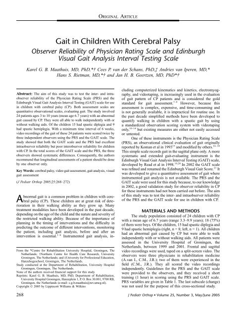
ORIGINAL ARTICLE<strong>Gait</strong> <strong>in</strong> <strong>Children</strong> <strong>With</strong> <strong>Cerebral</strong> <strong>Palsy</strong>Observer Reliability of Physician Rat<strong>in</strong>g Scale and Ed<strong>in</strong>burghVisual <strong>Gait</strong> Analysis Interval Test<strong>in</strong>g ScaleKarel G. B. Maathuis, MD, PhD,*† Cees P. van der Schans, PhD,‡ Andries van Iperen, MD,*Hans S. Rietman, MD,*† and Jan H. B. Geertzen, MD, PhD*†Abstract: The aim of this study was to test the <strong>in</strong>ter- and <strong>in</strong>traobserverreliability of the Physician Rat<strong>in</strong>g Scale (PRS) and theEd<strong>in</strong>burgh Visual <strong>Gait</strong> Analysis Interval Test<strong>in</strong>g (GAIT) scale for use<strong>in</strong> children with cerebral palsy (CP). Both assessment scales arequantitative observational scales, evaluat<strong>in</strong>g gait. The study <strong>in</strong>volved24 patients ages 3 to 10 years (mean age 6.7 years) with an abnormalgait caused by CP. They were all able to walk <strong>in</strong>dependently with orwithout walk<strong>in</strong>g aids. Of the children 15 had spastic diplegia and 9had spastic hemiplegia. <strong>With</strong> a m<strong>in</strong>imum time <strong>in</strong>terval of 6 weeks,video record<strong>in</strong>gs of the gait of these 24 patients were scored twice bythree <strong>in</strong>dependent observers us<strong>in</strong>g the PRS and the GAIT scale. Thestudy showed that both the GAIT scale and the PRS had excellent<strong>in</strong>traobserver reliability but poor <strong>in</strong>terobserver reliability for childrenwith CP. In the total scores of the GAIT scale and the PRS, the threeobservers showed systematic differences. Consequently, the authorsrecommend that longitud<strong>in</strong>al assessments of a patient should be doneby one observer only.Key Words: cerebral palsy, video gait assessment, gait analysis, visualgait assessment(J Pediatr Orthop 2005;25:268–272)From the *Centre for Rehabilitation University Hospital, Gron<strong>in</strong>gen, TheNetherlands; †Northern Centre for Health Care Research, UniversityGron<strong>in</strong>gen, The Netherlands; and ‡University for Professional Education,Hanzehogeschool, Gron<strong>in</strong>gen, The Netherlands.Study conducted at the Department of Rehabilitation, University HospitalGron<strong>in</strong>gen, Gron<strong>in</strong>gen, The Netherlands.None of the authors received f<strong>in</strong>ancial support for this study.Repr<strong>in</strong>ts: Karel G. B. Maathuis, MD, PhD, Department of Rehabilitation,University Hospital Gron<strong>in</strong>gen, Hanzeple<strong>in</strong> 1, P. O. Box 30.001, 9700 RB,Gron<strong>in</strong>gen, the Netherlands (e-mail: c.g.b.maathuis@rev.umcg.nl).Copyright Ó 2005 by Lipp<strong>in</strong>cott Williams & Wilk<strong>in</strong>sAbnormal gait is a common problem <strong>in</strong> children with cerebralpalsy (CP). These children are at great risk of deterioration<strong>in</strong> their walk<strong>in</strong>g ability as they grow up. Manytreatment modalities have been developed <strong>in</strong> the past decade,depend<strong>in</strong>g on the age of the child and the nature and severity ofthe restricted walk<strong>in</strong>g ability. Because of the importance ofplann<strong>in</strong>g <strong>in</strong> the tim<strong>in</strong>g of <strong>in</strong>terventions and the difficulty <strong>in</strong>predict<strong>in</strong>g the outcome of different <strong>in</strong>terventions, monitor<strong>in</strong>gthe patient, <strong>in</strong>clud<strong>in</strong>g gait analysis, before and after an<strong>in</strong>tervention is essential. 1–6 Instrumented gait analysis, <strong>in</strong>clud<strong>in</strong>gcomputerized k<strong>in</strong>ematics and k<strong>in</strong>etics, electromyography,and videotap<strong>in</strong>g, is <strong>in</strong>creas<strong>in</strong>gly used <strong>in</strong> the evaluationof gait pattern of CP patients and is considered the goldstandard for gait assessment. 7–9 However, because thisassessment is complex, expensive, and time-consum<strong>in</strong>g andis not generally available, it is impractical for rout<strong>in</strong>e use. Inthe past decade simplified methods have been developed toquantify walk<strong>in</strong>g <strong>in</strong> children with a spastic gait by us<strong>in</strong>ga standardized observation scor<strong>in</strong>g system with videotap<strong>in</strong>gonly, 10–14 but exist<strong>in</strong>g measures are either not easily accessedor untested.One of these <strong>in</strong>struments is the Physician Rat<strong>in</strong>g Scale(PRS), an observational cl<strong>in</strong>ical evaluation of gait orig<strong>in</strong>allyreported by Koman et al <strong>in</strong> 1993 15 and modified by others. 16–18This simple scale records gait <strong>in</strong> the sagittal plane only. A moresystematic and extended gait-evaluat<strong>in</strong>g <strong>in</strong>strument is theEd<strong>in</strong>burgh Visual <strong>Gait</strong> Analysis Interval Test<strong>in</strong>g (GAIT) scale,developed by Read et al <strong>in</strong> 1998. 19,20 In 2002 the GAIT scalewas ref<strong>in</strong>ed and renamed the Ed<strong>in</strong>burgh Visual <strong>Gait</strong> Score. 21 Itwas developed to give a quantitative assessment of gait where<strong>in</strong>strumented gait analysis is not available. The PRS and theGAIT scale were used for this study because, to our knowledge<strong>in</strong> 2002, a good validation study for observer reliability <strong>in</strong> CPfor these <strong>in</strong>struments had not been carried out before. The aimof this study was to test the <strong>in</strong>ter- and <strong>in</strong>traobserver reliabilityof the PRS and the GAIT scale for use <strong>in</strong> children with CP.MATERIALS AND METHODSThe study population consisted of 24 children with CPwith a mean age of 6.7 years (range 3.3–9.9 years); 18 (75%)of them were boys. Of the children, 15 had spastic diplegia and9 had spastic hemiplegia (right, n = 8; left, n = 1). All childrenhad an abnormal gait caused by CP but were able to walk<strong>in</strong>dependently with or without walk<strong>in</strong>g aids. All patients wereassessed <strong>in</strong> the University Hospital of Gron<strong>in</strong>gen, theNetherlands, between 1999 and 2001. Frontal and sagittalvideo record<strong>in</strong>gs were used, taped on a split-screen video. Theobservers were three physicians <strong>in</strong> rehabilitation medic<strong>in</strong>e(A.van I., C.M., J.R.); two of them were experienced <strong>in</strong> thefield (C.M., J.R.). They all scored the video record<strong>in</strong>gs<strong>in</strong>dependently. Guidel<strong>in</strong>es for the PRS and the GAIT scalewere provided to the observers, and they received a shorttra<strong>in</strong><strong>in</strong>g (1 hour) <strong>in</strong> scor<strong>in</strong>g us<strong>in</strong>g the PRS and GAIT scale.PRS variables are given <strong>in</strong> Table 1. The last subscale (change)was not used for the purpose of this cross-sectional study.268 J Pediatr Orthop Volume 25, Number 3, May/June 2005
J Pediatr Orthop Volume 25, Number 3, May/June 2005Observer Reliability of Two <strong>Gait</strong> ScalesTABLE 1. Physician Rat<strong>in</strong>g Scale 15Def<strong>in</strong>ition Right LeftCrouchSevere (.20° hip, knee, ankle) 0 0Moderate (5–20° hip, knee, ankle) 1 1Mild (,5° hip, knee, ankle) 2 2None 3 3KneeRecurvatum .5° 0 0Recurvatum 0–5° 1 1Neutral (no recurvatum) 2 2Foot contactToe 0 0Toe-heel 1 1Flat 2 2Occasional heel-toe 3 3Heel-toe 4 4ChangeWorse 21 21None 0 0Better 1 1GAIT scale variables are given <strong>in</strong> Table 2. It conta<strong>in</strong>s 17variables of observation dur<strong>in</strong>g gait at six anatomic levels(foot, ankle, knee, hip, pelvis, and trunk), <strong>in</strong>clud<strong>in</strong>g sagittal 22and frontal 23 observations. Record<strong>in</strong>gs are made us<strong>in</strong>g a threepo<strong>in</strong>tord<strong>in</strong>al scale: 0 (normal), 1 (moderate deviation), and 2(marked deviation). Both sides of the patients were scoredseparately.Observers were recommended to use slow-motion facilities,to stop or repeat the video if necessary, and to take theirtime. They were <strong>in</strong>structed not to measure degrees directlyfrom the video screen but to give their best visual estimate. Forall patients, either with hemiplegia or diplegia, both sides werescored. All video record<strong>in</strong>gs were scored twice us<strong>in</strong>g both thePRS and GAIT scale with a m<strong>in</strong>imal time <strong>in</strong>terval of 6 weeksto avoid any effects of memory; this also corresponds withcl<strong>in</strong>ical practice.Statistical AnalysisAll statistical analyses were performed us<strong>in</strong>g SPSS 11.0.Reliability analysis was done us<strong>in</strong>g analysis of variance(ANOVA). As we were <strong>in</strong>terested <strong>in</strong> the <strong>in</strong>ter- and <strong>in</strong>traobserverreliability of the total scores and <strong>in</strong> the sources ofTABLE 2. Ed<strong>in</strong>burgh Visual <strong>Gait</strong> Analysis Interval Test<strong>in</strong>g Scale 25MovementSagittal 2 1 0 1 2FOOTMovementFrontal 2 1 0 1 2FOOT1 foot clearance none reduced full n.a n.a 5 stance positionh<strong>in</strong>d foot <strong>in</strong> load.15valgus6–15valgus5–0–5neutral6–15varus.15varus2 <strong>in</strong>itial contact toe flat foot heel n.a n.a 6 foot progression .15 ir 6–15 ir 5–0–5 6–15 er .15 erangleneutral3 heel lift none early normal delayed n.a4 max dorsiflexionh<strong>in</strong>d foot <strong>in</strong> stance.10plan10–0–9plan/dor10–20dor21–30 dor .30dorKNEEKNEE7 term<strong>in</strong>al sw<strong>in</strong>g .30flex15–30flex0–15flex.0hyperextn.apartcap irall cap ir neutral allcap erpartcap er8 peak stance kneeextension.30flex16–30flex9 peak knee flexion .80 65–80<strong>in</strong> sw<strong>in</strong>gflex flexHIP11 peak hip .30 16–30extension <strong>in</strong> stance flex flex12 peak hip flexion .75 51–75<strong>in</strong> sw<strong>in</strong>gflex flexPELVIS14 pelvic rotation .15 6–15midstancefwd fwdTRUNK16 peak sagittalposition <strong>in</strong> stanceTOTAL.15fwd6–15fwd10 kneeprogressionangle mid-stance0–15flex1–10hyperext.10hyperext60–64 30–59 .30flex flex flexHIP15–0–15 n.a n.a 13 positionflex/ext<strong>in</strong> sw<strong>in</strong>g30–50 15–29 ,15flex flex flexPELVIS5–0–5 6–15neutral bwd5–0–5 6–15neutral bwd.15bwd.15bwd.15add5–15add4–0–9 10–20add/abd abd.20abd15 contralateral drop<strong>in</strong> stance marked mod normal n.a n.aTRUNK17 max lateralshift <strong>in</strong> stance marked mod neutral n.a n.aTOTALScore 2 means marked deviation, score 1 is moderate deviation, score 0 is normal range.n.a, not available; plan, plantarflexion; dor, dorsiflexion; flex, flexion; hyperext, hyperextension; fwd, forward rotation; bwd, backward rotation; ir, <strong>in</strong>ternal rotation; er, externalrotation; part cap, only a part of the knee cap is visible; all cap, whole knee cap is visible; add, adduction; abd, abduction; lat, lateral; mod, moderate.q 2005 Lipp<strong>in</strong>cott Williams & Wilk<strong>in</strong>s 269
Maathuis et al J Pediatr Orthop Volume 25, Number 3, May/June 2005variance (child, observer, and repetition), for each total scorewe chose this method, not kappa statistics of the separateitems. Total GAIT scale and PRS and subscores of both scalesfor the right and left side were taken as <strong>in</strong>dependent factors.For each <strong>in</strong>dependent factor the estimated variances werecalculated. Post hoc comparison of differences between thethree observers was done us<strong>in</strong>g the Friedman test. P , 0.05was considered statistically significant.RESULTSTwo subjects were excluded from analysis: <strong>in</strong> one patientthe frontal video imag<strong>in</strong>g failed; <strong>in</strong> the other the sagittal onefailed. The 22 patients who rema<strong>in</strong>ed for the reliabilityanalysis were assessed at random by the observers. On theright as well as the left side, both observer and child proved tobe significant sources of variance <strong>in</strong> the GAIT scale and <strong>in</strong> thePRS. Repetition was not a significant source of variance (Table3); <strong>in</strong> most cases it even approached P = 1. In fact, both theGAIT scale and the PRS showed excellent <strong>in</strong>traobserverreliability. The <strong>in</strong>terobserver reliability of both assessmentscales, the GAIT scale and the PRS, was considered poor.Post hoc analysis showed considerable differencesbetween the three observers. The mean (SD) scores for eachobserver are given <strong>in</strong> Table 4; box plots are shown <strong>in</strong> Figure 1.In Table 5, the GAIT scale is subdivided <strong>in</strong>to sevensubscales: the first five subscales correspond to the differentanatomic levels, and the last two correspond to the differentdirections of observ<strong>in</strong>g gait (frontal and sagittal views). Onlywith the ankle subscale on the left side did the observer appearnot to be a significant factor <strong>in</strong> the source of variation. In allother GAIT subscales the observer appeared to be a significantsource of variance.TABLE 4. Total Scores of GAIT Scale and PRS of All ThreeObserversObserver 1(AvI)Mean (sd)Observer 2(CM)Mean (sd)Observer 3(JR)Mean (sd)P Values*GAIT scale right 9.1 (5) 10.3 (5.2) 13.5 (6.7) ,0.001GAIT scale left 7.8 (6.8) 8.5 (6.9) 11.1 (9.4) 0.003PRS right 5.5 (1.9) 4.4 (2.1) 4.4 (2.2) ,0.001PRS left 6.1 (2.4) 5.4 (2.7) 5.5 (3.0) 0.004*Friedman test.relevant because the differences <strong>in</strong> the outcome of 1.1 <strong>in</strong> thePRS and 4.4 <strong>in</strong> the GAIT scale (Table 4) for the same personshould be clearly visible when observ<strong>in</strong>g gait. Besides, if thesame difference <strong>in</strong> the PRS and GAIT scale could be measuredas a result of an <strong>in</strong>tervention <strong>in</strong> CP patients, it would beconsidered a cl<strong>in</strong>ically relevant difference.The total scores of the GAIT scale and the PRS, betweenthe three observers, also showed systematic differences. Thereason is not clear. Probably, angle estimation of the differentanatomic levels from a video screen was done systematicallydifferent between the observers.Although the PRS is used often <strong>in</strong> research, we onlyfound one study <strong>in</strong> the literature 23 that reported <strong>in</strong>terobserverreliability; we found no study that reported <strong>in</strong>traobserverreliability data. Corry et al 23 studied the <strong>in</strong>terobserver reliabilityof the PRS <strong>in</strong> a group of 20 CP children with a dynamiccomponent <strong>in</strong> spastic equ<strong>in</strong>us, treated by serial cast<strong>in</strong>g orDISCUSSIONThe differences <strong>in</strong> mean total scores of the threeobservers were considerable and were considered cl<strong>in</strong>icallyTABLE 3. Sources of Variation <strong>in</strong> the GAIT Scale and PRSSS MS P Value Variance EstimatesGAIT scale rightObserver 240 120 ,0.001 2.6Repetition 0.2 0.2 0.846 0Child 3904 186 ,0.001 30.1GAIT scale leftObserver 162 81 ,0.001 1.7Repetition 0.1 0.1 0.941 0Child 7571 361 ,0.001 59.2PRS rightObserver 21 10.5 ,0.001 0.2Repetition 0.4 0.4 0.395 0Child 509 24 ,0.001 4.0PRS leftObserver 14.6 7.3 ,0.001 0.1Repetition 0 0 1 0Child 872 42 ,0.001 6.8SS, sum of squares; MS, mean square.FIGURE 1. Box plots of GAIT scale and PRS. The ends of therectangle reflect the <strong>in</strong>terquartile range; the horizontal l<strong>in</strong>e <strong>in</strong>the rectangle reflects the median value; the whiskers <strong>in</strong>dicatethe m<strong>in</strong>imum and maximum values.270 q 2005 Lipp<strong>in</strong>cott Williams & Wilk<strong>in</strong>s
J Pediatr Orthop Volume 25, Number 3, May/June 2005Observer Reliability of Two <strong>Gait</strong> ScalesTABLE 5. Observer Variance of Subscores of the GAIT ScaleSS MS P ValueVarianceEstimatesGAIT subscoreAnkle (6)Right 13.2 6.6 0.002 0.1Left 3.0 1.5 0.254 0Knee (4)Right 17.5 8.7 ,0.001 0.2Left 16.1 8.0 ,0.001 0.2Hip (3)Right 13.5 6.7 ,0.001 0.1Left 9.5 4.7 ,0.001 0Pelvis (2)Right 13.9 6.9 ,0.001 0.1Left 14.7 7.3 ,0.001 0.2Trunk (2)Right 11.2 5.6 ,0.001 0.1Left 8.4 4.2 ,0.001 0.1Frontal (6)Right 68.0 34.0 ,0.001 0.7Left 53.8 26.9 ,0.001 0.6Sagittal (11)Right 109.1 54.6 ,0.001 1.2Left 43.7 21.8 ,0.001 0.4SS, sum of squares; MS, mean square.Values <strong>in</strong> parentheses represent the number of items on the GAIT scale.Botul<strong>in</strong>um tox<strong>in</strong> A. They found a moderate agreement <strong>in</strong>crouch assessment and <strong>in</strong> the overall impression <strong>in</strong> gait change<strong>in</strong> the affected side us<strong>in</strong>g the PRS (weighted kappa test 0.55–0.67). The agreement <strong>in</strong> the knee assessment was poor. The‘‘change’’ section was added to provide a more discrim<strong>in</strong>at<strong>in</strong>gdifference. Unfortunately, no <strong>in</strong>formation was given about the<strong>in</strong>terobserver variation of the total PRS. Moreover, only twoobservers were used <strong>in</strong> Corry et al’s study.Post hoc analysis of the results of our two mostexperienced observers (observers 2 and 3) showed no differencesbetween these observers for the PRS. In other words,the <strong>in</strong>formation concern<strong>in</strong>g the <strong>in</strong>terobserver reliability for thisscale may have been <strong>in</strong>fluenced by the number of observersused <strong>in</strong> the study and the degree of experience <strong>in</strong> observ<strong>in</strong>ggait. This may expla<strong>in</strong> the different results between the Corryet al study, which <strong>in</strong>cluded only two observers, and ours, <strong>in</strong>which three observers took part.In 1999 Read et al 20 presented data about the <strong>in</strong>ter- and<strong>in</strong>traobserver reliability of the GAIT scale <strong>in</strong> a studypopulation of four patients with CP and one ‘‘normal’’ person.Because of the very small group, these data are of limitedvalue, however. In 2003 Read et al 24 reported a good <strong>in</strong>tra- and<strong>in</strong>terobserver reliability for the Ed<strong>in</strong>burgh Visual <strong>Gait</strong> Scoresystem. They used kappa statistics for their calculations; we,however, were more <strong>in</strong>terested <strong>in</strong> the source of variance. In ourpopulation the child and the observer proved to be a significantfactor <strong>in</strong> this respect for the source of variation.We are aware of some limitations <strong>in</strong> our study. First, weused a fixed video camera fram<strong>in</strong>g for record<strong>in</strong>g the sagittalgait. This means that only a small part of the gait is availablefor evaluation of gait <strong>in</strong> the sagittal direction. We tried toapproach daily practice as much as possible. <strong>With</strong> the techniqueused, it is possible to reproduce the study <strong>in</strong> any consult<strong>in</strong>groom <strong>in</strong> a hospital. Second, the children did not wearstandardized cloth<strong>in</strong>g at the time of the measurement. In somevideo record<strong>in</strong>gs, children wore a T-shirt or sweater, whichmight have <strong>in</strong>fluenced the ability to estimate the amount offlexion and extension <strong>in</strong> the hip, pelvis, and trunk <strong>in</strong> the GAITscale. To exclude such possible failures we calculated the<strong>in</strong>terobserver reliability of each anatomic level itself and thesubscores of the frontal and sagittal plane. Our hypothesis wasthat the reliability of the assessments of the foot, ankle, andknee level would have been better compared with the hip,pelvis, and trunk level (see Table 5). It appeared that onlythe subscore of the left ankle was not a significant factor forthe source of variation. Of the 22 left sides we tested, 8 sideswere not affected sides and were scored almost equally by allobservers, compared with only 1 unaffected right side. Thismight expla<strong>in</strong> the difference <strong>in</strong> <strong>in</strong>terobserver reliability betweenthe subscores at ankle level, respectively, on the rightand left side. All other subscores of the GAIT scale, <strong>in</strong>clud<strong>in</strong>gthe frontal and sagittal plane, showed poor <strong>in</strong>terobserver reliabilitydata. Post hoc analysis of the more affected side only ofall patients showed similar results (data not presented).Noonan et al 6 reported a <strong>in</strong>terobserver reliability studyof patients with CP evaluated with <strong>in</strong>strumented gait analysisat four different centers. The results were poor.In 2003 editorials <strong>in</strong> the Journal of Pediatric Orthopaedics,Gage22 and Wright 25 made contrast<strong>in</strong>g comments about<strong>in</strong>terobserver variability <strong>in</strong> gait analysis. They agreed that weneed a careful assessment of the analysis of the pathology ofthe CP child. The importance of <strong>in</strong>strumented gait analysis isclear for this assessment, but it is only one of the methods ofexam<strong>in</strong>ation and should be seen as complementary to thegeneration of the problem list, the physical exam<strong>in</strong>ation, andradiographs. Further research <strong>in</strong>to the different sources thatcontribute to the variability <strong>in</strong> gait analysis will be necessary.To evaluate the gait of a CP patient <strong>in</strong> the consult<strong>in</strong>g room bymeans of video record<strong>in</strong>g is very useful. To score gait withPRS took about 5 m<strong>in</strong>utes; the GAIT scale took 25 m<strong>in</strong>utes perpatient on average. We also recommend that longitud<strong>in</strong>alassessments of a patient should be done by the same observer.REFERENCES1. Calderon-Gonzalez R, Calderon-Sepulveda R, R<strong>in</strong>con-Reyes M, et al.Botul<strong>in</strong>um tox<strong>in</strong> A <strong>in</strong> management of cerebral palsy. Pediatr Neurol.1994;4:284–288.2. Cook RE, Schneider I, Hazlewood ME, et al. <strong>Gait</strong> analysis alters decisionmak<strong>in</strong>g<strong>in</strong> cerebral palsy. J Pediatr Orthop. 2003;23:292–295.3. Gage JR. The role of gait analysis <strong>in</strong> the treatment of cerebral palsy[editorial]. J Pediatr Orthop. 1994;14:701–702.4. Hailey D, Tomie J. An assessment of gait analysis <strong>in</strong> the rehabilitation ofchildren with walk<strong>in</strong>g difficulties. Disabil Rehabil. 2000;6:275–280.5. Molenaers G, Desloovere K, Eyssen M, et al. Botul<strong>in</strong>um tox<strong>in</strong> A treatmentof cerebral palsy: an <strong>in</strong>tegrated approach. Eur J Neurol. 1999;6:S1–S7.6. Noonan KJ, Halliday S, Brown R, et al. Interobserver variability of gaitanalysis on patients with cerebral palsy. J Pediatr Orthop. 2003;23:279–287.7. Drou<strong>in</strong> LM, Malou<strong>in</strong> F, Richards CL, et al. Correlation between the grossmotor function measure and gait spatiotemporal measures <strong>in</strong> children withneurological impairments. Dev Med Child Neurol. 1996;38:1007–1019.q 2005 Lipp<strong>in</strong>cott Williams & Wilk<strong>in</strong>s 271
Maathuis et al J Pediatr Orthop Volume 25, Number 3, May/June 20058. Kirkpatrick M, Wytch R, Cole G, et al. Is the objective assessment ofcerebral palsy gait reproducible? J Pediatr Orthop. 1994;14:705–708.9. Sutherland DH, Kaufman KR, Wyatt MP, et al. Injections of botul<strong>in</strong>umA tox<strong>in</strong> <strong>in</strong>to the gastrocnemius muscle of patients with cerebral palsy: a3-dimensional motion analysis study. <strong>Gait</strong> Posture. 1996;4:269–279.10. Boyd NR, Graham HK. Objective measurement of cl<strong>in</strong>ical f<strong>in</strong>d<strong>in</strong>gs <strong>in</strong> theuse of botul<strong>in</strong>um tox<strong>in</strong> type A for the management of children withcerebral palsy. Eur J Neurol. 1999;6:523–535.11. Boyd NR, Graham JEA, Nattrass GR, et al. Medium term outcomeresponsecharacterisation and risk factor analysis of Botul<strong>in</strong>um tox<strong>in</strong> typeA <strong>in</strong> the management of spasticity <strong>in</strong> children with cerebral palsy. Eur JNeurol. 1999;6:S37–S45.12. Eastlack ME, Arvidson J, Snyder-Mackler L, et al. Interrater reliabilityof videotaped observational gait-analysis assessments. Phys Ther. 1991;71:465–472.13. Flett PJ, Stern LM, Waddy H, et al. Botul<strong>in</strong>um tox<strong>in</strong> A versus fixed caststretch<strong>in</strong>g for dynamic calf tightness <strong>in</strong> cerebral palsy. J Paediatr ChildHealth. 1999;35:71–77.14. Ubhi T, Bhakta BB, Ives HL, et al. Randomised double-bl<strong>in</strong>d placebocontrolledtrial of the effect of botul<strong>in</strong>um tox<strong>in</strong> on walk<strong>in</strong>g <strong>in</strong> cerebralpalsy. Arch Dis Child. 2000;83:481–487.15. Koman LA, Mooney JF, Smith B, et al. Management of cerebral palsy withbotul<strong>in</strong>um-A tox<strong>in</strong>: prelim<strong>in</strong>ary <strong>in</strong>vestigation. J Pediatr Orthop. 1993;4:489–495.16. Chutorion AM, Root L. Management of spasticity <strong>in</strong> children withbotul<strong>in</strong>um-A tox<strong>in</strong>. Int Pediatr. 1994;9:35–42.17. Denislic M, Meh D. Botul<strong>in</strong>um tox<strong>in</strong> <strong>in</strong> the treatment of cerebral palsy.Neuropediatrics. 1995;26:249–252.18. Graham HK, Aoki KR, Autti-Rämö, et al. Recommendations for the useof botul<strong>in</strong>um tox<strong>in</strong> type A <strong>in</strong> the management of cerebral palsy. <strong>Gait</strong>Posture. 2000;11:67–79.19. Kerr AM, Hazlewood ME, L<strong>in</strong>den van der Ml, et al. The Ed<strong>in</strong>burgh Visual<strong>Gait</strong> Score as an outcome measure after surgical <strong>in</strong>tervention cerebralpalsy. <strong>Gait</strong> Posture. 2002;16:S116.20. Read HS, Hillmann SJ, Hazlewood ME, et al. The Ed<strong>in</strong>burgh Visual <strong>Gait</strong>Analysis Interval Test<strong>in</strong>g (GAIT) scale. <strong>Gait</strong> Posture. 1999;10:63.21. Read HS, Hazlewood ME, Hillmann SJ, et al. A visual gait analysis scorefor use <strong>in</strong> cerebral palsy: the Ed<strong>in</strong>burgh Visual <strong>Gait</strong> Score. <strong>Gait</strong> Posture.2002;16:S115–S116.22. Gage JR. Con: <strong>in</strong>terobserver variability of gait analysis [editorial].J Pediatr Orthop. 2003;23:290–291.23. Corry IS, Graham HK. Botul<strong>in</strong>um tox<strong>in</strong> A compared with stretch<strong>in</strong>g casts<strong>in</strong> the treatment of spastic equ<strong>in</strong>es: a randomised prospective trial.J Pediatr Orthop. 1998;18:304–311.24. Read HS, Hazlewood ME, Hillman SJ, et al. Ed<strong>in</strong>burgh Visual <strong>Gait</strong> Score.J Pediatr Orthop. 2003;23:296–301.25. Wright JG. Pro: <strong>in</strong>terobserver variability of gait analysis [editorial].J Pediatr Orthop. 2003;23:288–289.272 q 2005 Lipp<strong>in</strong>cott Williams & Wilk<strong>in</strong>s


