View - ResearchGate
View - ResearchGate View - ResearchGate
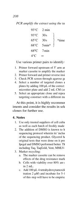
208 Caldwell et al.PCR amplify the extract using the targeting screening reaction mixture:Use various primer pairs to identify the targeted clones (refer to Fig. 4):1. Primer forward upstream of 5′ arm and primer reverse inside the drug resistancemarker cassette to amplify the marker and to exclude single crossing over.2. Primer forward and primer reverse inside target locus to amplify the deleted region.3. Check PCR screen through agarose gel electrophoresis.4. Select a number of targeted clones and grow in 24-well flat bottom microtiterplates by adding 100 µL of the correct cell suspension from the 96-well flat bottommicrotiter plate and add 2 mL CM (see Notes 8 and 9).5. Select an appropriate clone and repeat the transfection procedure using a secondtargeting construct with a different marker cassette.At this point, it is highly recommended to perform the phenotype experimentsand consider the results in selecting specific heterologous knockoutclones for further use.4. Notes93°C 2 min93°C 30s65°C 30s68°C 5min*68°C 7min4° C ∞⎫⎪⎬⎪⎭35 cycles*time increases: 20 s each cycle1. Use only trusted suppliers of cell culture reagents and always test new lot numbersas well as each batch of freshly made CM.2. The addition of DMSO is known to help with difficult spots and it is used in thesequencing protocol wherein its’ inclusion increases both the quality and the lengthof the sequencing product. Glycerol has sometimes been used for PCR, but in theoriginal tests that were done on it previously, it was found that additives such asIpegal and DMSO performed better. This effect was seen with multiple polymerasesincluding Taq, TaqGold, Vent, MMLV-RT, and Superscript RT.3. Marker recycling:a. The marker cassette can be removed from the knockout clones to exclude sideeffects of the drug resistance marker and to reuse the marker.b. Cells with viability over 80% are diluted to a concentration of 0.6 × 10 6 cellsin 2 mL.c. Add 100 µL 4-tetrahydroxytamoxifen (40 µM) to the 2 mL of cells (final concentration2 µM) and incubate for 5–12 h at 41°C. The concentration and timingof this step will have to be empirically determined based on the targeted locus.
- Page 384: Identifying Differentially Expresse
- Page 388: Identifying Differentially Expresse
- Page 392: Identifying Differentially Expresse
- Page 396: Identifying Differentially Expresse
- Page 400: Identifying Differentially Expresse
- Page 404: 11Gene Function Analysis Using the
- Page 408: Gene Function Analysis Using the Ch
- Page 412: Gene Function Analysis Using the Ch
- Page 416: Gene Function Analysis Using the Ch
- Page 420: Gene Function Analysis Using the Ch
- Page 424: Gene Function Analysis Using the Ch
- Page 428: Gene Function Analysis Using the Ch
- Page 432: Gene Function Analysis Using the Ch
- Page 438: 210 Caldwell et al.8. Thawing cells
- Page 442: 212 Zhang et al.Going one step beyo
- Page 446: 214 Zhang et al.Fig. 2. Generation
- Page 450: 216 Zhang et al.Perform PCR cycles,
- Page 454: 218 Zhang et al.Fig. 4. Schematic m
- Page 458: 220 Zhang et al.Fig. 5. Replacement
- Page 462: 13Construction of Simple and Effici
- Page 466: DNA Vector-Based shRNA-Expression S
- Page 470: DNA Vector-Based shRNA-Expression S
- Page 474: DNA Vector-Based shRNA-Expression S
- Page 478: DNA Vector-Based shRNA-Expression S
- Page 482: DNA Vector-Based shRNA-Expression S
208 Caldwell et al.PCR amplify the extract using the targeting screening reaction mixture:Use various primer pairs to identify the targeted clones (refer to Fig. 4):1. Primer forward upstream of 5′ arm and primer reverse inside the drug resistancemarker cassette to amplify the marker and to exclude single crossing over.2. Primer forward and primer reverse inside target locus to amplify the deleted region.3. Check PCR screen through agarose gel electrophoresis.4. Select a number of targeted clones and grow in 24-well flat bottom microtiterplates by adding 100 µL of the correct cell suspension from the 96-well flat bottommicrotiter plate and add 2 mL CM (see Notes 8 and 9).5. Select an appropriate clone and repeat the transfection procedure using a secondtargeting construct with a different marker cassette.At this point, it is highly recommended to perform the phenotype experimentsand consider the results in selecting specific heterologous knockoutclones for further use.4. Notes93°C 2 min93°C 30s65°C 30s68°C 5min*68°C 7min4° C ∞⎫⎪⎬⎪⎭35 cycles*time increases: 20 s each cycle1. Use only trusted suppliers of cell culture reagents and always test new lot numbersas well as each batch of freshly made CM.2. The addition of DMSO is known to help with difficult spots and it is used in thesequencing protocol wherein its’ inclusion increases both the quality and the lengthof the sequencing product. Glycerol has sometimes been used for PCR, but in theoriginal tests that were done on it previously, it was found that additives such asIpegal and DMSO performed better. This effect was seen with multiple polymerasesincluding Taq, TaqGold, Vent, MMLV-RT, and Superscript RT.3. Marker recycling:a. The marker cassette can be removed from the knockout clones to exclude sideeffects of the drug resistance marker and to reuse the marker.b. Cells with viability over 80% are diluted to a concentration of 0.6 × 10 6 cellsin 2 mL.c. Add 100 µL 4-tetrahydroxytamoxifen (40 µM) to the 2 mL of cells (final concentration2 µM) and incubate for 5–12 h at 41°C. The concentration and timingof this step will have to be empirically determined based on the targeted locus.


