with p, N. Izzo, A. M. GRAYBIEL* and J, p, BOLAM to electron on ...
with p, N. Izzo, A. M. GRAYBIEL* and J, p, BOLAM to electron on ...
with p, N. Izzo, A. M. GRAYBIEL* and J, p, BOLAM to electron on ...
- No tags were found...

Create successful ePaper yourself
Turn your PDF publications into a flip-book with our unique Google optimized e-Paper software.
Printed in20,Britainpp. 1987p, N. <str<strong>on</strong>g>Izzo</str<strong>on</strong>g>, A. M. <str<strong>on</strong>g>GRAYBIEL*</str<strong>on</strong>g> <str<strong>on</strong>g>and</str<strong>on</strong>g> J, p, <str<strong>on</strong>g>BOLAM</str<strong>on</strong>g><str<strong>on</strong>g>to</str<strong>on</strong>g> <str<strong>on</strong>g>electr<strong>on</strong></str<strong>on</strong>g>the<strong>on</strong> secti<strong>on</strong>s of 80---1 00 f1 m>:lLa, ...,x\.ll1J
578 P. N. <str<strong>on</strong>g>Izzo</str<strong>on</strong>g> ettpf"hn,,-,'"'t'> more reliable <str<strong>on</strong>g>and</str<strong>on</strong>g> morein secti<strong>on</strong>sof cat <str<strong>on</strong>g>and</str<strong>on</strong>g> ferret forebrain <str<strong>on</strong>g>and</str<strong>on</strong>g> describe in this paperthe modificati<strong>on</strong>s of thethe morneur<strong>on</strong>sin the caudate nucleus.Tissue from 3 catsm<strong>on</strong>ths) was used inanaesthetized200-500 ml 0.9%approXImately 21EXPERIMENTAL PROCEDURESassembliesseparating the slides <str<strong>on</strong>g>with</str<strong>on</strong>g> thesee The secti<strong>on</strong>s were detIVdlra1ledembedded <strong>on</strong> microscope slidesACM, Fluka).For sub-1 % (0.5 h)incubatedFac<str<strong>on</strong>g>to</str<strong>on</strong>g>rs influencing impregnati<strong>on</strong>The most important fac<str<strong>on</strong>g>to</str<strong>on</strong>g>rimpregnati<strong>on</strong> of neur<strong>on</strong>s was the1""\P'~P""'H""nitrate soluti<strong>on</strong>thefor the silver nitratein theGolgi-impregnati<strong>on</strong> procedure for single
Substance P <str<strong>on</strong>g>and</str<strong>on</strong>g> enkephalin neur<strong>on</strong>s in the caudate nucleus 579penetrati<strong>on</strong> enhancement techniques (freeze-thawing ordetergent treatment).Light <str<strong>on</strong>g>and</str<strong>on</strong>g> <str<strong>on</strong>g>electr<strong>on</strong></str<strong>on</strong>g> microscopyAfter curing the resin for 48 h at 60°C all secti<strong>on</strong>s wereexamined in the light microscope. The immunostaining <str<strong>on</strong>g>and</str<strong>on</strong>g>Golgi-impregnati<strong>on</strong> were assessed <str<strong>on</strong>g>and</str<strong>on</strong>g> neur<strong>on</strong>s that wereboth Golgi-impregnated <str<strong>on</strong>g>and</str<strong>on</strong>g> immunostained were identified.Selected neur<strong>on</strong>s in the cat material were drawn <str<strong>on</strong>g>with</str<strong>on</strong>g>the aid of a microscope drawing tube (Leitz) <str<strong>on</strong>g>and</str<strong>on</strong>g> measurementsof the size of perikarya <str<strong>on</strong>g>and</str<strong>on</strong>g> dendrites were made fromthese drawings using a Bioquant image analysis system. Noaccount was taken for shrinkage of the tissue.To c<strong>on</strong>firm the presence of immunoreactivity in theperikarya of Golgi-impregnated neur<strong>on</strong>s, some neur<strong>on</strong>swere re-embedded in blocks of resin suitable for resecti<strong>on</strong>ing.From these, I pm secti<strong>on</strong>s for light microscopy<str<strong>on</strong>g>and</str<strong>on</strong>g> ultrathin (silver/grey) secti<strong>on</strong>s for <str<strong>on</strong>g>electr<strong>on</strong></str<strong>on</strong>g> microscopywere cut. The latter secti<strong>on</strong>s were collected <strong>on</strong> Formvarcoatedsingle-slot grids counterstained <str<strong>on</strong>g>with</str<strong>on</strong>g> lead citrate <str<strong>on</strong>g>and</str<strong>on</strong>g>examined in a Philips EM201 <str<strong>on</strong>g>electr<strong>on</strong></str<strong>on</strong>g> microscope. Toimprove c<strong>on</strong>trast for <str<strong>on</strong>g>electr<strong>on</strong></str<strong>on</strong>g> microscopy, the tissue wasstained en bloc <str<strong>on</strong>g>with</str<strong>on</strong>g> 1% uranyl acetate (in 70% ethanol)during dehydrati<strong>on</strong>.RESULTSThe degree <str<strong>on</strong>g>and</str<strong>on</strong>g> quality of Golgi-impregnati<strong>on</strong> wassimilar <str<strong>on</strong>g>to</str<strong>on</strong>g> that seen <str<strong>on</strong>g>with</str<strong>on</strong>g> c<strong>on</strong>venti<strong>on</strong>al Golgi techniques.The impregnated neur<strong>on</strong>s appeared sometimesin groups but were often isolated (Figs 1 B, <str<strong>on</strong>g>and</str<strong>on</strong>g>3A). Perikarya, dendrites, dendritic spines <str<strong>on</strong>g>and</str<strong>on</strong>g> ax<strong>on</strong>swere all impregnated (Figs 1 <str<strong>on</strong>g>and</str<strong>on</strong>g> 3). The types ofneur<strong>on</strong>s that were impregnated were similar <str<strong>on</strong>g>to</str<strong>on</strong>g> thosedescribed <strong>on</strong> previous occasi<strong>on</strong>s in rat, cat <str<strong>on</strong>g>and</str<strong>on</strong>g>m<strong>on</strong>key (see Refs 3, 10, 28, 40 <str<strong>on</strong>g>and</str<strong>on</strong>g> 52). Although themajority of impregnated neur<strong>on</strong>s were the mediumsizedensely spiny type (see Figs I <str<strong>on</strong>g>and</str<strong>on</strong>g> 3), other typesincluding large neur<strong>on</strong>s <str<strong>on</strong>g>and</str<strong>on</strong>g> medium-size aspiny neur<strong>on</strong>swere also observed. No obvious differences wereseen between the caudate nucleus <str<strong>on</strong>g>and</str<strong>on</strong>g> putamen, orbetween the cat <str<strong>on</strong>g>and</str<strong>on</strong>g> ferret.ImmunostainingSubstance P. At the light microscopic level substanceP-immunoreactivity in the neostriatum wasremarkably uneven. In the caudate <str<strong>on</strong>g>and</str<strong>on</strong>g> particularlyin its rostral part, high c<strong>on</strong>centrati<strong>on</strong>s of immunoreacti<strong>on</strong>product were present in patches of irregularshape (see Fig. 1 B). These regi<strong>on</strong>s represent thestriosomal compartments of the caudate nucleus <str<strong>on</strong>g>and</str<strong>on</strong>g>corresp<strong>on</strong>d <str<strong>on</strong>g>to</str<strong>on</strong>g> AChE-poor regi<strong>on</strong>sY Detail of thestructure of substance P-positive/AChE-poor patches<str<strong>on</strong>g>and</str<strong>on</strong>g> their relati<strong>on</strong>ship <str<strong>on</strong>g>to</str<strong>on</strong>g> Golgi-impregnated neur<strong>on</strong>swill be the subject of a separate communicati<strong>on</strong>.Most of the immunoreacti<strong>on</strong> product was associated<str<strong>on</strong>g>with</str<strong>on</strong>g> neur<strong>on</strong>al perikarya <str<strong>on</strong>g>and</str<strong>on</strong>g> sometimes proximaldendrites (Fig. I B) . The immunoreactive neur<strong>on</strong>swere of medium-size <str<strong>on</strong>g>and</str<strong>on</strong>g> the majority weresimilar <str<strong>on</strong>g>to</str<strong>on</strong>g> the type I substance P-positive neur<strong>on</strong>described in the rat. 5 The neur<strong>on</strong>al staining occurredpredominantly in the striosomes (as defined by thedense neuropil staining; see Fig 1 B), but there werealso many immunoreactive perikarya <str<strong>on</strong>g>with</str<strong>on</strong>g> no apparentassociati<strong>on</strong> <str<strong>on</strong>g>with</str<strong>on</strong>g> the striosomes. Immunoreactiveax<strong>on</strong> or fibre-like structures were also present; thesewere predominantly in the superficial layers of thetissue <str<strong>on</strong>g>and</str<strong>on</strong>g> appeared evenly distributed throughoutthe secti<strong>on</strong>s.Immunoreactive neur<strong>on</strong>s, in which indentati<strong>on</strong>s ofthe nucleus were visible in the light microscope, wererarely seen. These neur<strong>on</strong>s probably corresp<strong>on</strong>d <str<strong>on</strong>g>to</str<strong>on</strong>g>the type 2 substance P-immunoreactive neur<strong>on</strong>s observedin the rat. 5 The apparent rarity of these cellsprobably reflects their low density in the striatum <str<strong>on</strong>g>and</str<strong>on</strong>g>the difficulty in identifying indented nuclei in the lightmicroscope.Methi<strong>on</strong>ine enkephalin. [Met]enkephalin immunoreacti<strong>on</strong>product was present <str<strong>on</strong>g>with</str<strong>on</strong>g>in medium-sizeperikarya, their proximal dendrites <str<strong>on</strong>g>and</str<strong>on</strong>g> many apparentlyisolated dendrites (Fig. 3A, B). In additi<strong>on</strong>, insome areas terminal staining occurred in the mostsuperficial layers of the secti<strong>on</strong>s. Perikaryal stainingwas most prominent in the colchicine-treated cat. Thedistributi<strong>on</strong> of the immunoreactive neur<strong>on</strong>s wasdifferent for colchicine <str<strong>on</strong>g>and</str<strong>on</strong>g> n<strong>on</strong>-colchicine animals ashas been reported previously;27 in the latter animalsthe immunoreactive-neur<strong>on</strong>s tended <str<strong>on</strong>g>to</str<strong>on</strong>g> avoid thestriosomes (i.e. AChE-poor z<strong>on</strong>es) while in thecolchicine-treated cat they were distributed moreevenly throughout the caudate nucleus <str<strong>on</strong>g>and</str<strong>on</strong>g> putamen.Specificity <str<strong>on</strong>g>and</str<strong>on</strong>g> c<strong>on</strong>trols. The specificity of the antibodiesused in the present experiments has beencharacterized previously'4.37 (see also Refs 5, 27 <str<strong>on</strong>g>and</str<strong>on</strong>g>47). In secti<strong>on</strong>s that were incubated by the immunocy<str<strong>on</strong>g>to</str<strong>on</strong>g>chemicalprocedure but <str<strong>on</strong>g>with</str<strong>on</strong>g> omissi<strong>on</strong> of theprimary antibodies, no specific immunostainingoccurred. In additi<strong>on</strong>, some secti<strong>on</strong>s from each animalwere incubated <str<strong>on</strong>g>with</str<strong>on</strong>g> other antisera raised inrabbits (directed against glutamate decarboxylase,tyrosine hydroxylase, soma<str<strong>on</strong>g>to</str<strong>on</strong>g>statin) <str<strong>on</strong>g>and</str<strong>on</strong>g> m<strong>on</strong>oc1<strong>on</strong>alantibodies (directed against choline acetyltransferase).The patterns of staining obtained <str<strong>on</strong>g>with</str<strong>on</strong>g> theseantibodies were markedly different from those obtained<str<strong>on</strong>g>with</str<strong>on</strong>g> the anti-substance P or anti-enkephalinantibodies. The pattern of staining described above istherefore specific <str<strong>on</strong>g>to</str<strong>on</strong>g> the substance P <str<strong>on</strong>g>and</str<strong>on</strong>g> [Met]enkephalinantibodies we used <str<strong>on</strong>g>and</str<strong>on</strong>g> not a result of othersteps in the procedure.Golgi-impregnati<strong>on</strong> combined <str<strong>on</strong>g>with</str<strong>on</strong>g> immunostainingA <str<strong>on</strong>g>to</str<strong>on</strong>g>tal of 33 Golgi-impregnated neur<strong>on</strong>s that werealso immunoreactive for substance P were identified(25 in cat, 8 in ferret). In the [Met]enkephalinimmunoreactedmaterial 55 Golgi-impregnated immunoreactiveneur<strong>on</strong>s were identified (41 in cat <str<strong>on</strong>g>and</str<strong>on</strong>g>14 in ferret). The immunoreacti<strong>on</strong> end productappeared as a brown homogenous staining of theperikarya <str<strong>on</strong>g>and</str<strong>on</strong>g> was easily distinguishable from thegrey/black sec<strong>on</strong>dary Golgi deposit. Immunoreacti<strong>on</strong>product was identified in Golgi-impregnated neur<strong>on</strong>sat the light microscopic level when (I) the gold-<str<strong>on</strong>g>to</str<strong>on</strong>g>ningof the perikarya was pale (Fig. I B), (2) Golgiimpregnateddendrites arose from n<strong>on</strong>-impregnated
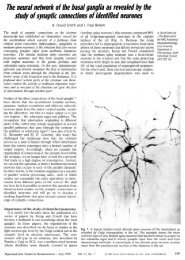
Fig. I. (A) Drawing of a Golgi-impregnated, gold-<str<strong>on</strong>g>to</str<strong>on</strong>g>ned neur<strong>on</strong> that is also immunoreactive for substanceP. Micrographs of this neur<strong>on</strong> are shown in subsequent figures. The unshaded dendrite <strong>on</strong> the leftwas not Golgi-impregnated but expressed immunoreactivity; see the dendrite labelled d in (B) <str<strong>on</strong>g>and</str<strong>on</strong>g> (C).This neur<strong>on</strong> was found <str<strong>on</strong>g>with</str<strong>on</strong>g>in a substance P-immunoreactive "patch", the boundaries of which are shownby the solid black lines above <str<strong>on</strong>g>and</str<strong>on</strong>g> below the neur<strong>on</strong>. The dendritic spines were drawn free-h<str<strong>on</strong>g>and</str<strong>on</strong>g> <str<strong>on</strong>g>and</str<strong>on</strong>g> aretherefore an approximati<strong>on</strong> of the true density. The neur<strong>on</strong> shows the characteristics of medium-sizedensely spiny neur<strong>on</strong>s, i.e. medium-size perikary<strong>on</strong>, sec<strong>on</strong>dary <str<strong>on</strong>g>and</str<strong>on</strong>g> higher order dendrites densely laden<str<strong>on</strong>g>with</str<strong>on</strong>g> spines. (B) Partial light microscopic pho<str<strong>on</strong>g>to</str<strong>on</strong>g>m<strong>on</strong>tage of the same neur<strong>on</strong> (N) as in (A). Note the highdensity of spines <strong>on</strong> the sec<strong>on</strong>d <str<strong>on</strong>g>and</str<strong>on</strong>g> higher order dendrites <str<strong>on</strong>g>and</str<strong>on</strong>g> the n<strong>on</strong>-impregnated but immunoreactivedendrite (d) emerging from the left of the perikary<strong>on</strong>. Note the darker appearance of the neuropil below<str<strong>on</strong>g>and</str<strong>on</strong>g> lower right of the perikary<strong>on</strong>; this is due <str<strong>on</strong>g>to</str<strong>on</strong>g> the high density of substance P-immunoreactive structuresforming the "patch". Three immunoreactive perikarya are indicated by stars. A n<strong>on</strong>-impregnated,n<strong>on</strong>-immunoreactive neur<strong>on</strong> (n) <str<strong>on</strong>g>and</str<strong>on</strong>g> a capillary (c) are labelled for correlati<strong>on</strong> <str<strong>on</strong>g>with</str<strong>on</strong>g> <str<strong>on</strong>g>electr<strong>on</strong></str<strong>on</strong>g> micrograph(C). (C) Low power <str<strong>on</strong>g>electr<strong>on</strong></str<strong>on</strong>g> micrograph of the perikary<strong>on</strong> of the Golgi-impregnated, immunostainedneur<strong>on</strong> (N) <str<strong>on</strong>g>and</str<strong>on</strong>g> its immunostained proximal dendrite (d). Bars: (A) 50/Lm; (B) 25/Lm; (C), 10/Lm.Fig. 2. (A) Medium power <str<strong>on</strong>g>electr<strong>on</strong></str<strong>on</strong>g> micrograph of the same neur<strong>on</strong> (N) shown in Fig. I. Note the roundnucleus <str<strong>on</strong>g>with</str<strong>on</strong>g> smooth unindented nuclear envelope <str<strong>on</strong>g>and</str<strong>on</strong>g> the relatively small area of cy<str<strong>on</strong>g>to</str<strong>on</strong>g>plasm, features thatare typical of medium-size densely spiny neur<strong>on</strong>s. The cy<str<strong>on</strong>g>to</str<strong>on</strong>g>plasm is darker than the surrounding neuropilbecause of the Golgi-deposit <str<strong>on</strong>g>and</str<strong>on</strong>g> the immunoreacti<strong>on</strong> product. The proximal dendrite (d) is <strong>on</strong>lyimmunostained. The boxed areas <str<strong>on</strong>g>and</str<strong>on</strong>g> (B) <str<strong>on</strong>g>and</str<strong>on</strong>g> (C) are shown at higher power. (B <str<strong>on</strong>g>and</str<strong>on</strong>g> C) Higher power<str<strong>on</strong>g>electr<strong>on</strong></str<strong>on</strong>g> micrographs of the regi<strong>on</strong>s shown by boxes in (A). In both micrographs the <str<strong>on</strong>g>electr<strong>on</strong></str<strong>on</strong>g> densegranules of the sec<strong>on</strong>dary Golgi-deposit are indicated by arrowheads. The immunoreacti<strong>on</strong> product(arrows) appears as an amorphous <str<strong>on</strong>g>electr<strong>on</strong></str<strong>on</strong>g> dense material, often associated <str<strong>on</strong>g>with</str<strong>on</strong>g> organelle membranes<str<strong>on</strong>g>and</str<strong>on</strong>g> often in clumps. An immunoreactive profile in the neuropil is also shown in (B). Bars: (A) 2/Lm;(B <str<strong>on</strong>g>and</str<strong>on</strong>g> C) 0.5 /Lm.Fig. 3. (A) Low power light micrograph of a secti<strong>on</strong> through the caudate nucleus of the cat, processed<str<strong>on</strong>g>to</str<strong>on</strong>g> dem<strong>on</strong>strate [Met]enkephalin-immunoreactivity <str<strong>on</strong>g>and</str<strong>on</strong>g> then Golgi-impregnated. The field c<strong>on</strong>tains manyimmunoreactive perikarya, some of which are indicated by open arrows, <str<strong>on</strong>g>and</str<strong>on</strong>g> several unstained neur<strong>on</strong>sindicated by stars. The neur<strong>on</strong>s labelled NI <str<strong>on</strong>g>and</str<strong>on</strong>g> N2 are both immunoreactive <str<strong>on</strong>g>and</str<strong>on</strong>g> Golgi-impregnated. Theperikary<strong>on</strong> of NI was <strong>on</strong>ly partially impregnated by the Golgi deposit (small arrow in (B» which enabledeasy identificati<strong>on</strong> of the immunoreacti<strong>on</strong> product. Two of the dendrites (arrowheads) of this neur<strong>on</strong> arein the plane of the micrograph <str<strong>on</strong>g>and</str<strong>on</strong>g> are densely laden <str<strong>on</strong>g>with</str<strong>on</strong>g> spines. The perikary<strong>on</strong> of neur<strong>on</strong> N2 was notimpregnated but <strong>on</strong>e of its dendrites is impregnated (small arrows) <str<strong>on</strong>g>and</str<strong>on</strong>g> is densely laden <str<strong>on</strong>g>with</str<strong>on</strong>g> spines.(B) High power light micrograph of Golgi-impregnated, [Met]enkephalin-immunoreactive neur<strong>on</strong> NI in(A). The arrowheads indicate part of the perikary<strong>on</strong> that is partially impregnated. [Met]enkephalinimmunoreactiveneur<strong>on</strong>s are indicated by open arrows <str<strong>on</strong>g>and</str<strong>on</strong>g> n<strong>on</strong>-immunoreactive neur<strong>on</strong>s by stars.(C) Drawings of the two neur<strong>on</strong>s in (A) <str<strong>on</strong>g>and</str<strong>on</strong>g> (B) . The unshaded parts of the cells are those regi<strong>on</strong>s thatwere not Golgi-impregnated but displayed [MetJenkephalin-immunoreactivity. The dendrites indicated byarrows or arrow heads are the same as those that appear in light micrograph (A). Bars (A) 25 /Lm; (B)12.5/L m; (C) 50/Lm.580
A581
582
cFig. 3.583
P. N. lzzo et al.indistinCornpa:nSC)D of 10LJ()JQ:ll-aC~O()slt wasthe cell membrane. Inimmunoreacti<strong>on</strong>an <str<strong>on</strong>g>electr<strong>on</strong></str<strong>on</strong>g>dense associated <str<strong>on</strong>g>with</str<strong>on</strong>g> the external membranesof subcellular<str<strong>on</strong>g>and</str<strong>on</strong>g> the internal side of theThe "'''....,vH'ua.caudate nucleus, revealed no 2>.',l"OL!1differences between cross-secti<strong>on</strong>al area<str<strong>on</strong>g>and</str<strong>on</strong>g> dimensi<strong>on</strong>s of cell numbers <str<strong>on</strong>g>and</str<strong>on</strong>g> di-1"'\rlrn~~ru <str<strong>on</strong>g>and</str<strong>on</strong>g>DISCUSSION-D(ISllWe neur<strong>on</strong>sAll neur<strong>on</strong>s that were both<str<strong>on</strong>g>and</str<strong>on</strong>g>substance P-immunoreactive were medium-sizedneur<strong>on</strong>s <str<strong>on</strong>g>and</str<strong>on</strong>g>characteristics of the class ofneur<strong>on</strong> that is described as medium-sized <str<strong>on</strong>g>and</str<strong>on</strong>g>1 <str<strong>on</strong>g>and</str<strong>on</strong>g>The relative aUVa!Ha~"'"examinati<strong>on</strong> of the P-ljC)lgl,-mn.pl~egnated neur<strong>on</strong>s In the<str<strong>on</strong>g>electr<strong>on</strong></str<strong>on</strong>g> showed that the ofthese neur<strong>on</strong>s all had round unindented nuclei surroundedbywhich was poor<str<strong>on</strong>g>to</str<strong>on</strong>g> any slgmti,caI1tneur<strong>on</strong>s the ax<strong>on</strong> initialthis from the "",,,,,",V,., ...,fAndendrite.5substance Ptheneur<strong>on</strong>sAs <str<strong>on</strong>g>with</str<strong>on</strong>g> the substance P-immunostained neur<strong>on</strong>s,all neur<strong>on</strong>s thatforthe medium-sizewere present <strong>on</strong> sec<strong>on</strong>ldar'yorder dendrites <str<strong>on</strong>g>with</str<strong>on</strong>g> of up <str<strong>on</strong>g>to</str<strong>on</strong>g> 16,um of dendrite. Whentheax<strong>on</strong> initial segment was seen <str<strong>on</strong>g>to</str<strong>on</strong>g> emerge from theor a dendrite.Our results dem<strong>on</strong>strate that thetypeexpresses substance P-Iikethe type of<str<strong>on</strong>g>and</str<strong>on</strong>g> areas medium-sizeneur<strong>on</strong>s. These observati<strong>on</strong>s supportthe c<strong>on</strong>clusi<strong>on</strong>s of ultrastructural studies ofsubstance P-immunoreactive 5 <str<strong>on</strong>g>and</str<strong>on</strong>g>immunoreactive neur<strong>on</strong>s l8 ,43 in whichstrated that both types of neur<strong>on</strong> havestructural characteristic of medium-sizeneurtypeof striatalneur<strong>on</strong> 44 ,48 neurochemical or immunochemicalstudies have shown the presence of bothsubstance <str<strong>on</strong>g>and</str<strong>on</strong>g> orrelatedin striatal outputways.Thea similar soma<str<strong>on</strong>g>to</str<strong>on</strong>g>dendriticqw~stJl<strong>on</strong> of whether substance P <str<strong>on</strong>g>and</str<strong>on</strong>g>co-exist in the neur<strong>on</strong>s. Oneneur<strong>on</strong>s of a similar soma<str<strong>on</strong>g>to</str<strong>on</strong>g>dendriticwould have similar chemical characteristics. Indeedal. 42 have dem<strong>on</strong>strated that apODulatJl<strong>on</strong> of medium-sized striatal neur<strong>on</strong>s expressbothhowever the
Substance P <str<strong>on</strong>g>and</str<strong>on</strong>g> en kephalin neur<strong>on</strong>s in the 585caudateneur<strong>on</strong>spelPtI~:leS have different distributi<strong>on</strong>s.Substance P-immunoreactive neur<strong>on</strong>slocatedI.e. in z<strong>on</strong>es that are AChEsegmen<str<strong>on</strong>g>to</str<strong>on</strong>g>f therich in substancehave restricted distributi<strong>on</strong>s ofThe chemical differences betweenmedi urn-sizein differences in ax<strong>on</strong>al rYlr,rr\hr\lAcnlsoma<str<strong>on</strong>g>to</str<strong>on</strong>g>dendriticsimilar. Theredi vided <strong>on</strong> theneur<strong>on</strong>sevidencemay be subofthe patterns of arborizati<strong>on</strong> ofof striatal neur<strong>on</strong>s labelledof horseradish perneur<strong>on</strong>s<str<strong>on</strong>g>with</str<strong>on</strong>g>ax<strong>on</strong>al arbors. Both types had extensivecollaterals in the of their dendritic trees butrhttpr'pn,('p
586 P. N. <str<strong>on</strong>g>Izzo</str<strong>on</strong>g> et al.7. Brownstein M. J., Mroz E. A., Kizer J. S., Palkovits M. <str<strong>on</strong>g>and</str<strong>on</strong>g> Leeman S. E. (1976) Regi<strong>on</strong>al distributi<strong>on</strong> of substanceP in the brain of the rat. Brain Res. 116, 299-305.8. Brownstein M . J., Mroz E. A., Tappaz M. L. <str<strong>on</strong>g>and</str<strong>on</strong>g> Leeman S. E. (1977) On the origin of substance P <str<strong>on</strong>g>and</str<strong>on</strong>g> glutamicacid decarboxylase (GAD) in the substantia nigra. Brain Res. 135, 315-323.9. Chang H. T., Wils<strong>on</strong> C. J. <str<strong>on</strong>g>and</str<strong>on</strong>g> Kitai S. T. (1981) Single neostriatal efferent ax<strong>on</strong>s in the globus pallidus; a light <str<strong>on</strong>g>and</str<strong>on</strong>g><str<strong>on</strong>g>electr<strong>on</strong></str<strong>on</strong>g> microscopic study. Science 213, 915-918.10. Chang H. T., Wils<strong>on</strong> C. J. <str<strong>on</strong>g>and</str<strong>on</strong>g> Kitai S. T. (1982) A Golgi study of rat neostriatal neur<strong>on</strong>s: light microscopic analysis.J. comp. Neurol. 208, 107-126.11. Chesselet M.-F. <str<strong>on</strong>g>and</str<strong>on</strong>g> Graybiel A. M . (1983) Met-enkephalin-like <str<strong>on</strong>g>and</str<strong>on</strong>g> dynorphin-like immunoreactivities of the basalganglia of the cat. Life Sci. 33, 37-40.12. Correa F. M. A., Innis R. B., Hester L. D. <str<strong>on</strong>g>and</str<strong>on</strong>g> Snyder S. H. (1981) Diffuse en kephalin innervati<strong>on</strong> from caudate <str<strong>on</strong>g>to</str<strong>on</strong>g>globus pallidus. New·osci. Lell. 25, 63-68.13. Cuello A. c., Del Fiacco M., Paxinos G., Somogyi P. <str<strong>on</strong>g>and</str<strong>on</strong>g> Priestly J . V. (1981) Neuropeptides in stria<str<strong>on</strong>g>to</str<strong>on</strong>g>-nigral pathways.1. Neural Trans. 51, 83-96.14. Cuello A. c., Galfre G. <str<strong>on</strong>g>and</str<strong>on</strong>g> Milstein C. (1979) Detecti<strong>on</strong> of substance P in the central nervous system by a m<strong>on</strong>ocl<strong>on</strong>alantibody. Proc. natn. A cad. Sci. U.s.A . 76, 3532-3536.15. Cuello A. C. <str<strong>on</strong>g>and</str<strong>on</strong>g> Paxinos G. (1978) Evidence for a l<strong>on</strong>g leu-enkephalin striopallidal pathway in rat brain. Nature 271,178-180.16. Cuello A. C. <str<strong>on</strong>g>and</str<strong>on</strong>g> Kanazawa A. (1978) The distributi<strong>on</strong> of substance P immunoreactive fibres in the rat central nervoussystem. 1. comp. Neurol. 178, 129-157.17. Del Fiacco M ., Paxinos G. <str<strong>on</strong>g>and</str<strong>on</strong>g> Cuello A. C. (1982) Neostriatal enkephalin-immunoreactive neur<strong>on</strong>s project <str<strong>on</strong>g>to</str<strong>on</strong>g> theglobus pallidus. Brain Res. 231, 1-17.18. DiFiglia M., Ar<strong>on</strong>in N. <str<strong>on</strong>g>and</str<strong>on</strong>g> Martin J. B. (1982) Light <str<strong>on</strong>g>and</str<strong>on</strong>g> <str<strong>on</strong>g>electr<strong>on</strong></str<strong>on</strong>g> microscopic localizati<strong>on</strong> of immunoreactiveleu-enkephalin in the m<strong>on</strong>key basal ganglia. 1. Neurosci. 2, 303-320.19. Fairen A., Peters A. <str<strong>on</strong>g>and</str<strong>on</strong>g> Saldanha J. (1977) A new procedure for examining Golgi impregnated neur<strong>on</strong>s by light <str<strong>on</strong>g>and</str<strong>on</strong>g><str<strong>on</strong>g>electr<strong>on</strong></str<strong>on</strong>g> microscopy. 1. Neurocy<str<strong>on</strong>g>to</str<strong>on</strong>g>l. 6, 311-337.20. Feger J. <str<strong>on</strong>g>and</str<strong>on</strong>g> Crossman A. R. (1984) Identificati<strong>on</strong> of different subpopulati<strong>on</strong>s of neostriatal neur<strong>on</strong>s projecting <str<strong>on</strong>g>to</str<strong>on</strong>g> theglobus pallidus or substantia nigra in the m<strong>on</strong>key: a retrograde fluorescent double-Iabeling study. Neurosci. Lell. 49,7-12.21. Freund T. F., Martin K . A. c., Smith A. D . <str<strong>on</strong>g>and</str<strong>on</strong>g> Somogyi P. (1983) Glutamate decarboxylase-immunoreactive terminalsof Golgi-impregnated axoax<strong>on</strong>ic cells <str<strong>on</strong>g>and</str<strong>on</strong>g> of presumed basket cells in synaptic c<strong>on</strong>ta
Sutlstance P <str<strong>on</strong>g>and</str<strong>on</strong>g> en~:ep.hal:min the caudateP. C. <str<strong>on</strong>g>and</str<strong>on</strong>g> Cuello A. prcl1e(:hcms <str<strong>on</strong>g>to</str<strong>on</strong>g> the enl<str<strong>on</strong>g>to</str<strong>on</strong>g>p,edunl:ul.ar neuc1eus, the<str<strong>on</strong>g>and</str<strong>on</strong>g> theAfsharpoor S. T. (1986) glLltalmate d,ecarb()xylasle- leucine enkephalin-, methi<strong>on</strong>ineenl