pdf - British Journal of Medical Practitioners
pdf - British Journal of Medical Practitioners
pdf - British Journal of Medical Practitioners
- No tags were found...

Create successful ePaper yourself
Turn your PDF publications into a flip-book with our unique Google optimized e-Paper software.
BJMPVolume 3 Number 1March 2010<strong>British</strong> <strong>Journal</strong> <strong>of</strong> <strong>Medical</strong> <strong>Practitioners</strong>www.bjmp.orgISSN: 1757-85151
<strong>British</strong> <strong>Journal</strong> <strong>of</strong> <strong>Medical</strong> <strong>Practitioners</strong>Volume 3 Number 1 (March 2010)http://www.bjmp.orgEditorial BoardManaging Editors• Dr Javed Latoo, UK• Dr Nadeem Mazi-Kotwal, UK<strong>Medical</strong> Editor• Dr M.Y. Latoo, UKAssociate Editors• Pr<strong>of</strong>essor Ken Brummel-Smith, USA• Dr Nasseer Masoodi, USA• Dr Ramesh Mehta, UKAssistant Editor• Dr Mehraj Shah, UKEditorial Advisors• Pr<strong>of</strong> Raman Bedi, Director <strong>of</strong> Global Child Dental HealthTaskforce, UK• Dr Francis Dunne, Consultant Psychiatrist and HonorarySenior Lecturer, UK• Pr<strong>of</strong> Jorg Haier, Pr<strong>of</strong>essor <strong>of</strong> Surgery, Germany• Dr Amir Jaffer, Associate Pr<strong>of</strong>essor <strong>of</strong> Internal Medicine,USA• Pr<strong>of</strong> Rajan Madhok,<strong>Medical</strong> Director <strong>of</strong> NHS Manchester,UK• Pr<strong>of</strong> Elisabeth Paice, Dean Director <strong>of</strong> Postgraduate<strong>Medical</strong> & Dental Education for London, UK• Pr<strong>of</strong> Arnie Purushotham, Pr<strong>of</strong>essor <strong>of</strong> Surgery, UK• Pr<strong>of</strong> Khalid J Qazi, Pr<strong>of</strong>essor <strong>of</strong> clinical Medicine, USA• Dr Abid Rajah, Consultant Anaesthetics and Critical CareMedicine, UK• Pr<strong>of</strong> A A Riaz, Pr<strong>of</strong>essor <strong>of</strong> Surgery, UK• Pr<strong>of</strong> Robert Thomas, Pr<strong>of</strong>essor <strong>of</strong> Oncology, UK• Dr Yili Zhou, Neurologist and Interventional PainManagement Specialist, USAEditorial BoardInternal Medicine and allied Specialties• Dr John Ellis Agens, Jr, Associate Pr<strong>of</strong>essor <strong>of</strong> Medicine,USA• Dr Mohammed Azher, Consultant Physician, UK• Dr Rajith deSilva, Consultant Neurologist, UK• Dr Indrajit Gupta, Consultant Physician, UK• Dr Roop Kaw, Assistant Pr<strong>of</strong>essor <strong>of</strong> Internal Medicine,USA• Pr<strong>of</strong> MS Khuroo, Internal Medicine & Gastroenterologist,India• Dr Ajay Kumar, <strong>Medical</strong> Director, Internal MedicinePreoperative Center, US• Pr<strong>of</strong> Claudio Puoti, Chief, Internal Medicine and LiverUnit, Marino, Italy• Pr<strong>of</strong> G V Sherbet, Cancer and Molecular Medicine, UKSurgery and allied Specialties• Pr<strong>of</strong> Leif Bergkvist, Pr<strong>of</strong>essor <strong>of</strong> Surgery, Sweden• Mr Habib Charfare, Consultant Surgeon, UK• Mr Sanjiv Manjure, Consultant Orthopaedic Surgeon, UK• Mr Patrick Omotoso, Consultant Surgeon, UK• Mr Harbinder Sharma, Consultant Surgeon and Urologist,UK• Mr Manoj Sood, Consultant Orthopaedic Surgeon, UKAnaesthesia and Critical Care Medicine• Dr Leena Ali, Consultant Anaesthetist, UK• Dr Mehmood A Durrani, Vice Chair <strong>of</strong> Anaesthesia andChief <strong>of</strong> Cardiiac Anaesthesia, USA• Dr Faisal Salim, Consultant Anaesthetics, UKPsychiatry• Dr Saad Ghalib, Consultant Psychiatrist , UK• Dr Chris McEvedy, Consultant Psychiatrist, UK• Dr Kabir Padamsee, Consultant Child Psychiatrist, UK• Pr<strong>of</strong> Malcolm Weller, Emeritus Consultant Psychiatrist,UKFamily Medicine• Dr Anita Sharma, Family Physician, UKPaediatrics• Dr Raghvan Kadalraja, Consultant Paediatrician, UKGynaecology & Obstetrics• Mr Dilip Patil, Consultant Obstetrician & Gynaecologist,UKRadiology• Dr M I Shaikh, Consultant Radiologist, UKResearch & Development Advisors• Dr Sam Tothill, Associate Dean <strong>of</strong> the Faculty <strong>of</strong> Medicine& Biosciences Crainfield University, UK• Dr Mohammed Wasil,Assistant Director <strong>of</strong> Research &Development & Clinical Fellow Crainfield University ,UK© BJMP.org1
Statistical Advisor• Dr Richard Ibbotson, UKLegal Advisor• Fazl Syed, Consultant International law, UKAttorney at Law -New York USA, Solicitor-Supreme Court<strong>of</strong> England & Wales-UKOther Editorial StaffMarketing Advisors• Dr Mohamed Abeid, EgyptTrainee Editors• Dr Farida Jan, UK• Dr Minaz Mazi Kotwal, UK• Dr Minal Mistry, UKPro<strong>of</strong> Readers• Dr Nazneen Ala, UK• Dr Nicholas Harris, UK• Dr Susan Hay, UK• Dr Maryam Naeem, UK• Dr Natasha Quader, UK• Dr Simon Wilkinson, UKInstructions to authorsPlease visit: http://bjmp.org/content/guidance-authorsSubmit an articlePlease visit: http://bjmp.org/content/submit-articlesContact usPlease visit: http://www.bjmp.org/contactPublishersJMN <strong>Medical</strong> Education Ltd10 The MaplesKempstonBedford, United KingdomMK427JXThe <strong>British</strong> <strong>Journal</strong> <strong>of</strong> <strong>Medical</strong> <strong>Practitioners</strong> (BJMP) is aquarterly peer-reviewed online international medical journalpublished by JMN <strong>Medical</strong> Education Ltd UK. Theinformation, opinions and views presented in the <strong>British</strong><strong>Journal</strong> <strong>of</strong> <strong>Medical</strong> <strong>Practitioners</strong> reflect the views <strong>of</strong> the authorsand contributors <strong>of</strong> the articles and not <strong>of</strong> the <strong>British</strong> <strong>Journal</strong> <strong>of</strong><strong>Medical</strong> <strong>Practitioners</strong> or the Editorial Board or its publishers.The <strong>British</strong> <strong>Journal</strong> <strong>of</strong> <strong>Medical</strong> <strong>Practitioners</strong> and/or itspublisher cannot be held responsible for any errors or for anyconsequences arising from the use <strong>of</strong> the information containedin this journal.© BJMP.org2
<strong>British</strong> <strong>Journal</strong> <strong>of</strong> <strong>Medical</strong> <strong>Practitioners</strong>Volume 3 Number 1 (March 2010)EditorialBisphosphonates and atypical femur fractures 4Nasseer A MasoodiResearch ArticleThe Exeter Trauma Stem: Early results <strong>of</strong> a new cemented Hemiarthroplasy for femoral neck fracture 6David Cash , Jens Bayer , Karl Logan , James WimhurstEducation in the Foundation Programme: what doctors are doing and why 11MJ Keogh , JM Findlay , S Sithamparanathan , D MathesonPredictors Of Difficult Intubation: Study In Kashmiri Population 16Arun Kr. Gupta , Mohamad Ommid , Showkat Nengroo , Imtiyaz Naqash , Anjali MehtaA comparison <strong>of</strong> different methods <strong>of</strong> assessing cosmetic outcome following breast-conserving surgery and factors influencingcosmetic outcomeCharfare H , MacLatchie E, Cordier C , Bradley M, Eadie C, Byrtus A , Burnet K, Chapman D, Wishart GC , Purushotham AD19Review ArticleRole <strong>of</strong> Chronic Bacterial and Viral Infections in Neurodegenerative, Neurobehavioural, Psychiatric, Autoimmune and FatiguingIllnesses: Part 2Garth L. Nicolson , Jörg Haier24Chemical and physical restraint use in the older person 34John Ellis AgensWhat if the ‘sexual headache’ is not a joke? 40Margaret J RedelmanViewpointPsychiatry in descent 45Francis J DunneE-InterviewInterview with Pr<strong>of</strong>essor Elisabeth Paice 50MiscellneousSit, Listen, Learn! 52Shamim Sadiq© BJMP.org3
<strong>British</strong> <strong>Journal</strong> <strong>of</strong> <strong>Medical</strong> <strong>Practitioners</strong>, March 2010, Volume 3, Number 1BJMP 2010;3(1):311EditorialBisphosphonates and atypical femur fracturesNasseer A MasoodiBisphosphonates, which have been on the market for roughly adecade, have raised safety concerns in the past. Several caseseries and multiple individual case reports suggest that somesubtrochanteric and femoral shaft fractures may occur inpatients who have been treated with long-termbisphosphonates. Several unique clinical and radiographicfeatures are emerging. Recent media spotlight in the UnitedStates (US), implying that long-term use <strong>of</strong> alendronate couldcause spontaneous femur fractures in some women, hasreignited the debate about the safety <strong>of</strong> bisphosphonates. Thequestion posed: is the risk <strong>of</strong> bisphosphonate-associated fractures sogreat that treatment should be stopped?Postmenopausal women with osteoporosis are commonlytreated with the bisphosphonate class <strong>of</strong> medications, one <strong>of</strong> themost frequently prescribed medications in the US. Whilealendronate therapy has been shown to decrease the risk <strong>of</strong>vertebral and femoral neck fractures in postmenopausalosteoporotic patients, recent reports have associated long-termalendronate therapy with low-energy subtrochanteric anddiaphyseal femoral fractures in a number <strong>of</strong> patients. In the pastfour years reports have been published implying that long-termbisphosphonate therapy could be linked to atraumatic femoraldiaphyseal fractures. 1, 2 According to two studies reportedrecently at the American Association <strong>of</strong> Orthopedic Surgeons2010 Annual Meeting, an unusual type <strong>of</strong> bone fracture hasbeen reported in women who have taken bisphosphonates forosteopenia and osteoporosis for more than four years. 3, 4 Thefirst report was published in 2005. Odvina et al 5 reported onnine patients who sustained atypical fractures, including somewith delayed healing, while receiving alendronatetherapy. These authors raised the concern that long-termbisphosphonate therapy may lead to over-suppression <strong>of</strong> boneremodelling, an impaired ability to repair skeletalmicr<strong>of</strong>ractures, and increased skeletal fragility. There have beenother reports <strong>of</strong> "peculiar" fractures - i.e. low-energy femurfractures that are typically transverse or slightly oblique,diaphyseal, or subtrochanteric, with thickened cortices and aunicortical beak - in patients who have been on long-termbisphosphonate treatment. 1-4, 6In a small prospective study, Lane et al 3 obtained bone biopsiesfrom the lateral femurs <strong>of</strong> 21 postmenopausal women withfemoral fractures. Twelve <strong>of</strong> the women had been onbisphosphonate therapy for an average duration <strong>of</strong> 8.5 years,and nine had no history <strong>of</strong> bisphosphonate use. They foundthat the heterogeneities <strong>of</strong> the mineral/matrix ratio weresignificantly reduced in the bisphosphonate group by 28%, andthe crystallinity <strong>of</strong> the bone was significantly reduced by 33%(p < 0.05). The authors concluded that this suggestedsuppression <strong>of</strong> bone turnover, resulting in a loss <strong>of</strong>heterogeneity <strong>of</strong> the tissue properties, which may be acontributing factor to the risk <strong>of</strong> atypical fractures that we arestarting to see. It is believed that long-term alendronateadministration may inhibit normal repair <strong>of</strong> microdamagearising from severe suppression <strong>of</strong> bone turnover (SSBT),which, in turn, results in accumulation <strong>of</strong> microdamage. Thisprocess would lead to brittle bone and the occurrence <strong>of</strong>unexpected stress fractures, characteristically at thesubtrochanter <strong>of</strong> femur. The typical presentation <strong>of</strong> thesefractures consist <strong>of</strong> prodromal pain in the affected leg and/or adiscrete cortical thickening on the lateral side <strong>of</strong> the femur inconventional radiological examination or the presentation witha spontaneous transverse subtrochanteric femur with typicalfeatures. The morbidity <strong>of</strong> atypical femoral fractures,particularly when bilateral, is high. Surgical intervention isgenerally required and healing may not be achieved for severalyears. Despite the lack <strong>of</strong> conclusive evidence <strong>of</strong> a causalrelationship with bisphosphonate therapy, the currentconsensus is that treatment should be discontinued in patientswho develop these fractures. In view <strong>of</strong> the high frequency <strong>of</strong>bilateral involvement, imaging <strong>of</strong> the contralateral femoral shaftwith X-rays, MRI, or an isotope bone scan should beperformed. MRI and bone scanning havegreater sensitivity thanradiography for an incipient stressfracture. If lateral corticalthickening and/or an incipient stress fracture is seen,prophylactic surgical fixation should be considered. Suppressedbone formation in these patients provides a possible rationalefor the use <strong>of</strong> anabolic skeletal agents, such as parathyroidhormone peptides, but at the present time the efficacy <strong>of</strong> thisapproach remains to be established. Parathyroid hormone notonly has activated bone-formation markersin trials in humansbut has also enhanced the healing <strong>of</strong> fracturesin studies inanimals.The question <strong>of</strong> whether these fractures are causally linked tobisphosphonate therapy is widely debated but as yetunresolved. Consequences <strong>of</strong> long-term suppression <strong>of</strong> bone© BJMP.org4
<strong>British</strong> <strong>Journal</strong> <strong>of</strong> <strong>Medical</strong> <strong>Practitioners</strong>, March 2010, Volume 3, Number 1turnover include increased mineralization <strong>of</strong> bone, alterations inthe composition <strong>of</strong> its mineral/matrix composite and increasedmicro damage, all <strong>of</strong> which may reduce bone strength. Whilstthese lend biological plausibility to a causal association,however, they do not constitute direct evidence. The bilateralfractures seen in many patients corroborate the suspicion thatpatients with bisphosphonate-associated stress fractures carrysome other risk factor in addition to taking thedrug. Micr<strong>of</strong>ractures,inadequate mineralization, and outdatedcollagen are some <strong>of</strong> the candidate causes. However, untilfurther studies can provide definitive evidence<strong>of</strong>bisphosphonate-associated fractures, it is premature toattributeatypical fractures to over-suppression <strong>of</strong> bone turnoveralone,while disregarding secondary and patient-relatedfactors. Many experts believe that prolonged suppression <strong>of</strong>bone remodelling with alendronate may be associated with anew form <strong>of</strong> insufficiency fracture <strong>of</strong> the femur. Studies havenot shown if the entire class <strong>of</strong> medications produce a similarresult, but patients who have been treated with anybisphosphonate for an extended period <strong>of</strong> time should beconsidered at risk.A wealth <strong>of</strong> information from well-designed clinical trials clearlyshows that, as a class, bisphosphonates are highly effective atlimiting the loss <strong>of</strong> bone mass, deterioration <strong>of</strong> bone microarchitecture, and increased fracture risk that occur withaging. The benefit/risk ratio <strong>of</strong> bisphosphonate therapy inpatients at high risk <strong>of</strong> fracture remains overwhelmingly positivebecause <strong>of</strong> the very low incidence <strong>of</strong> atypical femoralfractures. Current estimates suggest that alendronate prevents200 clinical fractures if 4000 women are treated over three yearsand will cause one femur fracture over the same course <strong>of</strong>time. 7 A study by Schilcher et al 8 found that the incidencedensity <strong>of</strong> a stress fracture for a patient on bisphosphonate was1/1000 per year (95% CI: 0.3-2), which is acceptableconsidering that bisphosphonate treatment is likely to reducethe incidence density <strong>of</strong> any fracture by 15/1000. 9 Nevertheless,limitation <strong>of</strong> treatment duration to five years in the firstinstance, with evaluation <strong>of</strong> the need to continue therapythereafter, may be appropriate in clinical practice. The FractureIntervention Trial Long-term Extension (FLEX), in whichpostmenopausal women who had received alendronate therapyfor five years were randomised to continue receivingalendronate for five additional years or switched to placebo,provided clinical evidence that the effect <strong>of</strong> bisphosphonatetherapy was maintained after discontinuation <strong>of</strong> therapy. 7,10Women who are being treated with bisphosphonates shouldtake a drug holiday if they have been on them for fiveyears. Patients in whom bisphosphonate therapy is discontinuedshould typically follow up with bone mineral densitymeasurements at 1- to 2-year intervals, with some expertsadvocating periodic measurement <strong>of</strong> biochemical markers <strong>of</strong>bone turnover to detect loss <strong>of</strong> the antiresorptiveeffect. Additional research is necessary to determine the exactcorrelation between the use <strong>of</strong> bisphosphonates andspontaneous or low-energy trauma fractures.Competing InterestsNone declaredAuthor DetailsNasseer A Masoodi MD, CMD, CPE, FACP Assistant Pr<strong>of</strong>essor Clinical SciencesFSU College <strong>of</strong> Medicine, Tallahassee, FL. Courtesy Assistant Pr<strong>of</strong>essor GeriatricsUF College <strong>of</strong> Medicine, Gainesville, FL. <strong>Medical</strong> Director Health Services ACVInc, Dowling Park, FL, USA.CORRESSPONDENCE: Nasseer A Masoodi MD, CMD, CPE, FACP AssistantPr<strong>of</strong>essor Clinical Sciences FSU College <strong>of</strong> Medicine, Tallahassee, FL. <strong>Medical</strong>Director Health Services ACV Inc, Dowling Park, FL, USA.Email: nmasoodi@acvillage.netREFERENCES1. Goh S-K, Yang KY, Koh JSB, et al. Subtrochanteric insufficiencyfractures in patients on alendronate therapy: a caution. <strong>Journal</strong> <strong>of</strong> Boneand Joint Surgery B. 2007; 89(3): 349–353.2. Neviaser AS, Lane JM, Lenart BA, Edobor-Osula F, Lorich DG. Lowenergyfemoral shaft fractures associated with alendronate use. <strong>Journal</strong><strong>of</strong> Orthopedic Trauma. 2008; 22(5): 346–350.3. American Association <strong>of</strong> Orthopedic Surgeons (AAOS) 2010 AnnualMeeting: Abstract 241, presented March 10, 2010.4. American Association <strong>of</strong> Orthopedic Surgeons (AAOS) 2010 AnnualMeeting: Abstract 339, presented March 11, 2010.5. Odvina CV, Zerwekh JE, Rao DS, Maalouf N, Gottschalk FA, Pak CY.Severely suppressed bone turnover: a potential complication <strong>of</strong>alendronate therapy. J Clin Endocrinol Metab. 2005; 90(3):1294-1301.6. Kwek EBK, Goh SK, Koh JSB, Png MA, Howe TS. An emergingpattern <strong>of</strong> subtrochanteric stress fractures: a long-term complication <strong>of</strong>alendronate therapy? Injury. 2008; 39(2): 224–231.7. Black DM, Schwartz AV, Ensrud KE, et al., FLEX Research Group.Effects <strong>of</strong> continuing or stopping alendronate after 5 years <strong>of</strong> treatment:the Fracture Intervention Trial Long-term Extension (FLEX): arandomized trial. JAMA. 2006; 296(24):2927-2938.8. Schilcher J, Aspenberg P. Incidence <strong>of</strong> stress fractures <strong>of</strong> the femoralshaft in women treated with bisphosphonate. Acta Orthop. 2009 Aug;80(4): 413-5.9. Black DM, Cummings SR, Karpf DB, Cauley JA, Thompson DE,Nevitt MC, Bauer DC, Genant HK, Haskell WL, Marcus R, Ott SM,Torner JC, Quandt SA, Reiss TF, Ensrud KE. Randomised trial <strong>of</strong>effect <strong>of</strong> alendronate on risk <strong>of</strong> fracture in women with existingvertebral fractures. Fracture Intervention Trial Research Group. Lancet.1996; 348(9041): 1535–41.10. Bone HG, Hosking D, Devogelaer JP, et al., Alendronate Phase IIIOsteoporosis Treatment Study Group. Ten years' experience withalendronate for osteoporosis in postmenopausal women. N Engl J Med.2004; 350(12): 1189-1199.© BJMP.org5
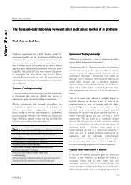
<strong>British</strong> <strong>Journal</strong> <strong>of</strong> <strong>Medical</strong> <strong>Practitioners</strong>, March 2010, Volume 3, Number 1BJMP 2010;3(1):303ResearchArticleThe Exeter Trauma Stem: Early results <strong>of</strong> a new cemented Hemiarthroplasy forfemoral neck fractureDavid Cash , Jens Bayer , Karl Logan and James WimhurstABSTRACTIntroduction: The Exeter Trauma Stem (ETS) is a new monoblock unipolar prosthesis with no independent published series using this implant. Thisstudy prospectively evaluates the first 50 ETS hemiarthroplasties performed as a primary treatment for fractured neck <strong>of</strong> femur at Norfolk and NorwichUniversity Hospital.Methods: Patient demographics and operative details were recorded from the patient notes. Radiographic evaluation involved the Barrack cementationgrading system, Dorr’s criteria and leg length measurement. All patients were sent an Oxford Hip Score questionnaire between two and four monthspostoperatively with 100% response rate.Results: Two thirds <strong>of</strong> cement mantles were Barrack grade A and B. Twenty-eight patients had lengthening <strong>of</strong> the operated limb with a mean <strong>of</strong> 12mm (5-30) including one irreducible prosthesis. Further complications included three deaths and one deep infection. The average Oxford Hip Score was 27.2.Discussion: Patient demographics were similar to previous studies. There was no statistical difference between the cement mantles and those <strong>of</strong> anotherpublished study using the Exeter stem. The major difficulty evident from this study was correct positioning <strong>of</strong> the prosthesis with regards to leg-length.Post-operative hip scores were similar to other studies as was the mortality rate.Conclusion: Post-operative functional and radiographic scoring <strong>of</strong> the ETS prosthesis were encouraging but care is needed with regards to correctpositioning <strong>of</strong> this prosthesis to attain equal leg lengths. Subsequent to the results <strong>of</strong> this study, a trialling system has been added to the instrumentationwhich the authors recommend in conjunction with pre-operative templating.KEYWORDSExeter Trauma Stem, Cemented hemiarthroplasty, Hip fractures, Leg length inequality, Barrack Grading, Oxford Hip Score.INTRODUCTIONThe Western world is experiencing a rapid increase in theincidence <strong>of</strong> femoral neck fractures, from 50000 fractures in1990 to a projected 120000 in 2015 1 as the age <strong>of</strong> thepopulation increases. Hip fractures account for approximately20 percent <strong>of</strong> orthopaedic bed occupancies in Britain at a totalcost <strong>of</strong> up to £25000 per patient 1 .Around half <strong>of</strong> these fracturesare intracapsular in nature <strong>of</strong> which two thirds are displaced.The ideal surgical treatment for displaced intracapsular femoralneck fractures remains controversial with studies indicating alack <strong>of</strong> consensus among treatment centres 2,3 . Options includereduction with internal fixation, cemented or cementless hemiarthroplastyand total hip replacement. Internal fixation is lesstraumatic than arthroplasty but has a higher re-operationrate 4,5 whilst cemented femoral prostheses are associated with alower rate <strong>of</strong> revision compared to cementless implants. Inaddition there are statistically significant improvements in painscores, walking ability, use <strong>of</strong> walking aids and activities <strong>of</strong> dailyliving within the cemented group 6,7 . The cementation processmay however be associated with increased morbidity due to fatembolisation and increased length <strong>of</strong> operation 8 .Treatment planning for intracapsular fractures, therefore, needsto take into account the patient’s medical fitness and activitylevel as well as the cost-effectiveness <strong>of</strong> the procedure.Figure 1: Exeter Trauma Stem (ETS) Implant6
<strong>British</strong> <strong>Journal</strong> <strong>of</strong> <strong>Medical</strong> <strong>Practitioners</strong>, March 2010, Volume 3, Number 1The Exeter Trauma Stem is a new monoblock unipolar implantusing an intermediate size 1.5, forty millimetre <strong>of</strong>fset Exeterstem with a large head sized to match the patient’s anatomy(Figure 1, 2).All fifty procedures were performed with the patient in thelateral position via the modified lateral approach with the gluteiincised at the musculotendinous junction. Cefuroxime wasgiven on induction in each instance followed by two postoperative doses at eight and sixteen hours after the procedure.Patients were scored by the hospital protocol for risk <strong>of</strong>thrombosis and were administered aspirin or subcutaneous lowmolecularweight heparin as appropriate. All drains wereremoved between twenty-four and forty-eight hours andpatients were mobilised within one day <strong>of</strong> operation as painallowed.Patient demographics and operative details were gathered bothfrom the patients’ notes and from the ORSOS computerisedtheatre system.Figure 2: X-ray <strong>of</strong> ETS with correct length. Neck cut has beenmade 1cm above lesser trochanter with shoulder <strong>of</strong> prosthesissunk below greater trochanter to ensure equal leg lengthAs yet there are no independent published series <strong>of</strong> the results <strong>of</strong>using this implant. Purported advantages <strong>of</strong> the ETS includethe use <strong>of</strong> a tried and tested polished, tapered stainless steel stemwith which many primary hip surgeons are familiar, ease <strong>of</strong>‘cement-in-cement’ revision to a total hip replacement shouldthe patient develop acetabular erosion and the relatively lowcost <strong>of</strong> £240 compared to many contemporary cementedimplants.This study prospectively evaluates the first 50 ETShemiarthroplasties performed at the Norfolk and NorwichUniversity Hospital, UK over a six month period providing anindication <strong>of</strong> early outcomes and complications involved withthe use <strong>of</strong> this prosthesis.METHODPatients presenting to our unit with a displaced intracapsularfemoral neck fracture who were sufficiently active to get out <strong>of</strong>their home independently, had an ASA grade <strong>of</strong> 1 or 2 and werenot significantly cognitively impaired were treated with acemented ETS prosthesis. In addition, patients with displacedintracapsular fractures associated with significant comminution<strong>of</strong> the medial femoral neck precluding the use <strong>of</strong> our standardcalcar-bearing Austin Moore (Stryker Howmedica OsteonicsLtd) hemiarthroplasty were also treated with an ETS regardless<strong>of</strong> functional capability and medical condition.The first fifty patients who underwent ETS hemiarthroplasty asa primary treatment for fractured neck <strong>of</strong> femur were includedin the study. Four patients were excluded. Two <strong>of</strong> these patientshad an ETS performed due to failure <strong>of</strong> cancellous screwfixation and two as part <strong>of</strong> a two stage revision for infecteduncemented prosthesis.Radiographic evaluation involved the Barrack 9 cementationgrading system, Dorr’s criteria 10,11 including varus/valgusalignment <strong>of</strong> the prosthesis and leg length measurement..Measurements <strong>of</strong> length and varus/valgus were performed usingthe PACS (GE <strong>Medical</strong> Systems 2005) digital imaging systemby two orthopaedic registrars independent <strong>of</strong> one another.Finally all fifty patients were sent an Oxford Hip Score 12 atbetween two and four months postoperatively. Three patientsdied before the questionnaires were sent and <strong>of</strong> the remainingforty seven, there was a 98% response rate with 44questionnaires completed solely by the patient and a further twocompleted with the aid <strong>of</strong> a carer.RESULTS1. Patient Demographics and operative detailsOf the fifty patients in the study, thirty six were female andfourteen male. The mean age was 78 (range 38 to 99). Fortyfour ETS hemiarthroplasties were performed due to patientfitness and activity levels (Type 1 patients) with six undertakenin frail patients due to fracture extension into the calcar (Type2). All type 1 patients were ASA grade 1 or 2 with all type 2patients ASA grade 2-4. All type 1 patients had a mini-mentaltest score <strong>of</strong> 10/10 with type 2 patients ranging from 0-7.The mean delay to surgery was 26 hours (9-58). Eightprocedures were performed by consultants, thirty eight byregistrars (training years three to six) and four by the traumafellow under supervision by a senior. The mean operative timewas sixty four minutes and the mean haemoglobin drop was 2.6g/dl 3 . Seven patients required post operative transfusion <strong>of</strong>either two or three units <strong>of</strong> packed cells.Thirty four <strong>of</strong> the patients mobilised unaided pre-injury witheight using one stick, four using two sticks and four using aframe. Using the four categories above, the average drop inmobility from injury to discharge was 1.6 levels.© BJMP.org7
<strong>British</strong> <strong>Journal</strong> <strong>of</strong> <strong>Medical</strong> <strong>Practitioners</strong>, March 2010, Volume 3, Number 1The average hospital stay was 8.6 days (range 5-69) with thirtyfive patients discharged to their own house, four to their ownresidential home and eleven to a rehabilitation ward.2. Radiographic EvaluationThe cement mantle was firstly evaluated using Barrack’sgrading:-grade A: medullary canal completely filled w/ cement (whiteout).grade B: a slight radiolucency exists at the bone cementinterface.grade C: a radiolucency <strong>of</strong> more than 50% at the bone cementinterface.grade D: radiolucency involving more than 100% <strong>of</strong> theinterface between bone and cement in any projection, includingabsence <strong>of</strong> cement distal to the stem tipPost-operative radiographic evaluation according to this systemshowed that 54% <strong>of</strong> cement mantles were Barrack grade B (27cases) with the majority <strong>of</strong> the remainder grade C (12 cases) andgrade A (eight cases). Two were graded as D.Dorr’s criteria were employed firstly to assess whether there wasan adequate cement thickness <strong>of</strong> 3mm in Gruen zones 3 and 7and <strong>of</strong> one centimetre distal to the tip <strong>of</strong> the prosthesis. Thirtyfourprosthesis scored 3/3, nine scored two, four scored one andtwo scored none.Dorr’s criteria also assess position <strong>of</strong> the prosthesis using the APradiograph. Ten prostheses were placed in a neutral positionrelated to the femoral shaft. Seven were placed in 1-2 degrees <strong>of</strong>varus, twenty-seven were placed in 1-2 degrees <strong>of</strong> valgus andfive were placed in 3-6 degrees <strong>of</strong> valgus.There were equal leg length measurements in nineteen patientspost-operatively with two patients left 5-10mm short on theoperated side. Twenty-eight patients were left long with a meanlengthening <strong>of</strong> 12mm (5-30) and <strong>of</strong> these five were left between20 and 30mm long one <strong>of</strong> which was irreducible and needed tobe revised on the table.3. Post-operative operative ScoringThe Oxford Hip Score contains 6 questions relating to pain andsix relating to function and mobility which are scored 1 pointfor the best outcome and five for the poorest (Score 12-60).The average pain score was 12.0 and the average functionalscore was 15.2 giving an overall score <strong>of</strong> 27.2. The type 1patients fared better with an average score <strong>of</strong> 25.3, the averagescore for type 2 patients was 44.3There was one superficial wound infection requiring antibiotictherapy and one early deep infection requiring open washout intheatre which resolved the infection in combination withantibiotic therapy.There were three deaths (one CVA, one MI and one frompneumonia) all <strong>of</strong> which occurred between 30-90 days from theoperative procedure.DISCUSSIONThe cohort <strong>of</strong> patients included in this study was similar toother studies with regards to male:female ratio, age andcognitive function 4,5 . The patients also experienced a delay tosurgery and length <strong>of</strong> operation similar to previous studies 4,7 .The length <strong>of</strong> inpatient stay, however, was markedly better at8.6 days compared to approximately fourteen to twenty-onedays cited in the literature 13,14 .The length <strong>of</strong> operation,post-operative mobility andtransfusion requirements were also similar to studies evaluatinghemiarthroplasty outcomes 4,5 .Post-operative radiographic evaluation showed greater than50% <strong>of</strong> cement mantles were Barrack grade B with the majority<strong>of</strong> the remainder grade C (24%) and A (16%). There was nostatistical difference between our findings and those <strong>of</strong> an 8-12year study <strong>of</strong> the Exeter stem in total hip replacement 15 . Thetwo Barrack D grade cement mantles were in patients whobecame unwell intra-operatively and the decision was taken notto pressurise during cementation.Figure 3: Original ETS broach with squared <strong>of</strong>f handle, notallowing intra-operative triallingThe major difficulty evident from this study is the correctpositioning <strong>of</strong> the ETS prosthesis with regards to restoration <strong>of</strong>accurate leg length which the authors believe was due to tworeasons. Firstly, the original set for the Exeter Trauma Stemcomes with one femoral broach (Fig 3) which does not allowtrial reduction. Therefore positioning <strong>of</strong> the prosthesis requiredintra-operative estimation <strong>of</strong> the correct leg length which can bedifficult with hip fractures as the leg length is abnormal at thecommencement <strong>of</strong> surgery. Therefore the centre <strong>of</strong> rotation <strong>of</strong>the femoral head on the injured side was approximated bycomparison with the contralateral side on the pelvic APradiograph and referenced against the level <strong>of</strong> the greatertrochanter during the procedure.4. ComplicationsThe one immediate complication was the need for an on-tablerevision due to an irreducible prosthesis.Secondly, because the large monoblock head <strong>of</strong> the ETS ismatched to the patient’s own femoral head anatomy, the© BJMP.org8
<strong>British</strong> <strong>Journal</strong> <strong>of</strong> <strong>Medical</strong> <strong>Practitioners</strong>, March 2010, Volume 3, Number 1diameter <strong>of</strong> the ETS head is generally around 15-30mm widerthan the 28mm heads commonly used with the Exeter stem inelective hip arthroplasty. Therefore care must be taken to sinkthe stem by a corresponding amount if a similar neck cut isused or the femoral neck osteotomy should be made at a moredistal level. This <strong>of</strong>ten involves positioning the shoulder <strong>of</strong> theETS stem below the level <strong>of</strong> the greater trochanter. This canmislead surgeons who are familiar with the Exeter stem asplacing the ETS stem in a similar position to that employedwith smaller head elective arthroplasty results in limblengthening. Figure 4 shows a leg length discrepancy <strong>of</strong> 15mmdespite a low neck cut as the stem has not been sunksufficiently. This led to 56% <strong>of</strong> patients being left with truelengthening <strong>of</strong> the operated limb and one prosthesis irreducible.It is difficult to assess whether this is a common problem in theliterature with other hemiarthroplasties used for femoral neckfractures as none <strong>of</strong> the comparable studies comment on clinicalor radiographic assessment <strong>of</strong> leg length.Figure 4: X-ray <strong>of</strong> ETS with limb lengthening. Although theneck cut has been made relatively low in relation to the lessertrochanter, the shoulder <strong>of</strong> the prosthesis slopes marginallyabove the greater trochanter, inadvertently lengthening theoperated limb.One major advantage to the tapered Exeter stem is the ease withwhich conversion to a total hip replacement can be performedusing an in-cement technique 16 . Many <strong>of</strong> the patients includedin this study were below the age <strong>of</strong> 70 and a proportion couldbe expected to outlive the prosthesis especially with regards toacetabular erosion 4 . Whilst none <strong>of</strong> this cohort has requiredrevision for loosening, the irreducible Exeter implant wasrevised on-table using this technique without furthercomplication.Post operative Oxford Hip Scores were encouraging with nodifference between our mean score <strong>of</strong> 27.2 and other studiesevaluating both cemented hemiarthroplasty and total hipreplacement following femoral neck fracture 12,17,18 .The mortality rate was 6% six to twelve months post surgerywith all three deaths more than one month post surgery andapparently unrelated to the surgery itself. Overall mortality ratesfollowing neck <strong>of</strong> femur fracture are approximately thirtypercent at one year however studies specifically looking atoutcomes following cemented hemiarthroplasty in the fit andactive patient have found mortality rates similar to this study 5,19 .Costing around £240, the ETS is a relatively cheap prosthesis incomparison to cemented bipolar prosthesis depite the additionalexpense <strong>of</strong> a cement restrictor, bone cement, cement gun andcement pressurisers.In conclusion, the Exeter Trauma Stem (ETS) is an effectivemethod <strong>of</strong> treating displaced intracapsular neck <strong>of</strong> femurfractures with encouraging post-operative functional, pain andradiographic scoring outcomes. The message highlighted by thisstudy is that additional care is needed with regards to thecorrect positioning <strong>of</strong> the prosthesis to ensure the restoration <strong>of</strong>limb length. Subsequent to discussion with the Strykerrepresentative regarding the results <strong>of</strong> this study, a secondgeneration trialling system has been added to the set with amodular broach. The authors suggest that not only should thesemodular broaches be used, but also accurate pre-operativeplanning is needed to ensure equal leg lengths post-operatively.Competing InterestsAuthor would like to state that none <strong>of</strong> the authors involved with thispaper have any financial or personal relationship with Stryker or anyother companies that could inappropriately influence this study.Author DetailsDAVID CASH, JENS BAYER, KARL LOGAN: Specialist Registrars,Orthopaedic Department, Norfolk and Norwich HospitalJAMES WIMHURST, Consultant orthopaedic Surgeon, OrthopaedicDepartment, Norfolk and Norwich HospitalCORRESSPONDENCE: Mr. D Cash, Specialist Registrar,Orthopaedic Dept, Addenbrooke’s Hospital, Long Road, CambridgeCB2 0QHEmail: davecash@doctors.org.ukREFERENCES1. Parrott S : The economic cost <strong>of</strong> hip fracture in the UK (2000)www.dti.gov.uk/files/file21463.<strong>pdf</strong>2. Crossman PT, Khan RJK, MacDowell A, Gardner AC, Reddy NS, KeeneGS A survey <strong>of</strong> the treatment <strong>of</strong> displaced intracapsular femoral neckfractures in the UK. Injury, 33(2002): 383-3863. Anderson GH, Harper WM, Gregg PJ: Management <strong>of</strong> the intracapsularfractures <strong>of</strong> the proximal femur in 1990 : a cause for concern? J Bone JointSurg (Br) 73B(Suppl 1) (1991) : 734. Parker MJ, Khan RJK, Crawford J, Pryor GA: Hemiarthroplasty versusinternal fixation for displaced intracapsular hip fractures in the elderly. JBone Joint Surg (Br), 84(8) (2002): 1150-55. Davison JNS, Calder SJ, Anderson GH, Ward G, Jagger C, Harper WM,Gregg PJ: Treatment for displaced intracapsular fracture <strong>of</strong> the proximalfemur. J Bone Joint Surg (Br) 83 (2001) : 206-16. Khan RJK, MacDowell A, Crossman PT, Keene GS: Cemented oruncemented hemiarthroplasty for displaced intracapsular fractures <strong>of</strong> thehip – a systematic review. Injury 33 (2002) : 13-177. Keating JF, Grant A, Masson M, Scott NW and Forbes JF: Randomizedcomparison <strong>of</strong> reduction and fixation, bipolar hemiarthroplasty, and totalhip arthroplasty. Treatment <strong>of</strong> displaced intracapsular hip fractures inhealthy older patients. J Bone Joint Surg Am, 88(2) (2006): 249-60,8. Parker MJ: The management <strong>of</strong> intracapsular fractures <strong>of</strong> the proximalfemur. J Bone Joint Surg (Br) 82 (2000) : 937-41© BJMP.org9
<strong>British</strong> <strong>Journal</strong> <strong>of</strong> <strong>Medical</strong> <strong>Practitioners</strong>, March 2010, Volume 3, Number 19. Barrack RL, Mulroy, RD Jr and Harris WH: Improved cementingtechniques and femoral component loosening in young patients with hiparthroplasty. A 12-year radiographic review. J Bone Joint Surg Br, 74(3)(1992.): 385-910. Dorr LD, Luckett M and Conaty JP: Total hip arthroplasties in patientsyounger than 45 years. A nine to ten-year follow-up study. Clin OrthopRelat Res, (260) (1990.): 215-911. Dorr LD, Takei GK and Conaty JP: Total hip arthroplasties in patientsless than forty-five years old. J Bone Joint Surg Am, 65(4) (1983): 474-912. Dawson J, Fitzpatrick R, Carr A, and Murray D: Questionnaire on theperceptions <strong>of</strong> patients about total hip replacement. J Bone Joint Surg Br,78(2) (1996.): 185-9013. Department <strong>of</strong> Health. Hospital episode statistics, England: Financialyear 1993-1994; volume 1. London: HMSO, 1994.14. Hay D, Parker MJ: Hip fracture in the immobile patient. J Bone JointSurg (Br) 85 (2003) : 1037-915. Williams HDW, Browne G, Gie GA, Ling RSM, Timperley AJ,Wendover NA: The Exeter universal cemented femoral component ateight to twelve years J Bone Joint Surg (Br) 84 (2002) 324-3416. W W Duncan, M J W Hubble, A J Timperley and G A Gie: Cement incement femoral revision with the Exeter hip. J Bone Joint Surg (Br) 88(Suppl 2) (2006) : 23917. Mishra V, Thomas G, Sibley TF: Results <strong>of</strong> displaced subcapitalfractures treated by primary total hip replacement. Injury 35(2) (2004):157-60.18. Wazir NN, Mukundala VV, Choon DSK: Early results <strong>of</strong> prosthetic hipreplacement for femoral neck fractures in active elderly patients. J OrthopSurg 14 (2006) 43-619. Keating JF, Grant A, Massom M, Scott NW, Forbes JF: Displacedintracapsular hip fractures in fit, older people: a randomised comparison<strong>of</strong> reduction and fixation, bipolar hemiarthroplasty and total hiparthroplasty. Health Technol Assess 9 (2005) 1-65© BJMP.org10
<strong>British</strong> <strong>Journal</strong> <strong>of</strong> <strong>Medical</strong> <strong>Practitioners</strong>, March 2010, Volume 3, Number 1BJMP 2010;3(1):306ResearchArticleEducation in the Foundation Programme: what doctors are doing and whyMJ Keogh , JM Findlay , S Sithamparanathan and D MathesonAbstractThe Foundation Programme details the first two years <strong>of</strong> training for UK doctors in the UK. Thereafter, trainees are expected to apply for highlycompetitive specialist training posts. Our study aimed to clarify and quantify the educational activities currently used by Foundation doctors during thistwo year period, and to assess their motivational and deterring factors towards such educational activities.Method: A fourteen point questionnaire was posted at random to 100 Foundation Year 1 and 2 (50 FY1 and 50 FY2) doctors across five Trent Deaneryhospitals. The questionnaire assessed involvement in the following voluntary educational activities: courses, conferences, higher qualifications, e-learningpackages and personal reading. It also sought their underlying attitudes.Results: Response rate was 49.0% (49/100), comprising 34 (68%) FY1 and 15 (30%) FY2. Overall 89.8% <strong>of</strong> respondents engaged in voluntaryeducational activities. The most common (89.8%) was the e-learning package (FY1 85.3%, FY2 100%) followed by society membership (73.5% (FY164.7%, FY2 93.3%), courses (69.4%) (FY1 55.9%, FY2 100%), sitting higher qualifications (36.7%) (FY1 14.7%, FY2 86.7%) and attending conferences(14.3%) (FY1 14.7%, FY2 13.3%). The mean total cost incurred by doctors for these activities was £581 in FY1 and £1842 in FY2.The most common deterrents to pursuing voluntary education were a lack <strong>of</strong> study leave (42.9%) (FY1 38.2%, FY2 53.3%), lack <strong>of</strong> annual leave (22.4%)(FY1 23.5% FY2 20.0%) and expense (20.4%) (FY1 17.6%, FY2 26.7%).The most common motivating factor was the belief they would help candidates achieve a specialist training post (67.3%) (FY1 58.8%, FY2 86.7%). Only8.2% (FY1 11.8%, FY2 0.0%) engaged primarily to improve their medical competence.Discussion: Our study is the first to quantify the voluntary educational activities <strong>of</strong> Foundation doctors. Most popular are e-learning packages —outstripping courses, higher qualification revision and conferences — highlighting their increasing popularity as a viable and accessible educational tool.The primary deterrent to pursuing voluntary educational activities is lack <strong>of</strong> study leave, <strong>of</strong> concern as entitlements to this continue to decrease.Interestingly, Foundation doctors are not motivated primarily by the educational benefits <strong>of</strong> these activities, but rather by their perceived ability to helpattain a specialist training post. This highlights the concerning potential for voluntary educational activities to become a badge <strong>of</strong> attendance, underminingtheir intrinsic educational value and outcome.The implementation <strong>of</strong> Modernising <strong>Medical</strong> Careers (MMC)significantly altered the structure <strong>of</strong> postgraduate medicaleducation in the UK. MMC oversees the training <strong>of</strong> all UKdoctors from the outset <strong>of</strong> their career, the first two years <strong>of</strong>which comprise the Foundation Programme. Successfulcompletion <strong>of</strong> the Foundation Programme is based upondoctors’ Foundation Portfolios in which they must demonstrateachievement <strong>of</strong> essential competences and work-basedassessments. Doctors are also encouraged to attain additionalcompetencies and to develop their portfolio further. Voluntaryeducational activities undertaken outside the workplace formthe basis <strong>of</strong> this.Application into Specialist Training following the FoundationProgramme is highly competitive, with an average <strong>of</strong> threeapplicants for each post in 2008 1 . Points-based shortlistingcriteria are used to select candidates, and are based upon thecontents <strong>of</strong> the Foundation Portfolio and application form.This means that points can be scored for activities not requiredfor completion <strong>of</strong> the Foundation Programme, such as RoyalCollege membership examinations and course attendance.Foundation Programme doctors undertake voluntary activitiesto improve their portfolios however no quantifiable evidencecurrently exists as to what doctors undertake in this respect.We aimed, therefore, to determine firstly what voluntaryeducational activities Foundation doctors are undertaking. Wealso aimed to establish their underlying motivating anddeterring factors, financial costs incurred, and use <strong>of</strong> annual andstudy leave and ‘specialty taster days’, to assess the overall extentand impact <strong>of</strong> portfolio activities. The authors hope the resultsare useful in informing medical students and Foundationtrainees <strong>of</strong> the scope <strong>of</strong> activities <strong>of</strong> their peers, and in advisingsupervisors <strong>of</strong> the activities <strong>of</strong> their trainees.MethodsA two page anonymous questionnaire was posted at random to100 Foundation doctors across five hospitals in East MidlandsDeanery (50 Foundation Year 1, 50 Foundation Year 2). SeeAppendix 1DemographicsThe first section <strong>of</strong> the questionnaire asked for the sex andgrade <strong>of</strong> respondents (Foundation Year 1 (FY1), or FoundationYear 2 (FY2))ActivitiesRespondents were directly asked whether they were attendingcourses or conferences, using on-line e-learning packages,© BJMP.org11
<strong>British</strong> <strong>Journal</strong> <strong>of</strong> <strong>Medical</strong> <strong>Practitioners</strong>, March 2010, Volume 3, Number 1joining pr<strong>of</strong>essional bodies/societies or sitting higherpr<strong>of</strong>essional examinations such as royal college membershipexaminations/higher degrees.CostDoctors were asked how much money (excluding that <strong>of</strong>teaching allowances) and days <strong>of</strong> annual leave they used on theabove activities. They were also asked how many <strong>of</strong> theirallowed ‘specialty taster days’ they had taken during each year.Motivating and deterring factorsDoctors were asked to rank from a list the motivating anddeterring factors determining what activities they wereundertaking.Of the courses attended, 25.5% pertained to teaching, 25.5%to advanced life support and 18.0% to surgical skills. Theremaining 31% <strong>of</strong> courses related to a variety <strong>of</strong> other interestssuch as anaesthetic skill days, expedition medicine courses, andsub speciality specific courses such as movement disorderworkshops and laparoscopic surgery.CostThe mean amount spent by Foundation Year 1 Doctors onthese activities was £581 (range £0 - £3100) Foundation Year 2Doctors spent significantly more at £1842 (range £0 - £3500).The mean cost per activity is shown in figure 2.Pr<strong>of</strong>essional developmentDoctors were finally asked to rank which educational activitiesthey thought would make them a better overall Foundationdoctor.ResultsResponse rate was 49% with 49 doctors returning thequestionnaire. Of these 69.4% (n=34) were Foundation Year 1(FY1) and 30.6% (n=15) were Foundation Year 2 (FY2), with53.1% female and 46.9% male.ActivitiesOverall 89.8% (n=44) <strong>of</strong> respondents were engaged involuntary educational activity (FY1 85.3%, FY2 100%). Themost common mode (89.8%, n=44) was e-learning packages(FY1 85.3% (n=29), FY2 100% (n=15)) followed by joining/becoming a member <strong>of</strong> pr<strong>of</strong>essional bodies or societies ie BMAetc (73.5%, n=36) (FY1 64.7% (n=22), FY2 93.3% (n=14)),followed by courses (69.4%, n=34) (FY1 55.9% (n=19), FY2100% (n=15)), undertaking higher qualifications (36.7%) (FY114.7% (n=5), FY2 86.7% (n=13)) and attending conferences(14.3%) (FY1 14.7% (n=5), FY2 13.3% (n=2))– See figure 1.Fig 2 – A graph to show the mean amount <strong>of</strong> money spent byfoundation year 1 and 2 doctors on each mode <strong>of</strong> educationalactivity.The mean number <strong>of</strong> days <strong>of</strong> annual leave used by doctors forthese activities was 2.8 in FY1 and 5.3 in FY2, thereforecombining to average 8.1 days in total that would be used overthe whole foundation programme. Of their five allowed ‘taster– days’ the mean number attended was 1.3 and 2.9 by FY1 andFY2 doctors respectively. Only 20.4% <strong>of</strong> doctors took their fullentitled allowance.Motivating and deterring factorsThe most common factor motivating Foundation doctors toundertake portfolio educational activities was the belief theywould help candidates achieve a specialist training post(67.3%). Only 12.2% engaged primarily out <strong>of</strong> personalinterest with 8.2% to improve their medical competence (SeeTable 1).The most common deterrents were a lack <strong>of</strong> study leave(42.9%), lack <strong>of</strong> annual leave (22.4%) and expense (20.4%)(See Table 2).Pr<strong>of</strong>essional developmentFig 1 – A graph to show the percentage <strong>of</strong> Foundation year 1and 2 doctors involved in each mode <strong>of</strong> voluntary educationalactivity.The final section <strong>of</strong> the questionnaire asked respondents whicheducational activity they felt was most influential in makingthem a better Foundation doctor. Interestingly 83.7%(n=41)(FY1 88.2% (n=30), FY2 73.3%( n=11)) felt on-callexperience was most influential, with only 6.1% (FY1 2.9%© BJMP.org12
<strong>British</strong> <strong>Journal</strong> <strong>of</strong> <strong>Medical</strong> <strong>Practitioners</strong>, March 2010, Volume 3, Number 1(n=1), FY2 13.3% (n=2)) citing courses, 6.1 % (FY1 2.9%(n=1), FY2 13.3% (n=2)) e-learning packages and 4.1% (FY12.9% (n=1), FY2 6.7% (n=1)) qualifications (Fig 3).Primary Motivating Factor FY1 FY2 Overall(%) n (%) n (%) nImprove chance <strong>of</strong> specialisttraining post58.8 20 86.7 13 67.3 33Personal interest 14.7 5 6.7 1 12.2 6To improve medicalcompetencies11.8 4 0 0 8.2 4On advice <strong>of</strong> seniors 11.8 4 6.7 1 10.2 5Other 2.9 1 0 0 2 1TOTAL 100 34 100 15 100 49Table 1 – A table to show the primary motivating factors <strong>of</strong> foundationdoctors to undertake voluntary portfolio educational activities.Primary Deterring FactorFY1 Doctors FY2 Doctors Overall(%) n (%) n (%) nLack <strong>of</strong> study leave 38.2 13 53.3 8 42.9 21Lack <strong>of</strong> annual leave 23.5 8 20 3 22.4 11Financial expense 17.6 6 26.7 4 20.4 10Lack <strong>of</strong> career choice 11.8 4 0 0 8.2 4Not relevant to Foundationdoctors8.8 3 0 0 6.1 3Other 0 0 0 0 0 0TOTAL 100 34 100 15 100 49Table 2 – A table to show the primary deterring factors listed byfoundation doctors that deter them from undertaking voluntaryeducational portfolio activities.Pr<strong>of</strong>essional developmentThe final section <strong>of</strong> the questionnaire asked respondents whicheducational activity they felt was most influential in makingthem a better Foundation doctor. Interestingly 83.7%(n=41)(FY1 88.2% (n=30), FY2 73.3%( n=11)) felt on-callexperience was most influential, with only 6.1% (FY1 2.9%(n=1), FY2 13.3% (n=2)) citing courses, 6.1 % (FY1 2.9%(n=1), FY2 13.3% (n=2)) e-learning packages and 4.1% (FY12.9% (n=1), FY2 6.7% (n=1)) qualifications (Fig 3).The academic conference was ranked least influential by 89.8%(n=44) (FY1 85.3% (n=29), FY2 100% (n=15)) <strong>of</strong> respondents,followed by 6.1% (n=3) (FY1 8.8% (n=3), FY2 0.0% (n=0))citing courses, and 4.8% (FY1 5.8% (n=2), FY2 0.0% (n=0)) e-learning packages (Fig 3).DiscussionThis survey suggests that Foundation doctors undertakenumerous activities at significant personal expense to expandtheir portfolios, and are primarily motivated by a belief that thiswill increase their chance <strong>of</strong> obtaining higher specialist trainingposts.Fig 3 – The above graph was the response <strong>of</strong> Foundationdoctors when asked which activities they thought were mostand least influential in making them a better foundation doctor.Educational activities and opportunitiesThe advent <strong>of</strong> the European Working Time Directive and NewDeal document 2 have resulted in junior doctors workingconsiderably fewer hours than in previous years. This has ledsome authors to conclude that the quality <strong>of</strong> learningopportunities in the working environment has reduced 3 .With89.8% <strong>of</strong> Foundation doctors in this survey activelyundertaking some form <strong>of</strong> educational activity outside <strong>of</strong> work,this suggests that Foundation doctors may be going some wayto re-dressing this balance. It may also come as a surprising yetreassuring figure to Foundation Programme educationalsupervisors who may be unaware <strong>of</strong> the education <strong>of</strong> theirtrainees outside <strong>of</strong> work.We found the most popular mode <strong>of</strong> educational activity to bethe e-learning package. E-learning is an effective and extensivelyemployed method for both distance learning 4 , and as anadjunct to “traditional” lecture-based techniques across severaldisciplines. It has also been shown to be a well received andpractical method <strong>of</strong> supplementary education for doctors 5 andour study suggests this is particularly true for the Foundationyears. The reasons why e-learning is popular in this group wasnot explored, but its low cost, easily accessible and modularnature may have some part to play. As medical schools continueto utilise this modality to a greater extent, its follow-throughinto the Foundation years and postgraduate medical educationin general is inevitable. With such high uptake, e-learningpackages are a promising format for delivering education to thisgroup.Popular courses undertaken by Foundation doctors related toobtaining teaching skills, or advanced life support. This suggeststhat Foundation doctors place a high emphasis on teaching andtraining, and on recognising and managing acutely ill patients.These are two core objectives <strong>of</strong> the Foundation Programme.However, one could also argue that doctors undertaking coursesoutside work to achieve essential competencies casts doubt onthe ability <strong>of</strong> the Foundation Programme to deliver them. We© BJMP.org13
<strong>British</strong> <strong>Journal</strong> <strong>of</strong> <strong>Medical</strong> <strong>Practitioners</strong>, March 2010, Volume 3, Number 1submit that educational supervisors are in a prime position toappraise this issue.The least popular mode <strong>of</strong> activity in our survey was theattendance <strong>of</strong> a medical conference. It was also regarded as leastinfluential by 89.7% <strong>of</strong> respondents. There is a global shortage<strong>of</strong> medical academics 6 , and as conferences serve to introducejunior doctors to academic medicine and research, perhapsacademic doctors should take a more prominent role inpromoting conferences as an educational activity.Time and moneyDoctors incur the majority <strong>of</strong> their costs attending courses withFoundation Year 1 and 2 doctors spending £365 and £1120respectively on this area (fig 2). This highlights the possibilitythat Foundation doctors may be prone to financial exploitationby a growing number <strong>of</strong> courses which are <strong>of</strong>ten unvalidated. Assenior advice was the primary motivating factor for only 10.2%<strong>of</strong> activities, this suggests that educational supervisors could playa greater role in assessing, appraising and advising their traineeson the courses best suited for them and their pr<strong>of</strong>essionaldevelopment.The overall financial cost incurred for all portfolio educationalactivities was £581 for FY1 and £1842 for FY2. Whilst previousestimates have been made in this area, this is the first specific tothe Foundation Programme and to include non-mandatoryoutlay, and represents 3 % and 7% <strong>of</strong> the basic salary for FY1and FY2 doctors before tax. As our survey found financialexpense to be a significant deterrent to portfolio activity (20.4%<strong>of</strong> respondents), a potentially serious implication is that expensewill limit the uptake <strong>of</strong> postgraduate education in the future.From the authors’ own experience such pr<strong>of</strong>essional costs arenot explained to medical students and that this issue meritsmore attention in undergraduate education.A lack <strong>of</strong> study leave was highlighted as the main deterringfactors to educational portfolio activities (42.9%). This is <strong>of</strong>particular interest as only 20.4% <strong>of</strong> Foundation doctors usetheir full ‘taster-day’ entitlement. These ‘taster days’ are afundamental aspect <strong>of</strong> the Foundation Programme, <strong>of</strong>feringdoctors the opportunity to explore a specialty for up to five daysper year. However, whilst doctors fail to utilise them, they takean average <strong>of</strong> 8.1 days’ annual leave over the two yearprogramme for educational purposes.The reasons behind this are unclear, but may be due to a lack <strong>of</strong>awareness <strong>of</strong> these ‘taster days’. With a lack <strong>of</strong> study leavehindering educational activities, a potential solution might befor doctors to have the option to utilise ‘taster days’ as a form <strong>of</strong>study leave.Pr<strong>of</strong>essional education and motivationBetween 1998 and 2005, the number <strong>of</strong> medical students in theUK has risen by 57% 7 . Increasing numbers <strong>of</strong> doctors anddecreasing working hours may reduce the amount <strong>of</strong> on-callexperience for those in the Foundation Programme. However,it is this on-call experience that is regarded by the vast majority(83.7% in this study) as the most important educationalmodality in making them a better foundation doctor. Althoughtime and money are perceived as barriers to portfolioeducational activities it appears that doctors value this on-callexperience above all. With key aims <strong>of</strong> the FoundationProgramme being training and emergency competence, effortsmust be made to preserve this experience.Whilst Foundation doctors are engaging in numerous portfolioactivities, their underlying motivations are interesting. Itappears this group are primarily motivated not by theeducational benefits <strong>of</strong> these activities, but rather by theirperceived ability to help attain a specialist training post. Thiscould suggest that the educational portfolio is at risk <strong>of</strong>becoming a ‘tick-box’ means for career progression, rather thanaddressing limitations, exploring interests and aspiring toclinical excellence. This contrasts with the conclusions <strong>of</strong> themost recent assessment <strong>of</strong> postgraduate medical education inthe UK 8 .As competition for jobs appears to be driving Foundationdoctors to undertake educational activities it remains unclearwhether engaging in these activities to obtain jobs, rather thancompetencies, reduces their validity and educational outcomes.Furthermore it is unclear whether trainees will be more likely toachieve their overriding aim <strong>of</strong> obtaining a specialist trainingpost through these activities. Determining the career outcomes<strong>of</strong> doctors undertaking these activities will provide an evidencebase, allowing educational supervisors to optimally advise theirtrainee in portfolio educational activities.ConclusionsThis is a baseline survey quantifying portfolio educationalactivities in the Foundation Programme, applicable to traineesand supervisors alike. Whilst the latter are well aware <strong>of</strong>assessments such as DOPS (Direct Observation <strong>of</strong> ProceduralSkills) and CbD’s (Case-based Discussions), they are <strong>of</strong>ten lessaware <strong>of</strong> the voluntary educational activities <strong>of</strong> their trainees.Our study would suggest that Foundation Programme doctorsare a cohort driven to undertake numerous voluntaryeducational activities, albeit largely to achieve career progressionrather than accrue educational benefit. To this end theyundertake activities such as e-learning, courses and higherqualifications at the expense <strong>of</strong> conferences. For this they spendsignificant amounts <strong>of</strong> money and leave, yet continue to site alack <strong>of</strong> traditional study leave as a barrier to further educationaldevelopment. The authors would suggest that further work isneeded to develop the role <strong>of</strong> educational supervisors in theFoundation Programme in harnessing the motivation <strong>of</strong> theirtrainees, and guiding them appropriately.© BJMP.org14
<strong>British</strong> <strong>Journal</strong> <strong>of</strong> <strong>Medical</strong> <strong>Practitioners</strong>, March 2010, Volume 3, Number 1Key Points• Foundation Doctors spend significant amounts <strong>of</strong>time and money on voluntary educational activities.• Foundation Doctors are primarily driven toundertake these activities due to the belief that it willhelp them obtain specialist training posts.• A lack <strong>of</strong> study leave is the primary barrier tovoluntary education.• The academic medical conference is viewed as theactivity least likely to improve medical competence,whereas on-call experience is regarded as the mostlikely.• Foundation Programme educational supervisors arebest placed to guide their trainees towards the mostappropriate educational modalitiesCompeting InterestsNone DeclaredAuthor DetailsM J Keogh, BMedSci (Hons), BMBS (Hons). Research Fellow, University <strong>of</strong>Auckland, New ZealandJ M Findlay, BMedSci (Hons), BMBS (Hons). Core Surgical Trainee, JohnRadcliffe Hospital, Oxford, UKS Sithamparanathan, BMedSci (Hons), BMBS (Hons) Core <strong>Medical</strong> Trainee,Surrey, UKA Looseley, BMedSci (Hons), BMBS. Intensive Care Registrar, Mona ValeHospital, Sydney, Australia.D Matheson, Lecturer in <strong>Medical</strong> Education, University <strong>of</strong> Nottingham, UKCORRESSPONDENCE: M J KEOGH BMedSci (Hons), BMBS (Hons).Research Fellow, University <strong>of</strong> Auckland, New Zealand 670 Mount Eden Road,Auckland, New ZealandEmail: mikekeogh@doctors.org.ukREFERENCES1. MMC, Modernising <strong>Medical</strong> Careers. Specialty competition ratios, 2008.http://www.mmc.nhs.uk/Docs/TABLE%20for%20competion%20ratios%20page%20_2_.<strong>pdf</strong>.2. The Department <strong>of</strong> Health, L., Hours <strong>of</strong> work <strong>of</strong> doctors in training; thenew deal. 1991.3. Scallan, S., Education and the working patterns <strong>of</strong> junior doctors in theUK: a review <strong>of</strong> the literature. Med Educ, 2003. 37(10): p. 907-12.4. Sitzmann T, K.K., Stewart D, Wisher R, The comparative effectiveness <strong>of</strong>web-based and classroom instruction: a meta-analysis. Personnel Psychology,2006. 59: p. 623-664.5. Autti T, A.H., Vehmas T, Laitalainen V, Kivisaari L, E-learning is a wellacceptedtool in supplementary training among medical doctors: anexperience <strong>of</strong> obligatory radiation protection training in healthcare. ActaRadiol, 2007. 48(5): p. 508-513.6. Pritchard, L., International rescue. Med Educ, 2005. 39(2): p. 122-4.7. Higher Education Funding Council for England. Increasing medicalstudent numbers in England (Report 01/31). Bristol: HEFCE, 2001.8. MMC, MMC Inquiry. Aspiring to excellence. Final report <strong>of</strong> theIndependent inquiry into Modernising <strong>Medical</strong> Careers led by Pr<strong>of</strong>essor SirJohn Tooke. Aldridge presss, London, 2008.Appendix 1Educational Activities in the Foundation Programme –QuestionnaireWhat is your current grade (F1 or F2)? F1 F2 What sex are you ? Male Female 1) Which <strong>of</strong> the following educational activities have youundertaken as an F1/F2 Doctor outside <strong>of</strong> that deemedmandatory by your employing deanery or hospital?Completed Online/e-learning packages Which? __________________________________________What was the personal financial cost to you? _____________Attended courses (eg ATLS, ALS etc) Which?__________________________________________What was the personal financial cost to you? _____________Attended local, regional or national conferences Which? __________________________________________What was the personal financial cost to you?______________Attempted higher academic qualifications eg MRCP, MRCS exams Which? __________________________________________What was the personal financial cost to you? _____________Joined any pr<strong>of</strong>essional bodies or societies Which? ________________________________________What was the personal financial cost to you? ____________2) From the list below, please choose the option that has moststrongly motivated you to undertake the activities you haveoutlined in question 1To further my personal knowledge/interest in an areaTo improve my overall ability/achieve my competencies asFoundation Doctor To increase my chance <strong>of</strong> obtaining a specialist training (ST1) postin my chosen areaI have been advised to undertake certain activities by seniors Free text/other ______________________________________3) From the list below, please choose the main reason why youhave not engaged in further educational activities to those youlisted in question 1Due to financial expense There is a lack <strong>of</strong> study leave There is a lack <strong>of</strong> annual leave I am still not decided on a firm career choice I don’t think these activities are needed by Foundation doctors. Other ____________________________________________4) From the list <strong>of</strong> choices below, please mark which activity youfeel will most improve your overall ability as a foundationdoctor, and which you feel will have the least effect? (Pleaserespond with ‘M’ for most and ‘L’ for least)Online/e-learning packages Attending courses (eg ATLS, ALS etc) Attending local, regional or national conferences Sitting higher academic qualifications eg MRCP, MRCS exams On call in hospital experience 5) How many <strong>of</strong> your allowed specialty taster days have you usedthis year?_________________________________________________6) How many days <strong>of</strong> annual leave/ holiday have you used this yearto undertake voluntary educational activities?_________________________________________________© BJMP.org15
<strong>British</strong> <strong>Journal</strong> <strong>of</strong> <strong>Medical</strong> <strong>Practitioners</strong>, March 2010, Volume 3, Number 1BJMP 2010;3(1):307ResearchArticlePredictors <strong>of</strong> Difficult Intubation: Study In Kashmiri PopulationArun Kr. Gupta , Mohamad Ommid , Showkat Nengroo , Imtiyaz Naqash and Anjali MehtaAbstractAirway assessment is the most important aspect <strong>of</strong> anaesthetic practice as a difficult intubation may be unanticipated. A prospective study was done tocompare the efficacy <strong>of</strong> airway parameters to predict difficult intubation. Parameters studied were degree <strong>of</strong> head extension, thyromental distance, interincisor gap, grading <strong>of</strong> prognathism, obesity and modified mallampati classification. 600 Patients with ASA I& ASA II grade were enrolled in the study. Allpatients were preoperatively assessed for airway parameters. Intra-operatively all patients were classified according to Cormack and Lehane laryngoscopicview. Clinical data <strong>of</strong> each test was collected, tabulated and analyzed to obtain the sensitivity, specificity, positive predictive value & negative predictivevalue. Results obtained showed an incidence <strong>of</strong> difficult intubation <strong>of</strong> 3.3 % <strong>of</strong> patients. Head and neck movements had the highest sensitivity (86.36%);high arched palate had the highest specificity (99.38%). Head and neck movements strongly correlated for patients with difficult intubation.KEYWORDSIntubation, Anaesthesia, LaryngoscopyIntroductionThe fundamental responsibility <strong>of</strong> an anesthesiologist is tomaintain adequate gas exchange through a patent airway.Failure to maintain a patent airway for more than a few minutesresults in brain damage or death 1 . Anaesthesia in a patient witha difficult airway can lead to both direct airway trauma andmorbidity from hypoxia and hypercarbia. Direct airway traumaoccurs because the management <strong>of</strong> the difficult airway <strong>of</strong>teninvolves the application <strong>of</strong> more physical force to the patient’sairway than is normally used. Much <strong>of</strong> the morbidityspecifically attributable to managing a difficult airway comesfrom an interruption <strong>of</strong> gas exchange (hypoxia andhypercapnia), which may then cause brain damage andcardiovascular activation or depression 2 .Though endotracheal intubation is a routine procedure for allanesthesiologists, occasions may arise when even an experiencedanesthesiologist might have great difficulty in the technique <strong>of</strong>intubation for successful control <strong>of</strong> the airway. As difficultintubation occurs infrequently and is not easy to define,research has been directed at predicting difficult laryngoscopy,i.e. when is not possible to visualize any portion <strong>of</strong> the vocalcords after multiple attempts at conventional laryngoscopy. It isargued that if difficult laryngoscopy has been predicted andintubation is essential, skilled assistance and specialistequipment should be provided. Although the incidence <strong>of</strong>difficult or failed tracheal intubation is comparatively low,unexpected difficulties and poorly managed situations mayresult in a life threatening condition or even death 3 .Difficulty in intubation is usually associated with difficulty inexposing the glottis by direct laryngoscopy. This involves aseries <strong>of</strong> manoeuvres, including extending the head, opening themouth, displacing and compressing the tongue into thesubmandibular space and lifting the mandible forward. Theease or difficulty in performing each <strong>of</strong> these manoeuvres can beassessed by one or more parameters 4 .Extension <strong>of</strong> the head at the atlanto-occipital joint can beassessed by simply looking at the movements <strong>of</strong> the head,measuring the sternomental distance, or by using devices tomeasure the angle 5 . Mouth opening can be assessed bymeasuring the distance between upper and lower incisors withthe mouth fully open. The ease <strong>of</strong> lifting the mandible can beassessed by comparing the relative position <strong>of</strong> the lower incisorsin comparison with the upper incisors after forward protrusion<strong>of</strong> the mandible 6 . The measurement <strong>of</strong> the mento-hyoiddistance and thyromental distance provide a rough estimate <strong>of</strong>the submandibular space 7 . The ability <strong>of</strong> the patient to movethe lower incisor in front <strong>of</strong> the upper incisor tells us about jawmovement. The classification provided by Mallampati et al 8 andlater modified by Samsoon and Young 9 helps to assess the size<strong>of</strong> tongue relative to the oropharynx. Abnormalities in one ormore <strong>of</strong> these parameters may help predict difficulty in directlaryngoscopy 1 .Initial studies attempted to compare individual parameters topredict difficult intubation with mixed results 8,9 . Later studieshave attempted to create a scoring system 3,10 or a complexmathematical model 11,12 . This study is an attempt to verifywhich <strong>of</strong> these factors are significantly associated with difficultexposure <strong>of</strong> glottis and to rank them according to the strength<strong>of</strong> association.Materials & methodsThe study was conducted after obtaining institutional reviewboard approval. Six hundred ASA I & II adult patients,scheduled for various elective procedures under generalanesthesia, were included in the study after obtaining informedconsent. Patients with gross abnormalities <strong>of</strong> the airway wereexcluded from the study. All patients were assessed the eveningbefore surgery by a single observer. The details <strong>of</strong> airwayassessment are given in Table I.© BJMP.org16
<strong>British</strong> <strong>Journal</strong> <strong>of</strong> <strong>Medical</strong> <strong>Practitioners</strong>, March 2010, Volume 3, Number 1Table I: Method <strong>of</strong> assessment <strong>of</strong> various airway parameters(predictors)Airway ParametererModifiedMallampati ScoringMethod <strong>of</strong> assessmentClass I: Faucial pillars, s<strong>of</strong>t palate and uvulavisible.Class II: S<strong>of</strong>t palate and base <strong>of</strong> uvula seenClass III: Only s<strong>of</strong>t palate visible.Class IV: S<strong>of</strong>t palate not seenClass I & II : Easy IntubationClass III & IV: Difficult IntubationObesity Obese BMI (≥ 25)Non Obese BMI (< 25)Inter Incisor GapThyromentaldistanceDegree <strong>of</strong> HeadExtensionGrading <strong>of</strong>PrognathismDistance between the incisors with mouth fullyopen(cms)Distance between the tip <strong>of</strong> thyroid cartilage andtip <strong>of</strong> chin, with fully extended(cms)Grade I ≥ 90 ◦Grade II = 80 ◦ -90 ◦Grade III < 80 ◦Class A: - Lower incisor protruded anterior to theupper incisor.Class B: - Lower incisor brought edge to edgewith upper incisor but not anterior to them.Class C: - Lower incisors could be brought edgeto edge.Table II: The frequency analysis <strong>of</strong> predictor parametersAirway ParameterModified MallampatiScoringGroupClass 1&2Class 3&4Obesity Obese BMI (≥ 25)Non Obese BMI (< 25)Inter Incisor GapThyromental distanceClass I : >4cmClass II: 90˚)}Grading <strong>of</strong> PrognathismWide and Short neckHigh arched PalateProtruding IncisorsDifficult (class III)Easy (class I + II)Normal neck body ratio1:13Difficult (Ratio≥ 1:13)YesNoYesNoFrequency(%)96%4%28.7%71.3%93.5%6.5%94.6%5.4%16%84%96.1%3.9%86.9%13.1%1.9%98.1%4.2%95.8%In addition the patients were examined for the following.• High arched palate.• Protruding maximally incisor (Buck teeth)• Wide & short NeckDirect laryngoscopy with Macintosh blade was performed by ananaesthetist who was blinded to preoperative assessment.Glottic exposure was graded as per Cormack-Lehaneclassification 13 (Fig 1).Figure 1: Cormack-Lehane grading <strong>of</strong> glottic exposure on directlaryngoscopyGrade 1: most <strong>of</strong> the glottis visible; Grade 2: only the posteriorextremity <strong>of</strong> the glottis and the epiglottis visible; Grade 3: no part <strong>of</strong> theglottis visible, only the epiglottis seen; Grade 4: not even the epiglottisseen. Grades 1 and 2 were considered as ‘easy’ and grades 3 and 4 as‘difficult’.ResultsGlottic exposure on direct laryngoscopy was difficult in 20(3.3%) patients. The frequency <strong>of</strong> patients in various categories<strong>of</strong> ‘predictor’ variables is given in Table-II.The association between different variables and difficulty inintubation was evaluated using the chi-square test for qualitativedata and the student’s test for quantitative data and p
<strong>British</strong> <strong>Journal</strong> <strong>of</strong> <strong>Medical</strong> <strong>Practitioners</strong>, March 2010, Volume 3, Number 1As for as the predictors are concerned, different parameters forthe prediction <strong>of</strong> difficult airways have been studied. Restriction<strong>of</strong> head and neck movement and decreased mandibular spacehave been identified as important predictors in other studies.Mallampati classification has been reported to be a goodpredictor by many but found to be <strong>of</strong> limited value by others 14 .Interincisor gap, forward movement <strong>of</strong> jaw and thyromentaldistance have produced variable results in predicting difficultairways in previous studies 7,15 . Even though thyromentaldistance is a measure <strong>of</strong> mandibular space, it is influenced bydegree <strong>of</strong> head extension.There have been attempts to create various scores in the past.Many <strong>of</strong> them could not be reproduced by others or wereshown to be <strong>of</strong> limited practical value. Complicatedmathematical models based on clinical and/or radiologicalparameters have been proposed in the past 16 , but these aredifficult to understand and follow in clinical settings. Many <strong>of</strong>these studies consider all the parameters to be <strong>of</strong> equalimportance.Instead <strong>of</strong> trying to find ‘ideal’ predictor(s), scores or models,we simply arranged them in an order based on the strength <strong>of</strong>association with difficult intubation. Restricted extension <strong>of</strong>head, decreased thyromental distance and poor Mallampati classare significantly associated with difficult intubation.In other words patients with decreased head extension are atmuch higher risk <strong>of</strong> having a difficult intubation compared tothose with abnormalities in other parameters. The type <strong>of</strong>equipment needed can be chosen according to the parameterwhich is abnormal. For example in a patient with decreasedmandibular space, it may be prudent to choose devices whichdo not involve displacement <strong>of</strong> the tongue like the Bullardlaryngoscope or Fiber-optic laryngoscope. Similarly in patientswith decreased head extension devices like the McCoyLarngoscope are likely to be more successful.ConclusionREFERENCESERENCES1. Rose DK, Cohen MM. The airway: problems and predictions in18,500 patients. Can J Anaesth 1994; 41:372-83.2. Benum<strong>of</strong> JL: Management <strong>of</strong> the difficult airway: With specialemphasis on awake tracheal intubation. Anesthesiology 1991; 75: 1087-11103. Wilson ME, Spiegelhalter D, Robertson JA, Lesser P. Predictingdifficult intubation. Br J Anaesth 1988; 61(2):211-6.4. A.Vasudevan, A.S.Badhe. Predictors <strong>of</strong> difficult intubation – a simpleapproach. The Internet <strong>Journal</strong> <strong>of</strong> Anesthesiology 2009; 20(2)5. Tse JC, Rimm EB, Hussain A. Predicting difficult endotrachealintubation in surgical patients scheduled for general anesthesia: aprospective blind study. Anesth Analg 1995; 81(2):254-8.6. Savva D. Prediction <strong>of</strong> difficult tracheal intubation. Br JAnaesth 1994; 73(2):149-53.7. Butler PJ, Dhara SS. Prediction <strong>of</strong> difficult laryngoscopy: anassessment <strong>of</strong> the thyromental distance and Mallampati predictivetests. Anaesth Intensive Care 1992; 20(2):139-42.8. Mallampati SR, Gatt SP, Gugino LD, Desai SP, Waraksa B,Freiberger D, et al. A clinical sign to predict difficult trachealintubation: a prospective study. Can Anaesth Soc J 1985; 32(4):429-34.9. Samsoon GL, Young JR. Difficult tracheal intubation: a retrospectivestudy. Anaesthesia 1987; 42(5):487-90.10. Nath G, Sekar M. Predicting difficult intubation--a comprehensivescoring system. Anaesth Intensive Care 1997; 25(5):482-6.11. Charters P. Analysis <strong>of</strong> mathematical model for osseous factors indifficult intubation. Can J Anaesth 1994; 41(7):594-602.12. Arne J, Descoins P, Fusciardi J, Ingrand P, Ferrier B, Boudigues D,et al. Preoperative assessment for difficult intubation in general andENT surgery: predictive value <strong>of</strong> a clinical multivariate risk index. Br JAnaesth 1998; 80(2):140-6.13. Cormack RS, Lehane J. Difficult tracheal intubation inobstetrics. Anaesthesia 1984; 39(11):1105-11.14. Lee A, Fan LT, Gin T, Karmakar MK, Ngan Kee WD. Asystematic review (meta-analysis) <strong>of</strong> the accuracy <strong>of</strong> the Mallampati teststo predict the difficult airway. Anesth Analg 2006; 102(6):1867-78.15. Bilgin H, Ozyurt G. Screening tests for predicting difficultintubation. A clinical assessment in Turkish patients.Anaesth IntensiveCare 1998; 26(4):382-6.16. Naguib M, Malabarey T, AlSatli RA, Al Damegh S, SamarkandiAH. Predictive models for difficult laryngoscopy and intubation. Aclinical, radiologic and three-dimensional computer imaging study. CanJ Anaesth 1999; 46(8):748-59.This prospective study assessed the efficacy <strong>of</strong> variousparameters <strong>of</strong> airway assessment as predictors <strong>of</strong> difficultintubation. We have find that head and neck movements, higharched palate, thyromental distance & Modified Malampatticlassification are the best predictors <strong>of</strong> difficult intubation.Competing InterestsNone DeclaredAuthor DetailsARUN KUMAR GUPTA, Dept. Of Anaesthesiology, Rural <strong>Medical</strong> College,Loni, India, MOHAMED OMMID, Dept. Of Anaesthesiology, SKIMS, Soura,J&K, India, SHOWKAT NENGROO, Dept. Of Anaesthesiology, SKIMS,Soura, J&K, India, IMTIYAZ NAQASH, Dept. Of Anaesthesiology, SKIMS,Soura, J&K, India, ANJALI MEHTA, Dept. Of Anaesthesiology, GMC Jammu,J&K, India.CORRESSPONDENCE: ARUN KUMAR GUPTA, Assistant Pr<strong>of</strong>essor Dept. <strong>of</strong>Anaesthesiology & Critical Care Rural <strong>Medical</strong> College, Loni Maharashtra, India,413736Email: guptaarun71@yahoo.com© BJMP.org18
<strong>British</strong> <strong>Journal</strong> <strong>of</strong> <strong>Medical</strong> <strong>Practitioners</strong>, March 2010, Volume 3, Number 1BJMP 2010;3(1):310Research ArticleA comparison <strong>of</strong> different methods <strong>of</strong> assessing cosmetic outcome following breastconservingsurgery and factors influencing cosmetic outcomeCharfare H, MacLatchie E, Cordier C, Bradley M, Eadie C, Byrtus A, Burnet K, Chapman D,Wishart GC, Purushotham ADAbstractMethods to assess cosmesis following breast-conserving surgery are varied and assumed to yield similar results. The aim <strong>of</strong> this study was to compare threedifferent methods <strong>of</strong> cosmetic assessment following breast-conserving surgery and to assess the impact <strong>of</strong> certain factors on cosmetic outcome.One hundred and fifteen patients undergoing breast-conserving surgery had 3 view digital photographs taken for assessment <strong>of</strong> cosmesis at one year postsurgery.Subjective cosmetic assessment was performed by a 5 member panel and objective assessment by Breast Retraction Assessment (BRA) and NippleDeviation (ND). Factors including tumour size, percentage breast volume excised, location <strong>of</strong> tumour and number <strong>of</strong> breast operations performed wascorrelated with final cosmetic outcome.The majority <strong>of</strong> patients undergoing breast-conserving surgery demonstrated satisfactory cosmetic results. Inter-observer variation assessed using a kappastatistic for panel assessment gave a value <strong>of</strong> 0.42 with a 95% confidence interval (CI) <strong>of</strong> 0.37 to 0.47, indicating moderate agreement between observers.The kappa statistic for agreement between the three methods used for assessing cosmesis was -0.23 with 95% CI <strong>of</strong> -0.35, -0.11 indicating poorconcordance between the three methods used. These methods however, may be complementary to each other and therefore these observations merit furtherinvestigation. Tumour location, tumour size and the number <strong>of</strong> operations performed did not influence cosmetic outcome. However, cosmetic outcomewas related to percentage breast volume excised.Keywords: breast-conserving surgery, cosmetic assessmentIntroduction:Cosmetic outcome following breast-conserving surgery dependson various factors including location <strong>of</strong> the tumour, weight <strong>of</strong>the specimen excised, number <strong>of</strong> surgical procedures, volume <strong>of</strong>breast, length <strong>of</strong> scar and postoperative adjuvant treatment 1 .The best method <strong>of</strong> cosmetic assessment following breastconservingsurgery is still unclear. However various objectiveand subjective methods in combination are known to give agood assessment <strong>of</strong> cosmesis 2, 3, 4 . It has been shown thatphotographic assessment is as effective as live assessment in thepost-surgical setting 5 . Methods to assess cosmesis followingbreast-conserving surgery are varied and more recentlycomputer s<strong>of</strong>tware are being used to assess cosmesis followingbreast-conserving surgery.The aim <strong>of</strong> this study was to compare three different methods<strong>of</strong> cosmetic assessment following breast-conserving surgery andto assess the influence <strong>of</strong> various factors on final cosmeticoutcome.Methods:One hundred and fifteen patients underwent breast-conservingsurgery for carcinoma <strong>of</strong> breast by wide local excision and level2 axillary clearance. Following wide local excision, cavityshavings were taken to ensure adequate local excision. Breastdrainage was not used but suction drains were used routinelyfollowing axillary clearance. All patients received adjuvant breastradiotherapy (46 Gy, 23 fractions with a cavity boost <strong>of</strong> 12 Gyin 4 fractions) administered over a period <strong>of</strong> 6 weeks.Figure-1: Measurement <strong>of</strong> Breast Retraction Assessment 6 (reprintedwith permission from Elsevier, ref 6 (page 670), copyright 1999)Digital photographs were taken at one year in three views;frontal with arm by the side, frontal and oblique with armabducted to 90 degrees. The photographs were used forsubjective and objective assessment <strong>of</strong> cosmesis. The objectiveassessment <strong>of</strong> cosmesis was carried out using Breast Retraction19
<strong>British</strong> <strong>Journal</strong> <strong>of</strong> <strong>Medical</strong> <strong>Practitioners</strong>, March 2010, Volume 3, Number 1Assessment (BRA) and Nipple Deviation (ND). BRA wascalculated as indicated in figure 1 6 . ND was calculated as apercentage difference from suprasternal notch to nipple onnormal side compared with the operated side. BRA and NDwere then categorised into three groups; BRA: (excellent togood 6.5), ND: (difference <strong>of</strong> 10% poor). Subjectiveassessment was carried out using a panel consisting <strong>of</strong> aConsultant Breast Surgeon, Research Fellow, Secretary, BreastCare Nurse and Nurse Practitioner with each scoringindependently. The method described by Harris et al 7 with ascore <strong>of</strong> 9-10 for excellent (no visible difference between twobreasts), good (slight difference; score 7-8), fair (obviousdifference but no major distortion; score 4-6) and poor (majordistortion; score
<strong>British</strong> <strong>Journal</strong> <strong>of</strong> <strong>Medical</strong> <strong>Practitioners</strong>, March 2010, Volume 3, Number 1Examining the panel assessment using the kappa statistics forthe 115 patients assessed there was moderate agreementbetween the panel members (Kappa statistic <strong>of</strong> 0.42; 95%confidence interval <strong>of</strong> (0.37, 0.47). This suggests there ismoderate chance that the panel members will categorise eachpatient the same way. If one plots the panel assessmentgraphically one can see that excellent is used least by all and fairmost frequently (figure 4).Factors affecting cosmesis:1) Percentage breast volume excisedcosmetic outcome with each <strong>of</strong> the three methods <strong>of</strong> assessmentis shown in table 3. The location <strong>of</strong> tumour within the breastwas not significantly associated with cosmetic outcome (χ 2=1.86, p=0.39 for panel assessment), (p=0.23, Fisher’s exact testfor BRA) and (χ 2 =0.21, p=0.90 for ND).Table-2: Estimated percentage breast volume excised and cosmetic outcomePanel Assessment< 10% breastvolume excisedGood to excellent (%) 32 (65) 7 (35)Fair (%) 15 (31) 6 (30)Poor (%) 2 (4) 7 (35)Breast Retraction AssessmentGood to excellent (%) 32 (65) 10 (50)Fair (%) 16 (33) 10 (50)Poor (%) 1 (2) 0Nipple DeviationGood to excellent (%) 22 (45) 8 (40)Fair (%) 15 (31) 4 (20)Poor (%) 12 (24) 8 (40)> 10% breastvolume excised3) Number <strong>of</strong> breast operations:Figure -5: Effect <strong>of</strong> percentage breast volume excised on cosmeticoutcome using Panel assessment, BRA and NDFor panel assessment it appears that removal <strong>of</strong> a largerpercentage volume gives a poor cosmetic result and a smallerpercentage volume an excellent/good result (figure 5) as wouldbe expected clinically. This is supported by a Jonckhneere-Terpstra test for trend (=0.01). Using ND median percentagevolumes across the groups did not appear to differ (χ 2 =1.05p=0.59, Kruskal Wallis test). However, for BRA, only onepatient was classified as poor and no difference was seenbetween those with fair and good/excellent results (U=477,p=0.34). The median volume excised for different cosmeticoutcome using the three methods is shown in table 1.Table-1: Medians volumes for the three measurements.PanelassessmentBRANippledeviationPoor 157.56 (1 poor value) 100.61Fair 88.58 93.11 55.96Good/Excellent 68.33 76.55 81.33BRA= breast retraction assessmentThe percentage breast volume excised was then compared withcosmetic outcome using the three methods <strong>of</strong> assessment. Asshown in table 2, 45-65% <strong>of</strong> patients with 10% breastvolume was excised.2) Tumour location:Tumour location was divided into inner or outer quadrants <strong>of</strong>the breast. The distribution <strong>of</strong> tumours in the breast and theThe influence <strong>of</strong> number <strong>of</strong> operations (1 vs 2) was examinedfor each <strong>of</strong> the three methods <strong>of</strong> assessment. Using BRA andPanel assessment there was no significant difference in thecosmetic outcome for patients who underwent one or twooperations ( p=0.70 for panel assessment), (p=0.99, Fisher’sexact test for BRA). For ND there does appear to be a largerproportion in the poor group for those with two operations (p=0.30 Fisher’s exact test for ND). This is illustrated in Table 3.Table-3: Factors affecting cosmesisPercentage volume excisedPoor (median (IQR))Fair (median (IQR))Good/Excellent(median (IQR))LocationPoor (outer (n), inner (n))Fair (outer (n), inner (n))Good/Excellent(outer (n), inner (n))No. <strong>of</strong> OperationsPoor (One (n), Two (n))Fair (One (n), Two (n))Good/Excellent(One (n), Two (n))Tumour size (mm)Poor (median (IQR)Fair (median (IQR)Good/ Excellent(median (IQR)Panel BRA ND13.8(11.0,16.5)8.4 (4.4,10.4)5.8 (3.9,8.0)8, 222,847,89,124,648,812 (9, 15)11 (9, 19)12 (7, 15)-8.0 (4.6,11.6)6.9 (4.3,10.1)0,126,551,121,026,554,10-11 (8,15)12 (7, 15)8.5 (5.1,11.4)5.8 (3.9,9.4)7.2 (4.4,11.0)9,123,633,820,527,234,812 (10, 15)12 (8, 16)9 (6,14)Panel= panel assessment; BRA= breast retraction assessment; ND= nippledeviation; IQR= inter quartile range4) Tumour size:Table 3 shows the median tumour size and interquartile rangefor the three categories, good/ excellent, fair and poor and one© BJMP.org21
<strong>British</strong> <strong>Journal</strong> <strong>of</strong> <strong>Medical</strong> <strong>Practitioners</strong>, March 2010, Volume 3, Number 1can see that there is no significant difference in tumour size forthese categories using panel assessment (Jonckheere-Terpstrap=0.31) or BRA (U =873, p=0.55). However, using ND therewas evidence to suggest that large tumour size resulted in pooroutcome (Jonckheere-Terpstra, p=0.04).Thus, tumour size had a significant influence on the cosmeticoutcome when ND was used as the method <strong>of</strong> assessment.Discussion:Cosmetic outcome following breast-conserving surgery isassessed using a combination <strong>of</strong> subjective and objectivemethods. The subjective method uses a panel <strong>of</strong> members fromdifferent backgrounds to assess overall cosmesis. However,Pezner et al 10 showed relatively low level <strong>of</strong> agreement betweenobservers when a four-point scale was used for assessment <strong>of</strong>overall cosmesis. The objective methods, which mainly comparethe position <strong>of</strong> the nipple, are easy to reproduce but do not takeinto account skin changes and give poor assessment <strong>of</strong> cosmesisfor lower quadrant tumours.In this study the cosmetic outcome was assessed in 115 patientsone year post-operatively. The mean cosmetic result using thethree different methods <strong>of</strong> assessment was good to excellent in55% <strong>of</strong> the patients, which compares favourably with otherstudies reported in the literature 2, 4 . Looking at inter-observervariation for the panel assessment, moderate agreement wasfound between different panel members. This comparesfavourably with an earlier study that looked at cosmeticoutcome in the EORTC trial 22881/10882 6 . However, whenthe three methods <strong>of</strong> cosmetic assessment were compared witheach using kappa statistic there was poor concordance.Although some agreement was noted, this was likely to be dueto chance as the kappa statistic was low. It is difficult to explainthis finding as other authors 1, 6 have reported moderate to goodagreement between subjective and objective methods. Oneexplanation for this lack <strong>of</strong> agreement is that each methodassesses a different aspect <strong>of</strong> cosmesis.The two objective methods <strong>of</strong> cosmetic assessment (BRA andND) that are used to assess upward retraction <strong>of</strong> nipple havebeen found to be a very good determinant <strong>of</strong> cosmetic outcomeand are easy to reproduce according to Fujishiro et al 11 .Furthermore, evaluation <strong>of</strong> nipple position has also been shownto be moderately representative <strong>of</strong> overall cosmetic result 6 . BRAis a two dimensional measurement <strong>of</strong> nipple position and somecosmetic factors such as volume, shape or skin changes cannotbe accurately assessed 11 . This is probably the reason why BRAshows a better cosmetic outcome when compared withsubjective assessment by panel members. In this study only one(1%) patient was deemed to have a poor cosmetic outcomeusing BRA compared with 12 (10%) using panel assessment.after breast-conserving surgery and their self-assessment <strong>of</strong>cosmesis 12, 13 . This study shows that there is need to find areproducible method <strong>of</strong> cosmetic assessment which takes intoaccount all the limitations <strong>of</strong> the methods currently used. Morerecently computer s<strong>of</strong>tware like BCCT.core and BreastAnalysing Tool have been developed and early results usingthese s<strong>of</strong>tware are promising 14, 15 . There are various factors thatare known to affect cosmesis following breast-conservingsurgery. As expected larger percentage volume <strong>of</strong> excised breasttissue was associated with poorer cosmetic result. This wasparticularly evident from panel assessment. Such a relationshipwas less clear with BRA and ND. The effect <strong>of</strong> percentagevolume <strong>of</strong> breast tissue excised and the outcome is consistentwith a recent report that showed higher patient satisfaction ifestimated percentage breast volume excised was < 10% 16 .Cosmetic outcome based on tumour location varies dependingon the method <strong>of</strong> assessment used. BRA is adversely affected bytumour in the upper and outer quadrants <strong>of</strong> the breast,suggesting that surgery causes larger nipple deviation in thisquadrant, while panel assessment gives poor scores for tumourslocated in inferior quadrant 2, 11 . In this study only 19% <strong>of</strong>patients had tumours located in the inner quadrant and thesmall number may explain why, no significant difference incosmetic outcome was found. Tumour location or the number<strong>of</strong> operations performed did not appear to affect the cosmeticoutcome in this study. The volume <strong>of</strong> breast tissue exciseddepends on tumour size. Since the majority <strong>of</strong> tumours in thisstudy were small, the size <strong>of</strong> the tumour did not affect cosmeticoutcome except when nipple deviation was used. This onceagain indicates that these three methods <strong>of</strong> assessment may belooking at different aspects <strong>of</strong> cosmesis.In conclusion, cosmetic outcome following breast-conservingsurgery is an important, measurable end point. However, thebest method <strong>of</strong> assessment <strong>of</strong> cosmesis has not been devised 17 .Although, the objective methods are easier to apply andreproduce, they do not give a good assessment <strong>of</strong> globalcosmetic results. Panel Assessment however, does appear toprovide concordant results between different observers and maybe a useful, simple measure <strong>of</strong> cosmetic assessment followingbreast-conserving surgery.Competing interestsNone DeclaredAuthor detailsCHARFARE H, Bedford Hospital NHS Trust, UKMACLATCHIE E, CORDIER C, EADIE C, Western Infirmary, University <strong>of</strong>Glasgow, UKBRADLEY M, Department <strong>of</strong> Public Health and Primary Care, University <strong>of</strong>Cambridge, UKBYRTUS A, PURUSHOTHAM AD, Department <strong>of</strong> Academic Oncology, King’sCollege London, UKBURNET K, CHAPMAN D, WISHART GC, Addenbrooke’s Hospital,Cambridge, UKCORRESPONDENCE: H CHARFARE, Bedford Hospital NHS Trust, SouthWing, Kempston Road, Bedford Hospital NHS Trust, MK42 9DJE-mail: habib.charfare@bedfordhospital.nhs.ukA criticism <strong>of</strong> the current study is that patients’ perceptions <strong>of</strong>their own cosmetic outcome were not assessed. Previous studieshave shown a significant correlation between patient satisfaction© BJMP.org22
<strong>British</strong> <strong>Journal</strong> <strong>of</strong> <strong>Medical</strong> <strong>Practitioners</strong>, March 2010, Volume 3, Number 1REFERENCES1. Al-Ghazal SK, Blamey RW, Stewart Jet al. The cosmetic outcome inearly breast cancer treated with breast conservation. Eur J Surg Oncol1999; 26 (6): 566- 5702. Van Limbergen E, Rijnders A, van der Scheuren et al. Cosmeticevaluation <strong>of</strong> breast conserving treatment for mammary cancer. 2. Aquantitative analysis <strong>of</strong> the influence <strong>of</strong> radiation dose, fractionationschedules and surgical treatment techniques on cosmetic results.Radiother and Oncol 1989; 16: 253-2673. Christie DRH, O’Brien MY, Christie JA et al. A comparison <strong>of</strong>methods <strong>of</strong> cosmetic assessment in breast conservation treatment. TheBreast 1996; 5: 358- 3674. Ash D V, Benson E A, Sainsbury J R et al. Seven year follow-up on 334patients treated by breast-conserving surgery and short course <strong>of</strong> radicalpostoperative radiotherapy: a report <strong>of</strong> the Yorkshire breast cancergroup. Clin Onco (R Coll Radiol) 1995; 7 (2): 93-965. Eadie C, Herd A, Stallard S: An investigation into digital imaging inassessing cosmetic outcome after breast surgery. J Audiovisual Media inMedicine 2000; 23 (1): 12- 166. Vrieling C, Collette L, Bartelink E et al. Validation <strong>of</strong> the methods <strong>of</strong>cosmetic assessment after breast-conserving therapy in the EORTC ‘boost versus no boost’ trial. Int J Radiat Oncol Biol Phys 1999; 45 (3):667- 6767. Harris JR, Levene MB, Svensson G et al: Analysis <strong>of</strong> cosmetic resultsfollowing primary radiation therapy for stage I and II carcinoma <strong>of</strong> thebreast. Int J Radiat Oncol Biol Phys 1979; 5: 257- 2618. Siegel S, Castellan Jr NJ. Nonparametric statistics for the behaviouralsciences 2 nd Edition, McGraw-Hill, 1988, 284- 291.9. Altman DG. Practical statistics for medical research, Chapman & Hall,1991, 404.10. Pezner RD, Lipsett JA, Vora NL et al. Limited usefulness <strong>of</strong> observerbasedcosmesis scales employed to evaluate patients treatedconservatively for breast cancer. Int J Radiat Oncol Biol Phys 1985; 11:1117- 111911. Fujishiro S, Mitsumori M, Kokubo M et al. Cosmetic results andcomplications after breast-conserving therapy for early breast cancer.Breast Cancer 2000; 7 (1): 57- 6312. Al-Ghazal SK, Fallowfield L, Blamey RW. Patient evaluation <strong>of</strong>cosmetic outcome after conserving surgery for treatment <strong>of</strong> primarybreast cancer. Eur J Surg Oncol 1999; 25 (4): 344- 34613. Kaija H, Rauni S, Jorma I et al. Consistency <strong>of</strong> patient-and doctorassessed cosmetic outcome after conservative treatment <strong>of</strong> breast cancer.Breast cancer Res Treat 1997; 45 (3): 225- 22814. Fitzal F, Krois W, Trischler H et al. The use <strong>of</strong> breast symmetry indexfor objective evaluation <strong>of</strong> breast cosmesis. The Breast 2007; 16: 429-43515. Cardoso MJ, Cardoso J, Amaral N et al. Turning objective intosubjective: The BCCT.core s<strong>of</strong>tware for evaluation <strong>of</strong> cosmetic resultsin breast cancer conservative management. The Breast 2007; 16: 456-46116. Cochrane RA, Valasiadou P, Wilson ARM et al. Cosmesis andsatisfaction after breast-conserving surgery correlates with percentagebreast volume excised. Br J Surg 2003; 90: 1505- 150917. A Munshi, S Kakkar, R bhutani et al. Factors influencing cosmeticoutcome in breast conservation. Clin Oncol 2009; 21: 285-293© BJMP.org23
<strong>British</strong> <strong>Journal</strong> <strong>of</strong> <strong>Medical</strong> <strong>Practitioners</strong>, March 2010, Volume 3, Number 1BJMP 2010;3(1):301Review ArticleRole <strong>of</strong> Chronic Bacterial and Viral Infections in Neurodegenerative,Neurobehavioural, Psychiatric, Autoimmune and Fatiguing Illnesses: Part 2Garth L. Nicolson and Jörg HaierABSTRACTChronically ill patients with neurodegenerative and neurobehavioural and psychiatric diseases commonly have systemic and central nervous system bacterialand viral infections. In addition, other chronic illnesses where neurological manifestations are routinely found, such as fatiguing and autoimmune diseases,Lyme disease and Gulf War illnesses, also show systemic bacterial and viral infections that could be important in disease inception, progression or increasingthe types/severities <strong>of</strong> signs and symptoms. Evidence <strong>of</strong> Mycoplasma species, Chlamydia pneumoniae, Borrelia burgdorferi, human herpesvirus-1, -6 and -7and other bacterial and viral infections revealed high infection rates in the above illnesses that were not found in controls. Although the specific roles <strong>of</strong>chronic infections in various diseases and their pathogeneses have not been carefully determined, the data suggest that chronic bacterial and/or viralinfections are common features <strong>of</strong> progressive chronic diseases.ABBREVIATIONSAb Beta Amyloid; AD Alzheimer’s Disease; ADHD Attention-Deficit Hyperactivity Disorder; ALS Amyotrophic Lateral Sclerosis; ASD Autism SpectrumDisorders; EBV Epstein-Barr Virus; CFS Chronic Fatigue Syndrome; CFS/ME Chronic Fatigue Syndrome/Myalgic Encephalomyopathy; CI ConfidenceInterval; CMV Cytomegalovirus; CSF Cerebrospinal Fluid; CNS Central Nervous System; ELISA Enzyme Linked Immunoabsorbant Assay; GS Guillain-Barré Syndrome; GWI Gulf War Illnesses; HHV Human Herpes Virus; HSV Herpes Simplex Virus; MDD Major Depressive Disorder; ME MyalgicEncephalomyelitis; MRI Magnetic Resonance Imaging; MS Multiple Sclerosis; OCD Obsessive-Compulsive Disorder; PANDAS Paediatric AutoimmuneNeuropsychiatric Disorders Associated With Streptococci; PCR Polymerase Chain Reaction; PD Parkinson’s Disease; QOL Quality Of Life; TS Tourette’sSyndromeIntroductionIn the first part <strong>of</strong> this review we considered neurodegenerativeand neurobehavioural diseases and the findings that thesediseases commonly are associated with systemic and centralnervous system bacterial and viral infections. 1 In this secondpart we continue with psychiatric diseases, autoimmunediseases, fatiguing illnesses, and other chronic diseases wherechronic infections play an important role.Psychiatric diseasesBorrelia-associated psychiatric disordersIn addition to neurologic and rheumatologic symptoms Borreliaburgdorferi has been associated with several psychiatricmanifestations 2, 3 (see also below). Such infections can invadethe central nervous system and may cause or mimic psychiatricdisorders or cause a co-morbid condition. A broad range <strong>of</strong>psychiatric conditions have been associated with Lyme disease,including paranoia, dementia, schizophrenia, bipolar disorder,panic attacks, major depression, anorexia nervosa and obsessivecompulsivedisorder. 4-7 For example, depressive states amongpatients with late Lyme disease are fairly common, rangingfrom 26% to 66%. 3 It is not known whether B.burgdorferi contributes to overall psychiatric morbidity, butundiagnosed chronic Lyme disease caused by this spirochete isconsidered a differential diagnosis in patients with certainpsychiatric symptoms such as depressive symptoms, lack <strong>of</strong>concentration and fatigue.The neuropsychiatric sequelae <strong>of</strong> chronic Lyme disease remainsunclear. Studies were performed, some on large numbers <strong>of</strong>patients, to investigate whether a correlation exists betweenchronic Lyme disease (defined by seropositivity) and psychiatricdisorders. 8-11 Interestingly, different results were reported on theassociation between B. burgdorferi infection and psychiatricmorbidity. 8-11 For example, Hájek et al. 8 compared theprevalence <strong>of</strong> antibodies toB. burgdorferi in groups <strong>of</strong> psychiatricpatients and healthy subjects. Among the matched pairs, 33%<strong>of</strong> the psychiatric patients and 19% <strong>of</strong> the healthy comparisonsubjects were seropositive. In contrast, Grabe et al. 11 did notfind an association between Borrelia seropositivity and mentaland physical complaints. In 926 consecutive psychiatric patientsthat were screened for antibodies and compared with 884simultaneously recruited healthy subjects, seropositivepsychiatric patients were found to be significantly younger thanseronegative ones, and this was not found in the healthycontrols. 10 However, none <strong>of</strong>the psychiatric diagnosticcategories used in this study exhibited a stronger associationwith seropositivity. 10 These findings suggest a potentialassociation between B. burgdorferi infection and psychiatric© BJMP.org24
<strong>British</strong> <strong>Journal</strong> <strong>of</strong> <strong>Medical</strong> <strong>Practitioners</strong>, March 2010, Volume 3, Number 1morbidity, but fail to identify any specific clinical 'signature' <strong>of</strong>the infection. This might be due to the very low incidence in anendemic region (0.2%, CI 95% 0.0% to 1.1%) asdemonstrated in 517 patients hospitalized for psychiatricdiseases. 9In addition to serological data, clinical evidence for theassociation <strong>of</strong> psychiatric symptoms and post-Lyme disease hasalso been investigated. If mental and physical complaints inpatients were assessed with the von Zerssen's complaint scaleusing multivariate analyses, the data revealed that definitions <strong>of</strong>seropositivity were not associated with increased mental orphysical complaints. 11 In contrast, if the SF-36 was used todetermine Quality <strong>of</strong> Life (QOL) in post-Lyme patients, theaverage SF-36 physical component summary (40±9, range 29-44) and mental component summary (39±14, range 23-46) <strong>of</strong>the QOL assessment were worse than the general USApopulation, and they could be significantly improved by anti-Lyme antibiotics (46% versus 18%, p=0.007). 5 Barr etal. 12 examined the relation between complaints <strong>of</strong> memorydisturbance and measures <strong>of</strong> mood and memory functioning in55 patients with serological evidence <strong>of</strong> late-stage Lymeborreliosis. There was a significant correlation betweensubjective memory ratings and self-reported depression(p
<strong>British</strong> <strong>Journal</strong> <strong>of</strong> <strong>Medical</strong> <strong>Practitioners</strong>, March 2010, Volume 3, Number 1comprise only about 10% <strong>of</strong> the total brain cells, they respondrapidly to even minor pathological changes in the brain andmay contribute to neurodegeneration through the production<strong>of</strong> pro-inflammatory cytokines and free radicals. CNS infectionscould also activate microglia and cause similar events.Neuropsychiatric Movement DisordersGiles de la Tourette’s syndrome (TS) is a neurological conditionthat usually begins in childhood and results in involuntarysounds or words (vocal tics) and body movements (movementtics). An association between infection and TS has beenrepeatedly described. 32 Abrupt onset <strong>of</strong> the disease, usually afterinfection, was noted in up to 11% <strong>of</strong> these patients. 33-34 A rolefor streptococcal infections (PANDAS, see below) as causativeor mediating agent in TS was established several yearsago. 35 Additionally, the involvement <strong>of</strong> other infectious agents,such as B. Burgdorferi or M. pneumoniae, has been described incase reports and small studies. For example, comparing 29 TSpatients with 29 controls revealed significantly elevatedserological titers in TS patients (59% versus 3%). This higherproportion <strong>of</strong> increased serum titers, especially IgA titers,suggested a putative role for M. pneumoniae in a subgroup <strong>of</strong>patients with TS. 36 In predisposed persons, infection withvarious agents including M. pneumoniae should be considered asat least an aggravating factor, but an autoimmune reaction hasto be taken into account in TS patients. In addition, coinfectionswith toxoplasmosis have been described in a few casereports <strong>of</strong> obsessive-compulsive disorder (OCD). 37 Asmentioned above, streptococcal infections are likely to play apivotal role in these syndromes. 35The pathogenic mechanism may be secondary to an activation<strong>of</strong> the immune system, resulting in an autoimmuneresponse. This will be discussed in the next section.Autoimmune DiseasesInfections are associated with various autoimmuneconditions. 38-40 Autoimmunity can occur when infections likecell-wall-deficient bacteria are released from cells containingparts <strong>of</strong> cell membranes that are then seen as part <strong>of</strong> a bacterialantigen complex, or bacteria can synthesize mimicry antigens(glycolipids, glycoproteins or polysaccharides) that are similarenough in structure (molecular mimicry) to stimulateautoimmune responses against similar hostantigens. Alternatively, viral infections can weaken or kill cellsand thus release cellular antigens, which can stimulateautoimmune responses, or they can incorporate molecules likegangliosides into their structures.In addition to molecular mimicry, autoimmunity involvesseveral other complex relationships within the host, includinginflammatory cytokines, Toll-like receptor signalling, stress orshock proteins, nitric oxide and other stress-related free radicals,among other changes that together result in autoimmune38, 39disease.Guillain-Barré syndromeGuillain-Barré syndrome (GB) is a demyelinating autoimmuneneuropathy <strong>of</strong>ten associated with bacterial infections. 40Symptoms include pain, muscle weakness, numbness ortingling in the arms, legs and face, trouble speaking, chewingand swallowing. Of the types <strong>of</strong> infections found inGB, Campylobacter jejuni, Mycoplasma pneumonia andHaemophilus influenzae are <strong>of</strong>ten found. 39 For example, Tayloret al. 41 found serological evidence <strong>of</strong> C. jejuni in 5 <strong>of</strong> 7 patientswith GB and other motor neuropathies, and Gregson etal. 42 found anti-ganglioside GM1 antibodies that cross-reactedwith C. jejuni liposaccharide isolates. When infections wereexamined in GB cases in India, Gorthi et al. 43 found that 35%and 50% <strong>of</strong> GB patients had serological evidence <strong>of</strong> C.jejuni and M. penumoniae infections, respectively, while onethird<strong>of</strong> cases showed evidence <strong>of</strong> both infections. In JapanMori et al. 44 found that 13% <strong>of</strong> GB patients had antibodiesagainstHaemophilus influenzae . Autoantibodies stimulated byinfections found in GB patients can cross-react with nerve cellgangliosides (anti-GM1, anti-GM1b, anti-GD1a, among others),and these are thought to be important in the pathogenesis <strong>of</strong>GB. 45 Indeed, injection <strong>of</strong> C. jejuni lipo-oligosaccharide intorabbits induces anti-gangliosides and a neuropathy thatresembles acute motor axonal neuropathy. 46Viruses have also been found to be associated with GB. 40Examples are: CMV, 47 HIV, 48 herpes simplex virus, 49 West Nilevirus, 50 and HHV-6. 51Paediatric autoimmune neuropsychiatric disorders associatedwith Streptococci ('PANDAS')Streptococcal infections in children are usually benign and selflimited.In a small percentage <strong>of</strong> children, however, prominentneurologic and/or psychiatric sequelae can occur. Poststreptococcalbasal ganglia dysfunction has been reported withvarious manifestations, all <strong>of</strong> which fall into a relatively welldefinedsymptom complex or syndrome called paediatricautoimmune neuropsychiatric disorders associated withstreptococcal infection (PANDAS). 52Evidence from past studies indicates that adults and childrenwith a symptom course consistent with PANDAS experiencesubtle neuropsychological deficits similar to those <strong>of</strong> primarypsychiatric diagnosis <strong>of</strong> OCD and TS. 53 PANDAS are nowconsidered as a well-defined syndrome in which tics (motorand/or vocal) and/or OCD are consistently exacerbated intemporal correlation to a group A beta-hemolytic streptococcalinfection. However, the pathological relationship betweenOCD or tics/TS in childhood to antecedent group AStreptococci is still not fully understood. 52© BJMP.org26
<strong>British</strong> <strong>Journal</strong> <strong>of</strong> <strong>Medical</strong> <strong>Practitioners</strong>, March 2010, Volume 3, Number 1In an epidemiological investigation Leslie et al. 54 assessedwhether antecedent streptococcal infection(s) increase the risk<strong>of</strong> subsequent diagnosis <strong>of</strong> OCD, TS, other tic disorders,attention-deficit hyperactivity disorder (ADHD) or majordepressive disorder (MDD). Children with newly diagnosedOCD, TS, or tic disorder were more likely than controls tohave had a diagnosis <strong>of</strong> streptococcal infection in the previousyear (Odds Ratio=1.54, CI 95% 1.29-2.15). Previousstreptococcal infection was also associated with incidentdiagnoses <strong>of</strong> ADHD (Odds Ratio=1.20, CI 95% 1.06-1.35)and MDD (Odds Ratio=1.63, CI 95% 1.12-2.30). 54 Similarresults were found in a retrospective, cross-sectional,observational study <strong>of</strong> 176 children and adolescents with tics,TS, and related problems. 55 In a case-control study <strong>of</strong> children4 to 13 years old patients with OCD, TS, or tic these disorderswere more likely than controls to have had prior streptococcalinfection (Odds Ratio=2.22; CI 95% 1.05-4.69) in the 3months before onset date. The risk was higher among childrenwith multiple streptococcal infections within 12 months (OddsRatio=3.10; CI 95% 1.77-8.96). 56 Having multiple infectionswith group A beta-hemolytic Streptococcus within a 12-monthperiod was associated with an increased risk for TS (OddsRatio=13.6; CI 95% 1.93-51.0). Similar results were found inpatients with typical symptoms <strong>of</strong> Tourette's syndrome. 57 Thefrequency <strong>of</strong> elevated anti-streptolysin O titers was alsosignificantly higher (p=0.04) in patients with attention-deficithyperactivity disorder (64%) than in a control group (34%). 58Sydenham's chorea is one manifestation <strong>of</strong> post-streptococcalneuropsychiatric movement disorders. A pathogenic similaritybetween Sydenham's chorea, TS and other PANDAS has beensuggested since some patients can present with onediagnosis and then evolve with other neuropsychiatricconditions. 59 These observations support a role <strong>of</strong> group Astreptococcal infection and basal ganglia autoimmunity. Antibasalganglia antibodies that are associated with serologicevidence <strong>of</strong> recent streptococcal infection were found aspotential diagnostic markers for this group <strong>of</strong> disorders, whichincludes Sydenham's chorea as the prototype. 60However, contradictory results were also reported. 61 Forexample, an association between symptom exacerbations andnew group A beta-hemolytic streptococcus infections among 47paediatric patients with TS and/or OCD was not observed. 59 Inaddition, the failure <strong>of</strong> immune markers for streptococcalinfections to correlate with clinical exacerbations in a smallstudy <strong>of</strong> children with paediatric autoimmune neuropsychiatricdisorders raised concerns about the viability <strong>of</strong> autoimmunity asa pathophysiological mechanism in thesesyndromes. 62 However, in a second study the same groupreported that patients who fit published criteria for paediatricautoimmune neuropsychiatric disorders associated withstreptococcal infections represented a subgroup <strong>of</strong> those withchronic tic disorders and OCD. These patients may bevulnerable to group A beta-hemolytic Streptococcus infection as aprecipitant <strong>of</strong> neuropsychiatric symptom exacerbations. 63Taken together, these findings provide epidemiologic evidencethat some paediatric-onset neuropsychiatric disorders, includingOCD, tic disorders, ADHD, and MDD, may be, at leastpartially, related to prior streptococcal infections. Group A betahemolyticStreptococcus infections are likely not the only eventassociated with symptom exacerbations for PANDAS patients,but they appear to play a role at least in a subgroup <strong>of</strong> thesechildren. A potential genetic susceptibility for these postinfectiouscomplexes has been recently proposed. 64The recent recognition that these paediatric neurobehaviouralsyndromes have infectious and/or immunologic triggers haspointed to important new avenues for their management.Fatiguing illnessesChronic fatigue syndrome/myalgic encephalomyelitisChronic fatigue syndrome/myalgic encephalomyelitis(CFS/ME) is a fatiguing illness characterised by unexplained,persistent long-term disabling fatigue plus additional signs andsymptoms, including neurophysiological symptoms. 65 Brainimaging studies have shown that CFS/ME patients aredysfunctional in their ventral anterior cingulate cortex, and theyalso have other brain MRI abnormalities. 66, 67 In addition,CFS/ME patients also have immunological and inflammationabnormalities, such as alternations in natural killer cellfunction 68, 69 and cytokine pr<strong>of</strong>iles. 70, 71 In addition, thehypothalamo-pituitary-adrenal axis, which plays a major role instress responses, appears to be altered in CFS/ME. 72Most, if not all, CFS/ME patients have multiple chronicbacterial and viral infections. 73-80 For example, when patientswere examined for evidence <strong>of</strong> multiple, systemic bacterial andviral infections, the Odds Ratio for this was found to be 18 (CI95% 8.5-37.9, p< 0.001). 75 In this study CFS/ME patients hada high prevalence <strong>of</strong> one <strong>of</strong> four Mycoplasmaspecies (OddsRatio=13.8, CI 95% 5.8-32.9, p< 0.001) and <strong>of</strong>ten showedevidence <strong>of</strong> co-infections with differentMycoplasma species, C.pneumoniae (Odds Ratio=8.6, CI 95% 1.0-71.1, p< 0.01) andHHV-6 (Odds Ratio=4.5, CI 95% 2.0-10.2, p< 0.001). 75 In aseparate study the presence <strong>of</strong> these infections was also relatedto the number and severity <strong>of</strong> signs and symptoms in CFS/MEpatients, including neurological symptoms. 77 Similarly, Vojdaniet al. 76 foundMycoplasma species in a majority <strong>of</strong> CFS/MEpatients, but this has not been seen in all studies. 81 Interestingly,when European CFS/ME patients were examined forvarious Mycoplasma species, the most common species foundwas M. hominis, 82 whereas in North America the most commonspecies found was M. pneumoniae, 75, 77 indicating possibleregional differences in the types <strong>of</strong> infections in CFS/MEpatients. In addition to Mycoplasma species, CFS/ME patients© BJMP.org27
<strong>British</strong> <strong>Journal</strong> <strong>of</strong> <strong>Medical</strong> <strong>Practitioners</strong>, March 2010, Volume 3, Number 1are also <strong>of</strong>ten infected with B. burgdorferi, 80 and as mentioned75, 77, 83above, C. pneumoniae.Other infections are also found in CFS/ME patients, such asviral infections: CMV, 84 parvovirus B19, 78 enterovirus 79 andHHV-6. 75, 77, 85-88 For example, Ablashi et al. 88 found that 54%<strong>of</strong> CFS/ME patients had antibodies against HHV-6 earlyprotein, compared to 8% <strong>of</strong> controls. Similarly, Patnaik etal. 86 found that 77% <strong>of</strong> CFS/ME patients were positive forHHV-6 early antigen IgG or IgM antibodies, whereas only12% <strong>of</strong> control subjects had IgG or IgM antibodies to HHV-6early antigen. Recently a new retrovirus, XMRV, was found inmononuclear blood cells <strong>of</strong> 67% <strong>of</strong> 101 chronic fatiguesyndrome patients compared to only 3.7% <strong>of</strong> healthy controls.Cell culture experiments determined that the patient-derivedvirus was infectious and could possibly be transmitted. 89Gulf War illnessesGWI is a syndrome similar to CFS/ME. 90 In most GWIpatients the variable incubation time, ranging from months toyears after presumed exposure, the cyclic nature <strong>of</strong> the relapsingfevers and the other chronic signs and symptoms, and theirsubsequent appearance in immediate family members, areconsistent with an infectious process. 90, 91 GWI patients wereexposed to a variety <strong>of</strong> toxic materials including chemicals,radiochemicals and biologicals so not all patients are likely tohave infections as their main clinical problem. Neurologicalsymptoms are common in GWI cases. 90 Baumzweiger andGrove 92 have described GWI as neuro-immune disorder thatinvolves the central, peripheral and autonomic nervous systemsas well as the immune system. They attribute a major source <strong>of</strong>the illness to brainstem damage and central, peripheral andcranial nerve dysfunction from demyelination. They foundGWI patients have muscle spasms, memory and attentiondeficits, ataxia and increased muscle tone. 92Bacterial infections were a common finding in many GWIpatients. 90 Mycoplasmal infections were found in about one-half<strong>of</strong> GWI patients, and more than 80% <strong>of</strong> these cases were PCRpositive for M. fermentans. 90, 91, 93-95 In studies <strong>of</strong> over 1,500 U.S.and <strong>British</strong> veterans with GWI, approximately 45% <strong>of</strong> GWIpatients have PCR evidence <strong>of</strong> such infections, compared to 6%in the non-deployed, healthy population. Other infectionsfound in GWI cases at much lower incidence were Y.pestis, Coxiella burnetii and Brucella species. 90When we examined the immediate family members <strong>of</strong> veteranswith GWI who became sick only after the veteran returned tothe home, we found that >53% had positive tests formycoplasmal infections and showed symptoms <strong>of</strong> CFS/ME.Among the CFS/ME-symptomatic family members, most(>80%) had the same Mycoplasma fermentansinfection as theGWI patients compared to the few non-symptomatic familymembers who had similar infections (Odds Ratio=16.9, CI95% 6.0-47.6, p
<strong>British</strong> <strong>Journal</strong> <strong>of</strong> <strong>Medical</strong> <strong>Practitioners</strong>, March 2010, Volume 3, Number 1neuropsychiatric disease similar to some <strong>of</strong> theneurodegenerative diseases discussed in previous sections. 1Diagnostic laboratory testing for Lyme disease at various clinicalstages is not fool-pro<strong>of</strong>, and experts <strong>of</strong>ten use a checklist <strong>of</strong> signsand symptoms and potential exposures, along with multiplelaboratory tests to diagnose Lyme disease. 104 The laboratory testsinclude serology, Western blot analysis<strong>of</strong> B.burgdorferi associated bands, PCR analysis <strong>of</strong> blood and thenonspecific decrease in CD-57 natural killercells. Unfortunately, similar to other intracellularbacteria,Borrelia spirochetes are not always released into theblood circulation or other body fluids, making the very sensitivePCR method less than reliable for diagnosingLyme Borrelia with blood samples. Lebech and Hansen 106 foundthat only 40% <strong>of</strong> cerebrospinal fluid samples from patients withLyme neuroborreliosis were positive for B. burgdorferi by PCR.Co-infections in Lyme disease are important but, in general,have not received the attention that B. burgdorferi attracts. Some<strong>of</strong> the Lyme Disease co-infections on their own, such as M.fermentans, have been shown to produce signs and symptoms80, 102comparable to B. burgdorferi infections.The most common co-infections found in Lyme disease arespecies <strong>of</strong> Mycoplasma, mostly M. fermentans, present in amajority <strong>of</strong> cases. 80, 103, 107 In some cases multiple mycoplasmalinfections are present in patients with Lyme disease, 80 whileother common co-infectionsinclude Ehrlichia species, Bartonella species and Babesia species.103, 104, 108-Such co-infections are present in 10-40% <strong>of</strong> cases.112Ehrlichia and Bartonella species are usually found along94, 98, 108-with Mycoplasmaspecies in Lyme disease.111Bartonella species, such as B. henselae, 111 which also causescat-scratch disease, 113 are <strong>of</strong>ten found in neurological cases <strong>of</strong>100, 111Lyme disease.Protozoan co-infections have been found with B. burgdorferi,such as intracellular Babesia species. 100, 108, 109, 112, 114 Thecombination <strong>of</strong> Borrelia, Mycoplasma and Babesia infections canbe lethal in some patients, and ~7% <strong>of</strong> patients can havedisseminated intravascular coagulation, acute respiratory distresssyndrome and heart failure. 109BrucellosisBrucellosis is a nonspecific clinical condition characterized byintracellular Brucella species infection. 115 Approximately 40% <strong>of</strong>patients with Brucella spp. infections have a systemic, multiorganchronic form <strong>of</strong> brucellosis that is similar to CFS/ME inits multi-organ signs and symptoms. 115, 116 Brucella infectionscan invade the central nervous system and cause neurologicalsymptoms. 117Brucella species cause infections in animals, and <strong>of</strong>ten humansget the infections from prolonged contact with infectedanimals. Thus these bacteria are zoonotic, they are capable <strong>of</strong>being transmitted from animals to humans. Although there areat least eight species <strong>of</strong> Brucella that are pathogenic, only B.melitensis, B. abortus, B. suis and B. canis have been reported tobe pathogenic in humans. 116When CFS/ME patients were examined for the presence<strong>of</strong> Brucella spp. infections, approximately 10% showed evidenceby PCR <strong>of</strong> Brucella spp. infections (Odds Ratio=8.2, CI 95% 1-66, p
<strong>British</strong> <strong>Journal</strong> <strong>of</strong> <strong>Medical</strong> <strong>Practitioners</strong>, March 2010, Volume 3, Number 1concluded that infections are not involved in the pathogenesis<strong>of</strong> chronic diseases. Unfortunately, the tools available to findchronic infections are not optimal, and many patients are likelygo undiagnosed with chronic infections for purely technical1, 119-121reasons.In the history <strong>of</strong> medicine animal models <strong>of</strong> disease haveprovided useful information that could not be obtained throughclinical studies alone. Indeed, the field <strong>of</strong> chronic diseases couldbenefit from the greater use <strong>of</strong> relevant animal models. Wesuggest that to be useful, the pathogenesis <strong>of</strong> the animal models<strong>of</strong> disease must be similar to the pathogenesis <strong>of</strong> human diseaseand the animal models must have a similar response to therapyas humans. Thus such models are only relevant if they closelymimic human disease and its response to treatment. Forexample, the infection <strong>of</strong> non-human primates withneuropathologic microorganisms, such as Mycoplasmafermentans, resulted in brain infections and fatal diseases withclinically typical neurological signs and symptoms. 122 Theseprimates also respond to therapies that have been usedsuccessfully to treat humans. 93, 123 Thus this particular modelmay be useful if it can be reproucibly infected with specificmicroorganisms and later develop neurological signs andsymptoms that closely mimic chronic human neurologicaldiseases. Future efforts to determine the relationship betweenspecific infections and the pathogenesis <strong>of</strong> various chronicdiseases may well depend on the further development <strong>of</strong>relevant animal models.Competing InterestsNone declaredAuthor DetailsGARTH L. NICOLSON, Department <strong>of</strong> Molecular Pathology, The Institute forMolecular Medicine, Huntington Beach, California 92647, USAJORG HAIER, Department <strong>of</strong> General and Visceral Surgery, University Hospital,Münster 48149, GermanyCORRESSPONDENCE: PROF. GARTH L. NICOLSON, Office <strong>of</strong> thePresident, The Institute for Molecular Medicine, P.O. Box 9355, S. LagunaBeach, California, 92652 USA. Website: www.immed.orgEmail: gnicolson@immed.orgREFERENCES1. Nicolson GL, Haier J. Role <strong>of</strong> chronic bacterial and viral infections inneurodegenerative, neurobehavioral, psychiatric, autoimmune andfatiguing illnesses: Part 1. Br J Med Practit 2009; 2(4): 20-28.2. Fallon BA, Nields JA, Parsons B, et al. Psychiatric manifestations <strong>of</strong> Lymeborreliosis. J Clin Psychiatry 1993; 54: 263-268.3. Fallon BA, Nields JA. Lyme disease: a neuropsychiatric illness. Am JPsychiatry. 1994; 151: 1571-1583.4. Hassett AL, Radvanski DC, Buyske S, et al. Role <strong>of</strong> psychiatric comorbidityin chronic Lyme disease.Arthritis Rheum. 2008; 59: 1742-17499.5. Cameron D. Severity <strong>of</strong> Lyme disease with persistent symptoms. Insightsfrom a double-blind placebo-controlled clinical trial. Minerva Med. 2008;99: 489-496.6. Almeida OP, Lautenschlager NT. Dementia associated with infectiousdiseases. Int Psychogeriatr. 2005; 17(suppl 1): S65-S77.7. Hassett AL, Radvanski DC, Buyske S, et al. Psychiatric comorbidity andother psychological factors in patients with "chronic Lyme disease". Am JMed. 2009; 122: 843-850.8. Hájek T, Pasková B, Janovská D, et al. Higher prevalence <strong>of</strong> antibodies toBorrelia burgdorferi in psychiatric patients than in healthy subjects. Am JPsychiatry. 2002; 159: 297-301.9. Nadelman RB, Herman E, Wormser GP. Screening for Lyme disease inhospitalized psychiatric patients: prospective serosurvey in an endemicarea. Mt Sinai J Med. 1997; 64: 409-412.10. Hájek T, Libiger J, Janovská D, et al. Clinical and demographiccharacteristics <strong>of</strong> psychiatric patients seropositive for Borrelia burgdorferi.Eur Psychiatry. 2006; 21: 118-122.11. Grabe HJ, Spitzer C, Lüdemann J, et al. No association <strong>of</strong> seropositivityfor anti-Borrelia IgG antibody with mental and physical complaints. NordJ Psychiatry. 2008; 62: 386-391.12. Barr WB, Rastogi R, Ravdin L, Hilton E. Relations among indexes <strong>of</strong>memory disturbance and depression in patients with Lyme borreliosis.Appl Neuropsychol. 1999; 6: 12-18.13. Elkins LE, Pollina DA, Scheffer SR, Krupp LB. Psychological states andneuropsychological performances in chronic Lyme disease. ApplNeuropsychol. 1999; 6: 19-26.14. Niebuhr DW, Millikan AM, Cowan DN, et al. Selected infectiousagents and risk <strong>of</strong> schizophrenia among U.S. military personnel. Am JPsychiatry. 2008; 165: 99-106.15. Fellerh<strong>of</strong>f B, Laumbacher B, Mueller N, et al. Associations betweenChlamydophila infections, schizophrenia and risk <strong>of</strong> HLA-A10. MolPsychiatry. 2007; 12: 264-272.16. Fellerh<strong>of</strong>f B, Laumbacher B, Wank R. High risk <strong>of</strong> schizophrenia andother mental disorders associated with chlamydial infections: hypothesis tocombine drug treatment and adoptive immunotherapy. Med Hypotheses.2005; 65: 243-252.17. Loiselle CR, Wendlandt JT, Rohde CA, Singer HS. Antistreptococcal,neuronal and nuclear antibodies in Tourette syndrome. Pediatr Neurol.2003; 28: 119-125.18. Dickerson FB, Boronow JJ, Stallings C, et al. The catechol O-methyltransferase Val158Met polymorphism and herpes simplex virustype 1 infection are risk factors for cognitive impairment in bipolardisorder: additive gene-environmental effects in a complex humanpsychiatric disorder. Bipolar Disord. 2006; 8: 124-132.19. Dickerson FB, Boronow JJ, Stallings C, et al. Infection with herpessimplex virus type 1 is associated with cognitive deficits in bipolardisorder. Biol Psychiatry. 2004; 55: 588-593.20. Dickerson FB, Boronow JJ, Stallings C, et al. Association <strong>of</strong> serumantibodies to herpes simplex virus 1 with cognitive deficits in individualswith schizophrenia. Arch Gen Psychiatry. 2003; 60: 466-472.21. Buka SL, Cannon TD, Torrey EF, Yolken RH. Collaborative StudyGroup on the Perinatal Origins <strong>of</strong> Severe Psychiatric Disorders. Maternalexposure to herpes simplex virus and risk <strong>of</strong> psychosis among adult<strong>of</strong>fspring. Biol Psychiatry. 2008; 63: 809-815.22. Brown AS, Schaefer CA, Quesenberry CP Jr, et al. No evidence <strong>of</strong>relation between maternal exposure to herpes simplex virus type 2 and risk<strong>of</strong> schizophrenia? Am J Psychiatry. 2006; 163: 2178-2180.23. Fukuda R, Sasaki T, Kunugi H, Nanko S. No changes in paired viralantibody titers during the course <strong>of</strong> acute schizophrenia.Neuropsychobiology. 1999; 40: 57-62.24. Alexander RC, Cabirac G, Lowenkopf T, et al. Search for evidence <strong>of</strong>herpes simplex virus, type 1, or varicella-zoster virus infection inpostmortem brain tissue from schizophrenic patients. Acta PsychiatrScand.1992; 86: 418-420.25. Singh B, Bera NK, Nayak CR, Chaudhuri TK. Decreased serum levels<strong>of</strong> interleukin-2 and interleukin-6 in Indian Bengalee schizophrenicpatients. Cytokine. 2009; 47: 1-5.26. Schmitt A, Bertsch T, Tost H, et al. Increased serum interleukin-1betaand interleukin-6 in elderly, chronic schizophrenic patients on stableantipsychotic medication. Neuropsychiatr Dis Treat. 2005; 1: 171-177.27. Watanabe Y, Nunokawa A, Shibuya M, et al. Association study <strong>of</strong>interleukin 2 (IL2) and IL4 with schizophrenia in a Japanese population.Eur Arch Psychiatry Clin Neurosci. 2008; 258: 422-427.© BJMP.org30
<strong>British</strong> <strong>Journal</strong> <strong>of</strong> <strong>Medical</strong> <strong>Practitioners</strong>, March 2010, Volume 3, Number 128. Sun S, Wei J, Li H, et al. A family-based study <strong>of</strong> the IL3RA gene onsusceptibility to schizophrenia in a Chinese Han population. Brain Res.2009; 1268: 13-16.29. Ozbey U, Tug E, Kara M, Namli M. The value <strong>of</strong> interleukin-12B (p40)gene promoter polymorphism in patients with schizophrenia in a region<strong>of</strong> East Turkey. Psychiatry Clin Neurosci. 2008; 62: 307-312.30. Ozbey U, Tug E, Namli M. Interleukin-10 gene promoterpolymorphism in patients with schizophrenia in a region <strong>of</strong> East Turkey.World J Biol Psychiatry. 2009; 19: 1-8 [Epub ahead <strong>of</strong> print].31. Monji A, Kato T, Kanba S. Cytokines and schizophrenia: Microgliahypothesis <strong>of</strong> schizophrenia. Psychiatry Clin Neurosci. 2009; 63: 257-265.32. Müller N. Tourette's syndrome: clinical features, pathophysiology, andtherapeutic approaches.Dialogues Clin Neurosci. 2007; 9: 161-171.33. Janik P, Kalbarczyk A, Sitek M. Clinical analysis <strong>of</strong> Gilles de la Tourettesyndrome based on 126 cases.Neurol Neurochir Pol. 2007; 41: 381-387.34. Catarina Prior A, Tavares S, Figueiroa S, et al. Tics in children andadolescents: a retrospective analysis <strong>of</strong> 78 cases. An Pediatr (Barc). 2007;66: 129-134.35. Betancourt YM, Jiménez-León JC, Jiménez-Betancourt CS, Castillo VE.Autoimmune neuropsychiatric disorders associated to infection bystreptococcus in the paediatric age: PANDAS. Rev Neurol. 2003;36(suppl 1): S95-S107.36. Müller N, Riedel M, Blendinger C, et al. Mycoplasma pneumoniaeinfection and Tourette's syndrome.Psychiatry Res. 2004; 129: 119-125.37. Brynska A, Tomaszewicz-Libudzic E, Wolanczyk T. Obsessivecompulsivedisorder and acquired toxoplasmosis in two children. EurChild Adolesc Psychiatry. 2001; 10: 200-204.38. Girschick HJ, Guilherme L, Inman RD, et al. Bacterial triggers andautoimmune rheumatic diseases. Clin Exptl Rheumatol 2008; 26: S12-S17.39. Sherbet G. Bacterial infections and the pathogenesis <strong>of</strong> autoimmuneconditions. Br J Med Practit 2009; 2(1): 6-13.40. Bach JF. Infections and autoimmune diseases. J Autoimmun 2005;25(suppl): 74-80.41. Talyor BV, Phillips BA, Speed BR, et al. Serological evidence forinfection with Campylobacter jujuni in patients with multifocal motorneuropathy. J Clin Neurosci 1998; 5: 33-35.42. Gregson NA, Rees JH, Hughes RA. Reactivity <strong>of</strong> serum anti-GM1ganglioside antibodies with the lipopolysaccharide fractions <strong>of</strong>Campylobacter jejuni isolates from patients with Guillain-Barre syndrome(GBS). J Neuroimmunol 1997; 73: 28-36.43. Gorthi SP, Kapoor L, Chaudhry R, et al. Guillain-Barré syndrome:association with Campylobacter jujuniand Mycoplasma penumoniaeinfections in India. Natl Med India 2006: 19: 137-139.44. Mori M, Kuwabara S, Miyake M, et al. Haemophilus influenzaeinfection and Gullain-Barré syndrome. Brain 2000; 123: 2171-2178.45. Komagamine T, Yuki N. ganglioside mimicry as a cause <strong>of</strong> Gullain-Barré syndrome. CNS Neurol Discord Drug Targets 2006; 5: 391-400.46. Hughes RA, Cornblath DR. Gullain-Barré syndrome. Lancet 2005; 366:1653-1666.47. Igarashi O, Fujioka T, Kishi M, et al. Gullain-Barré syndrome withoptic neuritis and cytomegalovirus infection. J Peripher Nerv Syst 2005;10: 340-341.48. Brannagan TH, Zhou Y. HIV-associated Gullain-Barré syndrome. JNeurol Sci 2003; 15: 39-42.49. Saraf S, Singh RK. GB syndrome with herpes simplex infection. Indian JPediatr 2001; 68: 889-890.50. Ahmed S, Libman R, Wesson K, et al. Gullain-Barré syndrome: anunusual presentation <strong>of</strong> West Nile virus infection. Neurology 2000; 55:144-146.51. Galvan M, Rotola A, Govoni V, et al. Simultanous Gullain-Barrésyndrome and active human herpesvirus 6 infection in the central nervoussystem. J Clin Virol 2007; 38: 271-272.52. Shulman ST. Pediatric autoimmune neuropsychiatric disordersassociated with streptococci (PANDAS): update. Curr Opin Pediatr.2009; 21: 127-130.53. Hirschtritt ME, Hammond CJ, Luckenbaugh D, et al. Executive andattention functioning among children in the PANDAS subgroup. ChildNeuropsychol. 2009; 15: 179-194.54. Leslie DL, Kozma L, Martin A, et al. Neuropsychiatric disordersassociated with Streptococcal infection: a case-control study amongprivately insured children. J Am Acad Child Adolesc Psychiatry. 2008; inpress.55. Gabbay V, C<strong>of</strong>fey BJ, Babb JS, et al. Pediatric autoimmuneneuropsychiatric disorders associated with streptococcus: comparison <strong>of</strong>diagnosis and treatment in the community and at a specialty clinic.Pediatrics.2008; 122: 273-278.56. Mell LK, Davis RL, Owens D. Association between streptococcalinfection and obsessive-compulsive disorder, Tourette's syndrome, and ticdisorder. Pediatrics. 2005; 116: 56-60.57. Müller N, Riedel M, Straube A, et al. Increased anti-streptococcalantibodies in patients with Tourette's syndrome. Psychiatry Res. 2000;94: 43-49.58. Loiselle CR, Wendlandt JT, Rohde CA, Singer HS. Antistreptococcal,neuronal and nuclear antibodies in Tourette syndrome. Pediatr Neurol.2003; 28: 119-125.59. Luo F, Leckman JF, Katsovich L, et al. Prospective longitudinal study <strong>of</strong>children with tic disorders and/or obsessive-compulsive disorder:relationship <strong>of</strong> symptom exacerbations to newly acquired streptococcalinfections.Pediatrics. 2004; 113: e578-e585.60. Wills A, Dale R, Giovannoni G. Gluten Ataxia and Post-StreptococcalCentral Nervous System Syndromes: Emerging Immune-mediatedDisorders <strong>of</strong> the Central Nervous System? Curr Treat Options Neurol.2005; 7: 183-189.61. Hoekstra PJ, Kallenberg CG, Korf J, Minderaa RB. Is Tourette'ssyndrome an autoimmune disease? Mol Psychiatry. 2002; 7: 437-445.62. Singer HS, Gause C, Morris C, Lopez P. Tourette Syndrome StudyGroup. Serial immune markers do not correlate with clinical exacerbationsin pediatric autoimmune neuropsychiatric disorders associated withstreptococcal infections. Pediatrics. 2008; 121: 1198-1205.63. Kurlan R, Johnson D, Kaplan EL, et al. Streptococcal infection andexacerbations <strong>of</strong> childhood tics and obsessive-compulsive symptoms: aprospective blinded cohort study. Pediatrics. 2008; 121: 1188-1197.64. Mell LK, Davis RL, Owens D. Association between streptococcalinfection and obsessive-compulsive disorder, Tourette's syndrome, and ticdisorder. Pediatrics. 2005; 116: 56-60.65. Fukuda K, Strauss SE, Hickie I, et al.The Chronic Fatigue Syndrome, acomprehensive approach to its definition and study. Ann Internal Med1994; 121: 953-959.66. de Lange FP, Kalkman JS, Bleijenberg G, et al. Neural correlates <strong>of</strong> thechronic fatigue syndrome—an fMRI study. Brain 2004; 127: 1948-1957.67. Cook DB, Lange G, DeLuca J et al. Relationship <strong>of</strong> brain MRIabnormalities and physicial functional status in chronic fatigue syndrome.Intern J Neurosci 2001; 107: 1-6.68. Fletcher MA, Maher KJ, Klimas NG. Natural Killer cell function inchronic fatigue syndrome. Clin Appl Immunol Rev 2002; 2: 129-139.69. Nijs J, Fremont M. Intercellular immune dysfunction in myalgicencephalomyelitis/chronic fatigue syndrome: state <strong>of</strong> the art andtherapeutic implications. Expert Opin Ther Targets 2008; 12: 281-289.70. Skowera A, Cleare A, Blair D, et al. High levels <strong>of</strong> type 2 cytokineproducingcells in chronic fatigue syndrome, Clin Exp Immunol 2004;135: 294-302.71. Patarca R. Cytokines and chronic fatigue syndrome. Ann New YorkAcad Sci 2001; 933: 185-200.72. Tanriverdi F, Karaca Z, Unluhizarci K, Kelestimur F. The hypothalamopituitary-adrenalaxis in chronic fatigue syndrome and fibromyalgiasyndrome. Stress 2007; 10: 13-25.73. Nicolson GL, Nasralla M, Haier J, et al. Mycoplasmal infections inchronic illnesses: Fibromyalgia and Chronic Fatigue Syndromes, Gulf© BJMP.org31
<strong>British</strong> <strong>Journal</strong> <strong>of</strong> <strong>Medical</strong> <strong>Practitioners</strong>, March 2010, Volume 3, Number 1War Illness, HIV-AIDS and Rheumatoid Arthritis. Med Sentinel1999; 4:172-176.74. Nicolson GL, Haier J, Nasralla M, et al. Mycoplasmal infections inChronic Fatigue Syndrome, Fibromyalgia Syndrome and Gulf WarIllness. J Chronic Fatigue Syndr 2000; 6(3): 23-39.75. Nicolson GL, Nasralla M, Gan R, et al. Evidence for bacterial(Mycoplasma, Chlamydia) and viral (HHV-6) co-infections in chronicfatigue syndrome patients. J Chronic Fatigue Syndr 2003; 11(2): 7-20.76. Vojdani A, Choppa PC, Tagle C, et al. Detection <strong>of</strong> Mycoplasma genusand Mycoplasma fermentans by PCR in patients with Chronic FatigueSyndrome. FEMS Immunol Med Microbiol 1998; 22: 355-365.77. Nicolson GL, Gan R, Haier J. Multiple co-infections (Mycoplasma,Chlamydia, human herpesvirus-6) in blood <strong>of</strong> chronic fatigue syndromepatients: association with signs and symptoms. APMIS 2003; 111: 557-566.78. Seishima M, Mizutani Y, Shibuya Y, Arakawa C. Chronic fatiguesyndrome after human parvovirus B19 infection without persistentviremia. Dermatol 2008; 216: 341-346.79. Chia JK, Chia AY. Chronic fatigue syndrome is associated with chronicenterovirus infection <strong>of</strong> the stomach. J Clin Pathol 2008; 61: 43-48.80. Nicolson GL, Nicolson NL, Haier J. Chronic Fatigue Syndrome patientssubsequently diagnosed with Lyme Disease Borrelia burgdorferi: evidencefor Mycoplasma species co-infections. J Chronic Fatigue Syndr 2008;15(4): 5-17.81. Vernon SD, Shukia SK, Reeves WC. Absence <strong>of</strong> Mycoplasma speciesDNA in chronic fatigue syndrome. J Med Microbiol 2003; 52: 1027-1028.82. Nijs J, Nicolson GL, De Becker P, et al. High prevalence <strong>of</strong>mycoplasmal infections among European Chronic Fatigue Syndromepatients. Examination <strong>of</strong> four Mycoplasma species in Chronic FatigueSyndrome patients. FEMS Immunol Med Microbiol 2002; 34: 209-214.83. Chia JKS, Chia LY. Chronic Chlamydia pneumoniae infection: atreatable cause <strong>of</strong> Chronic Fatigue Syndrome. Clin Infect Dis 1999; 29:452-453.84. Beqaj SH, Lerner AM, Fitzheerald JT. Immunossay withcytomegalovirus early antigens from gene products p52 and CM2 (UL44and UL57) detects active infection in patients with chronic fatiguesyndrome. J Clin Pathol 2008; 61: 623-526.85. Sairenji T, Yamanishi K, Tachibana Y et al. Antibody responses toEpstein-Bar virus, human herpesvirus 6 and human herpesvirus 7 inpatients with chronic fatigue syndrome. Intervirol 1995; 38: 269-273.86. Patnaik M, Komar<strong>of</strong>f AL, Conley C, et al. Prevalence <strong>of</strong> IgM antibodiesto human herpesvirus-6 early antigen in patients with chronic fatiguesyndrome. J Infect Dis 1995; 172: 1164-1167.87. Wagner M, Krueger GRF, Ablashi DV, Whitman JE. Chronic fatiguesyndrome (CFS): a critical evaluation <strong>of</strong> testing for active humanherpesvirus-6 (HHV-6) infection: a review <strong>of</strong> data on 107 cases. J ChronicFatigue Syndr 1996; 5: 3-16.88. Ablashi DV, Eastman HB, Owen CB et al. Frequent HHV-6reactivation in multiple sclerosis (MS) and chronic fatigue syndrome(CFS) patients. J Clin Virol 2000; 16: 179-191.89. Lombardi VC, Ruscetti FW, Gupta JD, et al. Detection <strong>of</strong> a retrovirus,XMRV, in blood cells <strong>of</strong> patients with chronic fatigue syndrome. Science2009; online 10.1126/science 1179052.90. Nicolson GL, Berns P, Nasralla M et al. Gulf War Illnesses: chemical,radiological and biological exposures resulting in chronic fatiguingillnesses can be identified and treated. J Chronic Fatigue Syndr 2003;11(1): 135-154.91. Nicolson GL, Nasralla M, Nicolson NL, et al. High prevalence <strong>of</strong>mycoplasmal infections in symptomatic (Chronic Fatigue Syndrome)family members <strong>of</strong> mycoplasma-positive Gulf War Illness patients. JChronic Fatigue Syndr 2003; 11(2): 21-36.92. Baumzweiger WE, Grove R. Brainstem-Limbic immune dysregulation in111 Gulf War veterans: a clinical evaluation <strong>of</strong> its etiology, diagnosis andresponse to headache treatment. Intern J Med 1998; 1: 129-143.93. Nicolson GL, Nicolson NL. Diagnosis and treatment <strong>of</strong> mycoplasmalinfections in Persian Gulf War Illness-CFIDS patients. Intern J OccupatMed Immunol Tox 1996; 5: 69–78.94. Nicolson GL, Nicolson NL, Nasralla M. Mycoplasmal infections andChronic Fatigue Illness (Gulf War Illness) associated with deployment toOperation Desert Storm. Intern J Med 1998; 1: 80-92.95. Vojdani A, Franco AR. Multiplex PCR for the detection <strong>of</strong> Mycoplasmafermentans, M. hominis and M. penetrans in patients with ChronicFatigue Syndrome, Fibromyalgia, Rheumatoid Arthritis and Gulf WarIllness. J Chronic Fatigue Syndr 1999; 5: 187-197.96. Burgdorfer WA, Barbour AG, Hayes SF et al. Lyme disease – a tickbornespirochetosis? Science 1982; 216: 1317-1319.97. Miklossy J, Khalili K, Gern L, et al. Borrelia burgdorferi persists in thebrain in chronic Lyme neuroborreliosis and may be associated withAlzheimer’s Disease. J Alzheimer’s Dis 2004; 6: 639-649.98. MacDonald AB. Alzheimer’s Disease Braak Stage progressions:reexamined and redefined as Borreliainfection transmission throughneural circuits. Med Hypotheses 2007; 68: 1059-1064.99. Straubinger RK, Straubinger AF, Harter L, et al. Borrelia burgdorferimigrates into joint capsules and causes an upregulation <strong>of</strong> interleukin-8 insynovial membranes <strong>of</strong> dogs experimentally infected with ticks, Infectionand Immunity, . 65:1273-1285, 1997.100. Gale A, Ringdahl E. Tick-borne diseases. Am Fam Physician 2001; 64:461-466.101. Trelb J, Grauer MT, Haass A, et al. Chronic fatigue syndrome inpatients with Lyme borrellosis. Eur Neurol 2000; 43: 107-109.102. Coyle PK, Krupp LB, Doscher C, Amin K. Borrelia burgdorferireactivity in patients with severe persistent fatigue who are from a regionin which Lyme disease is endemic. Clin Infect Dis 1994; 18(suppl 1):S24-S27.103. Nicolson GL. Diagnosis and therapy <strong>of</strong> chronic systemic co-infectionsin Lyme Disease and other tick-borne infectious diseases. Townsend Lett2007; 285: 93-98.104. Verdon ME, Sigal LH. Recognition and management <strong>of</strong> Lyme Disease.Am Family Physician 1997; 56: 427-436.105. Hansel Y, Ackerl M, Stanek G. ALS-like sequelae in chronicneuroborreliosis. Wien Med Wochensch 1995; 147: 186-188.106. Lebech AM, Hansen K. Detection <strong>of</strong> Borrelia burgdorferi DNA inurine samples and cerebrospinal fluid samples from patients with early andlate Lyme neuroborreliosis by polymerase chain reaction. J Clin Microbiol1992; 30: 1646-1653.107. Eskow E, Adelson ME, Rao RV et al. Evidence for disseminatedMycoplasma fermentans in New Jersey residents with antecedent tickattachment and subsequent musculoskeletal symptoms, J Clin Rheumatol2003; 9: 77-87.108. Mitchell PD, Reed KD, H<strong>of</strong>kes JM. Immunoserologic evidence <strong>of</strong>coinfection to tick-borne pathogens <strong>of</strong> babesiosis, ehrlichiosis and Lymeborrelosis in human sera. J Clin Microbiol 1996; 34: 724-727.109. Burrascano JJ. Diagnostic hints and treatment guidelines for Lyme andother tick-borne diseases. In: Advanced Topics in Lyme Disease,Managing Lyme Disease, 16th edition, 5-8, 2008.110. Belongia EA, Reed KD, Mitchell PD, et al. Clinical andepidemiological features <strong>of</strong> early Lyme Disease and human granulocyticehrlichiosis in Wisconsin. Clin Infect Dis 1999; 29: 1472-1477.111. E. Eskow E, R.-V. Rao R-V, and E. Mordechai E. Concurrentinfection <strong>of</strong> the central nervous system byBorrelia burgdorferi andBartonella henselae. Arch Neurol 2001; 58: 1357-1363.112. Mylonakis E. When to suspect and how to monitor Babesiosis. AmFam Physican 2001; 63: 1969-1974.113. Armengol CE, Hendley JD. Cat-scratch disease encepthalopathy: acause <strong>of</strong> status epilepticus in school-aged children. J Pediatr 1999; 134:635-638.114. Krause PJ, Telford SR III, Spielman A, et al. Concurrent Lyme diseaseand babeiosis: evidence for increased severity and duration <strong>of</strong> illness.JAMA 1996; 275: 1657-1660.© BJMP.org32
<strong>British</strong> <strong>Journal</strong> <strong>of</strong> <strong>Medical</strong> <strong>Practitioners</strong>, March 2010, Volume 3, Number 1115. Corbel MJ. Brucellosis: an overview. Emerg Infect Dis 1997;3: 313-321.116. Young EJ. An overview <strong>of</strong> human brucellosis. Clin Infect Dis 1995; 21:283-290.117. McLean DR, N. Russell N, Khan Y. Neurobrucellosis: clinical andtherapeutic features. Clin Infect Dis 1992; 15: 582-590.118. Nicolson GL, Gan R, and Haier J. Evidence for Brucella species andMycoplasma species co-Infections in blood <strong>of</strong> Chronic Fatigue Syndromepatients. J Chronic Fatigue Syndr 2005; 12(2): 5-17.119. Nicolson GL. Chronic infections in neurodegenerative andneurobehavioral diseases. Lab Med 2008; 39(5): 291-299.120. Nicolson GL, Gan R, Nicolson NL, Haier J. Evidence forMycoplasma, Chlamydia pneunomiae and HHV-6 co-infections in theblood <strong>of</strong> patients with Autism Spectrum Disorders. J Neurosci Res 2007;85: 1143-1148.121. Greenlee JE, Rose JW. Controversies in neurological infectiousdiseases. Semin Neurol 2000; 20: 375-386. J Neurosci Res 2007; 85:1143-1148.122. Lo SC, Wear DJ, Shih WK, et al. Fatal systemic infections <strong>of</strong> nonhumanprimates by Mycoplasma fermentans (incognitus strain). ClinInfect Dis 1993;17(suppl 1): S283-S288.123. Lo S-C, Buchholz CL, Wear DJ, et al. Histopathology and doxycyclinetreatment in a previously healthy non-AIDS patient systemically infectedby Mycoplasma fermentans (incognitus strain). Mod Pathol 1991; 6: 750-754.© BJMP.org33
<strong>British</strong> <strong>Journal</strong> <strong>of</strong> <strong>Medical</strong> <strong>Practitioners</strong>, March 2010, Volume 3, Number 1BJMP 2010;3(1):302Review ArticleChemical and physical restraint use in the older personJohn Ellis AgensAbstractA restraint is a device or medication that is used to restrict a patient’s voluntary movement. Reported prevalence <strong>of</strong> physical restraint varies from 7.4% to17% use in acute care hospitals up to 37% in long term care in the United States. Prevalence <strong>of</strong> 34% psychotropic drug use in long term care facilities inthe United States has been reported; but use is decreasing, probably due to regulation. Use <strong>of</strong> restraints <strong>of</strong>ten has an effect opposite <strong>of</strong> the intended purpose,which is to protect the patient. The risk <strong>of</strong> using a restraint must be weighed against the risk <strong>of</strong> not using one, and informed consent with proxy decisionmakers should occur. Comprehensive nursing assessment <strong>of</strong> problem behaviours, a physician order when instituting restraints, and documentation <strong>of</strong> failure<strong>of</strong> alternatives to restraint is required. Ignorance about the dangers <strong>of</strong> restraint use results in a sincere, but misguided, belief that one is acting in thepatient’s best interest.Steps can be taken to reduce restraints before the need for restraints arises, when the need for restraints finally does arise, and whilethe use <strong>of</strong> restraints is ongoing.Keywordsphysical restraint, chemical restraint, aged care, antipsychotic agents, therapeutic use, psychotropic agents, treatment outcome, regulationsDefinition <strong>of</strong> restraint: a device or medication that is used to restrict a patient’s voluntary movement.Prevalence <strong>of</strong> physical restraints: up to 17% in acute care settings.Prevalence <strong>of</strong> chemical restraints: up to 34% psychotropic drug use in long term care facilities.Complications <strong>of</strong> restraints: include documented falls, decubitus ulcers, fractures, and death.Regulations: require documentation <strong>of</strong> indications plus failure <strong>of</strong> alternatives by a licensed pr<strong>of</strong>essional.Prevention <strong>of</strong> removal <strong>of</strong> life sustaining treatment: is a relatively clear indication for restraints.Informed consent: including consideration <strong>of</strong> risks, benefits, and alternatives is necessary in all cases.Barrier to reducing restraints: a misguided belief that, by use, one is preventing patient injury.Steps can be taken to limit their use: including an analysis <strong>of</strong> behaviours precipitating their use.Case studyA 79 year old female nursing home resident withfrontotemporal dementia and spinal stenosis has a chronicindwelling catheter for cauda equina syndrome and neurogenicbladder. Attempts to remove the catheter and begin straightcatheterization every shift were met by the patient becomingcombative with the staff. Replacing the catheter led to repeatedepisodes <strong>of</strong> the patient pulling out the catheter. The patientlacks decision making capacity to weigh the risks, benefits, andalternatives; but she clearly doesn’t like having a catheter in.The attending physician instituted wrist restraints pending ateam meeting. Unfortunately, attempts by the patient to getfree led to dislocation <strong>of</strong> both shoulders and discharge to thehospital.IntroductionA restraint is any device or medication used to restrict apatient’s movement. In the intensive care unit, for example, s<strong>of</strong>twrist restraints may be used to prevent a patient from removinga precisely placed endotracheal tube. A lap belt intended toprevent an individual from falling from a wheelchair in anursing home is a restraint if the patient is unable to readilyundo the latch.1 In the case study above <strong>of</strong> a catheterized,demented patient, if medication is used to prevent the patientfrom striking out at staff when performing or maintainingcatheterization, then the medication is considered a restraint.There is little data on efficacy and benefits <strong>of</strong> restraints 1. Evenwhen the indication to use a restraint is relatively clear, theoutcome is <strong>of</strong>ten opposite <strong>of</strong> the intention. Consider thatrestraints used for keeping patients from pulling out theirendotracheal tubes are themselves associated with unplannedself- extubation 2. Complications <strong>of</strong> restraints can be seriousincluding death resulting from medications or devices 3,4. Use <strong>of</strong>restraints should be reserved for documented indications,should be time limited, and there should be frequent reevaluation<strong>of</strong> their indications, effectiveness, and side effects ineach patient. Lack <strong>of</strong> a Food and Drug Administration (FDA)approved indication for use <strong>of</strong> medications as restraints inagitated, aggressive, demented patients has led torecommendations that medications in these situations be usedonly after informed consent with proxy decision makers 5 .<strong>Medical</strong>, environmental, and patient specific factors can be rootcauses <strong>of</strong> potentially injurious behavior to self or others as in thecase study above. To ensure consideration and possible© BJMP.org34
<strong>British</strong> <strong>Journal</strong> <strong>of</strong> <strong>Medical</strong> <strong>Practitioners</strong>, March 2010, Volume 3, Number 1amelioration <strong>of</strong> these underlying causes, the Center forMedicare and Medicaid Services (CMS ) in 2006 required faceto face medical and behavioral evaluation <strong>of</strong> a patient withinone hour after restraints are instituted by a physician (licensedindependent practitioner). As a result <strong>of</strong> controversysurrounding this rule, clarification <strong>of</strong> that rule in 2007 allowedfor a registered nurse or physician assistant to perform theevaluation provided that the physician is notified as soon aspossible 6 . In depth situational analysis <strong>of</strong> the circumstancessurrounding the use <strong>of</strong> restraints in individual cases as well aseducation <strong>of</strong> the patient, family, and caregivers may lead to theuse <strong>of</strong> less restrictive alternatives 7 .Frequency <strong>of</strong> restraint useFrequency <strong>of</strong> restraint use depends on the setting, the type <strong>of</strong>restraint, and the country where restraint use is being studied.In the acute care hospital setting, reported physical restraint usewas 7.4% to 17%.a decade ago 8 .Two decades ago, in long termcare facilities prevalence was reported as 28%-37%. 9 . There hasbeen a steady decline over the past several decades coincidentwith regulation such that, according to the Department <strong>of</strong>Health and Human Services, it is down to about 5% sincenewer CMS rules went into effect in 2007. In contrast, someEuropean nursing homes still report physical restraint use from26% to 56% 10,11 .Chemical restraint is slightly more prevalent than physicalrestraint with a prevalence <strong>of</strong> up to 34% in long term carefacilities in the US prior to regulations 12 .There is someindication that prevalence may be decreasing, some saymarkedly, perhaps as a result <strong>of</strong> governmentregulation 13,12 .Interestingly, one case-control study <strong>of</strong> morethan 71,000 nursing home patients in four states showed thatpatients in Alzheimer special care units were no less likely to bephysically restrained compared to traditional units.Furthermore, they were more likely to receive psychotropicmedication 14 .Complications <strong>of</strong> restraint useThe use <strong>of</strong> chemical and physical restraints is associated with anincrease in confusion, falls, decubitus ulcers, and length <strong>of</strong>stay 15,16 . Increase in ADL dependence, walking dependence, andreduced cognitive function from baseline has also beenreported 17 . Use <strong>of</strong> restraints <strong>of</strong>ten has an effect opposite theintended purpose <strong>of</strong> protecting the patient, especially when theintent is prevention <strong>of</strong> falls 18 . Physical restraints have evencaused patient deaths. These deaths are typically due toasphyxia when a patient, attempting to become free <strong>of</strong> therestraint, becomes caught in a position that restrictsbreathing 4,19 .Antipsychotic medications may be used as restraints in elderlypatients with delirium or dementia who become combative andendanger themselves and others; however, there is no FDAapproval for these drugs for this use 5 . In a meta-analysis, anincreased relative risk <strong>of</strong> mortality <strong>of</strong> 1.6 to 1.7 in the elderlyprompted the FDA to mandate a “black box” label on atypicalantipsychotic medications stating that they are not approved foruse in the behavioral manifestations <strong>of</strong> dementia 20 . Otherresearch suggests that conventional antipsychotics are just aslikely to cause death, if not more so 3 . Forensic research alsolinks antipsychotic medication and patient deaths 21 . Thereported relative risk <strong>of</strong> falls from these drugs is 1.7 22 . Given therisks, if antipsychotic medications are used at all, they need tobe prescribed as part <strong>of</strong> a documented informed-consentprocess. Education <strong>of</strong> patients, families <strong>of</strong> patients, and facilitystaff about the harms <strong>of</strong> restraints is a good first step in a plan toavoid or eliminate their use. Over the past several decades,regulations have arisen in the United States because <strong>of</strong>complications <strong>of</strong> restraints and a lack <strong>of</strong> clear evidencesupporting their use.The regulatory environment in the United StatesThe Omnibus Budget Reconciliation Act <strong>of</strong> 1987 (OBRA 87)resulted in regulations that specify the resident’s right to be free<strong>of</strong> the use <strong>of</strong> restraints in nursing homes when used for thepurpose <strong>of</strong> discipline or convenience and when not required totreat the resident’s medical symptoms 23,24 . OBRA87 relatedregulations also specified that uncooperativeness, restlessness,wandering, or unsociability were not sufficient reasons to justifythe use <strong>of</strong> antipsychotic medications. If delirium or dementiawith psychotic features were to be used as indications, then thenature and frequency <strong>of</strong> the behavior that endangered theresident themselves, endangered others, or interfered with thestaff’s ability to provide care would need to be clearlydocumented 24 . Comprehensive nursing assessment <strong>of</strong> problembehaviors, a physician order before or immediately afterinstituting a restraint, and documentation <strong>of</strong> the failure <strong>of</strong>alternatives to restraint are required before the use <strong>of</strong> a restraintis permitted. The restraint must be used for a specific purposeand for a specified time, after which reevaluation is necessary.The Joint Commission on Accreditation <strong>of</strong> HealthcareOrganizations (JCAHO) instituted similar guidelines that applyto any hospital or rehabilitation facility location where arestraint is used for physical restriction for behavioral reasons 25 .In response to the 1999 Institute <strong>of</strong> Medicine report, To Err isHuman, JCAHO focused on improving reporting <strong>of</strong> sentinelevents to increase awareness <strong>of</strong> serious medical errors. Not allsentinel events are medical errors, but they imply risk for errorsas noted in the revised 2007 JCAHO sentinel event definition:A sentinel event is an unexpected occurrence involving death orserious physical or psychological injury, or the risk there<strong>of</strong> 6 . TheJCAHO recommends risk reduction strategies that includeeliminating the use <strong>of</strong> inappropriate or unsafe restraints. Therecommendations for restraint reduction are prioritized alongwith items like eliminating wrong site surgery, reducing postoperativecomplications, and reducing the risk <strong>of</strong> intravenousinfusion pump errors 6 . It is clear that JCAHO considers placing© BJMP.org35
<strong>British</strong> <strong>Journal</strong> <strong>of</strong> <strong>Medical</strong> <strong>Practitioners</strong>, March 2010, Volume 3, Number 1restraints as a sentinel event to be monitored and reported.CMS and JCAHO have worked to align hospital and nursinghome quality assurance efforts especially with respect to thestandard concerning face to face evaluation <strong>of</strong> a patient withinone hour <strong>of</strong> the institution <strong>of</strong> restraints. They held ongoingdiscussions that resulted in revised standards for the use <strong>of</strong>restraints in 2009 26 . Among the agreed upon standards are:policies and procedures for safe techniques for restraint, face t<strong>of</strong>ace evaluation by a physician or other authorized licensedindependent practitioner within one hour <strong>of</strong> the institution <strong>of</strong>the restraint, written modification <strong>of</strong> the patient’s care plan, nostanding orders or prn use <strong>of</strong> restraints, use <strong>of</strong> restraints onlywhen less restrictive interventions are ineffective, use <strong>of</strong> the leastrestrictive restraint that protects the safety <strong>of</strong> the patient,renewal <strong>of</strong> the order for a time period not to exceed four hoursfor an adult, restraint free periods, physician or licensedindependent practitioner daily evaluation <strong>of</strong> the patient beforere-ordering restraint, continuous monitoring, anddocumentation <strong>of</strong> strategies to identify environmental orpatient specific triggers <strong>of</strong> the target behavior. The one hourface to face evaluation may be accomplished by a registerednurse provided that the attending physician is notified as soonas possible 26 .Indications for use <strong>of</strong> restraintsThe risk <strong>of</strong> using a restraint must be weighed against the risk <strong>of</strong>not using one when physical restriction <strong>of</strong> activity is necessaryto continue life-sustaining treatments such as mechanicalventilation, artificial feeding, or fluid resuscitation. Everyattempt should be made to allow earlier weaning from thesetreatments, thereby rendering the restraint unnecessary. Even incases where the indication is relatively clear, the risks, benefits,and alternatives must be weighed (see Figure). In an emergency,when it is necessary to get a licensed provider’s order for arestraint to prevent a patient from disrupting lifesaving therapyor to keep a patient from injuring others, an analysis <strong>of</strong> whatmay be precipitating the episode is essential. Are environmentalfactors such as noise or lighting triggering the behavior? Arepatient factors such as pain, constipation, dysuria, or poorvision or hearing triggering the disruptive behavior? Is there anacute medical illness? Is polypharmacy contributing?Psychotropic drugs and drugs with anticholinergic activity arecommon culprits. Patient, staff, family, and other health careproviders need to be queried.One must guard against perceiving the continued need for lifesustainingtreatment and the use <strong>of</strong> restraints as beingindependent factors, because that misconception can lead to avicious cycle. For example, a patient who has persistentdelirium from polypharmacy and needs artificial nutrition andhydration which perpetuates the need for continued chemicaland physical restraints. Correcting the polypharmacy and therestraint as a potential cause <strong>of</strong> the delirium can break the cycle.When restraints are indicated, one must use the least-restrictiverestraint to accomplish what is needed for the shortest period <strong>of</strong>time. Restraint-free periods and periodic reassessments areabsolutely required.A weaker indication is the use <strong>of</strong> restraints to prevent patientself-injury when the danger is not imminent. Such anindication exists when a patient repeatedly attempts unsafeambulation without assistance or when he or she cannot safelyambulate early in the process <strong>of</strong> rehabilitation fromdeconditioning or after surgery. In these cases, weighing therisks and benefits <strong>of</strong> the restraint is more difficult than whenconsidering restraints to maintain life-sustaining treatment.Even more difficult to justify is the use <strong>of</strong> restraints to restrictmovement to provide nonurgent care. An example might be apatient who repeatedly removes an occlusive dressing for anearly decubitus ulcer. In these cases, it is more fruitful to usealternatives to restraints. For example, considering alternativesto a urinary catheter is more important than documenting thatrestraints are indicated to keep the patient from pulling it out.If used, the specific indication, time limit, and plan for ongoingreevaluation <strong>of</strong> the restraint must be clearly documented.Effectiveness and adverse effects must be monitored. Restraintfreeperiods are also mandatory. The same is true for chemicalrestraints. Periodic trials <strong>of</strong> dosage reduction and outcome aremandatory.Barriers to reducing the use <strong>of</strong> restraintsPerceived barriers to reducing restraints can be thought <strong>of</strong> asopportunities to build relationships between patients,physicians, staff, patients’ families, and facility leaders. Alegitimate fear <strong>of</strong> patient injury, especially when the patient isunable to make his or her own decisions, is usually the rootmotivation to use restraints. Ignorance about the dangers <strong>of</strong>restraint use results in a sincere, but misguided, belief that oneis acting in the patient’s best interest 27 . Attempts to educatephysicians, patients, and staff may not have been made. Thesebarriers are opportunities for the community to work togetherin creative partnerships to solve these problems. Even incommunities where there are no educational institutions, thereare opportunities for educational leadership among physician,nursing, and other staff. Conversely, lack <strong>of</strong> commitment toreducing restraints by institutional leaders will tend to reinforcethe preexisting barriers. Regulatory intervention has been a keypart <strong>of</strong> gaining the commitment <strong>of</strong> institutional leadershipwhen other opportunities were not seized. On the other hand,competing regulatory priorities such as viewing a serious fallinjury as a ‘never event’ and simultaneously viewing institution<strong>of</strong> a restraint as a sentinel event may lead to reduced mobility <strong>of</strong>the patient 18 . An example <strong>of</strong> this would be the use <strong>of</strong> a lap beltwith a patient-triggered release. The patient may technically beable to release the belt, but the restricted mobility may lead todeconditioning and an even higher fall risk when the patientleaves the hospital. In the process <strong>of</strong> preventing the serious fallinjury or ‘never event’ there is, even at the regulatory level,© BJMP.org36
<strong>British</strong> <strong>Journal</strong> <strong>of</strong> <strong>Medical</strong> <strong>Practitioners</strong>, March 2010, Volume 3, Number 1intervention that may not be in the patient’s best interest. Thesegood intentions are, again, a barrier to the reduction <strong>of</strong> the use<strong>of</strong> restraints and an opportunity for physician leadership insystems based care collaboration. Physician leadership probablyneeds to extend beyond educational efforts. Evidence suggestseducation may be necessary but not sufficient to reduce the use<strong>of</strong> restraints 10 .Reducing the use <strong>of</strong> restraintsSteps can be taken to reduce the use <strong>of</strong> restraints before theneed for them arises, when the need for restraints finally doesarise, and while their use is ongoing.Programs to prevent delirium, falls in high-risk patients, andpolypharmacy are all examples <strong>of</strong> interventions that mayprevent the need for restraints in the first place. Attention toadequate pain control, bowel function, bladder function, sleep,noise reduction, and lighting may all contribute to a restraintfreefacility.When a restraint is deemed necessary, a sentinel event hasoccurred. Attempts to troubleshoot the precipitating factorsmust follow. Acute illness such as infection, cardiac, orrespiratory illness must be considered when a patient begins todemonstrate falls or begins to remove life-sustaining equipment.Highly individualized assessment <strong>of</strong> the patient <strong>of</strong>ten requiresinput from physical therapy, occupational therapy, social work,nursing, pharmacy, and family. If root causes are determinedand corrected, the need for restraints can be ameliorated andalternatives can be instituted.The least restrictive alternative should be implemented whenneeded. For example, a lowered bed height with padding on thefloor can be used for a patient who is at risk for falls out <strong>of</strong> bedin contrast to the use <strong>of</strong> bedrails for that purpose. Anotherexample is the use <strong>of</strong> a lap belt with a Velcro release as opposedto a vest restraint without a release. A third example is the use <strong>of</strong>a deck <strong>of</strong> cards or a lump <strong>of</strong> modeling clay to keep the patientinvolved in an alternative activity to the target behavior thatmay be endangering the patient or staff. Alternatives to the use<strong>of</strong> restraints need to be considered both when restraint use isinitiated and during their use. Judicious use <strong>of</strong> sitters has beenshown to reduce falls and the use <strong>of</strong> restraints 28 . When dangerto self or others from patient behaviors and restraints aredeemed necessary, a tiered approach has been recommended byAntonelli 29 beginning with markers and paper or a deck <strong>of</strong>cards for distraction and then proceeding up to hand mitts, lapbelts, or chair alarms if needed. Vest or limb restraints are thedefault only when other methods have been ineffective 29 .Literature from the mental health field provides some guidanceto those attempting to use the least intrusive interventions forolder patient behaviors that endanger themselves or others. Acombination <strong>of</strong> system-wide intervention, plus targeted trainingin crisis management to reduce the use <strong>of</strong> restraints has beendemonstrated to be effective in multiple studies 30 . In a recentrandomized controlled study, one explanation the author gives© BJMP.org37
<strong>British</strong> <strong>Journal</strong> <strong>of</strong> <strong>Medical</strong> <strong>Practitioners</strong>, March 2010, Volume 3, Number 1for the ineffectiveness the educational intervention is that theintervention was “at the ward level unlike other restraintreduction programs involving entire organizations.” 10 . Researchand clinical care in restraint reduction will likely need to beboth patient-centered and systems-based in the future.Case study revisitedOur 79 year old female with frontotemporal dementia andspinal stenosis noted in the above case pulls out her urinarycatheter. The physician is called and determines that thepatient’s urine has been clear prior to the episode, that she hasno fever, nor does she have evidence <strong>of</strong> acute illness. Thepatient is likely pulling the catheter out simply because <strong>of</strong> thediscomfort caused by the catheter itself since the patientsbehavior is at the same baseline as before the catheter wasinserted as determined by discussion with the staff. The patientis unable to inhibit her behavior because <strong>of</strong> the frontotemporaldementia. The physician places a call to the medical power <strong>of</strong>attorney and explains the risks <strong>of</strong> bladder infection, bladderdiscomfort, renal insufficiency, and overflow incontinence fromuntreated neurogenic bladder. This is weighed against the risk<strong>of</strong> frequent infections and bladder discomfort from a chronicindwelling urinary catheter, or damage to the urethra frompulling the catheter out. The option <strong>of</strong> periodic straightcatheterization is dismissed by the medical power <strong>of</strong> attorney asbeing too traumatic for this demented patient who becomesagitated during this procedure.The medical power <strong>of</strong> attorney considers the options and agreesto observation by the staff without the catheter overnight with ateam conference the next day. At the conference, it was notedthat overnight the patient had several episodes <strong>of</strong> overflowincontinence in spite being toileted every few hours whileawake. The patient had no signs <strong>of</strong> discomfort and was changedwhen found to be wet. A bladder scan done at the facilityshowed a few hundred cubic centimeters <strong>of</strong> residual urine afterthe patient was noted wet and changed. The team conferenceyielded the informed decision to continue checking the patientfrequently and changing when wet as well as frequent toiletingopportunities.The patient continued at baseline for twelve weeks until shedeveloped urinary sepsis and the patient’s medical power <strong>of</strong>attorney was contacted about additional care decisions.ConclusionA restraint is any device or medication used to restrict apatient’s movement. Complications <strong>of</strong> restraints can be seriousincluding death resulting from both medications and devices.Use <strong>of</strong> restraints should be reserved for documented indications,should be time limited, and there should be frequent reevaluation<strong>of</strong> their indications, effectiveness, and side effects ineach patient. Analysis <strong>of</strong> environmental and patient specific rootcauses <strong>of</strong> potentially self-injurious behavior can lead toreduction in the use <strong>of</strong> restraints. Education <strong>of</strong> the patients,families, and the health care team can increase the use <strong>of</strong> lessrestrictive alternatives.COMPETING INTERESTSNone declaredAUTHOR DETAILSJohn Ellis Agens Jr. MD FACP, Associate Pr<strong>of</strong>essor <strong>of</strong> Geriatrics at Florida StateUniversity College <strong>of</strong> Medicine, 1115 W. Call Street, Suite 3140-H, Tallahassee,Florida 32306-4300CORRESSPONDENCE: JOHN ELLIS AGENS JR. Associate Pr<strong>of</strong>essor <strong>of</strong>Geriatrics at Florida State University College <strong>of</strong> Medicine, 1115 W. Call Street,Suite 3140-H, Tallahassee, Florida 32306-4300Email: john.agens@med.fsu.eduREFERENCES1. Chaves ES, Cooper RA, Collins DM, Karmarkar A, Cooper R. Review <strong>of</strong>the use <strong>of</strong> physical restraints and lap belts with wheelchair users. AssistTechnol. Summer 2007;19(2):94-107.2. Chang LY, Wang KW, Chao YF. Influence <strong>of</strong> physical restraint onunplanned extubation <strong>of</strong> adult intensive care patients: a case-control study.Am J Crit Care. Sep 2008;17(5):408-415; quiz 416.3. Wang PS, Schneeweiss S, Avorn J, et al. Risk <strong>of</strong> death in elderly users <strong>of</strong>conventional vs. atypical antipsychotic medications. N Engl J Med. Dec 12005;353(22):2335-2341.4. Byard RW, Wick R, Gilbert JD. Conditions and circumstancespredisposing to death from positional asphyxia in adults. J Forensic LegMed. Oct 2008;15(7):415-419.5. Salzman C, Jeste DV, Meyer RE, et al. Elderly patients with dementiarelatedsymptoms <strong>of</strong> severe agitation and aggression: consensus statement ontreatment options, clinical trials methodology, and policy. J Clin Psychiatry.Jun 2008;69(6):889-898.6. JCAHO-Sentinelevents andAlerts. http://www.premierinc.com/safety/topics/patient_safety/links.jsp.Accessed August 13, 2009.7. Koch S. Case study approach to removing physical restraint. International<strong>Journal</strong> <strong>of</strong> Nursing Practice. 2001(7):156-161.8. Kow JV, Hogan DB. Use <strong>of</strong> physical and chemical restraints in medicalteaching units. CMAJ. Feb 8 2000;162(3):339-340.9. Hawes C, Mor V, Phillips CD, et al. The OBRA-87 nursing homeregulations and implementation <strong>of</strong> the Resident Assessment Instrument:effects on process quality. J Am Geriatr Soc. Aug 1997;45(8):977-985.10. Huizing AR, Hamers JP, Gulpers MJ, Berger MP. A cluster-randomizedtrial <strong>of</strong> an educational intervention to reduce the use <strong>of</strong> physical restraintswith psychogeriatric nursing home residents. J Am Geriatr Soc. Jul2009;57(7):1139-1148.11. de Veer AJ, Francke AL, Buijse R, Friele RD. The Use <strong>of</strong> PhysicalRestraints in Home Care in the Netherlands. J Am Geriatr Soc. Aug 132009.12. Hughes CM, Lapane KL. Administrative initiatives for reducinginappropriate prescribing <strong>of</strong> psychotropic drugs in nursing homes: howsuccessful have they been? Drugs Aging. 2005;22(4):339-351.13. Snowden M, Roy-Byrne P. Mental illness and nursing home reform:OBRA-87 ten years later. Omnibus Budget Reconciliation Act. PsychiatrServ. Feb 1998;49(2):229-233.14. Phillips CD, Spry KM, Sloane PD, Hawes C. Use <strong>of</strong> physical restraintsand psychotropic medications in Alzheimer special care units in nursinghomes. Am J Public Health. Jan 2000;90(1):92-96.15. Evans D, Wood J, Lambert L. Patient injury and physical restraintdevices: a systematic review. J Adv Nurs. Feb 2003;41(3):274-282.16. Frank C, Hodgetts G, Puxty J. Safety and efficacy <strong>of</strong> physical restraintsfor the elderly. Review <strong>of</strong> the evidence. Can Fam Physician. Dec1996;42:2402-2409.17. Engberg J, Castle NG, McCaffrey D. Physical restraint initiation innursing homes and subsequent resident health. Gerontologist. Aug© BJMP.org38
<strong>British</strong> <strong>Journal</strong> <strong>of</strong> <strong>Medical</strong> <strong>Practitioners</strong>, March 2010, Volume 3, Number 12008;48(4):442-452.18. Inouye SK, Brown CJ, Tinetti ME. Medicare nonpayment, hospitalfalls, and unintended consequences. N Engl J Med. Jun 42009;360(23):2390-2393.19. Karger B, Fracasso T, Pfeiffer H. Fatalities related to medical restraintdevices-asphyxia is a common finding. Forensic Sci Int. Jul 4 2008;178(2-3):178-184.20. Friedman JH. Atypical antipsychotics in the elderly with Parkinsondisease and the "black box" warning. Neurology. Aug 22 2006;67(4):564-566.21. Jusic N, Lader M. Post-mortem antipsychotic drug concentrations andunexplained deaths. Br J Psychiatry. Dec 1994;165(6):787-791.22. Guideline for the prevention <strong>of</strong> falls in older persons. AmericanGeriatrics Society, <strong>British</strong> Geriatrics Society, and American Academy <strong>of</strong>Orthopaedic Surgeons Panel on Falls Prevention. J Am Geriatr Soc. May2001;49(5):664-672.23. Elon RD. Omnibus Budget Reconciliation Act <strong>of</strong> 1987 and itsimplications for the medical director. Clin Geriatr Med. Aug1995;11(3):419-432.24. Elon R, Pawlson LG. The impact <strong>of</strong> OBRA on medical practice withinnursing facilities. J Am Geriatr Soc. Sep 1992;40(9):958-963.25. American Geriatrics Society. AGS position statement: restraint use.2008;www.americangeriatrics.org/products/positionpapers/restraintsupdate.shtml.Accessed July 17, 2009.26. "The Joint Commission issues revised 2009 accreditation requirements."Hospital Peer Review. . 2009.27. Moore K, Haralambous B. Barriers to reducing the use <strong>of</strong> restraints inresidential elder care facilities. J Adv Nurs. Jun 2007;58(6):532-540.28. Tzeng HM, Yin CY, Grunawalt J. Effective assessment <strong>of</strong> use <strong>of</strong> sittersby nurses in inpatient care settings. J Adv Nurs. Oct 2008;64(2):176-183.29. Antonelli MT. Restraint management: moving from outcome to process.J Nurs Care Qual. Jul-Sep 2008;23(3):227-232.30. Paterson B. Developing a perspective on restraint and the least intrusiveintervention. Br J Nurs. Dec 14-2007 Jan 10 2006;15(22):1235-124© BJMP.org39
<strong>British</strong> <strong>Journal</strong> <strong>of</strong> <strong>Medical</strong> <strong>Practitioners</strong>, March 2010, Volume 3, Number 1BJMP 2010;3(1):304Review ArticleWhat if the ‘sexual headache’ is not a joke?Margaret J RedelmanAbstractHeadaches and sexual activity are <strong>of</strong>ten treated with humour as a typical way for women to reject male advances. However, headaches associated with sexualactivity can be anything but a joke.HSA (headaches associated with sexual activity) are by definition benign conditions but the symptoms can be the same as in serious life threatening cerebralconditions and these need to be quickly excluded at the first presentation. Most sexual headaches are <strong>of</strong> a benign nature. However, the first time an HSAoccurs it can be a traumatic, frightening occurrence for the patient. HSA are capricious in nature with poorly understood pathophysiology and uncertaincourse <strong>of</strong> the condition. Patients need to have the situation clearly explained to them so that management can be optimal.However, good overall management <strong>of</strong> a patient with HSA should also include discussions about possible negative sexual consequences <strong>of</strong> the HSAexperience. Sexuality can be affected by HSA both during the active condition and subsequently. Sexuality must be addressed by the treating physician ifthe patient/couple are not to be left with an ongoing negative effect on their future sex life as a consequence <strong>of</strong> the HSA.Keywordssexual headache, sex, sexuality, headacheHeadaches associated with or occurring around sexual activityhave been recognized since the time <strong>of</strong> Hippocrates [1, 2] .Wolff [3] discussed headache during sexual activity in 1963.However, these headaches started to be formally reported in the1970s, first by Kitz in 1970 [4] and then Paulson [5] andMartin [6] in 1974. The first published study was by Lance in1976 [7] .ClassificationThis type <strong>of</strong> headache has been given many different names:benign sex headache (BSH), benign coital headache, coitalcephalgia, orgasmic cephalgia, primary headache associated withsexual activity (PHSA), coital ‘thunderclap’ headache, primarythunderclap headache (PTH), orgasmic headache (OH) andpreorgasmic headache.In 2004the International Headache Society [8] classified HSA asa distinct form <strong>of</strong> primary headache.These benign HSA arebilateral headaches, precipitated by sexual excitement(masturbation or coitus) occurring in the absence <strong>of</strong> anyintracranial disorder and which can be prevented or eased byceasing activity before orgasm. Type 1 consists <strong>of</strong> a bilateral,usually occipital, pressure-like headache that gradually increaseswith mounting sexual excitement. Type 2 headaches have anexplosive, throbbing quality and appear just before or at themoment <strong>of</strong> orgasm. These <strong>of</strong>ten start occipitally but maygeneralize rapidly [9] .However, there are individuals who experience patterns <strong>of</strong> HSAthat do not fall within the classifications and are included as asubgroup <strong>of</strong> HSA with unusual psychopathology [10] . Forexample, Paulson and Klawans [5] described a rare type posturalsexual headache after coitus, which is present on standing, easedby lying, accompanied by a low CSF pressure, and persists forseveral weeks.International national Headache Society diagnostic criteria - ICHD-2 (7) classification for HSA4.4 Primary headache associated with sexual activity4.4.1 Pre-orgasmic headacheA. Dull ache in the head and neck associated with awareness <strong>of</strong>neck and/or jaw muscle contraction and fulfilling criterion B.B. Occurs during sexual activity and increases with sexualexcitementC. Not attributed to another disorder4.4.2 Orgasmic headacheA. Sudden severe (“explosive”) headache fulfilling criteria BB. Occurs at orgasmC. Not attributed to another disorder7 Secondary headache disorder7.2.3 Headache attributed to spontaneous (or idiopathic) low CSFpressurePrevalenceHSA are not common but it is generally felt that they areunder-reported due to patient embarrassment [1] at tellinghealth pr<strong>of</strong>essionals when their headaches occur. Prevalence inthe general population is reported at around 1% [11, 12] and isgreater in men than in women, by 3-4 times [11, 13-16] . Thereappear to be two peak times <strong>of</strong> onset: in the early 20s and thenaround age 40 [17] . About 22% <strong>of</strong> HSA are Type 1 and 78% areType 2 [18] . The male:female ratio is the same for Type 1 andType 2 headache.© BJMP.org40
<strong>British</strong> <strong>Journal</strong> <strong>of</strong> <strong>Medical</strong> <strong>Practitioners</strong>, March 2010, Volume 3, Number 1PathophysiologyHSA are not clearly understood but by definition lack seriousunderlying disease. They are however, unpleasant, frightening,repetitive and episodic. The clinical characteristics <strong>of</strong> Type 1suggest a relationship with tension/muscular contractionheadaches [2, 13, 15, 16] . There is a significant association betweenthe risk <strong>of</strong> having more than one cluster <strong>of</strong> HSA and thepresence <strong>of</strong> tension headaches or migraine [11, 14-17, 19-21] .Biehl [11] concluded that the association between migraine andHSA is bilateral. The prevalence <strong>of</strong> migraine in HSA patients is25-47% [15, 16, 20] . Ostergaard [14] showed that the presence <strong>of</strong>concomitant migraine or tension headache was significantlyassociated with the recurrence <strong>of</strong> periods lasting weeks tomonths in which HSA occurred. Patients without anotherprimary headache <strong>of</strong>ten have only one HSA period or episodeand a more favourable prognosis . Migraine is co-morbid in30% <strong>of</strong> Type 2 as opposed to 9% with Type 1. Co-morbidity isalso seen in exertional headaches, 35% <strong>of</strong> Type 2 and 9% Type1 [17, 18] . There can be simultaneous onset <strong>of</strong> benign exertionalheadache (BEH) and HSA [22] as well as HSA after a history <strong>of</strong>BEH [16, 22] .Several drugs have been linked in case reports to sexualheadaches associated with neurologic symptoms:Amiodarone [23] , birth control pills [24] , pseudoephedrine [7] andcannabis [25] .An interesting more recent addition to HSA is thatresulting from the use <strong>of</strong> PDE5 medication to assist in erectiledifficulties [26, 27] .In type 2 headaches, increased intracranial pressure secondary toa Valsalva maneuver during orgasm has been proposed as apossible mechanism. Blood pressure may increase by 40-100mmHg systolic and 20-50mmHg diastolic during orgasm [7,28-30]. A possible disruption <strong>of</strong> autoregulation <strong>of</strong> the cerebralvasculature has also been proposed [31-33] .Classic presentationA male patient, middle-aged, in poor physical shape, mildly tomoderately overweight, and mildly to moderatelyhypertensive [34] . In women muscle contraction andpsychological factors are <strong>of</strong>ten involved [34] .The typical story is that the headache occurs during sexualactivity, is bilateral and stops or is less severe if sexual activitystops prior to orgasm. The duration varies from 5 minutes to 2hours if sexual activity stops and from 3 minutes to 4 hours,with the possibility <strong>of</strong> milder symptoms up to 48hours, ifactivity continues.Differential diagnosisWith the first episode it is absolutely mandatory to excludepotentially life threatening and disabling causes. A thoroughhistory and neurological examination with the option <strong>of</strong>imaging studies and CSF examination must be conducted.Type 2 explosive “thunderclap” headaches can be secondary tosubarachnoid haemorrhage, aneurysms without obviousrupture, intracerebral haemorrhage, pituitary apoplexy, venoussinus thrombosis, cervical artery dissection, subduralhaematoma, haemorrhage into an intracranial neoplasm [35] ,cerebral tumour [36], intracranial hypotension and hypertension,significant cervical spine disease, and ischaemic stroke [37-43] andthese serious conditions need to be excluded before an HSAdiagnosis can be given. HSA may present similarly toparoxysmal headaches caused by phaeochromocytoma [44] .Sexual intercourse is reported as a precipitating cause <strong>of</strong>subarachnoid haemorrhage in 3.8% to 12% <strong>of</strong> patients withbleeding from a ruptured aneurysm [35] .Course <strong>of</strong> the diseaseThe unpredictable clinical course falls into 2 temporal patterns:an episodic course with remitting bouts, and a chroniccourse [20] . In most cases the headaches occur in bouts that recurover periods <strong>of</strong> weeks to months before resolving [16, 45] .The episodic type is defined as a bout <strong>of</strong> at least 2 attacksoccurring in ≥ 50% <strong>of</strong> sexual activity followed by no attack for≥ 4 weeks despite continuing sexual activity. The chronic courseis defined as ongoing HSA attacks for ≥ 12 months withoutremission <strong>of</strong> ≥ 4 weeks [20] .Further uncertainty is experienced by the patient as HSA doesnot necessarily occur in every sexual encounter [7,19] . Acharacteristic <strong>of</strong> HSA is the sporadic vulnerability <strong>of</strong> patients tothe headache. Episodes can occur singly, in clusters or atirregular intervals. Recurrence can occur years later.The acute HSA attacks are usually short lasting but the overallduration <strong>of</strong> pain can vary widely [17] . The mean duration <strong>of</strong>severe pain in HSA is similar (30 minutes) in type 1 and type2 but the mean duration <strong>of</strong> milder pain is more prolonged withtype 2 (4 hours vs 1 hour). About 15% <strong>of</strong> patients suffer fromsevere pain for >4hours needing acute treatment. Severe paincontinuing for 2-24 hours occurs in up to 25% <strong>of</strong> patients withHSA [17] . Patients with episodic HSA compared to chronic HSAhave an earlier age at onset and tend to suffer more <strong>of</strong>ten fromconcomitant BEH [20] .About 30% <strong>of</strong> patients report headaches with masturbation aswell as intercourse. There are also reports <strong>of</strong> HSA occurringexclusively during masturbation [46,occurring with nocturnal emission [21] .47] and a case <strong>of</strong> thisOverall HSA occurs more commonly when the patient is tired,under stress or attempting intercourse for the second or thirdtime in close succession [48] . HSA appears in bouts lasting weeksto months and can disappear without specific treatment [14, 16] .The number <strong>of</strong> attacks within one bout ranges from 2 to 50 [17] .About 25% <strong>of</strong> patients suffer attacks without longer remissions.© BJMP.org41
<strong>British</strong> <strong>Journal</strong> <strong>of</strong> <strong>Medical</strong> <strong>Practitioners</strong>, March 2010, Volume 3, Number 1PrognosisPrognosis is usually good for HSA as it is a benign self- limitingdisorder and disappears without any specific treatment in themajority <strong>of</strong> patients [17] . It is usually better if there has been onlyone attack, especially if it was not associated with any other type<strong>of</strong> headache.Frese [20] concluded that episodic HSA occurs in approximately75% and chronic HSA in approximately 25% <strong>of</strong> patients.However even in chronic HSA, the prognosis is favourable,with remission rates <strong>of</strong> 69% in patients followed over 3 years.ManagementA thorough history and examination is mandatory in a firstattack.Referral is warranted if:Atypical story and suspicious examinationFirst episode <strong>of</strong> severe headache where headache still presentA recurrent episode <strong>of</strong> severe headache with longer than averagedurationNeck stiffness, photophobia or vomitingAltered consciousness or confusionFocal neurological signsPrevious history <strong>of</strong> AV malformation, neoplasms or neurosurgeryInvestigationsComputed tomographyMRILumbar punctureCerebral angiographyUrinary catecholamine<strong>Medical</strong> treatmentTurner [49] has provided a good review.Pre-emptive emptive treatmentPropanolol hydrochloride ( Inderal) is effective in theprophylaxis <strong>of</strong> HSA [19] . Naratriptan 2.5mg has been reported asuseful prior to sexual activity [50] but due to lower absorptionrates needs to be taken more than 60 minutes before sexualactivity [30] . Indomethacin 25-100mg can be taken 30-60minutes prior to sexual activity [15, 16, 45, 51] and for acute severepain management [20] but can cause serious gastrointestinal sideeffectsand is not tolerated by about 10% <strong>of</strong> headachepatients [52] .Acute treatmentTriptans shorten the attack in about 50% <strong>of</strong> patients [30] . Thereis an 80% response rate [30] . Analgesics (ibupr<strong>of</strong>en, dicl<strong>of</strong>enac,paracetamol, acetylsalicylic acid) given after onset <strong>of</strong> headacheare <strong>of</strong> limited or no value in nearly all patients [45] .Other triptans, ergots and benzodiazepines have also beenreported to have efficacy [5, 24, 53, 54] for acute and pre-emptivetreatment for those patients not tolerating indomethacin. Taken30 minutes before sexual activity they shorten orgasmicheadache attacks in 66% <strong>of</strong> users [30] .Long term prophylaxis for longer lasting bouts or continuedattacksOptions include indomethacin 25mg three times a day,propanolol 120-240mg per day, metoprolol 100-200 mg perday and diltiazem 180 mg per day [15, 19, 20, 22, 24, 45] . There isabout an 80% response rate [30] .Sexual managementTrauma due to pain associated with sexual activity has thepotential to affect immediate and long term satisfaction withsexual activity unless specifically addressed. HSA can be verydistressing for both patient and partner with the development<strong>of</strong> fears around sexual activity and orgasm. Patients may developpatterns <strong>of</strong> impaired sexual arousal. If these fears are notexposed and dealt with, sexual problems may occur. Secondaryavoidance behaviours may become established in therelationship leading to a decrease in couple’s physical affection,eroticism and sexual activity. Patients must be given theopportunity to talk about sexual fears in an ongoing way,especially if HSA is chronic.The social and relationship history will disclose areas <strong>of</strong> stresswhich should be evaluated and managed as best possible. Intype 1 HSA where neck and jaw tension may be a factor,conscious relaxation <strong>of</strong> these muscles during intercourse mayhelp [7] . Relaxation exercises especially concentrating on neckand shoulder tension can be done regularly and particularlybefore anticipating sexual activity.Individuals <strong>of</strong>ten sense early in the lovemaking process whetheror not HSA will occur and encouragement not to pursueorgasm on that occasion can be helpful. Some patients canterminate the headache by stopping the sexual activity orsuppressing orgasm and about 51% can lessen the intensity <strong>of</strong>pain by being more sexually passive [18] .Advice on continuing to engage with the partner despite ceasingor modifying one’s own sexual arousal needs to be given.Having a disappointed or resentful partner increases the distress<strong>of</strong> the condition so partner needs have to be discussed. Patients<strong>of</strong>ten have difficulty talking about sexual issues with both theirpartner and their doctor, therefore the doctor needs to be theone to raise the subject.A brief sexual history will outline the love-making practice andmodification to sexual positions, especially where neck tensionis exaggerated, may help. In one report, the advice to engage inintercourse more frequently but less strenuously resulted in areduction in headaches [5] .© BJMP.org42
<strong>British</strong> <strong>Journal</strong> <strong>of</strong> <strong>Medical</strong> <strong>Practitioners</strong>, March 2010, Volume 3, Number 1Avoiding sexual activity and strenuous activities until totallysymptom free has been recommended by some [13, 22, 24, 55] . Thismay be difficult to follow as the capricious nature <strong>of</strong> HSAmakes knowing when they have stopped difficult.ConclusionHSA are benign, but because they can mimic seriousconditions, patients need to be properly assessed beforereassurance is given and management <strong>of</strong> HSA started. Becausepain can alter sexual experience and behaviour around sexualityfor the patient and the couple, this aspect <strong>of</strong> patient wellbeingmust be addressed by the treating physician for good holisticmanagement. As not everyone is comfortable with addressingsexuality with patients, respectful acknowledgement <strong>of</strong> thesituation and appropriate referral can be a useful approachCOMPETING INTERESTSNone declaredAUTHOR DETAILSMARGARET J REDELMAN, MBBS MPsychotherapy, Sydney Centre forSexual and Relationship Therapy Consultant, 40 Grosvenor St, Bondi JunctionNSW 2022, Australia.CORRESSPONDENCE: MARGARET J REDELMAN, MBBSMPsychotherapy, Sydney Centre for Sexual and Relationship TherapyConsultant, 40 Grosvenor St, Bondi Junction NSW 2022, Australia.Email: redels@medemail.com.auREFERENCES1. Adams, F., The genuine works <strong>of</strong> Hippocrates. Vol. 94. 1848, London:Sydenham Society.2. Masters, W. and V. Johnson, Human sexual response. 1966, Boston:Little Brown.3. Wolff, H., Headache and other head pain. 1963, Oxford University Press:New York. p. 450-451.4. Kritz, K., Coitus as a factor in the pathogenesis <strong>of</strong> neurologicalcomplications. Cesk Neurol Neurochir, 1970. 33: p. 162-167.5. Paulson, G. and H. Klawans, Benign orgasmic cephalgia. Headache,1974. 13: p. 181-187.6. Martin, E., Headache during sexual intercourse (coital cephalalgia). Ir JMed Sci, 1974. 143: p. 342-345.7. Lance, J., Headache related to sexual activity. J Neurol NeurosurgPsychiatry, 1976. 39: p. 1126-1130.8. Society, I.H., The International Classification <strong>of</strong> Headache Disorders.Cephalalgia, 2004. 24: p. 37-39, 50-52, 58-59, 136.9. Cutrer, F. and C. Boes, Cough, exertional and sex headaches. Neurol ClinN Am, 2004. 22: p. 133-149.10.Chakravarty, A., Must all patients with headaches associated with sexualactivity fulfill ICHD-2-criteria? Headache, 2007. <strong>Journal</strong> compilation 2007:p. 436-438.11. Biehl, K., S. Evers, and A. Frese, Comorbidity <strong>of</strong> migraine and headacheassociated with sexual activity. Cephalalgia, 2007. 27: p. 1271-1273.12. Rasmussen, B. and J. Olesen, Symptomatic and non-symptomaticheadaches in a general population. Neurology, 1992. 42: p. 1225-1231.13. Lance, J., Benign coital headache. Cephalalgia, 1992. 12: p. 339.14. Ostergaard, J. and M. Kraft, Natural course <strong>of</strong> benign coital headache.BMJ, 1992. 305(7 November): p. 1129.15. Pascual, J., et al., Cough, exertional and sexual headaches. An analysis <strong>of</strong>72 benign and symptomatic cases. Neurology, 1996. 46: p. 1520-1524.16. Silbert, P., et al., Benign vascular sexual headache and exertionalheadache: interrelationships and long term prognosis. J Neurol NeurosurgPsychiatry, 1991. 54: p. 417-421.17. Frese, A., et al., Headache associated with sexual activity. Demography,clinical features and comorbidity. Neurology, 2003. 61: p. 796-800.18. Evers, S. and J. Lance, eds. Primary headache attributed to sexualactivity. 3rd ed. The Headaches, ed. J. Olesen and e. al. 2006, LippincottWilliams & Wilkins: Philadelphia, PA. 841-845.19. Johns, D., Benign sexual headache within one family. Arch Neurol,1986. 43: p. 1158-1160.20. Frese, A., et al., Headache associated with sexual activity: prognosis andtreatment options. Cephalalgia, 2007. 27: p. 1265-1270.21. Selwyn, D., A study <strong>of</strong> coital related headaches in 32 patients.Cephalalgia, 1985. 5(Suppl. 3): p. 300-301.22. Edis, R. and P. Silbert, Sequential benign sexual headaches andexertional headaches. Lancet, 1988. 1(8592): p. 993.23. Biran, I. and I. Steiner, Coital headaches induced by amiodarone.Neurology, 2002. 12(58): p. 501-502.24. Porter, M. and J. Jankovic, Benign coital cephalalgia. Arch Neurol,1981. 38: p. 710-712.25. Alvaro, L., I. Irionodo, and F. Villaverde, Sexual headache and stroke ina heavy cannabis smoker. Headache, 2002. 42: p. 224-226.26. Basson, R., et al., Efficacy and safety <strong>of</strong> Sildenafil Citrate in women withsexual dysfunction associated with female sexual arousal disorder. J <strong>of</strong>Women's Health and Gender-Based Medicine, 2002. 11(4): p. 367-377.27. Christiansen, E., et al., Long-term efficacy and safety <strong>of</strong> oral Viagra(Sildenafil Citrate) in men with erectile dysfunction and the effect <strong>of</strong>randomised treatment withdrawal. Int J <strong>of</strong> Impotence Research, 2000. 12: p.177-182.28. Calandre, L., A. Hernandez-Lain, and E. Lopez-Valdez, BenignValsalva's maneuver-related headaches: An MRI study <strong>of</strong> 6 cases. Headache,1996. 36: p. 251-253.29. Queiroz, L., Symptoms and therapies: Exertional and sexual headaches.Curr Pain Headache Rep, 2001. 5: p. 275-278.30. Frese, A., et al., Triptans in orgasmic headache. Cephalalgia, 2006. 26:p. 1458-1461.31. Heckmann, J., et al., Benign exertional headache/benign sexualheadache: A disorder <strong>of</strong> myogenic cerebrovascular autoregulation? Headache,1997. 37: p. 597-598.32. Evers, S., et al., The cerebral hemodynamics <strong>of</strong> headache associated withsexual activity. Pain 2003. 102: p. 73-78.33. Brilla, R. and S. Evers, A patient with orgasmic headaches converting toconcurrent orgasmic and benign exertional headaches. Cephalalgia, 2005.25: p. 1182-1183.34. Goldstein, J., Sexual aspects <strong>of</strong> headache, keeping current in thetreatment <strong>of</strong> headache. monograph series. 1985, New York: AyerstLaboratories.35. Lundberg, P. and P. Ostermann, The benign and malignant forms <strong>of</strong>orgasmic cephalgia. Headache, 1974. 13: p. 164-165.36. Martinez, J., C. Roig, and A. Arboix, Complicated coital cephalalgia,three cases with benign evolution. Cephalalgia, 1988. 8: p. 265-268.37. Edlow, J. and L. Caplan, Avoiding pitfalls in the diagnosis <strong>of</strong>subarachnoid haemorrhage. N Engl J Med, 2000. 342: p. 29-36.38. Raps, E., et al., The clinical spectrum <strong>of</strong> unruptured intracranialaneurysms. Arch Neurol, 1993. 50: p. 265-268.39. Dodick, D. and E. Wijdicks, Pituitary apoplexy presenting as athunderclap headache. Neurology, 1998. 50: p. 1510-1511.40. de Bruijn, S., J. Stam, and L. Kappelle, Thunderclap headache as firstsymptom <strong>of</strong> cerebral venous sinus thrombosis. CVST Study Group. Lancet,1996. 348: p. 1623-1625.41. Biousse, V., et al., Head pain in non-traumatic carotid artery dissection:a series <strong>of</strong> 65 patients. Cephalalgia, 1994. 14: p. 33-36.42. Schievink, W., et al., Spontaneous intracranial hypotension mimickinganeurysmal subarachnoid haemorrhage. Neurosurgery, 2001. 48: p. 513-516.43. SuttonBrown, M., W. Morrish, and D. Zochodne, Recurrent coital'thunderclap' headache associated with ischaemic stroke. Cephalalgia, 2006.© BJMP.org43
<strong>British</strong> <strong>Journal</strong> <strong>of</strong> <strong>Medical</strong> <strong>Practitioners</strong>, March 2010, Volume 3, Number 126: p. 1028-1030.44. Lance, J. and H. Hinterberger, Symptoms <strong>of</strong> phaeochromocytoma, withparticular reference to headache, correlated with catecholamine production.Arch Neurol, 1976. 33: p. 281-288.45. Frese, A., et al., eds. Prophylactic treatment and course <strong>of</strong> the disease inheadache associated with sexual activity. Preventative Pharmacotherapy <strong>of</strong>Headache Disorders, ed. J.e.a. Olesen. 2004, Oxford University Press:Oxford. 50-54.46. Vincent, F., Benign masturbatory cephalgia. Arch Neurology, 1982. 39:p. 673.47. Chakravarty, A., Primary headaches associated with sexual activity -some observations in Indian patients. Cephalalgia, 2005. 26: p. 202-207.48. Lance, J., When sex is a headache. BMJ, 1991. 303(27 July 1991): p.202-203.49. Turner, I. and T. Harding, Headache and Sexual Activity: A review.American Headache Society, 2008. <strong>Journal</strong> compilation: p. 1254-1256.50. Evans, R. and J. Pascual, Orgasmic headaches: Clinical features,diagnosis and management. Headache, 2000. 40: p. 491-494.51. Sands, G., Cough, exertional, and other miscellaneous headaches. MedClin North Am, 1991. 75: p. 733-747.52. Evers, S. and I. Husstedt, Alternatives in drug treatment <strong>of</strong> chronicparoxysmal hemicrania. Headache, 1996. 36: p. 429-432.53. Nutt, N., Sexually induced headaches. Br Med J, 1977. 1: p. 1664.54. Lewis, G., Orgasm headaches. J Indiana State Med Assoc, 1976. 69: p.785-788.55. Kim, J., Swimming headache followed by exertional and coitalheadaches. J Korean Med Sci, 1992. 7: p. 276-279© BJMP.org44
<strong>British</strong> <strong>Journal</strong> <strong>of</strong> <strong>Medical</strong> <strong>Practitioners</strong>, March 2010, Volume 3, Number 1BJMP 2010;3(1):305Psychiatry in descentViewpointFrancis J Dunne'The following article is another in a series <strong>of</strong> critical essays examining the current status <strong>of</strong> Psychiatry in the NHS'In therapy"Good advice is <strong>of</strong>ten a doubtful remedy but generally notdangerous since it has so little effect.’ Carl Jung (1875-1961)The word ‘therapy’, as defined by the Oxford Dictionary as ‘totreat medically’, is derived from the Greek therapeuein, meaningto minister. Nowadays it can denote any treatment frommassage therapy to music therapy. In mental health it hasbecome synonymous with counselling or psychotherapy. Drugtherapy, believe it or not, is included in the definition, thoughis frowned upon by many in the mental health industry, and is<strong>of</strong>ten the subject <strong>of</strong> derisory and ill-informed comments fromboth medical and non-medical practitioners. Many medicaldoctors who decide to embark on a career in psychotherapygenerally forfeit all their knowledge <strong>of</strong> physiology,biochemistry, anatomy, pharmacology and many other subjects,in the pursuit <strong>of</strong> an ideal that somehow all life’s problems canbe resolved through a particular brand <strong>of</strong> talking therapy. Onewonders why they spend many years in medical school and inpostgraduate teaching. Why devote all that time studyingsubjects, which have no relevance to common or gardenpsychotherapy? Would it not be more practical for those whospecifically want to pursue such a career in psychotherapy toenrol in a psychotherapy training college, and then ‘specialise’in whatever form <strong>of</strong> psychotherapy they aspire to? Suchindividuals, instead <strong>of</strong> wasting years training as medical doctors,could receive a diploma or certificate to practisepsychotherapy. Likewise, you do not need to be a neurosurgeonto become a neuroscientist, or a physician to study virology. Forsome reason, however, scientists, including innovators in thefields <strong>of</strong> medicine and surgery, seem to be disparaged by bothmedical and non-medical psychotherapists, and seen as personswho can only conceptualise individuals as molecules, or objectsto be examined with sophisticated machinery. Psychotherapyseems to induce a state <strong>of</strong> delusional intellectualism amongsome <strong>of</strong> its members, it would seem. Such intellectualism, if itbe described as such, portrays an affected and misguidedarrogance towards matters scientific. Yet curiously, publishedpapers in mental health journals or in the press, when writtenby ‘experts’ are <strong>of</strong>ten interspersed with the words ‘science’ or‘scientific’ even when they are little more than observations,studies, or comparisons between populations receiving aparticular mode <strong>of</strong> this therapy or that therapy. We are nottalking about advances in the treatment <strong>of</strong> neuroblastoma orother cancers here or a cure for dementia. It is one thing todescribe Addison’s disease; it is another to discover the cause.The panacea‘Nice people are those who have nasty minds.’ Bertrand Russell(1872-1970)The necessity for ‘therapy’ now seems to be deeply ingrained inour culture and the army <strong>of</strong> pop psychologists and psychiatrists,non-biological therapists, and agony aunts increases, it seems,by the day. In the media what is quoted as ‘research’ and passed<strong>of</strong>f as science, is <strong>of</strong>ten no more than a street survey, or opinionpoll on a current fad or passing headline grabber, rather likethose ‘we asked a hundred people’ questions posed on popularfamily quiz shows. The therapy bandwagon rolls on and is quitelucrative if you are fortunate enough to capture the market withyour own brand <strong>of</strong> snake oil cure to life’s woes. Admission isfree to the Mind Industry and furthermore, there are nocompulsory, nationally agreed standards for the conduct andcompetence <strong>of</strong> non-medical psychotherapists andcounsellors. Even if removed from the membership <strong>of</strong> theirpr<strong>of</strong>essional body for inappropriate conduct say, therapists cancontinue to practise, there being no legal means to preventthem from doing so. Most members <strong>of</strong> the public are unaware<strong>of</strong> this lack <strong>of</strong> statutory regulation. It is not surprising then thatmany ‘therapists’ flagrantly sell their product and any attemptto question the authenticity <strong>of</strong> a particular ‘cure’ is met withvitriol and feigned disbelief. After all, one has to guard one’ssource <strong>of</strong> income. The author Richard Dawkins was subject tosuch venom and hostility when he dared to question the reasonsand need for religion in his book The God Delusion. Woe betideany practitioner who dares to criticise the favourable results <strong>of</strong>‘carefully conducted positive outcome studies’ on, say, cognitivetherapy, even when one’s own clinical experience attests to theopposite. Of course, some therapies work, some <strong>of</strong> the time, butnot because <strong>of</strong> the outlandish claims made for them; rather,they work best when a ‘client’ harnesses the energy andmotivation to get better and ‘chooses’ one brand <strong>of</strong> therapy over© BJMP.org45
<strong>British</strong> <strong>Journal</strong> <strong>of</strong> <strong>Medical</strong> <strong>Practitioners</strong>, March 2010, Volume 3, Number 1another, or feels at ease with a therapist who is empathic andunderstanding, much as one might confide in a best friend,rather than any inherent benefit from the ‘therapy’itself. Certain therapies work because they have an intrinsicbehavioural component to them, for example, dialectic therapyfor ‘borderline personality’ disorder (as real a condition as‘sociopathic’ disorder), or cognitive behaviour therapy forobsessive-compulsive disorder and phobic disorders. With othertherapies one would almost have to admit feeling better giventhe enormous sums <strong>of</strong> money involved say, for a one-weekcourse in a therapeutic healing centre. After all, it would bepainful to admit an expensive holiday being a waste <strong>of</strong> timewhen a lot <strong>of</strong> hard-earned money has been spent.The enemy within‘Sorrow and silence are strong, and patient endurance isgodlike.’ Henry W Longfellow (1807-1882)Why does one who is vehemently opposed to psychiatry wantto become a psychiatrist? Do as many medically qualifiedpsychotherapists as non-medical therapists dismiss the role <strong>of</strong>biology in the causation <strong>of</strong> mental health disorders? Why do wespeak <strong>of</strong> anti-psychiatrists and not anti-cardiologists? Whatabout the claims for psychotherapy itself? Is it possibletruthfully to scientifically evaluate whether or not itworks? Criticism comes from within its own camp. Toparaphrase one well-known psychologist, ‘Psychotherapy maybe good for people, but I wish to question how farit changes them, and I strongly cast doubt on any assumptionthat it cures them’. 1 The irony now is that the therapiesthemselves are being ‘dumbed down’, sometimes aimed at ayounger audience to court popular appeal. Trite and stultifyingsound bites such as ‘getting in touch with your feelings’, ‘it’sgood to cry’, ‘promote your self-esteem’, ‘search for your innerchild’, and many other inane phrases flourish. Failure to displaydistress or intense emotional turmoil outwardly (say, after abereavement), is seen as weak, maladaptive, and abnormal,instead <strong>of</strong> being viewed as a strength, a mark <strong>of</strong> dignity, and animportant way <strong>of</strong> coping. The corollary <strong>of</strong> course, is thespectacle <strong>of</strong> some psychiatrists, because <strong>of</strong> their medicaltraining, endeavouring to explain every aspect <strong>of</strong> mental healthpsychopathology in terms <strong>of</strong> neurotransmitters andsynapses. And then there is the scenario <strong>of</strong> non-medical‘scientists’ critically evaluating and expounding on subjectscompletely outside their remit, for example, utteringpronouncements say, on the neuropharmacology <strong>of</strong> depression,or the reputed reduction in hippocampal volume caused byposttraumatic stress disorder, when they are not qualified to doso, having only a superficial knowledge <strong>of</strong> pharmacology and/orneuroimaging respectively. Instead <strong>of</strong> asking the engineer’sadvice on the safety strength <strong>of</strong> a steel column supporting abridge, why not ask the carpenter! The absurdity knows nobounds.It seems that all life’s problems are self-inflicted or caused by‘society’ or faulty upbringing. Back to the schizophrenogenicmother then. It is up to the client to seek the therapist’s helpand advice by way <strong>of</strong> talking cures to set him/her on the road torecovery. To be fair to non-medical therapists and laycounsellors, some psychiatrists do not believe in the genetics <strong>of</strong>,or neurobiological contribution to, mental health. Some evenbelieve mental illness to be a myth! Imagine an electrician whodoes not believe in electricity, or to compare like with like, anoncologist who does not believe in cancer. Many decades agothe psychiatrist Thomas Szasz described psychologyas pseudoscience and psychiatry as pseudomedicine 2 .Since thenothers have reinforced Szasz's conclusions. Who can blamethem? To illustrate by one example, many court cases(particularly in the forensic field) involve apsychiatrist/psychologist giving ‘expert’ testimony for thedefence with the prosecution in turn calling for apsychiatrist/psychologist to <strong>of</strong>fer a contradictory opinion onsay, the defendant’s fitness to plead. The prosecution says thedefendant is acting, the defence argues the defendant issuffering from a mental disorder. No surprises there as to whypsychiatry has descended into farce.Psychotherapy is all talk‘There is no art to find the mind’s construction in theface.’ William Shakespeare (1564-1616)One outspoken critic has had the courage, some might say theaudacity, to assert that the psychology/psychiatry therapy hoaxis still as widespread and dangerous as it was when theneurologist Sigmund Freud first invented what she describes as‘the moneymaking scam <strong>of</strong> psychoanalysis.’ 3 . Briefly, at thecore <strong>of</strong> psychoanalysis lies the principle that the id, ego andsuperego (not originally Freud’s terms) are considered to be theforces underlying the roots <strong>of</strong> psychological turmoil. The id, orpleasure principle, is in conflict with the superego or conscience(the conscious part <strong>of</strong> the superego) and the resultant outcomeis mediated by the ego. Any interference with this delicatebalance results in symptoms. However, this simplistic theoryhas come in for much criticism over the years and manyscholars now consider the claims <strong>of</strong> psychoanalysis as havinglittle credibility. It is not philosophy and it is certainly notscience. Research in this area is fraught with even moremethodological problems than say, with cognitive therapystudies. There is no way <strong>of</strong> testing analysts’ reports orinterpretations reliably, and their conclusions are speculativeand subjective. One eminent psychotherapist pronounced ‘asfar as psychoanalysis is concerned, the logistical problems <strong>of</strong>mounting a full-scale outcome study are probablyinsurmountable.’ 4 It is impossible to develop a truly validresearch protocol in either cognitive or psychoanalytictreatments to account for all the subtle, different variables thatmake individuals so unique. How can one research themind? There are no specific blood tests and brain investigationsthat diagnose mental illness in the same way one might© BJMP.org46
<strong>British</strong> <strong>Journal</strong> <strong>of</strong> <strong>Medical</strong> <strong>Practitioners</strong>, March 2010, Volume 3, Number 1diagnose neuroleptic malignant syndrome or Parkinson’s diseaserespectively, at least not yet. Measuring scales are a very crudeway <strong>of</strong> conducting research into mental health, and are notalways objective, particularly when researchers are keen to havea favourable result. This applies also to drug trials, I hasten toadd.Many people feel better simply by seeing and discussing theirtroubles with a friend, their physician, a member <strong>of</strong> the clergy,or their next-door neighbour for that matter. Such individualsare usually more than prepared to give considerable time tolistening sympathetically and <strong>of</strong>fering possible solutions to <strong>of</strong>tenintricate and personal problems. Nonetheless, talking about anegative experience or trauma does not necessarily alleviate thedistress or pain felt by that event. One wonders then why a‘client’ would be expected to get better simply by insistingchanging his/her ‘negative set’, for instance, by doinghomework exercises for the teacher/therapist. No doubtcountless individuals move in and out <strong>of</strong> therapy and supportgroups; some may even benefit from self-help books. However,it is the earnest fatuity in such books that is so tragically funny,and that people take them so seriously is even more worrying. 5Some ‘clients’ find therapy a waste <strong>of</strong> time, but since they donot return for their follow-up sessions it is assumed they arewell, or have moved on, or are simply unsuitable. On the otherhand, there are countless individuals who find an innerresilience to withstand and improve themselves through theirown volition, with a few prompts on the way, rather likefinding one’s way through unfamiliar territory with the aid <strong>of</strong> astreet map. Likewise, drug treatment is <strong>of</strong> very little value ifone’s relationships are in disarray, or an individual is in greatdebt, for instance. The ‘worried well’ simply require practicalhelp from appropriate advisors, not health care pr<strong>of</strong>essionalsand should they wish to spend money on counsellors andtherapists, that is for them to decide.Common sense and nonsense‘He who exercises his reason and cultivates it seems to be both in thebest state <strong>of</strong> mind and dear to the gods.’ Aristotle (384 -322 BC)We have now reached a point where minor setbacks andirritations are seen as obstacles to be treated. By adopting thisattitude we are succumbing to the might <strong>of</strong> the Therapies andMind Industry, eliminating those experiences that define whatit is to be human. Individuals freed from moral duty are nowpatients or victims. This abnegation, abdication and suffocation<strong>of</strong> individual responsibility for the sake <strong>of</strong> self-esteem is creatinga society which needs only to be placated and madecontent. 3 Anything that causes dismay or alarm is a trauma, andtherefore needs therapy. Any crime or misdemeanour is not ourfault. We have a psychological condition that absolves us fromevery sin or ailment. The opposite scenario is whether throughscientific ignorance or a refusal to acknowledge that the humangenome may play a part, perhaps both, some therapists accuseorganic theorists <strong>of</strong> being ‘too ready’ to favour biologicalmodels, believing that dysfunctions in neuronal circuits have nopart to play in ‘disorders <strong>of</strong> the psyche’. We are not all at themercy <strong>of</strong> our neurotransmitters, they cry. Neither view isaccurate. Psychoanalytic psychotherapy is no exceptioneither. The nub <strong>of</strong> psychoanalysis is the therapist’s analysis <strong>of</strong>transference and resistance, which distinguishes this form <strong>of</strong>psychotherapy from all other types. With this brand <strong>of</strong> therapyabsurd interpretations abound, leading one psychotherapist toopenly admit that ‘jargon is <strong>of</strong>ten used to lend a spurious air <strong>of</strong>pr<strong>of</strong>undity to utterances which are nothing <strong>of</strong> the kind’. 6 Theauthor Frederick Crews writes: ‘I pause to wonder at the curiouseagerness <strong>of</strong> some people to glorify Freud as the discoverer <strong>of</strong>vague general truths about human deviousness. It is hard todispute any <strong>of</strong> these statements about “humans”, but it is alsohard to see why they couldn't be credited as easily toShakespeare, Dostoevsky, or Nietzsche - if not indeed to Jesusor Saint Paul - as to Freud’. 7One particular concept that is difficult to sustain is thatrepressed memories <strong>of</strong> traumatic events lead to psychiatricdisorders. That such repressed memories in some instancesencompass sexual preferences towards one or other parent, iseven more perplexing to most people. The Oedipus and Electracomplexes, expounded by Freud and Jung respectively, werefounded on Greek mythology, hardly the basis for scientificstudy. Psychoanalysis set out to cure a disorder by uncoveringrepressed memories. However, traumatic memories by theirvery nature are actually difficult to ‘repress’. Of courseindividuals do forget. This is a normal part <strong>of</strong> the humancondition. Memories are recollected or resurrected byassociation <strong>of</strong> ideas; multiple-choice format questionnaires workon the same principle. Familiar sights, smells and sounds, asfamously depicted in Marcel Proust’s A La Recherché de TempsPerdu (‘and suddenly the memory revealed itself. The taste wasthat <strong>of</strong> the little piece <strong>of</strong> madeleine cake‘) <strong>of</strong>ten conjure uppreviously ‘forgotten’ memories, what used to be described asinvoluntary memory. Forgetting does not always equate withpsychopathology; forgetfulness is common and becomes morecommon with age. In psychiatric treatment electroconvulsivetherapy (ECT) is associated with a high prevalence <strong>of</strong> memorydisturbances, <strong>of</strong>ten irreparable. With organic disorders, memorychannels or traces are damaged, for example, through alcohol,or subcortical injury. 8 However, even in Alzheimer’s disease, atleast in the early stages, memories are <strong>of</strong>ten not totally erased, afact utilised in reminiscence therapy. Memories in healthy peopleare not suppressed or repressed. Not wanting to talk about somepainful issue is not necessarily ‘denial’, nor does it denote a fear<strong>of</strong> unleashing repressed/suppressed memories.After the Trauma‘We seldom confide in those who are better than ourselves.’ AlbertCamus(1913-1960)Mental health care workers <strong>of</strong>ten speak <strong>of</strong> posttraumatic stressdisorder where memories <strong>of</strong> an especially overwhelming and© BJMP.org47
<strong>British</strong> <strong>Journal</strong> <strong>of</strong> <strong>Medical</strong> <strong>Practitioners</strong>, March 2010, Volume 3, Number 1upsetting event are ever-present and particularly distressing,leading to panic feelings, flashbacks, and recurrentnightmares. Such memories may be easily evoked, sometimesmerely by watching a documentary, reading a news item,listening to a radio programme, and so forth. In other words,patients are all too quickly reminded <strong>of</strong> them - the memoriesare very vivid, not repressed. Often people simply do not wantto be reminded. They are not in denial – they are simplyavoiding the issue and should be allowed to do so. Whereasformerly such traumas were associated with catastrophic eventssuch as the Holocaust or major natural disasters, nowadays theterm posttraumatic has become over-inclusive. Some peoplehave ‘trauma’ imposed on them in the form <strong>of</strong> invidioussuggestions that they were subject to abuse <strong>of</strong> one form <strong>of</strong>another. On the contrary, there is no evidence that any <strong>of</strong>Freud’s patients who came to him without memories <strong>of</strong> abusehad ever suffered from sexual abuse. Furthermore, Freudensured that his theory <strong>of</strong> repression could not be easily tested,and in practice the theory became ‘unfalsifiable’. 9 Traumaticmemories <strong>of</strong> abuse are very difficult to forget, and patientsstruggle to suppress them, in the author’s experience.Undoubtedly, some memories are painful, and generallyspeaking, there are individuals who want to ‘forget the past’ inorder to ‘move on’, which would strike most <strong>of</strong> us as being areasonably healthy approach in certain circumstances. Manypatients, for instance, would want to ‘move on’ to a healthier,more satisfying relationship, change job, alter their lifestyles,and so forth. When it comes to major catastrophic events,memories are not preconscious or unconscious: they are very<strong>of</strong>ten disturbingly real, and very difficult to live with; in manycases time is the only ‘healer’. Some traumatic memories neverfade and in many cases no amount <strong>of</strong> talking will erase thepainful memories. Witness the Holocaust survivors and thosesubject to horrendous atrocities throughout the Pol Pot regime,for example.It is difficult to ascertain therefore whether so-called defencemechanisms such as repression or denial are truly separateentities operating in the human psyche, or merely part <strong>of</strong> aconscious natural survival instinct to ward <strong>of</strong>f painfulstimuli. How can such mechanisms be unconscious when it iscommonplace to hear <strong>of</strong> people ironically talking about ‘beingin denial’? Individuals who attempt to overcome their ownaddictions for example, are seen as suffering from a‘perfectionist complex’, and reluctant to admit their failings. Inother words, acknowledge you are unable to cope and are indenial about the true nature <strong>of</strong> your affliction and you will thenbe <strong>of</strong>fered a place in the recovery programme. 5 Therapists seedenial as a mechanism deployed to avoid the pain <strong>of</strong>acknowledging a problem and taking action to seek help. It isnot medical bodies but grass roots campaigners who areforemost in demanding that every ‘traumatic’ or ‘problematic’condition be medicalised, creating more opportunities forcounselling intervention. 10 Hence the new breed <strong>of</strong> disorders toinclude shyness, inattentiveness, road rage, trolley rage, sexaddiction, shopping addiction, internet addiction and so forth.Beyond therapy‘We are all born mad. Some remain so.’ Samuel Beckett (1906-1989)Talking therapy is now the new religious cult and is whatpeople have now turned to in order to find solace or answers(‘discover your real self’), and even cope with <strong>of</strong>teninconsequential day-to-day events. The constant, pervasiveemphasis on counselling diminishes the capacity <strong>of</strong> healthypeople to confront commonplace problems they encounter inordinary day life. Normal variants in behaviour are consideredpathological and ‘psychologised’ or ‘medicalised’. Psychobabbleprevails. We all need therapy or a pill. More and more‘disorders’ are being invented. The endless proliferation anddemand for ‘expertise’ in all areas <strong>of</strong> life is eroding thewillingness <strong>of</strong> those who are best positioned to <strong>of</strong>fer at leastmeasured advice, accumulated from years <strong>of</strong> experience. Thereare no ‘experts in living’ and some individuals need to steeraway from their excessive dependency and seeking self-approval<strong>of</strong> others who claim to be. Kierkegaard once wrote <strong>of</strong> people‘taking refuge in a depersonalized realm <strong>of</strong> ideas and doctrinesrather than confronting the fact that everyone is accountable tohimself for his life, character and outlook’. 11 In the words <strong>of</strong>John Stuart Mill, ‘Ask yourself whether you are happy, and youcease to be so.’Competing InterestsNone DeclaredAuthor DetailsFRANCIS J DUNNE, FRCPsych, Consultant Psychiatrist and Honorary SeniorLecturer, University College London, North East London Foundation Trust,United Kingdom.CORRESSPONDENCE: FRANCIS J DUNNE, FRCPsych, ConsultantPsychiatrist and Honorary Senior Lecturer, University College London, NorthEast London Foundation Trust, United Kingdom.Email: francis.dunne@nelft.nhs.ukREFERENCES1. Smail D. Why therapy doesn’t work and what should we do about it.London: Robinson; 2001.2. Szasz T. The myth <strong>of</strong> mental Illness. Foundations <strong>of</strong> a theory <strong>of</strong> personalconduct. Revised Edition. Harper Perennial; 1984.3. Dineen T. Manufacturing victims. What the psychology industry isdoing to people. London: Constable; 1999.4. Taylor D. Psychoanalytic contributions to the understanding <strong>of</strong>psychiatric Illness. In: The Scientific Principles <strong>of</strong> Psychopathology. London:Grune & Stratton; 1984.5. Kaminer W. I’m dysfunctional, you’re dysfunctional. The RecoveryMovement and other self-help fashions. Reading, MA: Addison-WesleyPublishing Company, INC.6. Storr A. The art <strong>of</strong> psychotherapy. Secker & Warburg and William.London: Heinemann <strong>Medical</strong> Books; 1981.7. Crews F. The memory wars. Freud’s Legacy in Dispute. New YorkReview; 1990.8. Dunne FJ. Subcortical dementia. Distinguishing it from corticaldementia may be worthwhile. Br Med J 1993: 307; 1-2.© BJMP.org48
<strong>British</strong> <strong>Journal</strong> <strong>of</strong> <strong>Medical</strong> <strong>Practitioners</strong>, March 2010, Volume 3, Number 19. Webster R. Why Freud was wrong. Sin, science and psychoanalysis.Orwell Press; 2005.10. Furedi F. Therapy culture. Cultivating vulnerability in an uncertain age.London Routledge; 2004.11. Gardiner P. Kierkegaard. A Very Short Introduction. Oxford UniversityPress, 2002.© BJMP.org49
<strong>British</strong> <strong>Journal</strong> <strong>of</strong> <strong>Medical</strong> <strong>Practitioners</strong>, March 2010, Volume 3, Number 1E-InterviewBJMP 2010;3(1):308Interview with Pr<strong>of</strong>essor Elisabeth PaiceI get great satisfaction out <strong>of</strong> developing and implementing newideas, especially when they work well enough to be taken up byothers. I think most doctors have a creative streak andsometimes bureaucracy can damp this down. One <strong>of</strong> thereasons why medical education and training is so enjoyable isthat it has to keep changing because <strong>of</strong> changes in the way theservice is developing. There are standards to be met, <strong>of</strong> course,and regulators to satisfy, but within those constraints there isplenty <strong>of</strong> room for innovation. The better the quality <strong>of</strong>education and training, the better and safer the care <strong>of</strong> patients.Pr<strong>of</strong>essor Elisabeth Paice is currently on secondment to NHSLondon having been appointed to the new post <strong>of</strong> ActingDirector <strong>of</strong> <strong>Medical</strong> and Dental Education from her role asDean Director at London Deanery. The new role will ensurethat the right number <strong>of</strong> doctors and dentists have the righttraining to deliver the service ambitions outlined in Healthcarefor London. Elisabeth will be leading on the <strong>Medical</strong> andDental Education Commissioning System (MDECS). This isthe name <strong>of</strong> the programme <strong>of</strong> work that will manage thechanges to postgraduate medical and dental training.She was born in Washington DC, brought up in Canada, andstudied medicine first at Trinity College Dublin and later atWestminster <strong>Medical</strong> School. She was the originator <strong>of</strong> the'Hospital at Night' concept; developed the 'Point <strong>of</strong> ViewSurveys'; chaired PMETB working parties on GenericStandards and the National Trainee Survey and has publishedvariously including on doctors in difficulty; workplace bullying;women in medicine. She was Chair <strong>of</strong> COPMeD, Conference<strong>of</strong> Postgraduate <strong>Medical</strong> Deans, from July 2006 to July 2008.How long have you been working in your speciality?I have been a full-time postgraduate dean since 1995. Beforethat I was a consultant rheumatologist for 13 years.Which aspect <strong>of</strong> your work do you find most satisfying?What achievements are you most proud <strong>of</strong> in your medicalcareer?As Dean Director <strong>of</strong> London, I have been very proud to leadpostgraduate medical and dental education in one <strong>of</strong> the world’sgreat cities, with its five world-renowned medical schools,numerous centres <strong>of</strong> clinical excellence, and over 10,000trainees. In order to understand trainees’ views, I introduced aregular survey through which they could voice their views aboutthe quality <strong>of</strong> training they were receiving. I was very pleasedwhen this formed the basis <strong>of</strong> the very successful NationalTrainee Doctor Survey, now embarking on its fourth iteration.This survey has enabled postgraduate deans across the UK toidentify departments where training is not meeting theminimum standards for training and to take appropriate action.Other achievements <strong>of</strong> which I am proud include thedevelopment <strong>of</strong> a multipr<strong>of</strong>essional team-based approach to out<strong>of</strong> hours services, known as the Hospital at Night initiative,which has improved patient safety while providing a solutionfor reducing the hours <strong>of</strong> junior doctors. Most recently I amdelighted with the success <strong>of</strong> London’s Simulation andTechnology-enhanced Learning Initiative (STeLI) whichrecently won the prestigious Health Service <strong>Journal</strong> Award forPatient Safety.Which part <strong>of</strong> your job do you enjoy the least?I least enjoy dealing with performance issues, whether internalto my staff or among trainees or their trainers.What t are your views about the current status <strong>of</strong> medicaltraining in your country and what do you think needs tochange?<strong>Medical</strong> education is recognized in the UK as being a vitalfactor in providing the high quality doctors necessary for a highquality health service. It needs to be better resourced, and inparticular every doctor with responsibility for educationalsupervision needs to have the training, the time, and the tools© BJMP.org50
<strong>British</strong> <strong>Journal</strong> <strong>of</strong> <strong>Medical</strong> <strong>Practitioners</strong>, March 2010, Volume 3, Number 1to do a good job. The way in which training has traditionallytaken place, known as the ‘apprenticeship model’, is no longersuitable because <strong>of</strong> restrictions on the hours <strong>of</strong> work. I am all infavour <strong>of</strong> these restrictions, because long hours have a negativeimpact on learning and pose a risk to the health and safety <strong>of</strong>both doctors and patients. But we need radical change in theway we depend on doctors in training to provide out <strong>of</strong> hourscover and we need to find robust ways to ensure they gain thepractical experience they need.How would you encourage more medical students intoentering your speciality?I would strongly encourage any medical student to considertaking an interest in medical education from the start. Whateverthe field <strong>of</strong> medicine that they enter, there will inevitably be anexpectation that they will teach the next generation <strong>of</strong> doctorsand <strong>of</strong> other healthcare pr<strong>of</strong>essionals. Teaching is increasinglybeing recognized as one <strong>of</strong> the duties <strong>of</strong> a doctor, and likeanything else, the more effort you put in, the more rewardingthe outcomes.What qualities do you think a good trainee should possess?Trainees need to have a solid grounding in the basic sciences,because it is the foundation on which their postgraduatetraining will build. They need to be both conscientious andcurious, doing what is required <strong>of</strong> them, but also going theextra mile in the search for knowledge. They should bemotivated by the desire to make a positive difference to the lives<strong>of</strong> others, because I believe that is the only motivation thatstands the test <strong>of</strong> time.What is the most important advice you could <strong>of</strong>fer to a newtrainee?Read the curriculum, establish what is expected <strong>of</strong> you andwhat you can expect from your seniors and your team, andengage with the educational programme.What qualities do you think a good trainer should possess?Kindness, honesty, expertise - and a passion for developingthese qualities in their juniors.Do you think doctors are over-regulated regulated compared withother pr<strong>of</strong>essions?No, it is a pr<strong>of</strong>ession in which we can potentially harm others,regulation is a necessity.Is there any aspect <strong>of</strong> current health policies in yourcountry that are de-pr<strong>of</strong>essionalising doctors? If yes whatshould be done to counter this trend?The responsibility for the pr<strong>of</strong>essionalism <strong>of</strong> a doctor lies withthe doctor. There are no policies in the UK that depr<strong>of</strong>essionalisedoctors.Which scientific paper/publication has influenced you themost?I have been heavily influenced by the body <strong>of</strong> work by CharlesCzeisler in the USA and Philippa Gander in New Zealandabout the impact <strong>of</strong> long hours and sleep deprivation on health,safety, errors and retention <strong>of</strong> learning <strong>of</strong> doctors in training.What single area <strong>of</strong> medical research in your specialityshould be given priority?Simulation technology.What is the most challenging area in your speciality thatneeds further development?Fitting adequate training into a 48 hour week withoutlengthening the duration <strong>of</strong> trainingWhich changes would substantially improve the quality <strong>of</strong>healthcare in your country?Improving the training <strong>of</strong> general practitionersDo you think doctors can make a valuable contribution tohealthcare management? If so how?All doctors need to learn to look after the system <strong>of</strong> care as wellas the patient in front <strong>of</strong> them. <strong>Medical</strong> leadership is crucial tomodernizing services. During training all doctors should beinvolved in quality improvement initiatives and all should learnhow to champion change effectively.How has the political environment affected your work?The most recent impact has come from the national policy tointroduce a separation between the commissioning <strong>of</strong> educationand its provision. This has meant a reorganization <strong>of</strong> the waywe work, with much <strong>of</strong> the work we did being commissionedfrom lead providers. While change is always disconcerting, thereare real benefits to be realized from this one, in particular abetter alignment between service and education planning.What are your interests outside <strong>of</strong> work?Looking after our four delightful grandchildrenIf you were not a doctor, what would you do?When I was at school I planned to write plays, but a medicalcareer has sated my appetite for drama.© BJMP.org51
<strong>British</strong> <strong>Journal</strong> <strong>of</strong> <strong>Medical</strong> <strong>Practitioners</strong>, March 2010, Volume 3, Number 1MiscellaneousBJMP 2010;3(1):309Sit, Listen, Learn !Shamim Sadiq(A Poem written by a doctor about ADHD)He'd try to sit, couldn't hold on for long,Fidgety, restless, frustration would only prolongTried hard to listen to parents and teacher,Distracted, voices sounding like a background clutterKept working on sitting listening and learningRealized wasn't at par with kids and his siblingThis sentence would redundantly echo in his head"Sit, listen, learn" you dumb head!!!"How come life can't be better than what I feel?"Why is it so hard for me to dealMy head hurts after constant listening,Nothing I do is gratifyingThey say, am not in same learning standard curve as other kidsMy parents are worried for me, not understanding my needsHave tried all avenues, anger, love , comfort, compassion,Yet everyday is a challenge for them to find a solutionThey interpreted his "not sitting still as restlessness",Not listening and disruptive behaviour as impulsivenessHis attention level considered as poor learning skillsparents embarrassed, trying to overcome his hills"Trust me”, He'd say, “you don’t understand, I'm trying my best"Parents instead kept echoing sit, listen and learn, and accept it as a testAll this felt repetitive and redundant in his head,Until someone said "maybe something is wrong with his brain instead"Suggested see a doctor who might help clear the clutter awayWho observed his behaviour without decision to change him right away,That's when he told the parents "Your child has had attention deficit disorder"They felt was a mental taboo, and asked not to speak about it louderThe doctor insisted on strict compliance and periodic follow-upMeds, mental stimulation exercises worked, felt no more like empty cupBefore he knew, he was sitting longer, nothing felt like clutterRealized the deficit had prevented him from thinking betterParents and doctors worked together, we salute them for the joint effort,helped him evolve into the person altogether differentHe listens to his inner and external suggestions alone and in group discussions,Has learned realities <strong>of</strong> life, applying them in every day decisionsSits down for hours working on his research projectsSit, listen, learn, now all sound real, not mystical actsCompeting InterestsNone declaredAuthor DetailsSHAMIM SADIQ MD, 2006 Vale St, Champaign, IL,61822, USA. Dr Shamim Sadiq works as a physician inthe USA and also writes in her past time.CORRESSPONDENCE: SHAMIM SADIQ MD,2006 Vale St, Champaign, IL, 61822, USAEmail: shamim_sadiq@hotmail.com© BJMP.org52